![Programme [Kyushu] web - institutfrancais.jp · 2020. 6. 30. · 9/1、8、15、29 14h30 - 16h00 アートから生まれる会話 中・上級 講師:ジョアン ¥ , (10回)](https://static.fdocuments.us/doc/165x107/5ff34e13073eb561dd21e949/programme-kyushu-web-2020-6-30-9181529-14h30-16h00-ffcoee.jpg)
6th International Antigen Processing and Presentation ... · 15:15 - 15:30 • Bénédicte Manoury...
31
6th International Antigen Processing and Presentation Workshop, Cargèse, Corsica, France, March 29 th to April 2 nd 2010 Programme: Monday 29/03/2010 14:00 - 16:00 – Arrival and Registration (http://www.iesc.univ-corse.fr/) 17:30 – Introduction to the meeting (Ken Rock, Philippe Pierre) 17:45 - In memoriam: Eli Sercarz (1934-2009) by Nilabh Shastri 18:00-19:00 - Lecture about Corsica’s history and culture: Cécile Breton, founder and director of Stantari, http://www.stantari.net/ 19:00: - Drinks Tuesday 30/03/2010 Antigen Presenting Cells Chair: Jose Villadangos 9:00 - 9:25 • Jose Villadangos (Walter and Eliza Hall Institute, Melbourne, Australia) 9:25 - 9:50 • Shannon Turley (Dana Farber Cancer Institute, Harvard Medical School, Boston, USA) 9:50 – 10:15 • Jacques Neefjes (The Netherlands Cancer Institute, Amsterdam, The Netherlands) 10:15 -10:40 • Break 10:40 – 11:05 • Günter Hammerling (DKFZ, Heidelberg, Germany)
Transcript of 6th International Antigen Processing and Presentation ... · 15:15 - 15:30 • Bénédicte Manoury...
6th International Antigen Processing and Presentation Workshop,
Cargèse, Corsica, France, March 29th to April 2nd 2010
Programme:
Monday 29/03/2010 14:00 - 16:00 – Arrival and Registration (http://www.iesc.univ-corse.fr/)
17:30 – Introduction to the meeting (Ken Rock, Philippe Pierre) 17:45 - In memoriam: Eli Sercarz (1934-2009) by Nilabh Shastri 18:00-19:00 - Lecture about Corsica’s history and culture: Cécile Breton, founder and director of Stantari, http://www.stantari.net/ 19:00: - Drinks
Tuesday 30/03/2010
Antigen Presenting Cells Chair: Jose Villadangos
9:00 - 9:25
• Jose Villadangos (Walter and Eliza Hall Institute, Melbourne, Australia) 9:25 - 9:50 • Shannon Turley (Dana Farber Cancer Institute, Harvard Medical School, Boston, USA) 9:50 – 10:15 • Jacques Neefjes (The Netherlands Cancer Institute, Amsterdam, The Netherlands) 10:15 -10:40 • Break 10:40 – 11:05 • Günter Hammerling (DKFZ, Heidelberg, Germany)
11:05– 11:30 • Victor Engelhard (University of Virginia School of Medicine, Charlottesville, USA) 11:30 – 11:45 • Natalio Garbi (DKFZ, Heidelberg, Germany) 11:45 – 12:00 • Jim Drake (Albany Medical College, Albany, USA) 12:00 – 12:10 • Jeff Colbert (Wellcome Trust Biocentre, University of Dundee, Dundee, UK) 12:10 – 12:20 • Edda Fiebiger (Harvard Medical School, Boston, USA) 12:30: – Lunch Ubiquitin, Pathogenesis and Antigen Presentation Chair: Evelina Gatti 16:00 – 16:25 • Paul Lehner (University of Cambridge, Cambridge, UK) 16:25 – 16:50 • Evelina Gatti (Centre d’Immunologie de Marseille-Luminy, Marseille, France) 16:50– 17:20 • Break 17:20- 17:35 • Lonnie Lybager (University of Arizona, Tucson ,USA) 17:35 -17:50 • Paul Roche (National Cancer Institute, Bethesda, USA) 17:50 -18:05 • Jacques Thibodeau (University of Montreal, Montreal, Canada) 18:05- 18:15 • Andrea Kniepert (University of Constance, Constance, Germany) 18h15 – 18h25
• Stephan Tenzer (University of Mainz, Mainz, Germany)
19:30 – Dinner

Wednesday 31/03/2010
Antigens, Proteasome and MHC class I (A) Chair: Nilabh Shastri
9:00 - 9:25 • Peter-Michael Kloetzel (Charité-Universitätsmedizin Berlin, Germany)
9:25 - 9:50 • Ken Rock (UMass, Worcester, USA) 9:50 – 10:15 • Sebastian Springer (Jacobs University Bremen, Germany) 10:15 -10:40 • Break 10:40 – 11:05 • Nilabh Shastri (University of California Berkeley, Berkeley, USA) 11:05– 11:30 • Benoit van den Eynde, (Ludwig Institute for Cancer Research, Brussels, Belgium). 11:30 – 11:45 • Marcus Goettrup (University of Constance, Constance, Germany) 11:45 – 12:00 • Raghavan Malini (University of Michigan, Ann Arbor, USA) 12:00 – 12:10 • Robert Tampe (Biocenter Frankfurt, Frankfurt, Germany) 12:10 – 12:20 • Ilka Hoof, (Utrecht University, Utrecht, The Netherlands) 12:20 – 12:30 • Luis Ferreira Moita (Institute of Molecular Medicine, Lisboa Portugal) 12:30: – Lunch
CD1 and APC regulation Chair: Albert Bendelac 14:00 – 14:25 • Albert Bendelac (University of Chicago, Chicago, USA) 14:25 – 14:50 • Michael Brenner (Harvard Medical School, Boston, USA) 14:50 – 15:15 • Anna-Maria Lennon (Institut Curie, Paris, France)
15:15 - 15:30 • Bénédicte Manoury (Institut Curie, Paris, France) 15:30 – 16h00 • Break 16h00
• Poster Session
19:30 – Dinner
Thursday 01/04/2010
Antigens, Proteasome and MHC class I (B) Chair: Jack Bennink 9:00 - 9:25 • Peter Cresswell (Yale University Medical School, New Haven, USA) 9:25 - 9:50
• Jon Yewdell (NIAID , NIH, Bethesda, USA) 9:50 – 10:15 • Peter Van Endert (Institut Necker, Paris, France) 10:15 -10:40 • Break 10:40 – 11:05
• Laurence Eisenlohr (Thomas Jefferson University, Philadelphia, USA) 11:05– 11:20
• Margarita Del Val (Centro de Biología Molecular Severo Ochoa, Madrid , Spain) 11:20-11h35
• Janet Connolly (Washington University School of Medicine, St-Louis, USA) 11h35 -11h55 • Tim Elliot (University of Southampton, Southampton, UK) 11h55– 12h05 • William Hildebrand (University of Oklahoma HSC, Oklahoma City, USA)
12h05-12h15 • Efstratios Stratikos (National Centre for Scientific Research, Agia Paraskevi, Greece) 12:30: – Lunch
Free afternoon 17h30 – 18:30
Plenary Lecture: Ira Mellman (Genentech, San Francisco, USA)
18:30 – Drinks and Concert 20h30 – Conference dinner
Friday 02/04/2010
Endocytosis, Autophagy and Antigen Presentation Chair: Christian Munz
9:00 - 9:25 • Christian Munz (Universität Zürich Medical School , Zurich , Switzerland)
9:25 - 9:50 • Michel Desjardins (University of Montreal, Montreal, Canada) 9:50 – 10:15 • Philippe Pierre (Centre d’Immunologie de Marseille-Luminy, Marseille, France) 10:15 -10:45 • Break 10:45 – 11h00 • Ted Hansen (Washington University, St-Louis , USA) 11:00 – 11:15 • David Lewinsohn (Oregon Health & Sciences University, Portland, USA) 11:20 – 11:35 11:15 – 11:25 • Frederic Ebstein (Charité-Universitätsmedizin Berlin, Germany) 11:25 – 11:35 • Ferry Ossendorp (Leiden University Medical Center, Leiden, The Netherlands) 11:35 – 11:45 • Anne Hosmalin (Institut Cochin, Paris, France) 11:45 – 11:55 • Melanie Harriff (Oregon Health & Sciences University, Portland, USA) 12h30: – Lunch
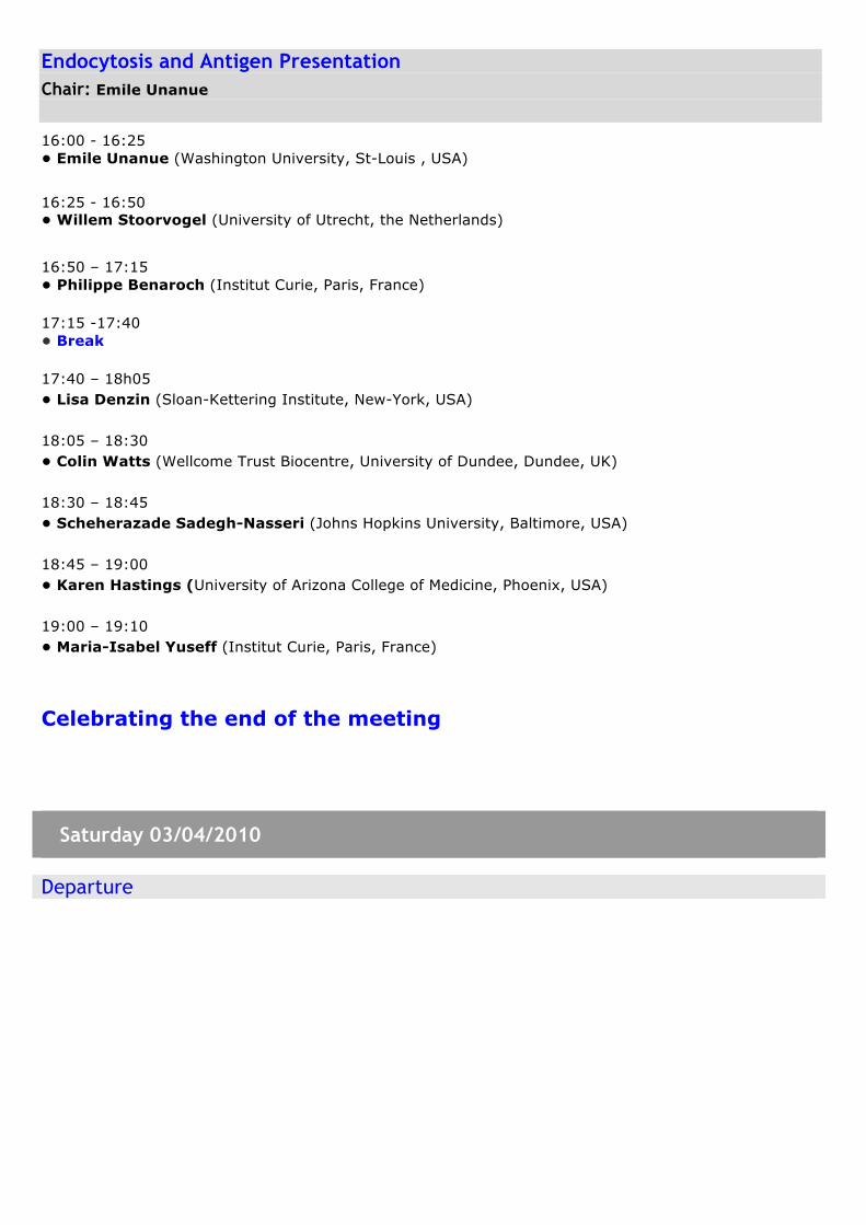
Endocytosis and Antigen Presentation Chair: Emile Unanue
16:00 - 16:25 • Emile Unanue (Washington University, St-Louis , USA)
16:25 - 16:50 • Willem Stoorvogel (University of Utrecht, the Netherlands)
16:50 – 17:15 • Philippe Benaroch (Institut Curie, Paris, France) 17:15 -17:40 • Break 17:40 – 18h05 • Lisa Denzin (Sloan-Kettering Institute, New-York, USA) 18:05 – 18:30 • Colin Watts (Wellcome Trust Biocentre, University of Dundee, Dundee, UK) 18:30 – 18:45 • Scheherazade Sadegh-Nasseri (Johns Hopkins University, Baltimore, USA) 18:45 – 19:00 • Karen Hastings (University of Arizona College of Medicine, Phoenix, USA) 19:00 – 19:10 • Maria-Isabel Yuseff (Institut Curie, Paris, France)
Celebrating the end of the meeting
Saturday 03/04/2010
Departure
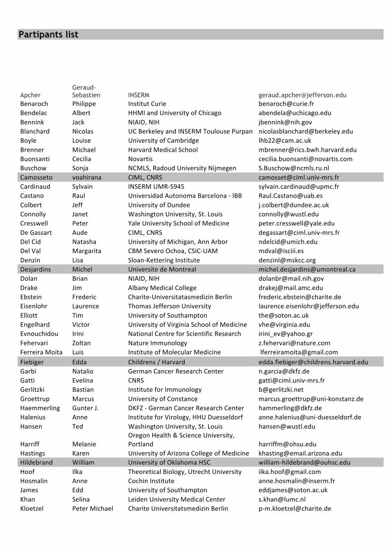
Partipants list
Apcher Geraud-Sebastien INSERM [email protected]
Benaroch Philippe InstitutCurie [email protected] Albert HHMIandUniversityofChicago [email protected] Jack NIAID,NIH [email protected] Nicolas UCBerkeleyandINSERMToulousePurpan [email protected] Louise UniversityofCambridge [email protected] Michael HarvardMedicalSchool [email protected] Cecilia Novartis [email protected] Sonja NCMLS,RadoudUniversityNijmegen [email protected] voahirana CIML,CNRS [email protected]‐mrs.frCardinaud Sylvain INSERMUMR‐S945 [email protected] Raul UniversidadAutonomaBarcelona‐IBB [email protected] Jeff UniversityofDundee [email protected] Janet WashingtonUniversity,St.Louis [email protected] Peter YaleUniversitySchoolofMedicine [email protected] Aude CIML,CNRS [email protected]‐mrs.frDelCid Natasha UniversityofMichigan,AnnArbor [email protected] Margarita CBMSeveroOchoa,CSIC‐UAM [email protected] Lisa Sloan‐KetteringInstitute [email protected] Michel UniversitedeMontreal [email protected] Brian NIAID,NIH [email protected] Jim AlbanyMedicalCollege [email protected] Frederic Charite‐UniversitatasmedizinBerlin [email protected] Laurence ThomasJeffersonUniversity [email protected] Tim UniversityofSouthampton [email protected] Victor UniversityofVirginiaSchoolofMedicine [email protected] Irini NationalCentreforScientificResearch [email protected] Zoltan NatureImmunology [email protected] Luis InstituteofMolecularMedicine [email protected] Edda Childrens/Harvard [email protected] Natalio GermanCancerResearchCenter [email protected] Evelina CNRS [email protected]‐mrs.frGerlitzki Bastian InstituteforImmunology [email protected] Marcus UniversityofConstance marcus.groettrup@uni‐konstanz.deHaemmerling GunterJ. DKFZ‐GermanCancerResearchCenter [email protected] Anne InstituteforVirology,HHUDuesseldorf anne.halenius@uni‐duesseldorf.deHansen Ted WashingtonUniversity,St.Louis [email protected]
Harriff MelanieOregonHealth&ScienceUniversity,Portland [email protected]
Hastings Karen UniversityofArizonaCollegeofMedicine [email protected] William UniversityofOklahomaHSC william‐[email protected] Ilka TheoreticalBiology,UtrechtUniversity [email protected] Anne CochinInstitute [email protected] Edd UniversityofSouthampton [email protected] Selina LeidenUniversityMedicalCenter [email protected] PeterMichael ChariteUniversitatsmedizinBerlin p‐[email protected]

Kniepert
Andrea
UniversityKonstanz
andrea.kniepert@uni‐konstanz.de
Laupeze Beatrice GSKBIOLOGICALS [email protected] Paul UniversityofCambridge [email protected]‐Dumenil Ana‐Maria InsermU932,InstitutCurie [email protected]
Lewinsohn DavidOregonHealth&ScienceUniversity,Portland [email protected]
Lu Xiuju NIAID,NIH [email protected]
Lukacs‐Kornek Veronika DanaFarberCancerInstitute veronika_lukacs‐[email protected]
Lybarger Lonnie UniversityofArizonaCollegeofMedicine [email protected] Raghavan UniversityofMichigan [email protected] Benedicte InstitutCurie [email protected] Ira Genentech,Inc. [email protected] Nawel INSERM [email protected] Christian UniversityofZurich [email protected] Jacques NetherlandsCancerInsitute [email protected] Ferry LeidenUniversityMedicalCenter [email protected] Ingeborg VUUniversityMedicalCentre [email protected] Philippe CIML [email protected]‐mrs.frRao Xiangyu UtrechtUniversity [email protected] Paul NationalCancerInstitute [email protected] Kenneth UMMS [email protected] Shereen TheUniversityofBirmingham [email protected]‐Nasseri Scheherazade JohnsHopkinsUniversity [email protected] Nilabh UniversityofCalifornia [email protected] Sebastian JacobsUniversityBremen s.springer@jacobs‐university.deStarck Shelley UniversityofCaliforniaBerkeley [email protected] Willem UniversityUtrecht [email protected] Efstratios NationalCentreforScientificResearch [email protected] Beverly WashingtonUniversity,St.Louis [email protected] Yuri ThomasJeffersonUniversity [email protected] Paul NCMLS,RadboudUniversity,Nijmegen [email protected] Robert BiocenterFrankfurt [email protected]‐frankfurt.deTaylor Graham UniversityofBirmingham [email protected]
Tenzer StefanInstituteofImmunology,UniversityofMainz tenzer@uni‐mainz.de
Thibodeau Jacques UniversitédeMontreal [email protected] Shannon DanaFarberCancerInstitute [email protected] Emil WashingtonUniversity,St.Louis [email protected] Benoit LudwigInstituteforCancerResearch [email protected] Hanneke UtrechtUniversity [email protected] Peter UniversiteParisDescartes peter.van‐[email protected] Sander NKIAmsterdam [email protected] Jose TheWalterandElizaHallInstitute [email protected] Anna LMUMunich,InstituteforImmunology, [email protected]‐muenchen.deWatts Colin UniversityofDundee [email protected] Mirjana INSERMU590,HopitalNecker [email protected] Jonathan NIAID,NIH [email protected] Maria‐Isabel InstituteCurie [email protected] Jianmin CRUKBirminghamCancerCenter [email protected]
Abstracts of talks and posters
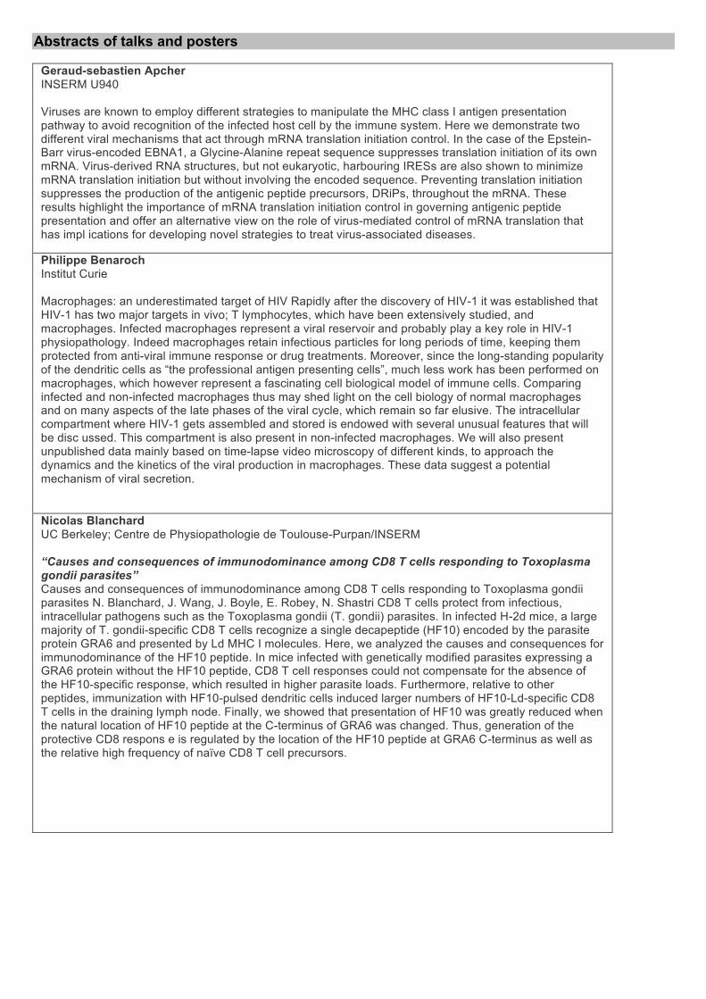
Geraud-sebastien Apcher INSERM U940 Viruses are known to employ different strategies to manipulate the MHC class I antigen presentation pathway to avoid recognition of the infected host cell by the immune system. Here we demonstrate two different viral mechanisms that act through mRNA translation initiation control. In the case of the Epstein-Barr virus-encoded EBNA1, a Glycine-Alanine repeat sequence suppresses translation initiation of its own mRNA. Virus-derived RNA structures, but not eukaryotic, harbouring IRESs are also shown to minimize mRNA translation initiation but without involving the encoded sequence. Preventing translation initiation suppresses the production of the antigenic peptide precursors, DRiPs, throughout the mRNA. These results highlight the importance of mRNA translation initiation control in governing antigenic peptide presentation and offer an alternative view on the role of virus-mediated control of mRNA translation that has impl ications for developing novel strategies to treat virus-associated diseases.
Philippe Benaroch Institut Curie Macrophages: an underestimated target of HIV Rapidly after the discovery of HIV-1 it was established that HIV-1 has two major targets in vivo; T lymphocytes, which have been extensively studied, and macrophages. Infected macrophages represent a viral reservoir and probably play a key role in HIV-1 physiopathology. Indeed macrophages retain infectious particles for long periods of time, keeping them protected from anti-viral immune response or drug treatments. Moreover, since the long-standing popularity of the dendritic cells as “the professional antigen presenting cells”, much less work has been performed on macrophages, which however represent a fascinating cell biological model of immune cells. Comparing infected and non-infected macrophages thus may shed light on the cell biology of normal macrophages and on many aspects of the late phases of the viral cycle, which remain so far elusive. The intracellular compartment where HIV-1 gets assembled and stored is endowed with several unusual features that will be disc ussed. This compartment is also present in non-infected macrophages. We will also present unpublished data mainly based on time-lapse video microscopy of different kinds, to approach the dynamics and the kinetics of the viral production in macrophages. These data suggest a potential mechanism of viral secretion.
Nicolas Blanchard UC Berkeley; Centre de Physiopathologie de Toulouse-Purpan/INSERM “Causes and consequences of immunodominance among CD8 T cells responding to Toxoplasma gondii parasites” Causes and consequences of immunodominance among CD8 T cells responding to Toxoplasma gondii parasites N. Blanchard, J. Wang, J. Boyle, E. Robey, N. Shastri CD8 T cells protect from infectious, intracellular pathogens such as the Toxoplasma gondii (T. gondii) parasites. In infected H-2d mice, a large majority of T. gondii-specific CD8 T cells recognize a single decapeptide (HF10) encoded by the parasite protein GRA6 and presented by Ld MHC I molecules. Here, we analyzed the causes and consequences for immunodominance of the HF10 peptide. In mice infected with genetically modified parasites expressing a GRA6 protein without the HF10 peptide, CD8 T cell responses could not compensate for the absence of the HF10-specific response, which resulted in higher parasite loads. Furthermore, relative to other peptides, immunization with HF10-pulsed dendritic cells induced larger numbers of HF10-Ld-specific CD8 T cells in the draining lymph node. Finally, we showed that presentation of HF10 was greatly reduced when the natural location of HF10 peptide at the C-terminus of GRA6 was changed. Thus, generation of the protective CD8 respons e is regulated by the location of the HF10 peptide at GRA6 C-terminus as well as the relative high frequency of naïve CD8 T cell precursors.

Louise Boyle University of Cambridge MHC class I expression is assisted by a number of proteins in the ER including calreticulin, ERp57, the TAP transporters, and tapasin. We have identified a new component that modulates MHC class I expression. When expressed in HeLa cells, this protein binds to peptide receptive HLA heavy chain /b2m heterodimers in the ER and slows export of the MHC class I. It alters the surface composition of MHC class I by increasing the expression of free HLA heavy chain (HC10 reactive) forms. Thus, unlike conventional MHC class I chaperones such as tapasin, the novel molecule does not appear to assist in the loading for peptide onto MHC class I molecules, but stabilises the expression of alternative/ empty forms of MHC class I. Its expression may have profound effects on the recognition of MHC class I by NK and T cells.
Michael Brenner Harvard Medical School REGULATION OF CD1 ANTIGEN PRESENTING COMPLEX STABILITY. For MHC class I and II molecules, the binding of specific peptide antigens is essential for assembly and trafficking and is at the center of their quality control mechanism. However, the role of lipid antigen binding in stabilization and quality control of CD1 heavy chain (HC)/β2m complexes is unclear. Further, the distinct trafficking and loading routes of CD1 proteins take them from mildly acidic pH in early endososmal compartments (pH 6.0) to markedly acidic pH in lysosomes (pH 5.0) and back to neutral pH of the cell surface (pH 7.4). Here, we present evidence that the stability of each CD1 HC/β2m complex is determined by the distinct pH optima that is identical to that of the intracellular compartments in which each CD1 isoform resides. While stable at acidic endosomal pH, complexes are only stable at cell surface pH 7.4 when bound to specific lipid antigens. The proposed model outlines a quality control program that allows lipid exchange at low endosomal pH withou t dissociation of the CD1 HC/β2m complex and then stabilizes the antigen
Sonja Buschow NCMLS, Radoud University Nijmegen Medical Centre Zooming in on human dendritic cells by whole cell and phagosome proteomics. Gene expression analysis is commonly used to study Dendritic Cell (DC) maturation. Proteomics however provides more direct insight into protein expression and - when applied on isolated organelles - it can also yield information on protein location. Here, we combined quantitative proteome and transcriptome analysis on a single batch of immature and cytokine cocktail matured human DCs and integrated resulting datasets at the pathway level. Proteins and genes differentially expressed during DC maturation partly mapped to identical but also to different pathway components demonstrating that RNA and protein expression data clearly support and complement each other. Exploiting this combined RNA and protein analysis we identified 5 dominant pathways, confirming the importance of cytokines, cell adhesion and migration in DC maturation. Interestingly, we discovered also a fundamental role for lipid metabolism in DC maturation. Identified pathways were enriched in existing maturation markers and now represent a rich data source to extract novel markers to improve DC vaccine design. Finally, we have also performed proteomics on human DC phagosomes, a key organelle for antigen presentation. Using label free quantification we could relate phagosomal protein abundance to that in whole DC, highlighting phagosome enriched proteins with potential important implications for particulate antigen presentation.
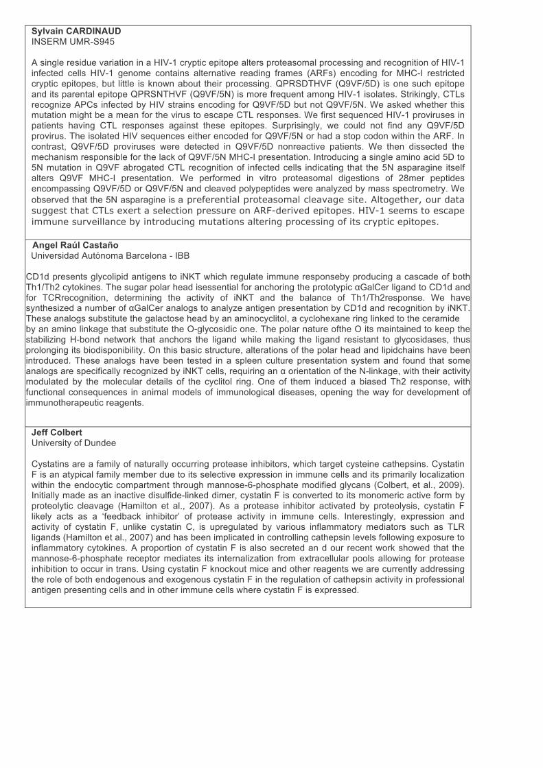
Sylvain CARDINAUD INSERM UMR-S945 A single residue variation in a HIV-1 cryptic epitope alters proteasomal processing and recognition of HIV-1 infected cells HIV-1 genome contains alternative reading frames (ARFs) encoding for MHC-I restricted cryptic epitopes, but little is known about their processing. QPRSDTHVF (Q9VF/5D) is one such epitope and its parental epitope QPRSNTHVF (Q9VF/5N) is more frequent among HIV-1 isolates. Strikingly, CTLs recognize APCs infected by HIV strains encoding for Q9VF/5D but not Q9VF/5N. We asked whether this mutation might be a mean for the virus to escape CTL responses. We first sequenced HIV-1 proviruses in patients having CTL responses against these epitopes. Surprisingly, we could not find any Q9VF/5D provirus. The isolated HIV sequences either encoded for Q9VF/5N or had a stop codon within the ARF. In contrast, Q9VF/5D proviruses were detected in Q9VF/5D nonreactive patients. We then dissected the mechanism responsible for the lack of Q9VF/5N MHC-I presentation. Introducing a single amino acid 5D to 5N mutation in Q9VF abrogated CTL recognition of infected cells indicating that the 5N asparagine itself alters Q9VF MHC-I presentation. We performed in vitro proteasomal digestions of 28mer peptides encompassing Q9VF/5D or Q9VF/5N and cleaved polypeptides were analyzed by mass spectrometry. We observed that the 5N asparagine is a preferential proteasomal cleavage site. Altogether, our data suggest that CTLs exert a selection pressure on ARF-derived epitopes. HIV-1 seems to escape immune surveillance by introducing mutations altering processing of its cryptic epitopes.
Angel Raúl Castaño Universidad Autónoma Barcelona - IBB
CD1d presents glycolipid antigens to iNKT which regulate immune responseby producing a cascade of both Th1/Th2 cytokines. The sugar polar head isessential for anchoring the prototypic αGalCer ligand to CD1d and for TCRrecognition, determining the activity of iNKT and the balance of Th1/Th2response. We have synthesized a number of αGalCer analogs to analyze antigen presentation by CD1d and recognition by iNKT. These analogs substitute the galactose head by an aminocyclitol, a cyclohexane ring linked to the ceramide by an amino linkage that substitute the O-glycosidic one. The polar nature ofthe O its maintained to keep the stabilizing H-bond network that anchors the ligand while making the ligand resistant to glycosidases, thus prolonging its biodisponibility. On this basic structure, alterations of the polar head and lipidchains have been introduced. These analogs have been tested in a spleen culture presentation system and found that some analogs are specifically recognized by iNKT cells, requiring an α orientation of the N-linkage, with their activity modulated by the molecular details of the cyclitol ring. One of them induced a biased Th2 response, with functional consequences in animal models of immunological diseases, opening the way for development of immunotherapeutic reagents.
Jeff Colbert University of Dundee Cystatins are a family of naturally occurring protease inhibitors, which target cysteine cathepsins. Cystatin F is an atypical family member due to its selective expression in immune cells and its primarily localization within the endocytic compartment through mannose-6-phosphate modified glycans (Colbert, et al., 2009). Initially made as an inactive disulfide-linked dimer, cystatin F is converted to its monomeric active form by proteolytic cleavage (Hamilton et al., 2007). As a protease inhibitor activated by proteolysis, cystatin F likely acts as a ‘feedback inhibitor’ of protease activity in immune cells. Interestingly, expression and activity of cystatin F, unlike cystatin C, is upregulated by various inflammatory mediators such as TLR ligands (Hamilton et al., 2007) and has been implicated in controlling cathepsin levels following exposure to inflammatory cytokines. A proportion of cystatin F is also secreted an d our recent work showed that the mannose-6-phosphate receptor mediates its internalization from extracellular pools allowing for protease inhibition to occur in trans. Using cystatin F knockout mice and other reagents we are currently addressing the role of both endogenous and exogenous cystatin F in the regulation of cathepsin activity in professional antigen presenting cells and in other immune cells where cystatin F is expressed.

Janet Connolly Washington University, St. Louis “A single peptide-MHC complex positively selects a diverse and specific CD8 T cell repertoire” A single peptide-MHC complex positively selects a diverse and specific CD8 T cell repertoire Janet M. Connolly1, Baomei Wang1, Tina Primeau1, Nancy Myers1, Henry Rohrs1, Michael Gross1, Lonnie Lybarger2, and Ted H. Hansen1 1Washington University School of Medicine, St. Louis, MO, and 2University of Arizona, Tucson, AZ Pathogen recognition by T cells is dependent upon their exquisite specificity for self major histocompatibility complex (MHC) molecules presenting a bound peptide. Although this specificity results from positive and negative selection of developing T cells in the thymus, the relative contribution of these two processes remains controversial. To address the relationship between the selecting peptide-MHC complex and the specificity of mature T cells, we generated transgenic mice that express a single peptide-MHC class I complex. Positive selection of CD8 T cells in these mice results in an MHC-specific repertoire. Furthermore, the T cells are strongly reactive with Kb occupied with endogenous peptides. This allowed the peptide specificity of T cells to be analyzed on an extensive panel of physiologic Kb-binding ligands. While selection on a single complex is peptide promiscuous, mature T cells are highly peptide-specific. Thus, positive selection imparts MHC and peptide specif icity on the peripheral CD8 T cell repertoire. Science:326, 871(2009)
Peter Cresswell Yale University School of Medicine “A role for GILT in cross-presentation of viral antigens” Antigenic peptides generated in the cytosol are translocated into the endoplasmic reticulum by the ATP-dependent ‘Transporter associated with Antigen Processing’ (TAP), where they bind to newly synthesized MHC class I molecules. TAP, together with the MHC class I molecule itself, and calreticulin, ERp57 and the specialized glycoprotein, tapasin, form the peptide loading complex (PLC). Tapasin is critical for loading MHC class I molecules with high affinity peptides. It functions within the PLC as a disulfide-linked, stable heterodimer with the thiol oxidoreductase ERp57, and this covalent interaction is required to support optimal PLC activity. The structure of the tapasin/ERp57 dimer revealed the basis for the stable dimerization of tapasin and ERp57 and provided the first example of a protein disulfide isomerase family member interacting with a substrate. Mutational analysis has mapped a conserved surface on tapasin that interacts with MHC class I molecules and is crit ical for the peptide loading function of the tapasin-ERp57 heterodimer. Further analysis of the MHC class I-tapasin interaction and the nature of the PLC will be presented which will further illuminate the processes involved in MHC class I peptide loading.
Natasha Del Cid University of Michigan, Ann Arbor All nucleated cells express surface MHC class I molecules to allow for the surveillance of viral and tumor antigens by CD8+ T cells. Cross-presentation is an important mechanism by which exogenous antigens derived from dying tumor cells or virally infected cells are presented on MHC class I molecules of certain antigen presenting cell subsets, thus initiating necessary CD8+ T cell responses. Heat shock proteins have been shown to increase the cross-presentation efficiency of antigenic peptides. We investigated the ability of calreticulin, an ER chaperone, to enhance the cross-presentation of peptides, fused soluble antigens, and bead-associated antigens to various antigen presenting cells. Using in vitro assays with bone marrow-derived GM-CSF differentiated dendritic cells (DC), bone marrow derived macrophages, and CD8+ splenic DC as antigen presenting cells, calreticulin did not enhance cross-priming of any of the i ndicated forms of antigen. In vivo studies suggested a role for calreticulin in enhancing antigen cross-priming, though the enhancement was not robust. A large number of receptors and ligands are involved in mediating the uptake and cross-priming of apoptotic cells. Thus, we are currently investigating calreticulin’s role in this network of signals by assessing its ability to enhance the cross-priming of antigens associated with apoptotic cells.

Margarita Del Val Centro de Biologia Molecular Severo Ochoa, CSIC-UAM “Furin-processed antigens targeted to the secretory route elicit functional TAP1-/- CD8+ T lymphocytes in vivo” Furin-processed antigens targeted to the secretory route elicit functional TAP1-/- CD8+ T lymphocytes in vivo Francisco Medina, Manuel Ramos, Salvador Iborra, Patricia de León, Marta Rodríguez-Castro, and Margarita Del Val Most pathogen-derived peptides recognized by CD8+ cytotoxic T lymphocytes are produced by proteasomes and delivered to the endoplasmic reticulum by the TAP transporters associated with antigen processing. Alternative proteases also produce antigenic peptides, but their actual relevance is unclear. There is a need to quantify the contribution of these supplementary pathways in vitro and in vivo. A well defined TAP-independent secretory route of antigen processing involves the trans-Golgi network protease furin. Quantitation of this route by using ovalbumin constructs encoded by vaccinia viruses indicates that it provides ca. one-third of all surface complexes of peptide and MHC class I molecules. Generation of the epitope carboxy terminus is a dramatic rate-limiting step, since bypassing it increased efficiency by at least 1,000-fold. Notably, the secretory construct activated a similar percentage of antigen-specific CD8+ T cells in wild type as in TAP1-deficient mice, which a llow only secretory routes but which have a 10-20-fold smaller CD8 compartment. Moreover, these TAP1-/- ovalbumin-specific CD8+ T lymphocytes accomplished elimination of epitope-bearing cells in vivo. The results obtained with this experimental system underscore the potential of secretory pathways of MHC class I antigen presentation to elicit functional CD8+ T-lymphocytes in vivo and support the hypothesis that non-cytosolic processing mechanisms may compensate in vivo for the lack of proteasome participation in antigen processing in persons genetically deficient in TAP, and thus contribute to pathogen control.
Lisa Denzin Sloan-Kettering Institute Peptide loading of major histocompatibility complex (MHC) class II (MHCII) molecules is directly catalyzed by HLA-DM. HLA-DO, another class II-like molecule, modulates DM function. The biological role of the DO-mediated regulation of DM activity in vivo remains unknown. To test the idea that DO modulation of the MHCII-self-peptide repertoire mediates self-tolerance, we generated non-obese diabetic (NOD) mice that expressed DO (NOD.DO) in DCs. Our studies showed that diabetes development was completely blocked in NOD.DO mice. NOD.DO animals selected a diabetogenic T cell repertoire and the numbers and function of regulatory T cells were normal. Indeed, immune system function in NOD.DO mice was equivalent to wild type NOD mice. NOD.DO DCs, however, presented an altered MHCII bound self-peptide repertoire, thereby preventing the activation of diabetogenic T cells and subsequent diabetes development. These studies sh ow that DO expression shapes the overall MHCII-self-peptide repertoire and in doing so promotes T cell tolerance.
Michel Desjardins Université de Montréal The immune response to viral infection involves the processing of viral proteins by the classical MHC class I pathway. This process is initiated by the degradation of proteins by the proteasome followed by the translocation of viral peptides in the lumen of the endoplasmic reticulum for loading on MHC class I molecules. Recent studies have shown that viral proteins can also be presented on MHC class II molecules by a process involving autophagy. It was described recently that Herpes virus-1 can inhibit autophagy, probably as a way to escape immune recognition. Our data indicate that the initial inhibition of autophagy by HSV-1 is followed by a host response characterized by the initiation of a novel form of autophagy involving the nuclear membrane. This process leads to the trafficking of viral proteins to lysosomes and their degradation in the vacuolar pathway (usually involved in the presentation on MHC class II m olecules). Interestingly nuclear membrane-derived autophagy enables the presentation of viral peptides on MHC class I molecules. The molecular characterization of this pathway is currently investigated.
Brian Dolan NIAID, NIH Following viral infection, cells rapidly present viral peptides to CD8+ T cells via the endogenous MHC class I antigen presentation pathway. The rapidity of antigen presentation of peptides from metabolically stable proteins clearly points to the contribution of Defective Ribosomal Products (DRiPs) to antigen processing, but we have made limited progress on biochemically defining peptidogenic DRiPs. Here, we describe a new tool to study DRiP antigen presentation where post-translational control of a protein’s stability is used to segregate the source of antigenic peptides into rapidly degraded proteins and true DRiPs. We find true DRiPs to be the major source of peptides antigen presentation, making up more than half of the peptide-MHC class I complexes generated by the antigen presenting cell. We also use this system to identify drugs and siRNAs that interfere with presentation of DRiP antigens specifically. Our data suggest a role for a surprising class of enzymes as key players in DRiP antigen presentation.
Jim Drake Albany Medical College “Physiological-Range Temperature Changes Modulate Cognate Antigen Processing and Presentation Mediated by Lipid Raft-Restricted Ubiquitinated B Cell Receptor Molecules” B cell receptor mediated antigen processing and presentation is critical to the initiation and control of a humoral immune response. Trafficking of internalized antigen-BCR (Ag-BCR) complexes to intracellular antigen processing compartments is driven by ubiquitination of the cytoplasmic domain of the BCR. Using a biochemical approach, it was established that ubiquitinated Ag-BCR complexes are restricted to plasma membrane lipid rafts. Since the structure of these lipid domains is sensitive to temperature, the impact of physiological range temperature changes (33-39°C) on lipid raft-dependent and independent BCR functions was investigated. While the kinetics of BCR internalization (a lipid raft independent event) are unaffected by temperature changes within this range, BCR signaling and ubiquitiation as well as BCR-mediated antigen processing are significantly impacted. When compared to 33°C (peripheral body temperature), the extent and duration of Ag-BCR ubiquitination is increased and prolonged at 37-39°C (normal to febrile temperature). As might be expected, increased temperature also accelerates the overall kinetics of Ag-BCR degradation. Surprisingly, incubation of B cells at 33°C profoundly slows the expression of peptide-class II complexes derived from the BCR-mediated processing of cognate antigen, while having essentially no effect of the kinetics of expression of peptide-class II complexes derived from fluid-phase internalized antigen. These results establish the effect of physiological range temperature changes on multiple lipid raft-dependent BCR functions including the processing and presentation of cognate antigen, suggesting one mechanism by which physiological temperature changes such as fever may impact the initiation and /or maturation of a humoral immune response.
Frederic Ebstein Charite-Universitatasmedizin Berlin/ Institut fur Biochemie “UBC6e/Derlin-1/VIMP are essential ERAD components for the processing of exogenous antigens for MHC class I cross-presentation by dendritic cells” UBC6e/Derlin-1/VIMP are essential ERAD components for the processing of exogenous antigens for MHC class I cross-presentation by dendritic cells. Frédéric Ebstein, Michael Seeger, Andrea Lehmann and Peter-Michael Kloetzel. Charité-Universitätsmedizin Berlin, Institut für Biochemie CCM. Dendritic cells (DC) are professional antigen-presenting cells with the unique ability to cross-present exogenous-derived peptides by MHC class I molecules. Despite extensive studies, the mechanisms promoting the delivery of ingested antigens into the cytoplasm remain to be fully elucidated. Here, we report that maturation of monocyte-derived DC is accompanied by increased expression of the endoplasmic reticulum (ER)-localised protein UBE2J1 (UBC6e). UBC6e is a novel ubiquitin-conjugating enzyme that lacks a homologue in yeast and is involved in ER-associated degradation pathway (ERAD) in higher eukaryotes. Interestingly, we show by small interfering RNA methods that gene silencing of UBC6e resulted in impaired cross-presentation of the human cytomegalovirus (HCMV)-derived pp65 (495-503) antigenic peptide by DC. These data are in agreement with previous observations suggesting a role for ERAD in the transfer of antigens from the endocytic compartments into the cytoplasm. Here , we further demonstrate that pp65 cross-presentation by DC is affected by knockdown of the ERAD-related components Derlin-1 and VIMP but not Derlin-2, -3, HRD1, gp96, Herp and sec61a1. We therefore propose that the retro-translocation of internalised antigens from membranes into the cytoplasm for pp65 cross-presentation relies on a ER-localised protein complex involving UBC6e, Derlin-1 and VIMP.

Laurence Eisenlohr Thomas Jefferson University Multiple endosomal (“exogenous”) and cytosolic (“endogenous”) processing pathways. This ensures a broad array of peptides, thereby maximizing the opportunity for CD4+ T cells to detect invasion. Previously we defined an endogenous MHC class II processing pathway that is both proteasome- and TAP- dependent and that accounts for a substantial portion of the class II-restricted response to influenza and vaccinia. Subsequent efforts have revealed several key aspects of this pathway: 1) Cytosolically generated peptides are delivered to the late endosome for loading onto class II. 2) The pathway is very tightly regulated; in the absence of innate signaling, it is essentially inoperable - an arrangement that limits presentation of self peptides and accentuates presentation of foreign peptides, analogous to what has been reported for exogenous class II-restricted presentation. 3) As has been reported for MHC class I-restricted cross-presentation, redistribution of TAP appears to be one mechanism for controlling activity of this pathway. 4) Evidence supports the notion that proteolytic susceptibility determines which pathway is productive for generation of a particular epitope. 5) On a global level, macroautophagy does not detectably contribute to the endogenous presentation of influenza and vaccinia virus, leaving the proteasome-/TAP-dependent pathway as the dominant route for endogenous presentation of class II-restricted epitopes derived from these two viruses. Tim Elliott
University of Southampton Calreticulin is a lectin chaperone of the endoplasmic reticulum (ER). In calreticulin-deficient cells, major histocompatibility complex (MHC) class I molecules travel to the cell surface associated with a suboptimal peptide load. We have shown that calreticulin exits the ER to accumulate in the ER-Golgi intermediate compartment (ERGIC) and the cis-Golgi, together with suboptimally loaded class I molecules. Calreticulin that lacks its C-terminal KDEL retrieval sequence assembles with the peptide loading complex but neither retrieves suboptimally loaded class I molecules from the cis-Golgi to the ER, nor supports optimal peptide loading. This identifies a critical quality-control step for MHC class I loading downstream of the peptide loading complex in which calreticulin plays an important role, and indicates a functional role of intracellular transport in the optimal loading of MHC class I molecules with antigenic peptide .
Victor Engelhard University of Virginia School of Medicine “Lymph node resident lymphatic endothelial cells mediate peripheral tolerance via Aire-independent direct antigen presentation” Peripheral immune tolerance is generally thought to result from cross-presentation of tissue-derived proteins by quiescent, tissue resident dendritic cells to self-reactive T cells that have escaped negative selection in the thymus, leading to anergy or deletion. We now demonstrate that lymph node-resident lymphatic endothelial cells (LN-LEC) also serve this function. LN-LEC directly express multiple proteins otherwise confined to specific peripheral tissues. They directly present an epitope derived from one of these, the melanocyte-specific protein tyrosinase, to specific CD8 T cells, leading to their deletion. Expression of peripheral tissue antigens by LN-LEC is independent of the autoimmune regulator, Aire, which has been shown to control a similar process in medullary thymic epithelial cells. LN-LEC express PD-L1, which as been shown to deliver an inhibitory signal to activated T cells and to induce anergy. Anti- PD-L1 blocking antibodies prevent T cell deletion, leading instead to robust T cell expansion. These results establish LN-LEC as important systemic mediators of peripheral immune tolerance.
Irini Evnouchidou National Centre for Scientific Research - Demokritos ERAP1 (ERAAP) is an ER aminopeptidase that plays crucial roles in antigenic peptide generation and destruction. Recently, large population studies have linked coding ERAP1 single nucleotide polymorphisms (SNPs) with predisposition to autoimmune diseases and virally induced cancer. We hypothesized that this link is due to ERAP1’s role in antigenic peptide generation, through the aberrant generation or destruction of key antigenic epitopes that initiate or sustain autoimmunity or elicit anti-viral responses. To test this hypothesis we overexpressed and purified allelic versions of ERAP1 and tested their ability to generate antigenic peptides in vitro. We found that, for several but not for all of the epitopes tested, mature antigenic peptide generation rates were dependent on the ERAP1 allele used and in patterns that were also epitope dependent. Furthermore, the generation rate of specific antigenic peptides suspected t o be linked with autoimmunity was highly dependent on the presence of the specific ERAP1 SNPs also linked with autoimmune disease. Our results suggest that ERAP1 SNPs may impose specificity changes in the enzyme. Furthermore, our findings provide support to the concept that antigenic peptide processing is the biochemical mechanism behind the link of ERAP1 SNPs and autoimmune disease predisposition.
Luis Ferreira Moita Institute Of Molecular Medicine “shRNA Dissection of Antigen Cross-Presentation Pathways” shRNA Dissection of Antigen Cross-Presentation Pathways Catarina Moita, Nir Hacohen and Luis F. Moita Effective immune responses against tumor antigens that are not endogenously expressed by dendritic cells (DCs) and against virus that do not infect antigen presenting cells (APCs) require extracellular antigens to stimulate CD8+ T cells via the MHC I pathway through a process known as cross-presentation. We have completed a screen for genes with a role in this process using a subset of an shRNA lentiviral library enriched for mouse kinases and phosphatases. In this screen we measured the proliferation of CD8+ T (OT-I) cells in response to the stimulation of bone marrow derived DCs (BMDCs) after incubation with OVA-expressing S. cerevisiae. After testing over 1000 genes in duplicate and two rounds of phenotypic validation, we have chosen ~80 genes that reproducibly caused increased or reduced rates of CD8+ T proliferation. We performed qPCR validation of the knockdowns and additional secondary screens on this group of 80 genes so that we could identify those that are specifically required for cross-presentation. Our current results suggest that the overwhelming majority of kinases and phosphatases involved in antigen presentation play a role in both canonical and antigen cross-presntation pathways.
Edda Fiebiger Childrens / Harvard Dendritic cells use IgE-receptor-mediated antigen presentation to shift immune responses towards Th2. The current literature suggest that basophils are the dominant antigen presenting cell for the initiation of T helper type 2 (Th2)-cell dependent immunity, while dendritic cell (DC) priming appears insufficient for this type of immune response. Here, we show that DCs can use antigen uptake via the high affinity receptor for IgE, Fc-epsilon-RI, to induce potent Th2 immune responses. Unlike humans, mice do not constitutively express Fc-epsilon-RI on dendritic cells. We therefore developed a transgenic animal model that expresses Fc-epsilon-RI on DCs (aTG mice). aTG-DCs express a chimeric FceRI that uses the murine common FcRg-chain for signaling and binds murine as well as human IgE via the human Fc-epsilon-RI a-chain. We show that the production of IL4 is a consequence of Fc-epsilon-RI-mediated antigen presentation but not observed after fluid phase uptake in vitro. In vivo, aTG animals suffer from exacerbated Th2-dependent chronic airway inflammation. In summary, we describe how DCs use a receptor-specific antigen uptake mechanism to modulate antigen presentation and the ensuing immune response. These findings are of high relevance for our basic understanding of the induction of Th2-type immune responses.
Natalio Garbi German Cancer Research Center “Analysis of Flt3-L expression and requirement for dendritic cell generation” Antigen presentation by dendritic cells (DCs) is critical in regulating immune responses. The requirements governing DC generation are only starting to be elucidated. Flt3-L is a growth/differentiation cytokine critical for DC development. However, little is known regarding FL expression or the source required for DC differentiation. To investigate this, we have generated FL reporter mice in which eGFP and luciferase are driven by the flt3l locus. We find that all organs express FL mRNA and protein, but the FL amount in bone marrow and spleen are surprisingly low. On a cellular level, immune and non-immune cells all express FL. We therefore investigated the source of FL necessary for DC differentiation. In bone marrow chimeras, we found that FL production by the hematopoietic compartment was both necessary and sufficient for conventional and plasmacytoid DC generation in spleen. In bone marrow, however, normal DC differe ntiation required FL produced by the non-hematopoietic and the hematopoietic compartments. These findings did not correlate with the amount of FL present in spleen and bone marrow. In summary, our studies reveal that although FL is ubiquitously expressed, its source rather than the total amount is the dominant factor for normal DC development.
Evelina Gatti CIML, CNRS UMR 6102, Marseille
Role of MARCH ubiquitin ligases in MHC intracellular transport. Aude De Gassart, Francesca De Angelis-Rigotti, Philippe Pierre and Evelina Gatti MARCH ubiquitin ligases have been recently identified on the basis of their homology to immunosuppressive viral factors. In mammalian cells, some MARCHs target immunorelevant receptors, promoting downregulation of specific antigen presentation molecules. Out of a family of 10 related ligases, we have identified MARCH I as the mediator of MHCII downregulation in human dendritic cells. MARCH VIII also targets MHCII and CD86, while MARCH IX acts on MHC I. MARCHs expression in human DCs is differentially regulated in response to specific environmental signals, such as TLR ligation or IL-10. The alteration of the correct expression pattern of these ligases could constitute for tumor cells a strategy to escape for immunosurveillance. To understand their function, we characterized the transcriptional regulation of MARCH E3 ligases in response to different conditions, in the attempt to explore their function in the global coordination between pathogen stimulation and antigen presentation up-regulation of DC function.
Bastian Gerlitzki Institute for Immunology In humans and mice naturally occurring regulatory T cells (nTregs) are a thymus-derived subset of T cells, crucial for the maintenance of peripheral tolerance by controlling not only autoreactive T cells but virtually all cells of the adaptive and innate immune system. Recent work strongly advocates the hypothesis that nTregs directly target dendritic cells (DC) in vivo to dampen an ongoing immune response. However, the mechanisms underlying this suppressive process are still elusive. Recent publications suggest that a crucial event of DC suppression is the inhibition of the expression of co-stimulatory molecules CD80 and CD86, which relies on CTLA-4. Here we show that the nTreg-mediated inhibition of DC primarily relies on the inhibition of antigen processing and that the suppression of co-stimulatory molecules is only a secondary process, reinforcing suppression. Importantly, the suppression of antigen processing by DC is independent from CTLA-4 and occurs within minutes upon cell-cell-contact. As T cell activation strictly relies on T cell receptor signalling upon binding of MHC-peptide complexes, the inhibition of antigen processing is fundamental to prevent the elucidation of an adaptive immune response. Hence, our data indicate that nTregs effectively control exuberant immune responses by modulating the capacity of DC to process antigens.
Marcus Groettrup Chair of Immunology, University of Constance “A selective inhibitor of the immunoproteasome subunit LMP7 blocks cytokine production and attenuates progression of autoimmune diseases” A selective inhibitor of the immunoproteasome subunit LMP7 blocks cytokine production and attenuates progression of autoimmune diseases Michael Basler1,2, Tony Muchamuel3, Monette A. Aujay3, Khalid W. Kalim1, Christoph Lauer1, Susan D. Demo3, Mark K. Bennett3, Christopher J. Kirk3, and Marcus Groettrup1,2 1Division of Immunology, Department of Biology, University of Constance, D-78457 Konstanz, Germany. 2Biotechnology Institute Thurgau (BITg) at Constance University, CH-8280 Kreuzlingen, Switzerland. 3Proteolix, Inc., South San Francisco, CA 94080, USA. The immunoproteasome is known to shape the antigenic repertoire presented on class I major histocompatibility complexes (MHC-I). However, a specific role for the immunoproteasome in regulating other facets of immune responses has not been established. We describe here the characterization of PR-957, a selective inhibitor of LMP7, the chymotrypsin-like subunit of the immunoproteasome. PR-957 blocked presentation of LMP7-specific, MHC-I-restricted antigens in vitro and in vivo. Selective inhibition of LMP7 by PR-957 blocked production of interleukin-23 by activated monocytes and interferon-gamma and interleukin-2 by T-cells and prevented Th17 differentiation in vitro. In mouse models of rheumatoid arthritis, PR-957 treatment reversed signs of disease and resulted in reductions in cellular infiltration, cytokine production and autoantibody levels. Also in mouse models of diabetes, colitis and multiple sclerosis treatment with PR-957 almost completely prevented the diseases . These studies reveal a unique role for LMP7 in controlling pathogenic immune responses and provide a therapeutic rationale for targeting LMP7 in autoimmune disorders. Currently, we are investigating the mechanistic base of how LMP7 affects cytokine production and Th17 differentiation. First insights into these mechanisms will be presented.

Anne Halenius Institute for Virology, HHU Duesseldorf
Human Cytomegalovirus Targets the MHC Class I Peptide Loading Complex Through Inhibition of Tapasin Gene Transcription Anne Halenius, Sebastian Hauka, Lars Dölken, Jan Stindt, Ulrich H. Koszinowski, Frank Momburg and Hartmut Hengel Tapasin stabilizes the peptide loading complec (PLC) and the peptide-receptive MHC I, thereby facilitating loading of high-affinity peptides onto MHC I. To cope with CD8+ T-cells human cytomegalovirus (HCMV) encodes for several post-translational strategies inhibiting MHC I antigen presentation, which have been extensively studied in transfected cells. In this study we analysed the assembly of the PLC in HCMV-infected cells at all stages throughout the protracted replication cycle. Metabolic labelling experiments revealed the virtual absence of tapasin incorporation into the PLC. In contrast, immunoblot analysis demonstrated a slow diminishment of tapasin steady-state levels in infected cells, suggesting a blocked synthesis rather than degradation. Tapasin mRNA levels were found to be continuously down-regulated during infection, however, the tapasin transcripts remained stable and long-lived. Taking advantage of a novel method, by which de novo transcribed RNA is selectively labelled and analysed, we found an immediate decline of tapasin transcription which was followed by a down-regulation of TAP2 gene expression, whereas TAP1, β2m and MHC I genes were up-regulated. Importantly, forced expression of tapasin restored the incorporation of tapasin into the PLC in HCMV-infected cells. The data indicate that HCMV essentially controls the MHC I antigen presentation pathway on a transcriptional level.
Guenter J. Haemmerling DKFZ - German Cancer Research Center “Immune synapse formation determines interaction forces between T cells and antigen presenting cells measured by atomic force microscopy” Immune synapse formation determines interaction forces between T cells and antigen presenting cells measured by atomic force microscopy. G.J. Hämmerling DKFZ - German Cancer Research Center, Heidelberg, Germany Recognition of antigen results in the formation of a so-called immune synapse (IS) at the T-cell/APC interface, which is crucial for T-cell activation. The molecular composition of the IS has been extensively studied, but little is known about the biophysics and interaction forces between T cells and APC. Here, we report the first measurement of interaction forces between T cells and APC employing atomic force microscopy (AFM). Dynamic analysis of T-cell/APC interaction by AFM revealed that in the presence of antigen (HEL peptide) the interaction forces increased from 1-2 nN at early time-points to a maximum of 14 nN after 30 min, and decreased again after 60 min. Surprisingly, very similar values were obtained for another T cell/APC pair involving T cells recognizing a MBP peptide. These data correlate with the kinetics of synapse formation that also reached a maximum after 30 min, as determined by high-throughput multispectral imaging flow cytometry. A small molecular inhibitor for LFA-1, BIRT377, strongly reduced the interaction forces, demonstrating the importance of LFA-1/ICAM-1-interactions in the IS for firm T-cell/ APC adhesion. In conclusion, using biophysical measurements, this study provides precise values for the interaction forces between T cells and APC and demonstrates that these forces develop over time and are highest when synapse formation is maximal.
Ted Hansen Washington University, St. Louis “MR1 restricts innate T cells in mycobacterial infection”. MR1 restricts innate T cell in mycobacterial infection. Ted H. Hansen, Shouxiong Huang, Wei Jen Chua, Steven Truscott, and Daniel Hoft Dept. of Path. and Immunol., Washington Univ. Sch. of Med; and Dept of Mol Micro, St Louis Univ Htlh Sci Ctr. St. Louis, Mo. USA Several nonclassical major histocompatibilty antigens (class Ib molecules) have emerged as key players in the early immune response to pathogens or stress. MR1 is a novel class Ib molecule whose physiological role was previously undefined. However, the Mr1 gene is evolutionarily conserved and controls the development of mucosal-associated invariant T (MAIT) cells. MAIT cells preferentially reside in mucosal tissues, and their development is dependent on commensal microbiota. Our recent studies provide direct evidence that MR1 presents ligand to MAIT cells, and that MR1 trafficking to endosomal compartments enhances MAIT cell activation. Furthermore, after mycobacterial infection in aerosol, Mr1 deficient mice were found to have higher bacterial burdens in the lung compared to wildtype mice. These cumulative findings implicate MR1 antigen presentation to MAIT cells as playing a key role in bacterial infection.

Melanie Harriff Oregon Health & Science University, Portland “Evidence for functional TAP in the M. tuberculosis phagosome” Mycobacterium tuberculosis (MTB) is an intracellular pathogen that can reside in both myeloid and non-myeloid cells in the lung. In myeloid cells such as DC, MTB survives and replicates in phagosomes that do not fuse with the lysosome. Recent studies have shown that the MTB phagosome is a competent antigen presenting organelle, as it contains HLA-I, TAP, and has the ability to directly elicit an IFN-γ response from HLA-E-restricted CD8+ T cells. In this study, we looked at the role of phagosomal TAP in the transport of peptides into the phagosome for subsequent loading on Class I molecules. Highly pure MTB phagosomes contained TAP and Class I, as well as other molecules involved in peptide loading in the endoplasmic reticulum (ER), such as ERp57 and Tapasin. ATP and TAP dependent peptide transport across the phagosome membrane was observed in MTB-phagosomes isolated from DCs. Furthermore, coupling of the luminal her pes-virus inhibitor of TAP, UL49.5, directly to MTB inhibited presentation of TB antigens to the HLA-E-restricted T-cell clone, D160 1-23. Our data are consistent with the hypothesis that the MTB phagosome is involved in presentation of MTB antigens, and show that phagosomal TAP is involved in the loading of these antigens. Karen Hastings University of Arizona College of Medicine “GILT and immune recognition of melanoma” Melanocyte differentiation antigens, including tyrosinase-related protein (TRP) 1, contain internal disulfide bonds and are presented by MHC class II molecules. These antigens are important for both the autoimmune destruction of melanocytes, which results in vitiligo, and the anti-melanoma immune response. Gamma-interferon inducible lysosomal thiol reductase (GILT) facilitates generation of class II-binding peptides by endocytic reduction of protein disulfide bonds. We show that GILT is required for efficient class II-restricted processing of a TRP1 epitope in vitro and accelerates the onset of spontaneous vitiligo in TRP1-specific TCR transgenic mice. GILT is also required to establish central tolerance to this self-antigen. Although similar numbers of CD4+ TRP1-specific T cells develop in GILT-deficient or TRP1-deficient mice, adoptive transfer of T cells that develop in the absence of TRP1, but not in the absence of GILT, induces vitiligo. T cells that develop in GILT-deficient mice proliferate in response to antigen, but fail to produce IL-2 in response to antigen or CD3/CD28. GILT’s role in antigen processing and its effects on T cell development and function are likely to be important both in the development of autoimmunity and for cancer immunotherapy.
William Hildebrand University of Oklahoma HSC In order to empower comparative analyses of peptide ligands eluted from MHC molecules, our laboratory pioneered the secretion of class I, and now class II, MHC molecules. Using this secreted MHC technology we have compared peptide ligands presented by the MHC of virus infected (influenza, HIV, WNV) to the ligands of uninfected cells. We have also systematically compared ligands from the MHC of cancerous and non-cancerous cells. Our comparisons demonstrate that each MHC molecule presents no more than a handful of virus-derived or cancer-specific peptide ligands. It also appears that, depending upon the pathogen or disease in question, molecules encoded at a particular MHC locus tend to present the brunt of the ligands sampled while the MHC encoded at other loci carry less of the antigen presenting load. When tested for immune recognition, we find that T cells respond in a definable hierarchy to these ligands and tha t the optimal T cell target is often a non-canonical (and therefore difficult to predict) peptide ligand. In summary, these data provide a perspective as to the number, nature, and immunogenicity of ligands sampled by the MHC molecules of infected and cancerous cells.
Ilka Hoof Theoretical Biology, Utrecht University "Which factors shape the HLA class I peptide repertoire?" Sampling self - What factors shape the HLA peptide repertoire? Ilka Hoof, Can Kesmir The peptide repertoire that is presented by the set of HLA class I molecules of an individual is formed by the different players of the antigen processing pathway, including the proteasome, TAP and the stringent binding environment of the HLA molecules. Peptide elution studies have shown that only a subset of the human proteome is sampled by the antigen processing machinery and represented on the cell surface. The aim of our study is to determine the factors relevant in shaping the HLA peptide repertoire. By combining peptide elution data and information on protein abundance and halflife, we illucidate the interplay between protein abundance and protein turnover in their effect on peptide presentation as well as the impact of protein length and density of predicted HLA binding peptides on sampling probability.
Anne Hosmalin Cochin Institute “Crosspresentation by dendritic cells from live cells induces protective immune responses in vivo” Crosspresentation by dendritic cells from live cells induces protective immune responses in vivo Matheoud, D., Feuillet, V., Perie, L., Vimeux, L., Hoeffel, G., *Baez, C., *Parent, I., Maranon, C., Bourdoncle, P., Renia, L., Blondel, A., Lucas, B., and Hosmalin, A.
Edd James University of Southampton Depletion of regulatory T cells in the CT26 murine tumour model stimulates a robust protective T cell response which is also protective to challenge with other tumours of different histological origins. We have characterised a cross-protective antigen, GSW11 (GGPESFYCASW) presented by H-2Dd. This antigen was found to be encoded within the env (gp90) gene of an endogenous retrovirus emv-1 (MuLV), previously shown to encode other CT26 antigens. Further characterisation of GSW11 identified the epitope to be susceptible to over-processing by ERAAP. Here we show that the generation of ERAAP knockdown CT26 tumours generate ~10-fold more GSW11 than wildtype and are rejected following in vivo challenge. Only ERAAP knockdown of >70% facilitates the rejection of the tumours in vivo. T cell responses in these immune mice are not only directed to GSW11 and (previously characterised) AH1 antigens, but to ERAAP knockdown CT26 specific antigens. ERAAP knockdown CT26 immune mice rechallenged with wildtype CT26 also reject the tumour suggesting a robust memory response is induced by ERAAP knockdown CT26 challenge. In addition, we show that T cell immunity directed to GSW11 and AH1 CT26 antigens are responsible for this rejection.
Selina Khan Leiden University Medical Centre Toll-like receptor ligand peptide targeting of synthetic long peptides to dendritic cells for efficient cross-presentation. Selina Khan, Cedrik Britten, Hermen S. Overkleeft, Gijsbert A. van der Marel, Dmitri V. Filippov, Cornelis J.M. Melief and Ferry Ossendorp. Dept. IHB, Leiden University Medical Centre, Leiden, the Netherlands. We have targeted peptide antigens to dendritic cells by the use of synthetic peptides chemically coupled to synthetic TLR ligands to study the impact on MHC class I and class II antigen presentation. furthermore, our data show that this type of targeting of peptides greatly improves antigen presentation and T-cell priming compared to free peptide. Vaccination of mice with the TLR- ligand peptide conjugates induced high numbers of functional CD8 and CD4 T-cells that could protect mice for aggressive melanoma. This potency relies on TLR signaling since peptide coupled to a non-functional TLR ligand was unable to support induction of specific T-cells. These data indicate that simultaneous encounter of antigen and a maturation signal are crucial for optimal T-cell activation by dendritic cells, and show the potency of TLR-L peptide conjugates as a vaccine modality.
Peter Michael Kloetzel Charite Universitatsmedizin Berlin “ERAP 1 and 2 trim epitope precursor peptides independent of MHC class I molecules and are part of the peptide loading complex” Urban, S.1, Textoris-Taube, K.1, Saveanu, L.2, Halenius, A.3, Reimann, B.1, Ebstein, F.1, Paschke, J.1, Schadendorf, D.4, Hengel, H.3, Paschen, A.4, Van Endert, P.2, Seifert, U.1, Kloetzel, P.-M.1 1 Institut für Biochemie, Charité - Universitätsmedizin Berlin CCM, Berlin, Germany 2 INSERM U580, Hôpital Necker, Paris, France 3 Institut für Virologie, Heinrich-Heine-Universität Düsseldorf, Düsseldorf, Germany 4 Klinik für Dermatologie, Universitätsklinikum Essen, Essen, Germany Efficient peptide loading of human MHC class I molecules frequently requires trimming of epitope precursor peptides by the aminopeptidases ERAP 1 and ERAP 2 in the endoplasmic reticulum (ER). However, it is unknown whether trimming of precursor peptides requires their prior binding to MHC class I proteins and there exists no experimental evidence whether ERAP 1 and 2 function as part of the peptide loading complex (PLC). Here we report that trimming of HCMV pp65 and melanoma cell Mart-1 derived epitope precursor peptides does not occur while bound to MHC class I molecules and that MHC class I binding protects epitopes against further destruction. Furthermore, we demonstrate that ERAP 1 and 2 are components of the PLC whereby interferon-γ induces enhanced recruitment of ERAP1 into the PLC. Thus trimming of precursor peptides and MHC binding of epitope peptides are consecutive events in the immune response that are orchestrated by the ERAP1/2 equipped PLC.

Andrea Kniepert University Konstanz "Involvement of the immunoproteasome in the development and progression of autoantibody-induced arthritis” Involvement of the immunoproteasome in the development and progression of autoantibody-induced arthritis Andrea Kniepert1, Michael Basler1,2, Falk Nimmerjahn3, and Marcus Groettrup1,2 1Division of Immunology, Department of Biology, University of Constance, D-78457 Konstanz, Germany. 2Biotechnology Institute Thurgau (BITg) at Constance University, CH-8280 Kreuzlingen, Switzerland. 3Laboratory of Experimental Immunology and Immunotherapy, Nikolaus-Fiebiger-Centre for Molecular Medicine, Medical Department III, University of Erlangen-Nuernberg, D-91054 Erlangen, Germany. Under inflammatory conditions the proteasome is known to change its composition through replacement of its proteolytic subunits β1, β2 and β5 by the interferon γ- and tumor necrosis factor α-induced alternative subunits β1i (LMP2), β2i (MECL1) and β5i (LMP7) forming the “immunoproteasome”. Whereas the MHC class I ligand-producing activity of the immunoproteasome has been studied extensively, recent findings suggest an additional function of the immunoproteasome in regulating cytokine production during inflammatory conditions. Here we examine the influence of the immunoproteasome subunits on the development of K/BxN-serum induced arthritis, representing a T cell independent mouse model for rheumatoid arthritis, in which arthritis is induced by transfer of autoantibodies. LMP2-, MECL1- as well as LMP7-knockout strains clearly developed less severe arthritis than wildtype mice. Similarly, wildtype mice treated with the LMP-7 specific inhibitor PR-957 showed milder sy mptoms and a slowed progression of the disease compared to untreated animals. These results point out a role of the immunoproteasome in inflammation, which might be independent of antigen presentation, and highlight the immunoproteasome as a potential drug target for autoimmune arthritis.
Paul Lehner University of Cambridge MHC class I molecules (MHC I) present peptides derived from intracellular proteins to cytotoxic T-lymphocytes (CTLs) and play a critical role in adaptive immunity. The expression of MHC I is regulated by both cellular and viral restriction factors. Indeed, to avoid CTL detection many viruses have acquired genes whose products prevent cell surface presentation of MHC I and their viral peptides. The ER-associated degradation (ERAD) pathway is appropriated by herpesvirus gene products, such as the US2 gene from human cytomegalovirus, to dislocate newly synthesised MHC I from the ER back to the cytosol for proteasomal degradation. Dislocation is ubiquitin-dependent and viral proteins which do not catalyse ubiquitination must recruit the host ubiquitin machinery. We recently undertook an siRNA ubiquitome screen to identify cellular E3 ligases involved in US2-mediated MHC I dislocation. This led to the identification of TRC8, a gene previously implicated in the pathogenesis of renal carcinomas, as a novel ERAD E3 ligase of the MHC I antigen presentation pathway. The role of TRC8 in MHC I dislocation, as well as the development of novel screens to identify additional substrates of this ligase will be presented.
Ana-Maria Lennon-Dumenil Inserm U932, Institut Curie “Spatio-temporal Regulation of Antigen Presentation and Dendritic Cell Migration" The immune system is constituted by different cell populations, which circulate between lymphoid organs and peripheral tissues as a result of their high migratory capacity. The success of the adaptive immune response relies on the ability of these cells to coordinate their individual immune function with cell motility. We have recently shown that Antigen (Ag) processing and migration of Ag Presenting Cells (APCs) are co-regulated by a protein complex that contains the MHC class II-associated Invariant Chain (Ii) and the actin-based motor protein Myosin II. While the association between Ii and Myosin II promotes Ag processing for presentation onto MHC class II molecules, it impairs APC motility, suggesting that the use of common regulators enables APCs to coordinate Ag processing and cell migration in time and space. In my talk, I will discuss our recent results on (1) the fundamental cell biological and physical mechanis ms that allow he coordination of Ag processing and APC migration by the Ii-Mysosin II complex and (2) their impact on Ag uptake, processing and presentation to T cells.

David Lewinsohn Oregon Health & Science University, Portland “Human mucosal associated invariant T cells detect Mycobacterium tuberculosis infected cells” Control of infection with Mycobacterium tuberculosis (Mtb) requires Th1-type immunity, of which CD8+ T cells play a unique role. High frequency Mtb-reactive CD8+ T cells are present in both Mtb-infected and uninfected humans. We show by limiting dilution analysis that non-classically restricted CD8+ T cells are universally present, but predominate in Mtb-unexposed individuals. Interestingly, these Mtb-reactive cells expressed the Vα7.2 TCR, were restricted by the HLA-Ib molecule MR1, and were activated in a TAP independent manner. These properties are all characteristics of mucosal associated invariant T cells (MAIT), an ‘innate’ T cell population of previously unknown function. Mtb-reactive MAIT cells are enriched in human lung, and respond to Mtb-infected lung epithelial cells. Finally, Mtb infection induces the surface expression of MR1 on lung epithelial cells. MAIT cells by virtue of their prevalence, location , and effector functions are poised to play a vital role in the control of tuberculosis.
Xiuju Lu NIAID, NIH “Visualizing MHC Class I Peptide Loading, Trafficking, and Presentation” Xiuju Lu, Jack R. Bennink, Jonathan W. Yewdell Laboratory of Viral Diseases, NIAID, NIH, Bethesda, MD We recently found that antigen presentation is compartmentalized: peptides generated from different endogenous viral gene products do not compete for binding to class I molecule. To better understand this phenomenon, and more generally, the process of loading of peptides onto class I molecules, we visualized class I peptide complexes in cells using the 25-D1.16 mAb specific for H2-Kb complexed with SIINFEKL. Surprisingly, under steady state conditions, we fail to detect Kb-SIINFEKL complexes in the endoplasmic reticulum (ER). Rather, the earliest secretory compartment stained by 25-D1.16 is the Golgi complex (GC). Since peptide loading occurs in the ER, this suggests that peptide MHC class I complexes rapidly exit the ER after loading. As expected, the pool of peptide MHC class I complexes in the GC is dynamic, requiring a continuous supply of class I molecules from the ER with Kb-SIINFEKL complexes exiting the GC within 20 to 40 min after blocking supply from the ER. I rrespective of origination from either cytosolic peptides or full-length proteins, Kb-SIINFEKL complexes localize in a perinuclear compartment upon BFA treatment. Generation of Kb-SIINFEKL complexes from either source is blocked by co-expression of either viral TAP inhibitor proteins or a mAb specific for SIINFEKL, demonstrating common features of the non-competitive compartments for peptide loading.
Lonnie Lybarger University of Arizona College of Medicine “Substrate selection mechanisms used by E3 ligases that affect antigen presentation” Viral and cellular members of the K3/MARCH family of E3 ubiquitin ligases can affect antigen presentation through diverse, ubiquitin-dependent mechanisms. A general feature of these molecules is their ability to initiate the degradation of multiple, often unrelated substrates. My lab has examined the basis for substrate recruitment/selection for viral and cellular E3 ligases within this family. Our results with the viral mK3 protein, which ubiquitinates MHC class I heavy-chains from within the peptide-loading complex, demonstrate that the precise position of a substrate within the loading complex, relative to mK3, is the major determinant of substrate selectivity. Thus, the loading complex here functions like an “adapter complex” to permit specific recognition and degradation of substrates which exhibit considerable sequence variability in their cytosolic domains. We are also investigating the role of adapter-mediated targeting in the selection of substrates by other MARCH family E3 ligases, including MARCH1, which can ubiquitinate multiple, seemingly unrelated substrates.
Benedicte Manoury Institut Curie Intracellular Toll-like receptor 3 (TLR3), TLR7 and TLR9 localise in endosomes and recognize single stranded RNA and nucleotides from viruses and bacteria. This interaction induces their conformational changes resulting in the production of proinflammatory cytokines and up regulation of cell surface molecules. TLR9 requires a proteolytic cleavage for its signaling. We have reported that myeloid and plasmacytoid dendritic cells (DCs) deficient for the asparagine endopeptidase (AEP), a cysteine lysosomal protease, showed a decrease in the secretion of proinflammatory cytokines in response to TLR9 stimulation in vitro and in vivo. Upon stimulation, full length TLR9 was cleaved into a 72 kDa-fragment and this processing was strongly reduced in DCs lacking AEP. We have demonstrated that a recruitment and/or boost in AEP activity that promote TLR9 cleavage was induced shortly after TLR9 stimulation and correlated with an incre ased acidification in endosomes and lysosomes. Furthermore, we have shown that mutating a putative AEP cleavage site in TLR9 strongly decreases its signaling in DCs suggesting perhaps a direct cleavage of TLR9 by AEP. Our results have identified a key endocytic protease playing a critical role in TLR processing and signaling. We are now investigating how intracellular TLR signaling is regulated and its impact on antigen presentation in DCs

Christian Munz University of Zurich “Viral regulation of macroautophagic antigen processing for MHC presentation” Major histocompatibility complex (MHC) class II molecules present products of lysosomal proteolysis to CD4+ T cells. Although extracellular antigen uptake is considered to be the main source of MHC class II ligands, cytoplasmic antigen transporting autophagosomes continuously fuse with multivesicular MHC class II-loading compartments (MIICs). This pathway is of functional relevance, because targeting of viral and tumor antigens for macroautophagy led to strongly enhanced MHC class II presentation. We suggest that macroautophagy constitutively and efficiently delivers cytosolic proteins for MHC class II presentation and can be harnessed for improved helper T cell stimulation. The relevance of this pathway is further emphasized by a growing list of pathogens that have developed mechanisms to block it. We could demonstrate that live influenza A virus infection causes accumulation of autophagosomes by blocking their fusion with lysosomes. Matrix protein 2 is sufficient and necessary for this inhibition of autophagosome degradation. Macroautophagy inhibition compromises cell survival of influenza virus infected cells, but does not influence viral replication. We propose that influenza A virus, which also encodes pro-apoptotic proteins, is able to determine the death of its host cell by inducing apoptosis and blocking macroautophagy. These mechanisms probably regulate the immunogenicity of this important pathogen
Ferry Ossendorp Leiden University Medical Center “Antigen storage in DC for prolonged MHC class I cross-presentation” Antigen storage compartments in mature dendritic cells facilitate sustained antigen cross-presentation in MHC class I N. van Montfoort1, M.G.M. Camps1, S. Khan1, S. Verbeek2, C.J.M. Melief1, F. Ossendorp1 1Leiden University Medical Center, Dept of Immunohematology and Blood Transfusion, Leiden, Netherlands, 2Leiden University Medical Center, Dept. of Human Genetics, Leiden, Netherlands Cross-priming by dendritic cells (DC) is crucial for CTL immunity against tumors and pathogens. We studied the efficiency and kinetics of protein antigen handling by DC after receptor-mediated uptake. Two distinct antigen loading vehicles were used that both are highly efficiently ingested and induce DC maturation: antigen-antibody complexes (targeting the IgG Fc receptor) and TLR ligand - long peptide conjugates. We observed that protein antigen was conserved for multiple days in DC. This was associated with long term presentation of peptides to CD8+ T cells in vitro and in vivo. Protein antigen-derived peptide presentation was rapidly restored after elution of peptides from MHC class I at the DC surface, indicating the presence of an intracellular antigen depot. The antigen depot was visualised by CM and EM. We characterized this depot as a lysosome-like organelle, distinct from classical MHC class II compartments and the recently described early endosomal loading compartments. Our compartment is long lived and behaves as a storage compartment rather than an MHC class I loading compartment. MHC class I antigen presentation from the intracellular storage depot was dependent on proteasomal degradation and the transporter associated with antigen processing (TAP). The storage compartment we have identified facilitates presentation of exogenously derived antigen by continuously supplying MHC class I ligands. This mechanism ensures efficient and sustained cross-presentation in MHC class I by DC, despite the rapid decay of MHC class I/peptide complexes from the cell surface.
Ingeborg Ouwehand VU university medical centre, Amsterdam Targeting mMGL-1 on murine DCs to enhance cross-presentation via an unknown presentation route Ingeborg Streng-Ouwehand, S. Kaur Singh, E. Saeland, S. van Vliet, W. Unger and Y. van Kooyk Molecular Cell Biology and Immunology, VU University Medical Centre, Amsterdam, The Netherlands The human C-type lectin MGL is expressed on DCs and macrophages. MGL recognizes terminal GalNAc-containing glycans on self-antigens and pathogens and plays a role in antigen uptake. In mice, two functional MGL-orthologues are described, MGL-1 and MGL-2, both expressed on tissue DC, macrophages and BM-DC. mMGL-1 displays exclusive specificity for LewisA and LewisX structures whereas mMGL-2 prefers Gal/GalNAc. The aim of this study is to determine the potential of targeting glycan modified antigen to mMGL1 to enhance antigen presentation and the induction of T cell responses both in-vitro as well as in-vivo. We found that targeting BM-DC with OVA-LewisX resulted in enhanced antigen-uptake and cross-presentation in-vitro as well as in-vivo, independent of TLR-signalling. Upon targeting pro-inflammatory cytokines are produced and naïve CD4 Tcells are skewed towards Th1 differentiation. We found that OVA-lewisX is targeted to early-endosomes where it enters a cross-presentation route which is not dependent of TAP-transporters or the endosomal protease Cathepsin S. Our results demonstrate the potency glycan modified antigens to be considered in the design of new immunotherapy strategies that focus on DC targeting
Philippe Pierre CIML, Marseille “Microbial detection controls defective ribosomal proteins degradation by autophagy and subsequent endogenous MHC II-restricted presentation in dendritic cells” Dendritic cells (DCs) are antigen presenting cells with the unique capacity to initiate primary immune responses. DCs have a remarkable pattern of differentiation (maturation) that exhibits highly specific mechanisms to control antigen presentation in response to microbial stimuli. In particular, expression, transport and loading of MHC class I and II molecules are strongly activated during DC maturation. MHC

Malini Raghavan University of Michigan “MHC Class I Assembly Factors: Interactions And Mechanisms Of Function” Assembly of major histocompatibility complex (MHC) class I molecules requires the functions of specific assembly factors, the transporters associated with antigen processing (TAP) and tapasin, and generic endoplasmic reticulum (ER) chaperones, including calreticulin, ERp57 and PDI. Our recent studies provide new understanding about the modes of function of the assembly factors tapasin and calreticulin. We showed that full-length tapasin (or the tapasin/TAP complex) has an intrinsic ability to recruit MHC class I molecules and promote MHC class I assembly, but also uses the generic folding factors ERp57 and calreticulin to enhance MHC class I recruitment and assembly. Calreticulin recruitment into the MHC class I assembly complex is dependent on its interactions with glycans, ERp57, and tapasin, and calreticulin is critical for maintaining the stabilities of both MHC class I heavy chains and of tapasin. We have also beg un to define distributions of tapasin-dependent and tapasin-independent HLA-B alleles, based on allelic frequencies in North American populations, studies that have implications for immune responses mediated by both cytotoxic T cells and natural killer cells.
Xiangyu Rao Utrecht University HLA class I genes are highly polymorphic which contributes to maximizing epitope diversity both in human individual and population level. On the other hand, big overlap of HLA ligand repertoires was observed leading to large fraction of promiscuous class I-restricted epitopes (Frahm et al 2007). To quantitatively explore HLA ligands promiscuity and its biological meaning, we analyzed both experimental verified HLA ligands from the Immune Epitopes Database and Analysis Resource (IEDB), as well as in silico predicted HLA ligands for a large HLA data set. As a result, majority of processed HLA class I ligands are promiscuous (70%). We also found that HLA-A ligands are more promiscuous than HLA-B ligands which may caused by their bigger ligands repertoire and shorter functional distance. In contrast to our hypothesis that the HLA AB haplotype including more different A-B allele pair as HLA functional heterozygous has benefit to be selected in human population, we did not find significant less promiscuous HLA ligands in evolutionary positively selected haplotypes compare to other haplotypes in the same human ethnic group.
Paul Roche National Cancer Institute Elucidating the mechanisms regulating the expression and turnover of MHC class II-peptide complexes (pMHC-II) on the surface of DCs and therefore their capacity to activate CD4 T cells is an important biological problem. We now show that ubiquitination of pMHC-II by the E3 ligase March-I prevents the return of internalized pMHC-II back to the plasma membrane of immature DCs and promotes degradation of these retained complexes in lysosomes. DC activation very rapidly terminates pMHC-II ubiquitination, and thus in mature DCs pMHC-II complexes recycle efficiently and are not degraded. DCs isolated from March-I-deficient mice express very high levels of pMHC-II on the plasma membrane even before DC activation and in these immature DCs pMHC-II degradation is also inhibited. Thus, the cellular distribution and stability of surface pMHC-II in DCs is regulated by ubiquitin-dependent inhibition pMHC-II recycling and subsequent ly sosomal degradation of internalized pMHC-II complexes
Kenneth Rock UMMS
“What does the MHC class I pathway actually monitor?” Diego Farfan and Kenneth L. Rock. The MHC class I pathway displays peptides from the expressed genes
in cells. This allows the adaptive immune system to monitor cells and detect ones that are synthesizing abnormal proteins. Our understanding of exactly what form(s) of cellular antigens are monitored by the class I pathway has changed over time. Experiments will be discussed that evaluate this issue.

Shereen Sabbah The University of Birmingham Defective processing and presentation of T cell epitopes by Kaposi’s sarcoma-associated herpes virus malignancies Kaposi’s sarcoma-associated herpes virus (KSHV) is an oncogenic human herpes virus which is associated with the development of the endothelial and B cell malignancies Kaposi’s sarcoma and primary effusion lymphoma (PEL), respectively. These malignancies express a subset of viral genes which potentially may be targeted by the immune response. Here we have identified the first CD4+ T cell responses to the KSHV genome maintenance protein LANA and used these to probe endogenous LANA presentation by PELs. LANA-specific T cells incubated with HLA matched PEL lines did not recognise endogenously expressed LANA, nor PELs engineered to overexpress LANA nor PELs fed with exogenous LANA protein. Only LANA directed into the endosomal compartment of the PELs stimulated relatively weak responses. By contrast, Epstein-Barr virus transformed B cells efficiently processed and presented these different forms of the LANA protein. We are currently investigating the role of other KSHV genes e xpressed in PELs which may regulate antigen processing and inhibit LANA presentation. We conclude that the class II antigen processing pathway of PELs is inefficient and provides an explanation for the poor control of these tumours in vivo.
Scheherazade Sadegh-Nasseri Johns Hopkins University “A Cell Free Antigen Processing System Successfully Yields Immunodominant Epitopes from Protein Antigens” A Cell Free Antigen Processing System Successfully Yields Immunodominant Epitopes from Protein Antigens AeRyon Kim, Isamu Z. Hartman, Tatiana Boronina, Robert N. Cole, and Scheherazade Sadegh-Nasseri. Departments of Pathology, and Department of Biological Chemistry, Johns Hopkins University School of Medicine, Baltimore, MD 21205, USA A Cell Free Antigen Processing System Successfully Yields Immunodominant Epitopes from Protein Antigens AeRyon Kim, Isamu Z. Hartman, Tatiana Boronina, Robert N. Cole, and Scheherazade Sadegh-Nasseri. Departments of Pathology, and Department of Biological Chemistry, Johns Hopkins University School of Medicine, Baltimore, MD 21205, USA We report the first in vitro processing system consisting of only five purified components that mimics cellular processing and yields physiologically processed immunodominant epitopes. A combination of purified MHC II, cathepsin B, cathepsin H, cathepsins S and HLA-DM are necessary and sufficient to generate the physiologically selected repertoire of immunodominant peptides. Testing this system for identification of immunodominant epitopes from influenza hemagglutinin HA1, and Collagen II, we demonstrate for both antigens that they bind HLA-DR1 as intact proteins or large fragments and binding is enhanced by DM. We clearly demonstrate that prior digestion of the proteins for even few minutes eliminates the possibility of dominant epitope capture by HLA-DR1. The role for DM is mostly critical in the selection of the physiological epitopes because peptides that form DM-sensitive complexes with DR1 are readily destroyed by the proteases while peptides that form DM-insensitiv e complexes survive the enzymatic degradation. These findings conclusively support a) the “bind first, cut/trim later” model in MHC class II antigen processing, and b) DM is the dominating factor in immunodominance. This system has also been used for de novo identification of immunodominant epitopes from unknown antigens such as H5N1 from influenza and liver stage malaria antigen with successful results.
Sebastian Springer Jacobs University Bremen "Molecular Mechanism of Peptide Selection by MHC Class I Molecules" We are interested in peptide binding and intracellular transport of MHC class I molecules, and their connection. We follow this with a multifaceted approach, in which our main expertise are biochemistry with isolated membrane fractions and microscopy. We have found that peptide-receptive class I molecules can exit the ER to the cis-Golgi, and that calreticulin plays a crucial role in retrieving them to the ER. In this cycle, class I molecules are accompanied by the proteins of the peptide loading complex, which is active in peptide transport and loading in the Golgi apparatus. The most important peptide loading chaperone, tapasin, selects for high-affinity peptides by accelerating the dissociation of an unstable intermediate of the peptide binding process. We have shown this directly in a novel in vitro system in the interior of purified ER microsomes. The molecular mechanism of action of tapasin is still unclear, but we think that it acts on a flexible section of the peptide binding site that we have identified. The flexibility of this regiopn is also monitored by the cellular quality control system. Based on this, we have constructed a class I mutant that travels to the cell surface in a peptide-independent fashion.
Shelley Starck University of California Berkeley Conventional and novel tRNAs shape the pMHC I repertoire MHC class I molecules present peptides on the cell surface for immune surveillance. These peptides are encoded not only in conventional AUG-initiated translational reading frames but also in non-AUG cryptic reading frames. Whether cells use the same or distinct machinery to translate cryptic peptides at non-AUG start codons is not known. Here, we characterize the molecular mechanism by which ribosomes initiate translation at cryptic non-AUG codons, such as CUG. By analysis of RNAs in the ribosome initiation complexes, we find that CUG start codons direct initiation with novel initiator tRNA(s). The cryptic tRNAs migrate as distinct bands relative to the canonical initiator Met-tRNAiMet and enhance CUG initiation using the toeprint assay. These data support the notion that a ribosome-based mechanism can supply antigenic precursors for pMHC I even when canonical Met-tRNAiMet initiation is inhibited, such as during stress or viral infection.
Willem Stoorvogel University Utrecht In immature DCs, MHC II is ubiquitinated after peptide loading. Ubiquitinated peptide-loaded MHC II is efficiently incorporated into the intraluminal vesicles (ILV) of multivesicular bodies (MVBs), for transfer to and degradation in lysosomes. As a consequence, immature DCs expose only little MHC II at their plasma membrane. In activated DCs, MHC II is increased at the cell surface by abrogating its ubiquitination. We also demonstrated evidence for an alternative sorting mechanism for MHC II at MVBs, which is triggered by antigen specific CD4+ T cells that are engaged in a cognate interaction with in antigen loaded DCs. At these conditions, DCs generate MVBs with intraluminal vesicles that are secreted as exosomes and transferred to the interacting T cells. In contrast to lysosomal targeting, sorting of MHC II into exosomes is independent of ubiquitination but rather correlated with incorporation into CD9-containing det ergent resistant membranes. We thus identified two distinct MVB pathways, one for lysosomal targeting and the other for the generation of exosomes.
Efstratios Stratikos National Centre for Scientific Research "Demokritos"
ERAP1 (ERAAP) and PLAP (IRAP) are two aminopeptidases that have been implicated in distinct pathways of antigen processing. ERAP1 has unique enzymatic properties that fit well with its role in antigen processing, being able to trim a large variety of long peptide sequences and efficiently accumulate correct length antigenic peptides for binding onto MHC class I molecules. In this study we evaluate the ability of PLAP to process antigenic peptide precursors in vitro and compare it to ERAP1. We find that, similarly to ERAP1, PLAP can trim a variety of long peptide sequences efficiently and, in most cases, accumulâtes appreciable amounts of correct length mature antigenic epitope. Again, similarly to ERAP1, PLAP can also destroy some of the epitopes tested by trimming them further to smaller products. However, the epitope generation and destruction properties of ERAP1 and PLAP appear disconcerted and epitope dependent, suggesting that these two enzymes may impose different selective pressures on epitope generation. Overall, although PLAP has the necessary enzymatic properties to participate in generating or destroying MHC class I presented peptides, its trimming behavior is distinct to ERAP1’s something that supports a separate role for these two enzymes in antigen processing.
Beverly Strong Washington University, St. Louis TLR-Induced Presentation of Type B pMHC Conformers From Protein Antigen Peptides bind MHC II in multiple conformations. The more stable conformers are termed type A and the less stable conformers type B. Type B epitopes were originally defined as pMHC that form when antigen is given to APC as peptide but not protein. Type B conformers are formed in early endosomes and are removed by H2-DM in late endosomes. Since type B-specific T cells are frequent (30-50% of CD4 T cells) and escape tolerance, it is critical to understand how type B responses could be protective during infection or detrimental in the context of inflammation-associated autoimmunity. We recently found that type B conformers can also be presented from protein when given along with some inflammatory stimuli such as TLR ligands or interferon-α/β. These findings are intriguing because presentation of peptide and type A pMHC are unaffected. We have also found that CD8α+ DC are specially suited for inducible presentation of type B pMHC from protein. We are analyzing TLR-induced changes in the endocytic pathway to determine if processing and/or presentation of protein is diverted to a H2-DM-negative compartment, allowing for presentation of type B conformers from protein antigen.

Yuri Sykulev Thomas Jefferson University When numbers matter less. The number of pMHCs per cell is an essential factor that determines the magnitude of T-cell response. We have shown, however, that CD8+ and CD4+ CTL stimulated at saturated densities of pMHCs still accumulate secretory vesicles at different compartments of immune synapse and demonstrate different kinetics of granule release. Slower granule release by CD4+ CTL was associated with inefficient killing of target cells. Consistent with this, CD8+ CTL show significantly faster kinetics of Ca2+ mobilization, regardless of how strong the signaling might be developed later in both CTL. Thus, quantitative difference in the kinetics of early signaling rather than the signaling magnitude determines qualitative differences in CTL degranulation.
Paul Tacken NCMLS, Radboud University Medical Centre Nijmegen Targeted co-delivery of antigen and TLR ligands to enhance antigen presentation Paul Tacken, Luis Javier Cruz, Annechien Lambeck, Ingrid Zeelenberg and Carl Figdor Targeted delivery of antigen to DC-specific surface receptors enhances antigen presentation. In addition, antigenicity is enhanced when antigen and TLR ligands enter APCs simultaneously, suggesting they must be co-delivered to obtain optimal immune responses. We explored whether specific delivery of ligands for intracellular TLR receptors to DCs via C-type lectin receptors enhances DC activation. Therefore, TLR ligands for TLR3 (poly I:C) and TLR7/8 (R848) were encapsulated within nanovaccine carriers. Subsequently, humanized antibodies were conjugated to the surface to specifically target DC-SIGN. Targeted delivery of TLR-ligands resulted in elevated expression of DC maturation markers and immune stimulatory cytokines IL-12, TNF-α and IL-6. Moreover, human DCs exposed to targeted TLR-ligands showed enhanced T cell stimulatory capacity. Next, we encapsulated the TLR ligands together with antigen in nanovaccine carriers followed by targeted delivery to DEC205 in mice. Targete d delivery enhanced antigen presentation irrespective of TLR ligands being present within the nanovaccine carriers. Although co-delivery of TLR ligands did not affect T cell proliferation, we are currently investigating whether it improves the quality of the T cell response.
Robert Tampe Biocenter Frankfurt
One tapasin molecule in the peptide-loading complex is sufficient for MHC I antigen presentation Sabine Hulpke, Maiko Iwai, David Parcej & Robert Tampé Institute of Biochemistry, Biocenter, Goethe-University Frankfurt The macromolecular peptide-loading complex (PLC) is a unique component of the adaptive immune system, catalyzing the display of the cellular proteome to cytotoxic T-cells. The PLC consists of the heterodimeric transporter associated with antigen processing TAP1/2, MHC I hc/β2m, calreticulin, ERp57 and the adapter protein tapasin (Tsn). Although a number of studies were undertaken, the exact composition of the PLC, especially the stoichiometry of tapasin required for MHC I loading is not known. To gain more insight into this topic, we tethered tapasin in fixed stoichiometry to a newly identified interaction hub of the PLC. These engineered PLCs are functional in peptide transport, recruitment of ERp57, and restoration of MHC I antigen presentation. PLCs with defined numbers of tapasin molecules were analyzed with regard to peptide loading of Tsn-dependent MHC I alleles in Tsn-negative cells. As expected, PLCs without tapasin showed relatively low MHC I surface expression, whi le two tapasin molecules symmetrically tethered within the PLC fully restored antigen presentation, similar to free tapasin. Strikingly, only one tapasin molecule linked either to TAP1 or TAP2 is sufficient for efficient MHC I loading. In addition, PLC dissociation and intracellular trafficking of tapasin is not required for antigen presentation.
Graham Taylor University of Birmingham While exogenously-acquired proteins are the major source of antigens feeding the MHC class II pathway in antigen presenting cells, some endogenously-expressed antigens also access this pathway. We have studied this process using Epstein-Barr virus (EBV)-coded nuclear antigen EBNA1, a protein naturally expressed in EBV-infected B lymphoblastoid cell lines (LCLs) and a source of multiple CD4+ T cell epitopes. Using CD4+ T cell clones against three indicator epitopes, we found that two epitopes were weakly displayed on the LCL surface while the third was undetectable, a pattern of limited epitope presentation that was maintained even when nuclear expression of EBNA1 was induced to high supra-physiological levels. Inhibitor and si-RNA studies showed that, of the two epitopes weakly presented under these conditions, one involved macroautophagy, the second involved antigen delivery to the MHC II pathway by another endogenous r oute. In contrast, when EBNA1 was expressed as a cytoplasmic protein, all three CD4 epitopes were processed and presented much more efficiently, and all involved macroautophagy. We conclude that EBNA1’s nuclear location limits its accessibility to the macroautophagy pathway and, in consequence, limits the level and range of EBNA1 CD4 epitopes naturally displayed on the infected cell surface.
Stefan Tenzer Institute of Immunology, University of Mainz “Antigen processing shapes HIV-specific CTL immunodominance” Antigen processing influences HIV-specific cytotoxic T lymphocyte immunodominance. Abtract: Although cytotoxic T lymphocytes (CTLs) in people infected with human immunodeficiency virus type 1 can potentially target multiple virus epitopes, the same few are recognized repeatedly. We show here that CTL immunodominance in regions of the human immunodeficiency virus type 1 group-associated antigen proteins p17 and p24 correlated with epitope abundance, which was strongly influenced by proteasomal digestion profiles, affinity for the transporter protein TAP, and trimming mediated by the endoplasmatic reticulum aminopeptidase ERAAP, and was moderately influenced by HLA affinity. Structural and functional analyses demonstrated that proteasomal cleavage ‘preferences’ modulated the number and length of epitope-containing peptides, thereby affecting the response avidity and clonality of T cells. Cleavage patterns were affected by both flanking and intraepitope CTL-escape mutations. Our analyses show that antigen processing shapes CTL response hierarchies and that viral ev olution modifies cleavage patterns and suggest strategies for in vitro vaccine optimization.
Jacques Thibodeau Université de Montreal “Regulation of MARCH1 and MHC II trafficking” MARCH1 triggers the down-regulation of MHC II molecules from the surface of antigen presenting cells. Expression of this E3 ubiquitin ligase is up-regulated by IL-10 (a known immunosuppressive cytokine) and down-regulated in dendritic cells activated through their Toll-like receptors (TLRs). Although MARCH1 was shown to ubiquitinate the DRbeta cytoplasmic tail, the mechanism by which MHC II molecules are redirected from the plasma membrane to the lysosomes remains to be demonstrated. In this context, we have investigated the role of TOLLIP, an adaptor protein involved in the transduction of signals through the TLRs as well as in the intracellular sorting of activated IL-1 receptor. Wild-type and mutant forms of TOLLIP were overexpressed in MHC II+ cell lines and the effect on HLA-DR expression was evaluated. The MARCH1 activity was found to be dependent on the protein levels of TOLLIP. Results on the impact of TOLLIP inh ibition in primary human cells will also be presented. Our structural and functional analyses showed that TOLLIP is crucial for the trafficking of MHC II molecules, suggesting a close interplay between receptors of the innate and adaptive immune responses.
Shannon Turley Dana Farber Cancer Institute
Peripheral mechanisms of T cell tolerance are required to protect vital tissues from autoimmune attack. Such mechanisms generally involve either the elimination or functional silencing of self-reactive T cells. Deletion of self-reactive T cells, which is arguably the best safeguard against T cell-mediated autoimmunity, relies on the presentation of peptide-MHC complexes to circulating T cells within secondary lymphoid organs. Previously, we and others reported that lymph node stromal cells (LNSCs) of non-hematopoietic origin can express and directly present peripheral tissue antigens. Furthermore, we demonstrated that presentation of endogenously expressed antigen by LNSCs results in deletion of antigen-specific CD8 T cells. Antigen presentation by LNSCs appears to represent an important mechanism for purging the peripheral repertoire of self-reactive T cells. Here, we will present new data describing the functions of di fferent LNSC subsets as they pertain to T cell reactivity under steady state and inflammatory conditions. Our data may shed light on the long-standing dilemma of how tolerance to self- and innocuous antigens is preserved under inflammatory conditions.

Emil Unanue Washington University, St. Louis “Intrinsic and extrinsic conditions influencing the diversity of epitopes presented from a protein antigen” Intrinsic and extrinsic conditions influencing the diversity of epitopes presented from a protein antigen. Emil R Unanue Washington University, St Louis Using hen egg-white lysozyme (HEL), we identify intracellular as well as extracellular events that determine the nature, and extent, of peptide selection and presentation. There is a diversity of intracellular vesicles in APC that influence presentation of different class II epitopes and that establish which peptides survive their intracellular sojourn. Whether some peptides are selected while others are eliminated depends on the sites of assembly of the peptide-MHC complex. Conformational isomers of a single peptide-MHC complex develop and some, the most unstable, can be edited in some vesicles and not in others. Importantly as the APC is activated, changes in residues can take place creating neo-peptides that are recognized by specific T cells. Depending on the state of the APC, three post translational modifications of HEL peptides are found: nitration of tyrosines, oxidation of tryptophans and arginine to citrulline modification. HEL can also give rise to epitopes pres ented by class I MHC molecules in situations where the APC is targetted by interferons. Finally neutrophils and Tregs have a profound influence on the epitopes selected for presentation as a result of their interactions with DC at the early stage of the response.
Benoit Van den Eynde Ludwig Institute for Cancer Research Ltd “New enzymes producing antigenic peptides presented by HLA class I molecules” Benoît Van den Eynde, Nicolas Parmentier, Benoît Guillaume, Alexandre Dalet We studied a proteasome-independent peptide derived form tumor protein MAGE-A3, and identified insulin-degrading enzyme as the protease producing both the C-terminus and the N-terminus of this peptide. Insulin-degrading enzyme is a cytosolic metallopeptidase not previously known to play a role in the class I processing pathway. The parental protein MAGE-A3 appears to be degraded along two parallel pathways involving either insulin-degrading enzyme or the proteasome, each pathway producing a distinct set of antigenic peptides presented by class I molecules. Using a series of novel antibodies recognizing catalytic subunits of the human proteasome in their native conformation, we also identified proteasomes that are intermediate between the standard proteasome and the immunoproteasome. They contain only one (ß5i) or two (ß1i and ß5i) of the three inducible catalytic subunits of the immunoproteasome. These intermediate proteasomes represent 30-54% of the proteasome content of human liver, colon, small intestine and kidney. They are also present in human tumor cells and dendritic cells. We studied the processing of a series of antigenic peptides by these intermediate proteasomes, and identified two tumor antigens that are processed exclusively either by intermediate proteasomes ß5i or by intermediate proteasomes ß1i-ß5i.
Hanneke van Deutekom Utrecht University Detailed genetic analysis has demonstrated that the MHC class I repertoire in chimpanzees is severely reduced compared to humans which suggest that chimpanzees have gone through a selective sweep. Studies on chimpanzees in captivity suggested that SIVcpz (a close relative to HIV-1) infected chimpanzees are quite resistant to develop AIDS. Therefore it was proposed that an SIV-like virus could have been the selective agent during this selective sweep. However a recent paper presenting data on a wild chimpanzee population described higher death rates for SIVspz infected animals compared to healthy ones. We investigate whether or not SIVcpz is the most likely agent to have caused this selective sweep in chimpanzee population by performing a comparative study on predicted presentation of common primate viruses on frequent chimpanzee MHC molecules. Additional results will be forthcoming.
Peter van Endert
Inserm U1013 and Université Paris Descartes, Paris REVISITING THE ORIGIN OF MHC MOLECULES AND THE ROLE OF THE TAP TRANSPORTERS IN PROTEASOME-DEPENDENT MHC CLASS I CROSS-PRESENTATION Loredana Saveanu1, Nawel Merzougui1, Mirjana Weimershaus1, Bénédicte Manoury2, Roland Kratzer1, Peter van Endert1 Cross-presentation of internalized antigens by dendritic cells (DCs) involves cellular compartments ranging from early and late endosomes over the endoplasmic reticulum (ER) to the cytosol, however the precise roles of these compartments and interactions between them remain poorly defined. We have recently reported that insulin-regulated aminopeptidase (IRAP), a trimming aminopeptidase, marks an endosomal compartment in DCs implicated in cross-presentation of particulate and receptor-targeted antigens. We now have investigated the role of IRAP in cross-presentation by defined DC subsets including CD8+ and CD8- conventional, and plasmacytoid DCs. We find that cross-presentation of particulate antigen by sorted splenic CD8+ DCs, or by equivalent DCs derived through Flt3-L incubation of bone marrow cells followed by sorting, is compromised by IRAP deficiency. Thus, the role of IRAP is not limited to inflammatory monocyte-derived DCs, as recently suggested (Segura et al., PNAS 106 (2009):20377), and extends to the DC subset considered to cross-present most efficiently. However, the role of IRAP in cross-presentation of different antigen forms and/or by different DC subsets varies according to the protocols used for DCs subset differentiation and sorting, suggesting that different DC subsets employ the multiple cellular cross-presentation pathways (e.g. IRAP-dependent, ERAP-dependent, «vacuolar») to different extents.
The dominant role of (a) TAP-dependent pathway(s) in cross-presentation is commonly interpreted as evidence for transport of cross-presented antigenic peptides into specialized cross-presentation compartments or into the perinuclear ER by the TAP transporters. We have re-examined this concept using TAP-deficient DCs. We find that TAP-deficient DCs pre-incubated overnight at 26°C, a method known to increase cell-surface export of class I molecules in such cells, results in cross-presentation of particulate antigens with an efficiency equal to or exceeding that of wild type cells. Surprisingly, restoration of cross-presentation by pre-incubation at 26°C is abolished by proteasome inhibitors. Because proteasome inhibition does not affect cross-presentation of a particulate antigen not requiring proteasomal proteolysis, this effect suggests that, in TAP-deficient DCs, cross-presented particulate antigens are degraded in the cytosol by the proteasome and gain access to (presumably recycling) MHC class I molecules in phagosomes and/or the ER in a TAP-independent manner. Interestingly, normalization of cell surface MHC class I molecules by pre-incubation of TAP-deficient DCs at 26°C does not restore cross-presentation of receptor-targeted antigen. Thus, the origin of MHC class I molecules and/or the mode of their peptide loading differs strikingly between the endosomal and phagosomal cross-presentation pathways. Moreover, phagosomal cross-presentation appears to employ a novel mechanism for peptide transport from the cytosol into an MHC class I loading compartment.
Sander van Kasteren University of Dundee/NKI Amsterdam
Proteases play an important role an antigen processing and additon of of protease inhibitors to antigen can alter its presentation. We have found an endosome-specific partial reduction of protease activity has a positive effect on antigen cross-presentation, but full protease inhibition has a negative effect on this event. MHC-II presentation is affected by these mechanisms in an antigen-dependent manner.
Jose Villadangos The Walter and Eliza Hall Institute Dendritic cells (DCs) comprise several subsets whose roles in presentation of antigens derived from pathogens, vaccines and self tissues, has only started to be elucidated. Differences in location, life cycle, and intrinsic abilities to capture, process and present antigens on their MHC class I and class II molecules, enable each DC subset to play distinct roles in immunity to infection, and maintenance of tolerance to self. Unexpected interactions among DC subsets have also been revealed. These interactions, which allow integration of the intrinsic abilities of different DC types, enhance the ability of the DC network to respond to multiple scenarios of infection. I will present our recent studies on the intracellular mechanisms that underpin the distinct antigen presenting functions of DC subtypes. Our observations have important implications for the design of prophylactic and therapeutic DC vaccines and contribute to understand the mechanisms causing immunosuppression duri ng systemic blood infections.
Anna Wahe LMU Munich, Institute for Immunology, AG Brocker "The Role of Small Rho GTPases in Antigen Presentation" Anna Waehe1, Nancy Luckashenak1, Cord Brakebusch2, Thomas Brocker1. 1Institute for Immunology, Ludwig-Maximilian-University, Munich, Germany. 2Biotech Research and Innovation Centre, University of Copenhagen, Copenhagen, Denmark. Cdc42, Rac1 and RhoA are members of the Rho GTPase-family, which have pivotal functions in processes such as actin organization, cell migration and proliferation. In addition, recent studies in dendritic cells (DC) overexpressing constitutively active or dominant negative forms also show an influence on endocytosis as well as phagocytosis of antigens and suggest a role of Rho GTPases in antigen presentation. To create a suitable model system to study the function of Rho GTPases in DC we used a LoxP/Cre recombinase approach to knock out Rho GTPases selectively in DC. Here, we show that DC from distinct Rho GTPase-flx mice show a defect in CD4+ and CD8+ T cell activation. To elucidate the underlying mechanisms, we studied the mode of antigen uptake using endocytic tracers and found it affected. Further, we investigated antigen processing as well as MHC loading and expression, which was altered. This study gives new insights on how members of the Rho GTPase family might contribute to the process of antigen presentation in DC on both the functional and the regulatory level.
Colin Watts University of Dundee "Assessing the protease requirements for class II MHC antigen processing in vitro and in vivo".

Mirjana Weimershaus INSERM U 580, Hopital Necker
The final step in antigen processing for presentation by MHC class I molecules is the aminoterminal trimming of precursor peptides in the endoplasmic reticulum (ER). The human ER is equipped with a pair of aminopeptidases with complementary specificities, ERAP1 and ERAP2, which presumably form heterodimers and trim HLA class I ligand precursors in a concerted manner. In order to investigate the functional significance of the physical interaction of ERAP1 and ERAP2, we produced recombinant ERAP1 and ERAP2 fused with carboxyterminal Jun and Fos leucine-zippers. Complementary zippers induced complete dimer formation, whereas identical zippers prevented stable interactions of the tagged peptidases. Peptide trimming by stabilized ERAP1-ERAP2 heterodimers was studied in comparison to a mix of the two monomeric peptidases. Preliminary results obtained in in vitro digestions with both artificial substrates and precursor peptides suggest that the stabilized heterodimer is significantly more efficient in epitope trimming than a mix of monomeric ERAP1 and ERAP2 unable to form dimers. This observation is not due to mere thermodynamic stabilisation but may involve positive cooperative effects between the heterodimers. Thus, allosteric interaction of ERAP1/ERAP2 in heterodimeric complexes may enhance the global efficiency of precursor peptide trimming in the human ER.
Jonathan Yewdell NIAID, NIH The Immunoribosome Is No Joke Cells typically express ~100,000 class I molecules; or approximately 1 per 30,000 cellular proteins. Given “one protein, one peptide” representation, immunosurveillance would be heavily biased towards the most abundant cell proteins. Cells use several mechanisms to prevent this, including the predominant use of defective ribosomal products (DRiPs) to generate peptides from nascent proteins, and as I will describe, compartmentalization of DRiP peptide generation to prevent competition from abundant cytosolic peptides. This provides one explanation for the exquisite ability of T cells to recognize peptides generated from otherwise undetected gene products
Maria-Isabel Yuseff Institute Curie B lymphocytes are professional Antigen-presenting cells that efficiently capture antigens (Ag) by means of their cell surface immunoglobulin, the B cell receptor (BCR). Engagement of the BCR with Ags tethered at the surface of Ag-presenting cells leads to the formation of an immunological synapse, which triggers B cell activation and promotes the acquisition of membrane-bound Ag for processing and presentation onto MHC class II molecules to CD4+ T cells. BCR-downstream signalling must coordinate both the uptake of Ag and its convergence with lysosomal/MHC-II-enriched vesicles for efficient Ag-processing to take place. We here show that BCR engagement by immobilized Ag drives polarized vesicular trafficking of MHC class II+ lysosomes towards incoming Ag. This process relies on the small GTPase CDC42, which is activated by BCR stimulation and orchestrates two main events: (1) re-orientates the microtubule network of BCR-st imulated cells towards the site of Ag internalization and (2) couples the transport of MHC II-carrying vesicles to this polarization event. Down-modulation of Cdc42 impairs MTOC and lysosomal polarized transport, directly compromising the efficiency of BCR-mediated Ag processing. Therefore, CDC42-dependent B cell polarization is essential for Ag processing and presentation.
Jianmin Zuo CRUK Birmingham Cancer Center “The mechanism of the EBV immune evasion protein BILF1” The mechanism of the EBV immune evasion protein BILF1 Jianmin Zuo and Martin Rowe Cancer research UK Birmingham Cancer Center University of Birmingham B15 2TT BILF1 has been identified as a EBV lytic cycle gene that is able to downregulate cell surface HLA-I expression, and to impair CD8+ T cell recognition of endogenously-processed antigen[1]. BILF1 encodes a seven transmembrane spanning protein with structural and functional characteristics of a constitutively signalling vGPCR[2]. BILF1 binds to the HLA-I/B2-microglobulin complex, can promote endocytosis of HLA-I from the cell surface, and markedly reduces the half-life of HLA-I molecules. Most recent data from mutant BILF1 proteins indicate that the mechanism of action of EBV vGPCR is more complex than was originally thought. Consistent with the fact that BILF1 can associate with immature HLA-I molecules in the ER shortly after synthesis, an additional mechanism of action might also involve diversion of HLA-I molecules for degradation before reaching the cell surface. References 1. Zuo J, Currin A, et al. (2009) PLoS Pathog 5: e1000255. 2. Paulsen SJ, Rosenkilde MM,et al. (2005) J Virol 79: 536-546.