631 msk ue
23
Upper Extremity Musculoskeletal Scintigraphy Remo George
-
Upload
ljmcneill33 -
Category
Education
-
view
143 -
download
0
Transcript of 631 msk ue

Upper Extremity Musculoskeletal
ScintigraphyRemo George

Shoulder Anatomy

Shoulder Girdle PathologiesThe anatomic lesions of the shoulder girdle complex involve: • sternoclavicular joint, • Acromioclavicular joint, • rotator cuff, • glenohumeral capsule,• glenoid labrum, and • humeral head.
West Point view

Arthroscopy (arthroscopic surgery)

An arthrogram is a test using X-rays to obtain a series of pictures of a joint after a contrast material (such as a dye, water, air, or a combination of these) has been injected into the joint.
Impingement Syndrome• common cause of shoulder pain• impingement of tendons or bursa from bones or spurs• Overhead activity, repeated activity, is a risk factor (painting,
lifting, swimming, tennis, and other overhead sports)• can lead to bursitis, rotator cuff tendinitis and tear.

Acromioclavicular Joint Injuries

Complex Regional Pain Syndrome (CRPS)(Reflex sympathetic dystrophy/ shoulder-hand syndrome)
• intense burning pain, stiffness, swelling, and discoloration (all may or may not be present)
• exaggerated response to a traumatic lesion or nerve damage
• stroke, spinal lesion, neck Sx or myocardial infarction
• 1/3rd – cause unknown

Complex Regional Pain Syndrome (CRPS)(Reflex sympathetic dystrophy/ shoulder-hand syndrome)
3-phase bone
imaging
Perfusion phase Bloodpool phase
Delayed phase
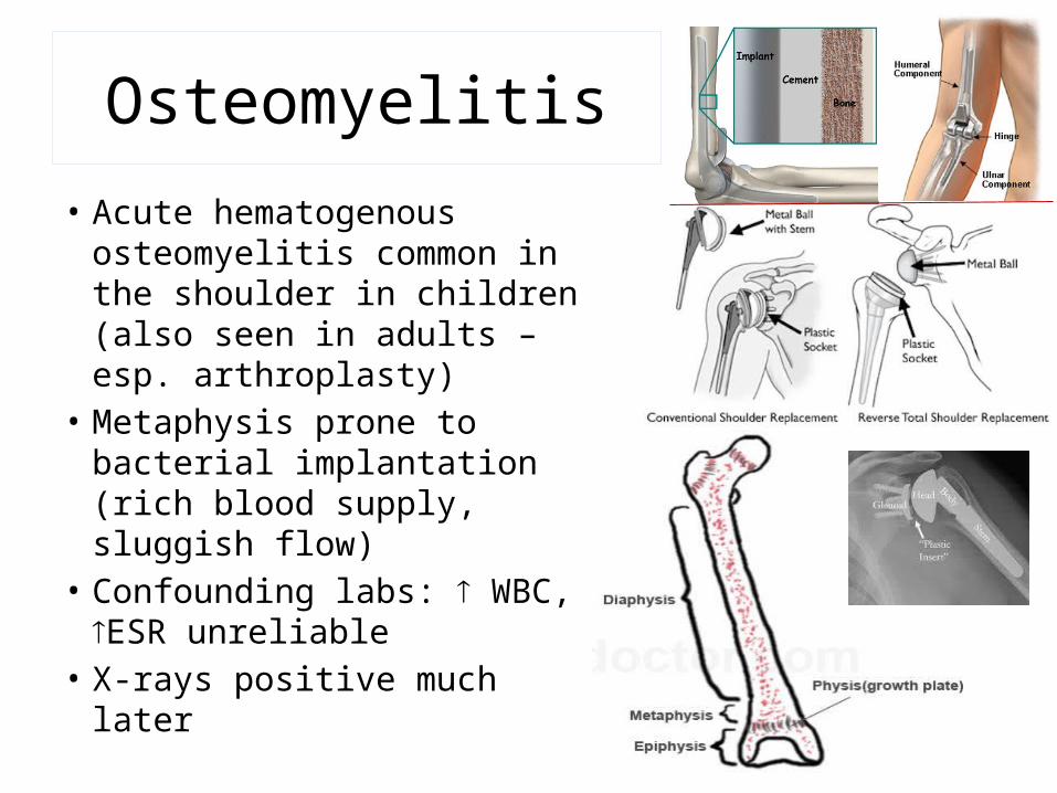
Osteomyelitis
• Acute hematogenous osteomyelitis common in the shoulder in children (also seen in adults – esp. arthroplasty)
• Metaphysis prone to bacterial implantation (rich blood supply, sluggish flow)
• Confounding labs: WBC, ESR unreliable
• X-rays positive much later

Osteomyelitis vs. Cellulitis
Osteomyelitis
3-phase 99mTc – MDP bone imaging shows increasing tracer accumulation that becomes focal in the delayed phase.
Cellulites
Multiphase bone imaging presents as a diffusely increased soft tissue and bony activity in early phases, with decreasing activity on later phases

Fig 2. Infantile and adult osteomyelitis: (A) Anterior delayed static image and (B) Posterior static image. Febrile infant withnegative plain radiographs protecting left upper extremity. Diffuse uptake in humeral shaft with photopenic defect in humeral head.(Just above arrow) is a common manifestation of osteomyelitis in neonates and infants,
(C) (D) Anterior and posterior static imagesof humeral head osteomyelitis in an adult, Diffuse increase uptake (Arrow).
Osteomyelitis: Adult vs. Child

WBC imaging: Osteomyelitis
• sensitivity & specificity for infection• assessment of infected prosthesis
99mTc-Sulfur colloid 99mTc-Sulfur colloid
Positive Negative

WBC imaging: Fever of Unknown Origin
(A) Anterior and (B) Posterior image of shoulder. Marked aeeumulation of labeled WBCs around shoulder (Arrow), (C) CT Scan--shows fluid in and around shoulder (Arrowheads) with soft tissue inflammation.
Fig 3. Shoulder Abscess--Indium 111 WBC scan: Elderly bedridden patient undergoing evaluation for fever of unknown origin.

Stress Fractures - HumerusRingmans lesion
• Cortical desmoid like lesion in the proximal humerus (esp. in gymnasts)
• Seen in antero-lateral aspect at pectoralis major tendon insertion site

Posttraumatic Myositis Ossificans
• Seen in contact sports (eg. karate)
• Early x-rays –ve, w/ soft tissue mass corresponding to hard palpable mass in area of pain.
• Pain relief by Sx removal of heterotopic bone
• TPBS used to age lesion/ confirm maturation.
Radiograph of the humerus, taken 6 weeks after injury, denoting maturing
myositis ossificans with increased peripheral mineralization

Avascular Necrosis (AVN)
• interruption of blood supply to bone edema intraosseous pressure
• Most common: Sickle cell disease AVN of humeral head
• Other reasons: traumatic interruption of blood supply, fat embolism, lupus vasculitis, and radiation osteonecrosis.
• Photopenic defects in bone scintigraphs (recent infarcts)
• MRI preferred

Fractures & Non-Union of Fracture
Better prognosis
Nonunion of humeral neck fracture: (A) Oblique delayed static view shows increased uptake at site of nonunion, reactive type. (Atopic photopenic)

Osteoid Osteoma
• Relatively common• Benign bone tumor in
adolescents and young adults
• Presented with pain• Majority seen in
diaphysis of long bones• Hot central nidus with
cooler periphery on TPBS
Osteoid osteoma of humeral shaft in a young adult male: (A) Blood pool image. Moderate hyperemia, with a central round focus of more intense uptake. (B) Delayed static image-- focal intense increased uptake. (C)(D) Plain radiograph and CT scan show cortical sclerosis and focal lucency. Definite nidus not visualized.

Overuse Elbow InjuriesOsteochondritis Dissecans (OCD)
• Results from repetitive compressive force between radial head and capitellum.
• most frequent site is capitellum of elbow
• If untreated, may lead to loose intraarticular bodies, loss of motion, and locking and clicking

• Overuse at the margin of extensor carpi radialis brevis muscle
Overuse Elbow InjuriesLateral Epicondylitis (Tennis elbow)
Medial Epicondylitis: Delayed static image shows focal increased uptake in medial epicondyle. Plain radiographs negative.

Overuse Elbow InjuriesMedial Epicondylitis (Golfer’s elbow)
• Secondary to overuse at the insertion of flexor and pronator muscle groups

Stress fracture• Occult fracture: fracture with or without
trauma
Occult radial head fracture: Normal elbow x-ray films following injury with persistent pain
(A) Blood pool images showsfocal hyperemia left radial head (Arrowhead). Injection site inright antecubital fossa, partially shielded. (B) Delayed staticimage anterior with intense uptake (Arrow). (C) Delayed lateralshows focal uptake in radial head (Arrow).
Ulnar Stress Fractures: (A) Delayed static images show bilateral mid-ulnar shafts stress fractures in a weight lifter, less focally fusiform than usually seen in shafts of long bones. (B) Ulnar styloid avuision fracture delayed static image shows focal uptake at styloid process.

Stress FractureRadial & Ulnar styloid avulsion fracture
• Involves the brachioradialis tendon insertion at the styloid process of radius
• Small bone fragments not visible in x-ray
Delayed static dorsal projection shows focal increased uptake at site of avulsion (Arrow).