6.3 Classification and Anatomy of Bones 150 and Bone 6.4 ...
27
MODULE 5: SKELETAL SYSTEM OUTLINE 6.1 Cartilage 147 6.1a Functions of Cartilage 147 6.1b Growth Patterns of Cartilage 148 6.2 Bone 148 6.2a Functions of Bone 148 6.3 Classification and Anatomy of Bones 150 6.3a General Structure and Gross Anatomy of Long Bones 150 6.4 Ossification 157 6.4a Intramembranous Ossification 157 6.4b Endochondral Ossification 157 6.4c Epiphyseal Plate Morphology 160 6.4d Growth of Bone 161 6.4e Blood Supply and Innervation 162 6.5 Maintaining Homeostasis and Promoting Bone Growth 163 6.5a Effects of Hormones 163 6.5b Effects of Vitamins 164 6.5c Effects of Exercise 165 6.5d Fracture Repair 165 6.6 Bone Markings 167 6.7 Aging of the Skeletal System 168 Cartilage and Bone 6 SKELETAL SYSTEM
Transcript of 6.3 Classification and Anatomy of Bones 150 and Bone 6.4 ...

MODULE 5: SKELETAL SYSTEM
O U T L I N E
6.1 Cartilage 147
6.1a Functions of Cartilage 1476.1b Growth Patterns of Cartilage 148
6.2 Bone 148
6.2a Functions of Bone 148
6.3 Classification and Anatomy of Bones 150
6.3a General Structure and Gross Anatomy of Long Bones 150
6.4 Ossification 157
6.4a Intramembranous Ossification 1576.4b Endochondral Ossification 1576.4c Epiphyseal Plate Morphology 1606.4d Growth of Bone 1616.4e Blood Supply and Innervation 162
6.5 Maintaining Homeostasis and Promoting Bone Growth 163
6.5a Effects of Hormones 1636.5b Effects of Vitamins 1646.5c Effects of Exercise 1656.5d Fracture Repair 165
6.6 Bone Markings 167
6.7 Aging of the Skeletal System 168
Cartilage and Bone
6 S K E L E T A L S Y S T E M
mck78097_ch06_146-172.indd 146mck78097_ch06_146-172.indd 146 2/14/11 3:40 PM2/14/11 3:40 PM

(b) Hyaline cartilage
(a)
Cartilages in nose
Cartilage in external ear
Cartilage of intervertebral disc
Respiratory tract cartilages in the lungs, trachea,
and larynx
Epiglottis
Larynx Lung Trachea
Articular cartilage of a joint
Costal cartilage
Pubic symphysis
Meniscus (padlike cartilage in knee joint)
Articular cartilage of a joint
Extracellular matrix
Lacuna (with chondrocyte)
Extracellular matrix Collagen fibers
Elastic fibers
Extracellular matrix Hyaline cartilage Fibrocartilage Elastic cartilage
Lacunae (with chondrocytes)
Lacunae (with chondrocytes)
(d) Elastic cartilage
(c) Fibrocartilage
LM 180x
LM 80x
LM 200x
Chapter Six Cartilage and Bone 147
Figure 6.1Distribution of Cartilage in an Adult. (a) Three types of cartilage are found within an adult. Photomicrographs show (b) hyaline cartilage, (c) fibrocartilage, and (d ) elastic cartilage.
Mention of the skeletal system conjures up images of dry, lifeless bones in various sizes and shapes. But the skeleton (skel e -ton;
skeletos = dried) is much more than a supporting framework for the soft tissues of the body. The skeletal system is composed of dynamic living tissues; it interacts with all of the other organ systems and continually rebuilds and remodels itself. Our skeletal system includes the bones of the skeleton as well as cartilage, ligaments, and other connective tissues that stabilize or connect the bones. Bones support our weight and interact with muscles to produce precisely controlled movements. This interaction permits us to sit, stand, walk, and run. Further, our bones serve as vital reservoirs for calcium and phospho-rus. Before concentrating on bone connective tissue, we first examine the cartilage components of the skeleton.
6.1 CartilageLearning Objectives: 1. Explain the functions of cartilage. 2. Describe the function and distribution of hyaline cartilage,
fibrocartilage, and elastic cartilage. 3. Explain both the interstitial and appositional growth of
cartilage.
Cartilage is found throughout the human body (figure 6.1). Cartilage is a semirigid connective tissue that is weaker than bone, but more flexible and resilient (see chapter 4). As with all connec-tive tissue types, cartilage contains a population of cells scattered throughout a matrix of protein fibers embedded within a gel-like ground substance. Chondroblasts (kon dro -blast; chondros = grit or gristle, blastos = germ) are the cells that produce the matrix of cartilage. Once they become encased within the matrix they have produced and secreted, the cells are called chondrocytes (kon dro -sı t; cyte = cell) and occupy small spaces called lacunae. These mature cartilage cells maintain the matrix and ensure that it remains healthy and viable. Mature cartilage is avascular (not penetrated by blood vessels) so nutrients must diffuse through the matrix. The three different types of cartilage—hyaline, elastic, and fibrocartilage—are described in detail in chapter 4, so only cartilage functions, locations, and growth will be discussed in this chapter.
6.1a Functions of Cartilage
Cartilage has three major functions in the body:
■ Supporting soft tissues. For example, C-shaped hyaline cartilage rings in the trachea support the connective tissue and musculature of the tracheal wall, fibrocartilage
entionn o off the skeletal system conjures up images of dry, lifelessbobones in various sizes and shapes. But the skeleton (skel e -ton;
skskeleletos = dried) is much more than a supporting framework for the=soft tissues of the body. The skeletal system is composed of dynamicliving tissues; it interacts with all of the other organ systems andcontinually rebuilds and remodels itself. Our skeletal system includesthe bones of the skeleton as well as cartilage, ligaments, and otherconnective tissues that stabilize or connect the bones. Bones supportour weight and interact with muscles to produce precisely controlledmovements. This interaction permits us to sit, stand, walk, and run.Further, our bones serve as vital reservoirs for calcium and phospho-rus. Before concentrating on bone connective tissue, we first examinethe cartilage components of the skeleton.
mck78097_ch06_146-172.indd 147mck78097_ch06_146-172.indd 147 2/14/11 2:36 PM2/14/11 2:36 PM

148 Chapter Six Cartilage and Bone
Study Tip!You can do a quick overnight experiment to demonstrate what
would happen to our body shape if the composition of our bones changed. Obtain the “wishbone” (fused clavicles) from a chicken or game hen and observe its physical characteristics. Next, place the bone in a glass container of vinegar. Let it stand overnight, and then examine the bone. You should see the following changes: (1) The bone is darker because the acid in the vinegar has dissolved the calcium phosphate in the bone, and (2) the bone is somewhat limp like a wet noodle because it has lost its strength due to the removal of the calcium phosphate from the bone.
provides both toughness and flexibility to the pubic symphysis and intervertebral discs, and flexible elastic cartilage supports the fleshy, external part of the ear called the auricle (aw ri-kl; auris = ear).
■ Providing a gliding surface at articulations (joints), where two bones meet.
■ Providing a model for the formation of most of the bones in the body. Beginning in the embryonic period, cartilage serves as a “rough draft” form that is later replaced by bone tissue.
6.1b Growth Patterns of Cartilage
Cartilage grows in two ways. Growth from within the cartilage itself is termed interstitial (in-ter-stish a l) growth. Growth along the cartilage’s outside edge, or periphery, is called appositional(ap-o -zish u n-a l) growth (figure 6.2).
Interstitial Growth
Interstitial growth occurs through a series of steps:
1. Chondrocytes housed in lacunae undergo mitotic cell division.
2. Following cell division, the two new cells occupy a single lacuna.
3. As the cells begin to synthesize and secrete new cartilage matrix, they are pushed apart and now reside in their own lacunae.
4. The new individual cells within their own lacunae are called chondrocytes. New matrix has been produced internally, and thus interstitial growth has occurred.
Appositional Growth
Appositional growth also occurs through a series of defined steps:
1. Stem cells at the internal edge of the perichondrium begin to divide, forming new stem cells and committed cells.
2. The committed cells differentiate into chondroblasts. 3. These chondroblasts, located at the periphery of the old
cartilage, begin to produce and secrete new cartilage matrix. As a result, they push apart and become chondrocytes, each occupying its own lacuna.
4. The new matrix has been produced peripherally, and thus appositional growth has occurred.
During early embryonic development, both interstitial and appositional cartilage growth occur simultaneously. However, interstitial growth declines rapidly as the carti-lage matures because the cartilage becomes semirigid as it matures, and the matrix is no longer able to expand. Further growth can occur only at the periphery of the tissue, so later growth is primarily appositional. Once the cartilage is fully mature, new cartilage growth typically stops entirely. From this point on, cartilage growth usually occurs only after injury to the cartilage.
WHAT DID YOU LEARN?
●1 How do the three cartilage types differ with respect to their locations and functions in the body?
●2 Compare and contrast interstitial and appositional growth of cartilage. In older cartilage, which type of growth predominates?
WW
6.2 BoneLearning Objective: 1. Explain the functions of bone.
The bones of the skeleton are complex, dynamic organs containing all tissue types. Their primary component is bone con-nective tissue, also called osseous (os e -u s) connective tissue (see chapter 4). In addition, they contain connective tissue proper (peri-osteum), cartilage connective tissue (articular cartilage), smooth muscle tissue (forming the walls of blood vessels that supply bone), fluid connective tissue (blood), epithelial tissue (lining the inside opening of blood vessels), and nervous tissue (nerves that supply bone). The matrix of bone connective tissue is sturdy and rigid due to deposition of minerals in the matrix, a process called calcifica-tion (kal si-fi-ka shu n), or mineralization.
6.2a Functions of Bone
Bone connective tissue and the bones that compose the skeletal system perform several basic functions: support and protection, movement, hemopoiesis, and storage of mineral and energy reserves.
Support and Protection
Bones provide structural support and serve as a framework for the entire body. Bones also protect many delicate tissues and organs from injury and trauma. The rib cage protects the heart and lungs, the cranial bones enclose and protect the brain, the vertebrae enclose the spinal cord, and the pelvis cradles some digestive, uri-nary, and reproductive organs.
Movement
Individual groups of bones serve as attachment sites for skeletal muscles, other soft tissues, and some organs. Bones of the skeleton function as levers that are pulled when skeletal muscles contract. The direction and magnitude of the forces generated by the skeletal muscles can be altered by bones. Potential movements range from powerful contractions needed for running and jumping to delicate, precise movements required to remove a splinter from the finger.
Hemopoiesis
The process of blood cell production is called hemopoiesis(he mo -poy-e sis; haima = blood, poiesi = making). Blood cells are produced in a connective tissue called red bone marrow, which is located in some spongy bone. Red bone marrow contains stem cells that form all of the formed elements in the blood.
mck78097_ch06_146-172.indd 148mck78097_ch06_146-172.indd 148 2/14/11 2:36 PM2/14/11 2:36 PM

Matrix
Chondrocyte
Lacuna
New matrix
Perichondrium Dividing undifferentiated stem cell
Chondroblast
Committed cells differentiating into chondroblasts
Chondrocyte
Mature chondrocyte
Perichondrium
Hyaline cartilage
Two cells produced by mitosis of one chondrocyte now occupy one lacuna.
Mitotic activity occurs in stem cells within the perichondrium.
RESULT: New chondrocytes and more matrix are produced as cartilage grows internally.
Committed cells produced by stem cell mitosis differentiate into chondroblasts.
Chondroblasts produce new matrix near the periphery and become chondrocytes. RESULT: New cells and more matrix are produced as cartilage grows peripherally.
New cartilage matrix
Older cartilage matrix
Undifferentiated stem cells
Chondrocyte within lacuna begins to exhibit mitotic activity.
Appositional Growth Interstitial Growth
Each cell produces new matrix and begins to separate from its neighbor.
New cartilage matrix
Older cartilage matrix
Chondroblasts secreting new matrix
Undifferentiated stem cells
Undifferentiated stem cells
Perichondrium
(b)(a)
1
2
3
4
1
2
3
4
Chapter Six Cartilage and Bone 149
Figure 6.2Formation and Growth of Cartilage. Cartilage grows either from within (interstitial growth) or at its edge (appositional growth). (a) In interstitial growth, chondrocytes within lacunae divide to form two chondroblasts; these cells grow, begin to produce new matrix, and push apart from each other, forming two new chondrocytes. (b) In appositional growth, cartilage grows when stem cells at the internal edge of the perichondrium divide. Differentiation of committed cells into chondroblasts results in the formation of new cartilage matrix and the differentiation of these cells into chondrocytes within the inner layer of the perichondrium.
mck78097_ch06_146-172.indd 149mck78097_ch06_146-172.indd 149 2/14/11 2:36 PM2/14/11 2:36 PM

Long bone (femur)
Flat bone (frontal bone)
Irregular bone (vertebra)
Short bone (tarsal bone)
150 Chapter Six Cartilage and Bone
Figure 6.3Classification of Bone by Shape. Four different classes of bone are recognized according to shape: long, short, flat, and irregular.
The locations of red bone marrow differ between children and adults. In children, red bone marrow is located in the spongy bone and the medullary cavity of most of the bones of the body. As children mature into adults, much of the red bone marrow degen-erates and turns into a fatty tissue called yellow bone marrow. As a result, adults have red bone marrow only in selected portions of the axial skeleton, such as the flat bones of the skull, the vertebrae, the ribs, the sternum (breastbone), and the ossa coxae (hip bones). Adults also have red bone marrow in the proximal epiphyses of each humerus and femur.
Storage of Mineral and Energy Reserves
More than 90% of the body’s reserves of the minerals calcium and phosphate are stored and released by bone. Calcium is an essential mineral for such body functions as muscle contraction, blood clot-ting, and nerve impulse transmission. Phosphate is needed for ATP utilization, among other things. When calcium or phosphate is needed by the body, some bone connective tissue is broken down, and the minerals are released into the bloodstream. In addition, potential energy in the form of lipids is stored in yellow bone mar-row, which is located in the shafts of long bones.
WHAT DID YOU LEARN?
●3 Briefly describe at least four functions of bone.
6.3 Classification and Anatomy of BonesLearning Objectives: 1. Identify the characteristics of long, short, flat, and irregular
bones. 2. Describe the gross anatomy of a long bone. 3. Compare the microscopic anatomy of compact bone and
spongy bone.
Bones of the human skeleton occur in various shapes and sizes, depending on their function. The four classes of bone as determined by shape are long bones, short bones, flat bones, and irregular bones (figure 6.3). Long bones have a greater length than width. These bones have an elongated, cylindrical shaft (diaphysis). This is the most common bone shape. Long bones are found in the upper limb (namely, the arm, forearm, palm, and fingers) and lower limb (thigh, leg, sole of the foot, and toes). Long bones vary in size; the small bones in the fingers and toes are long bones, as are the larger tibia and fibula of the lower limb. Short bones have a length nearly equal to their width. The external surfaces of short bones are covered by compact bone, and their interior is composed of spongy bone. Examples of short bones include the carpals (wrist bones) and tarsals (bones in the foot). Sesamoid bones, which are tiny, seed-shaped bones along the tendons of some muscles, are also classified as short bones. The patella (kneecap) is the largest sesamoid bone. Flat bones are so named because they have flat, thin sur-faces. These bones are composed of roughly parallel surfaces of compact bone with a layer of internally placed spongy bone. They provide extensive surfaces for muscle attachment and protect underlying soft tissues. Flat bones form the roof of the skull, the scapulae (shoulder blades), the sternum (breastbone), and the ribs.
WW
Irregular bones have elaborate, complex shapes and do not fit into any of the preceding categories. The vertebrae, ossa coxae (hip bones), and several bones in the skull, such as the ethmoid and sphenoid bones, are examples of irregular bones.
WHAT DO YOU THINK?
●1 Why is the rib classified as a flat bone instead of a long bone? Describe the features and functions of flat bones and long bones, then compare these to the features and functions of a rib.
6.3a General Structure and Gross Anatomy
of Long Bones
Long bones, the most common bone shape in the body, serve as a useful model of bone structure. Two examples of long bones are the femur (thigh bone) and the humerus (arm bone) (figure 6.4). A typical long bone contains the following parts:
■ One of the principal gross features of a long bone is its shaft, or diaphysis (dı -af i-sis; pl., diaphyses, dı -af i-se z; growing between). The elongated, usually cylindrical diaphysis provides for the leverage and major weight support of a long bone.
mck78097_ch06_146-172.indd 150mck78097_ch06_146-172.indd 150 2/14/11 2:36 PM2/14/11 2:36 PM

(a) Anterior view
Proximal epiphysis
Diaphysis (shaft)
Metaphysis
Metaphysis
Metaphysis
Distal epiphysis
(b) Sectional view (c)
Spongy bone (contains red bone marrow)
Compact bone
Endosteum
Medullary cavity (contains yellow bone marrow in adult)
Nutrient artery through nutrient foramen
Proximal epiphysis
Perforating fibers
Periosteum
Diaphysis
Epiphyseal line
Epiphyseal line
Distal epiphysis
Metaphysis
Articular cartilage
Articular cartilage
Chapter Six Cartilage and Bone 151
Figure 6.4Gross Anatomy of a Long Bone. Long bones support soft tissues in the limbs. The femur, the bone of the thigh, is shown in both (a) anterior and (b) sectional views. (c) A typical long bone, such as the humerus, contains both compact and spongy bone.
■ At each end of a long bone is an expanded, knobby region called the epiphysis (e-pif i-sis; pl., epiphyses, e-pif i-se z; epi = upon, physis = growth). The epiphysis is enlarged to strengthen the joint and provide added surface area for bone-to-bone articulation as well as tendon and ligament attachment. It is composed of an outer layer of compact bone and an inner layer of spongy bone. A proximal epiphysis is the end of the bone closest to the body trunk, and a distal epiphysis is the end farthest from the trunk.
■ The metaphysis (me -taf i-sis) is the region in a mature bone sandwiched between the diaphysis and the epiphysis. In a growing bone, this region contains the epiphyseal (growth) plate, thin layers of hyaline cartilage that provide for the continued lengthwise growth of the diaphysis. In adults, the
remnant of the epiphyseal plate is a thin layer of compact bone called the epiphyseal line.
■ The thin layer of hyaline cartilage covering the epiphysis at a joint surface is called articular cartilage. This cartilage helps reduce friction and absorb shock in movable joints.
■ The hollow, cylindrical space within the diaphysis is called the medullary cavity (marrow cavity). In adults, it contains yellow bone marrow.
■ The endosteum (en-dos te -u m; endo = within, osteon = bone) is an incomplete layer of cells that covers all internal surfaces of the bone, such as the medullary cavity. The endosteum contains osteoprogenitor cells, osteoblasts, and osteoclasts (figure 6.5), and is active during bone growth, repair, and remodeling.
mck78097_ch06_146-172.indd 151mck78097_ch06_146-172.indd 151 2/14/11 4:14 PM2/14/11 4:14 PM
(a) Periosteum Compact bone Periosteum
Endosteum
Fibrous layer
Circumferential lamellae
Cellular layer
Osteocyte in lacuna
Perforating fibers
Bone matrix
Endosteum
Osteoclast
Osteoprogenitor cell
Canaliculi
Osteocyte in lacuna
Osteoid
Osteoblasts
(b) Endosteum
Periosteum
Canaliculi
152 Chapter Six Cartilage and Bone
Study Tip!A long bone is similar in shape to a barbell: The barbell’s rounded
ends represent the epiphyses, and its cylindrical handle is the diaphysis.
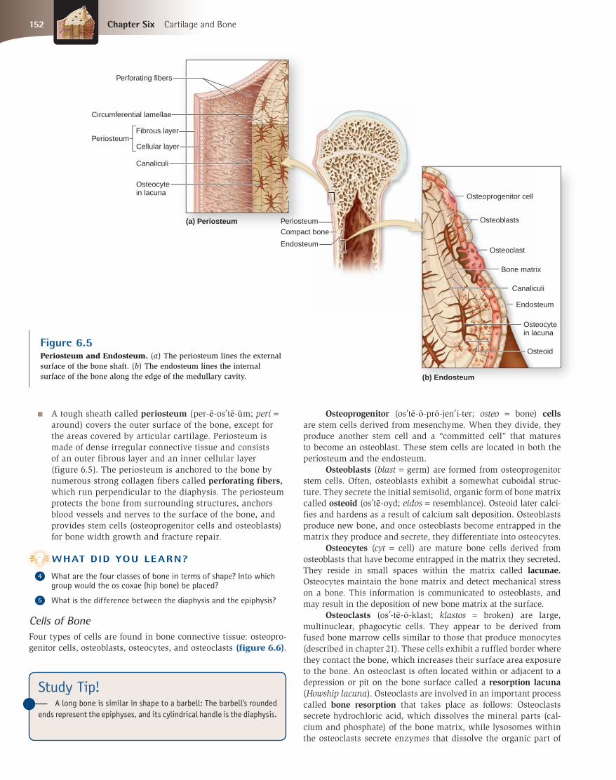
Figure 6.5Periosteum and Endosteum. (a) The periosteum lines the external surface of the bone shaft. (b) The endosteum lines the internal surface of the bone along the edge of the medullary cavity.
■ A tough sheath called periosteum (per-e -os te -u m; peri = around) covers the outer surface of the bone, except for the areas covered by articular cartilage. Periosteum is made of dense irregular connective tissue and consists of an outer fibrous layer and an inner cellular layer (figure 6.5). The periosteum is anchored to the bone by numerous strong collagen fibers called perforating fibers, which run perpendicular to the diaphysis. The periosteum protects the bone from surrounding structures, anchors blood vessels and nerves to the surface of the bone, and provides stem cells (osteoprogenitor cells and osteoblasts) for bone width growth and fracture repair.
WHAT DID YOU LEARN?
●4 What are the four classes of bone in terms of shape? Into which group would the os coxae (hip bone) be placed?
●5 What is the difference between the diaphysis and the epiphysis?
Cells of Bone
Four types of cells are found in bone connective tissue: osteopro-genitor cells, osteoblasts, osteocytes, and osteoclasts (figure 6.6).
WW
Osteoprogenitor (os te -o -pro -jen i-ter; osteo = bone) cellsare stem cells derived from mesenchyme. When they divide, they produce another stem cell and a “committed cell” that matures to become an osteoblast. These stem cells are located in both the periosteum and the endosteum.
Osteoblasts (blast = germ) are formed from osteoprogenitor stem cells. Often, osteoblasts exhibit a somewhat cuboidal struc-ture. They secrete the initial semisolid, organic form of bone matrix called osteoid (os te -oyd; eidos = resemblance). Osteoid later calci-fies and hardens as a result of calcium salt deposition. Osteoblasts produce new bone, and once osteoblasts become entrapped in the matrix they produce and secrete, they differentiate into osteocytes.
Osteocytes (cyt = cell) are mature bone cells derived from osteoblasts that have become entrapped in the matrix they secreted. They reside in small spaces within the matrix called lacunae. Osteocytes maintain the bone matrix and detect mechanical stress on a bone. This information is communicated to osteoblasts, and may result in the deposition of new bone matrix at the surface.
Osteoclasts (os -te -o -klast; klastos = broken) are large, multinuclear, phagocytic cells. They appear to be derived from fused bone marrow cells similar to those that produce monocytes (described in chapter 21). These cells exhibit a ruffled border where they contact the bone, which increases their surface area exposure to the bone. An osteoclast is often located within or adjacent to a depression or pit on the bone surface called a resorption lacuna (Howship lacuna). Osteoclasts are involved in an important process called bone resorption that takes place as follows: Osteoclasts secrete hydrochloric acid, which dissolves the mineral parts (cal-cium and phosphate) of the bone matrix, while lysosomes within the osteoclasts secrete enzymes that dissolve the organic part of
mck78097_ch06_146-172.indd 152mck78097_ch06_146-172.indd 152 2/14/11 2:36 PM2/14/11 2:36 PM

Osteoprogenitor cells develop into osteoblasts.
Osteoblast (forms bone matrix)
Some osteoblasts differentiate into osteocytes.
Osteocyte (maintains bone matrix)
Fused bone marrow cell
Endosteum
Lysosomes
Nuclei
Osteoclast
Ruffled border
Resorption lacuna
Osteocytes
Osteoblasts
Osteoclast
LM 400x
(c) Bone tissue
(a) Bone cells
(b) Osteoclast
Chapter Six Cartilage and Bone 153
Figure 6.6Types of Cells in Bone Connective Tissue. Four different types of cells are found in bone connective tissue. (a) Osteoprogenitor cells develop into osteoblasts, many of which differentiate to become osteocytes. (b) Bone marrow cells fuse to form osteoclasts. (c) A photomicrograph shows osteoblasts, osteocytes, and an osteoclast.
the matrix (described in the next section). The release of the stored calcium and phosphate from the bone matrix is called osteolysis(os-te -ol i-sis; lysis = dissolution, loosening). The liberated calcium and phosphate ions enter the tissue fluid and then the blood. Osteoclasts remove matrix and osteoblasts add to it, maintain-ing a delicate balance. Osteoblast and osteoclast activity may be affected by hormonal levels (discussed at the end of the chapter), the body’s need for calcium and/or phosphorus, and gravitational or mechanical stressors to bone. For example, when a person wears orth-odontic braces, osteoblasts and osteoclasts work together to modify the tooth-jaw junction, in response to the mechanical stress applied by the braces to the teeth and jaw. If osteoclasts resorb the bone to remove calcium salts at a faster rate than osteoblasts produce matrix to stimulate deposition, bones lose mass and become weaker; in con-trast, when osteoblast activity outpaces osteoclast activity, bones have a greater mass.
Composition of the Bone Matrix
The matrix of bone connective tissue has both organic and inor-ganic components. About one-third of bone mass is composed of organic components, including cells, collagen fibers, and ground substance. The collagen fibers give a bone tensile strength by resist-ing stretching and twisting, and contribute to its overall flexibility. The ground substance is the semisolid material that suspends
and supports the collagen fibers. The inorganic components of the bone provide its compressional strength. Calcium phosphate, Ca3(PO4)2, accounts for most of the inorganic components of bone. Calcium phosphate and calcium hydroxide interact to form crystals of hydroxyapatite (hı -drok se -ap-a -tı t), which is Ca10(PO4)6(OH)2. These crystals deposit around the collagen fibers in the extra-cellular matrix, leading to hardening of the matrix. The crystals also incorporate other salts, such as calcium carbonate, and ions, such as sodium, magnesium, sulfate, and fluoride, in the process of calcification.
Comparison of Compact and Spongy Bone
Two types of bone connective tissue are present in most of the bones of the body: compact bone (also called dense or cortical bone) and spongy bone (also called cancellous or trabecular bone). As their names imply, compact bone is solid and relatively dense, whereas spongy bone appears more porous, like a sponge. The arrangement of compact bone and spongy bone components dif-fers at the microscopic level. Spongy bone forms an open lattice of narrow plates of bone, called trabeculae (tra -bek u -le ; sing., trabecula, tra -bek u -la ; trabs = a beam). In a long bone, compact bone forms the solid external walls of the bone, and spongy bone is located internally, primarily within the epiphyses. In a flat bone of the skull, the spongy bone, also called diploë (dip lo -e ;
mck78097_ch06_146-172.indd 153mck78097_ch06_146-172.indd 153 2/14/11 4:17 PM2/14/11 4:17 PM

Spongy bone (diploë)
Flat bone of skull Periosteum
Periosteum Compact bone
154 Chapter Six Cartilage and Bone
CLINICAL VIEW
Osteitis DeformansOsteitis deformans (Paget disease of bone) was first described by Sir James Paget in 1877. The disease results from a disruption in the bal-ance between osteoclast and osteoblast function. It is characterized by excessive bone resorption (excessive osteoclast activity) followed by excessive bone deposition (excessive osteoblast activity). The resulting bone is structurally unstable and immature.
In osteitis deformans, the osteoclasts are anatomically and physiologically abnormal; they are five times larger than normal and may contain 20 or more nuclei (compared to about 3 to 5 nuclei in normal osteoclasts). These larger osteoclasts resorb bone at a higher rate than normal. In response to this excessive bone resorption, the osteoblasts (which are normal-sized) deposit additional bone, but this new bone is poorly formed, making it more susceptible to deformation and fractures.
Osteitis deformans most commonly occurs in the bones of the pelvis, skull, vertebrae, femur (thigh bone), and tibia (leg bone). Initial symptoms include bone deformity and pain. Eventually, the lower limb bones may be bowed, and the skull often becomes thicker and enlarged. Biochemical tests can measure the level of osteoclast activity. There
is no cure for osteitis deformans, but medications can reduce bone pain and bone resorption by osteoclasts.
Figure 6.7Flat Bones Within the Skull. These bones are composed of two layers of compact bone, with a region of spongy bone (diploë) sandwiched between. Both layers of compact bone are covered by periosteum.
diplous = double), is sandwiched between two layers of compact bone (figure 6.7).
Compact Bone Microscopic Anatomy Compact bone has an organized structure when viewed under the microscope. A cylin-drical osteon (os te -on; bone), or Haversian system, is the basic functional and structural unit of mature compact bone. Osteons run parallel to the diaphysis of the long bone. An osteon is a three-dimensional structure that has several components (figures 6.8 and 6.9a, b).
■ The central canal (Haversian canal) is a cylindrical channel that lies in the center of the osteon. Traveling within the central canal are the blood vessels and nerves that supply the bone.
■ Concentric lamellae (la-mel -e; sing., lamella, la-mel a; lamina = plate, leaf) are rings of bone connective tissue that surround the central canal and form the bulk of the osteon. The numbers of concentric lamellae vary among osteons. Each lamella contains collagen fibers oriented in one direction; adjacent lamellae contain collagen fibers oriented in alternating directions. In other words, if one lamella has collagen fibers directed superiorly and to the right, the next lamella will have collagen fibers directed superiorly and to the left. This alternating collagen fiber direction gives bone part of its strength and resilience.
■ Osteocytes are housed in lacunae and are found between adjacent concentric lamellae.
■ Canaliculi (kan-a-lik u-lı; sing., canaliculus, kan-a-lik u-lus; canalis = canal) are tiny, interconnecting channels within the bone connective tissue that extend from each lacuna, travel through the lamellae, and connect to other lacunae and the central canal. Canaliculi house osteocyte cytoplasmic projections that permit intercellular contact and communication. Thus, nutrients, minerals, gases, and wastes can travel through these passageways between the central canal and the osteocytes.
Several other structures are found in compact bone, but are not part of the osteon proper, including the following (see figure 6.8):
■ Perforating canals (Volkmann canals) resemble central canals in that they also contain blood vessels and nerves.
Lateral x-ray of a skull with Paget disease. White arrows
indicate areas of excessive bone deposition.
mck78097_ch06_146-172.indd 154mck78097_ch06_146-172.indd 154 2/14/11 2:36 PM2/14/11 2:36 PM

Collagen fiber orientation
Periosteum
External circumferential
lamellae
Osteon
Central canal
Central canal
Perforating fibers
Perforating canals
Trabeculae of spongy bone
Central canal
Artery Vein
Nerve
Canaliculi
Osteocyte
Concentric lamellae
Canaliculi
Lacuna
Osteon
Trabeculae
Space for bone marrow
Osteoclast
Osteoblasts aligned along trabecula of new bone
Osteocyte in lacuna
Interstitial lamellae
Canaliculi opening at surface
Endosteum
Cellular layer
Parallel lamellae
Fibrous layer
Interstitial lamellae
Chapter Six Cartilage and Bone 155
Figure 6.8Components of Bone. An expanded section of the humerus shows the arrangement of osteons within the compact bone in the diaphysis, and the relationship of the compact bone to both spongy bone and the medullary cavity.
mck78097_ch06_146-172.indd 155mck78097_ch06_146-172.indd 155 2/14/11 2:36 PM2/14/11 2:36 PM

Lacuna
Trabeculaof spongy
boneRed bonemarrowOsteoblasts
Osteon
Osteon
Central canal
Concentriclamellae
Canaliculi
Central canal
Lacunae
(b) Compact bone
(a) Compact bone
(c) Spongy bone
SEM 1040x
LM 75x
LM 25x
156 Chapter Six Cartilage and Bone
Figure 6.9Microscopic Anatomy of Bone. (a) SEM and (b) light micrograph of osteons in a cross section of bone. (c) Light micrograph of spongy bone.
However, perforating canals run perpendicular to the central canals and help connect multiple central canals, thus creating a vascular and innervation connection among the multiple osteons.
■ Circumferential lamellae are rings of bone immediately internal to the periosteum of the bone (external circumferential lamellae) or internal to the endosteum (internal circumferential lamellae). These two distinct regions appear during the original formation of the bone. Both external and internal circumferential lamellae run the entire circumference of the bone itself (hence, their name).
■ Interstitial lamellae are the leftover parts of osteons that have been partially resorbed. They often look like a “bite” has been taken out of them. The interstitial lamellae are incomplete and typically have no central canal.
Spongy Bone Microscopic Anatomy Spongy bone contains no osteons (figure 6.9c). Instead, the trabeculae of spongy bone are composed of parallel lamellae. Between adjacent lamellae are osteocytes resting in lacunae, with numerous canaliculi radiat-ing from the lacunae. Nutrients reach the osteocytes by diffusion through canaliculi that open onto the surfaces of the trabeculae. Note that the trabeculae often form a meshwork of crisscrossing bars and plates of bone pieces. This structure provides great resis-tance to stresses applied in many directions by distributing the stress throughout the entire framework. As an analogy, visualize the jungle gym climbing apparatus on a children’s playground. It is capable of supporting the weight of numerous children whether they are distributed throughout its structure or all localized in one area. This is accomplished because stresses and forces are distrib-uted throughout the structure.
WHAT DO YOU THINK?
●2 Long bones typically contain both compact bone and spongy bone. What benefit does spongy bone provide? Why wouldn’t you want compact bone throughout the entire bone?
WHAT DID YOU LEARN?
●6 What are some of the organic and inorganic components of bone?
●7 If the activity of osteoblasts exceeds the activity of osteoclasts, how is the mass of the bone affected?
●8 Compare the following spaces in bone: central canal, canaliculi, and lacunae. How are they similar and different? Where is each type located?
WW
Study Tip!The analogy of an archery target can help you remember the
components of an osteon:
The entire target represents the osteon.
The bull’s-eye of the target is the central canal.
The rings of the target are the concentric lamellae.
mck78097_ch06_146-172.indd 156mck78097_ch06_146-172.indd 156 2/14/11 2:36 PM2/14/11 2:36 PM

Chapter Six Cartilage and Bone 157
6.4 OssificationLearning Objectives: 1. Describe and compare the processes of intramembranous
ossification and endochondral ossification.2. Explain the components of bone that enable it to grow and
be remodeled.
Ossification (os i-fi-ka shu n; os = bone, facio = to make), or osteogenesis (os te -o -jen e -sis; osteo = bone, genesis = beginning), refers to the formation and development of bone connective tissue. Ossification begins in the embryo and continues as the skeleton grows during childhood and adolescence. Even after the adult bones have formed, ossification continues, as will be described later in this section. By the eighth through twelfth weeks of development, the skeleton begins forming from either thickened condensations of mesenchyme or a hyaline carti-lage model of bone. Thereafter, these models are replaced by hard bone.
6.4a Intramembranous Ossification
Intramembranous (in tra -mem bra -nu s) ossification literally means “bone growth within a membrane,” and is so named because the thin layer of mesenchyme in these areas is sometimes referred to as a membrane. Intramembranous ossification is also sometimes called dermal ossification, because the mesenchyme that is the source of these bones is in the area of the future dermis. Recall from chapter 4 that mesenchyme is an embryonic con-nective tissue that has mesenchymal cells and abundant ground substance. Intramembranous ossification produces the flat bones of the skull, some of the facial bones (zygomatic bone, maxilla), the mandible (lower jaw), and the central part of the clavicle (col-larbone). It begins when mesenchyme becomes thickened and condensed with a dense supply of blood capillaries, and continues in several steps (figure 6.10):
1. Ossification centers form within thickened regions of mesenchyme. Beginning at the eighth week of development, some cells in the thickened, condensed mesenchyme divide, and the committed cells that result then differentiate into osteoprogenitor cells. Some osteoprogenitor cells become osteoblasts, which secrete the semisolid organic components of the bone matrix called osteoid. Multiple ossification centers develop within the thickened mesenchyme as the number of osteoblasts increases.
2. Osteoid undergoes calcification. Osteoid formation is quickly followed by initiation of the process of calcification, as calcium salts are deposited onto the osteoid and then crystallize (solidify). Both organic matrix formation and calcification occur simultaneously at several sites within the condensed mesenchyme. When calcification entraps osteoblasts within lacunae in the matrix, the entrapped cells become osteocytes.
3. Woven bone and its surrounding periosteum form. Initially, the newly formed bone connective tissue is immature and not well organized, a type called woven bone, or primary bone. This woven bone is
eventually replaced by lamellar bone, or secondary bone. The mesenchyme that still surrounds the woven bone begins to thicken and eventually organizes to form the periosteum. The bone continues to grow, and new osteoblasts are trapped in the expanding bone. Additional osteoblasts are continually produced as mesenchymal cells grow and develop. Newly formed blood vessels also branch throughout this region. The calcified trabeculae and intertrabecular spaces are composed of spongy bone.
4. Lamellar bone replaces woven bone, as compact bone and spongy bone form. Lamellar bone replaces the trabeculae of woven bone. On the internal and external surfaces, spaces between the trabeculae are filled, and the bone becomes compact bone. Internally, the trabeculae are modified slightly and produce spongy bone. The typical structure of a flat cranial bone results: two external layers of compact bone with a layer of spongy bone in between.
6.4b Endochondral Ossification
Endochondral (en-do -kon dra l; endo = within, chondral = car-tilage) ossification begins with a hyaline cartilage model and produces most of the other bones of the skeleton, including those of the upper and lower limbs, the pelvis, the vertebrae, and the ends of the clavicle. Long bone development in the limb is a good example of this process, which takes place in the following steps (figure 6.11):
1. The fetal hyaline cartilage model develops. During the eighth to twelfth week of development, chondroblasts secrete cartilage matrix, and a hyaline cartilage model forms. Within this cartilage model, the chondroblasts have become chondrocytes trapped within lacunae. A perichondrium surrounds the cartilage.
2. Cartilage calcifies, and a periosteal bone collar forms. Within the center of the cartilage model (future diaphysis), chondrocytes start to hypertrophy (enlarge) and resorb (eat away) some of the surrounding cartilage matrix, producing larger holes in the matrix. As these chondrocytes enlarge, the cartilage matrix begins to calcify. Chondrocytes in this region die and disintegrate because nutrients cannot diffuse to them through this calcified matrix. The result is a calcified cartilage shaft with large holes in the place where chondrocytes once were.
As the cartilage in the shaft is calcifying, blood vessels grow toward the cartilage and start to penetrate the perichondrium around the shaft. Stem cells within the perichondrium divide to form osteoblasts. As the osteoblasts develop and this supporting connective tissue becomes highly vascularized, the perichondrium becomes a periosteum. The osteoblasts within the internal layer of the periosteum start secreting a layer of osteoid around the calcified cartilage shaft. The osteoid hardens and forms a periosteal bone collar around this shaft.
3. The primary ossification center forms in the diaphysis. A growth of capillaries and osteoblasts, called a
mck78097_ch06_146-172.indd 157mck78097_ch06_146-172.indd 157 2/14/11 2:36 PM2/14/11 2:36 PM

Mesenchymal cell
Collagen fiber
Ossification center
Osteoid
Osteoblast
Osteoblast Osteoid
Osteocyte
Newly calcified bone matrix
Mesenchyme condensing to form the periosteum
Trabecula of woven bone
Blood vessel
Periosteum
Osteoprogenitorcell
Compact bone
Spongy bone
Ossification centers form within thickened regions of mesenchyme.
1
Osteoid undergoes calcification.
2
Woven bone and surrounding periosteum form.
3
Lamellar bone replaces woven bone, as compact and spongy bone form.
4
Intramembranous Ossification
158 Chapter Six Cartilage and Bone
Figure 6.10Intramembranous Ossification.A flat bone in the skull forms from mesenchymal cells in a series of continuous steps.
mck78097_ch06_146-172.indd 158mck78097_ch06_146-172.indd 158 2/14/11 2:36 PM2/14/11 2:36 PM

Perichondrium
Developing periosteum
Epiphyseal capillaries
Calcified cartilage
Epiphyseal plate
Articular cartilage
Epiphyseal line (remnant of epiphyseal plate)
Spongy bone
Periosteal bone collar
Developing compact bone
Medullary cavity
Compact bone
Periosteum
Medullary cavity
Hyaline cartilage
Primary ossification center
Secondary ossification centers
Epiphyseal line
Deteriorating cartilage matrix
Blood vessel of periosteal bud
Epiphyseal blood vessel
Articular cartilage
Spongy bone
Epiphyseal plate
Fetal hyaline cartilage model develops.
1
Cartilage calcifies, and a periosteal bone collar forms around diaphysis.
2
Primary ossification center forms in the diaphysis.
3
Secondary ossification centers form in epiphyses.
4
Bone replaces cartilage, except the articular cartilage and epiphyseal plates.
5
Epiphyseal plates ossify and form epiphyseal lines.
6
Endochondral Ossification
Chapter Six Cartilage and Bone 159
Figure 6.11Endochondral Ossification. Endochondral ossification of a long bone occurs in progressive stages. Bone growth is complete when each epiphyseal plate has ossified and the epiphyseal line has formed. Depending on the bone, epiphyseal plate ossification occurs between the ages of 10 and 25 years.
periosteal bud, extends from the periosteum into the core of the cartilage shaft, invading the spaces left by the chondrocytes. The remains of the calcified cartilage serve as a template on which osteoblasts begin to produce osteoid. This region, where bone replaces cartilage in the center of the diaphysis of the hyaline cartilage model, is called the primary ossification center because it is the first major center of bone formation. Bone development extends in both directions toward the epiphyses from the primary ossification center. Healthy bone tissue quickly replaces the calcified, degenerating cartilage in the shaft. Most, but not all, primary ossification centers have formed by the twelfth week of development.
4. Secondary ossification centers form in the epiphyses. The same basic process that formed the primary ossification center occurs later in the epiphyses. Beginning around the time of birth, the hyaline cartilage in the center of each epiphysis calcifies and begins to degenerate. Epiphyseal blood vessels and osteoprogenitor cells enter each epiphysis. Secondary ossification centers form
as bone replaces calcified cartilage. Note that not all secondary ossification centers form at birth; some form later in childhood. As the secondary ossification centers form, osteoclasts resorb some bone matrix within the diaphysis, creating a hollow medullary cavity.
5. Bone replaces cartilage, except the articular cartilage and epiphyseal plates. By late stages of bone development, almost all of the hyaline cartilage is replaced by bone. At this point, hyaline cartilage is found only as articular cartilage on the articular surface of each epiphysis, and as a region called the epiphyseal (ep-i-fiz e -a l) plate, sandwiched between the diaphysis and the epiphysis.
6. Epiphyseal plates ossify and form epiphyseal lines. As the bone reaches its adult size, each epiphyseal plate ossifies. Eventually, the only remnant of each epiphyseal plate is an internal thin line of compact bone called an epiphyseal line. Depending upon the bone, most epiphyseal plates fuse between the ages of 10 and 25. (The last epiphyseal plates to ossify are those of the clavicle in the late 20s.)
mck78097_ch06_146-172.indd 159mck78097_ch06_146-172.indd 159 2/14/11 2:36 PM2/14/11 2:36 PM

In Depth
160 Chapter Six Cartilage and Bone
Study Tip!Endochondral bone growth is a tough process to learn and under-
stand. Before trying to remember every single detail, first learn these basics:
1. A hyaline cartilage model of bone forms.
2. Bone first replaces hyaline cartilage in the diaphysis.
3. Later, bone replaces hyaline cartilage in the epiphyses.
4. Eventually, bone replaces hyaline cartilage everywhere, except the epiphyseal plates and articular cartilage.
5. By a person’s late 20s, the epiphyseal plates have ossified, and lengthwise bone growth is complete.
Although adult bone size has been reached, the bone contin-ues to reshape itself throughout a person’s lifetime in a constant process of bone resorption and deposition called bone remodeling (discussed later in this section).
WHAT DO YOU THINK?
●3 Why does endochondral bone formation involve so many complex steps? Instead of having the hyaline cartilage model followed by the separate formation of the diaphysis and epiphyses, why can’t bone simply be completely formed in the fetus?
6.4c Epiphyseal Plate Morphology
Recall that the epiphyseal plate is a layer of hyaline cartilage at the boundary between an epiphysis and the diaphysis. The epiphyseal plate exhibits five distinct microscopic zones, which are continu-ous from the first zone (zone 1) nearest the epiphysis to the last zone (zone 5) nearest the diaphysis (figure 6.12).
1. Zone of resting cartilage. This zone is farthest from the medullary cavity of the diaphysis and nearest the
Bone Male Age at Epiphyseal Union (years)
Humerus, lateral epicondyle 11–16 (female: 9–13)
Humerus, medial epicondyle 11–16 (female: 10–15)
Humerus, head 14.5–23.5
Proximal radius 14–19
Distal radius 17–22
Distal fi bula and tibia 14.5–19.5
Proximal tibia 15–22
Femur, head 14.5–23.5
Distal femur 14.5–21.5
Clavicle 19–30
These two femurs came from individuals of different ages. (Left) In partial
union (arrows), the epiphyses are partially fused. This individual likely was
between the ages of 15 and 23 at death. (Right) No fusion has occurred
between the epiphyses and the diaphysis (see arrows), a category called
open. This individual likely was younger than 15 years of age.
Forensic Anthropology: Determining Age at Death
When the epiphyseal plates ossify, they fuse to and unite with the diaphysis. This process of epiphyseal plate ossification and fusion occurs in an orderly manner, and the timings of such fusions are well known. If an epiphyseal plate has not yet ossified, the diaphysis and epiphysis are still two separate pieces of bone. Thus, a skeleton that displays separate epiphyses and diaphyses (as opposed to whole fused bones) is that of a juvenile rather than an adult. Forensic anthropolo-gists utilize this anatomic information to help determine the age of skeletal remains.
Fusion of an epiphyseal plate is progressive, and is usually scored as follows:
■ Open (no bony fusion or union between the epiphysis and the
other bone end) ■ Partial union (some fusion between the epiphysis and
the rest of the bone, but a distinct line of separation may be seen)
■ Complete union (all visible aspects of the epiphysis are united to the rest of the bone)
When determining the age at death from skeletal remains, the skeleton will be older than the oldest complete union and younger than the youngest open center. For example, if one epiphyseal plate that typically fuses at age 17 is completely united, but another plate that typically fuses at age 19 is open, the skeleton is that of a person between the ages of 17 and 19.
Current standards for estimating age based on epiphyseal plate fusion have primarily used male skeletal remains. Female epiphyseal plates tend to fuse approximately 1 to 2 years earlier than those of males, so this fact needs to be considered when estimating the age of a female skeleton. Further, population differences may exist with some epiphyseal plate unions. With these caveats in mind, the accompanying table lists standards for selected epiphyseal plate unions.
CLINICAL VIEW:
mck78097_ch06_146-172.indd 160mck78097_ch06_146-172.indd 160 2/14/11 2:36 PM2/14/11 2:36 PM
Diaphysis
Epiphyses
Zone 1: Zone of resting cartilage
Zone 2: Zone of proliferating cartilage
Zone 3: Zone of hypertrophic cartilage
Zone 4: Zone of calcified cartilage
Zone 5: Zone of ossification
Epiphyses
Diaphyses
Epiphyseal plates
Epiphyseal plates
(a) Epiphyseal plate (b) X-ray of a hand
LM 70x
Chapter Six Cartilage and Bone 161
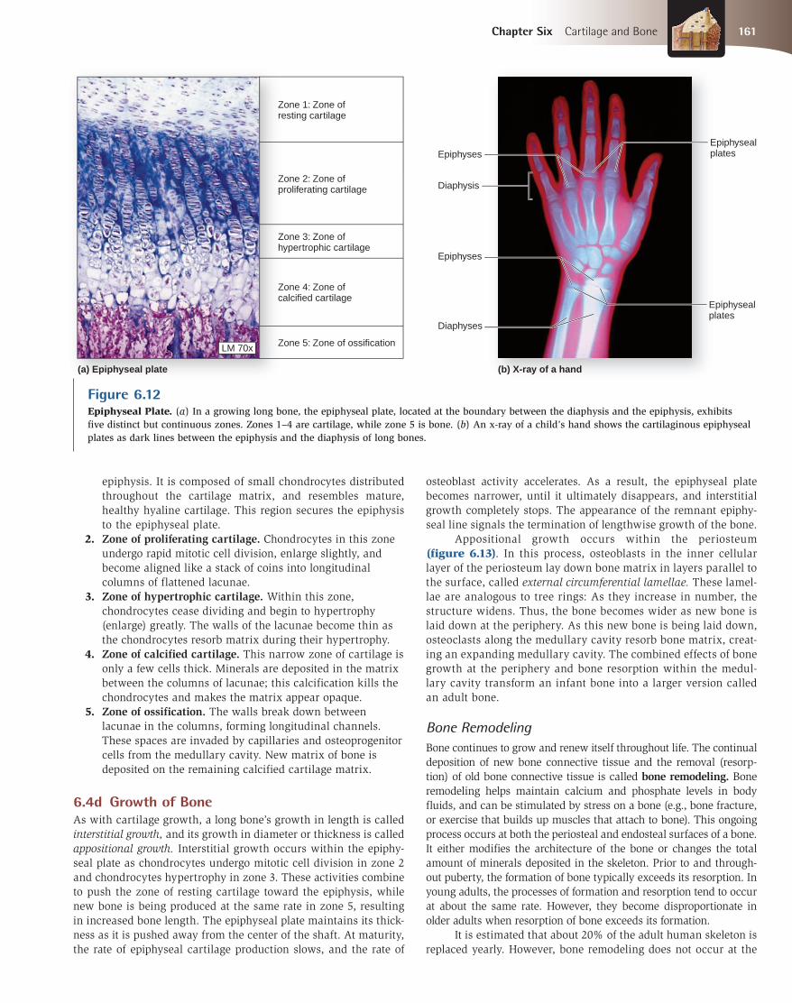
Figure 6.12Epiphyseal Plate. (a) In a growing long bone, the epiphyseal plate, located at the boundary between the diaphysis and the epiphysis, exhibits five distinct but continuous zones. Zones 1–4 are cartilage, while zone 5 is bone. (b) An x-ray of a child’s hand shows the cartilaginous epiphyseal plates as dark lines between the epiphysis and the diaphysis of long bones.
epiphysis. It is composed of small chondrocytes distributed throughout the cartilage matrix, and resembles mature, healthy hyaline cartilage. This region secures the epiphysis to the epiphyseal plate.
2. Zone of proliferating cartilage. Chondrocytes in this zone undergo rapid mitotic cell division, enlarge slightly, and become aligned like a stack of coins into longitudinal columns of flattened lacunae.
3. Zone of hypertrophic cartilage. Within this zone, chondrocytes cease dividing and begin to hypertrophy (enlarge) greatly. The walls of the lacunae become thin as the chondrocytes resorb matrix during their hypertrophy.
4. Zone of calcified cartilage. This narrow zone of cartilage is only a few cells thick. Minerals are deposited in the matrix between the columns of lacunae; this calcification kills the chondrocytes and makes the matrix appear opaque.
5. Zone of ossification. The walls break down between lacunae in the columns, forming longitudinal channels. These spaces are invaded by capillaries and osteoprogenitor cells from the medullary cavity. New matrix of bone is deposited on the remaining calcified cartilage matrix.
6.4d Growth of Bone
As with cartilage growth, a long bone’s growth in length is called interstitial growth, and its growth in diameter or thickness is called appositional growth. Interstitial growth occurs within the epiphy-seal plate as chondrocytes undergo mitotic cell division in zone 2 and chondrocytes hypertrophy in zone 3. These activities combine to push the zone of resting cartilage toward the epiphysis, while new bone is being produced at the same rate in zone 5, resulting in increased bone length. The epiphyseal plate maintains its thick-ness as it is pushed away from the center of the shaft. At maturity, the rate of epiphyseal cartilage production slows, and the rate of
osteoblast activity accelerates. As a result, the epiphyseal plate becomes narrower, until it ultimately disappears, and interstitial growth completely stops. The appearance of the remnant epiphy-seal line signals the termination of lengthwise growth of the bone. Appositional growth occurs within the periosteum (figure 6.13). In this process, osteoblasts in the inner cellular layer of the periosteum lay down bone matrix in layers parallel to the surface, called external circumferential lamellae. These lamel-lae are analogous to tree rings: As they increase in number, the structure widens. Thus, the bone becomes wider as new bone is laid down at the periphery. As this new bone is being laid down, osteoclasts along the medullary cavity resorb bone matrix, creat-ing an expanding medullary cavity. The combined effects of bone growth at the periphery and bone resorption within the medul-lary cavity transform an infant bone into a larger version called an adult bone.
Bone Remodeling
Bone continues to grow and renew itself throughout life. The continualdeposition of new bone connective tissue and the removal (resorp-tion) of old bone connective tissue is called bone remodeling. Bone remodeling helps maintain calcium and phosphate levels in body fluids, and can be stimulated by stress on a bone (e.g., bone fracture, or exercise that builds up muscles that attach to bone). This ongoing process occurs at both the periosteal and endosteal surfaces of a bone. It either modifies the architecture of the bone or changes the total amount of minerals deposited in the skeleton. Prior to and through-out puberty, the formation of bone typically exceeds its resorption. In young adults, the processes of formation and resorption tend to occur at about the same rate. However, they become disproportionate in older adults when resorption of bone exceeds its formation. It is estimated that about 20% of the adult human skeleton is replaced yearly. However, bone remodeling does not occur at the
mck78097_ch06_146-172.indd 161mck78097_ch06_146-172.indd 161 2/14/11 2:36 PM2/14/11 2:36 PM

Infant Child
Bone resorbed by osteoclasts
Bone deposited by osteoblasts
Adult Young adult
Medullary cavity
Periosteum
Periosteum
Branch of nutrient artery
Periosteal arteries
Nutrient artery (in nutrient foramen)
Metaphyseal artery
Metaphyseal artery
Epiphyseal artery
Epiphysealartery
Articular cartilage
Compact bone
Medullary cavity (contains yellow bone marrow)
Epiphyseal line
Articular cartilage
Fibrous layer
Cellular layer
162 Chapter Six Cartilage and Bone
Figure 6.13 Appositional Bone Growth. A bone increases in diameter as new bone is added to the surface. At the same time, some bone may be removed from the inner surface to enlarge the marrow cavity.
same rate everywhere in the skeleton. For example, the compact bone in our skeleton is replaced at a slower rate than the spongy bone. The distal part of the femur (thigh bone) is replaced every 4 to 6 months, while the diaphysis of this bone may not be com-pletely replaced during an individual’s lifetime.
6.4e Blood Supply and Innervation
Bone is highly vascularized (meaning it is supplied by many blood vessels), especially in regions containing red bone marrow. Blood vessels enter bones from the periosteum. A typical long bone such as the humerus has four major sets of blood vessels (figure 6.14). Nutrient blood vessels, called the nutrient artery and the nutrient vein, supply the diaphysis of a long bone. Typically, only one nutrient artery enters and one nutrient vein leaves the bone via a nutrient foramen in the bone. These vessels branch and extend along the length of the shaft toward the epiphyses and into the cen-tral canal of osteons within compact bone and the marrow cavity. Metaphyseal blood vessels (metaphyseal arteries and metaphyseal veins) provide the blood supply to the diaphyseal side of the epiphyseal plate, which is the region where new bone ossifica-tion forms bone connective tissue to replace epiphyseal plate cartilage. Epiphyseal arteries and epiphyseal veins provide the blood supply to the epiphyses of the bone. In early childhood, the cartilaginous epiphyseal plate separates the epiphyseal and metaphyseal vessels. However, once an epiphyseal plate ossi-fies and becomes an epiphyseal line, the epiphyseal vessels and metaphyseal vessels anastomose (interconnect) through channels formed in the epiphyseal line (see figure 6.14 for examples). Periosteal blood vessels (periosteal arteries and periosteal veins) provide blood to the external circumferential lamellae and the superficial osteons within the compact bone at the external edge of the bone. These vessels and the accompanying periosteal nerves penetrate the diaphysis and enter the perforating canals at many locations. Nerves that supply bones accompany blood vessels through the nutrient foramen and innervate the bone as well as its perios-teum, endosteum, and marrow cavity. These are mainly sensory nerves that signal injuries to the skeleton.
Figure 6.14Arterial Supply to a Mature Bone. Four major sets of blood vessels supply the humerus, a long bone: nutrient arteries and veins, metaphyseal arteries and veins, epiphyseal arteries and veins, and periosteal arteries and veins.
mck78097_ch06_146-172.indd 162mck78097_ch06_146-172.indd 162 2/14/11 2:36 PM2/14/11 2:36 PM

Chapter Six Cartilage and Bone 163
WHAT DID YOU LEARN?
●9 What is intramembranous ossification? What bones form by this process?
●10 Identify the locations of the primary and secondary ossification centers in a long bone.
●11 How could a physician determine whether a patient had reached full height by examining x-rays of his or her bones?
●12 Name the five zones in an epiphyseal plate and the characteristics of each.
6.5 Maintaining Homeostasis and Promoting Bone GrowthLearning Objectives: 1. Explain the effects of hormones, vitamins, and exercise on
bone growth and maintenance. 2. Describe the steps involved in healing bone fractures.
Bone growth and maintenance normally depend upon both hormones and vitamins (table 6.1).
6.5a Effects of Hormones
Hormones control and regulate growth patterns in bone by altering the rates of osteoblast and osteoclast activity. Growth hormone, also called somatotropin (so ma -to -tro pin), is produced by the anterior pituitary gland. It affects bone growth by stimulating the formation of
WW
another hormone, somatomedin (so ma -to -me din), which is produced by the liver. Somatomedin directly stimulates growth of cartilage in the epiphyseal plate. Thyroid hormone, secreted by the thyroid gland, stimulates bone growth by influencing the basal metabolic rate of bone cells. Together, growth hormone and thyroid hormone, if maintained in proper balance, regulate and maintain normal activity at the epiphyseal plates until puberty. If a child’s growth hormone and/or thyroid hormone levels are chronically too low, then bone growth is adversely affected, and the child will be short in stature. Another thyroid gland hormone is calcitonin (kal-si-to nin; calx = lime, tonos = stretching), which is secreted in response to ele-vated levels of calcium in the blood. Calcitonin encourages calcium deposition from blood into bone and inhibits osteoclast activity. Parathyroid hormone is secreted and released by the para-thyroid glands in response to reduced calcium levels in the blood. Ultimately, parathyroid hormone increases the blood calcium lev-els, so other body tissues can utilize this calcium. Parathyroid hor-mone stimulates osteoclasts to resorb bone and thereby increase calcium levels in the blood. Sex hormones (estrogen and testosterone), which begin to be secreted in great amounts at puberty, dramatically accelerate bone growth. Sex hormones increase the rate of bone formation by osteoblasts in ossification centers within the epiphyseal plate, resulting in increased length of long bones and increased height. The appearance of high levels of sex hormones at puberty also signals the beginning of the end for growth at the epiphyseal plate. Eventually, more bone is produced at the epiphyseal plate than the cartilage within the plate can support. As a result, the thickness of the epiphyseal plate cartilage begins to diminish, and eventually it disappears altogether, leaving behind the epiphyseal line. Older individuals (who have a normal reduction in sex hormones) also may experience a decrease in bone mass as they age.
Achondroplastic DwarfismAchondroplasia (a-kon-dro-pla ze-a) is characterized by abnormal conversion of hyaline cartilage to bone. The most common form is achondroplastic dwarfism, in which the long bones of the limbs stop growing in childhood, while the other bones usually continue to grow normally. Thus, an individual with achondroplastic dwarf-ism is short in stature but generally has a large head. Often the forehead is prominent, and the nose is flat at the bridge. Those affected may have bowlegs and lordosis (exaggerated curvature of the lumbar spine). Most individuals are about 4 feet tall. Their intelligence and life span are within normal range.
Achondroplastic dwarfism results from a failure of chondrocytes in the second and third zones of the epiphyseal plate (see figure 6.12a) to multiply and enlarge, leading to inadequate endochondral ossifi-cation. Most cases result from a spontaneous mutation during DNA replication. Thus, even parents who are of normal height and have no family history of dwarfism may have a child with achondroplastic dwarfism. Children of an achondroplastic dwarfism parent also may inherit the disorder. This is because it is an autosomal dominant condition, meaning that a child may inherit only one defective gene from a parent (as opposed to having both genes defective) to express the condition. This condition differs from pituitary dwarfism, which results when the pituitary gland produces insufficient growth hormone or none at all. In pituitary dwarfism, the growth of all the bones is stunted, so the individual is short in stature but has normal proportions throughout the skeletal system.
CLINICAL VIEW Table 6.1 Effects of Hormones and Vitamins on Bone Maintenance and Growth
HORMONES
Growth hormone Stimulates liver to produce the hormone somatomedin, which causes cartilage proliferation at epiphyseal plate and resulting bone elongation; too little growth hormone results in short stature in the child
Thyroid hormone Stimulates bone growth by stimulating metabolic rate of osteoblasts; too little thyroid hormone results in short stature
Calcitonin Promotes calcium deposition in bone and inhibits osteoclast activity
Parathyroid hormone Increases blood calcium levels by encouraging bone resorption by osteoclasts
Sex hormones (estrogen and testosterone)
Stimulate osteoblasts; promote epiphyseal plate growth and closure
Glucocorticoids If levels are chronically too high, bone resorption occurs and signifi cant bone mass is lost
VITAMINS
Vitamin A Activates osteoblasts
Vitamin C (ascorbic acid) Promotes collagen production
Vitamin D Promotes absorption of calcium and phosphate into blood; helps with calcifi cation of bone
mck78097_ch06_146-172.indd 163mck78097_ch06_146-172.indd 163 2/14/11 2:36 PM2/14/11 2:36 PM

Bowinglower limblong bones
Hot spots
(a) Normal bone scan (b) Abnormal bone scan (numerous "hot spots")
164 Chapter Six Cartilage and Bone
Finally, abnormal amounts of certain hormones can affect bone maintenance and growth. As mentioned earlier, chronically low levels of growth hormone and/or thyroid hormone in a child inhibit bone growth and result in short stature. Another example are the glucocorticoids, a group of hormones produced by the adrenal cortex. Normal glucocorticoid levels tend not to have any major effects on bone growth or mass. However, if glucocorticoid levels are chronically too high, they stimulate bone resorption and can lead to significant loss of bone mass.
6.5b Effects of Vitamins
A continual dietary source of vitamins is required for normal bone growth. For example, vitamin A activates osteoblasts, while vitamin C is required for normal synthesis of collagen, the primary organic component in the bone matrix. Vitamin D stimulates the absorption and transport of calcium and phosphate ions into the blood. It also is necessary for the calcification of bone. As calcium and phosphate levels rise in the blood, calcitonin is secreted, which encourages the deposition of these minerals into bone.
Radiograph of a 10-month-old with rickets.
RicketsRickets is a disease caused by a vitamin D deficiency in childhood and characterized by overproduction and deficient calcification of osteoid. Due to the lack of vitamin D, the digestive tract is unable to absorb calcium and phosphorus, minerals needed for the hardening of the osteoid during the formation of bone.
Rickets usually develops in children, and results in bones that are poorly calcified and exhibit too much flexibility. Patients with rickets acquire a bowlegged appearance as their weight increases and the bones in their legs bend. In addition to skeletal deformities, rickets is characterized by disturbances in growth, hypocalcemia (an abnormally low level of calcium in the blood), and sometimes tetany (cramps and muscle twitches), usually caused by low blood calcium. The condition is often accompanied by irritability, listlessness, and generalized muscular weakness. Fractures frequently occur in patients with rickets.
During the Industrial Revolution, the incidence of rickets increased as children were forced to work indoors in factories. These children had little exposure to sunlight and were usually malnourished as well. (Recall from chapter 5 that the body can manufacture its own vitamin D when the integument is exposed to sunlight.) Rickets continues to occur in some developing nations, and recently the incidence has increased in urban areas of the United States. Researchers have discovered that these children spend much of their time indoors and typically do not drink enough milk, opting for soft drinks instead. So, unfortunately, a disease that is easily preventable is making a comeback in the United States due to poor dietary and lifestyle habits among the nation’s youth.
Bone ScansBone scans are tests that can detect bone pathologies sooner than standard x-rays, while exposing the patient to only a fraction of the radiation of a normal x-ray. The patient is injected intravenously with a small amount of a radioactive tracer compound that is absorbed by bone. A scanning camera then detects and measures the radiation emitted from the bone. This information is converted into a diagram or photograph that can be read like an x-ray. In these films, normal bone tissue is a consistent gray color, while darker areas are “hot spots” indicating increased metabolism, and lighter areas are “cold spots” indicating decreased metabolism. Abnormalities that can be detected by a bone scan include fractures, decalcifica-tion of bone, osteomyelitis, degenerative bone disease, and Paget disease. Bone scans are also used to determine whether cancer has metastasized to bone, to identify bone infections, to monitor the progress of bone grafts and degenerative bone disorders, to evaluate unexplained bone pain or possible fracture, and to monitor response to therapy of a cancer that has spread to bone.
CLINICAL VIEW CLINICAL VIEW
mck78097_ch06_146-172.indd 164mck78097_ch06_146-172.indd 164 2/14/11 2:36 PM2/14/11 2:36 PM

Compound (open)
Greenstick Oblique
Comminuted
Transverse Spiral
Colles
Pott
Chapter Six Cartilage and Bone 165
6.5c Effects of Exercise
Mechanical stress, in the form of exercise, is required for normal bone remodeling. In response to mechanical stress, bone has the ability to increase its strength over a period of time by increas-ing the amounts of mineral salts deposited and collagen fibers synthesized. Stress also increases the production of the hormone calcitonin, which helps inhibit bone resorption by osteoclasts and encourage bone deposition by osteoblasts. Mechanical stresses that significantly affect bone result from repeated skeletal muscle contraction and gravitational forces. Typically, the bones of athletes become noticeably thicker as a result of repetitive and stressful exercise. Weight-bearing activi-ties, such as weight lifting or walking, help build and retain bone mass. In contrast, lack of mechanical stress weakens bone through both demineralization of the bone matrix and reduction of collagen formation. For example, if a person has a fractured bone in a cast or is bedridden, the mass of the unstressed bone decreases in the immobilized limbs. While in space, astronauts must exercise so that the lesser gravity won’t weaken their bones. Research has shown that regular weight-bearing exercise can increase total bone mass in adolescents and young adults prior to its inevitable reduction later in life. In fact, recent studies have shown that even 70- and 80-year-olds who perform moderate weight training can increase their bone mass.
6.5d Fracture Repair
Bone has great strength, and yet it may break as a result of unusual stress or a sudden impact. Breaks in bones, called fractures, are classified in several ways. They can be named from the cause: stress, trauma, or pathology. A stress fracture is a thin break caused by recent increased physical activity in which the bone experiences repetitive loads (e.g., as seen in some runners). Stress fractures tend to occur in the weight-bearing bones (e.g., pelvis and lower limb). A traumatic fracture is a result of impact or excess stress to the bone, and a pathologic fracture usually occurs in bone that has been weakened by disease, such as when the vertebrae fracture in some-one with osteoporosis (a bone condition discussed in the next sec-tion). Fractures are categorized by the amount of soft tissue damage associated with the fracture. In a simple fracture, the broken bone does not penetrate the skin, while in a compound fracture, one or both ends of the broken bone pierce the overlying skin and body tis-sues. Fractures are also known through eponyms (such as Colles or Pott), which have specific characteristics describing these fractures. Most often fractures are classified by the description of the fracture. Table 6.2 shows the some of the different classifications of fractures, and figure 6.15 illustrates some of the most common types. Often many fracture classifications are used to describe a single fracture. For instance, a Colles fracture is a complete, transverse fracture of the distal radius with posterior displacement. The healing of a simple fracture takes about 2 to 3 months, whereas a compound fracture takes longer to heal. Fractures heal much more quickly in young children (average healing time, 3 weeks) and become slower to heal as we age. In the elderly, the normal thinning and weakening of bone increases the incidence of fractures, and some severe fractures never heal without surgical intervention. Bone fracture repair can be described as a series of steps (figure 6.16):
1. A fracture hematoma forms. A bone fracture tears blood vessels inside the bone and within the periosteum, causing bleeding, and then a fracture hematoma forms from the clotted blood.
Figure 6.15Types of Bone Fractures. Selected bone fractures listed in table 6.2 are illustrated here.
2. A fibrocartilaginous (soft) callus forms. Regenerated blood capillaries infiltrate the fracture hematoma due to an increase in osteoblasts in both the periosteum and the endosteum near the fracture site. First, the
mck78097_ch06_146-172.indd 165mck78097_ch06_146-172.indd 165 2/14/11 2:37 PM2/14/11 2:37 PM

Hematoma
Periosteum
Compact bone Fibrocartilaginous
(soft) callus Regenerating blood vessels
Hard callus
Primary bone
Medullary cavity
Compact bone at break site
A fracture hematoma forms. 1 A fibrocartilaginous (soft) callus forms.
2 A hard (bony) callus forms. 3 The bone is remodeled. 4
166 Chapter Six Cartilage and Bone
Figure 6.16Fracture Repair. The repair of a bone fracture occurs in a series of steps.
fracture hematoma is reorganized into an actively growing connective tissue called a procallus. Fibro blasts within the procallus produce collagen fibers that help connect the broken ends of the bones. Chondroblasts in the newly growing connective tissue form a dense regular connective tissue associated with the cartilage. Eventually, the procallus becomes a fibrocartilaginous (soft) callus (kal u s; hard skin). The fibrocartilaginous callus stage lasts at least 3 weeks.
3. A hard (bony) callus forms. Within a week, osteoprogenitor cells in areas adjacent to the fibrocartilaginous callus become osteoblasts and produce trabeculae of primary bone. The fibrocartilaginous callus is then replaced by this bone, which forms a hard (bony) callus. The trabeculae of the hard callus continue to grow and thicken for several months.
4. The bone is remodeled. Remodeling is the final phase of fracture repair. The hard callus persists for at least 3
Table 6.2 Classifi cation of Bone Fractures
Classifi cation Fracture Description Classifi cation Fracture Description
Cause Stress Fracture result of repeated stressful impact such as running (these fractures are diffi cult to see on x-rays)
Descriptive(cont'd)
Depressed Broken part of the bone forms a concavity (as in skull fracture)
Pathology Weakening of a bone caused by disease processes (e.g., cancer)
Displaced Fractured bone parts are out of anatomic alignment
Trauma Fracture due to impact or increased tension or torsion on bone from outside source
Epiphyseal Epiphysis is separated from the diaphysis at the epiphyseal plate
Soft tissue involvement
Simple (closed)
Bone does not break through the skin Greenstick Partial fracture; convex side of bone breaks—the other side is bent
Compound (open)
Broken ends of the bone protrude through the skin
Hairline Fine crack in which sections of the bone remain aligned (common in the skull)
Eponymns Colles Fracture of the distal end of the radius; produces a “dinner fork” deformity
Impacted One fragment of bone is fi rmly driven into the other
Pott Fracture at the distal end of the fi bula and malleolus of the tibia
Incomplete Fracture extends only partway across the bone
Descriptive Avulsion Bone or bone fragments accompany soft tissue as it is pulled from its origin or insertion
Linear Fracture is parallel to the long axis of the bone
Comminuted Bone is splintered into several small pieces between the main parts
Oblique Diagonal fracture at an angle between linear and transverse
Complete Bone is broken into two or more pieces Spiral Fracture spirals around axis of long bone; results from twisting stress
Compression Bone is squashed (may occur in a vertebra during a fall)
Transverse Fracture at right angles to the long axis of the bone
mck78097_ch06_146-172.indd 166mck78097_ch06_146-172.indd 166 2/14/11 2:37 PM2/14/11 2:37 PM

Trochanter Head
Epicondyle
Tubercle
Condyle
Femur
Fissure
Ramus
Sulcus
Tuberosity
Head
Fossa
Fossa
Trochlea
Skull, anterior view
Process
Foramen
Spine
Line
Foramen
Pelvis
Humerus
Epicondyle
Ramus
Crest
Condyle
Canal
Ramus
Sinus
Skull, sagittal view
Alveolus
Facet
Large, smooth, rounded articulating oval structure
Projections for tendon and ligament attachment
Pointed, slender process
Massive, rough projection found only on the femur Trochanter
Head
Facet
Tuberosity
Fissure
Alveolus (pl., alveoli )
Sulcus
Tubercle
Fossa (pl., fossae)
Trochlea
Foramen (pl., foramina)
Spine
Line
Epicondyle
Crest
Ramus (pl., rami )
Process
Sinus
Condyle
General Structure
Anatomical Term
Description
Articulating surfaces
Canal
Small, flat, shallow articulating surface
Prominent, rounded epiphysis
Smooth, grooved, pulley-like articular process
Deep pit or socket in the maxillae or mandible
Flattened or shallow depression
Narrow groove
Narrow, prominent, ridgelike projection
Projection adjacent to a condyle
Low ridge
Depressions
Small, round projection
Large, rough projection
Any marked bony prominence
Angular extension of a bone relative to the rest of the structure
Openings and spaces
Passageway through a bone
Narrow, slitlike opening through a bone
Rounded passageway through a bone
Cavity or hollow space in a bone
Chapter Six Cartilage and Bone 167
to 4 months as osteoclasts remove excess bony material from both exterior and interior surfaces. Compact bone replaces primary bone. The fracture usually leaves a slight thickening of the bone (as detected by x-ray); however, in many instances healing occurs with no obvious thickening.
WHAT DID YOU LEARN?
●13 What are the effects of growth hormone and parathyroid hormone on bone growth and/or bone mass?
●14 Which vitamins help regulate bone growth?
●15 A fracture is diagnosed when the bone has broken through the skin.
WW
6.6 Bone MarkingsLearning Objective: 1. Identify the anatomic terms that describe the surface
features of bone.
Distinctive bone markings, or surface features, character-ize each bone in the body. Projections from the bone surface mark the point where tendons and ligaments attach. Sites of articulation between adjacent bones are smooth, flat areas. Depressions, grooves, and tunnels through bones indicate sites where blood vessels and nerves either lie alongside or penetrate the bone. Anatomists use spe-cific terms to describe these elevations and depressions (figure 6.17).
Figure 6.17Bone Markings. Specific anatomic terms describe the characteristic identification marks for bones.
mck78097_ch06_146-172.indd 167mck78097_ch06_146-172.indd 167 2/14/11 2:37 PM2/14/11 2:37 PM

(b) Osteoporotic bone (a) Normal bone
SEM 30x SEM 20x
168 Chapter Six Cartilage and Bone
Knowing the names of bone markings will help you learn about specific bones in chapters 7 and 8. For example, know-ing that foramen means “hole” or “passageway” will tell you to look for a hole when trying to find the foramen magnum on the skull. Likewise, you can usually correctly assume that any smooth, oval prominence on a bone is called a condyle. Refer back tofigure 6.17 frequently for assistance in learning the bones and their features. For professional criminologists, pathologists, and anthropol-ogists, bones can tell an intricate anatomic story. Bone markings on skeletal remains indicate where the soft tissue components once were, often allowing an individual’s height, age, sex, and general appearance to be determined.
WHAT DID YOU LEARN?
●16 What is the anatomic name for a narrow, slitlike opening through a bone?
WW
6.7 Aging of the Skeletal SystemLearning Objective:
1. Explain the changes in bone architecture and bone mass related to age.
Aging affects bone connective tissue in two ways. First, the tensile strength of bone decreases due to a reduced rate of protein synthesis, which in turn results in decreased ability to produce the organic portion of bone matrix. Consequently, the percentage of inorganic minerals in the bone matrix increases, and the bones of the skeleton become brittle and susceptible to fracture. Second, bone loses calcium and other minerals (demineral-ization). The bones of the skeleton become thinner and weaker, resulting in insufficient ossification, a condition called osteope-nia (os te -o -pe ne -a ; penia = poverty). Aging causes all people to become slightly osteopenic. This reduction in bone mass
OsteoporosisOsteoporosis, meaning “porous bones,” is a disease that results in decreased bone mass and microarchitectural changes that lead to weak-ened bones that are prone to fracture. Both bone matrix and calcium are lost, particularly in metabolically active spongy bone. The occurrence of osteoporosis is greatest among the elderly, especially Caucasian women, and the severity is closely linked to age and the onset of menopause. Postmenopausal women are at risk because (1) women have less bone mass than men, (2) women begin losing bone mass earlier and faster in life (sometimes as early as 35 years of age), and (3) postmenopausal women no longer produce significant amounts of estrogen, which appears to help protect against osteoporosis by stimulating bone growth. Although the condition does affect men, osteoporosis in men is typically less severe than in women for the reasons just mentioned.
As a result of osteoporosis, the incidence of fractures increases, most frequently in the wrist, hip, and vertebral column, and usually as the result of a normal amount of stress. Wrist fractures occur at the distal end of the radius (Colles fracture), and hip fractures usually occur at the neck of the femur. The weight-bearing regions of the vertebrae lose spongy bone and are more easily compressed, leading to a loss of height and sometimes to compression fractures of the vertebral bodies.
Although diagnosis and monitoring of osteoporo-sis have been simplified, a cure remains elusive. The best treatment seems to be prevention. Young adults should maintain good nutrition and physi-cal activity to ensure adequate bone density, thus allowing for the normal, age-related loss later in life. Calcium supplements may help maintain bone health, but by themselves will not stimulate new bone growth.
Medical treatments involve two strategies: (1) slowing the rate of bone loss and (2) attempt-ing to stimulate new bone growth. Formerly, because of the link between estrogen and bone growth, hormone replacement therapy (HRT) was widely used to slow bone degeneration in
postmenopausal women. Unfortunately, new studies have linked estrogen supplementation to increased risk of cardiovascular (heart and blood vessel) problems, such as stroke and heart attacks, as well as increases in blood clots in the lung (pulmonary emboli). These significant complications of HRT have substantially limited its usefulness in therapy and prevention.
Bisphosphonates, selective estrogen receptor modulators, calcitonin analogs, and parathyroid hormone (PTH) are alternative osteoporosis medications. Bisphosphonates—including alendronate (brand name: Fosamax), pamidronate (Aredia), risedronate (Actonel), and ibandro-nate (Boniva)—work by interfering with osteoclast function and thus retarding the removal of bone during remodeling. Selective estrogen receptor modulators, such as raloxifene (Evista), result in slowing bone resorption without the adverse effects seen with HRT. Calcitonin analogs (Miacalcin) act by reducing bone resorption. PTH in the form of teriparatide (Forteo) increases osteoclast activity as well as absorption of calcium from the intestines and kidneys. These treatments do slow bone resorption and some even add bone density, but they do have possible drawbacks such as abnormal serum calcium levels, impaired kidney function, and osteonecrosis of the jaw in some people. Calcium intake can be tightly controlled at times when taking these medications.
CLINICAL VIEW
mck78097_ch06_146-172.indd 168mck78097_ch06_146-172.indd 168 2/14/11 2:37 PM2/14/11 2:37 PM

Chapter Six Cartilage and Bone 169
Clinical Terms
may begin as early as 35–40 years of age, when osteoblast activity declines while osteoclast activity continues at previous levels. During each successive decade of their lives, women lose roughly more of their skeletal mass than do men. Different parts of the skeleton are affected unequally. Vertebrae, jaw bones, and epiphyses lose large amounts of mass, resulting in reduced height, loss of teeth, and fragile limbs. A significant percentage of older women and a smaller proportion of older men suffer from
osteoporosis (os te -o -po -ro sis; poros = pore, osis = condition), in which bone mass becomes reduced enough to compromise nor-mal function (see Clinical View).
WHAT DO YOU THINK?
●4 What major differences might we expect when comparing bone composition in a 65-year-old man with that of his 13-year-old granddaughter?
■ The skeletal system is composed of bones, cartilage that supports the bones, and ligaments that bind together, support, and stabilize bones.
6.1 Cartilage 147 ■ Cartilage contains cells embedded within a matrix of protein fibers and a gel-like ground substance.
6.1a Functions of Cartilage 147
■ Cartilage provides support for soft tissues, a sliding surface for bone, and a model for formation of most of the bones of the body.
6.1b Growth Patterns of Cartilage 148
■ Cartilage growth includes both interstitial growth (growth from within preexisting cartilage) and appositional growth (growth around the periphery of cartilage).
6.2 Bone 148 ■ Bones are organs that contain multiple tissue types, the most abundant being bone (osseous) connective tissue.
6.2a Functions of Bone 148
■ Bone performs the following functions: support and protection, movement, hemopoiesis, and storage of minerals and energy.
chondroma Benign (noncancerous) tumor derived from cartilage cells.
chondrosarcoma Malignant (cancerous) tumor derived from cartilage cells.
hyperostosis Excessive formation of bone tissue.osteogenesis imperfecta Also known as “brittle bone disease”;
inherited condition that affects collagen fiber distribution and organization. It occurs due to impaired osteoblast function, and results in abnormal bone growth, brittle bones, continuing deformation of the skeleton, and increased susceptibility to fracture.
osteoma Benign tumor in lamellar bone, often in the jaw or the skull.
osteomalacia Vitamin D deficiency disease in adults characterized by gradual softening and bending of the bones as a result of decreased mineral content; although bone mass is still present, it is demineralized.
osteomyelitis Infection and inflammation within both the bone marrow and neighboring regions of the bone.
osteosarcoma The most common and malignant bone sarcoma; arises from bone-forming cells (osteoblasts) and chiefly affects the ends of long bones.
Chapter Summary
6.3 Classifi cation and
Anatomy of Bones 150
■ Bones are categorized by shape as long, short, flat, or irregular.
6.3a General Structure and Gross Anatomy of Long Bones 150
■ A long bone contains the following parts: diaphysis, epiphyses, metaphysis, articular cartilage, medullary cavity, periosteum, and endosteum.
■ Osteoblasts synthesize and secrete osteoid, the matrix of bone prior to its calcification.
■ Osteocytes are mature bone cells that reside in lacunae.
■ Osteoclasts are large, multinucleated cells involved in bone resorption.
■ An osteon is the basic unit of structure and function of mature compact bone.
■ An osteon contains a central canal that houses blood vessels and nerves, concentric bone layers called lamellae, osteocytes in lacunae, and thin channels called canaliculi.
6.4 Ossifi cation 157 ■ Ossification is the process of bone connective tissue formation.
6.4a Intramembranous Ossifi cation 157
■ In intramembranous ossification, bone forms from mesenchyme.
6.4b Endochondral Ossifi cation 157
■ Endochondral ossification uses a hyaline cartilage model that is gradually replaced by newly formed osseous tissue.
(continued on next page)
mck78097_ch06_146-172.indd 169mck78097_ch06_146-172.indd 169 2/14/11 2:37 PM2/14/11 2:37 PM

170 Chapter Six Cartilage and Bone
6.4 Ossifi cation (continued) 6.4c Epiphyseal Plate Morphology 160
■ The epiphyseal plate contains five zones where cartilage grows and is replaced by bone.
6.4d Growth of Bone 161
■ Lengthwise bone growth is called interstitial growth, while a bone increases in diameter through appositional growth at the periosteum.
■ The continual deposition of new bone tissue and resorption of old bone tissue is called bone remodeling.
6.4e Blood Supply and Innervation 162
■ Four categories of blood vessels develop to supply a typical bone: nutrient vessels, metaphyseal vessels, epiphyseal vessels, and periosteal vessels.
Challenge Yourself
MatchingMatch each numbered item with the most closely related lettered item.
______ 1. flat bone of skull
______ 2. osteon
______ 3. spongy bone
______ 4. epiphysis
______ 5. osteoid
______ 6. parathyroid hormone
______ 7. endosteum
______ 8. osteoclasts
______ 9. vitamin D
______ 10. hydroxyapatite
Multiple ChoiceSelect the best answer from the four choices provided.
______ 1. The immature cells that produce osteoid are called
a. osteocytes. b. osteoblasts. c. osteoclasts. d. osteons.
______ 2. Hyaline cartilage is found in all of the following structures except the
a. trachea. b. larynx. c. pubic symphysis. d. fetal skeleton.
______ 3. A small space within compact bone housing an osteocyte is termed a
a. lamella. b. lacuna. c. canaliculus. d. medullary cavity.
a. end of a long bone
b. formed by intramembranous ossification
c. organic components of bone matrix
d. stimulates osteoclasts to become active
e. lines medullary cavity
f. calcium phosphate/hydroxide crystals
g. responsible for bone resorption
h. increases calcium absorption in intestine
i. formed from trabeculae
j. contains concentric lamellae
6.5 Maintaining
Homeostasis and Promoting
Bone Growth 163
6.5a Effects of Hormones 163
■ Growth hormone, thyroid hormone, calcitonin, and sex hormones stimulate bone growth.
■ Parathyroid hormone stimulates osteoclast activity.
6.5b Effects of Vitamins 164
■ Vitamins A and C are essential for bone growth and remodeling. Vitamin D is needed for calcium and phosphorus absorption and calcification of bone.
6.5c Effects of Exercise 165
■ Stress in the form of exercise strengthens bone tissue by increasing the amounts of mineral salts deposited and collagen fibers synthesized.
6.5d Fracture Repair 165
■ A fracture is a break in a bone that can usually be healed if portions of the blood supply, endosteum, and periosteum remain intact.
6.6 Bone Markings 167 ■ Specific names denote bone markings such as projections, elevations, depressions, and passageways.
6.7 Aging of the Skeletal
System 168
■ Due to aging, the tensile strength of bone decreases, and bone loses calcium and other minerals (demineralization).
mck78097_ch06_146-172.indd 170mck78097_ch06_146-172.indd 170 2/14/11 2:37 PM2/14/11 2:37 PM

Chapter Six Cartilage and Bone 171
______ 4. Endochondral ossification begins with a ______ model of bone.
a. dense regular connective tissue b. hyaline cartilage c. fibrocartilage d. elastic cartilage
______ 5. Production of new bone ______ as a result of increased sex hormone production at puberty.
a. is not affected b. slows down c. increases slowly d. increases rapidly
______ 6. An epiphyseal line appears when a. epiphyseal plate growth has ended. b. epiphyseal plate growth is just beginning. c. growth in bone diameter is just beginning. d. a primary ossification center first develops.
______ 7. The condition of inadequate ossification that may accompany aging and is a result of reduced calcification is called
a. osteopenia. b. osteomyelitis. c. osteitis. d. osteosarcoma.
______ 8. A fracture of the distal end of the radius that produces a characteristic “dinner fork” deformity is a ______ fracture.
a. displaced b. Colles c. Pott d. stress
______ 9. The femur is an example of a a. flat bone. b. long bone. c. irregular bone. d. short bone.
______ 10. A large, rough projection of a bone is termed a a. fossa. b. tuberosity. c. ramus. d. tubercle.
Content Review 1. Identify the three types of cartilage, describing the
extracellular matrix of each type.
2. Describe the structure of the periosteum, and list its functions.
3. Describe the characteristics of articular cartilage, the medullary cavity, and endosteum in a long bone.
4. Describe the microscopic anatomy of compact bone.
5. Why is spongy bone able to withstand stress in an area such as the expanded end of a long bone?
6. What is ossification? What is the difference between intramembranous and endochondral ossification?
7. List the steps involved in endochondral ossification.
8. List the four types of arteries that are found in a long bone, and what portions of the bone each artery supplies.
9. Discuss the effect of exercise on bone mass.
10. What are the steps in fracture repair?
Developing Critical Reasoning 1. Marty fell off his skateboard and suffered a broken leg.
A cast was put on the leg for 6 weeks. After the bone healed and the cast was removed, an enlarged, bony bump remained at the region of the fracture. Eventually, this enlargement disappeared, and the leg regained its normal appearance. What happened from the time the cast was removed until the enlargement disappeared?
2. Elise is 14 and lives in an apartment in the city. She does not like outdoor activities, so she spends most of her spare time watching TV, playing video games, drinking soft drinks, and talking to friends on the phone. One afternoon, Elise tries to run down the stairs while talking on the phone, and falls, breaking her leg. Although she appears healthy, her leg takes longer to heal than expected. What might cause the longer healing time?
3. Connor is a healthy, active 7-year-old who fell while climbing on a bar apparatus in the playground, breaking his forearm near the wrist. The doctor told Connor’s father that the fracture would require insertion of screws to align ends of the broken bones and ensure proper growth in the future. Why was the physician taking special care with the healing of this fracture?
Answers to “What Do You Think?”
1. Ribs are best classified as flat bones because they have parallel surfaces of compact bone with internally placed spongy bone. Flat bones tend to be relatively thin (like the rib) and are both light and strong. In contrast, long bones typically have a cylindrical diaphysis, while ribs are flattened.
2. Spongy bone is lighter and able to withstand stresses applied from many directions. In addition, hemopoietic
tissue resides in the spaces within some spongy bone. Compact bone is very strong but weighs more than spongy bone. A bone made entirely of compact bone would be too heavy to move and too metabolically expensive to maintain, partly due to the increased musculature necessary to move it.
mck78097_ch06_146-172.indd 171mck78097_ch06_146-172.indd 171 2/14/11 2:37 PM2/14/11 2:37 PM

172 Chapter Six Cartilage and Bone
3. The numerous complex steps in endochondral bone formation ensure that a working bone may be formed for a newborn and later develop into a working adult bone. Having a bone collar, epiphyseal plates, and constant bone remodeling ensures that the bone can reshape itself, grow in both width and length, and develop a medullary cavity so that it will not weigh too much.
4. The 13-year-old will likely have several active epiphyseal plates (indicating that the bones are still growing in length), while the 65-year-old’s bones will have stopped growing in length. Typically, the 65-year-old will have less bone mass and be at greater risk for osteopenia than the 13-year-old. (However, staying active will help the 65-year-old maintain bone mass and help ward off osteoporosis.)
www.mhhe.com/mckinley3 Enhance your study with practice tests and activities to assess your understanding. Your instructor may also recommend the interactive eBook, individualized learning tools, and more.
mck78097_ch06_146-172.indd 172mck78097_ch06_146-172.indd 172 2/14/11 2:37 PM2/14/11 2:37 PM

![r (M7.3) ] 12,000 6.0 6 6.1 6.2 6.3 6.4 6.5 6.6 7 …...r (M7.3) ] 12,000 6.0 6 6.1 6.2 6.3 6.4 6.5 6.6 7 (M7.3) /JåJi(— 45 (vq) 33,000 1000m 20m DI-I: 20—10m —-10m 0 -20m -20](https://static.fdocuments.us/doc/165x107/5e38843ec7f8c0136410d017/r-m73-12000-60-6-61-62-63-64-65-66-7-r-m73-12000-60-6-61.jpg)