5th Edition Copyright © Prentice Hall 20072-1 Psychology Stephen F. Davis Emporia State University...
104
Copyright © Prentice Hall 2007 2-1 5th Edition Psychology Stephen F. Davis Emporia State University Joseph J. Palladino University of Southern Indiana PowerPoint Presentation by Cynthia K. Shinabarger Reed Tarrant County College This multimedia product and its contents are protected under copyright law. The following are prohibited by law: any public performance or display, including transmission of any image over a network; preparation of any derivative work, including the extraction, in whole or in part, of any images; any rental, lease, or lending of the program.
-
date post
22-Dec-2015 -
Category
Documents
-
view
214 -
download
0
Transcript of 5th Edition Copyright © Prentice Hall 20072-1 Psychology Stephen F. Davis Emporia State University...

Copyright © Prentice Hall 2007 2-1
5th Edition
PsychologyStephen F. Davis
Emporia State University Joseph J. Palladino
University of Southern IndianaPowerPoint Presentation by
Cynthia K. Shinabarger ReedTarrant County College
This multimedia product and its contents are protected under copyright law. The following are prohibited by law:any public performance or display, including transmission of any image over a network;
preparation of any derivative work, including the extraction, in whole or in part, of any images;any rental, lease, or lending of the program.
Copyright © Prentice Hall 2007 2-2
5th Edition
Behavioral Neuroscience
Chapter 2

Copyright © Prentice Hall 2007 2-3
Biology and Behavior
• In their efforts to understand the brain and the rest of the nervous system, many researchers have adopted an evolutionary perspective which focuses on the role a particular physical structure or behavior plays in helping an organism adapt to its environment over time.

Copyright © Prentice Hall 2007 2-4
Charles Darwin
• 1859: The Origin of Species: By means of Natural selection or the preservation of favoured races in the struggle for life.
• As Miami University approaches our 200th year in “2009” others will be celebrating the 150th anniversary of Darwin’s important book, The Origin of Species.
Copyright © Prentice Hall 2007 2-5
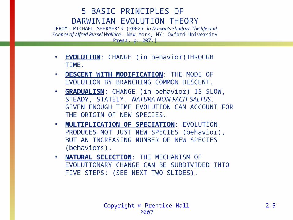
5 BASIC PRINCIPLES OF DARWINIAN EVOLUTION THEORY
[FROM: MICHAEL SHERMER’S (2002) In Darwin’s Shadow: The life and Science of Alfred Russel Wallace. New York, NY: Oxford University Press, p. 207.]
• EVOLUTION: CHANGE (in behavior)THROUGH TIME.• DESCENT WITH MODIFICATION: THE MODE OF
EVOLUTION BY BRANCHING COMMON DESCENT.• GRADUALISM: CHANGE (in behavior) IS SLOW,
STEADY, STATELY. NATURA NON FACIT SALTUS. GIVEN ENOUGH TIME EVOLUTION CAN ACCOUNT FOR THE ORIGIN OF NEW SPECIES.
• MULTIPLICATION OF SPECIATION: EVOLUTION PRODUCES NOT JUST NEW SPECIES (behavior), BUT AN INCREASING NUMBER OF NEW SPECIES (behaviors).
• NATURAL SELECTION: THE MECHANISM OF EVOLUTIONARY CHANGE CAN BE SUBDIVIDED INTO FIVE STEPS: (SEE NEXT TWO SLIDES).

Copyright © Prentice Hall 2007 2-6
Biology and Behavior
• Darwin maintained that evolution unfolds according to the principle of natural selection: the process by which inherited characteristics that lead to an advantage in adapting to the environment are more likely to be passed on (through genetic material) to future generations.

Copyright © Prentice Hall 2007 2-7
FIVE STEPS OF NATURAL SELECTION
• 1. POPULATIONS [behaviors] TEND TO INCREASE INDEFINITELY IN A GEOMETRIC RATIO. [FROM OBSERVATION]
• 2. IN A NATURAL ENVIRONMENT, HOWEVER, POPULATION [behavior] NUMBERS STABILIZE AT A CERTAIN LEVEL. [FROM OBSERVATION]
• 3. THERE MUST BE A “STRUGGLE FOR EXISTENCE” SINCE NOT ALL ORGANISMS [behaviors] PRODUCED CAN SURVIVE. [FROM INFERENCE]
• 4. THERE IS VARIATION IN EVERY SPECIES [behaviors]. [FROM OBSERVATION]
• 5. IN THE STRUGGLE FOR EXISTENCE, THOSE VARIATIONS THAT ARE BETTER ADAPTED TO THE ENVIRONMENT LEAVE BEHIND MORE OFFSPRING THAN THE LESS WELL ADAPTED INDIVIDUALS, ALSO KNOWN AS DIFFERENTIAL REPRODUCTIVE SUCCESS. [FROM INFERENCE]
Copyright © Prentice Hall 2007 2-8
Biology and Behavior
• In addition to studying the process of natural selection, researchers focus on discovering the actual genetic material responsible for the physical structure or behavior under investigation.
• The researchers who study the biological basis of animal and human behavior are working in an area called behavioral neuroscience.
Copyright © Prentice Hall 2007 2-9
Biology and Behavior
• This relatively new term focuses attention on the relation between biological factors and behavior.
• The term implies that these scientists represent several disciplines including psychology (especially physiological psychologists), biology, medicine, and others.
Copyright © Prentice Hall 2007 2-10
Biology and Behavior
• To survive, human beings must be able to perform three interrelated activities: sensing events, or stimuli; processing stimuli; and responding to stimuli.
• A stimulus is a feature in the environment—such as a traffic light, a sign, an alarm, or the smell of smoke—that may provoke a response.

Copyright © Prentice Hall 2007 2-11
Biology and Behavior:THE VAKTC MODEL
• V: Visual– Eyes “light”
• A: Auditory– Ears “sound”
• K: Kinesthetic– Whole body in motion
• T: Tactile– Touch/Skin “feeling”
• C: Chemio-Receptive– Smell and Taste

Copyright © Prentice Hall 2007 2-12
Levitin, Daniel J. (2006). This is your brain on music: The science of a human obsession
. New York: Dutton.Levitin is a “neuroscientist” with an
evolutionary psychology theoretical perspective. This book describes the evolutionary origins of music.
Levitin’s home page is: http://www.psych.mcgill.ca/faculty/levitin.html
His other home page is: http://www.psych.mcgill.ca/levitin/

Copyright © Prentice Hall 2007 2-13
Biology and Behavior
• Receptors are specialized cells of the nervous system that sense stimuli.
• The second activity in the chain is interpreting, or processing, the information that reaches the receptors.
• This processing typically takes place in the brain.

Copyright © Prentice Hall 2007 2-14
Biology and Behavior
• Once we’ve processed and understood the sensory input, we may need to respond to it.
• Therefore, the third activity occurs when the brain sends messages to the muscles to produce a response.

Copyright © Prentice Hall 2007 2-15
The Nervous System• The activities of sensing, processing, and
responding are coordinated and controlled by the nervous system, which has two major divisions: the central nervous system (CNS) and the peripheral nervous system (PNS).
• The CNS consists of the brain and spinal cord; the PNS connects the outer (or periphery) portions of the body with the CNS.
Copyright © Prentice Hall 2007 2-16
The Nervous System
• The PNS consists of all the parts of the nervous system that are outside the CNS.
• The two major divisions of the PNS are the somatic nervous system and the autonomic nervous system.

Copyright © Prentice Hall 2007 2-17
Divisions of the Nervous System
• Central Nervous System– Brain– Spinal cord
• Peripheral Nervous System– Somatic – Autonomic
• Sympathetic• Parasympathetic

Copyright © Prentice Hall 2007 2-18
The Nervous System
• The somatic nervous system of the PNS makes contact with the environment.
• It consists of nerves that connect receptors to the spinal cord and brain, as well as nerves that go to and from the brain and spinal cord to the muscles.

Copyright © Prentice Hall 2007 2-19
The Nervous System
• Nerves that carry information from receptors to the brain and spinal cord are called afferent (sensory) nerves.
• Nerves that carry information from the brain and spinal cord to the muscles are called efferent (motor) nerves.
• Because the responses we make are often planned and organized, the somatic division is said to be a voluntary system—that is, under our conscious control.
Copyright © Prentice Hall 2007 2-20
The Nervous System
• The autonomic nervous system of the PNS affects our organs and glands in ways that regulate bodily functioning.
• Because the autonomic nervous system operates without our conscious awareness, it is described as an automatic, or involuntary, system.
• The autonomic nervous system has two main components: the sympathetic nervous system and the parasympathetic nervous system.
Copyright © Prentice Hall 2007 2-21
The Nervous System
• The sympathetic nervous system mobilizes the body in times of stress or danger.
• The parasympathetic nervous system slows the processes that have been accelerated by activation of the sympathetic nervous system.
• These effects return the body to a more normal or balanced state of functioning characterized by an optimal range of physiological processes called homeostasis.
Copyright © Prentice Hall 2007 2-22
The Nervous System
• The spinal cord serves as the body’s information superhighway.
• Information that is not processed solely within the spinal cord itself is sent to the brain via ascending pathways; information that is sent back down from the brain follows descending pathways.
Copyright © Prentice Hall 2007 2-23
The Nervous System
• Within the CNS, interneurons connect neurons to each other.
• When information provided by the sensory nerves does not have to travel all the way to the brain to produce a response, automatic behaviors known as reflexes are produced.
Copyright © Prentice Hall 2007 2-24
The Endocrine System
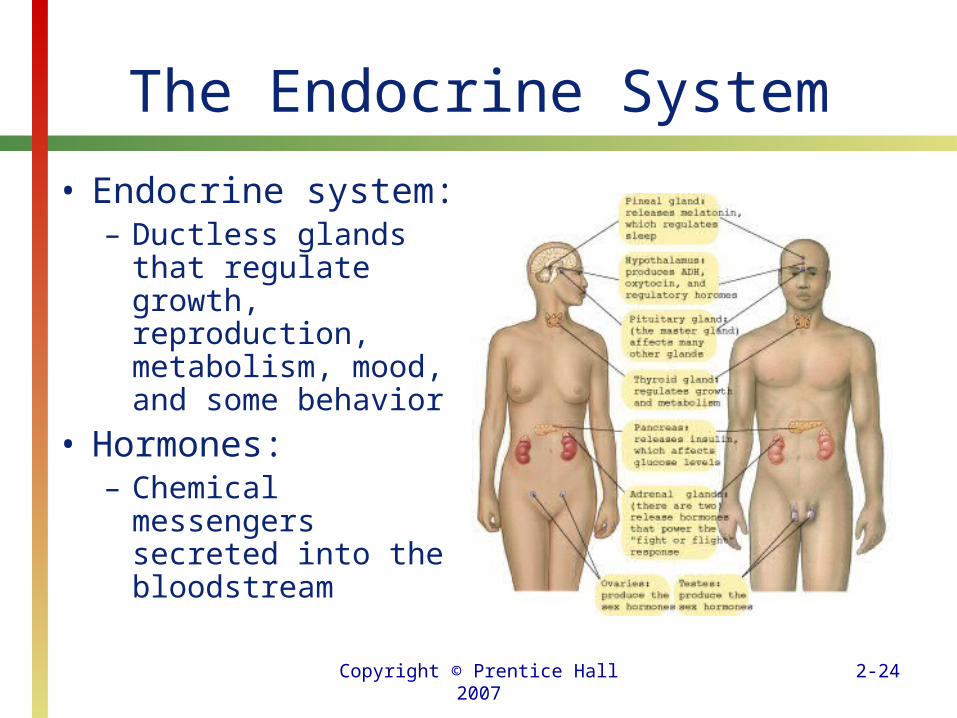
• Endocrine system:– Ductless glands that
regulate growth, reproduction, metabolism, mood, and some behavior
• Hormones: – Chemical messengers
secreted into the bloodstream
Copyright © Prentice Hall 2007 2-25
The Endocrine System
• The endocrine system consists of glands that produce and secrete (release) chemicals known as hormones.
• When stimulated, endocrine glands secrete hormones into the bloodstream.
• The pineal gland, located deep in the center of the brain, produces the hormone melatonin, especially at night.
• This hormone is important in regulating our sleep–wake cycle.
Copyright © Prentice Hall 2007 2-26
The Endocrine System
• Located near the stomach and small intestine, the pancreas secretes one of the best-known hormones, insulin.
• The cells of our body require insulin to use blood sugar (called glucose); without insulin, cells do not receive adequate nourishment from the available glucose.

Copyright © Prentice Hall 2007 2-27
The Endocrine System
• The hypothalamus is both an endocrine gland and a key center for a wide variety of behaviors related to survival.
• The hypothalamus signals its close neighbor, the pituitary, to release hormones that have a range of effects.

Copyright © Prentice Hall 2007 2-28
The Endocrine System
• The pituitary gland is often called the master gland because its secretions control many other glands.
• The pituitary is responsible for release of somatotropin, a growth hormone that acts directly on bones and muscles to produce the growth spurt that accompanies puberty.

Copyright © Prentice Hall 2007 2-29
The Endocrine System
• The thyroid gland secretes thyroxine, which regulates the body’s growth and metabolic rate.
• The gonads—ovaries in women and testes in men—produce sex hormones (androgens in men; estrogens in women) that activate reproductive organs and structures at puberty.
• These hormones also affect the appearance of secondary sex characteristics like facial and body hair, change of voice, and breast development.

Copyright © Prentice Hall 2007 2-30
The Endocrine System
• When you experience stress, the adrenal glands secrete epinephrine and norepinephrine (originally called adrenaline and noradrenaline, respectively), which power sympathetic nervous system activity.
• These hormones help us respond to stress by producing the fight-or-flight response.
Copyright © Prentice Hall 2007 2-31
Neurons: Basic Cells of the Nervous System
• The cells that make up the nervous system are called neurons.
• Neurons are composed of:– dendrites that receive signals from
adjacent neurons, – a cell body or soma, – an axon that transmits signals, and – terminal buttons that contain
neurotransmitters.

Copyright © Prentice Hall 2007 2-32
Structure of a Neuron
Copyright © Prentice Hall 2007 2-33
Neurons: Basic Cells of the Nervous System
• One of the major differences among neurons is found in their axons.
• Some axons are surrounded by a myelin sheath, which is a fatty protein substance.
• Myelin is whitish in appearance, which accounts for the whitish appearance of the spinal cord with its long myelin-covered axons.
Copyright © Prentice Hall 2007 2-34
Neurons: Basic Cells of the Nervous System
• The myelin sheath serves as a kind of living electrical tape that insulates the axons, thus preventing short circuits between neurons.
• In addition, it allows the signal to move along the axon faster.
Copyright © Prentice Hall 2007 2-35
Neurons: Basic Cells of the Nervous System
• Myelin does not cover the entire length of any axon; it is interrupted by what are called nodes of Ranvier.
• A nerve impulse “jumps” successively from one node of Ranvier to the next, resulting in transmission that is up to 100 times faster than neural impulses on unmeylinated axons.
Copyright © Prentice Hall 2007 2-36
Neurons: Basic Cells of the Nervous System
• The myelin sheath is composed of glial cells (from the Greek word for “glue”), another special type of cell found in the nervous system.
• Glial cells have several functions: removing waste, occupying vacant space when neurons die, guiding the migration of neurons during brain development, and insulation.
Copyright © Prentice Hall 2007 2-37
Neurons: Basic Cells of the Nervous System
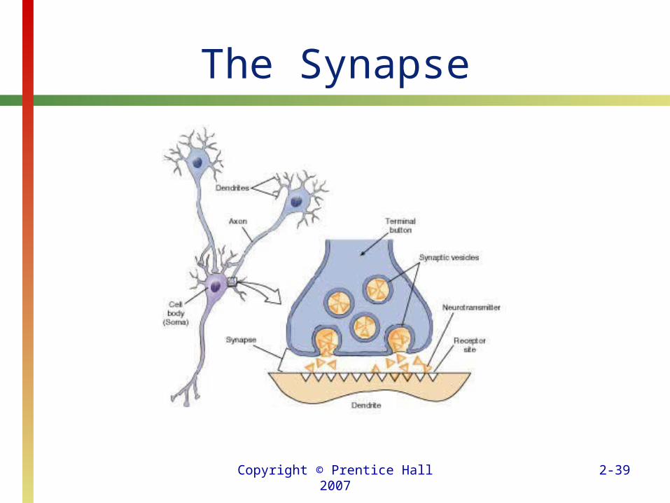
• Because a neural signal is sent from one neuron to the next through the terminal buttons of the axons, the most common arrangement is for a neuron’s terminal buttons to be near, but not touching, the receptive dendrites of neighboring neurons.
• The membrane on the side that sends the message is the presynaptic membrane and the membrane on the receiving side of the synapse is the postsynaptic membrane.
Copyright © Prentice Hall 2007 2-38
Neurons: Basic Cells of the Nervous System
• The most common arrangement at the end of an axon consists of a terminal button to send the signal, a dendrite to receive the signal, and the gap between the two, which is the synapse.
• Neurotransmitters are special chemicals stored in vesicles of the terminal buttons at the ends of axons.
Copyright © Prentice Hall 2007 2-39
The Synapse
Copyright © Prentice Hall 2007 2-40
Neurons: Basic Cells of the Nervous System
• When the electrical signal reaches the terminal buttons, it causes the vesicles in the terminal button to release a chemical signal in the form of a neurotransmitter into the synapse.
• As the neurotransmitter enters the synapse, it contacts the postsynaptic membrane (usually the dendrite) of the next neuron.
Copyright © Prentice Hall 2007 2-41
Neurons: Basic Cells of the Nervous System
• The neuron that is receiving the neurotransmitter may become more likely to transmit the message to subsequent neurons; this process is called excitation.
• In other instances, the neuron that receives the neurotransmitter becomes less likely to transmit the message to subsequent neurons; this process is called inhibition.
Copyright © Prentice Hall 2007 2-42
Neurons: Basic Cells of the Nervous System
• Dopamine is a neurotransmitter that controls arousal levels and plays a significant role in motor movement.
• Dopamine also is involved in brain pathways that are responsible for reward and punishment.
• Serotonin plays a role in weight regulation, sleep, depression, suicide, obsessive–compulsive disorder, aggression, and a wide range of other disorders and behavior problems.
Copyright © Prentice Hall 2007 2-43
Neurons: Basic Cells of the Nervous System
• Acetylcholine controls activity in brain areas related to attention, learning, and memory.
• Glutamate is called upon quite frequently to keep the lines of communication among neurons open, engage in passing along information, and may well play a role in learning.
• Excessive levels of glutamate may cause neurons to become overexcited, and they may die as a consequence.

Copyright © Prentice Hall 2007 2-44
Neurons: Basic Cells of the Nervous System
• GABA (gamma-aminobutyric acid) is an inhibitory neurotransmitter that is widely distributed throughout the brain and the spinal cord.
• The damping effect of inhibitory neurotransmitters is necessary to create a balance in the brain.

Copyright © Prentice Hall 2007 2-45
Neurons: Basic Cells of the Nervous System
• Norepinephrine (which is also a hormone) induces physical and mental arousal and heightens our mood.
• It is found in the autonomic nervous system and is part of the power behind the fight-or-flight response.

Copyright © Prentice Hall 2007 2-46
Neurons: Basic Cells of the Nervous System
• Synapses must be cleared, and cleared rapidly, before additional signals can be transmitted.
• The synapse is cleared in one of two ways, depending on the particular neurotransmitter involved.
• In the first method, breakdown, the neurotransmitter is broken down and removed from the synapse.
Copyright © Prentice Hall 2007 2-47
Neurons: Basic Cells of the Nervous System
• After the neurotransmitter affects the next neuron, an enzyme breaks it down.
• The second method for clearing the synapse, reuptake, involves taking the neurotransmitter back into the vesicles of the terminal buttons from which it came.

Copyright © Prentice Hall 2007 2-48
Neurons: Basic Cells of the Nervous System
• Most drugs exert their effects by influencing the operation of a neurotransmitter: some drugs increase the effectiveness of neurotransmitters; other drugs reduce their effectiveness.
• Drugs that promote or enhance the operation of a neurotransmitter are called agonists.
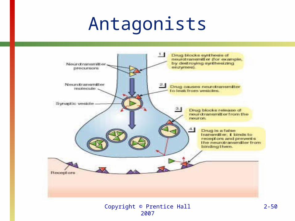
• Drugs that oppose or inhibit the operation of a neurotransmitter are called antagonists.

Copyright © Prentice Hall 2007 2-49
Agonists
Copyright © Prentice Hall 2007 2-50
Antagonists
Copyright © Prentice Hall 2007 2-51
Neurons: Basic Cells of the Nervous System
• Neuromodulators can influence the transmission of signals between neurons.
• The release and action of neurotransmitters are confined to synapses in a specific area; the distribution of neuromodulators is more widespread.

Copyright © Prentice Hall 2007 2-52
Neurons: Basic Cells of the Nervous System
• For example, neuromodulators can have simultaneous effects on diverse brain regions; their activity may be indirect and longer-lasting.
• Some neuromodulators produce their effects by facilitating the release of neurotransmitters; others inhibit the release of neurotransmitters.
• One of the best-known neuromodulators, morphine, relieves pain.
Copyright © Prentice Hall 2007 2-53
Neurons: Basic Cells of the Nervous System
• If you examine the chemicals on the outside of the neuron’s semipermeable cell membrane and compare them with the chemicals on the inside of the cell membrane, you will notice a difference in small electrically charged particles called ions.
• The two types of ions, positive (+) and negative (-), resemble the two poles or ends of a battery.
Copyright © Prentice Hall 2007 2-54
Neurons: Basic Cells of the Nervous System
• When a neuron is not sending or receiving a signal, it is in a resting state, with more negative ions on the inside than on the outside.
• Relative to the outside, the inside of the neuron is about 270 millivolts (a millivolt, mV, is 1/1,000th of a volt) when the neuron is in the resting state.

Copyright © Prentice Hall 2007 2-55
Neurons: Basic Cells of the Nervous System
• Because of this unequal distribution of ions, the neuron is polarized, like a battery.
• This 270-mV difference in electric charge between the inside and outside of a neuron at rest is the resting potential.
Copyright © Prentice Hall 2007 2-56
Neurons: Basic Cells of the Nervous System
• When a neurotransmitter enters the synapse it may result either in
– depolarization (the neuron becomes less negatively charged) or,
– hyperpolarization (the neuron becomes more negatively charged).

Copyright © Prentice Hall 2007 2-57
Neurons: Basic Cells of the Nervous System
• When excitatory neurotransmitters occupy appropriate receptor sites, they cause the cell membrane to allow positive ions to pass inside.
• The increase of positive ions on the inside of the neuron causes the resting potential to drop.
• This change, which brings the potential closer to zero, is depolarization.

Copyright © Prentice Hall 2007 2-58
Neurons: Basic Cells of the Nervous System
• If enough of the neurotransmitter is present to cause the dendrite and soma to depolarize to between 265 and 260 mV, the neuron generates its own electrical signal.
• At this threshold (the minimum amount of change required for the neuronal response to occur) the axon membrane suddenly allows large quantities of positive ions to rush inside.

Copyright © Prentice Hall 2007 2-59
Neurons: Basic Cells of the Nervous System
• In less than a millisecond the neuron changes from 260 to 130 mV, completely reversing its electrical nature or polarity.
• This reversal along the axon is the neural signal and is called an action potential, or all-or-none response.

Copyright © Prentice Hall 2007 2-60
Neurons: Basic Cells of the Nervous System
• Once the dendrite and soma reach the threshold, the action potential spreads rapidly down the axon until it reaches the terminal buttons, where it causes the release of a neurotransmitter.
• At the same time that the action potential is being transmitted, the initiating neurotransmitter is being cleared out of the synapse.

Copyright © Prentice Hall 2007 2-61
How Neurons Communicate
Copyright © Prentice Hall 2007 2-62
Neurons: Basic Cells of the Nervous System
• Removal of the neurotransmitter causes the receiving neuron to return to a resting state and allows it to generate another action potential—that is, to fire again.
• When the neuron is being reset—called the refractory period—the neuron cannot fire again.
• The action potential and the refractory period occur within 2 milliseconds.

Copyright © Prentice Hall 2007 2-63
Neurons: Basic Cells of the Nervous System
• Not all neurons respond to the presence of a neurotransmitter by depolarizing or generating an action potential; the result may be just the opposite.
• In these cases the neurotransmitter is inhibitory, and causes additional negative ions to cross the cell membrane and enter the neuron.
• When inhibition occurs, the neuron becomes more negative than it was during the resting state (hyperpolarized), making an action potential harder, if not impossible, to generate.

Copyright © Prentice Hall 2007 2-64
The Brain: A Closer Look
• In the 1800s a German physician and anatomist Franz Joseph Gall developed the pseudoscience phrenology (“science of the mind”).
• Gall believed that various skills and personality characteristics (which he called mental “faculties”) could be located on the brain.
• This early attempt to understand the brain contained an element of truth as Gall believed that skills and characteristics could be localized in certain areas of the brain.
Copyright © Prentice Hall 2007 2-65
The Brain: A Closer Look
• In 1861, the French physician Paul Broca used a technique for understanding the brain—the clinical or case study method.
• Broca discovered an area on the left hemisphere of the brain that is responsible for the ability to produce speech.
• In recognition of his discovery, this speech area is called Broca’s area.

Copyright © Prentice Hall 2007 2-66
The Brain: A Closer Look
• Phineas Gage’s story is one of the most famous cases of survival from massive brain injury.
• Gage, a railroad foreman, was working with explosives in Cavendish, Vermont, on September 13, 1848, trying to clear a railroad right-of-way through granite bedrock.

Copyright © Prentice Hall 2007 2-67
The Brain: A Closer Look
• Gage’s attention was distracted from the task at hand and a tamping iron became a 13¼ -pound, 3-foot 7-inch rocket that shot through the left side of Gage’s face and exited through his head.
• Before the accident, Gage had been an excellent worker who got along well with others and carried through with his plans.
Copyright © Prentice Hall 2007 2-68
The Brain: A Closer Look
• After the accident, he made plans he never carried out, used gross profanity, refused to listen if what others said interfered with what he wanted, and was very moody.
• The study of people who have suffered brain damage like Phineas Gage provides abundant information about brain functioning.

Copyright © Prentice Hall 2007 2-69
The Brain: A Closer Look• In 1904, brain researchers created a device that
made studying certain brain structures possible. • Before the invention of this device, structures
that were deep in the brain could be examined only by removing or damaging the tissue that covered them.
• The stereotaxic instrument holds the head in a fixed position and allows an electrode (a fine piece of specially treated wire) to be inserted into a specified area of a patient’s brain.

Copyright © Prentice Hall 2007 2-70
The Brain: A Closer Look
• The electrode is thin enough that it does not damage tissue as it passes through.
• The electrode can record electrical brain activity, stimulate brain activity with a mild electric current, or destroy a brain area by passing a strong electric current through it.

Copyright © Prentice Hall 2007 2-71
The Brain: A Closer Look
• More recently developed techniques have enabled neuroscientists to examine brain functions and anatomy without resorting to autopsy or invasive stereotaxic surgery.
• In 1929 Hans Berger developed the electroencephalograph (EEG), a device that monitors and records the brain’s electrical activity.

Copyright © Prentice Hall 2007 2-72
The Brain: A Closer Look• Brain waves (identified by Greek letters)
are distinguished by their frequency, which is measured in cycles per second (called hertz and abbreviated Hz), and their amplitude (the height of the wave on the EEG record), which reflects strength.
• Brain researchers have labeled a number of different types of brain waves; each is generally associated with a particular state of consciousness.

Copyright © Prentice Hall 2007 2-73
The Brain: A Closer Look
• Alpha waves are fast brain waves (8 to 12 Hz) that are not high in amplitude.
• The brain generally produces alpha waves when the individual is in a calm, relaxed state and is not concentrating on anything in particular.

Copyright © Prentice Hall 2007 2-74
The Brain: A Closer Look
• Beta waves are very fast brain waves (13 to 30 Hz), but are not high in amplitude.
• They are associated with mental activity such as reading, taking notes in class, or answering test questions.

Copyright © Prentice Hall 2007 2-75
The Brain: A Closer Look
• Theta waves are slow brain waves (3.5 to 7 Hz) that are irregular in frequency and low in amplitude.
• When we are in a light stage of sleep or daydreaming, our brains are likely to produce this type of wave; however, theta waves are normal in waking children up to 13 years of age.

Copyright © Prentice Hall 2007 2-76
The Brain: A Closer Look
• Delta waves are the slowest brain waves (below 3.5 Hz) and the highest in amplitude.
• Delta waves are quite common in infants up to 1 year of age and in the deepest stages of sleep.

Copyright © Prentice Hall 2007 2-77
The Brain: A Closer Look
• The advent of computers has led to major advances in the study of the brain.
• Positron emission tomography (PET) is an imaging technique that involves monitoring the metabolic activity of the brain.
Copyright © Prentice Hall 2007 2-78
The Brain: A Closer Look
• Computerized axial tomography (CT or CAT) involves the production of a large number of x-rays interpreted by a computer.
• Magnetic resonance imaging (MRI) involves the use of radio waves and a strong magnetic field to produce a signal that can be interpreted by a computer.

Copyright © Prentice Hall 2007 2-79
The Brain: A Closer Look
• Functional magnetic resonance imaging (fMRI) is a modification of the standard MRI procedure that allows both structural and temporal images of the brain to be gathered.

Copyright © Prentice Hall 2007 2-80
The Brain
• The brain is divided into the hindbrain, the midbrain, and the forebrain.
Copyright © Prentice Hall 2007 2-81
The Brain
Copyright © Prentice Hall 2007 2-82
The Brain: A Closer Look
• The major components of the hindbrain are the medulla, the pons, and the cerebellum.
• From an evolutionary perspective, these are the oldest parts of the brain, and they have important survival functions.
Copyright © Prentice Hall 2007 2-83
The Brain: A Closer Look
• The medulla (short for medulla oblongata) contains our respiratory center, which keeps us breathing, especially when we are asleep.
• The medulla also controls heart rate, vomiting, swallowing, yawning, and blood circulation.

Copyright © Prentice Hall 2007 2-84
The Brain: A Closer Look
• The pons connects the two halves of the brain at the hindbrain level; this part of the hindbrain is important for sleep and arousal.
• The cerebellum coordinates skilled movement sequences that deal with objects in motion.
Copyright © Prentice Hall 2007 2-85
The Brain: A Closer Look• Together the hindbrain and midbrain are known
as the brain stem because they form the stem, or stalk, on which the remainder of the brain rests.
• The midbrain also is composed of nerve pathways that go to and from higher brain centers.
• Psychologists have found that this complex network of fibers, known as the reticular formation, is very important in controlling our level of arousal or alertness.

Copyright © Prentice Hall 2007 2-86
The Brain: A Closer Look
• The forebrain is a part of the brain that is divided into two distinct halves with duplicate structures in each half.
• These two halves or hemispheres are connected by a wide band of fibers known as the corpus callosum (“hard body”).
Copyright © Prentice Hall 2007 2-87
The Brain: A Closer Look
• The two hemispheres of the forebrain communicate with each other through the corpus callosum.
• The cerebral cortex (cerebrum) is the convoluted (wrinkled) outer layer of the brain.
Copyright © Prentice Hall 2007 2-88
The Brain: A Closer Look
• Deep down in the brain—below the cortex—is the basal ganglia, a series of interconnected structures that play a significant role in motor movement.
• The limbic system is a group of interrelated subcortical structures that are involved in the regulation of emotions and motivated behaviors such as hunger, thirst, aggression, and sexual behavior.
Copyright © Prentice Hall 2007 2-89
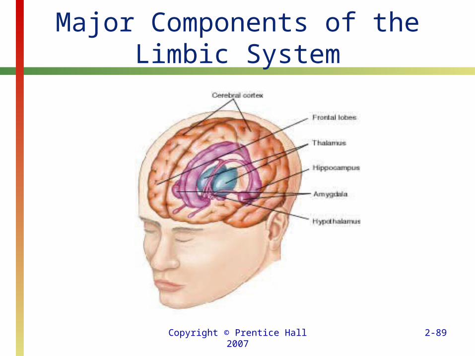
Major Components of the Limbic System

Copyright © Prentice Hall 2007 2-90
The Brain: A Closer Look
• The thalamus sends sensory information to the cerebral cortex and other parts of the brain.
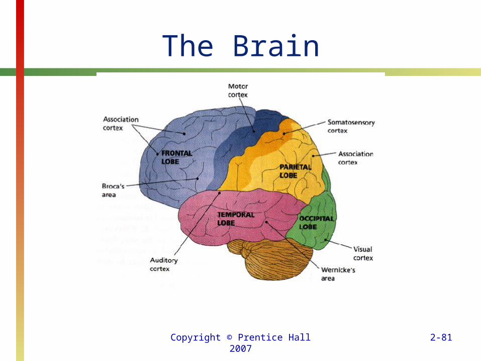
• Each cerebral hemisphere has four specific areas, called lobes—the frontal, temporal, parietal, and occipital lobes.

Copyright © Prentice Hall 2007 2-91
The Brain: A Closer Look• The frontal lobes are responsible for
language, movement, reasoning, planning, problem-solving, and personality.
• The major responsibility of the parietal lobes is to process every sensation except smell.
• The parietal lobes can also be thought of as a sensory integrator because they are responsible for body position as well as our sensory cortex or somatosensory strip.

Copyright © Prentice Hall 2007 2-92
The Brain: A Closer Look
• The temporal lobes have several functions, including the processing of auditory information; in most people the left side interprets the meaning of speech (Wernicke’s area).
• These lobes also play a significant role in learning, memory, and emotions.
• The occipital lobes’ primary responsibility is to processes visual information.

Copyright © Prentice Hall 2007 2-93
The Brain: A Closer Look
• Neuroscientists have focused on the brain structures responsible for language as well as the problems that develop when these areas are damaged.
• Broca’s area (in most people it is located in the left frontal lobe) is the key area involved in language production.

Copyright © Prentice Hall 2007 2-94
The Brain: A Closer Look
• Wernicke’s area (in most people it is located in the left temporal lobe) is the key area involved in understanding language.
• Damage to either or both areas can have significant effects on a person’s language abilities.
Copyright © Prentice Hall 2007 2-95
The Brain: A Closer Look
• The term aphasia refers to a loss of the ability to speak or understand written or spoken language.
• Damage to Broca’s area results in nonfluent aphasia.
• People with this type of aphasia have difficulty producing speech, although they generally understand what others say to them.

Copyright © Prentice Hall 2007 2-96
The Brain: A Closer Look• In 1874, the German neurologist Carl
Wernicke (1848–1905) identified a second brain area that plays a significant role in language.
• Damage to Wernicke’s area results in language problems called fluent aphasia.
• In contrast to the speech of people with damage to Broca’s area, damage to Wernicke’s area results in fluent-sounding, but meaningless speech.

Copyright © Prentice Hall 2007 2-97
The Brain: A Closer Look
• Broca’s and Wernicke’s aphasias are the two most common, but there are other types.
• For example, optic aphasia is the inability to read more than one letter at a time, whereas a person suffering from word deafness cannot understand spoken language despite the presence of normal hearing and reading abilities.
Copyright © Prentice Hall 2007 2-98
The Brain: A Closer Look
• Apraxias are deficits in nonverbal skills.
• Apraxias involve damage to the right hemisphere.
• Depending on the site of the damage, one might observe a dressing apraxia, in which a person has trouble putting clothing on one side of the body, or a constructional apraxia, in which a person cannot copy a simple drawing.
Copyright © Prentice Hall 2007 2-99
The Brain: A Closer Look
• In addition to apraxias, the right hemisphere controls prosody, the ability to express emotion.
• People suffering from motor aprosodia speak in a flat monotone regardless of their real feelings.
• Such people simply cannot display emotions.
Copyright © Prentice Hall 2007 2-100
The Brain: A Closer Look
• In the early 1960s, two neurosurgeons, Philip Vogel and Joseph Bogen, discovered that cutting the corpus callosum reduced seizures in untreatable epileptic patients.
• Even though we do not know exactly why this operation controls seizures, it is still performed as a last resort in severe cases of epilepsy.

Copyright © Prentice Hall 2007 2-101
The Brain: A Closer Look
• Research by Nobel Prize winner Roger Sperry and his colleague Michael Gazzaniga showed that in people with a severed corpus callosum, the two hemispheres appeared to be doing different things.
Copyright © Prentice Hall 2007 2-102
The Brain: A Closer Look
• Studies of split brain patients support the conclusion that the left hemisphere is involved in speech and language production.
• Although the right hemisphere has limited language functions, it is essential for adding emotional content to our speech.
• It is also important for spatial abilities such as recognizing complex geometric patterns.

Copyright © Prentice Hall 2007 2-103
The Brain: A Closer Look
• Plastic can be molded and changed into many forms.
• It is a pliable material that can take on different forms and even functions.
• In some ways, the same can be said of the brain, which can change remarkably over time.

Copyright © Prentice Hall 2007 2-104
The Brain: A Closer Look
• One reason the brain, especially in children, can change in response to experiences (including removal of an entire hemisphere) is that humans do not come into this world with a fully developed, hard-wired brain.
• New data indicates that it is possible for new neurons to develop in the human brain (neurogenesis).