Review on the Protozoan Parasite Perkinsus olseni (Lester and

5.2.1 Perkinsus sp. Infections of Marine Molluscs - 1
5.2.1 Perkinsus sp. Infections of Marine Molluscs
Kimberly Reece1 and Christopher Dungan2
1 Virginia Institute of Marine Science
College of William and Mary P.O. Box 1346
Gloucester Point, VA 23062 [email protected]
2 Maryland DNR
Cooperative Oxford Laboratory 904 S. Morris Street Oxford, MD 21654
[email protected] A. Name of Disease and Etiological Agent
In the earliest-reported cases in the USA (Mackin et al. 1950), the name of the disease in Crassostrea virginica oysters was popularly abbreviated as ‘dermo’, in reference to the now-obsolete identification of its protistan agent, Perkinsus marinus, as Dermocystidium marinum (=Labyrinthomyxa marina). Recent evidence suggests that the current placement of the genus Perkinsus among the Apicomplexa may be incorrect, because member species also show consistent affinities with the dinoflagellates, or with a proposed new alveolate protozoan phylum Perkinsozoa. Since 1981, several new Perkinsus spp. parasites and diseases have been described and reported in a variety of bivalve and gastropod molluscs, worldwide (reviewed by Villalba et al. 2004). Other currently described Perkinsus species include P. olseni from Australian abalone, P. atlanticus from European clams, P. chesapeaki and P. andrewsi from Chesapeake Bay clams, P. mediterraneus from Mediterranean flat oysters, and P. qugwadi (incertae sedis) from Pacific Canada scallops. Perkinsus olseni is proposed to be a priority synonym of P. atlanticus (Murrell et al. 2002). Perkinsus chesapeaki and P. andrewsi are proposed as synonymous clam parasite species, with the former name in priority (Burreson et al. 2005). Affinities of P. qugwadi with the genus Perkinsus are tenuous (Bower et al. 2003).
B. Known Geographic and Host Ranges of the Diseases
Perkinsus marinus (Mackin et al.1950) infections (dermo disease) occur widely in C. virginica oysters along the Gulf of Mexico and Atlantic coasts of North America, from Tabasco, Mexico to Maine, USA. The disease was reported once in moribund, introduced C. virginica from Hawaii waters (Kern et al. 1973).
February 2006

5.2 Perkinsus sp. Infections of Marine Molluscs - 2
Perkinsus chesapeaki (McLaughlin et al. 2000) and P. andrewsi (Coss et al. 2001) infections are reported at high respective prevalences in Chesapeake Bay Mya arenaria, Tagelus plebeius, and Macoma balthica clams. Perkinsus olseni (Lester and Davis 1981) infections are reported in moribund southern Australia abalone Haliotis rubra, H. laevigata, H. cyclobates, and H. scalaris. Similar parasites are reported in diverse bivalve molluscs from northern, southern, and eastern Australian coasts, as well as from northern New Zealand waters. Perkinsus atlanticus (Azevedo 1989) infections are reported in moribund Ruditapes decussatus, Venerupis philippinarum, V. pullastra, and V. aureus clams from the Atlantic coast of the Iberian Peninsula. Similar infections are reported in Crassostrea angulata and Ostrea edulis oysters from the Iberian Atlantic and Mediterranean coasts, and in Italian R. decussatus and Callista chione clams. Perkinsus mediterraneus (Casas et al. 2004) infections are reported in commercially cultured Ostrea edulis flat oysters from the western Mediterranean, Balearic Islands. Perkinsus qugwadi (incertae sedis) (Bower et al. 1998) infections were transiently reported in Japanese scallops, Patinopecten yessoensis, grown in Canadian Pacific waters. Infections by undescribed Perkinsus sp. with affinities to P. olseni and P. atlanticus are reported in moribund V. philippinarum clams from the Japan, Korea, and China coasts; as well as in Paphia undulata clams from the Thailand coast.
C. Epizootiology
The severity of oyster dermo disease cycles seasonally in enzootic North American waters, with highest infection prevalences, infection intensities, and disease mortality during late summer and fall. Similar seasonality characterizes Perkinsus spp. infections in several Chesapeake Bay clam species. Perkinsus marinus proliferates maximally at temperatures above 20 °C and salinities above 15 ppt. Since molluscan hosts are nominal osmoconformers, disease morbidity and mortality are reduced during low-salinity periods in affected estuaries, but infections persist in oysters over-wintering in 5 ppt waters. Mortality in affected oyster populations can reach 100%, with 30-50% mortality in the first year of exposure, and cumulative mortality of 75-100% during the second year of exposure. Direct waterborne transmission of infections occurs. During periods of high-salinity Chesapeake Bay epizootics, >90% of juvenile oysters planted both near and distant (>1 km) from infected oyster populations, harbored infections after 12 weeks of exposure. In Gulf of Mexico waters, the parasitic gastropod Boonea impressa serves as an active vector in transmitting parasite cells between oyster hosts.
D. Disease signs Reduced growth rate occurs prior to the onset of mortality.
February 2006
5.2.1 Perkinsus sp. Infections of Marine Molluscs - 3
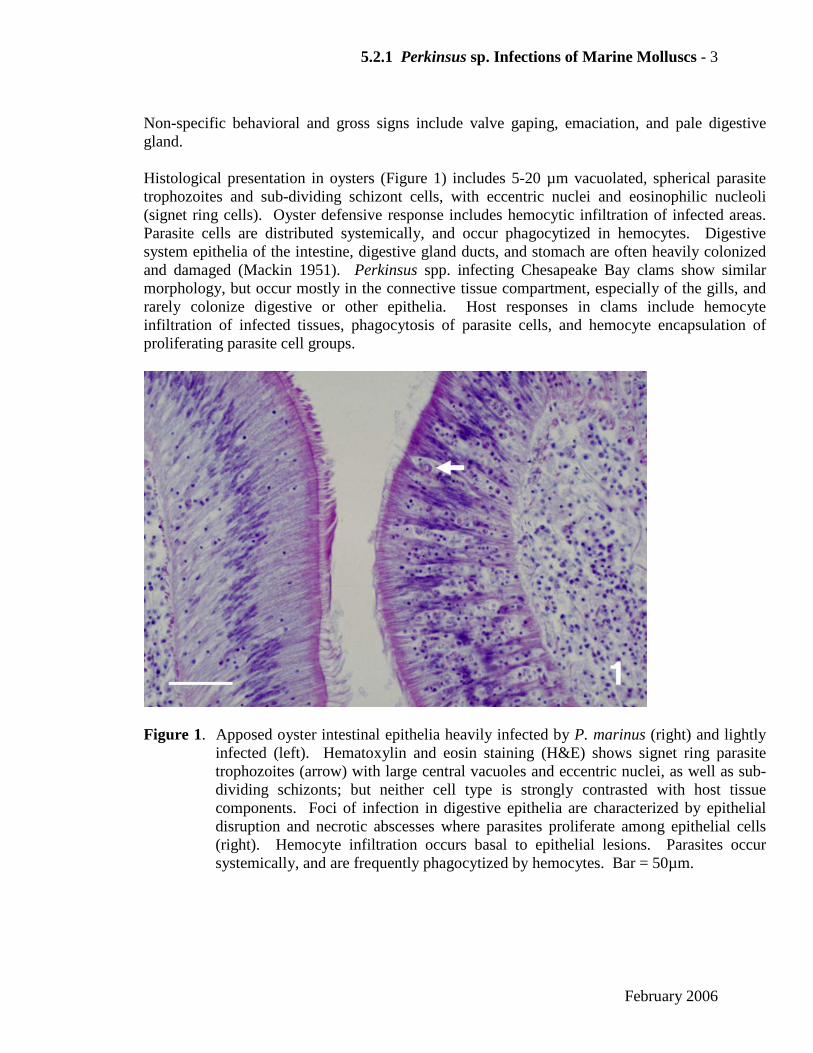
Non-specific behavioral and gross signs include valve gaping, emaciation, and pale digestive gland. Histological presentation in oysters (Figure 1) includes 5-20 µm vacuolated, spherical parasite trophozoites and sub-dividing schizont cells, with eccentric nuclei and eosinophilic nucleoli (signet ring cells). Oyster defensive response includes hemocytic infiltration of infected areas. Parasite cells are distributed systemically, and occur phagocytized in hemocytes. Digestive system epithelia of the intestine, digestive gland ducts, and stomach are often heavily colonized and damaged (Mackin 1951). Perkinsus spp. infecting Chesapeake Bay clams show similar morphology, but occur mostly in the connective tissue compartment, especially of the gills, and rarely colonize digestive or other epithelia. Host responses in clams include hemocyte infiltration of infected tissues, phagocytosis of parasite cells, and hemocyte encapsulation of proliferating parasite cell groups.
Figure 1. Apposed oyster intestinal epithelia heavily infected by P. marinus (right) and lightly infected (left). Hematoxylin and eosin staining (H&E) shows signet ring parasite trophozoites (arrow) with large central vacuoles and eccentric nuclei, as well as sub-dividing schizonts; but neither cell type is strongly contrasted with host tissue components. Foci of infection in digestive epithelia are characterized by epithelial disruption and necrotic abscesses where parasites proliferate among epithelial cells (right). Hemocyte infiltration occurs basal to epithelial lesions. Parasites occur systemically, and are frequently phagocytized by hemocytes. Bar = 50µm.
February 2006

5.2 Perkinsus sp. Infections of Marine Molluscs - 4
E. Disease Diagnostic Procedures
Table 1. Mollusc Perkinsus sp. infection diagnostic methods attributes. Increasing relative assay sensitivities are categorized (1-4). Due to small (section or tissue) sample size, and inherent attendant sampling error, histological methods are excluded from the level-4 sensitivity category.
Method Relative sensitivity 1
Pathogen specificity
Presumptive diagnosis2
Confirmatory diagnosis
Gross signs 0 - -
Histopathology 1-2 genus ++ ++
Histological immunoassay 3 genus ++ ++
ELISA assays 3-4 genus ++ ++
RFTM assays 1. hemolymph 2. solid tissue subsample 3. whole body burden
1-3 1-3 4
genus *
++ ++ +++
++ ++ +++
PCR assays 3-4 genus* or spe +++ +++
ISH assays 3 genus * +++ +++
1 0-4 increasing sensitivity * except Perkinsus qugwadi 2 applicability: - unsuitable, + limited utility, ++ standard method,
+++ recommended method
1. Ray’s high-salt fluid thioglycollate medium (RFTM) assay Non-differentially detects all Perkinsus species, except P. qugwadi (incertae sedis), which enlarge but do not proliferate in RFTM. Spherical, enlarged parasite hypnospores or pre-zoosporangia measure 10-250 µm, with refractile walls that stain blue-black in 20-33% (v/v) Lugol’s iodine (Figure 2). Possible analytes include solid tissue sub-samples, hemolymph, and whole host bodies. Choice depends on desired level of sensitivity, quantitative results quality, and tolerance for lethal sampling (see Ray 1963; Bushek et al. 1994 for reviews and methods). Optimum solid tissue sub-sample types are selected according to parasite tissue tropisms, where known: rectum or mantle for P. marinus infections in oysters; gill or labial palp for Perkinsus spp. infections in Chesapeake Bay clams.
February 2006

5.2.1 Perkinsus sp. Infections of Marine Molluscs - 5
The most sensitive and only strictly quantitative RFTM assay is the whole body burden (WBB) assay (see g.3 below). Wet weights of shucked host tissues are recorded before homogenization and incubation at 20-27 °C in 30-50 volumes of RFTM for 4-7 d. Host tissues are then hydrolyzed at 60 °C in 2M NaOH, leaving killed, unlysed, enlarged parasite cells. Parasite cell counts are normalized to initial host tissue wet weights (Bushek et al. 1994). Since Perkinsus species are aerobes, anaerobic gradients in RFTM are not required.
a. Re-hydrate fluid thioglycollate medium with dextrose (Difco 0256-, BBL 01-140, Sigma
A0465) in distilled water plus 20 g/L NaCl. Boil briefly to dissolve.
b. Aliquot RFTM to assay tubes or bulk containers. Autoclave to sterilize. If bulk RFTM medium is autoclaved, aliquot aseptically to sterile assay tubes or culture plate wells, before use.
c. Fortify autoclaved RFTM to 50 µg/ml chloramphenicol (mutagen), or to 100 U/ml
penicillin and 100 µg/ml streptomycin.
d. Before use, fortify RFTM aliquots with 50-200 U/ml nystatin, applied as a surface overlay, where possible.
e. Aseptically inoculate RFTM aliquots with solid tissue sub-samples, hemolymph samples,
or whole oyster homogenates, at ≤ 0.1 g tissue/3 ml RFTM.
f. Cap or cover RFTM assay containers. Incubate at 20-28 °C for 7-4 d, respectively.
g.1 Observe RFTM-incubated tissues, unstained, for thick-walled, refractile, vacuolated, 10-250 µm spherical, enlarged parasite hypnospore cells, using a microscope equipped for contrasting live cells. Rank and select optimal inocula for parasite in vitro propagation.
or
g.2 Transfer and macerate solid tissue sub-samples in pools of 20-33% Lugol’s iodine on microscope slides, add additional drops of dilute Lugol’s iodine to cover macerated tissue, and coverslip. Suspension samples (hemolymph) may be iodine-stained in microplate wells, or on filter membranes.
or g.3 WBB assay. Pellet suspended solids by centrifugation at 1,500 x g for 10 min.
Conservatively aspirate and discard RFTM supernate. Hydrolyze oyster tissue homogenates in 5-10 volumes of 2M NaOH, at 60 °C for 1-6 h, vortexing every 30 min., until tissue lysis is complete. Pellet parasite hypnospores by centrifugation at 1,500 x g for 10 min. Aspirate and discard NaOH supernate. Resuspend parasite cell pellet and wash 3x for 10 min. at 1,500 x g, in 0.1M phosphate-bufferd saline (PBS) + 0.5% (w/v) bovine serum albumin (BSA) (PBS/BSA). Resuspend washed pellet in 1 ml of PBS/BSA. Stain
February 2006

5.2 Perkinsus sp. Infections of Marine Molluscs - 6
entire cell suspension with 20% filtered Lugol’s iodine for microscopic enumeration, or quantitatively dilute parasite cell suspension sub-samples in PBS/BSA, to yield countable dilutions (100-400 cells), before staining and enumerating parasite cells. Estimate total parasite cells/oyster from enumerated sample dilution factors, and normalize total parasite cells/g. host tissue wet weight. See Bushek et al. 1994; La Peyre et al. 2003 for review and details.
h. Examine stained preparations for 10-250 µm smooth, spherical cells with refractile walls
that are stained blue-black. (Figure 2). Staining may vary with iodine penetration. Distinguish pollen grains and possible iodine crystals by morphology.
2. Histopathology Their low-contrast H&E staining characteristics render Perkinsus species detection in histological sections a less-sensitive diagnostic method than RFTM, or other, assays. See D. Disease Signs and Figure 1.
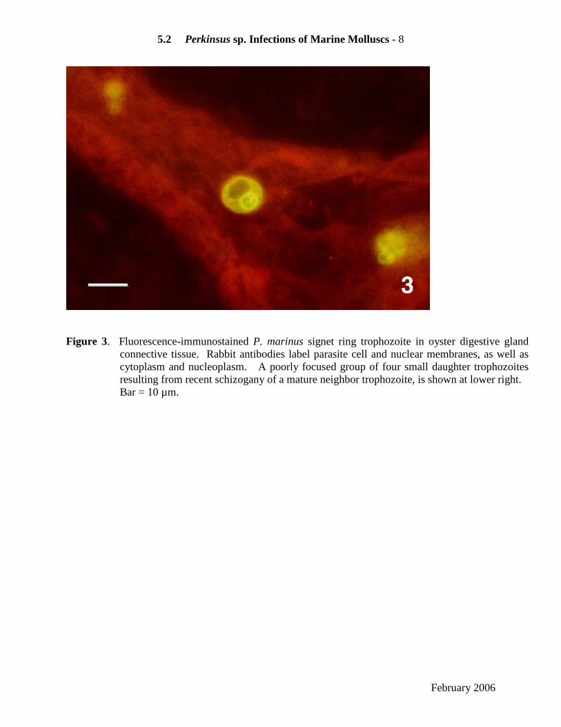
3. Histological immunoassays Fluorescent or colorimetric antibody labeling increases immuno-stained parasite contrast, and enhances detection sensitivity of histological assays. Neither polyclonal nor monoclonal antibodies developed to date to P. marinus immunogens are parasite species-specific (Dungan and Roberson 1993) (Figure 3). Limited quantities of rabbit anti- P. marinus antibodies are available by request ([email protected]).
a. De-wax paraffin sections, and hydrate to 0.1M phosphate-buffered saline (PBS), pH 7.2.
b. Block sections with PBS + 0.05% (v/v) Tween-20 (PBST) + 2% (w/v) bovine serum albumin (BSA) + 1% normal rabbit serum.
c. Incubate sections 30-60 min. in 10-20 µg/ml primary antibody IgG diluted in PBST + 1%
BSA (PBST/BSA). d. Wash sections in PBST.
e. Incubate sections 30-60 min. in darkened, 1-4 µg/ml affinity-purified secondary
antibody/FITC conjugate, diluted in PBST/BSA. f. Wash sections in PBST.
g. Counterstain sections with 0.01-0.05% Evan’s blue.
h. Dip sections in PBST to rinse.
i. Coverslip in pH 9-buffered glycerol, or equivalent mounting medium, and observe under
fluorochrome-appropriate (490 nm for FITC) epifluorescence excitation.
February 2006

5.2.1 Perkinsus sp. Infections of Marine Molluscs - 7
Figure 2. RFTM-enlarged Perkinsus sp. hypnospores stained with Lugol’s iodine. 2a) Following WBB
assay NaOH hydrolysis of oyster tissues. Bar = 50µm. 2b) In macerated Mya arenaria labial palp tissue with heavy infection. Incomplete iodine access or activity leaves many parasite hypnospores lightly stained or unstained. Bar = 250 µm.
February 2006
5.2 Perkinsus sp. Infections of Marine Molluscs - 8
Figure 3. Fluorescence-immunostained P. marinus signet ring trophozoite in oyster digestive gland
connective tissue. Rabbit antibodies label parasite cell and nuclear membranes, as well as cytoplasm and nucleoplasm. A poorly focused group of four small daughter trophozoites resulting from recent schizogany of a mature neighbor trophozoite, is shown at lower right. Bar = 10 µm.
February 2006

5.2.1 Perkinsus sp. Infections of Marine Molluscs - 9
4. ELISA assays Polyclonal antibody-based analyte-capture ELISA assays are reported to detect P. marinus extracellular secretions (ECP) in tissue homogenates from oysters with infection intensities of one parasite cell/g oyster tissue (Ottinger et al. 2001). The specificity of polyclonal antibodies against P. marinus ECP was not tested against other Perkinsus spp. or marine mollusc protistan associates. Monoclonal murine IgG1 antibodies against P. marinus in vitro trophozoites also label P. atlanticus cells, but do not recognize several other protistan oyster pathogens (Romestad et al. 2001). A competitive ELISA assay using these antibodies is reported to detect 1,000 parasite cells in 50 µl of oyster tissue homogenate. P. marinus ECP capture ELISA assay (Ottinger et al. 2001) a. Coat ELISA plate wells with 50 µl of 40 µg/ml anti-P. marinus extracellular proteins (ECP)
IgG in coating buffer for 1 h at 21 °C. b. Wash plate wells 3x with 0.1M TRIS-buffered saline (TBS, pH 7.2) + 0.1% (v/v) Tween-20
(TTBS).
c. Block assay wells 1 h with 240 µl of TTBS + 1% (w/v) BSA (TTBS/BSA). d. Wash plate wells, as above. e. Add 50 µl/well of oyster tissue homogenates or positive-control ECP samples, serially-
diluted in TTBS/BSA, and incubate for 2 h at 21 °C. f. Wash plate wells, as above.
g. Add 50 µl/well of 30 µg/ml biotinylated anti-P. marinus ECP IgG in TTBS/BSA, for 1 h at
21 °C. h. Wash plate wells, as above.
i. Add 50 µl/well of streptavidin-horseradish peroxidase (HRPO) conjugate, diluted 1:1,000 in
TTBS/BSA, for 1 h at 21 °C. j. Wash plate wells, as above.
k. Add 50 µl/well of ABTS chromagenic HRPO enzyme substrate; incubate reaction for 30 min
in the dark at 21 °C. l. Read assay well absorbances of 405-nm light; using ECP standard and negative-control well
absorbances to interpret diagnostic sample well absorbances.
February 2006

5.2 Perkinsus sp. Infections of Marine Molluscs - 10
5. PCR assays Perkinsus genus-specific primers have been designed (Casas et al. 2002) to target the internal transcribed (ITS) region of all described Perkinsus species except P. qugwadi (incertae sedis) (Figure 4a). These primers amplify Perkinsus spp. DNA isolated from cultured cells, as well as from DNA extracts of infected host tissue, and are able to detect less than 0.01 genome equivalents in a reaction (Figure 4b). DNA sequence analysis of the amplified ITS fragment enables species identification by comparison to GenBank (HTTP://WWW.NCBI.NLM.NIH.GOV/ENTREZ/) deposited ITS region sequences for Perkinsus species. In addition, several species-specific PCR assays have been developed. Most of the species-specific primers also target portions of the ribosomal RNA gene complex. Many assays are available to specifically detect P. marinus DNA (Marsh et al. 1995; Robledo et al. 1998; Yarnall et al. 2000; Audemard et al. 2004). To quantify the amount of parasite present in the host or the environment, Marsh et al. (1995) and Yarnall et al. (2000) developed semi-quantitative PCR assays, while Audemard et al. (2004) recently developed a very sensitive real-time PCR assay for quantification of Perkinsus spp. DNA in environmental samples. In addition, a multiplex PCR assay was developed to allow simultaneous and differential detection of Haplosporidium nelsoni, H. costale and P. marinus, all parasites of the eastern oyster Crassostrea virginica (Penna et al. 2001). Species-specific PCR assays have also been developed for other Perkinsus species. Nested ITS region primers were developed to diagnose the Perkinsus sp. infecting Japanese short-necked clams (Hamaguchi et al. 1998), which, based on the DNA sequence of the ITS region, is likely to be P. olseni. Several PCR primer pairs were designed to specifically target the non-transcribed spacer (NTS) region of P. olseni/P. atlanticus (de la Herrán et al. 2000; Robledo et al. 2000; Murrell et al. 2002) and Coss et al. (2001) developed a specific PCR assay targeting the NTS region for P. andrewsi to discriminate this species from P. marinus and P. atlanticus. Perkinsus genus PCR assay a. Extract genomic DNA from cultured cells or infected host tissue using the Qiagen (QiagenTM, Valencia, California) DNeasy tissue kit. Alternatively, DNA can be extracted using phenol/chloroform based (Reece et al. 1997) or CTAB-based DNA extraction methods (Carlini and Graves 1999).
b. Quantify DNA spectrophotometrically.
c. PCR amplification
1. Primers: Perk ITS 85 (5’CCGCTTTGTTTRGATCCC3’) Perk ITS 750 (5’ACATCAGGCCTTCTAATGATG3’)
2. Reagent concentrations for 25µl reactions. 10 ng (cultured cell)-100 ng (infected host tissue) of genomic DNA. 20 mM Tris-HCl (pH 8.4) 50 mM KCl, 1.5 mM MgCl2 0.2 mM each of dATP, dGTP, dCTP, dTTP 25 pmol of each primer (above) 0.625 units of Taq DNA polymerase
February 2006

5.2.1 Perkinsus sp. Infections of Marine Molluscs - 11
0.2 mg/ml of BSA
3. Cycling parameters (modified from Casas et al. 2002) Initial denaturation of 4 min at 94 °C 40 cycles of:
1 min at 94 °C 1 min at 55 °C 1 min at 72 °C
Final extension of 10 min at 72 °C
d. PCR products are electrophoresed on 2% agarose (in 1x TBE) gels, stained with ethidium bromide, and visualized using UV light.
6. SSU rRNA ISH assays A genus-Perkinsus DNA probe was developed to specifically target SSU rRNA sequences of Perkinsus species and was shown to hybridize specifically to a variety of Perkinsus spp. cells in paraffin-embedded histological sections of host species (Elston et al. 2004) (Figure 5). It does not hybridize, however, to Perkinsus qugwadi (incertae sedis), or to parasitic dinoflagellates or haplosporidian species. Perkinsus probe: Perksp700DIG (5’CGCACAGTTAAGTRCGTGRGCACG3’), probe is 5’ end-labeled with digoxigenin. In situ hybridizations are performed as previously described (Stokes and Burreson 1995, 2001), with modifications indicated below. a. Dewax paraffin-embedded tissue sections mounted on positively charged slides in xylene,
and rehydrate to 70% ethanol. b. Wash slides in P buffer (50 mM Tris-HCl, pH 7.5; 5 mM EDTA) and treat with 125 µg/ml
pronase solution in P buffer for 30 min. at 37 °C for permeablization, followed by two washes in P buffer and a 10 min incubation in 2X SSC.
c. Prehybridize in a standard prehybridization solution for 1 h at 42 °C. d. Hybridize overnight in a humid chamber at 42 °C in a solution with a probe concentration of
7 ng/ul in prehybridization buffer.
e. Wash in a series of solutions descending from 2x SSC at 21°C to 0.5x SSC at 42 °C. f. Immunological detection of the digoxigenin-labeled probe is done with an anti-digoxigenin
antibody-alkaline phoshatase conjugate and NBT/BCIP substrate, following blocking and rinsing.
February 2006

5.2 Perkinsus sp. Infections of Marine Molluscs - 12
Figure 4. Ethidium bromide-stained PCR gels demonstrating the specificity and sensitivity of the
Perkinsus genus-specific primer pair PerkITS-85/ PerkITS-750 (PerkITS) (a) Specificity of PerkITS primers tested on: no DNA (lane N), P. marinus (lane 1), P. chesapeaki (lane 2), P. olseni (lane 3), Hematodinium perezi (lane 4), Pfeisteria piscicida (lane 5), Pf. schumwayae (lane 6), Amphidinium carterae (lane 7), Crypthecodinium cohnii (lane 8), Karlodinium micrum (lane 9), Peridinium foliaceum (lane 10) and Prorocentrum micans (lane 11) DNAs. (b) Sensitivity of PerkITS primers tested on: no DNA (N), and serial dilutions of P. marinus DNA isolated from a determined (counted) number of cultured cells. DNA in each reaction was estimated to correspond with the following genome equivalents: 8 (lane 1), 0.8 (lane 2), 0.08 (lane 3), 0.008 (lane 4). Lanes L: 100-bp DNA size standards. Gel photos courtesy of C. Audemard.
February 2006

5.2.1 Perkinsus sp. Infections of Marine Molluscs - 13
Figure 5. In situ hybridization of genus Perkinsus SSU rRNA gene probe (violet-blue) to a heavy P.
marinus infection in C. virginica intestinal epithelium and visceral connective tissues. Both parasite nuclear rDNA and cytoplasmic rRNA share sequence homology with, and hybridize, the digoxigenin-conjugated oligonucleotide DNA probe. Large ciliates infesting the intestinal lumen and epithelium counterstain with Bismark brown. Bar = 100 µm.
February 2006

5.2 Perkinsus sp. Infections of Marine Molluscs - 14
F. Procedures for Detecting Subclinical Infections
1. Histological immunoassays have been successfully employed to visualize initial lesions in experimental oysters. In situ DNA/RNA probes should be similarly useful.
2. ELISA immunoassays may detect infections of one parasite cell/g oyster tissue. 3. RFTM whole body burden assays detect one parasite cell/host.
4. PCR assay amplification may detect subclinical infections when potentially rare parasite DNA
target templates are present in amplified, 50-150 ng DNA sub-samples. Multiple-copy gene templates, such as rRNA gene targets, may permit detection of infections when <1 parasite genome complement is present in the amplified DNA sample.
G. Procedures for Transportation and Storage of Samples to Ensure Maximum Viability or Preservation of the Etiological Agent 1. RFTM assays
a. Ship or transport live molluscs humidified and refrigerated (10-20 °C) to an analytical facility, or process aseptically where collected.
b. Aseptically dissect and inoculate antimicrobial-fortified RFTM tubes or plate wells with
selected tissue biopsies, to minimize contaminant microbial over-growth and sample cross-contamination of assay tubes/wells.
c. In vitro Perkinsus sp. parasite cultures are successfully initiated in isohaline
DME:Ham’s F-12, or other culture media, from hypnospores enlarged in RFTM at 27 °C for 48-96 h (Dungan et al. 2002).
2. PCR assays
a. Aseptically, and with due precautions against sample cross-contamination, excise optimal host tissue biopsy for parasite detection in extracted DNA. Alcohol-flame instruments and dissect each mollusc on a disposable, virgin plastic weigh boat or Petri dish. Sodium hypochlorite oxidation and hydrolysis of work surface and instrument contaminants between samples is an alternative decontamination method.
b. Preserve tissue for subsequent DNA extraction by dehydration in 10 volumes of
uncontaminated, 95-100% ethanol in sterile, virgin screw-capped containers. Store samples at or below 21 °C.
3. Histological assays
a. Excise desired 5 mm-thick tissue samples, place in labeled histological cassettes, and fix immediately for 48 h in Davidson’s, or other, fixative solution.
February 2006

5.2.1 Perkinsus sp. Infections of Marine Molluscs - 15
b. Decant fixative; replace with 70% ethanol. Infiltrate, embed, section, and stain tissue samples.
References
Audemard, C., K. S. Reece, and E. M. Burreson. 2004. Real-time PCR for the detection
and quantification of the protistan parasite Perkinsus marinus in environmental waters. Applied and Environmental Microbiology 70:6611-6618.
Bower, S., E. Burreson, and K. Reece. 2003. Annex 10: Review of Molecular Techniques Used to
Differentiate the Various Species/Isolates of Perkinsus. Pages 54-60 in 2003 Report of the Working Group on Pathology and Diseases of Marine Organisms. ICES CM 2003/F:03 Ref. ACME, International Council for the Exploration of the Sea, Copenhagen, Denmark. 95 pp. www.ices.dk/iceswork/wgdetail.asp?wg=wgpdmo
Burreson, E. M., K. S. Reece, and C. F. Dungan. 2005. Molecular, morphological, and
experimental evidence support the synonymy of Perkinsus chesapeaki and Perkinsus andrewsi. Journal of Eukaryotic Microbiology 52:258-270.
Bushek, D., S. E. Ford, and S. K. Allen. 1994. Evaluation of methods using Ray’s fluid
thioglycollate medium for diagnosis of Perkinsus marinus infection in the eastern oyster Crassostrea virginica. Annual Review of Fish Diseases 4:201-217.
Carlini, D. B., and J. E. Graves. 1999. Phylogenetic analysis of cytochrome C oxidase I
sequences to determine higher-level relationships within the coleiod cephalopods. Bulletin of Marine Science 64:57-76.
Casas, S. M., A. Villalba, and K. S. Reece. 2002. Study of perkinsosis in the carpet shell
clam Tapes decussatus in Galicia (NW Spain). I. Identification of the aetiological agent and in vitro modulation of zoosporulation by temperature and salinity. Diseases of Aquatic Organisms 50:51-65.
Coss, C. A., J. A. F. Robledo, G. M. Ruiz, and G. R. Vasta. 2001. Description of
Perkinsus andrewsi n. sp. isolated from the Baltic clam (Macoma balthica) by characterization of the ribosomal RNA locus, and development of a species-specific PCR-based diagnostic assay. Journal of Eukaryotic Microbiology 48:52-61.
De la Herrán, R., M. A. Garrido-Ramos, J. I. Navas, C. Ruiz Rejón, and M. Ruiz Rejón.
2000. Molecular characterization of the ribosomal RNA gene region of Perkinsus atlanticus: its use in phylogenetic analysis and as a target for a molecular diagnosis. Parasitology 120:345-353.
Dungan, C. F., and B. S. Roberson. 1993. Binding specificities of mono- and polyclonal
antibodies to the protozoan oyster pathogen Perkinsus marinus. Diseases of Aquatic Organisms 15:9-22.
Dungan, C. F., R. M. Hamilton, K. L. Hudson, C. B. McCollough, and K. S. Reece. 2002.
Two epizootic diseases in Chesapeake Bay commercial clams, Mya arenaria and Tagelus plebius. Diseases of Aquatic Organisms 50:67-78.
February 2006

5.2 Perkinsus sp. Infections of Marine Molluscs - 16
Elston, R. A., C. F Dungan, T. R. Meyers, and K. S. Reece. 2004. Perkinsus sp. infection
risk for Manila clams, Venerupis philippinarum (Admas and Reeve, 1850) on the Pacific coast of North and Central America. Journal of Shellfish Research 23:101-105.
Hamaguchi, M., N. Suzuki, H. Usuki, and H. Ishioka. 1998. Perkinsus protozoan
infection in short-necked clam Tapes (= Ruditapes) philippinarum in Japan. Fish Pathology 33:473-480.
La Peyre, M. K., A. D. Nickens, A. K. Volety, G. S. Tolley, and J. L. La Peyre. 2003.
Environmental significance of freshets in reducing Perkinsus marinus infection in eastern oysters Crassostrea virginica: potential management applications. Marine Ecology Progress Series 248:165-176.
Mackin, J. G., H. M.Owen, and A. Collier. 1950. Preliminary note on the ocurrence of a
new protistan parasite, Dermocystidium marinum n. sp. in Crassostrea virginica (Gmelin). Science 111:328-329.
Mackin, J. G. 1951. Histopathology of infection of Crassostrea virginica (Gmelin) by
Dermocystidium marinum Mackin, Owen and Collier. Bulletin of Marine Science of the Gulf and Caribbean 1:72-87.
Marsh, A. G., J. D. Gauthier, and G. R. Vasta. 1995. A semiquantitative PCR assay for
assessing Perkinsus marinus infections in the eastern oyster, Crassostrea virginica. Journal of Parasitology 81:577-583.
Murrell, A., S. N. Kleeman, S. C. Barker, and R. J. G. Lester. 2002. Synonymy of
Perkinsus olseni Lester & Davis, 1981 and Perkinsus atlanticus Azevedo, 1989 and an update on the phylogenetic position of the genus Perkinsus. Bulletin of the European Association of Fish Pathologists 22:258-265.
Ottinger, C. A., T. D. Lewis, D. A. Shapiro, M. Faisal, and S. L. Kaattari. 2001.
Detection of Perkinsus marinus extracellular proteins in tissues of the eastern oyster Crassostrea virginica: potential use in diagnostic assays. Journal of Aquatic Animal Health 13:133-141.
Penna, M. S., M. Khan, and R. A. French. 2001. Development of a multiplex PCR for the
detection of Haplosporidium nelsoni, Haplosporidium costale and Perkinsus marinus in the eastern oyster (Crassostrea virginica, Gmelin 1971). Molecular and Cellular Probes 15:385-390.
Ray, S. M. 1963. A review of the culture method for detecting Dermocystidium
marinum, with suggested modifications and precautions. Proceedings of the National Shellfisheries Association 54:55-69.
Reece, K. S., D. Bushek, and J. E. Graves. 1997. Molecular markers for population
genetic analysis of Perkinsus marinus. Molecular Marine Biology and Biotechnology 6:197-206.
February 2006

5.2.1 Perkinsus sp. Infections of Marine Molluscs - 17
Robledo, J. A. F., J. D. Gauthier, C. A. Coss, A. C. Wright, and G. R. Vasta . 1998. Species- specificity and sensitivity of a PCR- based assay for Perkinsus marinus in the eastern oyster, Crassostrea virginica: a comparison with the fluid thioglycollate assay. Journal of Parasitology 84:1237-1244.
Robledo, J. A. F., C. A. Coss, and G. R. Vasta. 2000. Characterization of the ribosomal
RNA locus of Perkinsus atlanticus and development of a polymerase chain reaction-based diagnostic assay. Journal of Parasitology 86:972-978.
Romestand, B., J. Torrielles, and P. Roch. 2001. Production of monoclonal antibodies
against the Protozoa, Perkinsus marinus: estimation of parasite proliferation in vitro. Aquatic Living Resources 14:351-357.
Stokes, N. A., and E. M. Burreson. 1995. A sensitive and specific DNA probe for the
oyster pathogen Haplosporidium nelsoni. Journal of Eukaryotic Microbiology 42:350-357.
Stokes, N. A., and E. M. Burreson. 2001. Differential diagnosis of mixed
Haplosporidium costale and Haplosporidium nelsoni infections in the eastern oyster, Crassostrea virginica, using DNA probes. Journal of Shellfish Research 20:207-213.
Villalba, A., K. S. Reece, M. Camino Ordás, S. M. Casas, and A. Figueras. 2004.
Perkinsosis in molluscs: a review. Aquatic Living Resources 17:411-423. Yarnall, H. A., K. S. Reece, N. A. Stokes, and E. M. Burreson. 2000. A quantitative
competitive polymerase chain reaction assay for the oyster pathogen Perkinsus marinus. Journal of Parasitology 86:827-837.
February 2006