5,200 128,000 150M
21
Selection of our books indexed in the Book Citation Index in Web of Science™ Core Collection (BKCI) Interested in publishing with us? Contact [email protected] Numbers displayed above are based on latest data collected. For more information visit www.intechopen.com Open access books available Countries delivered to Contributors from top 500 universities International authors and editors Our authors are among the most cited scientists Downloads We are IntechOpen, the world’s leading publisher of Open Access books Built by scientists, for scientists 12.2% 128,000 150M TOP 1% 154 5,200
Transcript of 5,200 128,000 150M
Selection of our books indexed in the Book Citation Index
in Web of Science™ Core Collection (BKCI)
Interested in publishing with us? Contact [email protected]
Numbers displayed above are based on latest data collected.
For more information visit www.intechopen.com
Open access books available
Countries delivered to Contributors from top 500 universities
International authors and editors
Our authors are among the
most cited scientists
Downloads
We are IntechOpen,the world’s leading publisher of
Open Access booksBuilt by scientists, for scientists
12.2%
128,000 150M
TOP 1%154
5,200
14
Distribution of SDF1-3’A, GNB3 C825T and MMP-9 C-1562T Polymorphisms in HSC CD34+
from Peripheral Blood of Patients with Hematological Malignancies
Ben Nasr Moufida2 and Jenhani Faouzi1,2 1Cellular Immunology and Cytometry and Cellular Therapy Laboratory,
National Blood Transfusion Center, 2Immunology Unit research, Faculty of Pharmacy, Monastir
Tunisia
1. Introduction
Mobilized peripheral blood stem cells (MPBSC) have nearly replaced bone marrow (BM). So, they become the primary source of hematopoietic grafts especially for patients with hematological malignancies undergoing aggressive myelosuppressive or myeloablative chemotherapy. It allows faster engraftment and equivalent disease-free survival compared with bone marrow cells [Siena S et al, 2000; To LB et al, 1997; Roberto M. Lemoli and Alessandra D’Addio, 2008].
Some reports suggested that hematopoietic stem cell mobilization involves a complex interplay between adhesion molecules, cytokines, proteolytic enzymes such as MMP-9 and MMP-2, stromal cells and chemokines among them (e.g,; SDF-1/CXCR4) play a central role [Roberto M. Lemoli and Alessandra D’Addio, 2008; Tsevee Lapidot and Isabelle Petit, 2002]. It has been reported that increased secretion of SDF-1 downmodulates CXCR4 on CD34+ cells, thus preventing the homing of hematopoietic progenitors to the bone marrow [Signoret N et al, 1997]. Moreover, Dlubek D et al, have observed a negative correlation between mobilization capacity and a reduced expression of CXCR4 on mobilized HPC CD34+ in the leukapheresis product [Dlubek D et al, 2006].
These data suggested a central role for CXCR4 and SDF-1 on mobilization of hematopoietic stem cell as well as their homing to the bone marrow [Dlubek D et al, 2006].
The reason for poor mobilization of hematopoietic stem cells that occur in many donors or patients is fully recognized and patients’ characteristics (age, BMI, mobilization regimen, diagnosis and clinical status or ulterior therapy) did not explain the whole thing.
Benboubker and his colleagues identified an association of a polymorphism in the SDF-1 gene, designated as SDF1-3’A, with the rate of mobilization of HPCs CD34+ into peripheral blood [Benboubker L et al, 2001]. Hence, we hypothesized that individual genetic factors might explain, at least in part, this variability and that polymorphism analysis can be used to anticipate CD34+ cells mobilization.
www.intechopen.com

Advances in Hematopoietic Stem Cell Research
300
So, identifying SNPs predictive of poor or good response to G-CSF or any mobilization regimen, in terms of number of CD34+ cells mobilized, might be useful in discussing the possibility of using a different mobilizing agent or a different source of CD34+ cells for auto-HSCT and allo-HSCT.
In this issue, we proposed to study the distribution of three genetic polymorphisms: SDF1-3’A, MMP-9 C-1562T and GNB3 C825T in Tunisian patients with malignant hematological diseases who underwent stem cell mobilization for autologous transplantation compared to a group of healthy allogenic PBPC donors.
2. Materials and methods
2.1 Study population
250 subjects (144 men, 106 women) admitted to the Cellular Immunology and Cytometry and Cellular Therapy Laboratory of National Blood Transfusion Center of Tunis –Tunisia, for autologous PBPC mobilization were enrolled.
Our patients can be divided in 4 subgroups distributed as follows: Group 1: 85 Non-Hodgkin’s Lymphoma (57 men, 28 women) which comprises 80 Diffuse B Cell Lymphoma, 4 Mantle Cell Lymphoma and a patient with Follicular Lymphoma.
Group 2: 87 Multiple Myeloma (48 men, 39 women).
Group 3: 63 Hodgkin’s disease (31 men, 32 women).
Group 4: composed of 15 patients with Acute Myeloid Leukemia (9 men, 6 women).
Besides, a group composed of 41 subjects (24 men, 17 women) with mean age of 32 years (range 12-63 years) designated for peripheral blood stem cells (PBSC) mobilization. They were visiting the Cellular Immunology and Cytometry and Cellular therapy Laboratory of National Blood Transfusion Center of Tunis–Tunisia as allogenic donors for stem cell transplantation.
Then, a group of 165 healthy blood donors visiting the Blood Transfusion Service of National Blood Transfusion Center of Tunis -Tunisia served as a control group was enrolled in the study. Whole details concerning the subjects will be resumed in Table 1.
Written informed consent was obtained from all subjects according to a protocol approved by the ethical committee for scientific and medical research of the National Blood Transfusion Center and National Bone marrow transplantation center of Tunis (Tunisia) in accordance with the Declaration of Helsinki.
Circulating hematopoietic progenitors CD34+ were evaluated daily by flow cytometry and PBSC collections or apheresis were begun when peripheral CD34+ cells were ~20 cells/µl. Apheresis was usually performed daily using continuous flow blood cell separators COBE SPECTRA and MCS+.
2.2 DNA extraction and genotyping
Genomic DNA was prepared from EDTA anticoagulated peripheral blood by using a common salting-out procedure [Miller SA et al, 1988].
www.intechopen.com

Distribution of SDF1-3’A, GNB3 C825T and MMP-9 C-1562T Polymorphisms in HSC CD34+ from Peripheral Blood of Patients with Hematological Malignancies
301
PATIENTS PBSC DONORS
Total
<2x10e6
CD34+/kg
>2x10e6
CD34+/
kg
p Total
<3x10⁶
CD34/k
g
≥3x10⁶
CD34/k
g
Age (years) Median 40.58
33.25
(12-63)
32.25
(15-57)
33.5
(12-63)
Range 12-64
Male 144 27 117 NS
24 6 11
Female 106 26 80
17 6 6 18
Diagnosis
NHL (non Hodgkin’s
lymphoma) 25 60
Diffuse large Cell
Lymphoma 80
FL(follicular
lymphoma) 1
ML (mantle Cell
lymphoma) 4
Hodgkin’s Disease 63 14 49
Multiple Myeloma 87 12 77
AML (acute myeloid
leukemia) 15 7 8
Prior radiotherapy 62 19 23
Prior chemotherapy 250
time from last chemotherapy to
mobilization
< 1 month 121 -
1 to 2 months 20
2 to 3 months 4
> 3 months 5
Chemomobilization
Rituximab ESHAP/
rituximab DSHAP 59 -
rituximab CHOP 2
ICE/ RICE 21
Others 168
Mobilization regimen
growth factor only
Lenograstim
(Granocyte®) 80 -
filgrastim (Neupogen®) 75 -
G/C [endoxan+ G-CSF] 95 -
Table 1. patients and healthy allogenic PBPC donors charachteristics Abbreviations: G-CSF, granulocyte colony-stimulating factor; G/C, G-CSF- chemotherapy; ICE, ifosfamide, carboplatin, etoposide; ESHAP/DHAP, etoposide, cytarabine, methylprednisolone,
www.intechopen.com
Advances in Hematopoietic Stem Cell Research
302
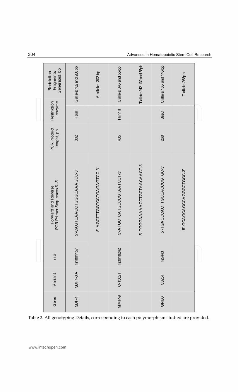
2.3 Genotyping
The reaction mixture consisted of 1µl PCR buffer 10x, 2 mM of MgSo4, 0.2 mM of each dNTP, 400mM of each primer, and 0,5units/reaction Taq DNA polymerase (Bio Basic Inc).
The reaction conditions were: For SDF1–3’A an initial denaturation at 95°C for five minutes, then 35 cycles at 94°C for 30 seconds, at 58°C for 30 seconds, at 72°C for 1min, and finally extension at 72°C for 7 minutes.
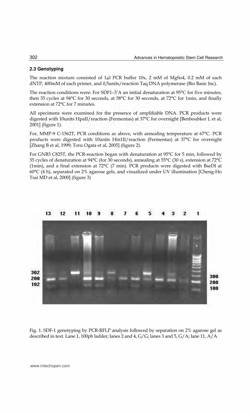
All specimens were examined for the presence of amplifiable DNA. PCR products were digested with 10units HpaII/reaction (Fermentas) at 37°C for overnight [Benboubker L et al, 2001] (figure 1).
For, MMP-9 C-1562T, PCR conditions as above, with annealing temperature at 67°C. PCR products were digested with 10units Hin1II/reaction (Fermentas) at 37°C for overnight [Zhang B et al, 1999; Toru Ogata et al, 2005] (figure 2).
For GNB3 C825T, the PCR-reaction began with denaturation at 95°C for 5 min, followed by 35 cycles of denaturation at 94°C (for 30 seconds), annealing at 55°C (30 s), extension at 72°C (1min), and a final extension at 72°C (7 min). PCR products were digested with BseDI at 60°C (4 h), separated on 2% agarose gels, and visualized under UV illumination [Cheng-Ho Tsai MD et al, 2000] (figure 3)
Fig. 1. SDF-1 genotyping by PCR-RFLP analysis followed by separation on 2% agarose gel as described in text. Lane 1, 100pb ladder; lanes 2 and 4, G/G; lanes 3 and 5, G/A; lane 11, A/A
www.intechopen.com

Distribution of SDF1-3’A, GNB3 C825T and MMP-9 C-1562T Polymorphisms in HSC CD34+ from Peripheral Blood of Patients with Hematological Malignancies
303
Fig. 2. MMP-9 genotyping by PCR-RFLP analysis followed by separation on 2% agarose gel as described in text. Lanes 1 and 8, 100 pb ladder; lanes 2 and 6, C/C; lanes 3 and 9, C/T; lane 5, T/T.
Fig. 3. GNB3 C825T genotyping by PCR-RFLP analysis followed by separation on 2% agarose gel as described in text. Lane 1, 100 pb ladder; lanes 2 and 3, C/T; lane 4, T/T; lane 5, C/C.
www.intechopen.com
Advances in Hematopoietic Stem Cell Research
304
Table 2. All genotyping Details, corresponding to each polymorphism studied are provided.
www.intechopen.com
Distribution of SDF1-3’A, GNB3 C825T and MMP-9 C-1562T Polymorphisms in HSC CD34+ from Peripheral Blood of Patients with Hematological Malignancies
305
2.4 Statistical analysis
Allele and genotype frequencies of the studied polymorphisms in patients and healthy controls were formulated by direct counting. Statistical analysis was performed using SPSS software (SPSS 16.0 for windows; SPSS Inc., Chicago, IL.).
The allele frequencies of SDF1-3’A, GNB3 C825T and MMP-9C-1562T polymorphisms were tested for the Hardy–Weinberg equilibrium of the whole group or subgroups of patients and were compared to the respective frequencies of the control group using the Pearson chi-square test or Fisher’s exact test when appropriate. The same test was applied to compare the genotype frequency between patients and controls. Association of the allelic frequencies with the clinico-pathologic parameters was evaluated by χ2 test. The odds ratios (OR) and 95% confidence intervals (CI) were calculated too. P<0.05 was required for statistical significance.
3. Results
3.1 Patient‘s distributions according to their CD34+ cell yield and failure rates
Overall 83% of patients included in this study collected ≥2x10⁶ CD34+ cells/kg after a
maximum of 4 aphereses, among them 20% collected 2-5x10⁶ CD34cells/kg, and 63% collected
≥5x10⁶ CD34 cells/kg. Beside, 10% are remobilizers as they did not achieve the threshold of
CD34+ cell yield of 2x10⁶ CD34/kg within 4 apheresis days and are subjects to another
mobilization protocol. Among them, the group of NHL represented the highest rate (40%), the
lower ones, the group of MM and AML, which represented respectively 19% and 13%. By
contrast, others are designed as first mobilizers (90%) since they have already collected
≥2x10⁶CD34+ cells/kg after a maximum of 4 aphereses days. Amongst them the group of
multiple myeloma was the most frequent (40%), thereafter the group of Non-Hodgkin’s
lymphoma (34%) and Hodgkin’s disease with 26%. For the patients included in this study,
mobilization failure was defined as <2x10⁶ CD34+ cells/kg obtained within 4 apheresis days.
So, especially MM patients collected ≥5x10⁶CD34+ cells/kg and contained the highest CD34+
cell yield (8,89x10⁶ CD34/kg for MM, and 5,51x10⁶ CD34/kg for the others patients).
Furthermore, the fact that MM patients had higher yield of CD34+ cells compared to NHL and
HD is likely since that NHL and HL patients are frequently more heavily pretreated with
cytotoxic chemotherapy than patients with MM [Iskra Pusic et al, 2008] (figure 4).
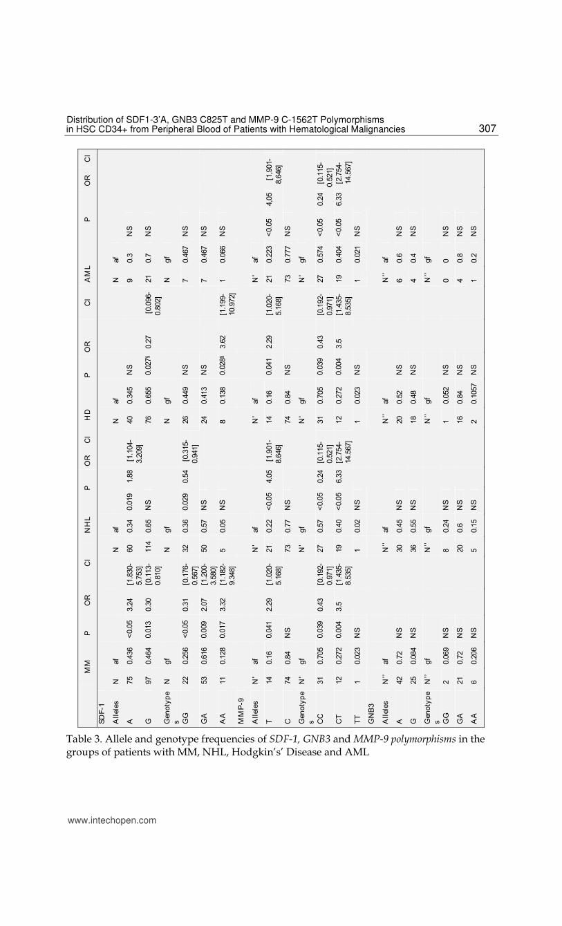
3.2 Analysis of the studied polymorphisms in the 4 subgroups of patients according to disease: A comparison between healthy donors of PBSC and patients
According to this study, SDF1-3’A and MMP-9 C-1562 T polymorphisms were significantly different between the patients and healthy controls (table 3). Particularly, we found significant differences in all the allelic and genotypic frequencies of the SDF1-3’A polymorphism in the MM group (p<0.05; OR=3.245 CI (95%) [1.830-5.753] for A allele; p= 0.017; OR= 3.324 CI (95%) [1.182-9.348]; p= 0.009; OR= 2.072 CI (95%) [1.200-3.580] for AA and GA genotypes, respectively).
Concerning the MMP-9 C-1562 T polymorphism its distribution was significantly different in the same MM group of patients compared to the control group, significant differences were observed exclusively for the T allele (p=0.041; OR=2.295 CI (95%) [1.020-5.168]) and also for the CC and CT genotypes (p= 0.039; p= 0.004; Table 3).
www.intechopen.com

Advances in Hematopoietic Stem Cell Research
306
NHL
40%
AML
13%
MM
19% HD
28%
A number of first mobilization and remobilization in database Distribution of remobilizers in
the 4 subgroup of patients
Distribution of good and poor mobilizers of PBPC CD34+ in the study population and by sex
Fig. 4. Overview of autologous stem cell transplantation database by disease as well as the distribution of good/poor mobilizers of PBSC CD34+ within the study population and by sex is already represented
www.intechopen.com
Distribution of SDF1-3’A, GNB3 C825T and MMP-9 C-1562T Polymorphisms in HSC CD34+ from Peripheral Blood of Patients with Hematological Malignancies
307
Table 3. Allele and genotype frequencies of SDF-1, GNB3 and MMP-9 polymorphisms in the groups of patients with MM, NHL, Hodgkin’s’ Disease and AML
www.intechopen.com
Advances in Hematopoietic Stem Cell Research
308
In table 3 are provided: all genotypic and allelic frequencies according to each polymorphism studied and corresponding to all patients. Distribution of genotypic and allelic frequencies by each disease included in this study. Then, all frequencies are calculated by statistical software SPSS 16.0 as well as p value and odd ratios (OR) are provided.
For the group of NHL, the distribution of the SDF1-3’A polymorphism was significantly different between patients and healthy controls especially for the A allele which seemed to be associated to this disease (p=0,019). Moreover, a decrease in GG genotype frequency compared to the control group was observed too reaching a statistically significance (p=0.029).
Concerning the MMP-9 C-1562T polymorphism, like the MM group, high significant
differences were seen especially for the T allele (P<0.05; OR=4.055; CI (95%) [1.901-8.646])
and CT genotypes (P<0.05; OR=6.333; CI (95%) [2.754-14.567]). Similar results were obtained
concerning the distribution of the MMP-9 C-1562T polymorphism in the group of Hodgkin’s
disease where significant differences were found in the T allele and CT genotype
frequencies (p<0.05; Table 3).
While, the distribution of the SDF1-3’A polymorphism was not significantly different between
the group of patients with AML and the control group, MMP-9 C-1562T distribution was
significantly different essentially for the T allele (p=0.019, OR= 7.298, CI (95%) [1.511-35.249])
and the CT genotypes (p=0.004, OR= 12.444, CI (95%) [2.485-62.319]) Table 3.
So the presence of the MMP-9 C-1562T might be associated with this disease.
When considering the GNB3 C825T polymorphism, we observed that the TT genotype was
more frequent in patient with MM and NHL with respectively 20.69% and 15.15% compared
to the Hodgkin’s disease group (only 10.52%). Whereas, the CC genotype was more frequent
in the NHL group (24.24%) (Table 3).
3.3 Association of the SDF1-3’A allele with a good mobilizing capacity
As the clinicians have defined mobilization failure as <2x10⁶ CD34+ cells/kg obtained
within 4 apheresis days, two mainly group of patients emerged: the subjects with a good
capacity of mobilization who collected ≥2x10⁶ CD34+ cells/kg obtained within 4 apheresis
days. Others with a poor mobilizing capacity and didn’t collect 2x10⁶ CD34+ cells/kg
within 4 apheresis days. For the healthy allogenic PBSC donors, the mobilization failure was
defined as <3x10⁶ CD34+ cells/kg obtained within 4 apheresis days.
When considering the SDF1-3’A polymorphism, significant difference was observed in the
SDF1-3’A allele carriers and GG carriers (p=0.023). A higher concentration of CD34+ cells in
the leukapheresis products was detected in SDF1-3’A positive patients compared to GG
homozygous subjects
Besides, a lower increase in the GG genotypes was observed in the “poor” mobilizer group
compared to the “good” ones reaching a statistical significance (p=0.023; OR =0.494; CI
(95%) [0.268-0.912]) (Table 4).
Thus, the SDF1-3’A allele carriers, especially the SDF1-3’AA homozygous individuals in the
group of healthy allogenic PBSC donors had a better mobilization potential (table 4).
www.intechopen.com
Distribution of SDF1-3’A, GNB3 C825T and MMP-9 C-1562T Polymorphisms in HSC CD34+ from Peripheral Blood of Patients with Hematological Malignancies
309
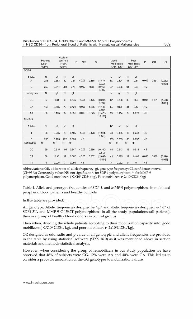
Abbreviations: OR, odds ratio; af, allele frequency; gf, genotype frequency; CI, confidence interval (CI=95%); Corrected p value; NS, not significant; *, for SDF-1 polymorphism; ** for MMP-9 polymorphism, Good mobilizers (>2X10⁶ CD34/kg), Poor mobilizers (<2x10⁶CD34/kg)
Table 4. Allele and genotype frequencies of SDF-1, and MMP-9 polymorphisms in mobilized peripheral blood patients and healthy controls
In this table are provided:
All genotypic Allelic frequencies designed as “gf” and allelic frequencies designed as “af” of SDF1-3’A and MMP-9 C-1562T polymorphisms in all the study populations (all patients), then in a group of healthy blood donors (as control group)
Then when, dividing the whole patients according to their mobilization capacity into: good mobilizers (>2X10⁶ CD34/kg), and poor mobilizers (<2x10⁶CD34/kg).
OR designed as odd radio and p value of all genotypic and allelic frequencies are provided in the table by using statistical software (SPSS 16.0) as it was mentioned above in section materials and methods-statistical analysis.
However, when considering the group of remobilizers in our study population we have observed that 48% of subjects were GG, 12% were AA and 40% were GA. This led us to consider a probable association of the GG genotypes to mobilization failure.
www.intechopen.com

Advances in Hematopoietic Stem Cell Research
310
For the MMP-9 C-1562T polymorphism, significant difference was obtained with CT
genotypes between the two groups (p=0.004; OR= 0.297; CI (95%) [0.125-0.703]).
For the GNB3 C825T polymorphism, we didn’t observe any difference between the 2 groups
of poor and good mobilizers.
This let us consider that there’s no association between GNB3 C825T polymorphism and the
capacity of mobilization of hematopoietic stem cells.
For the group of healthy PBSC donors, and with respect to our classification according to
mobilization failure (<3x10⁶ CD34/kg within 4 apheresis days), we have found an important
association of SDF1-3’A distribution with higher mobilization yield of hematopoietic stem cells
CD34+ reaching a higher statistical significance (p=0.001; OR=12.6; table 5).
Besides, we have observed a similar increase in the SDF1-3’G allele in the intermediate to
poor mobilizers’ subgroup reaching a statistical significance (p=0.035; OR=1.25; table 4).
Similarly, the association was already observed when comparing the genotypic frequencies
between the two subgroups.
The AA genotype was absent in the poor mobilizer subgroup, then was highly increased in
the other subgroup reaching a statistical significance (p=0.035; OR=1.25).
While, the GG genotype was more represented in the poor mobilizers and the differences
were significant too (p=0.001; OR=0.079; table 4).
Healthy allogenic PBSC Donors
Good mobilizers
Poor mobilizers
P OR CI
N af
N af
32 0,55
5 0,208 0,001§ 12,6 [2,407-65,953]
26 0,45
19 0,792 0,035§ 1,25 [1,045-1,495] N gf
N gf
3 0,10
7 0,583 0,001§ 0,079 [0,015-0,415] 20 0,69
5 0,417 NS
6 0,20
0 0 0,035§ 1,25 [1,045-1,495]
N' af
N' af
7 0,17
6 0,23 0,633 (NS)
33 0,82
20 0,77 NS
N' gf
N' gf
13 0,65
7 0,54 NS
7 0,35
6 0,46 NS
0 0
0 0 NS
Abbreviations: OR, odds ratio; af, allele frequency; gf, genotype frequency; CI, confidence interval
(CI=95%); Corrected p value; NS, not significant; *, for SDF-1 polymorphism; ** for MMP-9
polymorphism, for healthy allogenic PBPC donors: Good mobilizers (>3X10⁶ CD34/kg), Poor
mobilizers (<3x10⁶CD34/kg)
Table 5. Allele and genotype frequencies of SDF-1, and MMP-9 polymorphisms in
mobilized peripheral blood of healthy allogenic PBSC donors
www.intechopen.com
Distribution of SDF1-3’A, GNB3 C825T and MMP-9 C-1562T Polymorphisms in HSC CD34+ from Peripheral Blood of Patients with Hematological Malignancies
311
4. Discussion
In the present study, we investigated the effect of polymorphisms in the genes SDF-1, GNB3 and MMP-9 on the outcome of mobilization of peripheral blood stem cells for autologous transplantation by using a PCR-RFLP analysis.
We observed a significant association for SDF-1 and MMP-9 polymorphisms exclusively in patients with MM, NHL and Hodgkin’s disease suggesting that these polymorphisms are fair candidate gene variants to these 3 hematological diseases.
In fact, Association of these polymorphisms to cancer has been previously reported by many investigators [De Oliveira KB et al, 2009; Rabkin CS et al, 1999].
Our results were in agreement with other studies suggesting that SDF1-3’A polymorphism is a genetic determinant of NHL [Gabriela Gonçavales de Olivera Cavassin et al, 2004]. Furthermore; as the SDF1-3’A polymorphism is situated in the mRNAs of 3’UTR region (untranslated region) which has been identified as an important regulator of the mRNA transcript, as well as the translated product [Catia Andreassi and Antonella Riccio, 2004; Marilyn Kozak, 2004; Gavin S. Wilkie et al, 2003].
The second polymorphism studied encoded for MMP-9, J. Arai et al, have reported that SDF-1 mRNAs abundantly expressed in stromal cells from the lymph nodes of patients with malignant lymphoma, so that 3’A carriers NHL are good candidates for presenting proliferation of neoplasic cells in the lymph nodes since that SDF-1 variant is associated with an increase of SDF-1 levels [J. Arai et al, 2000; Gabriela Gonçavales de Olivera Cavassin et al, 2004].
De Oliveira KB et al, when studying distribution of SDF1-3’A polymorphism have reported also a significant difference in genotype distribution between NHL patients (GG: 51.4%; GA: 47.1%; AA: 1.5%) compared to healthy controls (GG: 65.6%; GA: 28.9%; AA: 5.5%). Whereas, they didn’t find any significant differences in genotypes distributions with breast cancer and Hodgkin’s lymphoma [De Oliveira KB et al, 2009].
Moreover, previous reports on AIDS related non-Hodgkin’s lymphoma (NHL)
demonstrated that the CXCL12–3’A chemokine variant was associated with approximate
doubling of the NHL risk in heterozygotes and an approximately fourfold increase in
homozygotes [Rabkin CS et al, 1999; A Zafiropoulos et al, 2004]. Hence, this might let us
suggest the possible role of such variant in the pathogenesis of NHL.
In this present work, we did not find a significant association between SDF1-3’A
polymorphism and our group of patients with AML, this could be due to the lower number
of patients (15 patients).
However, Dommange et al, have reported the implication of SDF1-3’A polymorphism in the
clinical representation of acute myeloid leukemia in 86 patients with AML, as an association
between this polymorphism and the risk of tissue infiltration by malignant cell was
established by an increased release of the blast from the bone marrow in the blood in the
SDF1-3’A carriers suggesting that this SDF-1 variant is associated with clinical
representation of AML [A Zafiropoulos et al, 2004].
MMP-9 is a zinc-dependent proteinase, which is involved in numerous physiological and
pathological processes. In the present study, we reported the distribution of the functional
www.intechopen.com
Advances in Hematopoietic Stem Cell Research
312
MMP-9 polymorphism -1567 C/T in the promoter region of the MMP-9 gene in group of
patients with some haematological malignancies as well as in patients undergoing stem cell
mobilization.
Then, we observed that the T allele was highly associated to the susceptibility to the four
diseases studied (table 3). We have to investigate either this variant have major influence on
the circulating levels of MMP-9.
Concerning the group of MM, we observed a significant association in all allelic and
genotypic frequencies of SDF1-3’A polymorphism with statistical differences when
compared to control. Hence, as increased angiogenesis was related to the pathogenesis of
MM, and because SDF-1 chemokine induces increased VEGF production, which is
responsible for an angiogenic activity [Florence Dommange et al, 2006], we hypothesize that
the SDF1-3’A polymorphism might increase SDF-1 protein which would have a role in
developing angiogenesis and in the pathogenesis of the disease.
On the other hand, frequent distribution of the SDF-1 3’A allele in multiple myeloma
patients confirms the implication of SDF-1 in hematopoietic stem cells. This logical
consequence of the widely distribution of SDF-1 3’A allele proving that multiple myeloma
patient’s could be considered as good mobilizers.
For the GNB3 polymorphism we’ve observed that the TT genotype and the T allele
frequencies are more frequent especially in patients with MM (0.72 for Tallele frequency)
and NHL (0.45 for Tallele frequency) compared to healthy donors of PBSC ( peripheral
blood stem cells) (Table 3) which is far from the others populations [Maggie C.Y et al, 2004] .
Then, suggesting the possible relation with these diseases.
Maggie et al when studying the ethnic differences in the linkage disequilibrium and
distribution of single-nucleotide polymorphisms in 35 candidate genes for cardiovascular
diseases have reported that the frequency of the T allele of GNB3 polymorphism in Chinese
population is about 0.545. Then, such frequency is far from those of the French and of the
Spanish population (0.329 and 0.359) and more closer to our result in Tunisian population
[Yair Gazitt & Cagla Akai, 2004].
When interesting to the capacity of mobilization which was largely demonstrated to vary
from a subject to another, several studies have focused on such phenomena and have
reported that 10–30% of patients with hematological malignancies fail to mobilize PBSC
[Ingrid G. Winkler & Jean-Pierre Levesque, 2006] and either a small proportion of normal
donors (1–5%) fail to mobilize sufficient CD34+ cells.
Besides, many reports suggest that numerous factors are related to poorer mobilization
including age, gender, type of growth factor, dose of the growth factor and in the
autologous setting patient’s diagnosis, chemotherapy regimen and number of previous
chemotherapy cycles or radiation [Sugrue MW et al, 2001].
In our study we were interested in the possible implication of some genetic factors in
mobilization and as we’ve found an association with the SDF-1 3’A variant only, then we
supposed that this polymorphism is the only predictor of mobilization capacity of PBSC
CD34+.
www.intechopen.com

Distribution of SDF1-3’A, GNB3 C825T and MMP-9 C-1562T Polymorphisms in HSC CD34+ from Peripheral Blood of Patients with Hematological Malignancies
313
In fact, when analyzing the distribution of the two functional polymorphisms SDF-1 G801A
and MMP-9 C-1562T considering the two groups of “good” and “poor” mobilizers, we’ve
found an association only with SDF1-3’A polymorphism. While no association with capacity
of mobilization was observed with GNB3 C825T and MMP-9 C-1562T polymorphisms.
When observing the distribution of the two polymorphisms not only when considering the
mobilization capacity but also in relation to each studied disease enrolled in this work we’ve
found that the good mobilizer group was mainly composed of MM patients. Whereas the
poor mobilizer group contains Hodgkin’s disease who are considered in previous studies as
hard-to-mobilize patients [Benboubker L et al, 2001; Patrick J Stiff, 1999].
The fact that multiple myeloma patients mobilized better PBSC CD34+ (peripheral blood
stem cells) than the others groups seem to be related to their ulterior chemotherapy
(dexamethasone + thalidomide) and didn’t receive any radiation therapy unlike the HD and
NHL groups.
In the good mobilizer group composed of patients needing fewer apheresis than the other
group, genotypes frequencies for the GG,GA, AA represented respectively 30.6%, 58% and
11.4%, and corresponded respectively to 45.5%, 47% and 7.6% in the poor mobilizer’s
group, and significant differences were found for GG genotype (p=0.007) and for A allele
(p=0.009).
This confirms on the one hand that the SDF1-3’A allele was associated with good mobilizing
capacity not only in the group of patients but for instance in the group of healthy allogenic
PBSC donors (see table 5). Thus, our results regarding patients undergoing autologous
transplantation of haematopoietic stem cells concur with those reported by Benboubker et al
[Bogunia-Kubik K et al, 2009].
Moreover this deduction is already found in the group of healthy allogenic transplantation
donors as it was reported in the present study and by Bogunia-Kubik K et al who have
suggested that the SDF1-3’A allele was associated with a higher yield of CD34+ cells from
healthy donors of PBPC for allogeneic haematopoietic SCT (stem cell transplantation)
compared to GG homozygotes [Patrick J Stiff, 1999].
Recent studies by the same group underlined an association of the SDF1-3’A allele with
faster granulocyte and platelet recovery after transplantation. Therefore they suggested that
the SDF-1 gene polymorphism could be a useful tool of prognostic value for recipients of
autologous haematopoietic stem cells [A. Gieryng et al, 2010]. The allelic variant SDF1-3’A is
a result of the SNP rs1801157, which is located in a highly demethylated area of the 3’UTR
region. This SNP confers a G to A transition in the nucleotide position 801, resulting in a loss
of a methylation site, which could affect the methylating effect of G-CSF [Nagler A et al,
2004], and leading to a more decreased SDF-1 expression in healthy individuals carrying the
polymorphism.
So, it’s of interest to investigate either this variant have major influence on the circulating
levels of SDF-1 and its mRNA expression, one of our future’s interests.
Further studies examining how these three polymorphisms interact with disease risk factors
are needed.
www.intechopen.com
Advances in Hematopoietic Stem Cell Research
314
Interestingly, the possible implication of others genes involved of homing and migration process of CD34+ cells and for instance VCAM-1 to higher or lower mobilization yield of PBPC might emphasize new strategies for poor mobilizers subjects and lead to the identification of new biomarkers and/or therapeutic targets.
5. Conclusion
In the present study, we observed a significant association for CXCL12 and MMP-9 polymorphisms exclusively in patients with MM, NHL and Hodgkin's disease suggesting that these polymorphisms are fair candidate gene variants to these 3 hematological diseases.
Furthermore we've confirmed that the SDF1-3'A allele was highly associated to a good mobilizing capacity especially in the group of healthy allogenic PBSC donors where the analysis not biased by background disease or chemotherapy.
Besides, we suggested a possible association of GG genotypes to poorer mobilization is already deduced.
6. Acknowledgment
We thank all participant and all patients in this work
7. References
Catia Andreassi and Antonella Riccio. (2004). To localize or not to localize: mRNA fate is in 3’UTR ends. Trends in Cell Biology; Vol.19 No.9
J. Arai, M Yasukawa, Y. Yakushijin, T. Miyazaki, S. Fujita. (2000). Stromal cells in lymph nodes attract B-lymphoma cells via production of stromal cell-derived factor-1. Eur. J. Haematol; 64:323-32.
Benboubker L, Watier H, Carion A, Georget MT, Desbois I, Colombat P, et al. (2001). Association between the SDF1-3_A allele and high levels of CD34+ progenitor cells mobilized into peripheral blood in humans. Br J Haematol; 113:247–50.
Belvisi MG and Bottomley KM. (2003). The role of matrix metalloproteinases (MMPs) in the pathophysiology of chronic obstructive pulmonary disease (COPD): a therapeutic role for inhibitors of MMPs? Inflamm. Res. 52: 95-100.
Bogunia-Kubik K, Gieryng A, Dlubek D, Lange A. (2009). The CXCL12-3'A allele is associated with a higher mobilization yield of CD34 progenitors to the peripheral blood of healthy donors for allogeneic transplantation. Bone Marrow Transplant:273-8.
Dlubek D, Drabczak-Skrzypek D, Lange A. (2006). Low CXCR4 membrane expression on CD34+ cells characterizes cells mobilized to blood. Bone Marrow Transplant 37:19.
Florence Dommange,* Guillaume Cartron, Claire Espanel, Nathalie Gallay, Jorge Domenech, Lotfi Benboubker, et al for the GOELAMS Study Group. (2006). CXCL12polymorphism and malignant cell dissemination/tissue infiltration in acute myeloid leukemia. FASEB J; 20: 1296–1300
De Oliveira KB, Oda JM, Voltarelli JC, Nasser TF, Ono MA, Fujita TC, et al. (2009). CXCL12 rs1801157 polymorphism in patients with breast cancer, Hodgkin's lymphoma, and non-Hodgkin's lymphoma. J Clin Lab Anal, 23(6):387-93.
www.intechopen.com

Distribution of SDF1-3’A, GNB3 C825T and MMP-9 C-1562T Polymorphisms in HSC CD34+ from Peripheral Blood of Patients with Hematological Malignancies
315
Gabriela Gonçavales de Olivera Cavassin, Fernando Luiz De Luca, Nayara Delgado André, Dimas Tadeu Covas, Maria Helena Pelegrinelli Fungaro, Júlio César Voltarelli, and Maria Angelica Ehara Watanabe. (2004). Molecular investigation of the stromal cell-derived factor-1 chemokine in lymphoid leukemia and lymphoma patients from Brazil. Blood Cells, Molecules, and Disease; 33: 90-93.
Yair Gazitt, Cagla Akai. (2004). Mobilization of myeloma cells involves SDF-1/CXCR4 signaling and downregulation of VLA-4. Stem Cells; 22:65-73.
A. Gieryng, K. Bogunia-Kubik, and A. Lange. (2010). CXCL12 Gene Polymorphism and Hematologic Recovery After Transplantation of Peripheral Blood Progenitor Cells. Transplantation Proceedings, 42, 3280–3283
Marilyn Kozak. (2004). How strong is the case for regulation of the initiation step of translation by elements at the 3’ end of eukaryotic mRNAs? Gene; 343: 41–54
Tsevee Lapidot and Isabelle Petit. (2002). Current understanding of stem cell mobilization: the roles of chemokines, proteolytic enzymes, adhesion molecules, cytokines and stromal cells. Experimental Hematology; 30: 973-81.
Roberto M. Lemoli and Alessandra D’Addio. (2008). Hematopoietic stem cell mobilization. Haematologica; 93(3): 321.
Maggie C.Y., Ng Ying Wang, Wing-Yee So, Suzanne Cheng, Sophie Visvikis, Robert Y.L. Zee et al. (2004). Ethnic differences in the linkage disequilibrium and distribution of single-nucleotide polymorphisms in 35 candidate genes for cardiovascular diseases. Genomics, 83; 559–65.
Miller SA, Dykes DD, Polesky HF. (1988). A simple salting out procedure for extracting DNA from human nucleated cells. Nucleic Acids Res; 16:1215.
Nagler A, kollenstein-Ilan A, Amiel A, Avivi L. (2004). Granulocyte-colony stimulating factor generates epigenetic and genetic alterations in lymphocytes of normal volunteers donors of stem cells. Exp Hematol, 32 (1): 122-30.
Iskra Pusic, Shi Yuan Jiang, Scott Landua, Geoffrey L. Uy, Michael P. Rettig, Amanda F. Cashen, et al. (2008). Impact of Mobilization and Remobilization Strategies on Achieving Sufficient Stem Cell Yields for Autologous Transplantation. Biology of Blood and Marrow Transplantation; 14:1045-56.
Rabkin CS, Yang Q, Goedert JJ, et al. (1999). Chemokine and chemokine receptor gene variants and risk of non-Hodgkin’s lymphoma in human immunodeficiency virus-1-infected individuals. Blood; 93(6):1838–42.
Siena S, Schiavo R, Pedrazzoli P, Carlo-Stella C. (2000). Therapeutic relevance of CD34+ cell dose in blood cell transplantation for cancer therapy. J Clin Oncol. 18:1360–77.
Signoret N, Oldridge J, Pelchen-Matthews A, et al. (1997). Phorbol esters and SDF-1 induce rapid endocytosis and down modulation of the chemokine receptor CXCR4. J Cell Biol 139:651
Stetler-Stevenson WG. (2001). The role of matrix metalloproteinases in tumour invasion, metastasis, and angiogenesis. Surg Oncol Clin N Am; 10:383–92.
Patrick J Stiff. (1999). Management strategies for the hard-to-mobilize patient. Bone Marrow Transplantation; (23), Suppl. 2: 29–33
Sugrue MW, Willians K, Pollok BH, et al. (2001). Characterization and outcome of ‘‘hard to mobilize” lymphoma patient sundergoing autologous stem cell transplantation. Leukemia Lymphoma; 39:509–19
www.intechopen.com
Advances in Hematopoietic Stem Cell Research
316
To LB, Haylock DN, Simmons PJ, Juttner CA. (1997). The biology and clinical uses of blood stem cells. Blood; 89:2233–58.
Toru Ogata, Hidenori Shibamura, Gerard Tromp, Moumita Sinha, MStat, Katrina A. B. Goddard et al. (2005). Genetic analysis of polymorphisms in biologically relevant candidate genes in patients with abdominal aortic aneurysms. J Vasc Surg; 41: 1036–42.
Cheng-Ho Tsai, Hung-I Yeh, Yusan Chou, Hsin-Fu Liu, Tzu-Yao Yang , Jyh-Chwan Wang et al. (2000). G protein b3 subunit variant and essential hypertension in Taiwan – a case–control study. International Journal of Cardiology; 73 :191–95.
Gavin S. Wilkie, Kirsten S. Dickson and Nicola K. Gray. (2003). Regulation of mRNA translation by 5’- and 3’-UTR-binding factors .TRENDS in Biochemical Sciences; 28:182-88.
Ingrid G. Winkler and Jean-Pierre Levesque. (2006). Mechanisms of hematopoietic stem cell mobilization:When innate immunity assails the cells that make blood and bone. Experimental Hematology; 34:996–1009.
A Zafiropoulos, N Crikas, A M Passam and D A Spandidos. (2004). CXCL12–3’A in the development of sporadic breast cancer. J. Med. Genet; 41; e59
Zhang B, Ye S., Herrmann SM, Eriksson P, de Maat M, Evans A, et al. (1999). Functional polymorphism in the regulatory region of gelatinase B gene in relation to severity of coronary atherosclerosis. Circulation; 99:1788– 94.
www.intechopen.com

Advances in Hematopoietic Stem Cell Research
Edited by Dr. Rosana Pelayo
ISBN 978-953-307-930-1
Hard cover, 464 pages
Publisher InTech
Published online 27, January, 2012
Published in print edition January, 2012
InTech Europe
University Campus STeP Ri
Slavka Krautzeka 83/A
51000 Rijeka, Croatia
Phone: +385 (51) 770 447
Fax: +385 (51) 686 166
www.intechopen.com
InTech China
Unit 405, Office Block, Hotel Equatorial Shanghai
No.65, Yan An Road (West), Shanghai, 200040, China
Phone: +86-21-62489820
Fax: +86-21-62489821
This book provides a comprehensive overview in our understanding of the biology and therapeutic potential of
hematopoietic stem cells, and is aimed at those engaged in stem cell research: undergraduate and
postgraduate science students, investigators and clinicians. Starting from fundamental principles in
hematopoiesis, Advances in Hematopoietic Stem Cell Research assemble a wealth of information relevant to
central mechanisms that may regulate differentiation, and expansion of hematopoietic stem cells in normal
conditions and during disease.
How to reference
In order to correctly reference this scholarly work, feel free to copy and paste the following:
Ben Nasr Moufida and Jenhani Faouzi (2012). Distribution of SDF1-3’A, GNB3 C825T and MMP-9 C-1562T
Polymorphisms in HSC CD34+ from Peripheral Blood of Patients with Hematological Malignancies, Advances
in Hematopoietic Stem Cell Research, Dr. Rosana Pelayo (Ed.), ISBN: 978-953-307-930-1, InTech, Available
from: http://www.intechopen.com/books/advances-in-hematopoietic-stem-cell-research/hematopoietic-stem-
cells-mobilized-in-peripheral-blood-viability-apoptosis-cryopreservation-and-infl

© 2012 The Author(s). Licensee IntechOpen. This is an open access article
distributed under the terms of the Creative Commons Attribution 3.0
License, which permits unrestricted use, distribution, and reproduction in
any medium, provided the original work is properly cited.