5-Bromodeoxyuridine in vivo labelling of M13 DNA, and its use as a non-radioactive probe for...
12
Molecularand Cellular Probes(1987)1,109-120 5-Bromodeoxyuridinein vivo labellingofM13DNA,andits useasanon-radioactiveprobeforhybridization experiments HiroshiSakamoto, François Traincard,Tuyen Vo-Quang,*Thérèse Ternynck,*Jean-LucGuesdonandStratisAvrameas* LaboratoiredesSondesFroides, and *Unité d'Immunocytochimie, Institut Pasteur,28rueduDr.Roux,75724Paris Cedex 15,France (Received,Accepted21January1987) Wedescribethein vivo productionof5-bromodeoxyuridine- (5-BUM labelledM13DNA byathymine-requiring Escherichiacoli strain . Weshowthatthe5-BUdR-labelledM13single-strandedDNAisnotextruded intothe culturemedium,butaccumulates insidethebacterialcells .Onthebasisofthis observation,aprocedureinvolvingFPLCgelfiltrationalready reportedandusedforthe isolationofplasmidDNAhasbeenadaptedfortheisolationofatleast90% pure5-BUdR- labelledsingle-strandedDNA . AnM13probe,containingpartoftheHepatitisBVirus (HBV) genomewas constructed, andthecorresponding5-BUdR-labelledsingle-strandedDNAwasusedinhybridization experimentstodetecthomologous HBV targetDNA . Picogramamounts(10 -79 moles)oftheprobeitselforthetargetDNAcouldbe detected,bymonoclonalanti-5-BUdRantibodiesinanimmunoenzymatic assay . KEYWORDS : Nucleicacidshybridization,non-radioactiveprobe,monoclonal anti-5-bromodeoxyuridineantibody,5-bromodeoxyuridinein vivo labelling, thymine-requiring Escherichia coli/M13 DNAprobe . INTRODUCTION DNA-DNAandDNA-RNAhybridizationsarewellknownasessentialtoolsin modernmolecularbiology .Thesemethodsarenowappliedinthefieldofdiagnosis, forthedetectionofinfectiousagentsandtheidentificationofinheriteddiseases ."' Recently,newprocedureshavebeendeveloped,aimingatdetectingthe hybridizedprobebythemeansofimmunologicaloraffinityreagentsinsteadof radioisotopes .Twomainapproachesoftaggingthepolynucleotideshavebeenused 0890-8508/87/010109+12$03 .00/0 109 ©1987AcademicPressInc'(London)Ltd
Transcript of 5-Bromodeoxyuridine in vivo labelling of M13 DNA, and its use as a non-radioactive probe for...

Molecular and Cellular Probes (1987) 1, 109-120
5-Bromodeoxyuridine in vivo labelling of M13 DNA, and itsuse as a non-radioactive probe for hybridization
experiments
Hiroshi Sakamoto, François Traincard, Tuyen Vo-Quang,* ThérèseTernynck,* Jean-Luc Guesdon and Stratis Avrameas*
Laboratoire des Sondes Froides, and *Unité d'Immunocytochimie, InstitutPasteur, 28 rue du Dr. Roux, 75724 Paris Cedex 15, France
(Received, Accepted 21 January 1987)
We describe the in vivo production of 5-bromodeoxyuridine- (5-BUM labelled M13 DNAby a thymine-requiring Escherichia coli strain .
We show that the 5-BUdR-labelled M13 single-stranded DNA is not extruded into theculture medium, but accumulates inside the bacterial cells. On the basis of thisobservation, a procedure involving FPLC gel filtration already reported and used for theisolation of plasmid DNA has been adapted for the isolation of at least 90% pure 5-BUdR-labelled single-stranded DNA .
An M13 probe, containing part of the Hepatitis B Virus (HBV) genome was constructed,and the corresponding 5-BUdR-labelled single-stranded DNA was used in hybridizationexperiments to detect homologous HBV target DNA .
Picogram amounts (10 -79 moles) of the probe itself or the target DNA could bedetected, by monoclonal anti-5-BUdR antibodies in an immunoenzymatic assay .
KEYWORDS : Nucleic acids hybridization, non-radioactive probe, monoclonalanti-5-bromodeoxyuridine antibody, 5-bromodeoxyuridine in vivo labelling, thymine-requiringEscherichia coli/M13 DNA probe.
INTRODUCTION
DNA-DNA and DNA-RNA hybridizations are well known as essential tools inmodern molecular biology. These methods are now applied in the field of diagnosis,for the detection of infectious agents and the identification of inherited diseases ."'
Recently, new procedures have been developed, aiming at detecting thehybridized probe by the means of immunological or affinity reagents instead ofradioisotopes. Two main approaches of tagging the polynucleotides have been used
0890-8508/87/010109+12 $03 .00/0
109
© 1987 Academic Press Inc' (London) Ltd

1 10
H. Sakamoto et al.
so far: chemical modifications of the nucleic acid,3-7 or enzymatic incorporations ofa modified nucleotide in the neosynthetized probe ."
We have already described such an immunoenzymatic procedure, based on theuse of a monoclonal antibody specific for 5-bromodeoxyuridine (5-BUdR) . This baseanalog was incorporated into the DNA probe, either by nick-translation or by invivo labelling of double-stranded plasmid DNA . 9 The detection limit value was toohigh (50 pg), since the antibody recognizes the halogenated bases, mainly in single-stranded DNA, and therefore only poorly in the hybridized probe .
We, therefore, attempted to incorporate 5-BUdR into an in vivo-produced single-stranded phage M13 DNA probe . In order to achieve the best substitution ofthymidine by 5-BUdR into the nucleic acids of the phage, we developed andcharacterized a thymine-requiring strain of Escherichia coli. Subsequent conditionsfor purification and use of the resulting probe were set up, and showed the interestof the system for diagnostic purposes .
MATERIALS AND METHODS
Escherichia colt, M13 and plasmids
Strain T000701 (this study) is a thymine-dependent derivative of strain JM103 .' oPlasmid pCP10 is a pBR322 derivative, carrying the total Hepatitis B Virus (HBV)genome." Phage mHBV4 (this study) is an M13 mp8 derivative, carrying anHBV DNA fragment and a pBR322 DNA fragment .
Buffers
TE buffer10 and TBS-Tween9 have already been described . TBS-Tween-Gelatin isTBS-Tween containing 0 .5% (w/v) alimentary-grade gelatin . SSPE and Denhardt'ssolution were as reported earlier"
Antibodies
The anti-5-BUdR monoclonal antibody, described elsewhere,' was purified byaffinity chromatography on protein A-sepharose . Sheep anti-mouse IgG antibodieswere isolated from whole serum 12 and labelled with alkaline phosphatase aspreviously described ."
Growth conditions
M63, ML and 2YT media were prepared as previously described .'"- 14 All cultures wereperformed at 37 °C, in standard Erlenmeyer flasks, or in a 1 .5-I fermentor with air-sparging, pH regulator set at pH 7 and Antifoam 426R (Prolabo) . Escherichia coli

precultures were grown overnight in M63 medium . Thymine (Sigma, 0 . 1 mg ml - ')
was added when required . To prepare M13 phage stocks, strain JM103 was grownovernight in M63 medium and M13 phages were added to give a multiplicity ofinfection (mi) of 1 . The inoculum was incubated for 15 min at 37°C, then diluted 100-
fold in 2YT medium and grown for 6 h ; the culture was centrifuged and thesupernatant kept at +4° C. M13 phages could also be grown on strain TUC0701following this procedure, except that the 2YT medium was supplemented withthymine (0 .1 mg ml - ') .
Trimethoprim selection
The method was adapted from that described by Stacey & Simson . 15 Strain JM103
was grown to saturation in M63 medium containing thymine (0 .05 mg ml - ') andvarious trimethoprim concentrations (Sigma, 0005-0 .05 mg ml - '), then diluted andspread on M63 plates containing thymine (0 .05 mg ml -1 ) ; resulting colonies were
screened for thymine dependence on M63 plates containing various thymine
concentrations (0, 0 .01, 0 .05 or 01 mg ml 1 ) in order to select the most thymine-
dependent clones .
5-BUdR labelling conditions
The following conditions were set up as described in Results and Discussion . StrainT000701 was grown overnight in M63 medium containing thymine (01 mg ml - ')and M13 phages were added at an mi of 1 . The inoculum was incubated for 15 minat 37°C then diluted 100-fold in 2YT or ML medium containing 5-bromouracil(Sigma, 0 .3 mg ml -1 ) . Bacterial growth was followed and the cells harvested after 1 h(culture without pH regulation) or after 4 h (with pH regulation) of stationary phase .The bacterial pellet was further used for DNA isolation, as described below .
DNA isolation
General methods
Phenol extraction and ethanol precipitation were performed as described pre-viously ." Unlabelled M13 single-stranded DNA isolation was carried out as reportedearlier ."
5-BUdR-labelled DNA isolation
For the M13-labelled DNA isolation, the cells and phages were grown as describedabove. The cultures were centrifuged and the pellets were treated as alreadydescribed 10 for the rapid isolation of plasmid DNA (alkaline lysis method). After thephenol/chloroform and ethanol precipitation steps, the DNA was purified by FPLC
5-Bromodeoxyuridine-labelled DNA probe
11 1

112
H. Sakamoto et al .
on a Superose 6 column (Pharmacia) according to a previous report .' The FPLC stepseparates DNA from RNA and proteins, but the double-stranded and single-stranded DNA are co-eluted in the same peak. Following this, the single-strandedDNA could not be obtained totally pure (except by using rifampicin in the labellingculture, see Results and Discussion) : double-stranded DNA contamination wasusually less than 10% . Pure double-stranded DNA was prepared by treating thenucleic acids with S1 nuclease (Boehringer Mannheim, 5 units µg - ' of DNA,incubation during 2 h at 37 ° C) prior to the FPLC step . Single- or double-strandedDNA proportion were calculated by measuring the DNA peak surface area obtainedafter the FPLC step, with or without S1 nuclease treatment . For the isolation ofchromosomal-labelled DNA, the cells were grown as described above, but withoutthe addition of M13 phages . After centrifugation, the bacterial pellet was treated asdescribed elsewhere.".
Dot hybridization
Target DNA preparation
To 1 volume of DNA in TE buffer, 0 .1 volume of 1 M NaOH was added . The mixturewas left for 15 min on ice, then 0 .15 volume of 1 M NaH2 PO4 was added . Serialdilutions (1 µl each) were spotted on nitrocellulose membranes . The filters weredried at room temperature, then baked 2 h at +80 °C under vacuum .
Hybridization procedure
The membranes were incubated for 3 h at + 65°C in a sealed bag containing0 .2 ml cm -2 of prehybridization buffer (SSPE 6x, Dextran Sulphate 5% w/v, SDS0 .5% w/v Denhardt's solution 5x, sonicated and heat-denatured salmon-spermDNA 0 .1 mgml -1 ), then overnight at +65°C in a fresh prehybridization buffercontaining the labelled probe . As described in Results and Discussion, the concen-tration of the labelled probe was 2 tg ml - ', and the probe solution was notdenatured. After hybridization, the membranes were washed 3 times (5 min each) at+65°C in a wash buffer (SSPE 2x, SDS 0 .1 w/v), 3 times (15 min each) at +65 °C in amore stringent wash buffer (SSPE 0 . 1x, SDS 0-1% w/v), then once briefly at roomtemperature in TBS-Tween, and treated as described below .
Immunodetection of 5-BUdR-labelled DNA
The membranes were incubated 1 h at room temperature in a sealed bag containingthe anti-5-BUdR antibody (0 .01 mg ml - ' in TBS-Tween-gelatin, at 0 .2 ml cm -2),washed three times (10 min each) at room temperature in TBS-Tween, then furtherincubated 1 h at room temperature with the alkaline phosphatase-antibody conju-gate (0 .01 mg ml - ' in TBS-Tween-gelatin at 0 .2 ml cm -2), washed three times(10 min each) at room temperature in TBS-Tween, and incubated at room tempera-ture in the substrate buffer : 0 . 1 M Tris-HCI pH 9 .5, 0 .1 M NaCl, 50 mM MgCl 2 ,

5-Bromodeoxyuridine-labelled DNA probe
113
nitrobluetetrazolium (Sigma) 0 .3 mg ml - ', 5-bromo-4-chloro-3-indolyl-phosphate(Boehringer Mannheim) 0 . 15 mg ml - ' . The enzyme reaction was stopped by wash-ing the membranes in water . After staining, the membranes were stored dry, in thedark .
RESULTS AND DISCUSSION
Construction of a thymine-dependent Escherichia coli strain
In order to achieve the highest rate of thymidines substituted by 5-BUdR in thelabelled DNA, we chose to work with a strongly thymine-dependent (Thy - ) strain .
Thy- strain T000701 was derived from strain JM103 by trimethoprim selection(see Materials and Methods) and subsequent screening of the resulting colonies onM63 plates containing various thymine concentrations . Clones that were unstable,mucoid, or that required less than 0 .01 mg ml - ' of thymine for optimal growth werenot retained . According to these criteria, about 60% of the clones were discarded .One of the remaining clones, named T000701, was further characterized .
At least 0 .05 mg ml - ' of thymine had to be added to M63 liquid medium foroptimal growth of strain T000701 . Because of this high Thy- phenotype, thethymine naturally contained in rich medium was readily exhausted by the growth ofthis strain . However, care was taken to avoid important cell lysis caused by thyminestarvation, as will be discussed later . Bacteriophage M13 developed satisfactorily instrain T000701, as phages stocks of up to 10 10 pfu ml - ' were routinely obtained .
The strain T000701 required no special handling technique as has remainedstable for 18 months for both thymine dependence and M13 development .
5-BUdR incorporation into chromosomal DNA
A comparison of strains T000701 and JM103 harvested after overnight growth inrich medium containing 5-bromouracil (0 . 3 mg ml -1 ) showed that the Thy- strainT000701 incorporated 5-BUdR into its chromosomal DNA 100 times more effi-ciently than the Thy+ strain JM103. In a similar experiment, the incorporationkinetic was tested as follows : at different steps of the growth (Fig . 1), aliquots of theculture were removed, the chromosomal DNA was extracted, denatured, spottedon nitrocellulose membrane, and its detection limit value was measured using theanti-5-BUdR antibody . The detection limit values were the following : 8 pg at the endof the log phase (after 3 .5 h growth, Fig. 1), 2 pg at the beginning of the stationaryphase (after 5 h growth), and 1 pg latter on the stationary phase (after 7 h growth) .We observed that the incorporation of 5-BUdR by strain T000701 was notdetectable during the log phase, and it only started when growth became thymine-limited . This observation confirmed, for strain T000701, the preferential incorpora-tion of thymine over 5-bromouracil previously reported for strain TAU-bar by otherauthors."
The lower detection limit value for denatured 5-BUdR-labelled chromosomalDNA was 1 pg . The labelling proceeded for only part of a bacterial generation, andtherefore did not cover the entire chromosome . This observation could be related

114
H. Sakamoto et al .
10
Time (h)
Fig . 1 . Strain T000701 growth curves . Strain T000701 was grown in rich medium (A), with thymine01 mg ml ' :0) or 5-bromouracil (03 mg ml ' :0) added, as described in Materials and Methods . Thegrowth was followed by optical density at 600 nm .
to the results obtained by others on chromosomal replication under comparableconditions." If the chromosome had been fully substituted, the detection limitvalue would have been lower than 1 pg .
5-BUdR incorporation into M13 DNA
We found that 5-BUdR induces changes in the distribution of the labelled M13single-stranded DNA ; after overnight growth in 2YT medium supplemented withthymine (0 .1 mg ml - '), M13 single-stranded DNA inside T000701 cells representedonly 1-2% of the total intracellular M13 DNA, as in strain JM103 (S1 nuclease andFPLC analysis as described in Materials and Methods) . Replacing thymine by 5-bromouracil (0.3 mg ml - ') increased the content of intracellular M13 single-stranded DNA from 1-2% to 50-90% . During late log and early stationary phase,the pH of the culture medium varied drastically (from pH 7 to pH 9) leading tointensive cell lysis . Growth in a pH-regulated fermentor limited cell lysis . Then, moreintracellular 5-BUdR-labelled M13 single-stranded DNA accumulated, and there wasfive times more single-stranded DNA at late stationary phase, than at earlystationary phase (Fig . 2). The yield was 10 .tg of labelled intracellular M13 single-stranded DNA per ml of bacterial culture, which was of the same order of
a

Time (h)
Fig . 2 . Kinetics of the intracellular accumulation of labelled M13 DNA . Strains 7000701 and M13 mp8were grown with 5-bromouracil (0'3 mg ml ') and pH regulation, as described in Materials and Methods .Growth (A) was followed by optical density at 600 nm (OD600), and aliquots were removed (arrows1-5) . Intracellular M13 DNA was extracted, and M13 single-stranded DNA (O) or M13 double-strandedDNA (E7) was quantified as described in Materials and Methods .
magnitude as for unlabelled M13 single-stranded DNA, extracted from culturesupernatants by the standard procedure ." Only a small amount of double-strandedDNA accumulated inside the cells (usually less than 10% of the total intracellularM13 DNA, Fig . 2) . This accumulation could be totally inhibited by adding rifampicin(5 µg ml - ') to the culture at late log phase (not shown), but usually the rifampicinstep was omitted because the double-stranded DNA contamination was judgednegligible .
The 5-BUdR-labelled M13 DNA (both double and single stranded) was purified byFPLC, using a procedure described for plasmid purification ." The detection limitvalue was 0 .25 pg for the single-stranded DNA, and 4 pg for the double-strandedDNA (Fig. 3), confirming that the anti-5-BUdR antibody reacted far better with thelabelled single-stranded DNA rather than with the labelled double-stranded DNA .'When the labelled double-stranded DNA was denatured, its detection limit valuewas also 0 .25 pg (Fig . 3), which suggested that the labelling occurred in acomparable way on both single- and double-stranded DNA .
The labelling conditions were modified, in order to try to lower the detection limitvalue of the labelled probe. Changing the culture medium (ML, 2YT or M63) or the 5-bromouracil concentration (from 0 .15 mg ml - ' to 2 mg ml - ') did not lower thedetection limit value. When the labelling was performed in rich medium (MLcontaining 5-bromouracil at 0 .3 mg ml -' ) with pH regulation, we observed that thedetection limit value of the labelled probe was always 0 .25 pg, whenever thelabelling was stopped during the stationary phase . Moreover, when minimalmedium (M63 containing 5-bromouracil at 0'15 mg ml -1 ) was used, the detectionlimit value of the labelled probe was also 0 . 25 pg. Since the detection limit value wasthe same, whether the label was incorporated in cells grown in rich medium or in
5-Bromodeoxyuridine-labelled DNA probe
115
I
2
3
4
5
15t
a/
E 10oomo
0 5
i E
A
c 5 Zy 0
o
o . .D' . .0
0 . 1 0/
I
I
1
1
1
I
I
I0
2
3
4
5
6
7
8

116 H. Sakamoto et al.
P4
Fig. 3. lmmunoenzymatic detection of labelled Ml3 DNA. Labelled single-stranded DNA and labelled double-stranded DNA were prepared as described in Materials and Methods. Twofold serial dilutions of
labelled DNA were spotted on nitrocellulose membranes (I pi) and detected with the anti-5-BUdR antibody, as described in Materials and Methods. Labelled native double-stranded DNA is (A), iabeiled denatured double-stranded DNA is (B), labelled single-stranded DNA is (Ci, and negative control (denatured salmon-sperm DNA) is (-).
minimal medium (where the substitution rate is theoretically 1000/o), we concluded that the labelling was optimal in both cases, in terms of antibody detection ability. Finally, we preferentially used the 2YT medium containing 5-bromouracil (0.3 mg ml-‘), because these conditions gave the best yields of labelled Ml3 DNA. The final procedure is detailed in Materials and Methods.
Concerning the variations of intracellular Ml3 single-stranded DNA content, the data were interpreted as follows: 5-BUdR-labelled Ml3 single-stranded DNA was apparently not extruded outside the cells but accumulated inside, while unlabelled, and possibly poorly labelled single-stranded DNA, was normally packaged into capsids and extruded into the culture medium. The detection limit value of the labelled DNA extracted from the culture supernatant was high (100 pg), and still higher (600 pg) in the case of pH-regulated cultures. Cell lysis was thought to be the main source of labelled DNA in the supernatant, and the detection limit value was high because the labelled DNA was diluted by the unlabelled Ml3 single-stranded DNA produced in the supernatant during log phase.

Why the 5-BUdR-labelled single-stranded DNA was retained inside the cells wasnot clearly understood, but strain T000701 was able to produce normal M13phages, and preliminary results showed that some labelled single-stranded DNAwas retained before the culture became thymine-limited, suggesting that thyminestarvation was not the main factor leading to the retention .
In any case, this characteristic made it a very easy way to separate labelled fromunlabelled M13 single-stranded DNA, providing a fast and simple method for thelarge-scale purification of heavily labelled probe . These in vivo-labelled M13 probeswere found to be very stable, as they were successfully used after 1 year of storagein TE buffer at -20°C .
Using 5-BUdR-labelled M13 single-stranded DNA probes in dot-hybridizationexperiments
As already reported, 9 5-BUdR nick-translated plasmid probes gave a high detectionlimit value (50 pg) in hybridization experiments . Indeed, in these experiments, mostof the hybridized probe is in the double-stranded state, since the insert hybridizeswith the target, and the two complementary strands of the vector itself hybridizewith each other (autohybridization), leading to a hybridization network (Fig . 4b) .20The detection limit value was high because the anti-5-BUdR antibody recognizes the5-BUdR mainly in the labelled single-stranded DNA rather than in the labelleddouble-stranded DNA, as also demonstrated in this work (Fig . 3) .
To counteract this drawback, a hybridization procedure was developed in thisstudy, where the hybridized probe was present as much as possible in the single-stranded state (Fig . 4a) . Because the cloning vector is usually bigger than the clonedinsert (see example in Fig . 5), about 80% of the hybridized probe was in the single-stranded state, and thus still available for antibody detection . Concerning theinfluence on the detection limit value of the proportion of double-stranded DNA inthe labelled probe solution, we observed that up to 50% of double-stranded DNAprobe was not a drawback, as long as the probe solution was not denatured prior touse. In contrast, when the probe solution was denatured, we observed that thedetection limit value increased . When the proportion of double-stranded DNA in
h
(a )
5-Bromodeoxyuridine-label led DNA probe
117
11
(b)
-+-
Fig. 4. Hybridization scheme with 5-BUdR in vivo-labelled M13 single-stranded DNA (a), or with 5-BUdRnick-translated probe (b) . Hatched lines represent the target DNA, and dotted lines the probe DNA . Theinsert DNA cloned in the probe, and its complementary part in the target DNA are represented by solidlines . The spots schematize 5-BUdR molecules incorporated into the probe DNA . The anti-5-BUdRantibody is represented by the arrows .

118 H . Sakamoto et al .
E
H E
E
E
~-r
I I I I I I
PCP 10
B B
B B B
B
~b
t'- lmHBV 4
Ikb
a E
H B
Fig. 5. Construction of probe mHBV4 . The hatched areas represent the cloning vectors DNA (pBR322for pCP10 and mp8 for mHBV4), and the lines the hepatitis B virus DNA . The letters represent therestriction endonucleases sites for BamHl (B), EcoRl (E) and Hindlll (H) . Plasmid pCP10 was partiallydigested with BamHl, and one of the resulting fragment was inserted into the BamHl site of the M13cloning vector mp8, yielding the probe mHBV4 .
the probe solution was 50%, the increase was fourfold . This increase in detectionlimit value was interpreted as being caused by the autohybridization of the labelledprobe, leading to a hybridization network comparable to the one obtained withnick-translated plasmid probes .
The effect of the labelled probe concentration on the detection limit value wasinvestigated, using a 95% pure single-stranded DNA probe solution . At a probeconcentration of 2 µg ml - ', the detection limit value was fourfold lower than at0-2 µg ml - ', without any increase in background . Increasing the probe concentra-tion beyond 2 µg ml - ' did not further lower the detection limit value .
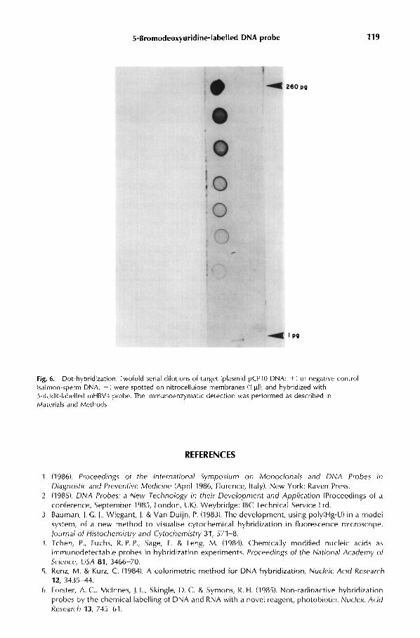
Using these optimal conditions (see Materials and Methods), the plasmid pCP10which carries the hepatitis B virus genome," and the probe mHBV4 (Fig . 5), we wereable to detect 1 pg (10 -19 moles) of target DNA in a dot-hybridization experiment(Fig . 6). This sensitivity theoretically enables the system to be used in the detectionof single copy eukaryotic gene, as reported by other authors with biotin-labelledprobes."
CONCLUSION
The results obtained in this work demonstrate the ability to use in vivo non-radioactive labelling as a tool for hybridization experiments . The double-strandedDNA contamination in the probe solution is not a drawback and does not interferewith the single-stranded DNA probe during the hybridization step . The detectionlimit value is related to the amount of single-stranded DNA available for detectionby the anti-5-BUdR antibody . The sensitivity of the system is comparable to those ofother non-radioactive nucleic acids labelling systems, but it is a cheaper and easierway of producing large quantities of labelled probes . Optimal labelling conditionsand probe isolation are simple enough to be reproduced in any laboratory . We arepresently trying to adapt the system to actual clinical diagnosis conditions .
ACKNOWLEDGEMENTS
We thank Dr G . Buttin for advice and critical reading of the manuscript, and Dr A Danchin for helpfuldiscussion .
5-Bromodeoxyuridine-labelled DNA probe 119
Fig. 6. Dot-hybridization. Twofold serial dilutions of target (plasmid pCPl0 DNA: +I or negative control (salmon-sperm DNA: -1 were spotted on nitrocellulose membranes (I pII, and hybridized with 5-BUdR-labelled mHBV4 probe. The lmmunoenzymatic detection was performed as described in Materials and Methods.
REFERENCES
1. (1986). Proceedings of the international Symposium on Monoclonals and DNA Probes in Diagnostic and Preventive Medicine (April 1986, Florence, Italy). New York: Raven Press.
2. (1985). DNA Probes: a New Technology in their Development and Application (Proceedings of a
conference, September 1985, London, UK). Weybridge: IBC Technical Service Ltd. 3. Bauman, J. C. J., Wiegant, J. & Van Duijn, P. (1983). The development, using poly(Hg-U) in a model
system, of a new method to visualise cytochemical hybridization in fluorescence mrcroscope.
journal of Histochemistry and Cytochemistry 31, 571-B 4. Tchen, P., Fuchs, R. P. P., Sage, E. & Leng, M. (1984). Chemically modified nucleic acids as
immunodetectable probes in hybridization experiments. Proceedings of the National Academy of Srience, USA 81, 3466-70.
5. Renz, M. & Kurz, C. (1984). A calorimetric method for DNA hybridization. Nucleic Acid Research 12, 3435-44.
6. Forster, A. C., Mclnnes, J. L., Skingle, D. C. & Symons, R. H. (1985). Non-radioactive hybridization probes by the chemical labelling of DNA and RNA with a novel reagent, photobiotin. Nurleir Arid Research 13. 745-61.

120
H. Sakamoto et al .
7 . Chollet, A . & Kawashima, E . H . (1985) . Biotin-labelled synthetic olygodeoxyribonucleotides synthe-sis and uses as hybridization probes . Nucleic Acid Research 13, 1529-41 .
8 . Langer, P . R ., Waldrop, A. A . & Ward, D. C . (1981). Enzymatic synthesis of biotin-labeled polynuc-leotides : Novel nucleic acid affinity probes . Proceedings of the National Academy of Science, USA78, 6633-7 .
9 . Traincard, F ., Ternynck, T ., Danchin, A. & Avrameas, S . (1983) . Une technique immunoenzymatiquepour la mise en évidence de l'hybridation moléculaire entre acides nucléiques . Annales of theInstitute Pasteur Immunology D 134, 399-405 .
10. Maniatis, T ., Fritsch, E. F . & Sambrook, J. (1982) . Molecular Cloning, A Laboratory Manual . NewYork: Cold Spring Harbor Laboratory .
11 . Dubois, M . F ., Pourcel, C ., Rousset, S ., Chany, C . & Tiollais, P. (1980) . Excretion of hepatitis B surfaceantigen particles from mouse cells transformed with cloned viral DNA . Proceedings of the NationalAcademy of Science, USA 77, 4549-53 .
12. Guesdon, J . L . & Avrameas, S. (1976) . Polyacrylamide-agarose beads for the preparation of effectiveimmunoabsorbents . Journal of Immunological Methods 11, 129-33 .
13. Avrameas, S . (1969) . Coupling of enzymes to proteins with glutaraldehyde. Immunochemistry 6,43-52 .
14 . Miller, J . H . (1972) . Experiments in Molecular Genetics . New York : Cold Spring Harbor Laboratory .15 . Stacey, K . A . & Simson, E . (1965) . Improved method for the isolation of thymine-requiring mutants
of Escherichia colt. Journal of Bacteriology 90, 554-5 .16. Vo-Quang, T., Malpiece, Y ., Buffard, D ., Kaminsky, P . A ., Vidal, D . & Strosberg, A . D . (1985) . Rapid
large-scale purification of piasmid DNA by medium or low pressure gel filtration . Application :construction of thermoamplifiable expression vector . Bioscience Report 5, 101-11 .
17. Marmur, J . (1961) . A procedure for the isolation of deoxyribonucleic acid from microorganisms .Journal of Molecular Biology 3, 208-18 .
18. Amersham International plc : M13 cloning kits, and M13 sequencing system .19. Hackett, P . Jr & Hanawalt, P . (1966). Selectivity for thymine over 5-bromouracil by a thymine
requiring bacterium . Biochim Biophy Acta 123, 356-63 .20. Alwine, J . C ., Kemp, D . J ., Parker, B . A ., Reiser, J ., Renart, J ., Stark, G . R . & Wahl, G. M . (1979) .
Detection of specific RNAs or specific fragments of DNAs by fractionation in gels and transfer todiazobenzyloxymethyl paper . Methods in Enzymology 68, 220-42 .
21 . Leary, J . J ., Brigati, D . J . & Ward, C . C . (1983) . Rapid and sensitive colorimetric method for visualizingbiotin-labeled DNA probes hybridized to DNA or RNA immobilized on nitrocellulose : bio-blots .Proceedings of the National Academy of Science, USA 80, 4045-9 .