496 IEEE TRANSACTIONS ON MEDICAL IMAGING, VOL. …wliu/TMI15_hash.pdf · 496 IEEE TRANSACTIONS ON...
11
496 IEEE TRANSACTIONS ON MEDICAL IMAGING, VOL. 34, NO. 2, FEBRUARY 2015 Towards Large-Scale Histopathological Image Analysis: Hashing-Based Image Retrieval Xiaofan Zhang, Student Member, IEEE, Wei Liu, Member, IEEE, Murat Dundar, Member, IEEE, Sunil Badve, and Shaoting Zhang*, Member, IEEE Abstract—Automatic analysis of histopathological images has been widely utilized leveraging computational image-processing methods and modern machine learning techniques. Both com- puter-aided diagnosis (CAD) and content-based image-retrieval (CBIR) systems have been successfully developed for diagnosis, disease detection, and decision support in this area. Recently, with the ever-increasing amount of annotated medical data, large-scale and data-driven methods have emerged to offer a promise of bridging the semantic gap between images and diagnostic informa- tion. In this paper, we focus on developing scalable image-retrieval techniques to cope intelligently with massive histopathological images. Specifically, we present a supervised kernel hashing tech- nique which leverages a small amount of supervised information in learning to compress a 10 000-dimensional image feature vector into only tens of binary bits with the informative signatures pre- served. These binary codes are then indexed into a hash table that enables real-time retrieval of images in a large database. Critically, the supervised information is employed to bridge the semantic gap between low-level image features and high-level diagnostic information. We build a scalable image-retrieval framework based on the supervised hashing technique and validate its per- formance on several thousand histopathological images acquired from breast microscopic tissues. Extensive evaluations are carried out in terms of image classification (i.e., benign versus actionable categorization) and retrieval tests. Our framework achieves about 88.1% classification accuracy as well as promising time efficiency. For example, the framework can execute around 800 queries in only 0.01 s, comparing favorably with other commonly used dimensionality reduction and feature selection methods. Index Terms—Breast lesion, hashing, high dimension, histopathological image analysis, large-scale image retrieval, supervised learning. I. INTRODUCTION B REAST cancer is the second most common cancer in women. In the United States, breast cancer alone ac- counted for 29% of all new cancer cases among women in Manuscript received August 18, 2014; revised September 24, 2014; accepted September 29, 2014. Date of publication October 09, 2014; date of current ver- sion January 30, 2015. This work was supported in part by the University of North Carolina at Charlotte, Charlotte Research Institute (CRI) , and in part by the Oak Ridge Associated Universities for the Ralph E. Powe Junior Faculty Enhancement Award . Asterisk indicates corresponding author. X. Zhang is with the Department of Computer Science, University of North Carolina, Charlotte, NC 28027 USA (e-mail: [email protected]). W. Liu is with the IBM T. J. Watson Research Center, Yorktown Heights, NY 10598 USA (e-mail: [email protected]). M. Dundar is with the Computer and Information Science Department, In- diana University–Purdue University, Indianapolis, IN 46202 USA. S. Badve is with the Department of Pathology, Indiana University, Indi- anapolis, IN 46202 USA. *S. Zhang is with the Department of Computer Science, University of North Carolina, Charlotte, NC 28027 USA (e-mail: [email protected]). Color versions of one or more of the figures in this paper are available online at http://ieeexplore.ieee.org. Digital Object Identifier 10.1109/TMI.2014.2361481 2013. Fortunately, over the past decade the death rate for female breast cancer has decreased by 17%, although the incidence of breast cancer has risen. This situation is largely the result of improvements in early detection and treatment [1]. The prein- vasive stages of breast cancer have two general histological categories: the lobular and ductal subtypes. Most work focuses on ductal preinvasive cancer, since approximately 80% of all diagnosed preinvasive and invasive breast cancers are of this subtype [2]. Pioneering work done by Page & Dupont divided intraductal lesions into three major classes [usual ductal hyper- plasia (UDH), atypical ductal hyperplasia (ADH), and ductal carcinoma in situ (DCIS)] and suggested that ADH and DCIS should be considered as precursor lesions [3]. On the other hand, there is little evidence to suggest that UDH is a precursor lesion. These differences in biology are manifested in the treat- ment of patients. The standard of care for patients diagnosed with UDH on core biopsy is routine follow-up, whereas those with ADH and DCIS are subjected to excisional biopsy, which can be associated with pain, discomfort, and scarring for the patient while also adding significantly to health-care costs. Classical examination methods include screening tests and biopsy. Compared to mammography, histopathology slides pro- vide more comprehensive information for diagnosis. However, manual examination of microscopic images is labor intensive and time consuming, and may depend on a subjective assess- ment by the pathologist, which poses a special challenge in the diagnosis of preinvasive breast cancer. Computer-aided diagnosis (CAD) systems have been widely used in an attempt to relieve the workload on pathologists and to offer more reliable and consistent analysis of histopathology im- ages. The appearance and widespread study of CAD systems for cytological diagnosis can be traced back to the last century [4]. Recently some studies have focused on analyzing high-resolu- tion images digitized from tissue histopathology slides [5]–[11]. In addition to the classifier-based CAD systems providing di- agnosis results or grading scores, content-based image retrieval (CBIR) has also been extensively investigated for decision sup- port in many clinical applications, including digital pathology [12]–[16]. Given an image database with diagnosis informa- tion, CBIR methods aim to retrieve and visualize images with morphological profiles most relevant to and consistent with the query image [17]. CBIR can also be used for classification pur- poses by considering the majority diagnosis of the retrieved im- ages as the most likely diagnosis. Traditional CBIR methods in medical files usually focus on small data sets that have only tens or hundreds of images. New opportunities and challenges arise with the ever-in- 0278-0062 © 2014 IEEE. Personal use is permitted, but republication/redistribution requires IEEE permission. See http://www.ieee.org/publications_standards/publications/rights/index.html for more information.
Transcript of 496 IEEE TRANSACTIONS ON MEDICAL IMAGING, VOL. …wliu/TMI15_hash.pdf · 496 IEEE TRANSACTIONS ON...
496 IEEE TRANSACTIONS ON MEDICAL IMAGING, VOL. 34, NO. 2, FEBRUARY 2015
Towards Large-Scale Histopathological ImageAnalysis: Hashing-Based Image Retrieval
Xiaofan Zhang, Student Member, IEEE, Wei Liu, Member, IEEE, Murat Dundar, Member, IEEE,Sunil Badve, and Shaoting Zhang*, Member, IEEE
Abstract—Automatic analysis of histopathological images hasbeen widely utilized leveraging computational image-processingmethods and modern machine learning techniques. Both com-puter-aided diagnosis (CAD) and content-based image-retrieval(CBIR) systems have been successfully developed for diagnosis,disease detection, and decision support in this area. Recently, withthe ever-increasing amount of annotated medical data, large-scaleand data-driven methods have emerged to offer a promise ofbridging the semantic gap between images and diagnostic informa-tion. In this paper, we focus on developing scalable image-retrievaltechniques to cope intelligently with massive histopathologicalimages. Specifically, we present a supervised kernel hashing tech-nique which leverages a small amount of supervised informationin learning to compress a 10 000-dimensional image feature vectorinto only tens of binary bits with the informative signatures pre-served. These binary codes are then indexed into a hash table thatenables real-time retrieval of images in a large database. Critically,the supervised information is employed to bridge the semanticgap between low-level image features and high-level diagnosticinformation. We build a scalable image-retrieval frameworkbased on the supervised hashing technique and validate its per-formance on several thousand histopathological images acquiredfrom breast microscopic tissues. Extensive evaluations are carriedout in terms of image classification (i.e., benign versus actionablecategorization) and retrieval tests. Our framework achieves about88.1% classification accuracy as well as promising time efficiency.For example, the framework can execute around 800 queriesin only 0.01 s, comparing favorably with other commonly useddimensionality reduction and feature selection methods.
Index Terms—Breast lesion, hashing, high dimension,histopathological image analysis, large-scale image retrieval,supervised learning.
I. INTRODUCTION
B REAST cancer is the second most common cancer inwomen. In the United States, breast cancer alone ac-
counted for 29% of all new cancer cases among women in
Manuscript received August 18, 2014; revised September 24, 2014; acceptedSeptember 29, 2014. Date of publication October 09, 2014; date of current ver-sion January 30, 2015. This work was supported in part by the University ofNorth Carolina at Charlotte, Charlotte Research Institute (CRI) , and in part bythe Oak Ridge Associated Universities for the Ralph E. Powe Junior FacultyEnhancement Award . Asterisk indicates corresponding author.X. Zhang is with the Department of Computer Science, University of North
Carolina, Charlotte, NC 28027 USA (e-mail: [email protected]).W. Liu is with the IBM T. J. Watson Research Center, Yorktown Heights, NY
10598 USA (e-mail: [email protected]).M. Dundar is with the Computer and Information Science Department, In-
diana University–Purdue University, Indianapolis, IN 46202 USA.S. Badve is with the Department of Pathology, Indiana University, Indi-
anapolis, IN 46202 USA.*S. Zhang is with the Department of Computer Science, University of North
Carolina, Charlotte, NC 28027 USA (e-mail: [email protected]).Color versions of one or more of the figures in this paper are available online
at http://ieeexplore.ieee.org.Digital Object Identifier 10.1109/TMI.2014.2361481
2013. Fortunately, over the past decade the death rate for femalebreast cancer has decreased by 17%, although the incidence ofbreast cancer has risen. This situation is largely the result ofimprovements in early detection and treatment [1]. The prein-vasive stages of breast cancer have two general histologicalcategories: the lobular and ductal subtypes. Most work focuseson ductal preinvasive cancer, since approximately 80% of alldiagnosed preinvasive and invasive breast cancers are of thissubtype [2]. Pioneering work done by Page & Dupont dividedintraductal lesions into three major classes [usual ductal hyper-plasia (UDH), atypical ductal hyperplasia (ADH), and ductalcarcinoma in situ (DCIS)] and suggested that ADH and DCISshould be considered as precursor lesions [3]. On the otherhand, there is little evidence to suggest that UDH is a precursorlesion. These differences in biology are manifested in the treat-ment of patients. The standard of care for patients diagnosedwith UDH on core biopsy is routine follow-up, whereas thosewith ADH and DCIS are subjected to excisional biopsy, whichcan be associated with pain, discomfort, and scarring for thepatient while also adding significantly to health-care costs.Classical examination methods include screening tests and
biopsy. Compared to mammography, histopathology slides pro-vide more comprehensive information for diagnosis. However,manual examination of microscopic images is labor intensiveand time consuming, and may depend on a subjective assess-ment by the pathologist, which poses a special challenge in thediagnosis of preinvasive breast cancer.Computer-aided diagnosis (CAD) systems have been widely
used in an attempt to relieve the workload on pathologists and tooffer more reliable and consistent analysis of histopathology im-ages. The appearance and widespread study of CAD systems forcytological diagnosis can be traced back to the last century [4].Recently some studies have focused on analyzing high-resolu-tion images digitized from tissue histopathology slides [5]–[11].In addition to the classifier-based CAD systems providing di-
agnosis results or grading scores, content-based image retrieval(CBIR) has also been extensively investigated for decision sup-port in many clinical applications, including digital pathology[12]–[16]. Given an image database with diagnosis informa-tion, CBIR methods aim to retrieve and visualize images withmorphological profiles most relevant to and consistent with thequery image [17]. CBIR can also be used for classification pur-poses by considering the majority diagnosis of the retrieved im-ages as the most likely diagnosis.Traditional CBIR methods in medical files usually focus
on small data sets that have only tens or hundreds of images.New opportunities and challenges arise with the ever-in-
0278-0062 © 2014 IEEE. Personal use is permitted, but republication/redistribution requires IEEE permission.See http://www.ieee.org/publications_standards/publications/rights/index.html for more information.

ZHANG et al.: TOWARDS LARGE-SCALE HISTOPATHOLOGICAL IMAGE ANALYSIS: HASHING-BASED IMAGE RETRIEVAL 497
creasing amount of patient data in the current era. Intuitively,larger databases provide more comprehensive informationand may improve the accuracy of CBIR systems. On theother hand, achieving an acceptable retrieval efficiency is achallenging task for large-scale data, especially when verylarge numbers of features are required to capture subtle imagedescriptors. In fact, CBIR methods usually suffer from the“curse of dimensionality” and low computational efficiencywhen using high-dimensional features in large databases. Al-though cloud and grid computing are a potential solution forefficient computing [18], [19], few efforts have been made todevelop computational and scalable algorithms for large-scalehistopathological image analysis.In this paper, we focus on scalable image-retrieval methods
for the image-guided diagnosis of preinvasive breast cancerusing several thousand breast-tissue images. In particular, wedesign a CBIR framework by leveraging high-dimensionaltexture features and hashing-based methods [20]–[24] forlarge-scale image retrieval. A kernel-based supervised hashingmodel is introduced to encode a high-dimensional image fea-ture vector to short binary bits using only a limited number oflabeled images. Such binary bits can significantly reduce theamount of memory required for storing the image database. Theproposed technique also allows for real-time querying of animage collection of millions of images, thanks to a hash tablecontaining only binary codes. A preliminary version of thiswork was reported in a conference paper [25], with 500 testingimages. In this paper, we improve the optimization scheme forefficiency and online updating, and also validate the proposedframework thoroughly on a larger data set. Specifically, wevalidate our framework in terms of both image classificationand retrieval on a breast-lesion data set containing 3121 imagesfrom 116 patients and achieve an accuracy of 88.1% in a 10-msquery time for around 800 testing images and a precision of83% in retrieval.The major contribution of this work is twofold. 1) A com-
prehensive and large-scale CBIR framework designed to ana-lyze histopathological images by leveraging high-dimensionaltexture features and hashing-based methods is successfully em-ployed for the image-guided diagnosis of preinvasive breastcancer using several thousand breast-tissue images. 2) An effi-cient updating and optimization scheme is proposed to improvethe supervised and kernelized hashing method. As a benefit, ourframework can handle new training samples more efficientlythan the traditional method.The paper is organized as follows. Section II reviews rele-
vant work in two categories, CAD systems and CBIR systems.Section III introduces the formulation and optimization of ourhashing-based image-retrieval framework for decision support.Section IV presents the experimental results. Section V summa-rizes our contributions and plans for future work.
II. RELATED WORK
In this section, we review three major categories of relevantwork: CAD systems for the analysis of histopathological im-ages, CBIR systems in medical image analysis, and hashingmethods for large-scale image retrieval.
A. Classifier-Based CAD for Histopathological Images
Classifier-based CAD systems consist mainly of imagepreprocessing, detection and/or segmentation, feature extrac-tion, machine learning-based classification, and postprocessingmethods. We briefly review relevant work mainly according tothe classification method, which is the focus of this paper. Forexample, Petushi et al. [26] employed adaptive thresholdingand morphological operations to segment cells and representhigh-density areas of different types of nuclei. These cells werethen classified with linear discriminant analysis (LDA) andforward/backward search methods. Yang et al. [27] employedfilter banks to model phenotypic appearance in histopatho-logical images, which were classified via a gentle boostingmechanism. Caicedo et al. [28] proposed to use SIFT to detectkey points and extract local descriptors, which are used toobtain a bag-of-words [29] and classified using a support vectormachine (SVM) with kernel functions. Basavanhally et al. [30]proposed to detect locations of nuclei using a combination ofregion growing and Markov random fields. Three graphs (i.e.,Voronoi diagram, Delaunay triangulation, and minimum span-ning tree) are constructed to describe the arrangement of cells.SVM is then employed to classify the high or low presence oflymphocytic infiltration that can be used to evaluate pheno-typic changes in breast cancer. Dundar et al. [31] proposed tosegment cells using a Gaussian mixture model (GMM) and awatershed algorithm and to describe individual cells by theirsize, shape, and nucleoli. After that, multiple-instance learning(MIL) with SVM was used to identify and classify the stage ofbreast lesion. Most of these methods use one type of featuresto classify histopathological images. It is also possible to fusemultiple features in classifiers for comprehensive information.For instances, Tabesh et al. [32] aggregated color, texture, andmorphometric cues at the global and histological object levelsfor k-nearest neighbors (kNN) and SVM-based classification.Doyle et al. [33] graded breast cancers with both graph-basedand texture features for SVM-based classification.
B. CBIR Systems for Medical Image Analysis
CBIR shows its importance in medical image analysis byproviding doctors with diagnostic aid in the form of visualizingexisting and relevant cases, along with diagnosis informa-tion. Therefore, clinical decision-support techniques such ascase-based reasoning or evidence-based medicine have a strongneed for retrieving images that can be valuable for diagnosis.For example, Comaniciu et al. [12] proposed a content-basedimage-retrieval system that supports decision making in clinicalpathology, in which a central module and fast color segmenterare used to extract features such as shape, area, and textureof the nucleus. System performance was assessed through aten-fold cross-validated classification and compared with thatof a human expert on a database containing 261 digitizedspecimens. Dy et al. [34] described a new hierarchical ap-proach of CBIR based on multiple feature sets and a two-stepapproach. The query image is classified into different classeswith best discriminative features between the classes. Thensimilar images are searched in the predicted class with thefeatures customized to distinguish subclasses. El-Naqa et al.[35] proposed a hierarchical learning approach that consists

498 IEEE TRANSACTIONS ON MEDICAL IMAGING, VOL. 34, NO. 2, FEBRUARY 2015
of a cascade of a binary classifier and a regression module tooptimize retrieval effectiveness and efficiency. They appliedthis to retrieve digital mammograms and evaluated it on adatabase of 76 mammograms. Greenspan et al. [36] proposeda CBIR system that consists of a continuous and probabilisticimage-representation scheme. It uses GMM and informa-tion-theoretic image matching via the Kullback-Leibler (KL)measure to match and categorize X-ray images by body region.Song et al. [37] designed a hierarchical spatial matching-basedimage-retrieval method using spatial pyramid matching toeffectively extract and represent the spatial context of patho-logical tissues. In the context of histopathological images frombreast tissues, Schnorrenberg et al. [38] extended the biopsyanalysis support system to include indexing and content-basedretrieval of biopsy slide images. A database containing 57breast-cancer cases was used for evaluation. Zheng et al. [13]designed a CBIR system to retrieve images and their associatedannotations from a networked microscopic pathology imagedatabase based on four types of image features. Akakin et al.[15] proposed a CBIR system using the multi-tiered approachto classify and retrieve microscopic images, which enables bothmulti-image query and slide-level image retrieval in order toprotect the semantic consistency among the retrieved images.As emphasized in [39], scalability is the key factor in CBIR
for medical image analysis. However, owing to the difficultiesin developing scalable CBIR systems for large-scale data sets,most previous systems have been tested on a relatively smallnumber of cases. With the goal of comparing CBIR methodson a larger scale, ImageCLEF and VISCERAL provide bench-marks for medical image-retrieval tasks [40]–[44]. Recently,Foran et al. [19] designed a CBIR system named ImageMinerfor comparative analysis of tissue microarrays by harnessing thebenefits of high-performance computing and grid technology.However, few attempts have beenmade to design computationaland scalable retrieval algorithms in this area, particularly for theanalysis of histopathological images.
C. Hashing Methods for Large-Scale Image Retrieval
Recently, hashing methods have been intensively investi-gated in the machine learning and computer vision communityfor large-scale image retrieval. Representative methods in-clude, but are not limited to, weakly-supervised hashing inkernel space [45], semi-supervised hashing [46] supervisedhashing [24], and compact kernel hashing with multiple fea-tures [47]. Among these methods, kernelized and supervisedhashing (KSH) [24] is generally considered the most effec-tive, achieving state-of-the-art performance with a moderatetraining cost. Therefore, this was chosen in our framework forscalable image retrieval. The central idea of KSH is to reducethe gap between low-level hash code similarity and high-levelsemantic (label) similarity by virtue of supervised training.In doing so, a similarity search in the binary code space canreveal the given semantics of examples. In other words, KSHdoes well in incorporating the given semantics into the learnedhash functions or codes, while the other hashing methods areinadequate in leveraging the semantics. Specifically, comparedto the unsupervised kernel hashing method [22], [24] and thesemi-supervised linear hashing method [23], [46], KSH shows
Fig. 1. Overview of our proposed system.
much higher search accuracy, as it takes full advantage ofsupervised information (originating from the semantics) that isnot well exploited by those unsupervised and semi-supervisedmethods. Even compared against competing supervised hashingmethods such as binary reconstructive embedding (BRE) [48]and minimal loss hashing (MLH) [49], KSH still shows clearaccuracy gains yet with much shorter training time. One majorlimitation of KSH is that optimization in order to obtain ac-curate hash functions is very time consuming. Therefore, wepropose to further improve the original KSH with an efficientupdating and optimization scheme that handles new trainingsamples on the fly.
III. METHODOLOGY
A. Overview of Scalable Image Retrieval Framework
Fig. 1 shows the proposed framework of our scalable imageretrieval-based diagnosis system. It includes offline learning andrun-time search. During the offline learning, we first extracthigh-dimensional visual features from digitized histopatholog-ical images. These features model texture and appearance in-formation based on SIFT [50] and are quantized with a bag-of-words [29]. The SIFT descriptor is an effective local texture fea-ture that uses the difference of Gaussian (DoG) detection resultand considers the gradient of pixels around the detected region.It can provide an informative description of cell appearance andis robust to subtle changes in staining color. It has been used inboth general computer vision tasks and histopathological imageanalysis [28], [51].Although these features can be used directly to measure the
similarity among images, computational efficiency is an issue,especially when searching in a large database (e.g., exhaustivelysearching k-nearest neighbors). Therefore, we employ a hashingmethod to compress these features into binary codes with tensof bits. Such short binary features allow easy mapping into ahash table for real-time search. Each feature is then linked to thecorresponding training images using an inverted index. Duringa run-time query, high-dimensional features are extracted fromthe query image and then projected to the binary codes. Witha hash table, searching for nearest neighbors can be achievedin a constant time, irrespective of the number of images. Theretrieved images (via inverted indices of nearest neighbors) canbe used to interpret this new case or for decision support basedon majority voting.

ZHANG et al.: TOWARDS LARGE-SCALE HISTOPATHOLOGICAL IMAGE ANALYSIS: HASHING-BASED IMAGE RETRIEVAL 499
Fig. 2. Visualization of desirable hash functions as a hyperplane.
B. Kernelized and Supervised Hashing
In this section, we introduce the key module for histopatho-logical image retrieval, a kernelized and supervised hashingmethod.1) Hashing Method: Given a set of image feature vectors
(in our case, is the high-dimen-sional texture feature extracted from the th histopathologicalimage), a hashing method aims to find a group of proper hashfunctions , each of which generates asingle hash bit to preserve the similarity of original features.Searching k-nearest neighbors using tens of bits is significantlyfaster than traditional methods (e.g., Euclidean distance-basedbrute-force search), owing to constant-time hash-table lookupsand/or efficient Hamming distance computation. Note thathashing methods are different from dimensionality-reductiontechniques, since a fundamental requirement of hashing is tomap similar feature vectors into the same bucket with highprobability. Fig. 2 visualizes desirable hash functions as ahyperplane to separate higher-dimensional features. Therefore,hashing methods need to ensure that the generated hash bitshave balanced and uncorrelated bit distributions, which leadsto maximum information at each single bit and minimumredundancy among all bits.2) Kernelized Hashing: Kernel methods can handle prac-
tical data that aremostly linearly inseparable. For histopatholog-ical images, linear inseparability is an important constraint thatneeds to be taken into account when building hashing methods.Therefore, kernel functions should be considered in hashingmethods [22] to map the feature vectors intohigher-dimensional space. A kernel function is denoted as
. The prediction function with kernelplugged in is defined as
(1)
where are feature vectors randomlyselected from , is the coefficient, and is the bias.The bits generated from hash functions using aim to keep
as much information as possible, so the hash functions shouldproduce a balanced distribution of bits, i.e., .Therefore, is set as the median of ,
Fig. 3. Supervised information is encoded in the label matrix .
which is usually approximated by the mean. Adding this con-straint into (1), we obtain
(2)
where . is, in which
.The vector is the most important factor that determines hash
functions. In traditional kernelized hashing methods, is de-fined as a random direction drawn from a Gaussian distribution[22], without using any other prior knowledge (i.e., no semanticinformation). This scheme works well for natural images, es-pecially scenes, because of large differences in their appear-ance. However, such differences are very subtle in histopatho-logical images. For example, identifying subtle differences be-tween benign and actionable categories may require character-izing cytoplasmic texture or nuclear appearance. This subtletymotivates us to leverage supervised information to design dis-criminative hash functions that are suitable for histopathologicalimage retrieval.3) Supervised Hashing: Intuitively, hashing methods min-
imize the Hamming distance of “neighboring” image pairs(e.g., close in terms of the Euclidean distance in the raw featurespace). “Neighboring” in our case is defined by its semanticmeaning, i.e., whether the two images belong to same categoryor not. Therefore, supervised information can be naturallyencoded as similar and dissimilar pairs. Specifically, we assignthe label 1 to image pairs when both are benign or actionable,and to pairs when one is benign and the other is actionable,as shown in Fig. 3. feature vectors are randomlyselected from to build the label matrix . Note that weneed to provide labels for only a small number of image pairs.Therefore, labeled data are explicitly constrained by both se-mantic information and visual similarities, whereas unlabeleddata are mainly constrained by visual similarities and implicitlyaffected by labeled data.Using this supervision scheme to bridge the semantic gap,hash functions are then designed to generate
discriminative hash bits based on Hamming distances. How-ever, direct optimization of the following Hamming distances
is nontrivial.Therefore, code inner products can be used to simplify the
500 IEEE TRANSACTIONS ON MEDICAL IMAGING, VOL. 34, NO. 2, FEBRUARY 2015
optimization process. As shown in [24], a Hamming distanceand a code inner product are actually equivalent
(3)
where are -bit hash codes and the symbol is thecode inner product.Therefore, the objective function to the binary codes is
defined as
(4)
where is the code matrix of
the labeled data and is a label matrix with 1 for similarpairs and for dissimilar pairs. denotes the Frobeniusnorm. Define as , .The inner product of code matrix can be represented as
for binarization.Therefore, the new objective function that offers a clearerconnection and easier access to the model parameter is
(5)
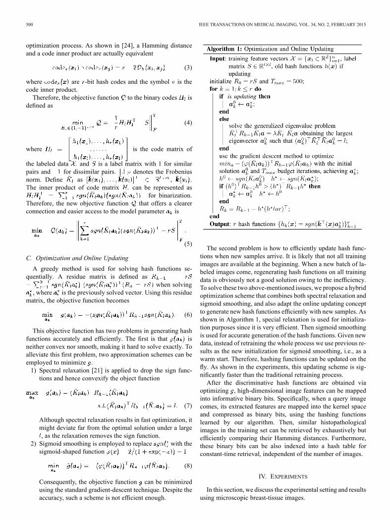
C. Optimization and Online Updating
A greedy method is used for solving hash functions se-quentially. A residue matrix is defined as
when solving, where is the previously solved vector. Using this residue
matrix, the objective function becomes
(6)
This objective function has two problems in generating hashfunctions accurately and efficiently. The first is that isneither convex nor smooth, making it hard to solve exactly. Toalleviate this first problem, two approximation schemes can beemployed to minimize .1) Spectral relaxation [21] is applied to drop the sign func-tions and hence convexify the object function
(7)
Although spectral relaxation results in fast optimization, itmight deviate far from the optimal solution under a large, as the relaxation removes the sign function.
2) Sigmoid smoothing is employed to replace with thesigmoid-shaped function
(8)
Consequently, the objective function can be minimizedusing the standard gradient-descent technique. Despite theaccuracy, such a scheme is not efficient enough.
The second problem is how to efficiently update hash func-tions when new samples arrive. It is likely that not all trainingimages are available at the beginning. When a new batch of la-beled images come, regenerating hash functions on all trainingdata is obviously not a good solution owing to the inefficiency.To solve these two above-mentioned issues, we propose a hybridoptimization scheme that combines both spectral relaxation andsigmoid smoothing, and also adapt the online updating conceptto generate new hash functions efficiently with new samples. Asshown in Algorithm 1, special relaxation is used for initializa-tion purposes since it is very efficient. Then sigmoid smoothingis used for accurate generation of the hash functions. Given newdata, instead of retraining the whole process we use previous re-sults as the new initialization for sigmoid smoothing, i.e., as awarm start. Therefore, hashing functions can be updated on thefly. As shown in the experiments, this updating scheme is sig-nificantly faster than the traditional retraining process.After the discriminative hash functions are obtained via
optimizing , high-dimensional image features can be mappedinto informative binary bits. Specifically, when a query imagecomes, its extracted features are mapped into the kernel spaceand compressed as binary bits, using the hashing functionslearned by our algorithm. Then, similar histopathologicalimages in the training set can be retrieved by exhaustively butefficiently comparing their Hamming distances. Furthermore,these binary bits can be also indexed into a hash table forconstant-time retrieval, independent of the number of images.
IV. EXPERIMENTS
In this section, we discuss the experimental setting and resultsusing microscopic breast-tissue images.

ZHANG et al.: TOWARDS LARGE-SCALE HISTOPATHOLOGICAL IMAGE ANALYSIS: HASHING-BASED IMAGE RETRIEVAL 501
Fig. 4. Key points detected by difference of Gaussian (marked as blue stars).
A. Experimental Setting
Breast-tissue specimens available for this study were col-lected on a retrospective basis from the IU Health PathologyLab (IUHPL) according to the protocol approved by the In-stitutional Review Board (IRB) for this study. All the slideswere imaged using a ScanScope registered digitizer (Aperio,Vista, CA, USA) available in the tissue archival service atIUHPL. 3121 images (around 2250 K pixels) were sampledfrom 657 larger region-of-interest images (e.g., )of microscopic breast tissue, which were gathered from 116patients. 53 of these patients were labeled as benign (UDH)and 63 as actionable (ADH/DCIS), based on the majoritydiagnosis of nine board-certified pathologists. To demonstratethe efficiency of our method, one fourth of all patients ineach category were randomly selected as the test set and theremainder used for training. Note that each patient may havedifferent number of images. Therefore, the number of testingimages is not fixed. The approximate number is about 700–900in each testing process. All the experiments were conductedon a 3.40 GHz CPU with four cores and 16 G RAM, in aMATLAB implementation.In each image, 1500–2000 SIFT descriptors were extracted
from key points detected by DoG [50] (Fig. 4). These descrip-tors were quantized into sets of cluster centers using bag-of-words, in which the feature dimension equals the number ofclusters. Specifically, we quantize them into high-dimensionalfeature vectors of length 10 000, to maximally utilize these mil-lions of cell-level texture features. We provide both qualitativeand quantitative evaluations for our proposed framework on twotasks, image classification (i.e., benign versus actionable cate-gory) and image retrieval, in terms of accuracy and computa-tional efficiency.In our system, classification is achieved using the majority
vote of the top images retrieved by hashing. We compare ourapproach with various classifiers that have been widely used insystems for histopathological image analysis. Specifically, kNNhas often been used as the baseline in analyzing histopatho-logical images [32], [18], owing to its simplicity and provedlower bound, despite the inefficiency in large-scale databases.The Bayesian method is another solution to ensemble statistics
Fig. 5. Comparison of classification accuracy with different dimensions of fea-tures (from 100 to 10000).
of all extracted features and minimize the classification metric,which shows its efficacy in classifying histopathological images[12]. Boosting methods are always employed to combine mul-tiple weak classifiers for higher accuracy [27], [18], [52]. SVMwith a nonlinear kernel is commonly used in histopathologicalimages because of its efficiency and the ability to handle lin-early inseparable cases [53], [28], [54], [9]. For fair comparison,all parameters and kernel selections of these compared methodswere optimized by cross-validation.In addition, we also compared our proposed method with sev-
eral dimensionality-reduction algorithms in terms of classifica-tion accuracy. Principal component analysis (PCA) has beenwidely used in this area to preserve variance of original features[55]. Graph embedding is a nonlinear dimensionality-reductionalgorithm that performs well in grading of lymphocytic infiltra-tion in breast cancer histopathology [30]. Since we usesupervised information in generating hash functions, a super-vised dimensionality reduction algorithm, neighborhood com-ponents analysis (NCA) [56], was also chosen for our experi-mental comparisons.
B. Evaluation of Image Classification
To demonstrate the benefit of high-dimensional features, allmethods are evaluated on multiple dimensions of SIFT quan-tization, ranging from 100 to 10 ,000. In our proposed frame-work, we use a hashing method to compress all features to 48bits (only six bytes), which is a suitable size for using a hashtable. To compare with dimensionality-reduction methods, wecompress all features into 48 dimensions. In contrast to hashingmethods using bits, results of other methods are based on floatsor doubles. Therefore, they need much more storage room thanhashing results do.Fig. 5 shows the quantitative results for the classification ac-
curacy. Most methods achieve better accuracy with higher-di-mensional features. This is very intuitive, as finer quantizationof SIFT features usually provides richer information. In partic-ular, since the SIFT interest points cover most nuclear regions
502 IEEE TRANSACTIONS ON MEDICAL IMAGING, VOL. 34, NO. 2, FEBRUARY 2015
Fig. 6. Comparison of the classification accuracy for different dimensionality-reduction methods.
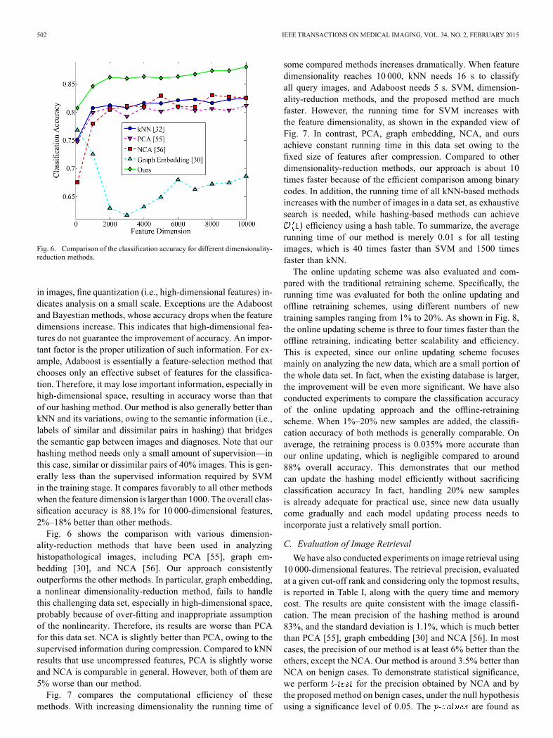
in images, fine quantization (i.e., high-dimensional features) in-dicates analysis on a small scale. Exceptions are the Adaboostand Bayestian methods, whose accuracy drops when the featuredimensions increase. This indicates that high-dimensional fea-tures do not guarantee the improvement of accuracy. An impor-tant factor is the proper utilization of such information. For ex-ample, Adaboost is essentially a feature-selection method thatchooses only an effective subset of features for the classifica-tion. Therefore, it may lose important information, especially inhigh-dimensional space, resulting in accuracy worse than thatof our hashing method. Our method is also generally better thankNN and its variations, owing to the semantic information (i.e.,labels of similar and dissimilar pairs in hashing) that bridgesthe semantic gap between images and diagnoses. Note that ourhashing method needs only a small amount of supervision—inthis case, similar or dissimilar pairs of 40% images. This is gen-erally less than the supervised information required by SVMin the training stage. It compares favorably to all other methodswhen the feature dimension is larger than 1000. The overall clas-sification accuracy is 88.1% for 10 000-dimensional features,2%–18% better than other methods.Fig. 6 shows the comparison with various dimension-
ality-reduction methods that have been used in analyzinghistopathological images, including PCA [55], graph em-bedding [30], and NCA [56]. Our approach consistentlyoutperforms the other methods. In particular, graph embedding,a nonlinear dimensionality-reduction method, fails to handlethis challenging data set, especially in high-dimensional space,probably because of over-fitting and inappropriate assumptionof the nonlinearity. Therefore, its results are worse than PCAfor this data set. NCA is slightly better than PCA, owing to thesupervised information during compression. Compared to kNNresults that use uncompressed features, PCA is slightly worseand NCA is comparable in general. However, both of them are5% worse than our method.Fig. 7 compares the computational efficiency of these
methods. With increasing dimensionality the running time of
some compared methods increases dramatically. When featuredimensionality reaches 10 000, kNN needs 16 s to classifyall query images, and Adaboost needs 5 s. SVM, dimension-ality-reduction methods, and the proposed method are muchfaster. However, the running time for SVM increases withthe feature dimensionality, as shown in the expanded view ofFig. 7. In contrast, PCA, graph embedding, NCA, and oursachieve constant running time in this data set owing to thefixed size of features after compression. Compared to otherdimensionality-reduction methods, our approach is about 10times faster because of the efficient comparison among binarycodes. In addition, the running time of all kNN-based methodsincreases with the number of images in a data set, as exhaustivesearch is needed, while hashing-based methods can achieve
efficiency using a hash table. To summarize, the averagerunning time of our method is merely 0.01 s for all testingimages, which is 40 times faster than SVM and 1500 timesfaster than kNN.The online updating scheme was also evaluated and com-
pared with the traditional retraining scheme. Specifically, therunning time was evaluated for both the online updating andoffline retraining schemes, using different numbers of newtraining samples ranging from 1% to 20%. As shown in Fig. 8,the online updating scheme is three to four times faster than theoffline retraining, indicating better scalability and efficiency.This is expected, since our online updating scheme focusesmainly on analyzing the new data, which are a small portion ofthe whole data set. In fact, when the existing database is larger,the improvement will be even more significant. We have alsoconducted experiments to compare the classification accuracyof the online updating approach and the offline-retrainingscheme. When 1%–20% new samples are added, the classifi-cation accuracy of both methods is generally comparable. Onaverage, the retraining process is 0.035% more accurate thanour online updating, which is negligible compared to around88% overall accuracy. This demonstrates that our methodcan update the hashing model efficiently without sacrificingclassification accuracy In fact, handling 20% new samplesis already adequate for practical use, since new data usuallycome gradually and each model updating process needs toincorporate just a relatively small portion.
C. Evaluation of Image Retrieval
We have also conducted experiments on image retrieval using10 000-dimensional features. The retrieval precision, evaluatedat a given cut-off rank and considering only the topmost results,is reported in Table I, along with the query time and memorycost. The results are quite consistent with the image classifi-cation. The mean precision of the hashing method is around83%, and the standard deviation is 1.1%, which is much betterthan PCA [55], graph embedding [30] and NCA [56]. In mostcases, the precision of our method is at least 6% better than theothers, except the NCA. Our method is around 3.5% better thanNCA on benign cases. To demonstrate statistical significance,we perform for the precision obtained by NCA and bythe proposed method on benign cases, under the null hypothesisusing a significance level of 0.05. The are found as

ZHANG et al.: TOWARDS LARGE-SCALE HISTOPATHOLOGICAL IMAGE ANALYSIS: HASHING-BASED IMAGE RETRIEVAL 503
Fig. 7. Comparison of the classification running time (seconds) with different dimensions of features, which means the average time of classifying hundreds oftest images.
Fig. 8. Comparison of the training time (seconds) using different optimizationschemes.
, , and at the range of top 10,20, and 30 retrievals, respectively, demonstrating that precisionvalues achieved by the proposed technique are indeed signif-icantly better than NCA on the benign cases. In addition, ourmethod is around 14% better than NCA in the actionable cases,resulting much higher average precision. In fact, most tradi-tional methods produce such highly unbalanced results as NCAdoes, i.e., the retrieval precision of the benign category is muchhigher than that of the actionable one. In contrast, our methoddoes not have this problem, owing to the supervised informa-tion and the optimization for balanced hash bits. Our frameworkis also computationally more efficient than traditional methods.The query time of our hashing method is a thousand times fasterthan kNN and ten times faster than other dimensionality-reduc-tion methods. Note that our method takes a constant time whenusing the hash table, independent of the number of feature di-mensions and the number of samples. Furthermore, the memorycost is also considerably reduced (10 000 times less than that ofkNN). Therefore, this method is more applicable to large-scaledatabases (millions of images) than are other methods.Fig. 9 shows our image-retrieval results. The top five relevant
images are listed for each query image. The differences betweencertain images in different categories are very subtle. Our accu-rate results demonstrate the efficacy of the proposed method.
Specifically, the features capturing local texture and appearanceare very robust to various image sizes, cell distributions, andocclusions by the blood. The supervised information also im-proves the retrieval precision by correlating binary code withdiagnosis information. These retrieved images are clinically rel-evant in potential (i.e., retrieved images belong to the same cat-egory as the query image) and thus can be useful for decisionsupport.
D. Discussions
We discuss the benefits of the algorithm, parameter sensi-tivity, implementation issues, and limitations here.Regarding the choice of high-dimensional features, around
1000 dimensions have usually been used for quantization bymany previous studies, a number that has been proved to achievegood accuracy. Using lower-dimensional features (e.g., 100) isnot accurate, while using higher-dimensional features is not effi-cient, and the improvement of accuracy could be marginal. Thisis consistent with our experimental results shown in Fig. 5, i.e.,a performance jump from 100 to 1000 dimensions. On the otherhand, when analyzing histopathological images, using high-di-mensional features (e.g., 10 000) implies nearly cell-level anal-ysis, which is actually beneficial for the accuracy, even thoughthe accuracy gain is not as big as jumping from 100 to 1000.Therefore, we have introduced hashing methods to harvest thebenefits of high-dimensional features, without sacrificing com-putational efficiency.Regarding supervised information, it significantly improves
classification accuracy thanks to the discriminative modeling ofthe hashing function in an attempt to bridge the semantic gap.In Fig. 10, we randomly selected 100 samples from benign andactionable categories and visualized their 48 hash bits. The dis-tributions of hash bits are clearly different between the two cat-egories, explaining the high accuracy for classification. We alsoquantitatively investigated the benefits of using supervised in-formation. Specifically, we evaluated our method when using10% to 100% supervision or training labels, as shown in Fig. 11.The gain in accuracy is very high (from 71% to nearly 87%)when the ratio of training labels increases from 10% to 40%,which demonstrates the efficacy of using supervised informa-tion. For more than 40% labels, the improvement of accuracybecomes marginal, reaching 88% accuracy when using 100%

504 IEEE TRANSACTIONS ON MEDICAL IMAGING, VOL. 34, NO. 2, FEBRUARY 2015
TABLE ICOMPARISON OF RETRIEVAL PRECISION FOR THE TOP 10, 20, AND 30 RESULTS, ALONG WITH THE MEMORY COST OF TRAINING DATA AND QUERY TIME OFALL TEST IMAGES. BOTH MEAN VALUES AND THE STANDARD DEVIATION (STD) OF 20 EXPERIMENTS ARE REPORTED. BEST PRECISION IN EACH ROW FOR
BENIGN AND ACTIONABLE CATEGORIES ARE HIGHLIGHTED
Fig. 9. Four examples of our image retrieval (query marked in red, and retrieved images marked in blue). First two rows are benign; the last two rows are action-able.
Fig. 10. Visualization of compressed hash bits. Their distribution well sepa-rates the begin and actionable categories.
labels. This means that our method needs only a small portionof labels to achieve high accuracy, owing to the unified frame-
Fig. 11. Classification accuracy when using 10%–100% supervision.
work of coupling Hamming distance optimization and super-vised information.

ZHANG et al.: TOWARDS LARGE-SCALE HISTOPATHOLOGICAL IMAGE ANALYSIS: HASHING-BASED IMAGE RETRIEVAL 505
Fig. 12. Classification accuracy with different lengths of hashing bits.
One of the most significant benefits of our proposed frame-work is the computational and storage efficiency. Comparing48 bits with Hamming distance or hash table is substantiallyfaster than using high-dimensional features. However, a naturalquestion is whether the length of hashing bits affects the ac-curacy and retrieval precision. Therefore, we evaluated the ef-fect of hashing-bit lengths ranging from 1 to 48. Theoretically, 1bit is sufficient for binary classification purpose, i.e., actionableversus benign. In fact, as shown in Fig. 12, using 8 bits alreadyachieves high accuracy for classification. However, such shortcode is not discriminative enough for image retrieval. For ex-ample, 8 bits can represent only 64 hash values. This means thatnearly 50 images are mapped into the same hash value, whichis an unordered list with zero Hamming distance. Retrievingthem may not be beneficial for decision support. On the otherhand, using more than 64 bits adversely affects computationalefficiency, since the hash table is no longer an option owing tomemory constraint. Therefore, we chose 48 bits for this task, en-suring sound accuracy for classification and high relevance forretrieval without sacrificing efficiency. We expect that our scal-able framework can be efficiently used for real-time queryingof very large databases.In the task of image retrieval, our method effectively re-
trieves images with morphological and architectural imagepatterns similar to the query image, as shown in Fig. 9. Thiscan be explained by the capability of the hashing function inleveraging both diagnostic information and visual similarities.In other words, hash bits can simultaneously encode localtextural features with semantic labels.
V. CONCLUSION
In this paper, we developed a scalable image-retrieval frame-work for intelligent histopathological image analysis. Specifi-cally, we employed hashing to achieve efficient image retrievaland presented an improved kernelized and supervised hashingapproach for real-time image retrieval. The potential applica-tions of our framework include image-guided diagnosis, de-cision support, education, and efficient data management. In
our future work, we will examine more types of features, espe-cially those features stemming from segmentation and architec-tures. Furthermore, we will incorporate appropriate feature-fu-sion techniques to design a hybrid hashing method such thatmultiple types of features can be systematically fused to boostimage-retrieval accuracy. We will also evaluate our frameworkin more applications in histopathological image analysis.
REFERENCES[1] R. Siegel, D. Naishadham, and A. Jemal, “Cancer statistics, 2013,”CA,
Cancer J. Clinicians, vol. 63, no. 1, pp. 11–30, 2013.[2] D. C. Sgroi, “Preinvasive breast cancer,” Annu. Rev. Pathol. Mech.
Disease, vol. 5, pp. 193–221, 2010.[3] D. L. Page, W. D. Dupont, L. W. Rogers, and M. S. Rados, “Atypical
hyperplastic lesions of the female breast. A long-term follow-up study,”Cancer, vol. 55, no. 11, pp. 2698–2708, 1985.
[4] B.Weyn, G. v. d.Wouwer, A. v. Daele, P. Scheunders, D. v. Dyck, E. v.Marck, and W. Jacob, “Automated breast tumor diagnosis and gradingbased on wavelet chromatin texture description,” Cytometry, vol. 33,no. 1, pp. 32–40, 1998.
[5] C. Bilgin, C. Demir, C. Nagi, and B. Yener, “Cell-graph mining forbreast tissuemodeling and classification,” inProc. IEEE Int. Conf. Eng.Med. Biol. Soc., 2007, pp. 5311–5314.
[6] M. N. Gurcan, L. E. Boucheron, A. Can, A. Madabhushi, N. M. Ra-jpoot, and B. Yener, “Histopathological image analysis, A review,”IEEE Rev. Biomed. Eng., vol. 2, pp. 147–171, Dec. 2009.
[7] J. Kong, O. Sertel, H. Shimada, K. L. Boyer, J. H. Saltz, and M. N.Gurcan, “Computer-aided evaluation of neuroblastoma on whole-slidehistology images: Classifying grade of neuroblastic differentiation,”Pattern Recognit., vol. 42, no. 6, pp. 1080–1092, 2009.
[8] P.-W. Huang and C.-H. Lee, “Automatic classification for pathologicalprostate images based on fractal analysis,” IEEE Trans. Med. Imag.,vol. 28, no. 7, pp. 1037–1050, Jul. 2009.
[9] P.-W. Huang and Y.-H. Lai, “Effective segmentation and classifica-tion for HCC biopsy images,” Pattern Recognit., vol. 43, no. 4, pp.1550–1563, 2010.
[10] P. Khurd, C. Bahlmann, P. Maday, A. Kamen, S. Gibbs-Strauss, E.M. Genega, and J. V. Frangioni, “Computer-aided Gleason grading ofprostate cancer histopathological images using texton forests,” in Proc.IEEE Int. Symp. Biomed. Imag., 2010, pp. 636–639.
[11] M. Veta, J. Pluim, P. v. Diest, and M. Viergever, “Breast cancerhistopathology image analysis: A review,” IEEE Trans. Biomed. Eng.,vol. 61, no. 5, pp. 1400–1411, May 2014.
[12] D. Comaniciu, P. Meer, and D. J. Foran, “Image-guided decisionsupport system for pathology,” Mach. Vis. Appl., vol. 11, no. 4, pp.213–224, 1999.
[13] L. Zheng, A. W. Wetzel, J. Gilbertson, and M. J. Becich, “Design andanalysis of a content-based pathology image retrieval system,” IEEETrans. Inf. Technol. Biomed., vol. 7, no. 4, pp. 249–255, Dec. 2003.
[14] H. Müller, N. Michoux, D. Bandon, and A. Geissbuhler, “A review ofcontent-based image retrieval systems in medical applications-clinicalbenefits and future directions,” Int. J. Med. Inf., vol. 73, no. 1, pp. 1–23,2004.
[15] H. C. Akakin and M. N. Gurcan, “Content-based microscopic imageretrieval system for multi-image queries,” IEEE Trans. Inf. Technol.Biomed., vol. 16, no. 4, pp. 758–769, Apr. 2012.
[16] A. Kumar, J. Kim, W. Cai, M. Fulham, and D. Feng, “Content-basedmedical image retrieval, A survey of applications to multidimen-sional and multimodality data,” J. Digital Imag., vol. 26, no. 6, pp.1025–1039, 2013.
[17] T. M. Lehmann, M. O. Güld, T. Deselaers, D. Keysers, H. Schubert, K.Spitzer, H. Ney, and B. B. Wein, “Automatic categorization of medicalimages for content-based retrieval and data mining,” Comput. Med.Imag. Graph., vol. 29, no. 2, pp. 143–155, 2005.
[18] L. Yang, W. Chen, P. Meer, G. Salaru, L. A. Goodell, V. Berstis, andD. J. Foran, “Virtual microscopy and grid-enabled decision support forlarge-scale analysis of imaged pathology specimens,” IEEE Trans. Inf.Technol. Biomed., vol. 13, no. 4, pp. 636–644, Jul. 2009.
[19] D. J. Foran et al., “Imageminer, A software system for comparativeanalysis of tissue microarrays using content-based image retrieval,high-performance computing, and grid technology,” J. Am. Med.Informat. Assoc., vol. 18, no. 4, pp. 403–415, 2011.
[20] M. Datar, N. Immorlica, P. Indyk, and V. S. Mirrokni, “Locality-sen-sitive hashing scheme based on p-stable distributions,” in Proc. Annu.ACM Symp. Computat. Geometry, 2004, pp. 253–262.

506 IEEE TRANSACTIONS ON MEDICAL IMAGING, VOL. 34, NO. 2, FEBRUARY 2015
[21] Y. Weiss, A. Torralba, and R. Fergus, “Spectral hashing,” in Adv.Neural Inf. Process. Syst., 2008, pp. 1753–1760.
[22] B. Kulis and K. Grauman, “Kernelized locality-sensitive hashing,”IEEE Trans. Pattern Anal. Mach. Intell., vol. 34, no. 6, pp. 1092–1104,Jun. 2012.
[23] J. Wang, S. Kumar, and S.-F. Chang, “Semi-supervised hashing forscalable image retrieval,” in Proc. IEEE Int. Conf. Comput. Vis. PatternRecognit., 2010, pp. 3424–3431.
[24] W. Liu, J. Wang, R. Ji, Y.-G. Jiang, and S.-F. Chang, “Supervisedhashing with kernels,” in Proc. IEEE Int. Conf. Comput. Vis. PatternRecognit., 2012, pp. 2074–2081.
[25] X. Zhang, W. Liu, and S. Zhang, “Mining histopathological imagesvia hashing-based scalable image retrieval,” in Proc. IEEE Int. Symp.Biomed. Imag., 2014, pp. 1111–1114.
[26] S. Petushi, F. U. Garcia, M. M. Haber, C. Katsinis, and A. Tozeren,“Large-scale computations on histology images reveal grade-differen-tiating parameters for breast cancer,” BMC Med. Imag., vol. 6, no. 1,p. 14, 2006.
[27] L. Yang, W. Chen, P. Meer, G. Salaru, M. D. Feldman, and D. J. Foran,“High throughput analysis of breast cancer specimens on the grid,” inProc. Int. Conf. Med. Image Comput. Comput. Assist. Intervent., 2007,pp. 617–625.
[28] J. C. Caicedo, A. Cruz, and F. A. Gonzalez, “Histopathology imageclassification using bag of features and kernel functions,” Artif. Intell.Med., pp. 126–135, 2009.
[29] J. Sivic and A. Zisserman, “Video Google: A text retrieval approachto object matching in videos,” in Proc. IEEE Int. Conf. Comput. Vis.,2003, pp. 1470–1477.
[30] A. N. Basavanhally, S. Ganesan, S. Agner, J. P. Monaco, M. D.Feldman, J. E. Tomaszewski, G. Bhanot, and A. Madabhushi,“Computerized image-based detection and grading of lymphocyticinfiltration in HER2+ breast cancer histopathology,” IEEE Trans.Biomed. Eng., vol. 57, no. 3, pp. 642–653, Mar. 2010.
[31] M. M. Dundar, S. Badve, G. Bilgin, V. Raykar, R. Jain, O. Sertel, andM. N. Gurcan, “Computerized classification of intraductal breast le-sions using histopathological images,” IEEE Trans. Biomed. Eng., vol.58, no. 7, pp. 1977–1984, Jul. 2011.
[32] A. Tabesh, M. Teverovskiy, H.-Y. Pang, V. P. Kumar, D. Verbel, A.Kotsianti, and O. Saidi, “Multifeature prostate cancer diagnosis andGleason grading of histological images,” IEEE Trans. Med. Imag., vol.26, no. 10, pp. 1366–1378, Oct. 2007.
[33] S. Doyle, S. Agner, A. Madabhushi, M. Feldman, and J. Tomaszewski,“Automated grading of breast cancer histopathology using spectralclustering with textural and architectural image features,” in Proc.IEEE Int. Symp. Biomed. Imag., 2008, pp. 496–499.
[34] J. G. Dy, C. E. Brodley, A. Kak, L. S. Broderick, and A. M. Aisen,“Unsupervised feature selection applied to content-based retrieval oflung images,” IEEE Trans. Pattern Anal. Mach. Intell., vol. 25, no. 3,pp. 373–378, Feb. 2003.
[35] I. El-Naqa, Y. Yang, N. P. Galatsanos, R. M. Nishikawa, and M. N.Wernick, “A similarity learning approach to content-based imageretrieval: Application to digital mammography,” IEEE Trans. Med.Imag., vol. 23, no. 10, pp. 1233–1244, Oct. 2004.
[36] H. Greenspan and A. T. Pinhas, “Medical image categorization andretrieval for PACS using the GMM-KL framework,” IEEE Trans. Inf.Technol. Biomed., vol. 11, no. 2, pp. 190–202, Mar. 2007.
[37] Y. Song, W. Cai, and D. Feng, “Hierarchical spatial matching for med-ical image retrieval,” in ACM Int. Workshop Med. Multimedia Anal.Retrieval, 2011, pp. 1–6.
[38] F. Schnorrenberg, C. Pattichis, C. Schizas, and K. Kyriacou, “Content-based retrieval of breast cancer biopsy slides,” Technol. Health Care,vol. 8, no. 5, pp. 291–297, 2000.
[39] X. S. Zhou, S. Zillner, M. Moeller, M. Sintek, Y. Zhan, A. Krishnan,and A. Gupta, “Semantics and CBIR: A medical imaging perspective,”in ACM Int. Conf. Content-Based Image Video Retrieval, 2008, pp.571–580.
[40] H. Müller, A. Geissbühler, and P. Ruch, “ImageCLEF 2004: Com-bining image and multi-lingual search for medical image retrieval,” inMultilingual Inform. Access for Text, Speech and Images. New York:Springer, 2005, pp. 718–727.
[41] H. Müller and J. Kalpathy-Cramer, “The imageCLEFmedical retrievaltask at ICPR 2010–Information fusion,” in Proc. IEEE Int. Conf. Pat-tern Recognit., 2010, pp. 3284–3287.
[42] L. Yang, R. Jin, L. Mummert, R. Sukthankar, A. Goode, B. Zheng, S. C.Hoi, andM. Satyanarayanan, “A boosting framework for visuality-pre-serving distance metric learning and its application to medical imageretrieval,” IEEE Trans. Pattern Anal. Mach. Intell., vol. 32, no. 1, pp.30–44, Jan. 2010.
[43] G. Langs, H. Müller, B. H. Menze, and A. Hanbury, “VISCERAL:Towards large data in medical imaging—Challenges and directions,”in MCBR-CDS MICCAI Workshop, 2013, vol. 7723.
[44] A. Hanbury, H. Müller, G. Langs, and B. H. Menze, “Cloud-basedevaluation framework for big data,” in FIA Book 2013,. New York:Springer, 2013, LNCS.
[45] Y. Mu, J. Shen, and S. Yan, “Weakly-supervised hashing in kernelspace,” in Proc. IEEE Int. Conf. Comput. Vis. Pattern Recognit., 2010,pp. 3344–3351.
[46] J. Wang, S. Kumar, and S.-F. Chang, “Semi-supervised hashing forlarge-scale search,” IEEE Trans. Pattern Anal. Mach. Intell., vol. 34,no. 12, pp. 2393–2406, Dec. 2012.
[47] X. Liu, J. He, D. Liu, and B. Lang, “Compact kernel hashing withmultiple features,” in Proc. ACM Int. Conf. Multimedia, 2012, pp.881–884.
[48] B. Kulis and T. Darrell, “Learning to hash with binary reconstructiveembeddings,” in Adv. Neural Inf. Process. Syst., 2009, pp. 1042–1050.
[49] M. Norouzi and D. M. Blei, “Minimal loss hashing for compact binarycodes,” in Int. Conf. on Mach. Learning, 2011, pp. 353–360.
[50] D. G. Lowe, “Distinctive image features from scale-invariant key-points,” Int. J. Comput. Vis., vol. 60, no. 2, pp. 91–110, 2004.
[51] J. Wang et al., “Bag-of-features based medical image retrieval viamultiple assignment and visual words weighting,” IEEE Trans. Med.Imag., vol. 30, no. 11, pp. 1996–2011, Nov. 2011.
[52] S. Doyle, M. Feldman, J. Tomaszewski, and A. Madabhushi, “Aboosted Bayesian multiresolution classifier for prostate cancer detec-tion from digitized needle biopsies,” IEEE Trans. Biomed. Eng., vol.59, no. 5, pp. 1205–1218, May 2012.
[53] O. Tuzel, L. Yang, P. Meer, and D. J. Foran, “Classification of hemato-logic malignancies using texton signatures,” Pattern Anal. Appl., vol.10, no. 4, pp. 277–290, 2007.
[54] K. Nguyen, A. K. Jain, and R. L. Allen, “Automated gland segmenta-tion and classification for Gleason grading of prostate tissue images,”in Proc. IEEE Int. Conf. Pattern Recognit., 2010, pp. 1497–1500.
[55] O. Sertel, J. Kong, U. V. Catalyurek, G. Lozanski, J. H. Saltz, andM. N. Gurcan, “Histopathological image analysis using model-basedintermediate representations and color texture: Follicular lymphomagrading,” J. Signal Process. Syst., vol. 55, no. 1–3, pp. 169–183, 2009.
[56] J. Goldberger, S. Roweis, G. Hinton, and R. Salakhutdinov, “Neigh-bourhood components analysis,” Adv. Neural Inf. Process. Syst., 2004.