42 Tushar Etal
4
735 Tushar et al., Int J Med Res Health Sci. 2014;3(3):735-738 International Journal of Medical Research & Health Sciences www.ijmrhs.com Volume 3 Issue 3 Coden: IJMRHS Copyright @2014 ISSN: 2319-5886 Received: 10 th Apr 2014 Revised: 8 th May 2014 Accepted: 19 th May 2014 Case report BILATERAL INTERNUCLEAR OPHTHALMOPLEGIA AS FIRST MANIFESTATION OF EXTRA PONTINE MYELINOLYSIS Tushar Kanti Bandyopadhyay 1 , * Rudrajit Paul 1 , Amit K Das 2 , Rathin drana th Sark ar 3 1 Assistant Professor, 2 Resident, 3 Prof essor , Depa rtmen t of Medic ine, Medica l College Kolka ta88, Colle ge Street , Kolkata, West Beng al *Corr espon ding author email: docr8 9@gmail.com ABSTRACT Extra pontin e mye linolys is (EPM) is a rare cli nical e ntity affecti ng an terior basal gang lia. This is one o f the osmo tic demy elinatio n synd romes . It occur s due to rap id corre ction o f hypon atremia and also r arely occur s in alcoholics. It gene rally pre sents with extrap yramidal symptoms. We here report a case of EPM in a 13 year old boy presenting with bilateral internuclear ophthalmopleg ia and ptosis. The patient also had generalised weakness, but no psychiatr ic sympt oms. The pat ient slow ly recov ered over six months. EPM can affect any age group, althou gh the elder ly are more li kely to be aff ected du e to frequ ent elec trolyt e abnormalities . Ocul ar move ment disorders or brains tem signs are rarely reported in EPM. When present, it can create diagnostic confusion with multiple sclerosis. We believe this is the first report of this entity from I ndia. The relevant literature regarding brainstem manifestations in myelinolysis syndromes is also discussed, along with the radiological findings. Keywords: Intern uclea r ophth almop legia, Extrap ontin e myelin olysi s, Ptos is, CIDP, Basal gangli a INTRODUCTION Extrapontine myelinolysis (EPM) is a rare clinical entity occur ring mainly after rapid corr ection of hyponatre mia . It is us uall y ass oci ate d wit h its counterpart: central pontine myelinolysis (CPM). 1 However, very rarely, EPM can occur in absence of CPM and this makes the diagnosis challenging. The clinic al manife statio ns of EPM vary and may ra nge from extrapyramidal features to neuropsychiatric manifestations. 1, 2 Such a typic al featu res, a long with the ra rity o f the e ntity often delay the di agnos is. We here report a case of EPM presenting with bilateral internuclear ophthalmoplegia (INO). To our knowledge, this is probably the first report of EPM presenting with INO from India. Other report ed cases from India have shown parkinsonian features and bulbar symptoms. 3 Another case was reported with flaccid quadruparesis. 4 CASE REPORT A13 yea r old boy pr ese nted wit h acute onset gener alised weak ness witho ut loss of consc iousn ess for two days. He had been admitted elsewhere with increa sing abd omina l pain and vomiting for twe nty day s. He wa s there documente d to be de hyd rat ed and resuscitated with intravenous fluids. He apparently impro ved with the conse rvativ e ma nagement bu t deteriorated again with severe generalized weakness and blurr ing of vision . With this comp laint, he was refer red to our tertiary car e center. At our centre, on admission, the boy was found to be severely weak with power 2-/5 in all four limbs. He could not turn in bed or lift his head from pillows. His abdomen was found to be distended and his parents comp lained o f seve re cons tipatio n for the las t ten DOI: 10.5958/2319-5886.2014.00428.7
-
Upload
editorijmrhs -
Category
Documents
-
view
230 -
download
0
Transcript of 42 Tushar Etal

8/12/2019 42 Tushar Etal
http://slidepdf.com/reader/full/42-tushar-etal 1/4
735
Tushar et al., Int J Med Res Health Sci. 2014;3(3):735-738
International Journal of Medical Research
&
Health Sciences
www.ijmrhs.com Volume 3 Issue 3 Coden: IJMRHS Copyright @2014 ISSN: 2319-5886Received: 10
thApr 2014 Revised: 8
thMay 2014 Accepted: 19
thMay 2014
Case report
BILATERAL INTERNUCLEAR OPHTHALMOPLEGIA AS FIRST MANIFESTATION OF EXTRA
PONTINE MYELINOLYSIS
Tushar Kanti Bandyopadhyay1,
*Rudrajit Paul
1, Amit K Das
2, Rathindranath Sarkar
3
1Assistant Professor,
2Resident,
3Professor, Department of Medicine, Medical College Kolkata88, College Street,
Kolkata, West Bengal
*Corresponding author email: [email protected]
ABSTRACT
Extrapontine myelinolysis (EPM) is a rare clinical entity affecting anterior basal ganglia. This is one of the
osmotic demyelination syndromes. It occurs due to rapid correction of hyponatremia and also rarely occurs in
alcoholics. It generally presents with extrapyramidal symptoms. We here report a case of EPM in a 13 year old
boy presenting with bilateral internuclear ophthalmoplegia and ptosis. The patient also had generalised weakness,
but no psychiatric symptoms. The patient slowly recovered over six months. EPM can affect any age group,
although the elderly are more likely to be affected due to frequent electrolyte abnormalities. Ocular movement
disorders or brainstem signs are rarely reported in EPM. When present, it can create diagnostic confusion with
multiple sclerosis. We believe this is the first report of this entity from India. The relevant literature regarding
brainstem manifestations in myelinolysis syndromes is also discussed, along with the radiological findings.
Keywords: Internuclear ophthalmoplegia, Extrapontine myelinolysis, Ptosis, CIDP, Basal ganglia
INTRODUCTION
Extrapontine myelinolysis (EPM) is a rare clinical
entity occurring mainly after rapid correction of
hyponatremia. It is usually associated with its
counterpart: central pontine myelinolysis (CPM).1
However, very rarely, EPM can occur in absence of CPM and this makes the diagnosis challenging. The
clinical manifestations of EPM vary and may range
from extrapyramidal features to neuropsychiatric
manifestations.1, 2
Such atypical features, along with
the rarity of the entity often delay the diagnosis. We
here report a case of EPM presenting with bilateral
internuclear ophthalmoplegia (INO). To our
knowledge, this is probably the first report of EPM
presenting with INO from India. Other reported cases
from India have shown parkinsonian features andbulbar symptoms.
3Another case was reported with
flaccid quadruparesis.4
CASE REPORT
A13 year old boy presented with acute onset
generalised weakness without loss of consciousness
for two days. He had been admitted elsewhere withincreasing abdominal pain and vomiting for twenty
days. He was there documented to be dehydrated and
resuscitated with intravenous fluids. He apparently
improved with the conservative management but
deteriorated again with severe generalized weakness
and blurring of vision. With this complaint, he was
referred to our tertiary care center.
At our centre, on admission, the boy was found to be
severely weak with power 2-/5 in all four limbs. He
could not turn in bed or lift his head from pillows. His
abdomen was found to be distended and his parents
complained of severe constipation for the last ten
DOI: 10.5958/2319-5886.2014.00428.7
8/12/2019 42 Tushar Etal
http://slidepdf.com/reader/full/42-tushar-etal 2/4
736
Tushar et al., Int J Med Res Health Sci. 2014;3(3):735-738
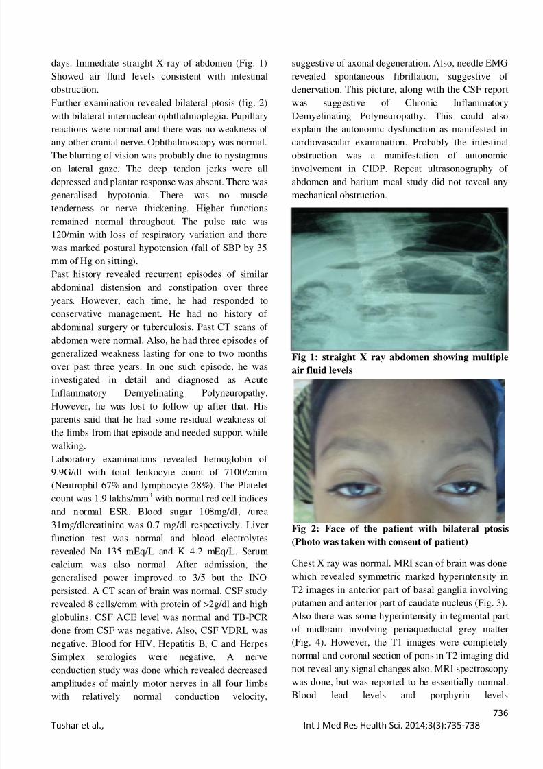
days. Immediate straight X-ray of abdomen (Fig. 1)
Showed air fluid levels consistent with intestinal
obstruction.
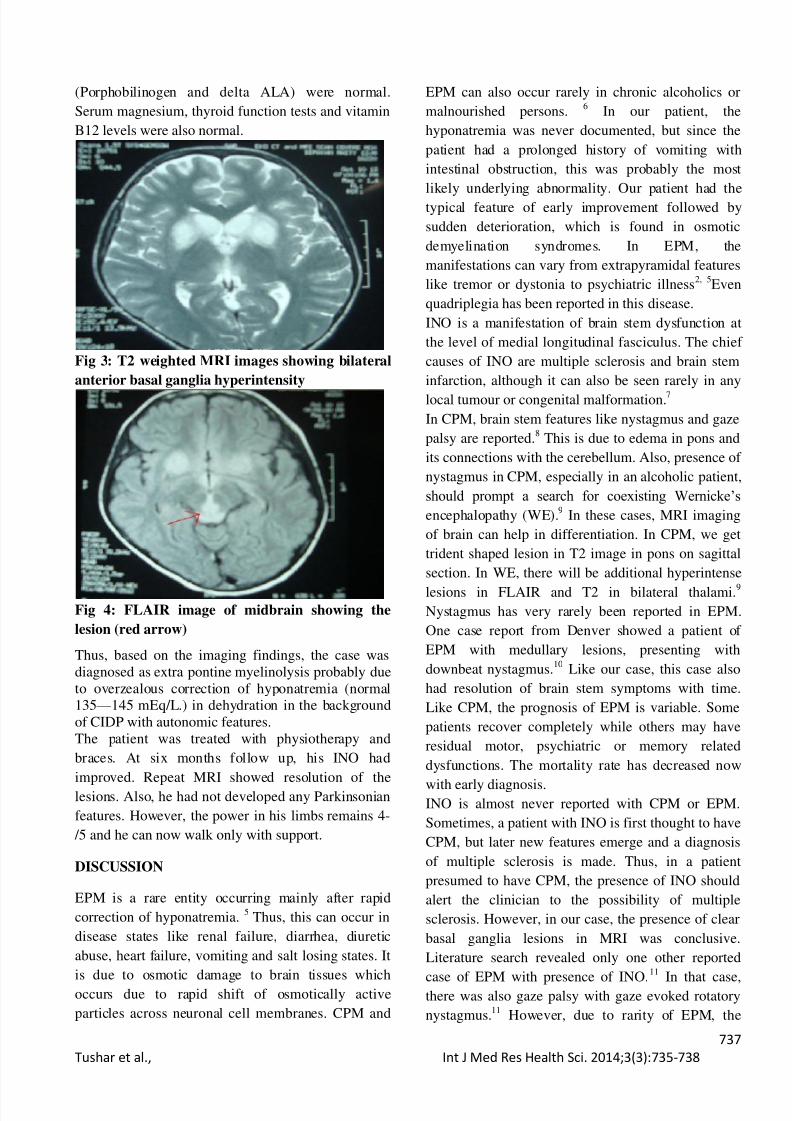
Further examination revealed bilateral ptosis (fig. 2)
with bilateral internuclear ophthalmoplegia. Pupillary
reactions were normal and there was no weakness of
any other cranial nerve. Ophthalmoscopy was normal.
The blurring of vision was probably due to nystagmus
on lateral gaze. The deep tendon jerks were all
depressed and plantar response was absent. There was
generalised hypotonia. There was no muscle
tenderness or nerve thickening. Higher functions
remained normal throughout. The pulse rate was
120/min with loss of respiratory variation and there
was marked postural hypotension (fall of SBP by 35
mm of Hg on sitting).
Past history revealed recurrent episodes of similar
abdominal distension and constipation over three
years. However, each time, he had responded to
conservative management. He had no history of
abdominal surgery or tuberculosis. Past CT scans of
abdomen were normal. Also, he had three episodes of
generalized weakness lasting for one to two months
over past three years. In one such episode, he was
investigated in detail and diagnosed as Acute
Inflammatory Demyelinating Polyneuropathy.
However, he was lost to follow up after that. His
parents said that he had some residual weakness of
the limbs from that episode and needed support while
walking.
Laboratory examinations revealed hemoglobin of
9.9G/dl with total leukocyte count of 7100/cmm
(Neutrophil 67% and lymphocyte 28%). The Platelet
count was 1.9 lakhs/mm3
with normal red cell indices
and normal ESR. Blood sugar 108mg/dl, /urea
31mg/dlcreatinine was 0.7 mg/dl respectively. Liver
function test was normal and blood electrolytesrevealed Na 135 mEq/L and K 4.2 mEq/L. Serum
calcium was also normal. After admission, the
generalised power improved to 3/5 but the INO
persisted. A CT scan of brain was normal. CSF study
revealed 8 cells/cmm with protein of >2g/dl and high
globulins. CSF ACE level was normal and TB-PCR
done from CSF was negative. Also, CSF VDRL was
negative. Blood for HIV, Hepatitis B, C and Herpes
Simplex serologies were negative. A nerve
conduction study was done which revealed decreasedamplitudes of mainly motor nerves in all four limbs
with relatively normal conduction velocity,
suggestive of axonal degeneration. Also, needle EMG
revealed spontaneous fibrillation, suggestive of
denervation. This picture, along with the CSF report
was suggestive of Chronic Inflammatory
Demyelinating Polyneuropathy. This could also
explain the autonomic dysfunction as manifested in
cardiovascular examination. Probably the intestinal
obstruction was a manifestation of autonomic
involvement in CIDP. Repeat ultrasonography of
abdomen and barium meal study did not reveal any
mechanical obstruction.
Fig 1: straight X ray abdomen showing multiple
air fluid levels
Fig 2: Face of the patient with bilateral ptosis
(Photo was taken with consent of patient)
Chest X ray was normal. MRI scan of brain was done
which revealed symmetric marked hyperintensity in
T2 images in anterior part of basal ganglia involving
putamen and anterior part of caudate nucleus (Fig. 3).
Also there was some hyperintensity in tegmental part
of midbrain involving periaqueductal grey matter
(Fig. 4). However, the T1 images were completely
normal and coronal section of pons in T2 imaging did
not reveal any signal changes also. MRI spectroscopywas done, but was reported to be essentially normal.
Blood lead levels and porphyrin levels
8/12/2019 42 Tushar Etal
http://slidepdf.com/reader/full/42-tushar-etal 3/4
737
Tushar et al., Int J Med Res Health Sci. 2014;3(3):735-738
(Porphobilinogen and delta ALA) were normal.
Serum magnesium, thyroid function tests and vitamin
B12 levels were also normal.
Fig 3: T2 weighted MRI images showing bilateral
anterior basal ganglia hyperintensity
Fig 4: FLAIR image of midbrain showing the
lesion (red arrow)
Thus, based on the imaging findings, the case was
diagnosed as extra pontine myelinolysis probably due
to overzealous correction of hyponatremia (normal
135 — 145 mEq/L.) in dehydration in the background
of CIDP with autonomic features.
The patient was treated with physiotherapy and
braces. At six months follow up, his INO had
improved. Repeat MRI showed resolution of the
lesions. Also, he had not developed any Parkinsonian
features. However, the power in his limbs remains 4-
/5 and he can now walk only with support.
DISCUSSION
EPM is a rare entity occurring mainly after rapid
correction of hyponatremia.5
Thus, this can occur in
disease states like renal failure, diarrhea, diuretic
abuse, heart failure, vomiting and salt losing states. It
is due to osmotic damage to brain tissues whichoccurs due to rapid shift of osmotically active
particles across neuronal cell membranes. CPM and
EPM can also occur rarely in chronic alcoholics or
malnourished persons.6
In our patient, the
hyponatremia was never documented, but since the
patient had a prolonged history of vomiting with
intestinal obstruction, this was probably the most
likely underlying abnormality. Our patient had the
typical feature of early improvement followed by
sudden deterioration, which is found in osmotic
demyelination syndromes. In EPM, the
manifestations can vary from extrapyramidal features
like tremor or dystonia to psychiatric illness2, 5
Even
quadriplegia has been reported in this disease.
INO is a manifestation of brain stem dysfunction at
the level of medial longitudinal fasciculus. The chief
causes of INO are multiple sclerosis and brain stem
infarction, although it can also be seen rarely in any
local tumour or congenital malformation.7
In CPM, brain stem features like nystagmus and gaze
palsy are reported.8
This is due to edema in pons and
its connections with the cerebellum. Also, presence of
nystagmus in CPM, especially in an alcoholic patient,
should prompt a search for coexisting Wernicke’s
encephalopathy (WE).9
In these cases, MRI imaging
of brain can help in differentiation. In CPM, we get
trident shaped lesion in T2 image in pons on sagittal
section. In WE, there will be additional hyperintense
lesions in FLAIR and T2 in bilateral thalami.9
Nystagmus has very rarely been reported in EPM.
One case report from Denver showed a patient of
EPM with medullary lesions, presenting with
downbeat nystagmus.10
Like our case, this case also
had resolution of brain stem symptoms with time.
Like CPM, the prognosis of EPM is variable. Some
patients recover completely while others may have
residual motor, psychiatric or memory related
dysfunctions. The mortality rate has decreased now
with early diagnosis.INO is almost never reported with CPM or EPM.
Sometimes, a patient with INO is first thought to have
CPM, but later new features emerge and a diagnosis
of multiple sclerosis is made. Thus, in a patient
presumed to have CPM, the presence of INO should
alert the clinician to the possibility of multiple
sclerosis. However, in our case, the presence of clear
basal ganglia lesions in MRI was conclusive.
Literature search revealed only one other reported
case of EPM with presence of INO.
11
In that case,there was also gaze palsy with gaze evoked rotatory
nystagmus.11
However, due to rarity of EPM, the

8/12/2019 42 Tushar Etal
http://slidepdf.com/reader/full/42-tushar-etal 4/4
738
Tushar et al., Int J Med Res Health Sci. 2014;3(3):735-738
ocular movement disorders in this disease have not
been well studied.
In INO, the lesions are usually found in paramedian
pontine tegmentum or periaqueductal region.12
In our
patient, the lesions in periaqueductal midbrain in MRI
accounted for the INO. In EPM, the typical MRI
features include symmetrical bilateral hyperintensity
in T2/FLAIR in putamen and caudate nucleus with
relative sparing of globus pallidus.1
Also T1 images
in these areas will be normal and this helps to
differentiate this condition from similar presentations
with CO poisoning. The diagnosis of EPM is mainly
clinical with added MRI findings.
Our patient recovered slowly over time. The MRI
lesions also resolved. This temporal profile of EPM
was also documented in other reported cases.8, 10
However, since our patient had underlying CIDP, he
did not regain full power of the limbs.
This is probably the second reported case of EPM
with INO. This case depicts the possible varied
presentation of osmotic demyelination syndromes
with brain stem signs.
CONCLUSION
Central nervous system osmotic demyelination is a
rare complication of electrolyte correction. It may
present with atypical features like ocular movement
disorders. Thus, clinicians should have a low
threshold for brain imaging if atypical neurological
signs appear in a patient of hyponatremia.
ACKNOWLEDGEMENT: the Principal of our
College for his guidance
Conflict of interest: Nil
REFERENCES
1. Sajith A, Ditchfield A, Katifi HA. Extrapontinemyelinolysis presenting as acute parkinsonism.
BMC Neurology 2006; 6:33
2. Seok JI, Lee DK, Kang MG, Park JH.
Neuropsychological findings of extrapontine
myelinolysis without central pontine
myelinolysis. Behav Neurol. 2007;18 :131-4
3. Panagariya A, Sureka RK, Udainiya DK.
Parkinsonism and recovery in central and
extrapontine myelinolysis. Neurol India.
2005;53:219-204. Nair SR, Ramli NM, Rahmat K, Mei-Ling ST.
Central pontine and extrapontine myelinolysis:
Diffusion weighted imaging and diffusion tensor
imaging on follow-up. Neurol India 2012;60:426-
8
5. Hsu M, Choi W. Extrapontine Myelinolysis: A
Case Report. J Emerg Crit Care Med. 2008; 19:
172-6
6. Yoon B, Shim YS, Chung SW. Central Pontine
and Extrapontine Myelinolysis After Alcohol
Withdrawal. Alcohol 2008; 43: 647 – 9
7. Obuchowska I, Mariak Z. Internuclear
ophthalmoplegia--causes, symptoms and
management. Klin Oczna. 2009;111:165-7
8. Kilinc M, Benli US, Can U. Osmotic
myelinolysis in a normonatremic patient. Acta
neurol. belg., 2002; 102: 87-9
9. Sutamnartpong P, Muengtaweepongsa S,
Kulkantrakorn K. Wernicke's encephalopathy and
central pontine myelinolysis in hyperemesis
gravidarum. J Neurosci Rural Pract. 2013; 4(1):
39 – 41
10. Neumann R, Pelak VS, Bennett J. Isolated
extrapontine myelinolysis with gaze-paretic and
downbeat nystagmus. 2000. Available online
from http://content.lib.utah.edu/cdm/ref/c
ollection/ehsl-nam/id/3922
11. Hawthorne KM, Compton C, Vaphiades MS,
Kline LB. Eye Movement Abnormalities in
Osmotic Demyelination Syndrome.2009.
Available online from http://content.lib.utah.edu/
utils/getfile/collection/ehsl-nam/id/114/filename
/68.pdf
12. Deleu D, Sokrab T, Salim K, El Siddig A, Hamad
AA. Pure isolated unilateral internuclear
ophthalmoplegia from ischemic origin: report of
a case and literature review. Acta Neurol Belg.
2005;105:214-7