
4.1 Plasma Membrane Structure and · PDF file4.1 Plasma Membrane ... • Carrier proteins...
19
8/30/2013 1 1 Chapter 04 Lecture and Animation Outline Copyright © The McGraw-Hill Companies, Inc. Permission required for reproduction or display. See separate PowerPoint slides for all figures and tables pre-inserted into PowerPoint without notes and animations. To run the animations you must be in Slideshow View. Use the buttons on the animation to play, pause, and turn audio/text on or off. Please Note: Once you have used any of the animation functions (such as Play or Pause), you must first click on the slide’s background before you can advance to the next slide. 2 4.1 Plasma Membrane Structure and Function • The plasma membrane separates the internal environment of the cell from its external environment. • It regulates the entrance and exit of molecules into and out of the cell. • The steady internal environment maintained is called homeostasis. 3 4.1 Plasma Membrane Structure and Function • Phospholipid bilayer with embedded proteins – Hydrophilic (water-loving) polar heads • Face inside and outside of cell (water present) – Hydrophobic (water-fearing) nonpolar tails • Face each other, away from water – Cholesterol (animal cells) controls excess fluidity
Transcript of 4.1 Plasma Membrane Structure and · PDF file4.1 Plasma Membrane ... • Carrier proteins...
8/30/2013
1
1
Chapter 04
Lecture and
Animation Outline
Copyright © The McGraw-Hill Companies, Inc. Permission required for reproduction or display.
See separate PowerPoint slides for all figures and tables pre-inserted into PowerPoint without notes and
animations.
To run the animations you must be in Slideshow View. Use the buttons on the animation to play, pause, and turn
audio/text on or off.
Please Note: Once you have used any of the animation functions (such as Play or Pause), you must first click on the slide’s background before you can advance to the next slide.
2
4.1 Plasma Membrane
Structure and Function
• The plasma membrane separates the internal environment of the cell from its external
environment.
• It regulates the entrance and exit of molecules
into and out of the cell.
• The steady internal environment maintained is
called homeostasis.
3
4.1 Plasma Membrane
Structure and Function
• Phospholipid bilayer with embedded proteins
– Hydrophilic (water-loving) polar heads
• Face inside and outside of cell (water present)
– Hydrophobic (water-fearing) nonpolar tails
• Face each other, away from water
– Cholesterol (animal cells) controls excess fluidity
8/30/2013
2
4
4.1 Plasma Membrane
Structure and Function
• Membrane proteins throughout membrane
may be:
– Peripheral proteins – associated with only one side of membrane
– Integral proteins – span the membrane
• Can protrude from one or both sides
• Embedded within the membrane
• Able to move laterally
5
4.1 Plasma Membrane
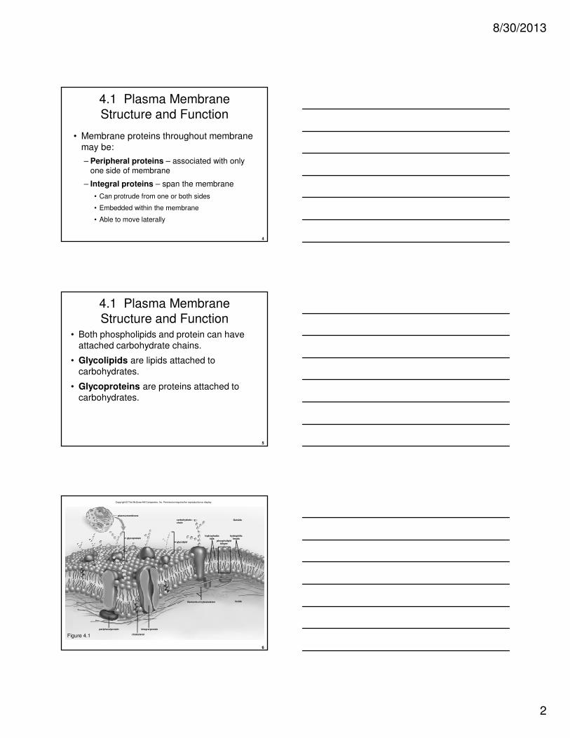
Structure and Function• Both phospholipids and protein can have
attached carbohydrate chains.
• Glycolipids are lipids attached to
carbohydrates.
• Glycoproteins are proteins attached to
carbohydrates.
6
Copyright © The McGraw-Hill Companies, Inc. Permission required for reproduction or display.
Outside
Inside
plasma membrane
glycolipid
glycoprotein
integral protein
cholesterol
peripheral protein
filaments of cytoskeleton
hydrophobictails
hydrophilicheads
phospholipidbilayer
carbohydratechain
Figure 4.1
8/30/2013
3
7
Functions of Membrane Proteins
• Channel proteins are involved in the passage
of solutes through the membrane.
– Substances simply move across the membrane.
– Some may contain a gate that must be opened in response to a signal.
• Carrier proteins allow the passage of a solute
by combing with it and help it move across the membrane.
8
Functions of Membrane Proteins
• Cell recognition proteins are glycoproteins.
– These proteins help the body recognize when it is being invaded by pathogens.
• Receptor proteins have a shape that allows
a specific molecule to bind.
– The binding causes the receptor to change
shape to initiate a cellular response.
9
Functions of Membrane Proteins
• Enzymatic proteins carry out metabolic
reactions directly.
– Example: the proteins of the electron transport chain, which carry out the final steps of aerobic respiration

8/30/2013
4
10
4.1 Plasma Membrane
Structure and Function• 5 Membrane Protein Functions
Copyright © The McGraw-Hill Companies, Inc. Permission required for reproduction or display.
Channel ProteinAllows a particularmolecule or ion tocross the plasma
membrane freely.Cystic fibrosis, aninherited disorder,is caused by afaulty chloride (Cl–)channel; a thick
mucus collects in airways and inpancreatic and liver ducts.
Carrier ProteinSelectively interactswith a specificmolecule or ion so
that it can cross the plasma membrane. The family of GLUT carriers transfers glucose in and out of the various cell types
of the body . Different carriers respond differently to blood levels of glucose.
b.a.
Figure 4.2
11
Copyright © The McGraw-Hill Companies, Inc. Permission required for reproduction or display.
c.
Cell RecognitionProtein The MHC(major histocompatibilitycomplex) glycoproteinsare different for eachperson, so organtransplants are difficultto achieve. Cells withforeign MHC glycoproteins are attacked by whiteblood cells responsiblefor immunity.
d. e.
Enzymatic ProteinCatalyzes a specificreaction. The membraneprotein, adenylatecyclase, is involved inATP metabolism. Cholerabacteria release a toxinthat interferes with theproper functioning ofadenylate cyclase, whicheventually leads tosevere diarrhea.
Receptor ProteinShaped in such a waythat a specificmolecule can bind toit. Some types ofdwarfism result notbecause the bodydoes not produceenough growthhormone, but becausethe plasma membranegrowth hormonereceptors are faultyand cannot interactwith growth hormone.
Figure 4.2
12
Copyright © The McGraw-Hill Companies, Inc. Permission required for reproduction or display.
Channel Protein
Allows a particular
molecule or ion to
cross the plasmamembrane freely .
Cystic fibrosis, an
inherited disorder,
is caused by afaulty chloride (Cl–)
channel; a thick
mucus collects in
airways and inpancreatic and
liver ducts.
Carrier Protein
Selectively interacts
with a specific
molecule or ion sothat it can cross the
plasma membrane.
The family of GLUT
carriers transfers glucose in and out of
the various cell types
of the body . Different
carriers respond differently to blood
levels of glucose.
b. c.
Cell Recognition
Protein The MHC
(major histocompatibility
complex) glycoproteinsare different for each
person, so organ
transplants are difficult
to achieve. Cells withforeign MHC
glycoproteins are
attacked by white
blood cells responsiblefor immunity.
d. e.
Enzymatic Protein
Catalyzes a specific
reaction. The membrane
protein, adenylatecyclase, is involved in
ATP metabolism. Cholera
bacteria release a toxin
that interferes with theproper functioning of
adenylate cyclase, which
eventually leads to
severe diarrhea.
Receptor Protein
Shaped in such a way
that a specific
molecule can bind toit. Some types of
dwarfism result not
because the body
does not produceenough growth
hormone, but because
the plasma membrane
growth hormonereceptors are faulty
and cannot interact
with growth hormone.
a.
Figure 4.2
8/30/2013
5
13
4.2 Permeability of the Plasma
Membrane
• The plasma membrane can regulate the passage of molecules into and out of the cell
because it is selectively permeable.
• Which molecules can freely cross the
membrane and which may require carrier proteins and/or energy depends on
– Size
– Nature of molecule – polarity, charge
14
4.2 Permeability of the Plasma
Membrane
• Small, uncharged molecules freely cross
membrane
– Examples: CO2, O2, glycerol, and alcohol
– Slip in between the hydrophilic heads and pass through hydrophobic tails
– Driven by the concentration gradient
15
4.2 Permeability of the Plasma
Membrane
• Concentration gradient
– More of a substance on one side of the
membrane
– Going “down” a concentration gradient
• From an area of higher to lower concentration
– Going “up” a concentration gradient
• From an area of lower to higher concentration
• Requires input of energy
8/30/2013
6
16
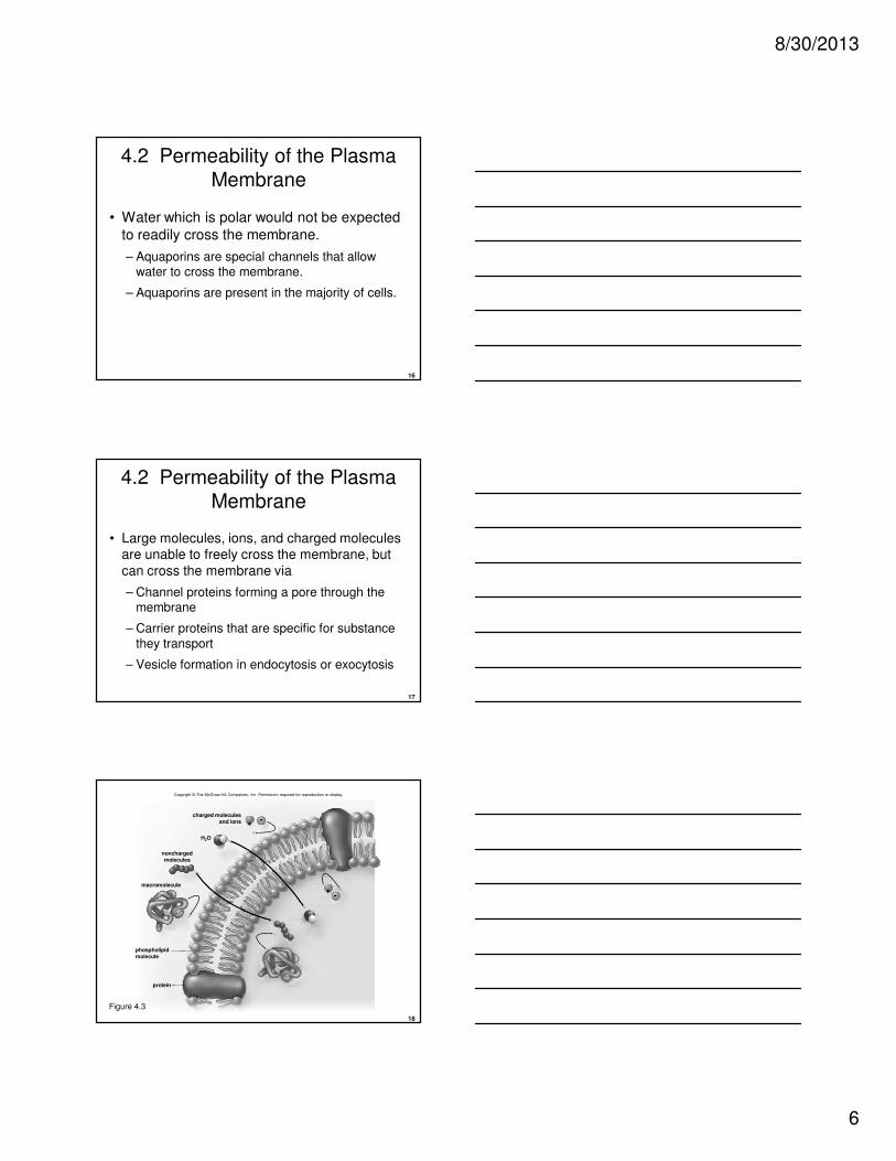
4.2 Permeability of the Plasma
Membrane
• Water which is polar would not be expected
to readily cross the membrane.
– Aquaporins are special channels that allow water to cross the membrane.
– Aquaporins are present in the majority of cells.
17
4.2 Permeability of the Plasma
Membrane
• Large molecules, ions, and charged molecules are unable to freely cross the membrane, but
can cross the membrane via
– Channel proteins forming a pore through the membrane
– Carrier proteins that are specific for substance
they transport
– Vesicle formation in endocytosis or exocytosis
18
macromolecule
H2O
protein
+
+-
-charged molecules
and ions
phospholipidmolecule
nonchargedmolecules
Copyright © The McGraw-Hill Companies, Inc. Permission required for reproduction or display.
Figure 4.3

8/30/2013
7
19
Passage of Molecules Into and
Out of the Cell
20
Diffusion and Osmosis
• Diffusion
– Movement of molecules from an area of higher
to lower concentration
• Down a concentration gradient
• Occurs until equilibrium is reached
– For example, when a crystal of dye is placed in water
the dye and water molecules move about until
equilibrium occurs
– Solution contains a solute (solid) and a solvent(liquid)
21
• Once the solute and solvent are evenly distributed, their molecules continue to move about, but there is no net movement of either one in any direction
Copyright © The McGraw-Hill Companies, Inc. Permission required for reproduction or display.
b. Diffusion of water and dye molecules c. Equal distribution of molecules resultsa. Crystal of dye is placed in the water
crystal dye
time time
8/30/2013
8
22
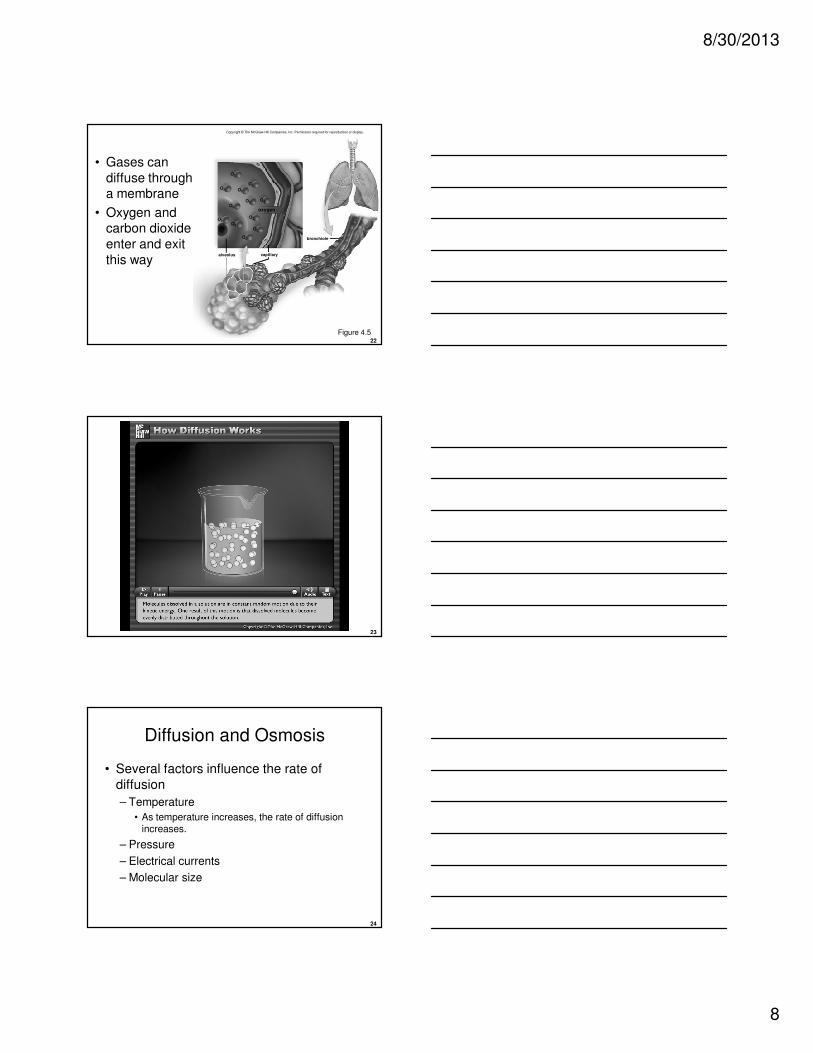
• Gases can
diffuse through a membrane
• Oxygen and carbon dioxide
enter and exit
this way
Copyright © The McGraw-Hill Companies, Inc. Permission required for reproduction or display.
capillaryalveolus
bronchiole
oxygen
O2
O2 O2
O2
O2
O2
O2O2
O2
O2
O2
O2
Figure 4.5
23
Please note that due to differing operating systems, some animations will not appear until the presentation is viewed in presentation mode (Slide Show view). You may see blank slides in the “Normal” or “Slide Sorter” views.
All animations will appear after viewing in Slide Show mode and playing each animation. Most animations will require the latest version of the Flash Player, which is available at http://get.adobe.com/flashplayer.
24
Diffusion and Osmosis
• Several factors influence the rate of
diffusion
– Temperature
• As temperature increases, the rate of diffusion increases.
– Pressure
– Electrical currents
– Molecular size

8/30/2013
9
25
Osmosis
• diffusion of water across a differentially
permeable membrane
– Diffusion always occurs from higher to lower concentration.
– Osmotic pressure is the pressure that
develops in a system due to osmosis.
• The greater the possible osmotic pressure, the more likely it is that water will diffuse in that direction.
26
• Membrane is not permeable to solute
Copyright © The McGraw-Hill Companies, Inc. Permission required for reproduction or display.
a.
10%
5%
< 10%
> 5%
solutewater
b.
c.
beaker
less water (higherpercentage of solute)
more water (lowerpercentage of solute)
more water (lowerpercentage of solute)
less water (higherpercentage of solute)
differentiallypermeablemembrane
thistletube
Figure 4.6
27
Osmosis
• Isotonic: the solute concentration is equal inside and outside of a cell
• Hypotonic: a solution has a lower solute concentration than the inside of a cell
• Hypertonic: a solution has a higher solute concentration than the inside of a cell
8/30/2013
10
28
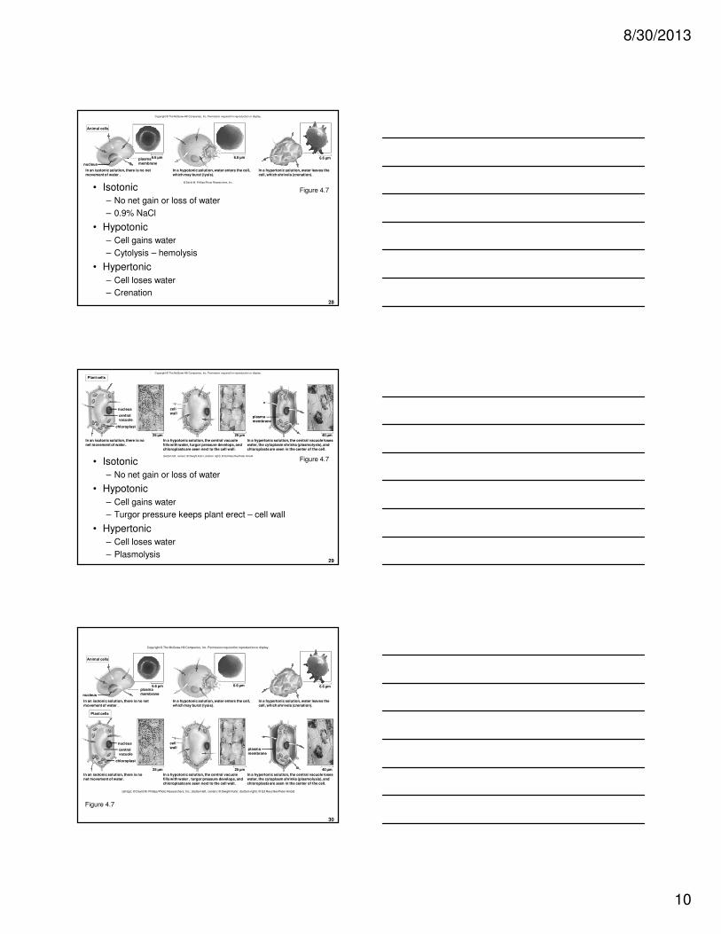
• Isotonic
– No net gain or loss of water
– 0.9% NaCl
• Hypotonic
– Cell gains water
– Cytolysis – hemolysis
• Hypertonic
– Cell loses water
– Crenation
Copyright © The McGraw-Hill Companies, Inc. Permission required for reproduction or display.
nucleus
6.6 µm 6.6 µm 6.6 µm
Animal cells
plasmamembrane
In an isotonic solution, there is no netmovement of water .
In a hypotonic solution, water enters the cell,which may burst (lysis).
In a hypertonic solution, water leaves thecell, which shrivels (crenation).
© David M. Phillips/Photo Researchers, Inc.
Figure 4.7
29
• Isotonic
– No net gain or loss of water
• Hypotonic
– Cell gains water
– Turgor pressure keeps plant erect – cell wall
• Hypertonic
– Cell loses water
– Plasmolysis
Copyright © The McGraw-Hill Companies, Inc. Permission required for reproduction or display.
chloroplast
nucleus
25 µm 40 µm25 µm
In an isotonic solution, there is nonet movement of water.
In a hypotonic solution, the central vacuolefills with water, turgor pressure develops, andchloroplasts are seen next to the cell wall.
In a hypertonic solution, the central vacuole loseswater, the cytoplasm shrinks (plasmolysis), andchloroplasts are seen in the center of the cell.
centralvacuole
cellwall
plasmamembrane
Plant cells
(bottom left, center): © Dwight Kuhn; (bottom right): © Ed Reschke/Peter Arnold
Figure 4.7
30
Copyright © The McGraw-Hill Companies, Inc. Permission required for reproduction or display.
Plant cells
chloroplast
nucleus
nucleus
6.6 µm 6.6 µm 6.6 µm
25 µm 40 µm25 µm
plasmamembrane
In an isotonic solution, there is no netmovement of water .
In a hypotonic solution, water enters the cell,which may burst (lysis).
In a hypertonic solution, water leaves thecell, which shrivels (crenation).
In an isotonic solution, there is nonet movement of water.
In a hypotonic solution, the central vacuolefills with water , turgor pressure develops, andchloroplasts are seen next to the cell wall.
In a hypertonic solution, the central vacuole loseswater, the cytoplasm shrinks (plasmolysis), andchloroplasts are seen in the center of the cell.
centralvacuole
cellwall plasma
membrane
Animal cells
(all top): © David M. Phillips/Photo Researchers, Inc.; (bottom left, center): © Dwight Kuhn; (bottom right): © Ed Reschke/Peter Arnold
Figure 4.7
8/30/2013
11
31
Transport by Carrier Proteins
• The plasma membrane impedes the passage of all but few substances.
• Substances enter or exit cells because of carrier proteins.
• Carrier proteins are specific.
– Combine with a molecule or ion to be transported across the membrane
– Change shape to move molecules across membranes
32
Transport by Carrier Proteins
• Carrier proteins are required for
– Facilitated Transport
– Active Transport
33
Facilitated Transport
• Facilitated transport explains the passage
of molecules such as glucose or amino acids.
– Neither molecule is lipid-soluble.
– Reversible combination and transport occurs.
– Like diffusion, ATP is not required because molecules are transported down their concentration gradient.

8/30/2013
12
34
Facilitated Transport
• Small molecules that are not lipid-soluble
• Molecules follow the concentration gradient
• Energy is not required
Inside
plasma
membranecarrier
protein
solute
Outside Figure 4.8
35
Active Transport
• Active Transport
– Molecules or ions combine with carrier proteins.
• Often called pumps
– Molecules move against the concentration
gradient
• Entering or leaving cell
• Accumulate either inside or outside the cell
– Energy and carrier proteins are required.
• Usually ATP is used
36
Active Transport
• Proteins in active transport are referred to as pumps.
• Proteins use energy to move molecules against the concentration gradient.
• Na+/K+ pump is especially important for nerve and muscle cells –it moves Na+ out and K+ into
cells.
• The carrier changes shape after phosphate
attaches, and then again after it detaches.

8/30/2013
13
37
Sodium-Potassium PumpCopyright © The McGraw-Hill Companies, Inc. Permission required for reproduction or display.
K+
Inside
carrierprotein
OutsideK+
K+
K+
1. Carrier has a shape that allowsit to take up 3 Na+.
Figure 4.9
38
Copyright © The McGraw-Hill Companies, Inc. Permission required for reproduction or display.
K+
P
ADPATP
K+ K+
K+
2. ATP is split, and phosphategroup attaches to carrier.
Figure 4.9
39
Copyright © The McGraw-Hill Companies, Inc. Permission required for reproduction or display.
K+
K+K+
K+
P
3. Change in shape results andcauses carrier to release 3 Na+
outside the cell.
Figure 4.9
8/30/2013
14
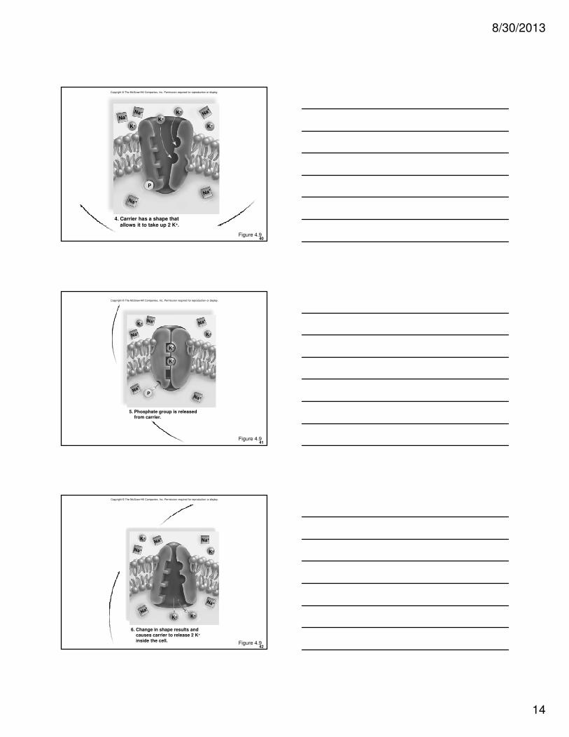
40
K+
K+
K+
K+
P
4. Carrier has a shape thatallows it to take up 2 K+.
Copyright © The McGraw-Hill Companies, Inc. Permission required for reproduction or display.
Figure 4.9
41
Copyright © The McGraw-Hill Companies, Inc. Permission required for reproduction or display.
K+
K+
K+
K+
P
5. Phosphate group is releasedfrom carrier.
Figure 4.9
42
Copyright © The McGraw-Hill Companies, Inc. Permission required for reproduction or display.
K+
K+
K+K+
6. Change in shape results andcauses carrier to release 2 K+
inside the cell. Figure 4.9
8/30/2013
15
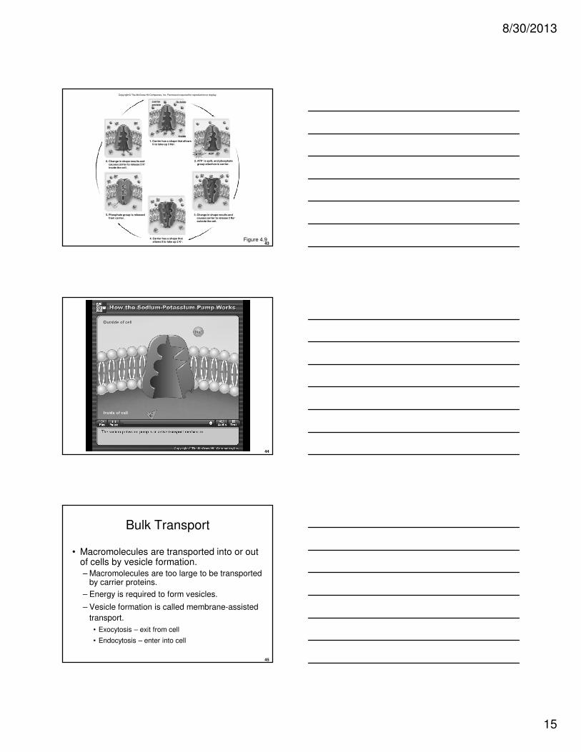
43
K+
K+
K+
K+
K+
K+K+
K+
K +
K+
K+
K+
K+
K+
K+
K+
K+K+
P
P
P
P
Inside
6. Change in shape results and
causes carrier to release 2 K+
inside the cell.
carrier
proteinOutsideK+
K+
K+
ADPATP
K+ K+
K+
3. Change in shape results and
causes carrier to release 3 Na+
outside the cell.
2. ATP is split, and phosphate
group attaches to carrier.
4. Carrier has a shape that
allows it to take up 2 K+.
5. Phosphate group is released
from carrier.
1. Carrier has a shape that allows
it to take up 3 Na+.
Copyright © The McGraw-Hill Companies, Inc. Permission required for reproduction or display.
Figure 4.9
44
Please note that due to differing operating systems, some animations will not appear until the presentation is viewed in presentation mode (Slide Show view). You may see blank slides in the “Normal” or “Slide Sorter” views.
All animations will appear after viewing in Slide Show mode and playing each animation. Most animations will require the latest version of the Flash Player, which is available at http://get.adobe.com/flashplayer.
45
Bulk Transport
• Macromolecules are transported into or out of cells by vesicle formation.– Macromolecules are too large to be transported
by carrier proteins.
– Energy is required to form vesicles.
– Vesicle formation is called membrane-assisted
transport.
• Exocytosis – exit from cell
• Endocytosis – enter into cell

8/30/2013
16
46
Exocytosis
– The vesicle fuses with plasma membrane as secretion occurs.
– The vesicle membrane becomes part of plasma
membrane.
– Cells of particular organs are specialized to produce and export molecules.
• Pancreatic cells release insulin or enzymes.
• Anterior pituitary cells release growth hormone.
47
Copyright © The McGraw-Hill Companies, Inc. Permission required for reproduction or display.
plasma membrane
Inside
Outside
secretoryvesicle
Figure 4.10
48
Endocytosis
– Cells take in substances by vesicle formation.
• Part of the plasma membrane invaginates to envelop the substance.
• The membrane then pinches off to form an intracellular vesicle.
– Three types of endocytosis
• Phagocytosis
• Pinocytosis
• Receptor-mediated endocytosis
8/30/2013
17
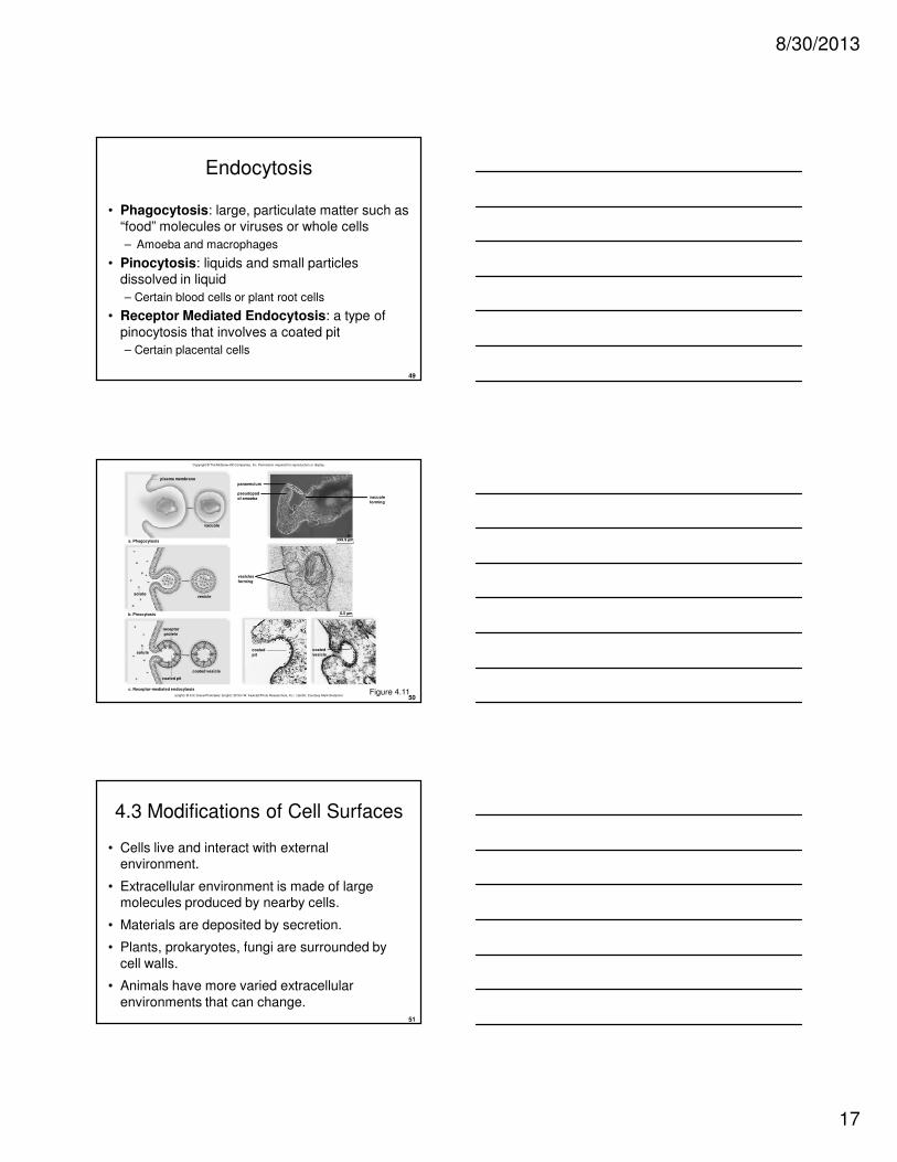
49
Endocytosis
• Phagocytosis: large, particulate matter such as “food” molecules or viruses or whole cells
– Amoeba and macrophages
• Pinocytosis: liquids and small particles dissolved in liquid
– Certain blood cells or plant root cells
• Receptor Mediated Endocytosis: a type of
pinocytosis that involves a coated pit
– Certain placental cells
50
Copyright © The McGraw-Hill Companies, Inc. Permission required for reproduction or display.
paramecium
solute
solute
a. Phagocytosis
b. Pinocytosis
vacuole
coated vesicle
plasma membrane
coated pit
c. Receptor-mediated endocytosis
399.9 µm
vesicle
vacuole
forming
pseudopod
of amoeba
0.5 µm
vesicles
forming
coated
vesiclecoated
pit
receptor
protein
a(right): © Eric Grave/Phototake; b(right): © Don W. Fawcett/Photo Researchers, Inc.; c(both): Courtesy Mark BretscherFigure 4.11
51
4.3 Modifications of Cell Surfaces
• Cells live and interact with external environment.
• Extracellular environment is made of large molecules produced by nearby cells.
• Materials are deposited by secretion.
• Plants, prokaryotes, fungi are surrounded by
cell walls.
• Animals have more varied extracellular
environments that can change.
8/30/2013
18
52
Cell Surfaces in Animals
• Animal cells have two different types of cell
surfaces.
– Extracellular matrix outside of cells
– Junctions that occur between cells
• Both can associate with the cytoskeleton and contribute to cell-to-cell communication
53
Extracellular Matrix
• A meshwork of proteins and polysaccharides closely associated with cells that produced them
• Common structural proteins in ECM
– Collagen resists stretching
– Elastin provide resilience to ECM
– Fibronectin is an adhesive protein that links integrin
54
Extracellular Matrix
• Polysaccharides made of amino sugars in
ECM attach to proteins called proteoglycans
– Proteoglycans attach to a long, centrally placed polysaccharide.
• Resist compression of ECM
• Assist cell signaling by regulating the passage of molecules through ECM to plasma membrane

8/30/2013
19
55
Junctions Between Cells
• Cell surfaces in certain tissues of animals
– Junctions Between Cells
• Adhesion Junctions
– Intercellular filaments between cells
• Tight Junctions
– Form impermeable barriers between cells
• Gap Junctions
– Plasma membrane channels are joined (allows
communication)
56
Plant Cell Walls
• All plant cells have a cell wall.
– It contains cellulose as the main component.
– Pectins allow the walls to stretch as cells grow.
– Noncellulose polysaccharides harden the wall as the cell matures.
– Pectin is abundant in the middle lamella, a
layer of adhesive substances that holds cells together.
57
Plant Cell Walls
• Plasmodesmata are narrow channels that penetrate the cell wall to connect adjacent cells.
• Each channel contains a strand of cytoplasm.
• Cytoplasm allows exchange of materials between cells.
– Only water and small solutes pass freely.
• Cytoplasm connects all the cells within a plant.


![Plasma Membrane [7.2] Goals: Understand the concept of homeostasis in relation to the plasma membrane Demonstrate and understand how the plasma membrane.](https://static.fdocuments.us/doc/165x107/5697c01d1a28abf838cd0a9a/plasma-membrane-72-goals-understand-the-concept-of-homeostasis-in-relation.jpg)


