
3enterococcus
13
308 Crit Rev Oral Biol Med 15(5):308-320 (2004) Introduction E nterococci inhabit the gastrointestinal tract, the oral cavity, and the vagina in humans as normal commensals. They can cause a wide variety of diseases in humans, infecting the urinary tract, bloodstream, endocardium, abdomen, biliary tract, burn wounds, and indwelling foreign devices (Jett et al., 1994). Enterococci now rank among the top three nosocomial bacterial pathogens (Richards et al., 2000; Wisplinghoff et al., 2003), and strains resistant to currently available antibiotics pose real therapeutic difficulties (Hunt, 1998). Up to 90% of enterococcal infections in humans are caused by Enterococcus faecalis. The majority of the remainder is caused by Enterococcus faecium, and infections with the other species are quite rare (Jett et al., 1994). Enterococci have also been impli- cated in endodontic infections. Although they make up only a small proportion of the initial flora of untreated teeth with necrotic pulps (Sundqvist, 1992), enterococci, particularly E. faecalis, have been frequently found in obturated root canals exhibiting signs of chronic apical periodontitis, isolated in 23- 70% of the positive cultures (Engström, 1964; Möller, 1966; Molander et al., 1998; Sundqvist et al., 1998; Peciuliene et al., 2000; Hancock et al., 2001) and often occur in monoculture (Sundqvist et al., 1998; Dahlén et al., 2000; Peciuliene et al., 2000; Hancock et al., 2001). Moreover, E. faecalis was among a group of bacteria cultured from periapical lesions refractory to endodontic treatment (Sunde et al., 2002). Enterococci can withstand harsh environmental condi- tions. As originally defined by Sherman (1937), enterococci can grow at 10°C and 45°C, at pH 9.6, in 6.5% NaCl broth, and sur- vive at 60°C for 30 minutes. E. faecalis can adapt to adverse con- ditions: Following pre-exposure to sublethal stress conditions, E. faecalis becomes less sensitive to normally lethal levels of sodium dodecyl sulfate, bile salts, hyperosmolarity, heat, ethanol, hydrogen peroxide, acidity, and alkalinity; further- more, 'cross-protection' is pronounced against diverse chal- lenges (Flahaut et al., 1996a,b,c, 1997). Starving E. faecalis cells maintain their viability for extended periods and become resis- tant to UV irradiation, heat, sodium hypochlorite, hydrogen peroxide, ethanol, and acid (Giard et al., 1996; Hartke et al., 1998). E. faecalis, moreover, can enter the viable but non-cul- tivable (VBNC) state, a survival mechanism adopted by a group of bacteria when exposed to environmental stress, and resuscitate upon returning to favorable conditions (Lleò et al., 2001). The ability of E. faecalis to tolerate or adapt to harsh envi- ronmental conditions may act as an advantage over other species. It may explain its survival in root canal infections, where nutrients are scarce and there are limited means of escape from root canal medicaments. In in vitro studies, E. faecalis has been shown to invade dentinal tubules (Akpata and Blechman, 1982; Haapasalo and Ørstavik, 1987; Ørstavik and Haapasalo, 1990; Love, 2001), whereas not all bacteria have this ability (Akpata and Blechman, 1982; Perez et al., 1993). In animal studies, where pure cultures of various bacteria were inoculated separately into root canals, E. faecalis, unlike others, was found to colonize the root canal in most cases and to survive without the support of other bacteria (Fabricius et al., 1982; Sobrinho et al., 1998). E. faecalis is resistant to the antimicrobial effects of calcium hydroxide (Byström et al., 1985; Haapasalo and Ørstavik, 1987; Ørstavik and Haapasalo, 1990; Distel et al., 2002), probably partly due to an effective pro- ton pump mechanism which maintains optimal cytoplasmic pH VIRULENCE F ACTORS OF ENTEROCOCCUS FAECALIS: RELATIONSHIP TO ENDODONTIC DISEASE Güven Kayaoglu* Gazi University, Faculty of Dentistry, Department of Endodontics and Conservative Treatment, 82. Sokak 06510 Emek, Ankara, Turkey; *corresponding author, [email protected] Dag Ørstavik NIOM, Scandinavian Institute of Dental Materials, Haslum, Norway ABSTRACT: Enterococcus faecalis is a micro-organism that can survive extreme challenges. Its pathogenicity ranges from life- threatening diseases in compromised individuals to less severe conditions, such as infection of obturated root canals with chronic apical periodontitis. In the latter situation, the infecting organisms are partly shielded from the defense mechanisms of the body. In this article, we review the virulence factors of E. faecalis that may be related to endodontic infection and the peri- radicular inflammatory response. The most-cited virulence factors are aggregation substance, surface adhesins, sex pheromones, lipoteichoic acid, extracellular superoxide production, the lytic enzymes gelatinase and hyaluronidase, and the toxin cytolysin. Each of them may be associated with various stages of an endodontic infection as well as with periapical inflam- mation. While some products of the bacterium may be directly linked to damage of the periradicular tissues, a large part of the tissue damage is probably mediated by the host response to the bacterium and its products. Key words: Enterococcus faecalis, virulence factors, endodontic infection, apical periodontitis.
-
Upload
alfredo-nicolas-a -
Category
Documents
-
view
17 -
download
0
Transcript of 3enterococcus

308 Crit Rev Oral Biol Med 15(5):308-320 (2004)
Introduction
Enterococci inhabit the gastrointestinal tract, the oral cavity,and the vagina in humans as normal commensals. They
can cause a wide variety of diseases in humans, infecting theurinary tract, bloodstream, endocardium, abdomen, biliarytract, burn wounds, and indwelling foreign devices (Jett et al.,1994). Enterococci now rank among the top three nosocomialbacterial pathogens (Richards et al., 2000; Wisplinghoff et al.,2003), and strains resistant to currently available antibioticspose real therapeutic difficulties (Hunt, 1998). Up to 90% ofenterococcal infections in humans are caused by Enterococcusfaecalis. The majority of the remainder is caused byEnterococcus faecium, and infections with the other species arequite rare (Jett et al., 1994). Enterococci have also been impli-cated in endodontic infections. Although they make up only asmall proportion of the initial flora of untreated teeth withnecrotic pulps (Sundqvist, 1992), enterococci, particularly E.faecalis, have been frequently found in obturated root canalsexhibiting signs of chronic apical periodontitis, isolated in 23-70% of the positive cultures (Engström, 1964; Möller, 1966;Molander et al., 1998; Sundqvist et al., 1998; Peciuliene et al.,2000; Hancock et al., 2001) and often occur in monoculture(Sundqvist et al., 1998; Dahlén et al., 2000; Peciuliene et al.,2000; Hancock et al., 2001). Moreover, E. faecalis was among agroup of bacteria cultured from periapical lesions refractory toendodontic treatment (Sunde et al., 2002).
Enterococci can withstand harsh environmental condi-tions. As originally defined by Sherman (1937), enterococci cangrow at 10°C and 45°C, at pH 9.6, in 6.5% NaCl broth, and sur-vive at 60°C for 30 minutes. E. faecalis can adapt to adverse con-
ditions: Following pre-exposure to sublethal stress conditions,E. faecalis becomes less sensitive to normally lethal levels ofsodium dodecyl sulfate, bile salts, hyperosmolarity, heat,ethanol, hydrogen peroxide, acidity, and alkalinity; further-more, 'cross-protection' is pronounced against diverse chal-lenges (Flahaut et al., 1996a,b,c, 1997). Starving E. faecalis cellsmaintain their viability for extended periods and become resis-tant to UV irradiation, heat, sodium hypochlorite, hydrogenperoxide, ethanol, and acid (Giard et al., 1996; Hartke et al.,1998). E. faecalis, moreover, can enter the viable but non-cul-tivable (VBNC) state, a survival mechanism adopted by agroup of bacteria when exposed to environmental stress, andresuscitate upon returning to favorable conditions (Lleò et al.,2001). The ability of E. faecalis to tolerate or adapt to harsh envi-ronmental conditions may act as an advantage over otherspecies. It may explain its survival in root canal infections,where nutrients are scarce and there are limited means ofescape from root canal medicaments.
In in vitro studies, E. faecalis has been shown to invadedentinal tubules (Akpata and Blechman, 1982; Haapasalo andØrstavik, 1987; Ørstavik and Haapasalo, 1990; Love, 2001),whereas not all bacteria have this ability (Akpata and Blechman,1982; Perez et al., 1993). In animal studies, where pure culturesof various bacteria were inoculated separately into root canals,E. faecalis, unlike others, was found to colonize the root canal inmost cases and to survive without the support of other bacteria(Fabricius et al., 1982; Sobrinho et al., 1998). E. faecalis is resistantto the antimicrobial effects of calcium hydroxide (Byström et al.,1985; Haapasalo and Ørstavik, 1987; Ørstavik and Haapasalo,1990; Distel et al., 2002), probably partly due to an effective pro-ton pump mechanism which maintains optimal cytoplasmic pH
VIRULENCE FACTORS OF ENTEROCOCCUS FAECALIS:RELATIONSHIP TO ENDODONTIC DISEASE
Güven Kayaoglu*
Gazi University, Faculty of Dentistry, Department of Endodontics and Conservative Treatment, 82. Sokak 06510 Emek, Ankara, Turkey; *corresponding author, [email protected]
Dag Ørstavik
NIOM, Scandinavian Institute of Dental Materials, Haslum, Norway
ABSTRACT: Enterococcus faecalis is a micro-organism that can survive extreme challenges. Its pathogenicity ranges from life-threatening diseases in compromised individuals to less severe conditions, such as infection of obturated root canals withchronic apical periodontitis. In the latter situation, the infecting organisms are partly shielded from the defense mechanisms ofthe body. In this article, we review the virulence factors of E. faecalis that may be related to endodontic infection and the peri-radicular inflammatory response. The most-cited virulence factors are aggregation substance, surface adhesins, sexpheromones, lipoteichoic acid, extracellular superoxide production, the lytic enzymes gelatinase and hyaluronidase, and thetoxin cytolysin. Each of them may be associated with various stages of an endodontic infection as well as with periapical inflam-mation. While some products of the bacterium may be directly linked to damage of the periradicular tissues, a large part of thetissue damage is probably mediated by the host response to the bacterium and its products.
Key words: Enterococcus faecalis, virulence factors, endodontic infection, apical periodontitis.

levels (Evans et al., 2002). Besides, E. faecalis, intrinsically or viaacquisition, may be resistant to a wide range of antibiotics(Leclercq, 1997; Hunt, 1998), which, if used, may shift the micro-bial flora in favor of E. faecalis.
We undertook a literature search for the virulence factorsof E. faecalis, which may relate to colonization of the host, com-petition with other bacteria, resistance against defense mecha-nisms of the host, and production of pathological changesdirectly through production of toxins or indirectly throughinduction of inflammation. The factors most extensively stud-ied are: aggregation substance, surface adhesins, sexpheromones, lipoteichoic acid, extracellular superoxide, gelati-nase, hyaluronidase, and cytolysin (hemolysin). Although notstrictly acting as virulence factors, AS-48 and other bacteriocinsare mentioned because of their possible contribution to thedominance of E. faecalis in persistent endodontic infections.From the data available, a model for the pathogenicity of E. fae-calis in endodontic infections has been developed (Fig.), wherethe elements of virulence factors and means of ecologicaladvantage for this organism have been integrated.
Aggregation SubstanceAggregation substance (AS) is a pheromone-responsive, plas-mid-encoded bacterial adhesin that mediates efficient contactbetween donor and recipient bacterium, facilitating plasmidexchange. While the AS is expressed by the donor cell, the bac-terial conjugation process requires that 'binding substance' (BS,the chromosomally encoded cognate ligand for AS) beexpressed on the surface of the recipient cell. A mutant strain ofE. faecalis lacking BS failed to act as a recipient in broth matingsyet retained recipient ability in filter membrane matings, whereno AS-BS interaction was necessary (Trotter and Dunny, 1990).AS is proteinaceous, appears as a hair-like structure on the cellsurface, and is incorporated into the 'older' parts of the cell wall(Wanner et al., 1989). Its expression on the cell surface may beinduced by serum (Kreft et al., 1992)
In addition to its adhesive function during the bacterial con-jugation process, AS mediates adhesion of E. faecalis to a varietyof eukaryotic cells in vitro, including renal tubular cells (Kreft etal., 1992) and intestinal epithelial cells (Olmsted et al., 1994). Thesequence analysis of the AS indicates two RGD motifs (arginine,glycine and aspartic acid, amino acids) that are thought to facili-tate adherence of the bacterium to the host cell via integrins, afamily of eukaryotic cell-surface receptors (Galli et al., 1990).However, analysis of recent data suggests that a non-RGD-dependent pathway of Asc10 (a group of AS proteins) mediatedcell internalization into enterocytes (Waters et al., 2003).
AS was also found to mediate binding to extracellularmatrix (ECM) proteins, including collagen type I. E. faecalisstrain OG1X(pAM721), constitutively expressing AS, binds tocollagen type I more than two times higher than the AS-nega-tive strain OG1X(pAM944) (Rodzinski et al., 2001). Binding tocollagen type I by bacteria may be of particular importance withrespect to endodontic infections, since this is the main organiccomponent of the dentin (Linde and Goldberg, 1993). A novelrepresentative of the AS family, Asa 373, differing in its proteinstructure from classic AS, was reported to exhibit some moder-ately conserved amino acid motifs when its database sequencewas compared with those of some other bacterial adhesins. Thegreatest similarity was with the amino acid sequence of Ag I/IIpolypeptides of oral streptococci (Muscholl-Silberhorn, 1999).The Ag I/II family of polypeptides has been found to mediate
collagen recognition in some oral streptococci, and this has beenassociated with their ability to invade dentin tubules (Love et al.,1997). However, there is no direct evidence to support a role forAsa 373 to mediate binding to collagen.
AS has been reported to promote direct, opsonin-indepen-dent binding of E. faecalis to human neutrophils via a comple-ment receptor-mediated mechanism (Vanek et al., 1999). As aconsequence of this special type of binding, E. faecalis-bearingAS was shown to be resistant to killing by human neutrophils,despite marked phagocytosis and neutrophil activation (Rakitaet al., 1999). Furthermore, both PMN-mediated extracellular
15(5):308-320 (2004) Crit Rev Oral Biol Med 309
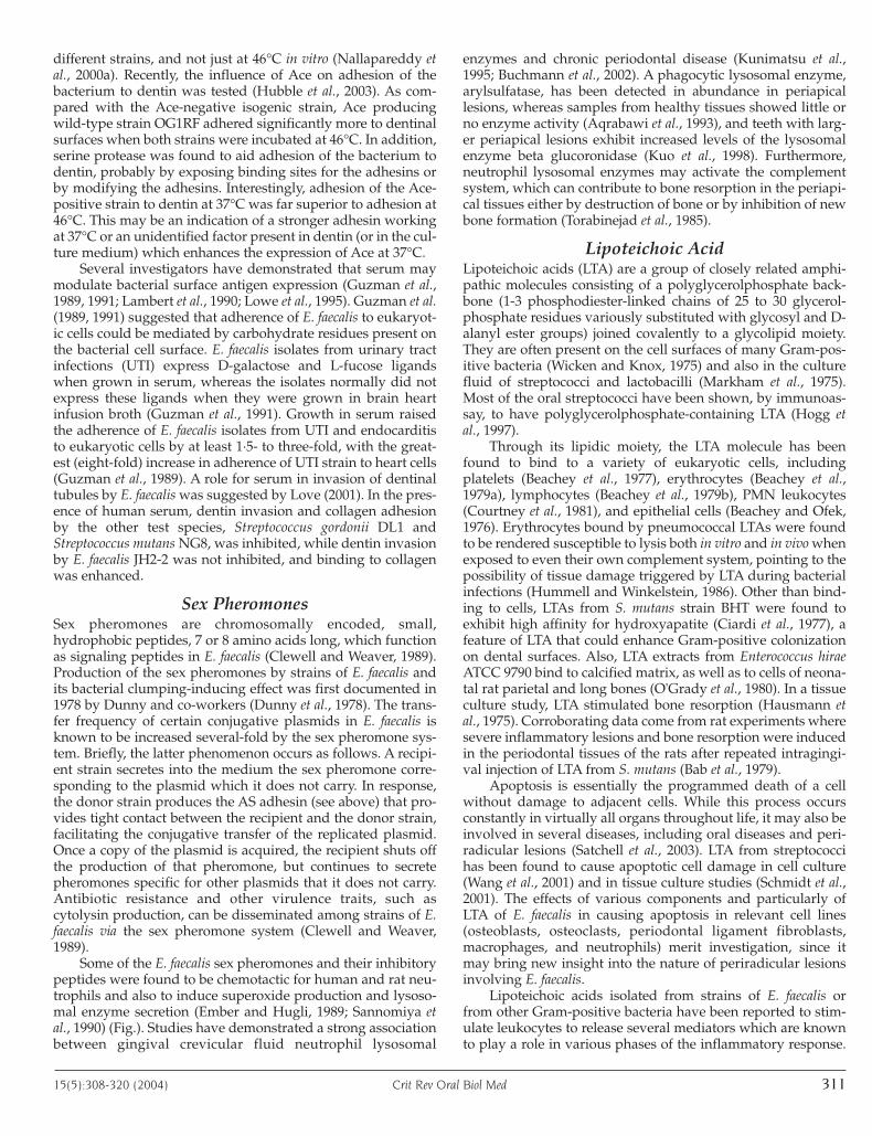
Figure. An endodontic disease model related to virulence factors of E.faecalis. The virulence factors of the bacterium inside the dentinaltubules and the root canal are released to the periradicular area,where they elicit leukocyte attraction or stimulate leukocytes to pro-duce inflammatory mediators or lytic enzymes. Some of the bacteriamay translocate to the periradicular lesion as well. The injurious viru-lence factors and leukocyte products are shown in the zone betweenthe interrupted lines. In a magnified window, the adhesion of the bac-terium to diverse elements of the dentin is depicted. Bacterial productsfighting other bacteria are also included. Note that names in blackboxes are the products of the bacterium. Abbreviations: Adh, surfaceadhesins; AS, aggregation substance; Bact, bacteriocins; BS, bindingsubstance; CP, collagen peptides; Cyl, cytolysin; Ef, Enterococcus fae-calis; Elas, elastase; Gel, gelatinase; Hya, hyaluronidase; H2O2,hydrogen peroxide; IFN-�, gamma interferon; IL, interleukin; LE, lyso-somal enzymes; LTA, lipoteichoic acid; NO, nitric oxide; O2
.-, super-oxide anion; PGE2, prostaglandin E2; SP, sex pheromones; and TNF,tumor necrosis factor. O2
.-: The dot denotes the presence of anunpaired electron, and the superscript denotes the negative charge.

superoxide production and phagosomal oxidant productionagainst the AS-expressing strains were higher than thoseagainst the control strains lacking AS (Rakita et al., 1999). Thisoxidative burst by the neutrophils may be a possible contribu-tion to tissue damage in case of infection with cells of E. faecalisexpressing AS (Fig.). AS was also reported to promote opsonin-independent adherence to and phagocytosis of E. faecalis byhuman macrophages as well, facilitating intracellular survivaltime in macrophages. However, AS suppresses the respiratoryburst triggered by macrophages, as indicated by reduced con-centrations of superoxide anion (Süßmuth et al., 2000). Theresponses to the AS-expressing E. faecalis by human neutrophilsand macrophages, therefore, appear to vary; however, it can beconcluded that AS serves as a protective factor in favor of thebacterium against the host defense mechanisms.
Superantigens are molecules produced by bacteria, virus-es, parasites, and yeasts which can induce inflammationthrough stimulation of T-lymphocytes, followed by massiverelease of inflammatory cytokines, resulting in tissue damage(Jappe, 2000). AS, in combination with BS, was reported to pos-sess superantigen activity (Schlievert et al., 1998). Cell extractsof AS- and BS-positive E. faecalis were found to induce T-cellproliferation, with subsequent release of tumor necrosis factorbeta (TNF-�) and gamma interferon (IFN-�), and to activatemacrophages to release tumor necrosis factor alpha (TNF-�)(Fig.). The stimulation of lymphocyte proliferation and the pro-duction of the cytokines were comparable with those occurringfollowing stimulation with the established staphylococcalsuperantigen toxic shock syndrome toxin-1 (TSST-1), as thepositive control (Schlievert et al., 1998). The cytokines TNF-�and TNF-� have been implicated in bone resorption(Stashenko, 1998), while IFN-� has been considered as an irre-placeable factor in host defense against infection and, at thesame time, as an inflammatory mediator (Billiau, 1996). IFN-�is well-known to potentiate respiratory burst responsiveness ofmacrophages to stimulants, resulting in increased productionof hydrogen peroxide and superoxide anion. IFN-� also stimu-lates the production of the cytotoxic agent nitric oxide (NO) bya variety of cells, including macrophages and neutrophils(Fig.), and may cause undesirable cell and tissue damage.
Results from animal studies concerning the role of AS in E.faecalis pathogenesis vary. In studies involving rabbits, AS pro-moted endocarditis (Chow et al., 1993; Schlievert et al., 1998),whereas this was not the case in a rat endocarditis model (Bertiet al., 1998), and AS did not affect the severity of the disease in arabbit endophthalmitis model (Jett et al., 1998). The issue of pro-motion of endocarditis by E. faecalis may be viewed in the gener-al context of an interaction between a bacterial adhesin and ahost target. The extracellular matrix of all mammalian tissuesconsists of glycoproteins (e.g., collagen, laminin, fibronectin) andproteoglycans that can be exploited by micro-organisms for col-onization and initiation of infection (Westerlund and Korhonen,1993). The ability of a bacterium to adhere to collagen has beenshown to play an important role in the pathogenesis of endo-carditis (Hienz et al., 1996). Since dentinal tissues (Linde andGoldberg, 1993) share common ECM proteins with the heart tis-sue (Bashey et al., 1992), a role for AS in endocarditis may alsohave relevance for endodontic infections. In epidemiologic stud-ies, AS has frequently been detected in clinical isolates (Ike et al.,1987; Elsner et al., 2000) but is rarely found among fecal isolatesfrom healthy volunteers (Coque et al., 1995), suggesting a possi-ble role for AS in human enterococcal infections.
Surface AdhesinsEnterococcal gene esp, encoding the high-molecular-weightsurface protein Esp, has been detected in abundance amongbacteremia and endocarditis isolates, but is rare in stool isolatesfrom healthy individuals (Shankar et al., 1999; Archimbaud etal., 2002). The contribution of the surface protein Esp to colo-nization and persistence of E. faecalis in urinary tract infectionshas been shown in an animal model (Shankar et al., 2001). Espis also associated with promotion of primary attachment andbiofilm formation of E. faecalis on abiotic surfaces (Toledo-Arana et al., 2001). Furthermore, biofilm formation by E. faecalishas also been observed on medicated dentinal walls, and thisform of organization could allow the bacteria to resist the bac-tericidal effect of calcium hydroxide medication in infected rootcanals (Distel et al., 2002).
The efaA gene was identified with the use of an antiserumfrom a patient with E. faecalis endocarditis (Lowe et al., 1995). Theamino acid sequence of the associated protein, EfaA, revealed 55to 60% homology to a group of streptococcal proteins known asadhesins. Thus, it was hypothesized that EfaA might be func-tioning as an adhesin in endocarditis. Production of EfaA bystrains of E. faecalis is common. In one study, the efaA gene wasdetected in all medical (blood, pus, urine, feces, hospital envi-ronment) and almost all food (milk, cheese, meat) isolates of E.faecalis (Eaton and Gasson, 2001). In an animal model, mutantswith the efaA gene showed prolonged survival, compared withE. faecalis strains bearing no efaA gene, indicating a role for theefaA gene in disease (Singh et al., 1998a). Recent studies suggestEfaA as a solute binding-protein receptor for a manganese trans-port system in E. faecalis. While manganese is required for thegrowth and survival of most micro-organisms, EfaA is stronglyexpressed in a manganese-ion-depleted environment, probablyfor the regulation of the cytoplasmic homeostasis of the cation(Low et al., 2003). The relatively low availability of manganese inserum (Krachler et al., 1999) and in dentin (Battistone et al., 1967)may induce expression of EfaA in vivo.
Studies have also focused on factors associated with thebinding of bacteria to extracellular matrix (ECM) proteins.Various strains of E. faecalis obtained from different clinicalmaterials were found to agglutinate strongly with ECM pro-teins, including collagen type I and type IV, and this was attrib-uted to the surface hydrophobicity of the cells (Zareba et al.,1997). The study by Xiao et al. (1998) indicated that a proteinwas involved in the binding of E. faecalis to ECM proteins fol-lowing growth in a stressful condition, defined as growth at46°C. The so-called 'conditional adherence' of the bacterium tocollagen, however, was impaired following pre-treatment witha protein-digesting enzyme or pre-incubation with solubleforms of collagen, or following digestion of the binding sub-strate with collagenase. The putative proteinaceous adhesin ofE. faecalis was subsequently identified as Ace, a collagen-bind-ing MSCRAMM (microbial surface component recognizingadhesive matrix molecules), which is structurally and function-ally similar to the collagen-binding protein Cna ofStaphylococcus aureus (Rich et al., 1999). It was recently shownthat the disruption of the ace gene impaired the conditionalbinding of E. faecalis to the ECM proteins (Nallapareddy et al.,2000b). Identification of Ace-specific antibodies in seraobtained from patients with enterococcal infections, and espe-cially from patients with E. faecalis endocarditis, indicated thatAce is commonly expressed in vivo during human infections by
310 Crit Rev Oral Biol Med 15(5):308-320 (2004)
different strains, and not just at 46°C in vitro (Nallapareddy etal., 2000a). Recently, the influence of Ace on adhesion of thebacterium to dentin was tested (Hubble et al., 2003). As com-pared with the Ace-negative isogenic strain, Ace producingwild-type strain OG1RF adhered significantly more to dentinalsurfaces when both strains were incubated at 46°C. In addition,serine protease was found to aid adhesion of the bacterium todentin, probably by exposing binding sites for the adhesins orby modifying the adhesins. Interestingly, adhesion of the Ace-positive strain to dentin at 37°C was far superior to adhesion at46°C. This may be an indication of a stronger adhesin workingat 37°C or an unidentified factor present in dentin (or in the cul-ture medium) which enhances the expression of Ace at 37°C.
Several investigators have demonstrated that serum maymodulate bacterial surface antigen expression (Guzman et al.,1989, 1991; Lambert et al., 1990; Lowe et al., 1995). Guzman et al.(1989, 1991) suggested that adherence of E. faecalis to eukaryot-ic cells could be mediated by carbohydrate residues present onthe bacterial cell surface. E. faecalis isolates from urinary tractinfections (UTI) express D-galactose and L-fucose ligandswhen grown in serum, whereas the isolates normally did notexpress these ligands when they were grown in brain heartinfusion broth (Guzman et al., 1991). Growth in serum raisedthe adherence of E. faecalis isolates from UTI and endocarditisto eukaryotic cells by at least 1·5- to three-fold, with the great-est (eight-fold) increase in adherence of UTI strain to heart cells(Guzman et al., 1989). A role for serum in invasion of dentinaltubules by E. faecalis was suggested by Love (2001). In the pres-ence of human serum, dentin invasion and collagen adhesionby the other test species, Streptococcus gordonii DL1 andStreptococcus mutans NG8, was inhibited, while dentin invasionby E. faecalis JH2-2 was not inhibited, and binding to collagenwas enhanced.
Sex PheromonesSex pheromones are chromosomally encoded, small,hydrophobic peptides, 7 or 8 amino acids long, which functionas signaling peptides in E. faecalis (Clewell and Weaver, 1989).Production of the sex pheromones by strains of E. faecalis andits bacterial clumping-inducing effect was first documented in1978 by Dunny and co-workers (Dunny et al., 1978). The trans-fer frequency of certain conjugative plasmids in E. faecalis isknown to be increased several-fold by the sex pheromone sys-tem. Briefly, the latter phenomenon occurs as follows. A recipi-ent strain secretes into the medium the sex pheromone corre-sponding to the plasmid which it does not carry. In response,the donor strain produces the AS adhesin (see above) that pro-vides tight contact between the recipient and the donor strain,facilitating the conjugative transfer of the replicated plasmid.Once a copy of the plasmid is acquired, the recipient shuts offthe production of that pheromone, but continues to secretepheromones specific for other plasmids that it does not carry.Antibiotic resistance and other virulence traits, such ascytolysin production, can be disseminated among strains of E.faecalis via the sex pheromone system (Clewell and Weaver,1989).
Some of the E. faecalis sex pheromones and their inhibitorypeptides were found to be chemotactic for human and rat neu-trophils and also to induce superoxide production and lysoso-mal enzyme secretion (Ember and Hugli, 1989; Sannomiya etal., 1990) (Fig.). Studies have demonstrated a strong associationbetween gingival crevicular fluid neutrophil lysosomal
enzymes and chronic periodontal disease (Kunimatsu et al.,1995; Buchmann et al., 2002). A phagocytic lysosomal enzyme,arylsulfatase, has been detected in abundance in periapicallesions, whereas samples from healthy tissues showed little orno enzyme activity (Aqrabawi et al., 1993), and teeth with larg-er periapical lesions exhibit increased levels of the lysosomalenzyme beta glucoronidase (Kuo et al., 1998). Furthermore,neutrophil lysosomal enzymes may activate the complementsystem, which can contribute to bone resorption in the periapi-cal tissues either by destruction of bone or by inhibition of newbone formation (Torabinejad et al., 1985).
Lipoteichoic AcidLipoteichoic acids (LTA) are a group of closely related amphi-pathic molecules consisting of a polyglycerolphosphate back-bone (1-3 phosphodiester-linked chains of 25 to 30 glycerol-phosphate residues variously substituted with glycosyl and D-alanyl ester groups) joined covalently to a glycolipid moiety.They are often present on the cell surfaces of many Gram-pos-itive bacteria (Wicken and Knox, 1975) and also in the culturefluid of streptococci and lactobacilli (Markham et al., 1975).Most of the oral streptococci have been shown, by immunoas-say, to have polyglycerolphosphate-containing LTA (Hogg etal., 1997).
Through its lipidic moiety, the LTA molecule has beenfound to bind to a variety of eukaryotic cells, includingplatelets (Beachey et al., 1977), erythrocytes (Beachey et al.,1979a), lymphocytes (Beachey et al., 1979b), PMN leukocytes(Courtney et al., 1981), and epithelial cells (Beachey and Ofek,1976). Erythrocytes bound by pneumococcal LTAs were foundto be rendered susceptible to lysis both in vitro and in vivo whenexposed to even their own complement system, pointing to thepossibility of tissue damage triggered by LTA during bacterialinfections (Hummell and Winkelstein, 1986). Other than bind-ing to cells, LTAs from S. mutans strain BHT were found toexhibit high affinity for hydroxyapatite (Ciardi et al., 1977), afeature of LTA that could enhance Gram-positive colonizationon dental surfaces. Also, LTA extracts from Enterococcus hiraeATCC 9790 bind to calcified matrix, as well as to cells of neona-tal rat parietal and long bones (O'Grady et al., 1980). In a tissueculture study, LTA stimulated bone resorption (Hausmann etal., 1975). Corroborating data come from rat experiments wheresevere inflammatory lesions and bone resorption were inducedin the periodontal tissues of the rats after repeated intragingi-val injection of LTA from S. mutans (Bab et al., 1979).
Apoptosis is essentially the programmed death of a cellwithout damage to adjacent cells. While this process occursconstantly in virtually all organs throughout life, it may also beinvolved in several diseases, including oral diseases and peri-radicular lesions (Satchell et al., 2003). LTA from streptococcihas been found to cause apoptotic cell damage in cell culture(Wang et al., 2001) and in tissue culture studies (Schmidt et al.,2001). The effects of various components and particularly ofLTA of E. faecalis in causing apoptosis in relevant cell lines(osteoblasts, osteoclasts, periodontal ligament fibroblasts,macrophages, and neutrophils) merit investigation, since itmay bring new insight into the nature of periradicular lesionsinvolving E. faecalis.
Lipoteichoic acids isolated from strains of E. faecalis orfrom other Gram-positive bacteria have been reported to stim-ulate leukocytes to release several mediators which are knownto play a role in various phases of the inflammatory response.
15(5):308-320 (2004) Crit Rev Oral Biol Med 311
These include the release of TNF-�, interleukin 1 beta (IL-1�),interleukin 6 (IL-6) (Bhakdi et al., 1991), and interleukin 8 (IL-8)(Saetre et al., 2001) by cultured human monocytes and byhuman whole-blood leukocytes, respectively, the release ofprostaglandin E2 (PGE2) by mouse peritoneal macrophages(Card et al., 1994), the release of lysosomal enzymes by rat peri-toneal macrophages (Harrop et al., 1980), and the generation ofsuperoxide anion by human monocytes (Levy et al., 1990) (Fig.).
These factors have all been detected in periapical samples,and each has a well-known tissue-damaging (TNF-�, IL-1�, IL-6, PGE2, lysosomal enzyme, superoxide anion) or leukocyte-attracting (IL-8) property.
Change in vascular permeability is also an importantphase in the course of inflammation, since extravasation ofplasma, succeeded by diapedesis of circulating leukocytes, fol-lows an increase in vascular permeability. LTA from S. aureuswas shown to increase the vascular permeability in mice, prob-ably through production of secondary mediators such aseicosanoids, platelet-activating factor, and histamine (Wada etal., 2000). A recent study indicates that streptococcal LTA up-regulates the expression of vascular endothelial growth factor(VEGF), a potent inducer of angiogenesis, vascular permeabili-ty, and edema, in macrophages and pulp cells (Telles et al.,2003). While an increase in vascular permeability is related toacute inflammation, angiogenesis is related more to chronicinflammation.
A proper autolytic activity appears to be necessary for theefficient killing of bacteria by cell-wall antibiotics. However,LTA has been found to inhibit the autolysis of isolated walls aswell as intact cells of the former Streptococcus faecalis, nowtermed Enterococcus hirae ATCC 9790 (Cleveland et al., 1976).Compared with the parent strain, autolytic-defective mutantsof E. hirae ATCC 9790 showed increased survival after exposureto cell-wall antibiotics. Moreover, they exhibited decreasedrates of autolysis when treated with detergents, suspended inlytic buffers, or when grown in medium depleted of essentialnutrients in the presence of increased levels of cellular LTA andlipids (Shungu et al., 1979). Thus, LTA appears to be associatedwith resistance against adverse conditions and may also beinvolved in resistance against root canal medicaments appliedduring endodontic treatment. Recently, LTA of E. faecalis wasreported to be doubled in quantity during the viable but non-cultivable (VBNC) state, suggesting a role for LTA during thisperiod (Signoretto et al., 2000).
D-alanylation of the cell-wall-associated LTA could beimportant in bringing about phenotypical advantages for thebacteria. A mutant strain of Streptococcus agalactiae, deficient inthe D-alanine moiety on the LTA, was more susceptible tokilling by macrophages and neutrophils than the wild-typestrain, and exhibited decreased virulence in animal models(Poyart et al., 2003). Insertional inactivation of the gene dltD(responsible for expression of the protein that incorporates D-alanine into LTA) in Lactobacillus casei 102S resulted inenhanced antimicrobial activity of the disinfectantscetyltrimethylammonium bromide and chlorhexidine(Debabov et al., 2000). The latter is also used as an endodonticdisinfectant.
Finally, LTA has been considered as a constituent of thebinding substance of E. faecalis that acts as the receptor on therecipient cell for aggregation substance produced by the donorcell. This presumption stems from experiments where free LTAisolated from E. faecalis inhibited pheromone-induced cell
clumping by acting as a competitive inhibitor of the cellular-binding substance (Ehrenfeld et al., 1986). Therefore, LTA canalso be regarded as a molecule contributing to the virulence ofE. faecalis through the facilitation of aggregate formation andplasmid transfer.
Extracellular Superoxide ProductionSuperoxide anion is a highly reactive oxygen radical involvedin cell and tissue damage in a variety of disorders, includinginflammatory diseases. Superoxide anion and other oxygenradicals exert a destructive effect on a wide variety of biologi-cal compounds such as lipids, proteins, and nucleic acids(Cross et al., 1987). While production of superoxide by neu-trophils and other phagocytic cells is essential for the killing ofmicro-organisms, it causes tissue damage at the site of inflam-mation. An altered balance between oxygen radical productionby phagocytic cells in periapical lesions and its elimination wassuggested to contribute to periapical damage and bone loss inchronic apical periodontitis (Marton et al., 1993). Superoxideanion has also been shown to be produced by osteoclasts andinvolved in bone resorption (Key et al., 1994). Furthermore,superoxide anion may react with a precursor in plasma to gen-erate a factor that is chemotactic for neutrophils (Petrone et al.,1980) (Fig.).
In addition to production by host cells, bacteria can alsoproduce superoxide anion. Production of superoxide by a clin-ical isolate of a Streptococcus D sp. strain was lytic for erythro-cytes (Falcioni et al., 1981). Extracellular superoxide productionhas been reported to be a common trait in strains of E. faecalis.Among a total of 91 clinical and community isolates and typestrains, 87 were found to produce detectable extracellularsuperoxide anion (Huycke et al., 1996). Isolates associated withbacteremia or endocarditis produced significantly higher extra-cellular superoxide than those from the stool of healthy sub-jects (Huycke et al., 1996). In a subcutaneous model, extracellu-lar superoxide production was found to enhance the in vivosurvival of E. faecalis in a mixed infection with Bacteroides frag-ilis (Huycke and Gilmore, 1997).
GelatinaseGelatinase is an extracellular zinc-containing metalloproteinasefrom E. faecalis which was first purified and described byBleiweis and Zimmerman (1964). It can hydrolyze gelatine, col-lagen, fibrinogen, casein, hemoglobin, insulin, certain E. faecalissex-pheromone-related peptides, and some other bioactivepeptides (Mäkinen et al., 1989).
Gelatinase, as a member of the matrix metalloproteinase(MMP) family, can also be produced by a wide variety of mam-malian cells, including inflammatory cells, epithelial cells,fibroblasts, osteoclasts, etc. Acting on substrates similar tothose of the bacterial gelatinase, host gelatinase plays a role innormal physiological processes, such as regulation of forma-tion and remodeling of tissues through its extracellular matrix-degrading functions. However, unregulated MMP activity hasbeen implicated in certain pathological states, such as invasionof cancer cells, arthritis, and periodontitis. Gelatinase(Gelatinase A, MMP-2; and Gelatinase B, MMP-9) levels wereelevated in oral rinses, crevicular fluid, and whole saliva sam-ples (Mäkelä et al., 1994) and in gingival biopsy specimens(Soell et al., 2002) from periodontitis patients compared withthose in healthy subjects. Inhibition of gelatinase decreases therate of bone resorption in tissue culture experiments (Hill et al.,
312 Crit Rev Oral Biol Med 15(5):308-320 (2004)

1994) and in experimental periodontal disease models(Ramamurthy et al., 2002). Recently, host gelatinase was report-ed to be higher in inflamed pulps and periapical lesions than inhealthy tissues (Shin et al., 2002). Host gelatinase was alsoshown to have a significant effect in the degradation of dentinorganic matrix (Tjäderhane et al., 1998). Certain peptides, gen-erated as a consequence of fragmentation of collagen, attractmonocytes (Postlethwaite and Kang, 1976), macrophages, andneutrophils (Riley et al., 1988; Laskin et al., 1994) to the site ofbreakdown (Fig.). Furthermore, the collagen peptides werefound to stimulate the release of destructive reactive oxygenspecies, hydrogen peroxide and the superoxide anion, and alsothe lytic enzymes, elastase and gelatinase, by macrophages(Laskin et al., 1994) (Fig.). By analogy, collagen hydrolysis bythe gelatinase of E. faecalis may therefore play an important rolein the pathogenesis of periapical inflammation.
A zinc-containing metalloproteinase from Legionella pneu-mophila hydrolyzes similar substrates as the gelatinase of E. fae-calis, and this enzyme has been associated with disease pro-gression due to its cytotoxic and tissue-destructive potentialand its inhibitory effects on phagocytes (Dowling et al., 1992).
Another condition where E. faecalis-derived gelatinase canproduce pathological alterations may be seen in the study byGold et al. (1975), where the gelatin-liquefying strain 2SaRinduced caries in rats, whereas this was not the case with non-proteolytic strains.
Animal studies indicate increased lethality of a gelatinase-producing E. faecalis strain compared with the isogenic straindeficient in gelatinase production (Singh et al., 1998b).
In epidemiologic studies with human clinical isolates of E.faecalis (those isolated from hospitalized patients with infectionat various sites), gelatinase production was detected in 45-68%of the isolates (Coque et al., 1995; Elsner et al., 2000; Kanemitsuet al., 2001), and the gelatinase activity was higher in clinicalisolates than in fecal isolates from healthy volunteers (Coque etal., 1995).
HyaluronidaseHyaluronidase acts on hyaluronic acid (hyaluronate, hyaluro-nan) and is mainly a degradative enzyme that is associatedwith tissue damage as the consequence of its activity. It is foundwidely in nature, from mammalian cells such as spermatozoato snake venom and to parasites such as leeches and hook-worms. It is produced in high quantities by streptococci andother bacteria as well (Hynes and Walton, 2000). Hyaluron-idase depolymerizes the mucopolysaccharide moiety of con-nective tissues, and so increases bacterial invasiveness. Strainsof Streptococcus pneumoniae with low or no hyaluronidase werefound to cause brain infections in mice only when they wereinoculated together with exogenous hyaluronidase(Kostyukova et al., 1995). Hyaluronidase was shown to be crit-ical for the dissemination of Treponema pallidum, which is thecausative micro-organism of syphilis (Fitzgerald and Repesh,1987). Hyaluronidase activity is detected in culture super-natants of Streptococcus intermedius isolated from human pus,indicating its potential role in tissue degradation (Takao et al.,1997).
Another role for hyaluronidase may be to supply nutrientsfor the bacteria, since the degradation products of its targetsubstrates are disaccharides that can be transported and metab-olized intracellularly by bacteria (Hynes and Walton, 2000).Hyaluronic acid as the substrate for hyaluronidase has also
been detected in dentin (Jones and Leaver, 1974; Chardin et al.,1990). Streptococci, isolated from carious dentin, can grow inmedium containing only hyaluronic acid, suggesting that thebacteria may derive the essential carbon for their growththrough hydrolysis of the substrate (Toto et al., 1968).Production of hyaluronidase by streptococci and a strain of E.faecalis isolated from carious dentin could play a role in tissuedestruction during the caries process (Parikh et al., 1965).Bacteria isolated from infected root canals associated with api-cal periodontitis also produce hyaluronidase, and thehyaluronidase activity appears to be related to the degree(acute and subacute) of clinical symptoms (Hashioka et al.,1994).
Hyaluronidase ('the spreading factor') is considered tofacilitate the spread of bacteria as well as their toxins throughhost tissues. In addition to its own damaging effect,hyaluronidase may also pave the way for the deleterious effectsof other bacterial toxins, thus increasing the magnitude of thedamage. The presence of micro-organisms, including E. faecalis,in periapical lesions (Abou-Rass and Bogen, 1998; Sunde et al.,2002) may also be related to the activity of a degrading bacter-ial enzyme such as hyaluronidase. It may act to facilitate themigration of bacteria from the root canal into the periapicallesion. Interestingly, a large number of species reported in theaforementioned studies are capable of producinghyaluronidase. However, due to the lack of studies concerningthe role of hyaluronidase in enterococcal virulence, the contri-bution of this factor to the apical periodontitis caused by ente-rococci remains hypothetical.
CytolysinFormerly called hemolysin, cytolysin, as expressed by variousisolates of E. faecalis, is most frequently a plasmid-encodedtoxin, but it may also be chromosomally encoded (Ike andClewell, 1992). Production and activation of cytolysin involvesa series of elaborate stages. The lytic factor precursors CylLL(the long subunit) and CylLS (the short subunit) are ribosoma-lly synthesized and modified post-translationally by CylM. Themodified peptides are then proteolytically cleaved and secret-ed from the cell by CylB, an ABC transporter. The secreted pep-tide subunits CylLL� and CylLS� are further cleaved and acti-vated extracellularly by CylA, a serine protease. Fully matureCylLL� and CylLS� are both required for the lysis of target cells(Haas and Gilmore, 1999). The cytolysin-producing bacteriumitself is protected from lysis by the cylI gene product, throughunknown mechanisms (Coburn et al., 1999).
Among the target cells of cytolysin are the erythrocytes(Basinger and Jackson, 1968; Miyazaki et al., 1993), PMNs andmacrophages (Miyazaki et al., 1993), and a broad range ofGram-positive, but not Gram-negative, organisms (Jackson,1971; Jett and Gilmore, 1990). It has been hypothesized that ifthe bacteriocin effect of cytolysin of E. faecalis favors coloniza-tion of the Gram-negatives, there could be a shift to a bacterialflora usually associated with periodontal disease (Jett andGilmore, 1990).
Recent studies investigated the influence of environmentalfactors on the expression of cytolysin genes. In one study, aquorum-sensing mechanism for production of cytolysin wasidentified (Haas et al., 2002). According to this study, the prod-ucts of two regulatory genes, cylR1 and cylR2, work together torepress the transcription of the cytolysin structural genes. Assoon as the level of one of the cytolysin subunits, CylLS� (the
15(5):308-320 (2004) Crit Rev Oral Biol Med 313

fully mature form), reaches an extracellular threshold, de-repression occurs and cytolysin expression is induced. Anotherstudy suggests that the genes cylLL and cylLS, encoding thestructural subunits of cytolysin, are regulated in response tochanging oxygen conditions, and increased amounts ofcytolysin are produced under anaerobic conditions (Day et al.,2003). From an endodontic point of view, this finding is impor-tant, in that cells of E. faecalis may encounter anaerobic condi-tions in the root canal following depletion of oxygen by aer-obes. Anaerobic conditions may also prevail in the layers ofbacterial biofilms in the root canal, and E. faecalis has the capac-ity to produce biofilms (Distel et al., 2002).
Epidemiological investigations partly support a role forcytolysin in disease occurrence. Ike et al. (1987) reported thatapproximately 60% of E. faecalis clinical isolates were hemolyt-ic, in contrast to only 17% of E. faecalis isolates derived fromfecal specimens from healthy individuals. In another study,cylA occurred more frequently among bacteremia isolates thanin isolates from cases of endocarditis or from stools fromhealthy subjects (Huycke and Gilmore, 1995). In contrast, thestudy by Coque et al. (1995) did not reveal any difference incytolysin incidence among E. faecalis isolates from endocarditis,bacteremia, or stool from healthy subjects. In another study,where only 16% of E. faecalis clinical isolates producedcytolysin, the role of this protein as a main virulence factor wasconcluded to be small (Elsner et al., 2000). However, resultsfrom a recent study suggested that silent cyl genes from clinicalisolates of E. faecalis may give a negative phenotypic profile (nohemolytic activity on blood agar plates), but environmentalfactors, such as those found in the infection site, may activatethe genes (Eaton and Gasson, 2001).
Data from animal models (Ike et al., 1984; Jett et al., 1992,1995; Chow et al., 1993; Singh et al., 1998b) and a nematodemodel (Garsin et al., 2001) suggest cytolysin to be an importantvirulence factor. In a rabbit endophthalmitis experiment,antibiotic treatment against E. faecalis together with cortico-steroid therapy was effective in cases of non-cytolytic strains inpreventing visual loss as a consequence of tissue damage,whereas this therapy was useless in the case of infection withthe cytolytic strain, suggesting a pathogenic role for cytolysinin endophthalmitis (Jett et al., 1995).
AS-48AS-48 is a plasmid-encoded peptide antibiotic originally isolat-ed from E. faecalis S-48 (Martinez-Bueno et al., 1990). AS-48 hasbeen shown to exert lytic activity toward a broad range ofGram-positive and Gram-negative bacteria (Galvez et al., 1989).The mode of action of AS-48 on target cells has been thought tobe through molecular electroporation due to its high net-posi-tive charge and through induction of ion permeation, which isaccompanied by the collapse of the cytoplasmic membranepotential (Galvez et al., 1991; Gonzalez et al., 2000). Results ofpolymerase chain-reaction (PCR) investigations on indepen-dently isolated bacteriocin-producing E. faecalis strains indicat-ed that bacteriocins produced by many E. faecalis strains wereclosely related or even identical to peptide AS-48 (Joosten et al.,1997).
Other BacteriocinsIn addition to the bacteriocins 'cytolysin' and 'AS-48', Bc-48(Lopez-Lara et al., 1991), enterocin 226NWC (Villani et al., 1993),enterocin 4 (Joosten et al., 1996), enterococcin EFS2 (Maisnier-Patin et al., 1996), bacteriocin 31 (Tomita et al., 1996), bacteriocin21 (Tomita et al., 1997), enterocin EJ97 (Galvez et al., 1998), ente-rocin 1071A and enterocin 1071B (Balla et al., 2000), and ente-rocin SE-K4 (Eguchi et al., 2001) have been isolated from vari-ous strains of E. faecalis and have been reported to haveinhibitory action mainly on Gram-positive bacteria.
ConclusionFor a bacterium to be pathogenic, it must essentially be able toadhere to, grow on, and invade the host. It must then survivehost defense mechanisms, compete with other bacteria, andproduce pathological changes. With the virulence factorsdescribed above, E. faecalis appears to possess the requisites toestablish an endodontic infection and maintain an inflammato-ry response potentially detrimental to the host. The virulencefactors of E. faecalis and their functions are summarized in theTable, and a model of the endodontic disease related to the vir-ulence factors is presented in the Fig.
Upon contamination of the root canal with the bacterium,it can colonize the dentinal walls, adhering to the mineral part,
probably through LTA,and to the collagenthrough AS and othersurface adhesins (Fig.). Itmay be that the mostinteresting among thesesurface adhesins is 'Ace',which is expressed bythe bacterium under dis-ease conditions and par-ticularly under stress(Rich et al., 1999).Bacteria face a variety ofstressful conditions inthe root canal, such asnutrient deficiency, tox-ins of other bacteria, andendodontic medica-ments. These conditionsmay modulate theadhesin expression of the
314 Crit Rev Oral Biol Med 15(5):308-320 (2004)
TABLEAn Overview of the Virulence Factors of E. faecalis and Their Functions
Function Factor References
Adhesion and colonization AS Kreft et al., 1992; Rodzinski et al., 2001other surface adhesins Rich et al., 1999; Shankar et al., 2001LTA Ciardi et al., 1977
Resistance to host defense AS Rakita et al., 1999; Süßmuth et al., 2000Inhibition on other bacteria cytolysin Jett and Gilmore, 1990
AS-48 Galvez et al., 1989other bacteriocins References in the text
Tissue damage LTA Hausmann et al., 1975; Bab et al., 1979extracellular superoxide anion Key et al., 1994gelatinase Mäkinen et al., 1989; Hill et al., 1994hyaluronidase Takao et al., 1997cytolysin Jett et al., 1992
Induction of inflammation sex pheromones Sannomiya et al., 1990; Ember and Hugli, 1989LTA Bhakdi et al., 1991; Card et al., 1994

bacterium. In addition, leakage of serum into the root canal caninduce the expression of AS and other carbohydrate moieties,thereby increasing the adhesiveness of the bacterium.Adhesion to dentin and penetration along dentinal tubules byE. faecalis may serve as a means of protection from endodonticmedicaments. An example is calcium hydroxide. When calci-um hydroxide is placed in the root canal, the pH decreasessharply toward deeper dentinal zones (Tronstad et al., 1981;Nerwich et al., 1993). Thus, bacteria that have penetrated moredeeply into the dentinal tubules and established footholdsperipheral to the main root canal are at an advantage. Anothermechanism by which E. faecalis survives may be through LTA,which has been associated with resistance of the bacteriumagainst a variety of lethal conditions (Shungu et al., 1979).
Since E. faecalis suppresses the growth of other bacteriawith its cytolysin, AS-48, and other bacteriocins, the activity ofthese toxins against Gram-positive and -negative bacteria canexplain, in part, the low number of other species in persistentendodontic infections where E. faecalis is dominant. The latterfactors are not believed to be pathogenic in humans. However,along with cytolysin, they facilitate the dominance of E. faecalisin a mixed infection and serve as means to obtain ecologicaladvantages which can result in disease in man.
The root canal is hardly a nutrient-rich medium, but E. fae-calis may derive the energy it needs from the hyaluronan pres-ent in the dentin through degradation by hyaluronidase. E. fae-calis may also feed on serum components present in the fluidin the dentinal tubules. Moreover, an inadequate apical seal ofroot fillings may allow serum to flow into the root canal.Therefore, it seems that, even in a well-debrided and coronal-ly well-sealed root canal, remaining or arriving cells of E. fae-calis may still grow and utilize local sources of energy andnutrients.
Production of extracellular superoxide and release of thelytic enzymes gelatinase and hyaluronidase and the toxincytolysin by E. faecalis can cause direct damage in the dentinalas well as in the periapical tissues (Fig.). In contrast, E. faecaliscan also induce host-mediated tissue damage in the periradic-ular tissues. Since cells of E. faecalis in the dentinal tubules can-not be reached and eliminated by the cells of the host defensesystem, they may elicit a permanent provocative effect on thesecells. PMN leukocytes, lymphocytes, monocytes, andmacrophages are stimulated by a group of virulence factors ofE. faecalis, which will contribute to the periradicular damage.
It has been proposed that, since strains of E. faecalis fre-quently harbor plasmids determining antibiotic resistance,cytolysin, and/or bacteriocin, they may represent a reservoir ofgenetic information available to other bacteria in the intestine(Clewell and Weaver, 1989). This applies to the root canalmicrobiota as well. While antibiotic resistance and other viru-lence traits can be disseminated by means of the sex-pheromone-responsive plasmid transfer among the strains ofE. faecalis, gene transfer is also possible from E. faecalis to bacte-ria of other species or even of other genera through sex-pheromone-independent conjugation. So far, there is no infor-mation on whether multiple strains of E. faecalis simultaneous-ly participate in endodontic infections in utilizing the sex-pheromone-related gene transfer. However, E. faecalis frequent-ly colonizes the root canal together with bacteria of otherspecies and/or genera, and it may use the latter pathway ofgene transfer. This is associated mainly with the transfer ofantibiotic resistance genes. Thus, bacteria resistant to multiple
antibiotics can be generated within the root canals, where E. fae-calis plays a pivotal role. It has been reported that micro-orga-nisms from the root canal can be seeded into the bloodstreamduring endodontic treatment, and this has the potential tobring about serious systemic diseases such as endocarditis,brain abscesses, and septicemia, particularly for compromisedpatients (Debelian et al., 1994, 1995). Although this may be arare clinical occurrence, there are reported cases related toendodontic infections and endodontic treatment (Henig et al.,1978; Lee, 1984; Green and Haisch, 1988). In this context, bacte-ria resistant to multiple antibiotics pose particular problems.Indeed, in marginal periodontitis refractory to conventionaltreatment, an increased prevalence of bacteria resistant toantibiotics may be found (Handal et al., 2003).
It cannot be excluded that bacteria may pass through theapical foramen to the periradicular lesion during the course ofendodontic infection and elicit host responses. However, thefocus of infection is the root canal and the dentinal tubules,which are inaccessible to the elements of the host defense sys-tem. Therefore, treatment or preventive procedures shouldmainly include local, rather than systemic, means. In additionto disinfectants, physical removal of cells of E. faecalis throughdebridement of the root canal remains essential, since rem-nants containing LTA may still sustain the inflammation.
The use of agents blocking the expression of virulencegenes or modulating their products may find a role in futuretreatments of persistent endodontic infections with E. faecalis.For example, sensitization of the bacteria to root canal medica-ments, which are otherwise ineffective, particularly throughtargeting the LTA synthesis or d-alanylation of the LTA chain,may be possible, but a better understanding of the regulation ofthe virulence genes is necessary.
Another possible preventive measure to avoid invasion ofthe dentinal tubules by E. faecalis may be through disruption ofthe dentinal collagen, the target for the adhesins. Enzymaticmodulation is one possible way of altering the collagen.Similarly, proteinaceous bacterial adhesins may be targeted byprotein-digesting agents such as trypsin. These methods havebeen tested in vitro and resulted in decreased adherence of E.faecalis to collagen-coated surfaces (Xiao et al., 1998).
In conclusion, this review has dealt with the virulence fac-tors of E. faecalis that may enable the bacterium to establish anendodontic infection and maintain a periradicular inflamma-tion. A model of endodontic disease related to these factors hasbeen proposed. The pathogenesis of the periradicular lesionsis definitely a very complex process that may involve a largenumber of host and microbial factors (Torabinejad et al., 1985;Stashenko, 1998; Takahashi, 1998). In the present context, onlythose aspects of the immune and inflammatory events likely tooccur within the periradicular lesion in relation to the viru-lence factors of E. faecalis have been discussed. It has beenestablished that the primary periradicular lesion is a conse-quence of a mixed microbial flora rather than solely of E. fae-calis. However, in apical periodontitis that persists despite rootcanal treatment, E. faecalis is frequently the dominant, some-times the only, pathogen, suggesting that this species alone hasthe potential to maintain root canal infection and periradicularlesion. A better understanding of the role of the virulence fac-tors of E. faecalis in endodontic infections may help in thedevelopment of new strategies to prevent or to eliminate theinfection by this species, thereby improving treatment resultsin endodontics.
15(5):308-320 (2004) Crit Rev Oral Biol Med 315

AcknowledgmentThe financial support of The Research Council of Norway is gratefully
acknowledged.
REFERENCES
Abou-Rass M, Bogen G (1998). Microorganisms in closed periapi-cal lesions. Int Endod J 31:39-47.
Akpata ES, Blechman H (1982). Bacterial invasion of pulpal dentinwall in vitro. J Dent Res 61:435-438.
Aqrabawi J, Schilder H, Toselli P, Franzblau C (1993). Biochemicaland histochemical analysis of the enzyme arylsulfatase inhuman lesions of endodontic origin. J Endod 19:335-338.
Archimbaud C, Shankar N, Forestier C, Baghdayan A, Gilmore MS,Charbonne F, et al. (2002). In vitro adhesive properties and vir-ulence factors of Enterococcus faecalis strains. Res Microbiol153:75-80.
Bab IA, Sela MN, Ginsburg I, Dishon T (1979). Inflammatorylesions and bone resorption induced in the rat periodontium bylipoteichoic acid of Streptococcus mutans. Inflammation 3:345-358.
Balla E, Dicks LM, Du Toit M, Van Der Merwe MJ, Holzapfel WH(2000). Characterization and cloning of the genes encodingenterocin 1071A and enterocin 1071B, two antimicrobial pep-tides produced by Enterococcus faecalis BFE 1071. Appl EnvironMicrobiol 66:1298-1304.
Bashey RI, Donnelly M, Insinga F, Jimenez SA (1992). Growth prop-erties and biochemical characterization of collagens synthe-sized by adult rat heart fibroblasts in culture. J Mol Cell Cardiol24:691-700.
Basinger SF, Jackson RW (1968). Bacteriocin (hemolysin) ofStreptococcus zymogenes. J Bacteriol 96:1895-1902.
Battistone GC, Feldman MH, Reba RC (1967). The manganese con-tent of human enamel and dentine. Arch Oral Biol 12:1115-1122.
Beachey EH, Ofek I (1976). Epithelial cell binding of group A strep-tococci by lipoteichoic acid on fimbriae denuded of M protein.J Exp Med 143:759-771.
Beachey EH, Chiang TM, Ofek I, Kang AH (1977). Interaction oflipoteichoic acid of group A Streptococci with human platelets.Infect Immun 16:649-654.
Beachey EH, Dale JB, Simpson WA, Evans JD, Knox KW, Ofek I, etal. (1979a). Erythrocyte binding properties of streptococcallipoteichoic acids. Infect Immun 23:618-625.
Beachey EH, Dale JB, Grebe S, Ahmed A, Simpson WA, Ofek I(1979b). Lymphocyte binding and T-cell mitogenic properties ofgroup A streptococcal lipoteichoic acid. J Immunol 122:189-195.
Berti M, Candiani G, Kaufhold A, Muscholl A, Wirth R (1998). Doesaggregation substance of Enterococcus faecalis contribute todevelopment of endocarditis? Infection 26:48-53.
Bhakdi S, Klonisch T, Nuber P, Fischer W (1991). Stimulation ofmonokine production by lipoteichoic acids. Infect Immun59:4614-4620.
Billiau A (1996). Interferon-�. Biology and role in pathogenesis. AdvImmunol 62:61-130.
Bleiweis AS, Zimmerman LN (1964). Properties of proteinase fromStreptococcus faecalis var. liquefaciens. J Bacteriol 88:653-659.
Buchmann R, Hasilik A, Nunn ME, Van Dyke TE, Lange DE (2002).PMN responses in chronic periodontal disease: evaluation bygingival crevicular fluid enzymes and elastase-alpha-1-pro-teinase inhibitor complex. J Clin Periodontol 29:563-572.
Byström A, Claesson R, Sundqvist G (1985). The antibacterial effectof camphorated paramonochlorophenol, camphorated phenoland calcium hydroxide in the treatment of infected root canals.Endod Dent Traumatol 1:170-175.
Card GL, Jasuja RR, Gustafson GL (1994). Activation of arachidon-
ic acid metabolism in mouse macrophages by bacterialamphiphiles. J Leukoc Biol 56:723-728.
Chardin H, Londono I, Goldberg M (1990). Visualization of gly-cosaminoglycans in rat incisor extracellular matrix using ahyaluronidase-gold complex. Histochem J 22:588-594.
Chow JW, Thal LA, Perri MB, Vazquez JA, Donabedian SM,Clewell DB, et al. (1993). Plasmid associated hemolysin andaggregation substance production contribute to virulence inexperimental enterococcal endocarditis. Antimicrob AgentsChemother 37:2474-2477.
Ciardi JE, Rölla G, Bowen WH, Reilly JA (1977). Adsorption ofStreptococcus mutans lipoteichoic acid to hydroxyapatite. Scand JDent Res 85:387-391.
Cleveland RF, Daneo-Moore L, Wicken AJ, Shockman GD (1976).Effect of lipoteichoic acid and lipids on lysis of intact cells ofStreptococcus faecalis. J Bacteriol 127:1582-1584.
Clewell DB, Weaver KE (1989). Sex pheromones and plasmid trans-fer in Enterococcus faecalis. Plasmid 21:175-184.
Coburn PS, Hancock LE, Booth MC, Gilmore MS (1999). A novelmeans of self-protection, unrelated to toxin activation, confersimmunity to the bactericidal effects of the Enterococcus faecaliscytolysin. Infect Immun 67:3339-3347.
Coque TM, Patterson ME, Steckelberg JM, Murray BE (1995).Incidence of hemolysin, gelatinase, and aggregation substanceamong enterococci isolated from patients with endocarditis andother infections and from feces of hospitalized and community-based persons. J Infect Dis 171:1223-1229.
Courtney H, Ofek I, Simpson WA, Beachey EH (1981). Charac-terization of lipoteichoic acid binding to polymorphonuclearleukocytes of human blood. Infect Immun 32:625-631.
Cross CE, Halliwell B, Borish ET, Pryor WA, Ames BN, Saul RL, etal. (1987). Oxygen radicals and human disease. Ann Intern Med107:526-545.
Dahlén G, Samuelsson W, Molander A, Reit C (2000). Identificationand antimicrobial susceptibility of enterococci isolated from theroot canal. Oral Microbiol Immunol 15:309-312.
Day AM, Cove JH, Phillips-Jones MK (2003). Cytolysin geneexpression in Enterococcus faecalis is regulated in response toaerobiosis conditions. Mol Genet Genomics 269:31-39.
Debabov DV, Kiriukhin MY, Neuhaus FC (2000). Biosynthesis oflipoteichoic acid in Lactobacillus rhamnosus: role of DltD in D-alanylation. J Bacteriol 182:2855-2864.
Debelian GJ, Olsen I, Tronstad L (1994). Systemic diseases causedby oral microorganisms. Endod Dent Traumatol 10:57-65.
Debelian GJ, Olsen I, Tronstad L (1995). Bacteremia in conjunctionwith endodontic therapy. Endod Dent Traumatol 11:142-149.
Distel JW, Hatton JF, Gillespie MJ (2002). Biofilm formation in med-icated root canals. J Endod 28:689-693.
Dowling JN, Saha AK, Glew RH (1992). Virulence factors of thefamily Legionellaceae. Microbiol Rev 56:32-60.
Dunny GM, Brown BL, Clewell DB (1978). Induced cell aggrega-tion and mating in Streptococcus faecalis: evidence for a bacterialsex pheromone. Proc Natl Acad Sci USA 75:3479-3483.
Eaton TJ, Gasson MJ (2001). Molecular screening of Enterococcusvirulence determinants and potential for genetic exchangebetween food and medical isolates. Appl Environ Microbiol67:1628-1635.
Eguchi T, Kaminaka K, Shima J, Kawamoto S, Mori K, Choi SH, etal. (2001). Isolation and characterization of enterocin SE-K4 pro-duced by thermophilic enterococci, Enterococcus faecalis K-4.Biosci Biotechnol Biochem 65:247-253.
Ehrenfeld EE, Kessler RE, Clewell DB (1986). Identification ofpheromone-induced surface proteins in Streptococcus faecalisand evidence of a role for lipoteichoic acid in formation of mat-ing aggregates. J Bacteriol 168:6-12.
316 Crit Rev Oral Biol Med 15(5):308-320 (2004)

Elsner HA, Sobottka I, Mack D, Claussen M, Laufs R, Wirth R(2000). Virulence factors of Enterococcus faecalis and Enterococcusfaecium blood culture isolates. Eur J Clin Microbiol Infect Dis19:39-42.
Ember JA, Hugli TE (1989). Characterization of the human neu-trophil response to sex pheromones from Streptococcus faecalis.Am J Pathol 134:797-805.
Engström B (1964). The significance of enterococci in root canaltreatment. Odontol Revy 15:87-106.
Evans M, Davies JK, Sundqvist G, Figdor D (2002). Mechanismsinvolved in the resistance of Enterococcus faecalis to calciumhydroxide. Int Endod J 35:221-228.
Fabricius L, Dahlén G, Holm SE, Möller ÅJ (1982). Influence ofcombinations of oral bacteria on periapical tissues of monkeys.Scand J Dent Res 90:200-206.
Falcioni GC, Coderoni S, Tedeschi GG, Brunori M, Rotilio G (1981).Red cell lysis induced by microorganisms as a cause of super-oxide- and hydrogen peroxide-dependent hemolysis mediatedby oxyhemoglobin. Biochim Biophys Acta 678:437-441.
Fitzgerald TJ, Repesh LA (1987). The hyaluronidase associated withTreponema pallidum facilitates treponemal dissemination. InfectImmun 55:1023-1028.
Flahaut S, Hartke A, Giard JC, Benachour A, Boutibonnes P,Auffray Y (1996a). Relationship between stress responsetowards bile salts, acid and heat treatment in Enterococcus fae-calis. FEMS Microbiol Lett 138:49-54.
Flahaut S, Frere J, Boutibonnes P, Auffray Y (1996b). Comparison ofthe bile salts and sodium dodecyl sulfate stress responses inEnterococcus faecalis. Appl Environ Microbiol 62:2416-2420.
Flahaut S, Benachour A, Giard JC, Boutibonnes P, Auffray Y(1996c). Defense against lethal treatments and de novo proteinsynthesis induced by NaCl in Enterococcus faecalis ATCC 19433.Arch Microbiol 165:317-324.
Flahaut S, Hartke A, Giard JC, Auffray Y (1997). Alkaline stressresponse in Enterococcus faecalis: adaptation, cross protection,and changes in protein synthesis. Appl Environ Microbiol 63:812-814.
Galli D, Lottspeich F, Wirth R (1990). Sequence analysis ofEnterococcus faecalis aggregation substance encoded by the sexpheromone plasmid pAD1. Mol Microbiol 4:895-904.
Galvez A, Maqueda M, Martinez-Bueno M, Valdivia E (1989).Bactericidal and bacteriolytic action of peptide antibiotic AS-48against Gram-positive and Gram-negative bacteria and otherorganisms. Res Microbiol 140:57-68.
Galvez A, Maqueda M, Martinez-Bueno M, Valdivia E (1991).Permeation of bacterial cells, permeation of cytoplasmic andartificial membrane vesicles, and channel formation on lipidbilayers of peptide antibiotic AS-48. J Bacteriol 173:886-892.
Galvez A, Valdivia A, Abriouel H, Camafeita E, Mendez E,Martinez-Bueno M, et al. (1998). Isolation and characterizationof enterocin EJ97, a bacteriocin produced by Enterococcus faecalisEJ97. Arch Microbiol 171:59-65.
Garsin D, Sifri CD, Mylonakis E, Qin X, Singh KV, Murray BE, et al.(2001). A simple model host for identifying Gram-positive viru-lence factors. Proc Natl Acad Sci USA 98:10892-10897.
Giard JC, Hartke A, Flahaut S, Benachour A, Boutibonnes P,Auffray Y (1996). Starvation-induced multiresistance inEnterococcus faecalis JH2-2. Curr Microbiol 32:264-271.
Gold OG, Jordan HV, van Houte J (1975). The prevalence of ente-rococci in the human mouth and their pathogenicity in animalmodels. Arch Oral Biol 20:473-477.
Gonzalez C, Langdon GM, Bruix M, Galvez A, Valdivia E,Maqueda M, et al. (2000). Bacteriocin AS-48, a microbial cyclicpolypeptide structurally and functionally related to mam-malian NK-lysin. Proc Natl Acad Sci USA 97:11221-11226.
Green JG, Haisch L (1988). Infective endocarditis and antibioticprophylaxis failure following an endodontic procedure. GenDent 36:131-133.
Guzman CA, Pruzzo C, LiPira G, Calegari L (1989). Role of adher-ence in pathogenesis of Enterococcus faecalis urinary tract infec-tion and endocarditis. Infect Immun 57:1834-1838.
Guzman CA, Pruzzo C, Plate M, Guardati MC, Calegari L (1991).Serum dependent expression of Enterococcus faecalis adhesinsinvolved in the colonization of heart cells. Microb Pathog 11:399-409.
Haapasalo M, Ørstavik D (1987). In vitro infection and disinfectionof dentinal tubules. J Dent Res 66:1375-1379.
Haas W, Gilmore MS (1999). Molecular nature of a novel bacterialtoxin: the cytolysin of Enterococcus faecalis. Med MicrobiolImmunol 187:183-190.
Haas W, Shepard BD, Gilmore MS (2002). Two-component regula-tor of Enterococcus faecalis cytolysin responds to quorum-sen-sing autoinduction. Nature 415:84-87.
Hancock HH, Sigurdsson A, Trope M, Moiseiwitsch J (2001).Bacteria isolated after unsuccessful endodontic treatment in aNorth American population. Oral Surg Oral Med Oral Pathol91:579-586.
Handal T, Caugant DA, Olsen I (2003). Antibiotic resistance in bac-teria isolated from subgingival plaque in a Norwegian popula-tion with refractory marginal periodontitis. Antimicrob AgentsChemother 47:1443-1446.
Harrop PJ, O'Grady RL, Knox KW, Wicken AJ (1980). Stimulationof lysosomal enzyme release from macrophages by lipoteichoicacid. J Periodontal Res 15:492-501.
Hartke A, Giard JC, Laplace JM, Auffray Y (1998). Survival of Entero-coccus faecalis in an oligotrophic microcosm: changes in mor-phology, development of general stress resistance, and analysisof protein synthesis. Appl Environ Microbiol 64:4238-4245.
Hashioka K, Suzuki K, Yoshida T, Nakane A, Horiba N, NakamuraH (1994). Relationship between clinical symptoms and enzyme-producing bacteria isolated from infected root canals. J Endod20:75-77.
Hausmann E, Lüderitz O, Knox K, Weinfeld N (1975). Structuralrequirements for bone resorption by endotoxin and lipoteichoicacid. J Dent Res 54(B):B94-B99.
Henig EF, Derschowitz T, Shalit M, Toledo E, Tikva P, Aviv T (1978).Brain abcess [sic] following dental infection. Oral Surg Oral MedOral Pathol 45:955-958.
Hienz SA, Schennings T, Heimdahl A, Flock JI (1996). Collagenbinding of Staphylococcus aureus is a virulence factor in experi-mental endocarditis. J Infect Dis 174:83-88.
Hill PA, Murphy G, Docherty AJ, Hembry RM, Millican TA,Reynolds JJ, et al. (1994). The effects of selective inhibitors ofmatrix metalloproteinases (MMPs) on bone resorption and theidentification of MMPs and TIMP-1 in isolated osteoclasts. J CellSci 107:3055-3064.
Hogg SD, Whiley RA, De Soet JJ (1997). Occurrence of lipoteichoicacid in oral streptococci. Int J Syst Bacteriol 47:62-66.
Hubble TS, Hatton JF, Nallapareddy SR, Murray BE, Gillespie MJ(2003). Influence of Enterococcus faecalis proteases and the colla-gen-binding protein, Ace, on adhesion to dentin. Oral MicrobiolImmunol 18:121-126.
Hummell DS, Winkelstein JA (1986). Bacterial lipoteichoic acid sen-sitizes host cells for destruction by autologous complement. JClin Invest 77:1533-1538.
Hunt CP (1998). The emergence of enterococci as a cause of noso-comial infection. Br J Biomed Sci 55:149-156.
Huycke MM, Gilmore MS (1995). Frequency of aggregation sub-stance and cytolysin genes among enterococcal endocarditisisolates. Plasmid 34:152-156.
15(5):308-320 (2004) Crit Rev Oral Biol Med 317

Huycke MM, Gilmore MS (1997). In vivo survival of Enterococcusfaecalis is enhanced by extracellular superoxide production. AdvExp Med Biol 418:781-784.
Huycke MM, Joyce W, Wack MF (1996). Augmented production ofextracellular superoxide by blood isolates of Enterococcus fae-calis. J Infect Dis 173:743-746.
Hynes WL, Walton SL (2000). Hyaluronidases of Gram-positivebacteria. FEMS Microbiol Lett 183:201-207.
Ike Y, Clewell DB (1992). Evidence that the hemolysin/bacteriocinphenotype of Enterococcus faecalis subsp. zymogenes can bedetermined by plasmids in different incompatibility groups aswell as by the chromosome. J Bacteriol 174:8172-8177.
Ike Y, Hashimoto H, Clewell DB (1984). Hemolysin of Streptococcusfaecalis subspecies zymogenes contributes to virulence in mice.Infect Immun 45:528-530.
Ike Y, Hashimoto H, Clewell DB (1987). High incidence ofhemolysin production by Enterococcus (Streptococcus) faecalisstrains associated with human parenteral infections. J ClinMicrobiol 25:1524-1528.
Jackson RW (1971). Bacteriolysis and inhibition of Gram-positivebacteria by components of Streptococcus zymogenes lysine. JBacteriol 105:156-159.
Jappe U (2000). Superantigens and their association with dermato-logical inflammatory diseases: facts and hypotheses. ActaDermatol Venereol 80:321-328.
Jett BD, Gilmore MS (1990). The growth-inhibitory effect of theEnterococcus faecalis bacteriocin encoded by pAD1 extends tothe oral streptococci. J Dent Res 69:1640-1645.
Jett BD, Jensen HG, Nordquist RE, Gilmore MS (1992).Contribution of the pAD1 encoded cytolysin to the severity ofexperimental Enterococcus faecalis endophthalmitis. InfectImmun 60:2445-2452.
Jett BD, Huycke MM, Gilmore MS (1994). Virulence of enterococci.Clin Microbiol Rev 7:462-478.
Jett BD, Jensen HG, Atkuri RV, Gilmore MS (1995). Evaluation oftherapeutic measures for treating endophthalmitis caused byisogenic toxin-producing and toxin-nonproducing Enterococcusfaecalis strains. Invest Ophthalmol Vis Sci 36:9-12.
Jett BD, Atkuri RV, Gilmore SM (1998). Enterococcus faecalis local-ization in experimental endophthalmitis: role of plasmid-encoded aggregation substance. Infect Immun 66:843-848.
Jones IL, Leaver AG (1974). Glycosaminoglycans of human den-tine. Calcif Tissue Res 16:37-44.
Joosten HM, Nunez M, Devreese B, Van Beeumen J, Marugg JD(1996). Purification and characterization of enterocin 4, a bacte-riocin produced by Enterococcus faecalis INIA 4. Appl EnvironMicrobiol 62:4220-4223.
Joosten HM, Rodriguez E, Nunez M (1997). PCR detection ofsequences similar to the AS-48 structural gene in bacteriocin-producing enterococci. Lett Appl Microbiol 24:40-42.
Kanemitsu K, Nishino T, Kunishima H, Okamura N, Takemura H,Yamamoto H, et al. (2001). Quantitative determination of gelati-nase activity among enterococci. J Microbiol Methods 47:11-16.
Key LL Jr, Wolf WC, Gundberg CM, Ries WL (1994). Superoxideand bone resorption. Bone 15:431-436.
Kostyukova NN, Volkova MO, Ivanova VV, Kvetnaya AS (1995). Astudy of pathogenic factors of Streptococcus pneumoniae strainscausing meningitis. FEMS Immunol Med Microbiol 10:133-137.
Krachler M, Rossipal E, Micetic-Turk D (1999). Concentrations oftrace elements in sera of newborns, young infants and adults.Biol Trace Elem Res 68:121-135.
Kreft B, Marre R, Schramm U, Wirth R (1992). Aggregation sub-stance of Enterococcus faecalis mediates adhesion to culturedrenal tubular cells. Infect Immun 60:25-30.
Kunimatsu K, Mine N, Muraoka Y, Kato I, Hase T, Aoki Y, et al.
(1995). Identification and possible function of cathepsin G ingingival crevicular fluid from chronic adult periodontitispatients and from experimental gingivitis subjects. J PeriodontalRes 30:51-57.
Kuo ML, Lamster IB, Hasselgren G (1998). Host mediators inendodontic exudates. I. Indicators of inflammation andhumoral immunity. J Endod 24:598-603.
Lambert PA, Shorrock PJ, Aitchison EJ, Domingue PA, Power ME,Costerton JW (1990). Effect of in vivo growth conditions uponexpression of surface protein antigens in Enterococcus faecalis.FEMS Microbiol Immunol 64:51-54.
Laskin DL, Soltys RA, Berg RA, Riley DJ (1994). Activation ofalveolar macrophages by native and synthetic collagen-likepolypeptides. Am J Respir Cell Mol Biol 10:58-64.
Leclercq R (1997). Enterococci acquire new kinds of resistance. ClinInfect Dis 24(1 Suppl):80S-84S.
Lee GT (1984). Septicaemia as a complication of endodontic treat-ment. J Dent 12:241-242.
Levy R, Kotb M, Nagauker O, Majumdar G, Alkan M, Ofek I, et al.(1990). Stimulation of oxidative burst in human monocytes bylipoteichoic acids. Infect Immun 58:566-568.
Linde A, Goldberg M (1993). Dentinogenesis. Crit Rev Oral Biol Med4:679-728.
Lleò MM, Bonato B, Tafi MC, Signoretto C, Boaretti M, Canepari P(2001). Resuscitation rate in different enterococcal species in theviable but non-culturable state. J Appl Microbiol 91:1095-1102.
Lopez-Lara I, Galvez A, Martinez-Bueno M, Maqueda M, ValdiviaE (1991). Purification, characterization, and biological effects ofa second bacteriocin from Enterococcus faecalis ssp. liquefaciens S-48 and its mutant strain B-48-28. Can J Microbiol 37:769-774.
Love RM (2001). Enterococcus faecalis—a mechanism for its role inendodontic failure. Int Endod J 34:399-405.
Love RM, McMillan MD, Jenkinson HF (1997). Invasion of dentinaltubules by oral streptococci is associated with collagen recogni-tion mediated by the antigen I/II family of polypeptides. InfectImmun 65:5157-5164.
Low YL, Jakubovics NS, Flatman JC, Jenkinson HF, Smith AW(2003). Manganese-dependent regulation of the endocarditis-associated virulence factor EfaA of Enterococcus faecalis. J MedMicrobiol 52:113-119.
Lowe AM, Lambert PA, Smith AW (1995). Cloning of anEnterococcus faecalis endocarditis antigen: homology withadhesins from some oral streptococci. Infect Immun 63:703-706.
Maisnier-Patin S, Forni E, Richard J (1996). Purification, partialcharacterisation and mode of action of enterococcin EFS2, anantilisterial bacteriocin produced by a strain of Enterococcus fae-calis isolated from a cheese. Int J Food Microbiol 30:255-270.
Mäkelä M, Salo T, Uitto VJ, Larjava H (1994). Matrix metallopro-teinases (MMP-2 and MMP-9) of the oral cavity: cellular originand relationship to periodontal status. J Dent Res 73:1397-1406.
Mäkinen PL, Clewell DB, An F, Mäkinen KK (1989). Purificationand substrate specificity of a strongly hydrophobic extracellu-lar metalloendopeptidase ('gelatinase') from Streptococcus fae-calis (strain OG1-10). J Biol Chem 264:3325-3334.
Markham JL, Knox KW, Wicken AJ, Hewett MJ (1975). Formationof extracellular lipoteichoic acid by oral streptococci and lacto-bacilli. Infect Immun 12:378-386.
Martinez-Bueno M, Galvez A, Valdivia E, Maqueda M (1990). Atransferable plasmid associated with AS-48 production inEnterococcus faecalis. J Bacteriol 172:2817-2818.
Marton IJ, Balla G, Hegedus C, Redi P, Szilagyi Z, Karmazsin L, etal. (1993). The role of reactive oxygen intermediates in thepathogenesis of chronic apical periodontitis. Oral MicrobiolImmunol 8:254-257.
Miyazaki S, Ohno A, Kobayashi I, Uji T, Yamaguchi K, Goto S
318 Crit Rev Oral Biol Med 15(5):308-320 (2004)

(1993). Cytotoxic effect of hemolytic culture supernatant fromEnterococcus faecalis on mouse polymorphonuclear neutrophilsand macrophages. Microbiol Immunol 37:265-270.
Molander A, Reit C, Dahlén G, Kvist T (1998). Microbiological statusof root-filled teeth with apical periodontitis. Int Endod J 31:1-7.
Möller ÅJ (1966). Microbiological examination of root canals andperiapical tissues of human teeth. Odontol Tidskr 74:1-380.
Muscholl-Silberhorn A (1999). Cloning and functional analysis ofAsa 373, a novel adhesin unrelated to the other sex pheromoneplasmid-encoded aggregation substances of Enterococcus fae-calis. Mol Microbiol 34:620-630.
Nallapareddy SR, Singh KV, Duh R-W, Weinstock GM, Murray BE(2000a). Diversity of ace, a gene encoding a microbial surfacecomponent recognizing adhesive matrix molecules, from dif-ferent strains of Enterococcus faecalis and evidence for produc-tion of Ace during human infections. Infect Immun 68:5210-5217.
Nallapareddy SR, Qin X, Weinstock GM, Höök M, Murray BE(2000b). Enterococcus faecalis adhesin, Ace, mediates attachmentto extracellular matrix proteins collagen type IV and laminin aswell as collagen type I. Infect Immun 68:5218-5224.
Nerwich A, Figdor D, Messer HH (1993). pH changes in root dentinover a 4-week period following root canal dressing with calci-um hydroxide. J Endod 19:302-306.
O'Grady RL, Harrop PJ, Knox KW, Wicken AJ (1980). Studies on thebinding of lipoteichoic acid to osseous tissue. J Periodontal Res15:206-215.
Olmsted SB, Dunny GM, Erlandsen SL, Wells CL (1994). A plasmidencoded surface protein on Enterococcus faecalis augments itsinternalization by cultured intestinal epithelial cells. J Infect Dis170:1549-1556.
Ørstavik D, Haapasalo M (1990). Disinfection by endodontic irri-gants and dressings of experimentally infected dentinaltubules. Endod Dent Traumatol 6:142-149.
Parikh SR, Toto PD, Grisamore TL (1965). Streptococcalhyaluronidase in dentin caries. J Dent Res 44:996-1001.
Peciuliene V, Balciuniene I, Eriksen HM, Haapasalo M (2000).Isolation of Enterococcus faecalis in previously root-filled canalsin a Lithuanian population. J Endod 26:593-595.
Perez F, Rochd T, Lodter JP, Calas P, Michel G (1993). In vitro studyof the penetration of three bacterial strains into root dentine.Oral Surg Oral Med Oral Pathol 76:97-103.
Petrone WF, English DK, Wong K, McCord JM (1980). Free radicalsand inflammation: superoxide-dependent activation of a neu-trophil chemotactic factor in plasma. Proc Natl Acad Sci USA77:1159-1163.
Postlethwaite AE, Kang AH (1976). Collagen- and collagen pep-tide-induced chemotaxis of human blood monocytes. J Exp Med143:1299-1307.
Poyart C, Pellegrini E, Marceau M, Baptista M, Jaubert F, LamyMC, et al. (2003). Attenuated virulence of Streptococcus agalactiaedeficient in D-alanyl-lipoteichoic acid is due to an increasedsusceptibility to defensins and phagocytic cells. Mol Microbiol49:1615-1625.
Rakita RM, Vanek NN, Jacques-Palaz K, Mee M, Mariscalco MM,Dunny GM, et al. (1999). Enterococcus faecalis bearing aggrega-tion substance is resistant to killing by human neutrophilsdespite phagocytosis and neutrophil activation. Infect Immun67:6067-6075.
Ramamurthy NS, Xu JW, Bird J, Baxter A, Bhogal R, Wills R, et al.(2002). Inhibition of alveolar bone loss by matrix metallopro-teinase inhibitors in experimental periodontal disease. JPeriodontal Res 37:1-7.
Rich RL, Kreikemeyer B, Owens RT, LaBrenz S, Narayana SV,Weinstock GM, et al. (1999). Ace is a collagen-binding MSCRAMMfrom Enterococcus faecalis. J Biol Chem 274:26939-26945.
Richards MJ, Edwards JR, Culver DH, Gaynes RP (2000).Nosocomial infections in combined medical-surgical intensivecare units in the United States. Infect Control Hosp Epidemiol21:510-515.
Riley DJ, Berg RA, Soltys RA, Kerr JS, Guss HN, Curran SF, et al.(1988). Neutrophil response following intratracheal instillationof collagen peptides into rat lungs. Exp Lung Res 14:549-563.
Rodzinski E, Marre R, Susa M, Wirth R, Muscholl-Silberhorn A(2001). Aggregation substance mediated adherence ofEnterococcus faecalis to immobilized extracellular matrix pro-teins. Microb Pathog 30:211-220.
Saetre T, Kahler H, Foster SJ, Lyberg T (2001). Aminoethyl-iso-thiourea inhibits leukocyte production of reactive oxygen speciesand proinflammatory cytokines induced by streptococcal cellwall components in human whole blood. Shock 15:455-460.
Sannomiya P, Craig RA, Clewell DB, Suzuki A, Fujino M, Till GO,et al. (1990). Characterization of a class of nonformylatedEnterococcus faecalis-derived neutrophil chemotactic peptides:the sex pheromones. Proc Natl Acad Sci USA 87:66-70.
Satchell PG, Gutmann JL, Witherspoon DE (2003). Apoptosis: anintroduction for the endodontist. Int Endod J 36:237-245.
Schlievert PM, Gahr PJ, Assimacopoulos AP, Dinges MM, StoehrJA, Harmala JW, et al. (1998). Aggregation and binding sub-stances enhance pathogenicity in rabbit models of Enterococcusfaecalis endocarditis. Infect Immun 66:218-223.
Schmidt H, Tlustochowska A, Stuertz K, Djukic M, Gerber J, SchutzE, et al. (2001). Organotypic hippocampal cultures. A model ofbrain tissue damage in Streptococcus pneumoniae meningitis. JNeuroimmunol 113:30-39.
Shankar N, Lockatell CV, Baghdayan AS, Drachenberg C, GilmoreMS, Johnson DE (2001). Role of Enterococcus faecalis surface pro-tein Esp in the pathogenesis of ascending urinary tract infec-tion. Infect Immun 69:4366-4372.
Shankar V, Baghdayan AS, Huycke MM, Lindahl G, Gilmore MS(1999). Infection-derived Enterococcus faecalis strains areenriched in esp, a gene encoding a novel surface protein. InfectImmun 67:193-200.
Sherman JM (1937). The streptococci. Bacteriol Rev 1:3-97.Shin SJ, Lee JI, Baek SH, Lim SS (2002). Tissue levels of matrix met-
alloproteinases in pulps and periapical lesions. J Endod 28:313-315.
Shungu DL, Cornett JB, Shockman GD (1979). Morphological andphysiological study of autolytic-defective Streptococcus faeciumstrains. J Bacteriol 138:598-608.
Signoretto C, Lleo MM, Tafi MC, Canepari P (2000). Cell wall chem-ical composition of Enterococcus faecalis in the viable but non-culturable state. Appl Environ Microbiol 66:1953-1959.
Singh KV, Coque TM, Weinstock GM, Murray BE (1998a). In vivotesting of an Enterococcus faecalis efaA mutant and use of efaAhomologs for species identification. FEMS Immunol MedMicrobiol 21:323-331.
Singh KV, Qin X, Weinstock GM, Murray BE (1998b). Generationand testing of mutants of Enterococcus faecalis in a mouse peri-tonitis model. J Infect Dis 178:1416-1420.
Sobrinho AP, Barros MH, Nicoli JR, Carvalho MA, Farias LM,Bambirra EA, et al. (1998). Experimental root canal infections inconventional and germ-free mice. J Endod 24:405-408.
Soell M, Elkaim R, Tenenbaum H (2002). Cathepsin C, matrix met-alloproteinases, and their tissue inhibitors in gingiva and gingi-val crevicular fluid from periodontitis-affected patients. J DentRes 81:174-178.
Stashenko P (1998). Etiology and pathogenesis of pulpitis and api-cal periodontitis. In: Essential endodontology. Ørstavik D, PittFord TR, editors. Wiltshire: Blackwell Science, pp. 42-67.
Sunde PT, Olsen I, Debelian GJ, Tronstad L (2002). Microbiota of
15(5):308-320 (2004) Crit Rev Oral Biol Med 319

periapical lesions refractory to endodontic therapy. J Endod28:304-310.
Sundqvist G (1992). Associations between microbial species in den-tal root canal infections. Oral Microbiol Immunol 7:257-262.
Sundqvist G, Figdor D, Persson S, Sjögren U (1998). Microbiologicanalysis of teeth with failed endodontic treatment and the out-come of conservative re-treatment. Oral Surg Oral Med OralPathol 85:86-93.
Süßmuth SD, Muscholl-Silberhorn A, Wirth R, Susa M, Marre R,Rodzinski E (2000). Aggregation substance promotes adher-ence, phagocytosis, and intracellular survival of Enterococcusfaecalis within human macrophages and suppresses respiratoryburst. Infect Immun 68:4900-4906.
Takahashi K (1998). Microbiological, pathological, inflammatory,immunological and molecular biological aspects of periradicu-lar disease. Int Endod J 31:311-325.
Takao A, Nagashima H, Usui H, Sasaki F, Maeda N, Ishibashi K, etal. (1997). Hyaluronidase activity in human pus from whichStreptococcus intermedius was isolated. Microbiol Immunol 41:795-798.
Telles PD, Hanks CT, Machado MA, Nör JE (2003). Lipoteichoicacid up-regulates VEGF expression in macrophages and pulpcells. J Dent Res 82:466-470.
Tjäderhane L, Larjava H, Sorsa T, Uitto VJ, Larmas M, Salo T (1998).The activation and function of host matrix metalloproteinasesin dentin matrix breakdown in caries lesions. J Dent Res 77:1622-1629.
Toledo-Arana A, Valle J, Solano C, Arrizubieta MJ, Cucarella C,Lamata M, et al. (2001). The enterococcal surface protein, Esp, isinvolved in Enterococcus faecalis biofilm formation. Appl EnvironMicrobiol 67:4538-4545.
Tomita H, Fujimoto S, Tanimoto K, Ike Y (1996). Cloning and genet-ic organization of the bacteriocin 31 determinant encoded onthe Enterococcus faecalis pheromone-responsive conjugativeplasmid pYI17. J Bacteriol 178:3585-3593.
Tomita H, Fujimoto S, Tanimoto K, Ike Y (1997). Cloning and genet-ic sequence analyses of the bacteriocin 21 determinant encodedon the Enterococcus faecalis pheromone-responsive conjugativeplasmid pPD1. J Bacteriol 179:7843-7855.
Torabinejad M, Eby WC, Naidorf IJ (1985). Inflammatory andimmunological aspects of the pathogenesis of human periapicallesions. J Endod 11:479-488.
Toto PD, Santangelo MV, Madonia JV (1968). Use of hyaluronicacid and chondroitin sulfate by bacterial isolates from cariousdentin. J Dent Res 47:1056-1061.
Tronstad L, Andreasen JO, Hasselgren G, Kristerson L, Riis I (1981).pH changes in dental tissues after root canal filling with calci-um hydroxide. J Endod 7:17-21.
Trotter KM, Dunny GM (1990). Mutants of Enterococcus faecalis defi-cient as recipients in mating with donors carrying pheromone-inducible plasmids. Plasmid 24:57-67.
Vanek NN, Simon SI, Jacques-Palaz K, Mariscalco MM, DunnyGM, Rakita RM (1999). Enterococcus faecalis aggregation sub-stance promotes opsonin independent binding to human neu-trophils via a complement receptor type 3-mediated mecha-nism. FEMS Immunol Med Microbiol 26:49-60.
Villani F, Salzano G, Sorrentino E, Pepe O, Marino P, Coppola S(1993). Enterocin 226NWC, a bacteriocin produced byEnterococcus faecalis 226, active against Listeria monocytogenes. JAppl Bacteriol 74:380-387.
Wada K, Fujii E, Ishida H, Yoshioka T, Muraki T (2000). Effect oflipoteichoic acid on dermal vascular permeability in mice. JPharmacol Exp Ther 294:280-286.
Wang PL, Shirasu S, Daito M, Ohura K (2001). Streptococcus mutanslipoteichoic acid induced apoptosis in cultured dental pulpcells from human deciduous teeth. Biochem Biophys Res Commun281:957-961.
Wanner G, Formanek H, Galli D, Wirth R (1989). Localization ofaggregation substances of Enterococcus faecalis after inductionby sex pheromones—an ultrastructural comparison usingimmunolabeling, transmission and high resolution scanningelectron microscopic techniques. Arch Microbiol 151:491-497.
Waters CM, Wells CL, Dunny GM (2003). The aggregation domainof aggregation substance, not the RGD motifs, is critical for effi-cient internalization by HT-29 enterocytes. Infect Immun71:5682-5689.
Westerlund B, Korhonen TK (1993). Bacterial proteins binding tothe mammalian extracellular matrix. Mol Microbiol 9:687-694.
Wicken AJ, Knox KW (1975). Lipoteichoic acids: a new class of bac-terial antigen. Science 187:1161-1167.
Wisplinghoff H, Seifert H, Tallent SM, Bischoff T, Wenzel RP,Edmond MB (2003). Nosocomial bloodstream infections inpediatric patients in United States hospitals: epidemiology, clin-ical features and susceptibilities. Pediatr Infect Dis J 22:686-691.
Xiao J, Höök M, Weinstock GM, Murray BE (1998). Conditionaladherence of Enterococcus faecalis to extracellular matrix pro-teins. FEMS Immunol Med Microbiol 21:287-295.
Zareba TW, Pascu C, Hryniewicz W, Wadstrom T (1997). Binding ofenterococci to extracellular matrix proteins. Adv Exp Med Biol418:721-723.
320 Crit Rev Oral Biol Med 15(5):308-320 (2004)


