3D Printing of Organs for Transplantation ... Printing of... · TISSUE ENGINEERING AND REGENERATION...
7
TISSUE ENGINEERING AND REGENERATION (JA WERTHEIM, SECTION EDITOR) 3D Printing of Organs for Transplantation: Where Are We and Where Are We Heading? Armando Salim Munoz-Abraham 1 & Manuel I. Rodriguez-Davalos 1 & Alessandra Bertacco 1 & Brian Wengerter 1 & John P. Geibel 1 & David C. Mulligan 1 Published online: 22 February 2016 # Springer International Publishing AG 2016 Abstract In the field of transplantation, the demand for organs continues to increase and has far outpaced the supply. This ever- growing unmet need for organs calls for innovative solutions in order to save more lives. The development of new technologies in the field of biomedical engineering might be able to provide some solutions. With the advent of 3D bioprinting, the potential development of tissues or organ grafts from autologous cells might be within the reach in the near future. Based on the tech- nology and platform used for regular 3D printing, 3D bioprinters have the ability to create biologically functional tissues by dis- pensing layer after layer of bioink and biogel that if left to mature with the proper environment will produce a functional tissue copy with normal metabolic activity. In the present day, 3D- bioprinted bladders, tracheal grafts, bone, and cartilage have proven to be functional after development and implantation in animal models and humans. Promising ongoing projects in dif- ferent institutions around the world are focused on the develop- ment of 3D-bioprinted organs such as the livers and kidneys with integrated vasculature, in order for the tissue to be able to thrive once it has been transplanted. This review focuses on the background, the present, and the future of 3D bioprinting and its potential role in transplantation. Keywords Organ engineering . Regenerative medicine . Tissue-engineered vascular grafts . 3D bioprinting . 3D printing Introduction Since the dawn of the era of transplantation more than five decades ago, the demand for organs has been rapidly increasing and has outpaced the supply. This subsequently has dramatically increased the waitlist for transplantation. There are many factors that cause this increased wait time, from the inadequate number of donors, the allocation parameters for organs based on geo- graphical locations, to the quality of available organs given the projections of very modest growth in donor numbers, and in- creasing donor age and comorbidities. The latter point remains a challenge as mortality in the waitlist continues to increase [1, 2]. In the year 2015, more than 120,000 patients alone in the USA are on the waiting list for organ transplantation. However, only ~ 18,000 patients have received a transplant in the first 6 months of the year, this represents only 12.5 % of the patients on the original donor list. Many others who did not receive a transplant during this time have died of organ failure while waiting [1]. However, the development of new technologies and the fast growth of biomedical engineering have helped to develop many potential new options that might provide solutions for the trans- plant crisis. This new field has the anachronisms of either organ engineering and/or regenerative medicine. Within organ engi- neering, there have been a lot of advances in the last decade, among them, is three-dimensional bioprinting (3D bioprinting). 3D bioprinting can be defined as the use of a technology or technique for the purpose of precise positioning of layers of cells and biological materials to support them in a three-dimensional fashion in order to resemble or replicate a functional tissue or Armando Salim Munoz-Abraham and Manuel I. Rodriguez-Davalos are first co-authors This article is part of the Topical Collection on Tissue Engineering and Regeneration * Manuel I. Rodriguez-Davalos [email protected] 1 Section of Transplantation and Immunology, Department of Surgery, Yale University - School of Medicine, 330 Cedar Street FMB 121, New Haven, CT 06510, USA Curr Transpl Rep (2016) 3:93–99 DOI 10.1007/s40472-016-0089-6
Transcript of 3D Printing of Organs for Transplantation ... Printing of... · TISSUE ENGINEERING AND REGENERATION...

TISSUE ENGINEERING AND REGENERATION (JAWERTHEIM, SECTION EDITOR)
3D Printing of Organs for Transplantation:Where Are We and Where Are We Heading?
Armando Salim Munoz-Abraham1& Manuel I. Rodriguez-Davalos1 &
Alessandra Bertacco1 & Brian Wengerter1 & John P. Geibel1 & David C. Mulligan1
Published online: 22 February 2016# Springer International Publishing AG 2016
Abstract In the field of transplantation, the demand for organscontinues to increase and has far outpaced the supply. This ever-growing unmet need for organs calls for innovative solutions inorder to save more lives. The development of new technologiesin the field of biomedical engineering might be able to providesome solutions. With the advent of 3D bioprinting, the potentialdevelopment of tissues or organ grafts from autologous cellsmight be within the reach in the near future. Based on the tech-nology and platform used for regular 3D printing, 3D bioprintershave the ability to create biologically functional tissues by dis-pensing layer after layer of bioink and biogel that if left tomaturewith the proper environment will produce a functional tissuecopy with normal metabolic activity. In the present day, 3D-bioprinted bladders, tracheal grafts, bone, and cartilage haveproven to be functional after development and implantation inanimal models and humans. Promising ongoing projects in dif-ferent institutions around the world are focused on the develop-ment of 3D-bioprinted organs such as the livers and kidneyswith integrated vasculature, in order for the tissue to be able tothrive once it has been transplanted. This review focuses on thebackground, the present, and the future of 3D bioprinting and itspotential role in transplantation.
Keywords Organ engineering . Regenerativemedicine .
Tissue-engineered vascular grafts . 3D bioprinting . 3Dprinting
Introduction
Since the dawn of the era of transplantation more than fivedecades ago, the demand for organs has been rapidly increasingand has outpaced the supply. This subsequently has dramaticallyincreased the waitlist for transplantation. There are many factorsthat cause this increased wait time, from the inadequate numberof donors, the allocation parameters for organs based on geo-graphical locations, to the quality of available organs given theprojections of very modest growth in donor numbers, and in-creasing donor age and comorbidities. The latter point remains achallenge as mortality in the waitlist continues to increase [1, 2].
In the year 2015, more than 120,000 patients alone in theUSA are on the waiting list for organ transplantation.However, only ~ 18,000 patients have received a transplantin the first 6 months of the year, this represents only 12.5 % ofthe patients on the original donor list. Many others who didnot receive a transplant during this time have died of organfailure while waiting [1].
However, the development of new technologies and the fastgrowth of biomedical engineering have helped to develop manypotential new options that might provide solutions for the trans-plant crisis. This new field has the anachronisms of either organengineering and/or regenerative medicine. Within organ engi-neering, there have been a lot of advances in the last decade,among them, is three-dimensional bioprinting (3D bioprinting).
3D bioprinting can be defined as the use of a technology ortechnique for the purpose of precise positioning of layers of cellsand biological materials to support them in a three-dimensionalfashion in order to resemble or replicate a functional tissue or
Armando Salim Munoz-Abraham and Manuel I. Rodriguez-Davalos arefirst co-authors
This article is part of the Topical Collection on Tissue Engineering andRegeneration
* Manuel I. [email protected]
1 Section of Transplantation and Immunology, Department of Surgery,Yale University - School of Medicine, 330 Cedar Street FMB 121,New Haven, CT 06510, USA
Curr Transpl Rep (2016) 3:93–99DOI 10.1007/s40472-016-0089-6

organ [3••]. The goal of 3D bioprinting is to reproduce a func-tioning tissue or organ with its natural microenvironment andarchitecture that canmimic the original organ and eventually canbe used to replace or assist the organ. At present, a variety ofdifferent 3D bioprinting concepts have been developed, amongthe best known are: Bioink 3D printing, Biomimicry, andAutonomous self-assembly [3••]. For the purpose of the presentarticle, we will focus on Bioink 3D printing.
In the field of transplantation, the use of 3D bioprintingpromises great opportunities for the development of differentbiological structures, from the creation of small vessels thatcan be used for vascular replacement grafts to the potentialcreation of implantable functional organs created from thecells of the recipient, thus eliminating the risk of organ rejec-tion. These printed organs can initially act as assist organs toincrease the quality of life of the recipient and eventually overtime could become viable replacements.
In the present article, we will review both current and po-tential new novel 3D bioprinting applications in the field ofmedicine and will address the present challenges associatedwith the implementation of the technology along with futurechallenges in the field that will need to be overcome prior toregular use of this technology.
Background
3D printing was born in the 1980s, when Charles Hullinvented stereolithography [4]. Stereolithography is a type ofprinting (Fig. 1) where a laser is used to solidify a polymermaterial extruded from a needle to form a solid 3D structure.
The instructions for the design came from computer softwarewhere a preset 3D shape order was sent to the printer [4].
In the following decade, biomedical scientists began torealize the potential for 3D printing applications in the biosci-ences. In 1999, the group lead by Atala and colleagues atWake Forest Institute for Regenerative Medicine was able todevise a 3D printer with the purpose of creating a syntheticbiodegradable 3D scaffold of a human bladder with seededautologous urothelial and muscle cells [5]. This is one of themost characteristic examples of how 3D bioprinting started togain shape and had a direct translational application.
In the present day, with the development of imaging tech-nologies, the ability to reproduce an organ in a 3D digitalformat with CT, MRI, or ultrasound leads to the idea of crea-tion of synthetic organs such as 3D-printed hepatic modelswith the primary purpose of reproducing the internal (vascularand biliary) anatomy of the liver and evaluate the volumes ofthe liver (Fig. 2). This innovative idea emerged to address theneed for an ideal pre-operative evaluation of patients that arecandidates for living donor liver transplantation (LDLT) [6].The basic idea was to transfer the digitized information of theCT or MRI imaging to a standard 3D printer and print a liverthat would resemble that of the donor graft. These modelshave already provided assistance for a global evaluation ofthe liver and minimize intraoperative complications [6].Collaborators from the Yale School of Medicine and theCenter for Engineering Innovation and Design (CEID) atYale have developed 3D plastic 1:1 scale models of the kneeand other articular structures with the goal to aid the orthope-dic surgeons to prepare for complicated surgeries in order tohave a better planning of the procedures [7]. Now, in ourinstitution, as well as others around the world, the next stepis being taken, from printing 3D anatomical models (Fig. 3) todeveloping 3D functional tissues and organs based on thesame principles of 3D printing.
3D Bioprinting Fundamentals
3D bioprinting consists of a set of techniques that transfer bio-logically active materials onto a substrate [8]. First of all, it isimportant to mention the basic components of a 3D bioprinter.(1) The printer head mount consists of a metal plate where theprint heads are attached and remotely controlled by a motor orseries of motors along the x, y, and z axes. (Fig. 4) (2) The printheads (either glass capillary or syringe shaped) are where the(3) bioink (biomaterial composed of living cells intended tocreate the 3D structure) or (4) biogel (extracellular matrixfluid/gel to support the cells) is contained and released. (5)The printing platform provides a flat surface where the petridish or biogel container is placed to support the new structureand will be positioned in the center of the platform bymotors inthe x and y axes in order for the print heads to extrude the
Fig. 1 Hull’s stereolithography process. Illustration from the originalpatent demonstrating the block diagram, schematic, and elevationalsectional view for practicing the invention. (From Hull [4])
94 Curr Transpl Rep (2016) 3:93–99
bioink or biogel for 3D printing. (6) The reservoir is where thebioink or biogel is contained inside the printer heads. Someprinters instead use individual cartridges or needles that areloaded onto the printer heads to inject/dispense the bioink orbiogel into the petri dish or container [3••, 9].
The 3D bioprinting process can be achieved by three differ-ent printer modalities based on the current technologies, knowas micro-extrusion bioprinting, inkjet bioprinting, or laserbioprinting as described in one of the most well-known manu-scripts on 3D bioprinting by Murphy and Atala [3••]. They canvary in the biological materials used for printing, the way thematerial is dispensed, and the resolution and detail of the struc-ture [3••]. Each of these technologies is described in detail inthe next section based on Murphy’s descriptions.
& Micro-extrusion Bioprinting—This type of 3D bioprintingis characterized by a temperature-controlled biomaterialdispensing system. It is based on the use of a standard3D printing set-up with printer heads capable of movingin the x, y, and z axes, a fiber-optic light-illuminated de-position area for photo-initiator activator and a piezoelec-tric humidifier. This system generates continuous beads of
biomaterial that is deposited in two dimensions, with sub-sequent layers placed bymoving on the 3 axis sequentiallythen move higher in y axis with a final product that resem-bles the original as a three-dimensional structure. The pro-cess is guided by computational software for exact posi-tioning of each of the heads to guide the extrusion of theprinting material. Micro-extrusion printers have provenvaluable for the creation of aortic valves and vascularstructures. The materials used are cell spheroids (smallcapsules where cells can grow), high viscosity hydrogels,and biocompatible co-polymers [3••, 10].
& Inkjet bioprinting—Inkjet printers work by either thermalor acoustic forces that are able to eject the liquid-containing cells onto the scaffold or a biogel base wherethe structure will be created. Thermal inkjet printers useelectricity to heat the print head to produce pulses of pres-sure that stimulate the droplets from the nozzle of the printhead to fall into the biogel. The advantages of thermalinkjet printers are high print speed, low cost, and wideavailability. The acoustic inkjet bioprinters contain apiezo-electric crystal that creates an acoustic wave insidethe print head that will further stimulate the disposition of
Fig. 2 Preoperatively 3D printerliver and actually explanted liverof a recipient (left). Preoperatively3D-printed right lobe and actualright lobe of a donor. (From Zein,N.N., et al. with permission fromWiley) [6]
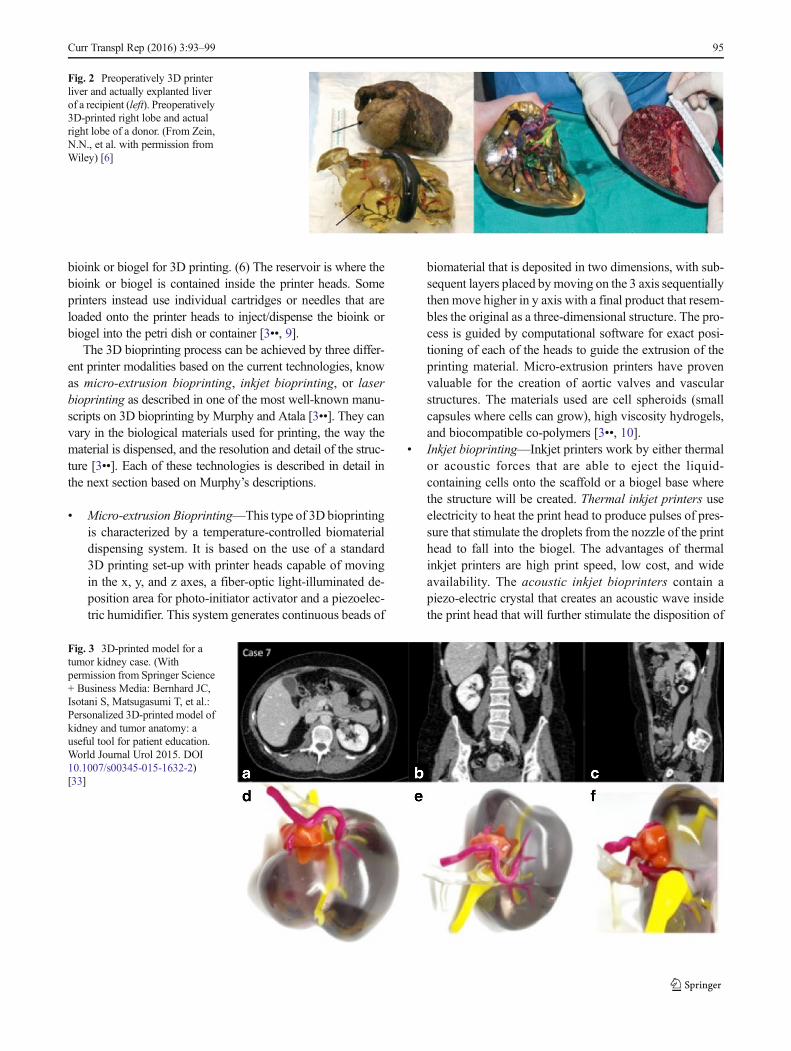
Fig. 3 3D-printed model for atumor kidney case. (Withpermission from Springer Science+ Business Media: Bernhard JC,Isotani S, Matsugasumi T, et al.:Personalized 3D-printed model ofkidney and tumor anatomy: auseful tool for patient education.World Journal Urol 2015. DOI10.1007/s00345-015-1632-2)[33]
Curr Transpl Rep (2016) 3:93–99 95

the cells into the scaffold or biogel base [3••]. The advan-tages of acoustic inkjet printers include the ability to createand control a uniform droplet size and high droplet direc-tionality. However, damage can be induced to the cellmembrane. Inkjet bioprinting is limited to low cell densi-ties to avoid nozzle clogging and reduce shear stress.Inkjet bioprinters have proven value for the regenerationof functional skin and cartilage in situ [10, 11].
& Laser-assisted Bioprinting—A Laser-assisted bioprinter(LAB) usually consists of a pulsed laser beam, a focusingsystem, a ribbon that has donor transport support that iscommonly made from glass covered with a laser-energy-absorbing layer (gold or titanium), and a layer of bioma-terial containing cells/hydrogel and a receiving substratefacing the ribbon. In LAB, the laser pulse will focus on theabsorbing layer to create high-pressure bubbles, whichwill propel cells toward the receiving substrate plate.LAB is compatible with wide range of biomaterial viscos-ities. LAB can produce high-resolution 3D structures withhigh cell viability. High cost of a LAB device hasprevented further basic research in the field [3••].
The 3D Bioprinting Process
The layer-by-layer basic fabrication of 3D printing is asfollows:
1. Pre-Processing—Generation of a computerized design ofthe structure—blue print
2. Processing
(a) A layer of hydrogel (extracellular matrix) is printedor preset in a petri dish or container, which will func-tion as the foundation base for the printed tissue.
(b) Bioink—usually made out of tissue spheroids or cul-tured cells. This bioink is loaded into the printerheads and disposed on the layer of hydrogel.
(c) Dispensing bioink or hydrogel repeatedly until manylayers are formed.
(d) As layers are built, the deposited bioink will fusetogether forming a 3D structure containing cellsand hydrogel.
(3) Maturation—Printed structure is then placed in an incu-bator and left to mature for 24 hours—1 week (depend-ing on the materials used) [12]
The printed tissue can then be used in pharmaceutical drugtesting or implantation in animal or in vitro models. In thefuture, hopefully, these printed tissues can be use for humantransplantation.
Present Success in 3D-Printed Tissues and Organs
As of the present day, this technology has been studied inacademia, research institutions, and by biotechnology firms(e.g., Organovo Holdings Inc., San Diego, CA, USA) for pos-sible use in tissue engineering applications, where tissues,organs, and body parts are built using 3D bioprinting tech-niques with the purpose of basic experimentation or the even-tual development of implantable organs and tissues.
The use of 3D bioprinting has already resulted in the suc-cessful printing of blood vessels and vascular networks [13],the bones [14], cartilage [15], ears [16], and tracheal grafts [17].In 2014, Bon Verweij and collaborators at the Utrecht MedicalCenter were able to replace a complete skull and successfullyimplant a 3D-printed synthetic component into a patient, withno adverse effects. Although this skull graft was synthetic, itdemonstrates the feasibility for implants to be custom-tailoredfor the patient in need, with the potential of the eventual crea-tion of a biological skull graft [18]. The use of 3D bioprinting inophthalmology is currently limited, but the potential is clear forthe generation of ocular tissues (e.g., conjunctiva, sclera, andcorneas) using 3D-bioprinting technologies in the future.
One of the greatest success stories written so far in the fieldof bioengineering and the creation of three-dimensional
Fig. 4 3D bioprinters around theworld. a 3D Organ Printer(Designed by Wake ForestInstitute for RegenerativeMedicine). b CommercializedNovogen MMX bioprinter TM(Designed by Organovo Inc). c3D Bioplotter (Designed byEnvisiontec, Germany). (FromSeol YJ, Kang HW, Lee SJ, et al.)[34]
96 Curr Transpl Rep (2016) 3:93–99

organs was done at the Wake Forest Institute for RegenerativeMedicine led by Atala et al. [5]. The idea was to providepatients with non-functional bladders an autologous bladderinstead of the traditional cystoplasty with gastrointestinal tis-sue. The team was able to engineer a human bladder by iso-lating autologous bladder urothelial and muscle cells,expanding the cells in vitro and seeding them to a bladderbiodegradable three-dimensional scaffold. Then, the tissue isanastomosed to the native bladder and covered with fibringlue as well as omentum [5]. They demonstrated that the im-plantation of bioengineered 3D organs is feasible after runninga clinical trial on seven patients in need of a cystoplasty. [5].
Manoor et al. were able to merge the field of cyberneticsand tissue engineering together in order to create a 3D-printedbionic ear as proof of concept. They printed a cell-seededhydrogel matrix in the anatomic geometry of human ear alongwith intertwined conducting polymer consisting of infusedsilver nanoparticles. This allowed in vitro culturing of carti-lage tissue around an inductive coil antenna in the ear, whichsubsequently enables detecting sound waves, thus exhibitingauditory sensing [16].
Although the whole idea behind 3D bioprinting is to createtissues and organs by assembling biological materials, the useof an aid, such as already created biological scaffolds, is alsobeing explored. The use of 3D extracellular matrix scaffolds,obtained by decellularization (which involves the use of adetergents perfused through the vasculature to remove cellularelements but allows ECM to remain intact) of allogenic orxenogeneic organs or tissues, is a promising approach forthe replacement of complex tissues and whole organs, sinceit provides a template with extracellular matrix that can sup-port cells and can contain the vascular network needed todeliver nutrients and oxygen for survival. Recent studies haveshown that decellularization of organs such as the livers,lungs, and heart is feasible. Song et al. recently incorporatedendothelial and epithelial cells into a decellularized kidneyECM scaffold in which the cells were integrated into the in-terstitial space and the vascular networks [19]. Yagi et al.described a successful decellularization technique in adult is-chemic porcine livers [20]. The aim of this technique is toobtain a liver matrix suitable for supporting functional hepa-tocytes as well as to maintain the functional vascular networkof the original organ including the biliary network.
The source of the organs for decellularization can be anissue. Use of porcine organs would vastly simplify logistics,enable precise quality control for obtaining decellularizedgrafts, avoid spread of human viruses, and indeed appears tobe the chosen approach for some tissues [21•] (Soto-Gutierrez2012). Even though any xenogeneic material creates the po-tential for immune complications, few have been observedthus far. Unrecovered human donor organs could also providea large quantity of potential scaffolds. Adult human organs,however, are rarely pristine, and their recovery introduces
many ethical questions. Still, it is fortunate that alternate op-tions exist, and determination of the best approach can be donewhen investigating clinical applications.
Current Developments by Tissue Type/Organ
In the following section, we will briefly describe outstandingwork presently being developed at different institutions fo-cused on particular tissues or organs that could offer potentialapplications for the field of transplantation.
Vascular Structures
Integral vascular structures are fundamental for a successfulorgan transplant. Autologous vascular conduits or the vascularstructures from deceased donor are frequently the first choicefor anastomosing the new organ to the recipient when neces-sary. They are sometimes not usable due to their length, diam-eter, or integrity. With the development of bioengineering,synthetic vascular grafts were successfully used for large di-ameter and high-flow grafts. However, these often come withcomplications such as vascular thrombosis and infection, lead-ing to longer post-operative hospital stays and increased treat-ment costs [22]. Tissue-engineered vascular grafts (TEVGs)offer an attractive theoretical alternative. Kurobe et al. de-scribed their experience with biodegradable vascular scaffoldswith seeded autologous cells that were implanted for congen-ital heart surgery with TEVG used as an extra-cardiac cavo-pulmonary connection and demonstrated that their grafts func-tioned without aneurismal change or graft rupture [23]. Thecreation of 3D vascular structures like these would signify agreat success. Actually, the creation of vascular conduits with-out scaffold is actually in progress. Norotte et al. described alayer-by-layer printing technique with the use of multicellularspheroids containing smooth muscle cells and fibroblastsalong with agarose rods, resulting in single- and double-layered small diameter vascular tubes [24]. In the field of liversurgery and transplantation, these TEVG conduits could bevery useful for the treatment of portal vein thrombosis andthe restoration of the intrahepatic portal flow, performing ameso-rex bypass from the superior mesenteric vein to donorleft portal vein. The TEVGs have also been discussed in theLDLT where often we need extension grafts for the drainageof segment Vand VIII branches to the middle hepatic vein orvena cava to avoid outflow issues in right lobe grafts as well asportal vein reconstructions.
Furthermore, the investigators of 3D bioprinting field arestudying out how to print branched vascular structures.Although it is still a complex task to achieve, some authorslike Visconti et al. have been able to design the adequateblueprints in order to guide the 3D bioprinters to create opti-mal branched vascular trees [25]. At our institution, ongoing
Curr Transpl Rep (2016) 3:93–99 97

efforts to develop these branched structures with 3D printingare already giving good results, and animal models are a pri-ority to evaluate their functionality. (Fig. 5). The other struc-ture that could have a potential benefit in the field is the engi-neering or 3D printing of bile ducts that could be used forrepair, bypasses, or as network for potential 3D livers.
Liver
An evenmore daring approach is the 3D printing of functionalliver tissue. Although to the present date there are no reports ofsuccessfully printed perfusable hepatic structures, early exper-iments have demonstrated promising data. For example,Robbins et al. with the use of the NovoGen MMXBioprinter ™ (Organovo Holdings, Inc., San Diego, CA,USA) demonstrated the feasibility of printing metabolicallyfunctional 3D hepatic structures and proving that the tissuewas capable of cell-cell interaction, protein production, andenzymatic activity. Also, they were able to enhance the com-plexity of the tissue by adding to the three-dimensional struc-ture hepatic stellate cells and endothelial cells [26]. The groupat Drexel led by Chang et al. developed a three-dimensionalliver micro-organ that consists of a microscale in vitro devicehousing a chamber of 3D liver cell-encapsuated hydrogel-based tissue that resembles the natural microenvironment ofthe hepatocyte in order to achieve biological functionality. Agreat enhancement of this system is that it included a dynamicperfusion in order to assess the cell metabolic function byperfusion of drugs [27]. Their model was developed to pro-vide NASAwith a liver tissue analog to assess drug pharma-cokinetic profiles in planetary environments. Although thesemodels are intended for drug or disease experimentation ratherthan for tissue transplantation, they set the basis to furtherunderstand the microenvironment of the liver in order to de-termine what interactions are needed to establish a fully
functional structure that can be transplanted into an animalor human. Further integration of all the 3D techniques current-ly in practice are needed to achieve the creation of a fullyfunctional bioprinted liver that could be transplanted. One ofthe most important elements of a successful model is the pres-ence of a vascular network to support the liver cells. Miller etal. published their experience with creating perfusable vascu-lar networks by printing 3D filament networks of carbohy-drate glass and used them as a template in engineered tissuescontaining living cells to generate cylindrical networks thatcould be lined with endothelial cells and perfused with blood.They were able to prove in a rat hepatocyte model that theperfused vascular channels were able to maintain the metabol-ic function of the cells [13].
Kidney
Although not much data has been published in kidney 3Dprinting, King et al. from Organovo recently presented theirin vitro model of a multicellular, three-dimensional tissuemodel of human kidney proximal tubule. In their printed mod-el, they were able to observe the interface between tubularepithelium and renal interstitial cells. Extensive endothelialnetworks were also observed [28]. Promising data such as thiscan be helpful to the field of transplantation, in order to assessimmunological models for rejection in the laboratory with 3Drenal tissue. The use of this model for that purpose has thepotential to modify the current immunosuppressant therapies.In terms of three-dimensional scaffolds for renal tissue engi-neering, many authors have demonstrated that the adequateuse of decellularization agents can preserve critical structuraland functional properties necessary for use of these three-dimensional scaffolds for promoting cellular repopulation oreven establish the adequate blue prints to instruct a 3D printerto design a brand new kidney [19, 29].
Future Horizons and Conclusion
The next step for tissue engineering and 3D printing is to con-tinue these extraordinary, mostly in vitro [30, 31] (Ott 2008, Ott2010), initial efforts by assessing in vivo organ viability andfunctionality in animal models. Development of such modelswith any attendant techniques and technologies will likely in-troduce further challenges and complications [32] (Lysaght2004). Knowledge gleaned from current in vitro pharmacologyand disease models will likely contribute to these efforts.
Better understanding of intercellular communication andtissue microenvironments will provide the blueprint for con-structing bio-artificial organs with native-like architecture andfunctionality. Although common themes emerge, ultimateprinting protocols will almost certainly be highly tissue- ororgan-specific for 3D printers to assemble physiologically
Fig. 5 Pathology slide of a printed vessel in the 3D Bioprinting lab atYale School of Medicine. (Courtesy of Dr. John Geibel)
98 Curr Transpl Rep (2016) 3:93–99

appropriate microstructures. As we have seen, decellularizedorgan scaffolds provide an excellent starting point.
Many issues remain unsolved for the majority of protocols;yet, it is encouraging to note the examples of already successfulimplantation (bladder and trachea). While complete ontogenicreplication is a lofty goal, this should also not impede consid-eration of bioengineered tissue for another application—name-ly, as bridge therapy while awaiting organ transplantation.Whether bioengineered tissue serves as organ supplement ororgan replacement may ultimately hinge on those age-old con-straints of patient disease and organ availability.
Compliance With Ethical Standards
Conflict of Interest Armando Salim Munoz-Abraham, Manuel I.Rodriguez-Davalos, Alessandra Bertacco, Brian Wengerter, John P. Geibel,and David C. Mulligan declare that they have no conflict of interest.
Human and Animal Rights and Informed Consent This article doesnot contain any studies with human or animal subjects performed by anyof the authors.
References
Papers of particular interest, published recently, have beenhighlighted as:• Of importance•• Of major importance
1. Services, U.S.D.o.H.H.S.U.S.D.o.H.H. Organ Procurement andTransplantation Network. OPTN [cited 2015 2015]; Availablefrom: http://optn.transplant.hrsa.gov/.
2. Mulligan D. Disparity, liver demand, and access to transplants. AmJ Transplant. 2015;15(7):1746–7.
3.•• Murphy SV, Atala A. 3D bioprinting of tissues and organs. NatBiotechnol. 2014;32(8):773–85. This review provides a great over-view of 3D bioprinting. The authors are pioneers in the field andDr. Atala is considered among the worlwide 3D bioprintingexperts.
4. Hull CW. In: Hull CW, editor. Apparatus for production of three-dimensional objects by stereolithography. United States: U.S.P.a.T.Office; 1986.
5. Atala A et al. Tissue-engineered autologous bladders for patientsneeding cystoplasty. Lancet. 2006;367(9518):1241–6.
6. Zein NN et al. Three-dimensional print of a liver for preoperativeplanning in living donor liver transplantation. Liver Transpl.2013;19(12):1304–10.
7. University, Y. Dr Mark Michalski is ready to print a 3-D brain(maybe yours). 2013 [cited 2015; Available from: http://news.yale.edu/2013/07/15/dr-mark-michalski-ready-print-3-d-brain-maybe-yours.
8. Mironov V, Reis N, Derby B. Review: bioprinting: a beginning.Tissue Eng. 2006;12(4):631–4.
9. Organovo. The Bioprinting Process. 2015 [cited 2015; Available from:http://www.organovo.com/science-technology/bioprinting-process.
10. Zhang, X. and Y. Zhang, Tissue Engineering Applications of Three-Dimensional Bioprinting. Cell Biochem Biophys, 2015.
11. Boland T et al. Application of inkjet printing to tissue engineering.Biotechnol J. 2006;1(9):910–7.
12. Guillemot F, Mironov V, Nakamura M. Bioprinting is coming of age:report from the International Conference on Bioprinting andBiofabrication in Bordeaux (3B'09). Biofabrication. 2010;2(1):010201.
13. Miller JS et al. Rapid casting of patterned vascular networks forperfusable engineered three-dimensional tissues. Nat Mater.2012;11(9):768–74.
14. Fedorovich NE et al. Organ printing: the future of bone regenera-tion? Trends Biotechnol. 2011;29(12):601–6.
15. Cui X et al. Direct human cartilage repair using three-dimensionalbioprinting technology. Tissue Eng Part A. 2012;18(11–12):1304–12.
16. Mannoor MS et al. 3D printed bionic ears. Nano Lett. 2013;13(6):2634–9.
17. Chang JW et al. Tissue-engineered tracheal reconstruction usingthree-dimensionally printed artificial tracheal graft: preliminary re-port. Artif Organs. 2014;38(6):E95–E105.
18. Utrecht, U. 3D-Printed Skull Impanted In Patient. 2014 [cited 20152015]; Available from: http://www.umcutrecht.nl/en/Research/News/3D-printed-skull-implanted-in-patient.
19. Song JJ et al. Regeneration and experimental orthotopic transplan-tation of a bioengineered kidney. Nat Med. 2013;19(5):646–51.
20. Yagi H et al. Human-scale whole-organ bioengineering for livertransplantation: a regenerative medicine approach. CellTransplant. 2013;22(2):231–42.
21.• Soto-Gutierrez A et al. Perspectives on whole-organ assembly:moving toward transplantation on demand. J Clin Invest.2012;122(11):3817–23. Their perspectives on tissue engineeringon how organ assembly should be approached is very interest-ing. Decellularization is a technique that might provide the an-swer for creating transplantable organs.
22. Udelsman BV, Maxfield MW, Breuer CK. Tissue engineering ofblood vessels in cardiovascular disease: moving towards clinicaltranslation. Heart. 2013;99(7):454–60.
23. Kurobe H et al. Concise review: tissue-engineered vascular graftsfor cardiac surgery: past, present, and future. Stem Cells TranslMed. 2012;1(7):566–71.
24. Norotte C et al. Scaffold-free vascular tissue engineering usingbioprinting. Biomaterials. 2009;30(30):5910–7.
25. Visconti RP et al. Towards organ printing: engineering an intra-organbranched vascular tree. Expert Opin Biol Ther. 2010;10(3):409–20.
26. Robbins, J.G., V; Min, P; Shepherd, B; Presnell, S., A novel in vitrothree-dimensional bioprinted liver tussue system for drugdevelopment. American Society of Experimental Biology, 2013.
27. Chang R et al. Biofabrication of a three-dimensional liver micro-organ as an in vitro drug metabolism model. Biofabrication.2010;2(4):045004.
28. King, S.C., O; Presnell S; Nguyen D,Design and charactherizationof a multicellular, three-dimensional tissue model of the humankidney proximal tubule. American Society of ExperimentalBiology, 2015.
29. Nakayama KH et al. Renal tissue engineering with decellularizedrhesus monkey kidneys: age-related differences. Tissue Eng Part A.2011;17(23–24):2891–901.
30. Ott HC et al. Perfusion-decellularized matrix: using nature's plat-form to engineer a bioartificial heart. Nat Med. 2008;14(2):213–21.
31. Ott HC et al. Regeneration and orthotopic transplantation of abioartificial lung. Nat Med. 2010;16(8):927–33.
32. Lysaght MJ, Hazlehurst AL. Tissue engineering: the end of thebeginning. Tissue Eng. 2004;10(1–2):309–20.
33. Bernhard JC, Isotani S, Matsugasumi T, et al.: Personalized 3Dprinted model of kidney and tumor anatomy: a useful tool for pa-tient education. World Journal Urol 2015. DOI 10.1007/s00345-015-1632-2
34. Seol YJ, Kang HW, Lee SJ, et al. Bioprinting technology and itsapplications. Eur J Cardiothorac Surg. 2014;46(3):342–8.
Curr Transpl Rep (2016) 3:93–99 99


![Ethical Controversies in Organ Transplantation · demand for organ transplantation and the supply of donor organs is growing [The economist 2008].The waiting list of the United Network](https://static.fdocuments.us/doc/165x107/603e691cbdfbcb7c012bb89a/ethical-controversies-in-organ-transplantation-demand-for-organ-transplantation.jpg)