3D printing for heart valve disease: a systematic review
10
SYSTEMATIC REVIEW Open Access 3D printing for heart valve disease: a systematic review Volkan Tuncay and Peter M. A. van Ooijen * Abstract Background: Current developments showed a fast-increasing implementation and use of three-dimensional (3D) printing in medical applications. Our aim was to review the literature regarding the application of 3D printing to cardiac valve disease. Methods: A PubMed search for publications in English with the terms “3D printing” AND “cardiac valve”, performed in January 2018, resulted in 64 items. After the analysis of the abstract and text, 27 remained related to the topic. From the references of these 27 papers, 7 papers were added resulting in a total of 34 papers. Of these, 5 were review papers, thus reducing the papers taken into consideration to 29. Results: The 29 papers showed that about a decade ago, the interest in 3D printing for this application area was emerging, but only in the past 2 to 3 years it really gained interest. Computed tomography is the most common imaging modality taken into consideration (62%), followed by ultrasound (28%), computer-generated models (computer-aided design) (7%), and magnetic resonance imaging (3%). Acrylonitrile butadiene styrene (4/14, 29%) and TangoPlus FullCure 930 (5/14, 36%) are the most used printing materials. Stereolithography (40%) and fused deposition modeling (30%) are the preferred printing techniques, while PolyJet (25%) and laser sintering (4%) are used in a minority of cases. The reported time ranges from 30 min to 3 days. The most reported application area is preoperative planning (63%), followed by training (19%), device testing (11%), and retrospective procedure evaluation (7%). Conclusions: In most cases, CT datasets are used and models are printed for preoperative planning. Keywords: Heart valves, Printing (three-dimensional), Stereolithography, Tomography (x-ray computed), Ultrasonography Key points Computed tomography is the standard imaging modality for cardiac valve printing, followed by ultrasound, computer-aided design, and magnetic resonance imaging. Stereolithography and fused deposition modeling are the preferred methods for cardiac valve printing. Acrylonitrile butadiene styrene and TangoPlus FullCure 930 are the most used printing materials. The most reported application area is preoperative planning, followed by training, device testing, and retrospective procedure evaluation. Background Although patient-specific three-dimensional (3D) visual- isation already provides good insight into the complex anatomy of a patient, in some cases, this is not sufficient, and more advanced techniques are required, such as the use of virtual and augmented reality but also 3D printing [1, 2]. On the one side, 3D printing allows the surgeon to hold and examine the structures printed in a tactile way, sometimes providing a better insight into the 3D anatomy. On the other side, a real-life-size 3D printed anatomy al- lows to test procedures by introducing the actual im- plants, wires, and instruments into the printed anatomy. The basis for 3D printing was laid in the 1980s. Medical applications arose from this new technology early on in the development, mainly in maxillofacial sur- gery. Although one paper already reported on the use of stereolithography (STL) printing of mitral valves based * Correspondence: [email protected] University of Groningen, University Medical Center Groningen, Hanzeplein 1, 9713GZ Groningen, The Netherlands European Radiology Experimental © The Author(s). 2019 Open Access This article is distributed under the terms of the Creative Commons Attribution 4.0 International License (http://creativecommons.org/licenses/by/4.0/), which permits unrestricted use, distribution, and reproduction in any medium, provided you give appropriate credit to the original author(s) and the source, provide a link to the Creative Commons license, and indicate if changes were made. Tuncay and Ooijen European Radiology Experimental (2019) 3:9 https://doi.org/10.1186/s41747-018-0083-0
Transcript of 3D printing for heart valve disease: a systematic review

SYSTEMATIC REVIEW Open Access
3D printing for heart valve disease: asystematic reviewVolkan Tuncay and Peter M. A. van Ooijen*
Abstract
Background: Current developments showed a fast-increasing implementation and use of three-dimensional (3D)printing in medical applications. Our aim was to review the literature regarding the application of 3D printing tocardiac valve disease.
Methods: A PubMed search for publications in English with the terms “3D printing” AND “cardiac valve”, performedin January 2018, resulted in 64 items. After the analysis of the abstract and text, 27 remained related to the topic.From the references of these 27 papers, 7 papers were added resulting in a total of 34 papers. Of these, 5 werereview papers, thus reducing the papers taken into consideration to 29.
Results: The 29 papers showed that about a decade ago, the interest in 3D printing for this application area wasemerging, but only in the past 2 to 3 years it really gained interest. Computed tomography is the most commonimaging modality taken into consideration (62%), followed by ultrasound (28%), computer-generated models(computer-aided design) (7%), and magnetic resonance imaging (3%). Acrylonitrile butadiene styrene (4/14, 29%) andTangoPlus FullCure 930 (5/14, 36%) are the most used printing materials. Stereolithography (40%) and fused depositionmodeling (30%) are the preferred printing techniques, while PolyJet (25%) and laser sintering (4%) are used in aminority of cases. The reported time ranges from 30min to 3 days. The most reported application area is preoperativeplanning (63%), followed by training (19%), device testing (11%), and retrospective procedure evaluation (7%).
Conclusions: In most cases, CT datasets are used and models are printed for preoperative planning.
Keywords: Heart valves, Printing (three-dimensional), Stereolithography, Tomography (x-ray computed),Ultrasonography
Key points
� Computed tomography is the standard imagingmodality for cardiac valve printing, followed byultrasound, computer-aided design, and magneticresonance imaging.
� Stereolithography and fused deposition modeling arethe preferred methods for cardiac valve printing.
� Acrylonitrile butadiene styrene and TangoPlusFullCure 930 are the most used printing materials.
� The most reported application area is preoperativeplanning, followed by training, device testing, andretrospective procedure evaluation.
BackgroundAlthough patient-specific three-dimensional (3D) visual-isation already provides good insight into the complexanatomy of a patient, in some cases, this is not sufficient,and more advanced techniques are required, such as theuse of virtual and augmented reality but also 3D printing[1, 2]. On the one side, 3D printing allows the surgeon tohold and examine the structures printed in a tactile way,sometimes providing a better insight into the 3D anatomy.On the other side, a real-life-size 3D printed anatomy al-lows to test procedures by introducing the actual im-plants, wires, and instruments into the printed anatomy.The basis for 3D printing was laid in the 1980s.
Medical applications arose from this new technologyearly on in the development, mainly in maxillofacial sur-gery. Although one paper already reported on the use ofstereolithography (STL) printing of mitral valves based
* Correspondence: [email protected] of Groningen, University Medical Center Groningen, Hanzeplein 1,9713GZ Groningen, The Netherlands
European RadiologyExperimental
© The Author(s). 2019 Open Access This article is distributed under the terms of the Creative Commons Attribution 4.0International License (http://creativecommons.org/licenses/by/4.0/), which permits unrestricted use, distribution, andreproduction in any medium, provided you give appropriate credit to the original author(s) and the source, provide a link tothe Creative Commons license, and indicate if changes were made.
Tuncay and Ooijen European Radiology Experimental (2019) 3:9 https://doi.org/10.1186/s41747-018-0083-0

on ultrasound (US) imaging in patients as early as 2000[3], the real interest for 3D printing in cardiovascular ap-plications started some years later.Building on the experience of the early adopters, the
use of 3D printing recently has enormously increased ina wide variety of medical applications. The field hasdemonstrated itself as an example of multidisciplinarycooperation where radiologists, surgeons, and mechan-ical/biomedical engineers all provide their specific ex-pertise in the different application areas [4]. Theseapplication areas vary from the printing of anatomicalmodels for teaching and training [5] to models to informthe patient about treatment and from the preoperativeevaluation of devices to the printing of guides and im-plants used during surgery. In recent years, cardiac anat-omy and especially congenital heart disease havebecome one of the focus areas of 3D printing to easilyvisualise and explore complex cardiovascular anatomy.However, other applications that could have a major im-pact on the field of cardiothoracic surgery, such as plan-ning of transcatheter aortic valve replacement (TAVR)and transcatheter mitral valve replacement (TMVR), arealso arising. 3D printing can be used to tackle some ofthe challenges in these interventions such as patient se-lection, prosthesis choice and sizing, and innovation invalve design. In this narrative review, we discuss thecurrent state of the art in this area from a technicalpoint of view by considering the constraints and possi-bilities of the 3D printing technique based on publishedwork that specifically focuses on 3D printing in cardiacvalve disease treatment. We will look at general topicssuch as data preparation, time requirements, printerpossibilities, and material properties relating to this spe-cific application area. Possible clinical applications fromthe literature will also be introduced.
Literature searchA PubMed search for publications in English with theterms “3D printing” AND “cardiac valve” showed thatinterest in this topic is certainly gaining. It was per-formed in January 2018. Although our initial search re-sulted in 64 items, after the analysis of the abstracts andtext, 27 remained valid and related to the review topic.From the references of these 27 papers, another 7 paperswere added resulting in a total of 34 papers. Of these,five were earlier review papers, of which most only men-tioned the specific case of 3D printing in cardiac valvediseases as a small subsection of their review, thus redu-cing the papers taken into consideration to 29. The 29papers clearly showed that about a decade ago, the inter-est in 3D printing for this application area was emerging,but only in the past 2 to 3 years it really gained interestresulting in a steep increase in the number of publica-tions (Fig. 1).
Source data and pre-processingA high-quality volume dataset with high resolution andno artefacts is required to allow for 3D printing. Thiscan be acquired by common modern radiological im-aging techniques provided that the proper reconstruc-tions and protocols are applied. Computed tomography(CT) is the most common imaging modality providingimage data for 3D printing in cardiac valve diseases (18of 29 papers, 62%) [6–23], followed by US (8 papers,28%) [9, 24–30], computer-generated models (compu-ter-aided design) (2 papers, 7%) [31, 32], and magneticresonance imaging (MRI) (1 paper, 3%) [34] (Fig. 2).The quality of printed models is highly depending on
the quality of the imaging dataset used. Cardiac motionand breathing artefacts have a negative impact on thesegmentation and thus the printed volume. Typically,high-resolution scans are used in combination withelectrocardiography gating, breath-hold, and/or respira-tory gating [35]. In order to allow 3D printing of struc-tures, they must have distinct tissue contrast in theimaging data [4].In CT, commonly 0.75- to 1-mm slice thickness with a
smoother kernel is used [4, 6, 35]. Scans with a higherresolution are less favourable since they introduce highernoise levels and require a more cumbersome segmenta-tion process [35]. Some studies reported the use ofmultiphase acquisition during the cardiac cycle to ensurethat the right phase can be reconstructed [20]. In MRI,standard cardiac imaging sequences can be used. How-ever, the lower resolution of MRI in comparison withCT can hamper the production of good-quality 3Dprints [35]. In US, the use of 3D scanning is required toobtain a proper 3D volume for segmenting the anatom-ical structures [24].Regardless of the modality used to acquire the 3D
datasets, structures of interest have to be segmented andtranslated into a surface model to enable 3D printing.Segmentation is the key process herein [33]. In somecases, the vessel wall is too thin to segment; extra thick-ness then should be added to the model since 3Dprinters have minimum thickness requirements [20].The most commonly described tool for segmentation
and creation of the STL file required for 3D printing isthe Mimics/3-Matic software combination (Materialise,Leuven, Belgium). Secondly, SolidWorks (Dassault Sys-tèmes SolidWorks Corporation, Vélizy-VillacoublayCedex, France) is also used frequently. Less common arethe 3D Slicer (Open source software package), AutoDeskMeshmixer (Autodesk, San Rafael, CA), and VascularModeling Toolkit (VMTK, Orobix, Bergamo, Italy).All used packages have in common is that they allow
to import the imaging data (according to the Digital Im-aging and COmmunications in Medicine (DICOM)standard) from modalities such as CT, US, and MRI and
Tuncay and Ooijen European Radiology Experimental (2019) 3:9 Page 2 of 10

Fig. 1 Number of publications on 3D printing in the application of cardiac valve assessment or replacement
Fig. 2 Frequency of use of different imaging modality providing the source data
Tuncay and Ooijen European Radiology Experimental (2019) 3:9 Page 3 of 10

transfer them to a 3D model. This model is realised bythe segmentation of the structures of interest after whicha surface representation is constructed. This surface re-construction is commonly exported in STL format fromthe modeling software and loaded into the software ofthe 3D printer. This software allows to create and cor-rect the model in order to ensure that it is printable andenables inclusion of required structures such as add-itional support material. After completion of the model,the data are resliced into print levels after which theycan be sent to the printer to be manufactured.
Printing materialsAlthough 3D printing is already known and used, oneof the major areas of concern when printing cardio-vascular structures such as the aorta, heart, andvalves is the limited availability of usable printingmaterials to obtain objects with vessel-like properties.Traditional phantoms would be constructed with rigidmodels of resins or glass, but these are not usefulwhen a more lifelike representation of the vessel wallis required. Therefore, the property requirements ofprinting materials for 3D printing of cardiovascularstructures should take into consideration the
flexibility of the material to mimic the vessel wall [11,34] (Fig. 3) and the transparent nature of the mate-rials [11] to allow for observation of instrumentswhen inserted and visual inspection of internalstructures.The literature shows that acrylonitrile butadiene
styrene or ABS (4/14, 29%) and TangoPlus FullCure930 (5/14, 36%) (Stratasys Ltd., Eden Prairie, MN,USA) are the most commonly used materials (Table 1).TangoPlus FullCure 930 is a commercially availabletranslucent rubber-like PolyJet photopolymer material.It can simulate different levels of hardness, elong-ation, and tear resistance. Because of the difficulty ofdirect printing in flexible materials, many papers de-scribe a process in which print casts and molds areprinted in other materials that are then dipped in, orcoated with, silicone to obtain flexible vessels andvalves with more accurate tissue properties [12, 27].A challenge with this method is that it must either bepossible to remove the silicone from the cast or moldafter hardening or the cast or mold should be printedin a dissolvable material. Few examples of customisedprinters also exist that directly print with (sanitary)silicone [11, 31].
Fig. 3 Example of the right ventricle outflow tract and main pulmonary artery print in flexible, non-transparent material in its normal (a) andsqueezed (b) form
Tuncay and Ooijen European Radiology Experimental (2019) 3:9 Page 4 of 10

An additional requirement of specific interest in the caseof the cardiac valves is the ability to print with multipletypes of material to obtain flexible vessel walls and valvesin combination with rigid calcified plaque deposits. Severalreports with successful outcomes have been publishedusing a transparent and flexible material for the vessel walland valves combined with an opaque, rigid material forthe calcified plaques [4, 7, 9, 17, 19, 20] (Fig. 4).For example, Vukicevic et al. [9] 3D printed
patient-specific mitral valves of three patients with mul-tiple materials to evaluate trans-catheter mitral valve re-pair procedures. They performed biomechanical tests on
different TangoPlus materials which were compared withthe mechanical properties of the porcine mitral valve tis-sue to select the most appropriate TangoPlus material fora specific region of the 3D model. Different TangoPlusmaterials were used on different parts of the model inorder to have the most realistic mechanical properties.
Printing techniquesSeveral printing techniques exist. Among them, most fre-quently used and well known are fused deposition model-ing (FDM), STL, PolyJet, and laser sintering. Details onthese techniques have been described extensively in the
Table 1 3D printing materials for cardiac valve replacement: intended uses and application areas
First author[reference number]
Intended use Application area Printing material Post-treatment material
Abdel-Sayed [11] Training Trans-apical aortic valve replacement Silicone Silicone coating
Biglino [34] Device testing Material testing forcardiovascular application
TangoPlus FullCure 930 None
Fujita [12] Preoperative planning Transcatheter aortic valve implantation Acrylonitrilebutadiene styrene
Silicone coating
Fujita [13] Retrospective procedureevaluation
Transcatheter aortic valve implantation TangoPlus FullCure 930 Silicone coating
Fujita [14] Preoperative planning Transcatheter aortic valve implantation Photopolymer resin None
Izzo [7] Preoperative planning Transcatheter native mitralvalve replacement
TangoPlus FullCure 930 None
Kalejs [31] Device testing Aortic valve replacement Silicone Silicone coating
Maragiannis [17] Training Aortic valve stenosis TangoPlus FullCure 930 Silicone coating
Mashari [26] Device testing Mitral valve models Moldstar 15 + Ecoflex 0030 Silicone coating
Ripley [20] Preoperative planning Transcatheter aortic valve implantation Clear flexible resin None
Sardari Nia [27] Preoperative planning Mitral valve intervention Acrylonitrile butadiene styrene Silicone coating
Vukicevic [9] Training Mitral valve intervention TangoPlus FullCure 930 None
Witschey [28] Preoperative planning Mitral valve intervention Acrylonitrile butadiene styrene None
Owais [29] Preoperative planning Mitral annuli Acrylonitrile butadiene styrene None
Fig. 4 a, b Examples of prints of the aortic valve in a flexible transparent material with calcifications in blue non-flexible material
Tuncay and Ooijen European Radiology Experimental (2019) 3:9 Page 5 of 10

literature [4, 5, 35, 36]. Based on the literature review per-formed, it is clear that STL is the preferred method forcardiac valve printing (40%), followed by FDM (30%). Thepreference for STL can be mainly explained by its abilityto print more easily with flexible and transparent materialsthan other techniques.In some cases, a dedicated setup was built to allow less
conventional printing materials or printing hardware. Oneexample setup in the literature was built with a syringefilled with (sanitary) silicone that was used to print asemi-transparent, flexible aortic root [31]. A high accuracycould be achieved (3.0% error along the x- and y-axes;4.1% error along the z-axis). Although this was a verycheap solution, printing and post-processing time of theprint were quite long (from 3 h and 20min to 3 days).
Time constraintsThe current printing process involves the followingsteps:
1) imaging data acquisition;2) segmentation of the anatomical structures;3) export of the segmented structures to STL;4) repair and improvement of the STL file;5) re-slicing and preparing for printing (e.g., definition
of support materials);6) printing process;7) post-printing (e.g., removal of support materials,
silicone dipping).
The time required for each of these steps is notmentioned in all papers, and those that do mostlyprovide only a total processing time for steps 2–7[11, 20, 24–26, 30, 34] (Table 2). The reported timeranges from 30 min for an FDM print of the mitralannulus [24] to 3 days for a dedicated FDM printerwith a syringe filled with silicone followed by siliconedip coating for a simplified heart model [11].Although the printing time is heavily depending on
the size and complexity of the printed structure, ex-perience shows that steps 6 and 7 are the mosttime-consuming. This especially holds in the case ofmolds and casts where extensive post-printing treat-ment is required, such as the application of the sili-cone (often with multiple coat dipping), andhardening of the material. Steps 2–5 are increasinglysupported by dedicated software tools allowing moreautomation in the process and guided workflows toensure a proper printing model.In general, the required time for the whole process of
segmentation, data cleaning and preparation, and theprinting itself greatly varies depending on the size of theprinted object, the printing technique used, and the re-quirement for post-printing treatment.
Possible printing issuesOne of the issues with 3D printing is the accuracy of the3D printed object in size and shape. The difference be-tween the 3D printed object and imaging modality mea-surements should be minimal. The print accuracy ishigh using current printers and software with reportedaccuracies of a mean difference between the measure-ment in CT and of the print of − 0.34 mm ± 1.3 mm [20]and 0.7 mm ± 0.3 mm, respectively, without significantdifferences between the CT measurement and the actualprint measurement [30]. Therefore, with careful designof the printing process, it is possible to print models thatresemble the real anatomy and can be used for pre-operative planning.Another issue is the removal of the support materials.
Different from most other 3D printed models, the vascu-lar models are complex in structure and must be hollowin order to gain access to the lumen with wires and de-vices. This can be achieved by either choosing a printingstrategy not requiring support material (such as binderjetting) or by removing the support material after print-ing. A soluble support material can be used in a multiplematerial printer. In that case, the object usually has to
Table 2 3D printing for cardiac valve replacement: required time for different printed objects with various printing techniques
First author [reference number] Printer Print method Post-treatment Printed object Time
Abdel-Sayed [11] FDM syringe with silicone FDM Dip-coating with silicone Simplified heart model 3 days
Biglino [34] PolyJet PolyJet None Descending aorta 12 h
Kalejs [31] Fab@Home FDM Dip-coating with silicone Aortic root model 200 min
Mahmood [25] Objet260 Connex PolyJet NA Mitral valve 90 min
Mahmood [24] Makerbot Replicator 2X FDM None Mitral annulus 30 min
Mashari [26] Makerbot Replicator 2X FDM Silicone casting Mitral valve 2–5 h
Muraru [30] Formiga P110 Laser sintering NA Tricuspid valve 90–120min
Ripley [20] Form 1 Plus SLT NA Aortic root 5 h
Owais [29] Makerbot Replicator 2X FDM None Aortic annulus 15 min
FDM fused deposition modeling, SLT stereolithography, NA not applicable
Tuncay and Ooijen European Radiology Experimental (2019) 3:9 Page 6 of 10
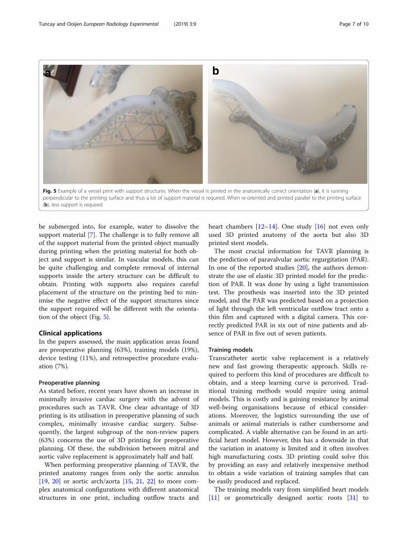
be submerged into, for example, water to dissolve thesupport material [7]. The challenge is to fully remove allof the support material from the printed object manuallyduring printing when the printing material for both ob-ject and support is similar. In vascular models, this canbe quite challenging and complete removal of internalsupports inside the artery structure can be difficult toobtain. Printing with supports also requires carefulplacement of the structure on the printing bed to min-imise the negative effect of the support structures sincethe support required will be different with the orienta-tion of the object (Fig. 5).
Clinical applicationsIn the papers assessed, the main application areas foundare preoperative planning (63%), training models (19%),device testing (11%), and retrospective procedure evalu-ation (7%).
Preoperative planningAs stated before, recent years have shown an increase inminimally invasive cardiac surgery with the advent ofprocedures such as TAVR. One clear advantage of 3Dprinting is its utilisation in preoperative planning of suchcomplex, minimally invasive cardiac surgery. Subse-quently, the largest subgroup of the non-review papers(63%) concerns the use of 3D printing for preoperativeplanning. Of these, the subdivision between mitral andaortic valve replacement is approximately half and half.When performing preoperative planning of TAVR, the
printed anatomy ranges from only the aortic annulus[19, 20] or aortic arch/aorta [15, 21, 22] to more com-plex anatomical configurations with different anatomicalstructures in one print, including outflow tracts and
heart chambers [12–14]. One study [16] not even onlyused 3D printed anatomy of the aorta but also 3Dprinted stent models.The most crucial information for TAVR planning is
the prediction of paravalvular aortic regurgitation (PAR).In one of the reported studies [20], the authors demon-strate the use of elastic 3D printed model for the predic-tion of PAR. It was done by using a light transmissiontest. The prosthesis was inserted into the 3D printedmodel, and the PAR was predicted based on a projectionof light through the left ventricular outflow tract onto athin film and captured with a digital camera. This cor-rectly predicted PAR in six out of nine patients and ab-sence of PAR in five out of seven patients.
Training modelsTranscatheter aortic valve replacement is a relativelynew and fast growing therapeutic approach. Skills re-quired to perform this kind of procedures are difficult toobtain, and a steep learning curve is perceived. Trad-itional training methods would require using animalmodels. This is costly and is gaining resistance by animalwell-being organisations because of ethical consider-ations. Moreover, the logistics surrounding the use ofanimals or animal materials is rather cumbersome andcomplicated. A viable alternative can be found in an arti-ficial heart model. However, this has a downside in thatthe variation in anatomy is limited and it often involveshigh manufacturing costs. 3D printing could solve thisby providing an easy and relatively inexpensive methodto obtain a wide variation of training samples that canbe easily produced and replaced.The training models vary from simplified heart models
[11] or geometrically designed aortic roots [31] to
Fig. 5 Example of a vessel print with support structures. When the vessel is printed in the anatomically correct orientation (a), it is runningperpendicular to the printing surface and thus a lot of support material is required. When re-oriented and printed parallel to the printing surface(b), less support is required
Tuncay and Ooijen European Radiology Experimental (2019) 3:9 Page 7 of 10

advanced flow models using pulsatile pumps allowing amore real-life simulation [5, 17] where devices can be in-troduced and deployed under lifelike conditions (Fig. 6).One study [32] described the printing of anMRI-compatible setup to allow scanning of mitral valvesof pigs in the natural state. They achieved this by design-ing and 3D printing valve-specific mounting materialsbased on premortem US intra-valve measurements.
Device testingDevice development and testing is also an important ap-plication of 3D printing. Biglino et al. [34] 3D printedmodels of the descending aorta with the same lumen di-mension but with different wall thicknesses and didcompliance tests. They used the distensibility knowledgeto build a right ventricular outflow tract model, whichwas used to simulate the pulmonary valve replacementprocedure for device testing. Kalejs and von Segesser[31] manufactured a real-life-size artic root model fortesting valved stents. Mashari et al. [26] created a 3Dmodel of the mitral valve from 3D US images of a pa-tient who underwent a percutaneous MitraClip oper-ation, a minimally invasive procedure to reduce themitral regurgitation. The model was then deployed in
the pulse-duplicator chamber filled with a blood-mimicking fluid for hemodynamic testing.
DiscussionNovel techniques such as 3D printing are investigated bydifferent research groups for specific clinical questions,and they all explore the technical requirements andshortcomings of the technique. While the topic iscurrent, the actual clinical benefit of 3D printing yet re-mains to be proven. However, technical developmentsare ongoing and the implementation of 3D printing forcardiac valve treatment is one of the more promisingclinical application areas. However, the application of 3Dprinting in cardiac valve replacement introduces add-itional requirements on the used printing material andthe nature of the printed structure. This review showsthat, with the advent of new flexible and transparent ma-terials and higher accuracy of 3D printers, an accuraterepresentation of the cardiac anatomy can be obtained.This 3D printed representation of the anatomy canalready be used in a variety of applications, especially fortraining purposes.It has been shown that for accurate 3D printing of
anatomy or pathology, correct segmentation is of vital
Fig. 6 Sample schematic setup of an experimental environment to test valves. The 3D printed valve would be included in the valveimplementation part inside the flow loop (blue lines)
Tuncay and Ooijen European Radiology Experimental (2019) 3:9 Page 8 of 10

importance. Although some of the segmentation work iscurrently automated, the input of experts is still neededfor validation and correction. Any small mistake in thesegmentation process could lead to erroneous prints.This could lead to the failure of the printing process it-self, but also to a 3D printed structure that is not accur-ate that, in turn, could be harmful to the patient whenused for preoperative planning. Thus, the segmentationstep should be conducted and/or supervised by an ex-pert in the anatomy being segmented, and quality con-trol of the source data should be in place. Also, thesegmentation tools should be validated and approved forclinical use. Although the majority of the reviewed pa-pers show the use of a validated commercial softwaretool, researchers may still rely on freeware andopen-source software which could hamper reliability ofthe segmentation, 3D model construction, and finallythe printing result.In this review, we aimed to explore the options, chal-
lenges, and possibilities of the 3D printing in the field ofcardiac valve replacement in order to give an insight intothe current state of the art and development in this spe-cific area of 3D printing. The low number of papersfound on this topic demonstrates its experimental na-ture. However, the published papers do show the pro-gress made in the past years allowing for clinicalapplication. This clinical application is currently mainlyin training and education, but the literature is promisingfor actual patient-specific clinical applications.Current technology allows for an accurate printing of
cardiac anatomy in materials that resemble the propertiesof the actual heart and vessels. The application of 3Dprinting in valve replacement planning could thereforeprovide new insights into many different ways for the dif-ferent stakeholders [33]. It can provide better insight intothe anatomy and allow preoperative training for the treat-ing physician [5]. For the patient, it can provide moreinsight into the disease and treatment options [4, 35]. Forthe manufacturer, it allows easier preclinical testing ofnew devices or instruments. And finally, for the educators,it can provide a wide variety of anatomical and patho-logical examples that would normally be unavailable.
Abbreviations3D: Three-dimensional; CT: Computed tomography; FDM: Fused depositionmodeling; MRI: Magnetic resonance imaging; PAR: Paravalvular aorticregurgitation; STL: Stereolithography; TAVR: Transcatheter aortic valvereplacement; TMVR: Transcatheter mitral valve replacement; US: Ultrasound
FundingThe authors state that this work did not receive any funding.
Availability of data and materialsAll relevant information can be obtained from the review paper itself.
Authors’ contributionsPvO and VT performed the literature research, interpreted the results, andwrote the manuscript. Both authors read and approved the final manuscript.
Ethics approval and consent to participateNot applicable.
Consent for publicationNot applicable
Competing interestsThe authors declare that they have no competing interests.
Publisher’s NoteSpringer Nature remains neutral with regard to jurisdictional claims inpublished maps and institutional affiliations.
Received: 3 July 2018 Accepted: 27 December 2018
References1. Sutherland J, Belec J, Sheikh A et al (2018) Applying modern virtual and
augmented reality technologies to medical images and models. J DigitImaging. https://doi.org/10.1007/s10278-018-0122-7 [Epub ahead of print]
2. Mitsouras D, Liacouras P, Imanzadeh A et al (2015) Medical 3D printing forthe radiologist. Radiographics 35:1965–1988
3. Binder TM, Moertl D, Mundigler G et al (2000) Stereolithographicbiomodeling to create tangible hard copies of cardiac structures fromechocardiographic data. J Am Coll Cardiol 35:230–237
4. Giannopoulos AA, Steigner ML, George E et al (2016) Cardiothoracicapplications of 3-dimensional printing. J Thorac Imaging 31:253–272
5. Vukicevic M, Mosadegh B, Min JK, Little SH (2017) Cardiac 3D printing andits future directions. JACC Cardiovasc Imaging 10:171–184
6. Dankowski R, Baszko A, Sutherland M et al (2014) 3D heart model printingfor preparation of percutaneous structural interventions: description of thetechnology and case report. Kardiol Pol 72:546–551
7. Izzo RL, O’Hara RP, Iyer V et al (2016) 3D printed cardiac phantom forprocedural planning of a transcatheter native mitral valve replacement. ProcSPIE Int Soc Opt Eng 9789
8. Little SH, Vukicevic M, Avenatti E, Ramchadani M, Barker CM (2016) 3Dprinted modeling for patient-specific mitral valve intervention: repair with aclip and a plug. JACC Cardiovasc Interv 9:973–975
9. Vukicevic M, Puperi DS, Jane Grande-Allen K, Little SH (2017) 3D printedmodeling of the mitral valve for catheter-based structural interventions. AnnBiomed Eng 45:508–519
10. Wang DD, Eng M, Greenbaum A et al (2016) Predicting LVOT obstructionafter TMVR. JACC Cardiovasc Imaging 9:1349–1352
11. Abdel-Sayed P, Kalejs M, von Segesser LK (2009) A new training set-up fortrans-apical aortic valve replacement. Interact Cardiovasc Thorac Surg 8:599–601
12. Fujita B, Kütting M, Scholtz S et al (2015) Development of an algorithm toplan and simulate a new interventional procedure. Interact CardiovascThorac Surg 21:87–95
13. Fujita B, Kütting M, Seiffert M et al (2016) Calcium distribution patterns ofthe aortic valve as a risk factor for the need of permanent pacemakerimplantation after transcatheter aortic valve implantation. Eur Heart JCardiovasc Imaging 17:1385–1393
14. Fujita T, Saito N, Minakata K, Imai M, Yamazaki K, Kimura T (2017)Transfemoral transcatheter aortic valve implantation in the presence of amechanical mitral valve prosthesis using a dedicated TAVI guidewire: utilityof a patient-specific three-dimensional heart model. Cardiovasc Interv Ther32:308–311
15. Gallo M, D’Onofrio A, Tarantini G, Nocerino E, Remondino F, Gerosa G (2016)3D-printing model for complex aortic transcatheter valve treatment. Int JCardiol 210:139–140
16. Hernández-Enríquez M, Brugaletta S, Andreu D et al (2017) Three-dimensionalprinting of an aortic model for transcatheter aortic valve implantation: possibleclinical applications. Int J Cardiovasc Imaging 33:283–285
17. Maragiannis D, Jackson MS, Igo SR et al (2015) Replicating patient-specificsevere aortic valve stenosis with functional 3D modeling. Circ CardiovascImaging 8:e003626
18. O’Neill B, Wang DD, Pantelic M et al (2015) Transcatheter caval valveimplantation using multimodality imaging: roles of TEE, CT, and 3D printing.JACC Cardiovasc Imaging 8:221–225
Tuncay and Ooijen European Radiology Experimental (2019) 3:9 Page 9 of 10

19. Qian Z, Wang K, Liu S et al (2017) Quantitative prediction of paravalvularleak in transcatheter aortic valve replacement based on tissue-mimicking3D printing. JACC Cardiovasc Imaging 10:719–731
20. Ripley B, Kelil T, Cheezum MK et al (2016) 3D printing based on cardiac CTassists anatomic visualization prior to transcatheter aortic valve replacement.J Cardiovasc Comput Tomogr 10:28–36
21. Schmauss D, Schmitz C, Bigdeli AK et al (2012) Three-dimensional printingof models for preoperative planning and simulation of transcatheter valvereplacement. Ann Thorac Surg 93:e31–e33
22. Schmauss D, Haeberle S, Hagl C, Sodian R (2015) Three-dimensional printingin cardiac surgery and interventional cardiology: a single-centre experience.Eur J Cardiothorac Surg 47:1044–1052
23. Sodian R, Schmauss D, Markert M et al (2008) Three-dimensional printingcreates models for surgical planning of aortic valve replacement afterprevious coronary bypass grafting. Ann Thorac Surg 85:2105–2108
24. Mahmood F, Owais K, Montealegre-Gallegos M et al (2014)Echocardiography derived three-dimensional printing of normal andabnormal mitral annuli. Ann Card Anaesth 17:279–283
25. Mahmood F, Owais K, Taylor C et al (2015) Three-dimensional printing ofmitral valve using echocardiographic data. JACC Cardiovasc Imaging 8:227–229
26. Mashari A, Knio Z, Jeganathan J et al (2016) Hemodynamic testing ofpatient-specific mitral valves using a pulse duplicator: a clinical applicationof three-dimensional printing. J Cardiothorac Vasc Anesth 30:1278–1285
27. Sardari Nia P, Heuts S, Daemen J et al (2017) Preoperative planning withthree-dimensional reconstruction of patient’s anatomy, rapid prototypingand simulation for endoscopic mitral valve repair. Interact CardiovascThorac Surg 24:163–168
28. Witschey WR, Pouch AM, McGarvey JR et al (2014) Three-dimensionalultrasound-derived physical mitral valve modeling. Ann Thorac Surg 98:691–694
29. Owais K, Pal A, Matyal R et al (2014) Three-dimensional printing of themitral annulus using echocardiographic data: science fiction or in theoperating room next door? J Cardiothorac Vasc Anesth 28:1393–1396
30. Muraru D, Veronesi F, Maddalozzo A et al (2017) 3D printing of normal andpathologic tricuspid valves from transthoracic 3D echocardiography datasets. Eur Heart J Cardiovasc Imaging 18:802–808
31. Kalejs M, von Segesser LK (2009) Rapid prototyping of compliant humanaortic roots for assessment of valved stents. Interact Cardiovasc Thorac Surg8:182–186
32. Stephens SE, Liachenko S, Ingels NB, Wenk JF, Jensen MO (2017) Highresolution imaging of the mitral valve in the natural state with 7 tesla MRI.PLoS One 12:e0184042
33. Vaquerizo B, Theriault-Lauzier P, Piazza N (2015) Percutaneous transcathetermitral valve replacement: patient-specific three-dimensional computer-basedheart model and prototyping. Rev Esp Cardiol (Engl Ed) 68:1165–1173
34. Biglino G, Verschueren P, Zegels R, Taylor AM, Schievano S (2013) Rapidprototyping compliant arterial phantoms for in-vitro studies and devicetesting. J Cardiovasc Magn Reson 15:2
35. Giannopoulos AA, Mitsouras D, Yoo SJ, Liu PP, Chatzizisis YS, Rybicki FJ(2016) Applications of 3D printing in cardiovascular diseases. Nat RevCardiol 13:701–718
36. Meier LM, Meineri M, Qua Hiansen J, Horlick EM (2017) Structural andcongenital heart disease interventions: the role of three-dimensionalprinting. Neth Heart J 25:65–75
Tuncay and Ooijen European Radiology Experimental (2019) 3:9 Page 10 of 10