3D printable SiO2 nanoparticle ink for patient specific ...
11
3D printable SiO 2 nanoparticle ink for patient specific bone regeneration† Uday Kiran Roopavath, * a Raghav Soni, a Urbashi Mahanta, b Atul Suresh Deshpande b and Subha Narayan Rath a Sodium alginate and gelatin are biocompatible & biodegradable natural polymer hydrogels, which are widely investigated for application in tissue engineering using 3D printing and 3D bioprinting fabrication techniques. The major challenge of using hydrogels for tissue fabrication is their lack of regeneration ability, uncontrolled swelling, degradation and inability to hold 3D structure on their own. Free hydroxyl groups on the surface of SiO 2 nanoparticles have the ability to chemically interact with alginate–gelatin polymer network, which can be explored to achieve the above parameters. Hence validating the incorporation of SiO 2 nanoparticles in a 3D printable hydrogel polymer network, according to the patient's critical defects has immense scope in bone tissue engineering. In this study, SiO 2 nanoparticles are loaded into alginate–gelatin composite hydrogels and chemically crosslinked with CaCl 2 solution. The effect of SiO 2 nanoparticles on the viscosity, swelling, degradation, compressive modulus (MPa), biocompatibility and osteogenic ability were evaluated on lyophilized scaffolds and found to be desirable for bone tissue engineering. A complex irregular patient-specific virtual defect was created and the 3D printing process to fabricate such structures was evaluated. The 3D printing of SiO 2 nanoparticle hydrogel composite ink to fabricate a bone graft using a patient-specific virtual defect was successfully validated. Hence this type of hydrogel composite ink has huge potential and scope for its application in tissue engineering and nanomedicine. 1. Introduction 3D printing is receiving huge attention from the whole world due to its high efficiency & precision for product development. 1 In recent years, this technology has been widely applied in the elds of automobiles, aerospace, the food industry and medical sciences. 2 Due to its high precision, 3D printing has a huge demand in medical science to develop reusable bio- instruments, patients-specic prosthetic and so/hard tissue implants. 2 Patient-specic implants are essential to obtain a facile customized t in to the defect site with greater accuracy. This technology involves a large amount of preoperative plan- ning from the surgeon depending on the CT or MRI scans of the patient. 3 Hence surgeons can plan for the better alignment of the implant in the defect site with greater accuracy. Layer by layer deposition of the materials into complex anatomical shapes from a 3D CAD model generated using CT/MRI scans is the main objective of 3D printing for patient-specic medicinal applications of fabricated tissues. 4 Polymers, ceramics, and metals have been successfully 3D printed for various biomedical applications using different 3D printing technologies like ster- eolithography (SLA), digital light processing (DLP), fused la- ment fabrication (FFF) and direct ink writing (DIW) etc. Extrusion 3D printing is a variant of fused lament fabrication technique where ceramics or polymers are extruded from a nozzle of a specic diameter into required 3D designs. 5 Poly- mer hydrogels like alginate, gelatin, chitosan, etc., are vastly investigated for 3D printing and 3D bioprinting purposes using various crosslinking mechanisms like ionic, temperature, pH, photo crosslinking, etc., for their application into tissue engineering. 6–8 Sodium alginate is a biocompatible & biodegradable natural polysaccharide, which is widely used as cell-laden hydrogel for bio-printing of engineered bone tissues. 9 Due to less cell reor- ganization peptides (RGD peptides), sodium alginate has a lack of cell adhesion sites and limited cell functioning. 7 Gelatin is another natural biomaterial which is highly used for tissue engineering applications. It a collagen derived polymer with a high number of RGD sequences that facilitate cell adhesion. 10 Alginate is usually crosslinked with CaCl 2 whereas, gelatin provides low temperature (4–14 C) gelation effect and undergoes temperature dependent crosslinking mechanism. a Regenerative Medicine and Stem Cell (RMS) Lab, Department of Biomedical Engineering, Indian Institute of Technology Hyderabad (IITH), Kandi (V), Sangareddy (M), Medak-502285, Telangana, India. E-mail: [email protected]; Fax: +91-40-2301-6032; Tel: +91-40-2301-7111 b Department of Material Science and Metallurgical Engineering, Indian Institute of Technology Hyderabad, Kandi, Medak-502285, Telangana, India † Electronic supplementary information (ESI) available. See DOI: 10.1039/c9ra03641e Cite this: RSC Adv. , 2019, 9, 23832 Received 14th May 2019 Accepted 17th July 2019 DOI: 10.1039/c9ra03641e rsc.li/rsc-advances 23832 | RSC Adv. , 2019, 9, 23832–23842 This journal is © The Royal Society of Chemistry 2019 RSC Advances PAPER Open Access Article. Published on 31 July 2019. Downloaded on 1/21/2022 12:40:12 PM. This article is licensed under a Creative Commons Attribution 3.0 Unported Licence. View Article Online View Journal | View Issue
Transcript of 3D printable SiO2 nanoparticle ink for patient specific ...

RSC Advances
PAPER
Ope
n A
cces
s A
rtic
le. P
ublis
hed
on 3
1 Ju
ly 2
019.
Dow
nloa
ded
on 1
/21/
2022
12:
40:1
2 PM
. T
his
artic
le is
lice
nsed
und
er a
Cre
ativ
e C
omm
ons
Attr
ibut
ion
3.0
Unp
orte
d L
icen
ce.
View Article OnlineView Journal | View Issue
3D printable SiO
aRegenerative Medicine and Stem Cell (
Engineering, Indian Institute of Techn
Sangareddy (M), Medak-502285, Telangan
Fax: +91-40-2301-6032; Tel: +91-40-2301-71bDepartment of Material Science and Meta
Technology Hyderabad, Kandi, Medak-5022
† Electronic supplementary informa10.1039/c9ra03641e
Cite this: RSC Adv., 2019, 9, 23832
Received 14th May 2019Accepted 17th July 2019
DOI: 10.1039/c9ra03641e
rsc.li/rsc-advances
23832 | RSC Adv., 2019, 9, 23832–238
2 nanoparticle ink for patientspecific bone regeneration†
Uday Kiran Roopavath, *a Raghav Soni,a Urbashi Mahanta,b
Atul Suresh Deshpande b and Subha Narayan Rath a
Sodium alginate and gelatin are biocompatible & biodegradable natural polymer hydrogels, which are
widely investigated for application in tissue engineering using 3D printing and 3D bioprinting fabrication
techniques. The major challenge of using hydrogels for tissue fabrication is their lack of regeneration
ability, uncontrolled swelling, degradation and inability to hold 3D structure on their own. Free hydroxyl
groups on the surface of SiO2 nanoparticles have the ability to chemically interact with alginate–gelatin
polymer network, which can be explored to achieve the above parameters. Hence validating the
incorporation of SiO2 nanoparticles in a 3D printable hydrogel polymer network, according to the
patient's critical defects has immense scope in bone tissue engineering. In this study, SiO2 nanoparticles
are loaded into alginate–gelatin composite hydrogels and chemically crosslinked with CaCl2 solution.
The effect of SiO2 nanoparticles on the viscosity, swelling, degradation, compressive modulus (MPa),
biocompatibility and osteogenic ability were evaluated on lyophilized scaffolds and found to be desirable
for bone tissue engineering. A complex irregular patient-specific virtual defect was created and the 3D
printing process to fabricate such structures was evaluated. The 3D printing of SiO2 nanoparticle
hydrogel composite ink to fabricate a bone graft using a patient-specific virtual defect was successfully
validated. Hence this type of hydrogel composite ink has huge potential and scope for its application in
tissue engineering and nanomedicine.
1. Introduction
3D printing is receiving huge attention from the whole worlddue to its high efficiency & precision for product development.1
In recent years, this technology has been widely applied in theelds of automobiles, aerospace, the food industry and medicalsciences.2 Due to its high precision, 3D printing has a hugedemand in medical science to develop reusable bio-instruments, patients-specic prosthetic and so/hard tissueimplants.2 Patient-specic implants are essential to obtaina facile customized t in to the defect site with greater accuracy.This technology involves a large amount of preoperative plan-ning from the surgeon depending on the CT or MRI scans of thepatient.3 Hence surgeons can plan for the better alignment ofthe implant in the defect site with greater accuracy. Layer bylayer deposition of the materials into complex anatomicalshapes from a 3D CAD model generated using CT/MRI scans is
RMS) Lab, Department of Biomedical
ology Hyderabad (IITH), Kandi (V),
a, India. E-mail: [email protected];
11
llurgical Engineering, Indian Institute of
85, Telangana, India
tion (ESI) available. See DOI:
42
the main objective of 3D printing for patient-specic medicinalapplications of fabricated tissues.4 Polymers, ceramics, andmetals have been successfully 3D printed for various biomedicalapplications using different 3D printing technologies like ster-eolithography (SLA), digital light processing (DLP), fused la-ment fabrication (FFF) and direct ink writing (DIW) etc.Extrusion 3D printing is a variant of fused lament fabricationtechnique where ceramics or polymers are extruded froma nozzle of a specic diameter into required 3D designs.5 Poly-mer hydrogels like alginate, gelatin, chitosan, etc., are vastlyinvestigated for 3D printing and 3D bioprinting purposes usingvarious crosslinking mechanisms like ionic, temperature, pH,photo crosslinking, etc., for their application into tissueengineering.6–8
Sodium alginate is a biocompatible & biodegradable naturalpolysaccharide, which is widely used as cell-laden hydrogel forbio-printing of engineered bone tissues.9 Due to less cell reor-ganization peptides (RGD peptides), sodium alginate has a lackof cell adhesion sites and limited cell functioning.7 Gelatin isanother natural biomaterial which is highly used for tissueengineering applications. It a collagen derived polymer witha high number of RGD sequences that facilitate cell adhesion.10
Alginate is usually crosslinked with CaCl2 whereas, gelatinprovides low temperature (4–14 �C) gelation effect andundergoes temperature dependent crosslinking mechanism.
This journal is © The Royal Society of Chemistry 2019
Paper RSC Advances
Ope
n A
cces
s A
rtic
le. P
ublis
hed
on 3
1 Ju
ly 2
019.
Dow
nloa
ded
on 1
/21/
2022
12:
40:1
2 PM
. T
his
artic
le is
lice
nsed
und
er a
Cre
ativ
e C
omm
ons
Attr
ibut
ion
3.0
Unp
orte
d L
icen
ce.
View Article Online
Hence at normal physiological temperature gelatin faces criticalchallenges with respect to crosslinking. In order to achievea simple and facile mode of cross-linking mechanism, gelatin isoen used in combination with various other polymers.Usually, methacrylate polymers are used for photo crosslinkingand alginate is used for covalent and ionic crosslinking.11 Thecombination of composite hydrogels using alginate and gelatinpolymers show good biocompatibility as oxidized alginate andgelatin undergo covalent bonding and can be ionically cross-linked. Gelatin provided the required RGD (Arg-Gly-Asp)peptides which facilitate enhanced cell adhesion property.Biofabrication of tissue gras using 3D printing with alginateand gelatin polymers still faces challenges as a very highconcentration of alginate and gelatin are required to achieve therequired viscosity, mechanical strength, and porosity. Achievinga certain level of micro porosity less than 100 mm, which isa crucial parameter for cell adhesion and proliferation is stilla challenge. Hence an alternate mechanism is required toachieve the required viscosity, mechanical strength, andporosity. Bioceramics like SiO2 nanoparticles are used incombination with various polymers as a composite material toimprove the mechanical strength of the polymers.12 SiO2
nanoparticles have free –OH groups on their surface which havestrong affinity to form a hydrogen bond with COO– groupspresent in biopolymers like sodium alginate, gelatin, agar, etc.13
In addition, it can be used for addition of growth factors orother bioactive molecules. Formation of a new hydrogen bondimproves mechanical strength and increases the viscosity ofhydrogel.14 A recent study reported that SiO2 nanoparticlespromotes osteo-conduction, improves osteoblast proliferationand induce osteogenic differentiation.15,16 The release of Si4+
ions from SiO2 nanoparticles are also reported to enhanceangiogenic ability of human endothelial cells.17 Hence incor-porating silica nanoparticles into alginate and gelatin hydrogelsappear to be a promising solution to achieve the requiredviscosity and mechanical strength for the 3D printed structures.Finally, by lyophilizing the 3D printed structures the requiredlevel of micro porosity can be obtained and even the shape ofthe scaffolds can also be maintained for easy handling of grasduring implantation. Moreover, SiO2 on its own has a highpotential in health care andmedical industry due to its ability tocarry various regenerative and cancer drugs. Validation of a 3Dprinting process of silica nanoparticles for bone tissue engi-neering application is not yet reported.
In this study, SiO2 nanoparticles are loaded into alginate–gelatincomposite hydrogels and chemically crosslinked with CaCl2 solu-tion. The effect of SiO2 nanoparticles on the viscosity, swelling,degradation, compressive modulus (MPa), biocompatibility andosteogenic ability are evaluated on lyophilized scaffolds. A complexirregular patient-specic virtual defect is created and the 3Dprinting process to fabricate such structures is evaluated.
2. Materials & methods2.1 Materials and methods
Sodium alginate and gelatin purchased from HIMEDIA,Hyderabad, India. Calcium chloride was obtained from SD Fine
This journal is © The Royal Society of Chemistry 2019
Chem. Limited, India. 10� PBS (Sigma Aldrich, India) wasdiluted to 1� PBS and used in experiments. For the synthesis ofSiO2 nanoparticles, reagents like tetraethyl orthosilicate (TEOS,99%, Alfa Aesar), ethanol (99.98%, Pharmco-Aaper) andammonia (30%, Sisco Research Laboratories) were used.
2.1.1 Synthesis of SiO2 nanoparticles. SiO2 nanoparticleswere synthesized using Stober process, under basic conditions. Inbrief, the synthesis was carried out bymixing 1.33 g of TEOS in 5.5 gethanol and allowed to stir at room temperature for 5 min. Later,a solution containing 5.5 g of ethanol, 0.5 g DI water, and 0.544 gNH4OH was added. The reaction was allowed to continue for 1 h atroom temperature. Next, the reaction mixture was ltered andwashed thoroughly with water and ethanol to obtain a solution withneutral pH and was dried overnight at 60 �C.18,19
2.1.2 Preparation of hydrogel. Alginate/gelatin/SiO2 (AGS)hydrogels were prepared by varying the concentration of SiO2
nanoparticle (0%, 2.5% and 7.5%) (w/v) and were named asgroup A, group B and group C respectively and used throughoutthe manuscript for better understanding. For this purpose,2.5% (w/v) of sodium alginate was mixed with differentconcentrations of SiO2 nanoparticles dispersed in water fol-lowed by stirring at room temperature until a homogeneoussolution was obtained. 8% (w/v) of gelatin was added to theabove solution under continuous stirring at 60 �C for 1 h.20 Thecompositions of SiO2 nanoparticles, sodium alginate andgelatin were listed in Table 1.
2.1.3 Preparation of lyophilized scaffolds. In vitro tests likeswelling, degradation and compression were performed on lyophi-lized hydrogel scaffolds prepared by a freeze casting method. Poly-ethylene cylindrical tube with diameter 5 mm, was lled with theprepared hydrogel and was frozen at �20 �C for 24 h. The frozenhydrogel was slowly extruded using a plunger andwas cut into smalluniform discs of height 5 mm with a surgical blade and chemicallycross-linked using CaCl2 (10 M) solution for 15 min. Crosslinkedscaffolds were again frozen at �20 �C for overnight and lyophilizedfor 24 h to form porous scaffolds.
2.2 Physico-chemical characterization
2.2.1 FT-IR spectroscopy. The lyophilized scaffolds werecrushed into ne powders and Fourier transform infrared (FT-IR) analysis of 0.1 g of powder samples was performed witha Tensor 37 FTIR spectrometer system (Bruker Optics, Ettlin-gen, Germany) equipped with OPUS soware (v.6.0 BrukerOptics, Ettlingen, Germany) for spectral acquisition andinstrumental control. Infrared spectra were obtained in therange between 4000 and 400 cm�1 at a data acquisition rate of4 cm�1 and by maintaining the working temperature at 25 �C.
2.2.2 Inductively coupled plasma-mass spectroscopy (ICP-MS). The mass percentage of silicon (Si 28) isotope in thescaffolds group B and group C with 2.5 and 5 wt% SiO2 nano-particles concentration was measured by induction coupledplasma-mass spectroscopy (ICP-MS, Bruker). The scaffolds weredigested in 5 ml of HNO3 and the volume was made to 30 mlwith deionized water and 0.5 ml of the digested scaffold solu-tion was further diluted to 25 ml using deionized water andused for ICP-MS analysis.
RSC Adv., 2019, 9, 23832–23842 | 23833

Table 1 Table showing sample compositions, number of SiO2 nanoparticles, viscosity (Pa s), swelling (wt%), degradation (wt%), compressivemodulus (MPa) and 3D printability of sample groups with different SiO2 concentrations
Samplegroup
Sodiumalginate(w/v%)
Gelatin(w/v%) SiO2 (wt%)
Number ofSiO2 nanoparticles
Viscosity (Pa s)(at shear rate10 s�1)
Swelling wt%(aer 72 hin PBS)
Degradation wt%(aer 72 h inPBS)
Compressivemodulus(MPa)
3Dprintability
Group A 2.5% 8% 0% 0 2.28 1268.24 � 30.08 61.05 � 4.26 32.57 � 0.98 NoGroup B 2.5% 8% 2.5% 8.92 � 1015 16 1204.59 � 16.38 57.18 � 1.35 39.49 � 2.76 YesGroup C 2.5% 8% 5% 17.85 � 1015 13.65 998.27 � 87.54 54.81 � 0.89 49.18 � 1.64 Yes
RSC Advances Paper
Ope
n A
cces
s A
rtic
le. P
ublis
hed
on 3
1 Ju
ly 2
019.
Dow
nloa
ded
on 1
/21/
2022
12:
40:1
2 PM
. T
his
artic
le is
lice
nsed
und
er a
Cre
ativ
e C
omm
ons
Attr
ibut
ion
3.0
Unp
orte
d L
icen
ce.
View Article Online
2.2.3 Surface morphology and rheology. Surfacemorphology of SiO2 nanoparticles was studied using Hitachi S-3400 scanning electron microscope (SEM) operating at 20 kVaccelerating voltage and 4.9 mm working distance. Thesynthesized SiO2 nanoparticles were rst dispersed in ethanoland drop cast on the sample stub. Aer drying, the sample wasgold sputtered to get a thin conductive layer. Surfacemorphology of lyophilized scaffolds without cells was examinedby SEM (Supra 40, ZEISS) at an accelerating voltage of 5 kV anda working distance of 12 mm. All scaffolds were sputter coatedwith 5 nm gold lm before SEM was performed. Rheology ofdifferent AGS hydrogels was analyzed by rheometer (Anton paar,MCR 72) with a shear rate of 0.01 at room temperature.
2.2.4 Swelling and degradation in PBS. In vitro tests likeswelling and degradation of lyophilized scaffolds were per-formed in PBS (1�, Sigma Aldrich). In a 6 well plate, lyophilizedscaffold with diameter 10 mm and thickness 5 mm was storedin 10 ml 1� PBS at 37 �C for 72 h. The swollen scaffolds weregently washed with deionized water and gently blotted witha tissue paper to remove the external adsorbed liquid andweighed. Swelling weight percent is calculated as
Sw ¼ ðWss �WlsÞWls
� 100
where,Wss ¼ weight of swollen scaffold aer 72 h,Wls ¼ weightof lyophilized scaffold.
To study the degradation behavior, swollen scaffolds werelyophilized for 24 h and the lyophilized scaffolds were weighed.Degradation weight percent is calculated as
Dw ¼ Wls �Wsls
Wls
� 100
where, Wls ¼ weight of lyophilized scaffold, Wsls ¼ weight ofswelled lyophilized scaffold.
2.2.5 Mechanical testing of lyophilized scaffolds.Compression test was performed on lyophilized scaffolds(diameter 5 mm & height 10 mm) with the help of UTM(Universal testing machine, Instron 5900 series). For all lyoph-ilized scaffolds, length (L) and diameter (D) was measured witha Vernier caliper before the compression test. The load of 10 kNand strain rate 1 mm min�1 was set during the test. Stress andstrain were calculated as
a ¼ P
p
�D
2
�2
23834 | RSC Adv., 2019, 9, 23832–23842
m ¼ L
lo
where a, P, D, L, m, and lo denote stress (MPa), Lload (N), Ddiameter
(mm), Llength (mm), strain, and gauge length respectively.Compressive modulus was then calculated form stress–straincurves using methods previously reported.21
2.3 Cell studies
In vitro biological tests of lyophilized scaffolds were carried outwith human umbilical cord mesenchymal stem cells (UMSCs).UMSCs were isolated from umbilical cord of a healthy adultfemale donor during childbirth.22 The experimental procedurewas accepted by the Institutional Ethics Committee (IEC), IITHyderabad (Indian Institute of Technology Hyderabad) inaccordance with the guidelines of ICMR-DBT for stem cellresearch 2017, India and informed consent was obtained fromthe patient. The isolated and cultured umbilical cord-derivedMSCs successfully differentiated into trilineage differentiationas described before.23,24 They were cultured in T75 asks(Corning, India) using DMEM (Dulbecco's modied Eagle'smedium, Sigma-Aldrich, India) supplemented with 10% FBS(fetal bovine serum, Sigma-Aldrich, India), 1% L-glutamine and1% antibiotic-antimycotic solution (penicillin–streptomycin,Invitrogen, Thermo Fischer, India) and maintained at 37 �Cwith the supply of 5% CO2 and 95% humidity in a CO2 incu-bator (Thermo Scientic Forma series-3131, India). Thenutrient medium was changed for every 48 h. Adherent cellswere trypsinized (0.25% trypsin–EDTA, Sigma-Aldrich, India)aer reaching 70–80% conuency and sub cultured untilpassage 5. For the entire cell culture experiments, cells withpassage 5 were used directly aer trypsinization. 50 000 cells for50 ml of media were seeded on each scaffold in a 24 well plate(Corning, India) and maintained at 37 �C with the supply of 5%CO2 in a CO2 incubator. The medium was changed for every 24hours during the complete study.23
2.3.1 Alamar blue and live/dead cell assay. The scaffoldswere sterilized overnight in a laminar air ow chamber using70% ethanol followed by UV sterilization for an hour. Alamarblue dye reduction assay (Bio Source International, Camarillo,CA, USA) was performed to determine the metabolic activity ofthe UMSCs on day 1, 7, 14 and 21 as described previously.25,26
Absorbance at 570 nm and 600 nmwas recorded by amicroplatereader (Enspire® multimode plate reader, PerkinElmer, MA,USA) and percentage of dye reduction was calculated. FDA(uorescein diacetate, Invitrogen, India) 2 mg ml�1 and 20 mg
This journal is © The Royal Society of Chemistry 2019

Paper RSC Advances
Ope
n A
cces
s A
rtic
le. P
ublis
hed
on 3
1 Ju
ly 2
019.
Dow
nloa
ded
on 1
/21/
2022
12:
40:1
2 PM
. T
his
artic
le is
lice
nsed
und
er a
Cre
ativ
e C
omm
ons
Attr
ibut
ion
3.0
Unp
orte
d L
icen
ce.
View Article Online
ml�1 PI (propidium iodide Invitrogen, India) in 1� PBS are usedas uorescent dyes for tagging live cells with green and deadcells with red respectively. Fluorescence microscopy imageswere obtained for all the three groups of scaffolds using a uo-rescent microscope (Apotome 2, Carl-Zeiss, Germany) on day 1and day 7. The assay was performed according to the manu-facturer's protocol and scaffolds with FDA dye solution wereincubated for 20 min at 37 �C in a CO2 incubator and 5 min atroom temperature for PI dye solution. Aer incubation scaffoldswere gently washed with 1� PBS and viewed undermicroscope.24
2.3.2 Cell proliferation and differentiation. The cellproliferation was evaluated by measuring the quantity of dsDNAusing pico green assay on day 1, 7, 14 and 21 respectively asdescribed previously.26 The scaffolds were lysed using lysisbuffer (10 mM tris, pH 7.0, 1 mM EDTA and 0.2% v/v Triton X-100; all from Sigma-Aldrich, USA). Aerwards, 100 ml of picogreen (Molecular Probes, Invitrogen GmbH, Karlsruhe, Ger-many) at 200� dilution in TE buffer was added to 50 ml of thesample and incubated at room temperature for 5 min withoutexposing to light. Excitation and an emission wavelength of485 nm and 520 nm was used to measure the uorescenceintensity using a microplate reader (Enspire® multimode platereader, PerkinElmer, MA, USA).
2.4 3D printing of patient-specic scull defect
Patient-specic CT scan of a healthy adult was obtained fromthe hospital (MNR hospital, Hyderabad, India) with the consentof the patient. As only a virtual osteotomy was performed noethical approval was required. The obtained scans were con-verted to DICOM images using InVesalius 3.1 (© 2007–2017Center for Information Technology Renato Archer CTI) so-ware. An image of 11 mm length and 11 mm breadth wasvirtually created in an irregular fashion as shown in Fig. 6 andexported into a STL le using “slicer” and “meshmixer” so-ware. Slicing of STL le and G-code conversion were done using“Repetier host” soware and 3D printed using BIOBOT (Allevi)3D printer. In brief, the 3D printing was performed by loadingthe prepared hydrogel inks into a syringe and extruded witha pressure of 35 Psi at a printing speed of 10 mm s�1. The inlldensity was kept 100% during the printing of the virtual defectmodel.
2.5 Statistical analysis
Results are presented as mean � standard deviation. GraphPadPrism soware (GraphPad Soware, San Diego, CA, USA) wasused to perform statistical analysis for all the results with n¼ 3.A one-way analysis of variance (ANOVA) followed by Bonferro-ni's post hoc test was used to extract the level of statisticalsignicance. P values < 0.05 were considered statisticallysignicant at a condence level of 95%.
3. Results
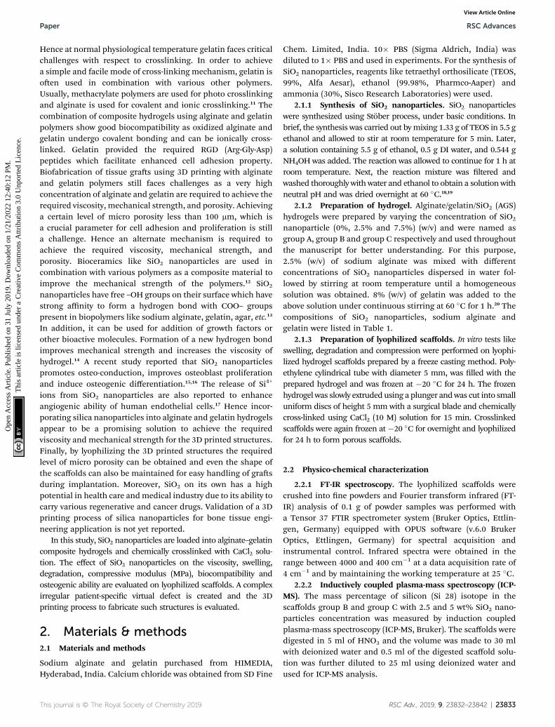
SiO2 nanoparticles were successfully integrated into the algi-nate–gelatin hydrogel system. Fig. 1A depicts the possible
This journal is © The Royal Society of Chemistry 2019
mechanism of SiO2 nanoparticles integration in the alginate–gelatin hydrogel system forming a SiO2 nanoparticle ink. Toconrm the chemical structure of the functional groups and tocheck the purity of the prepared samples, FTIR spectra of thelyophilized hydrogels and the lyophilized hydrogels aer 72hours immersion in 1� PBS are shown in Fig. 1A and Brespectively. The absorption band at around 799 cm�1 is arisingfrom the symmetric vibration of the Si–O bond. The bandappearing at 942 cm�1 is assigned to the asymmetric vibrationof Si–OH. The band at around 1080 cm�1 corresponds to theasymmetric stretching vibration of the Si–O–Si bond.17,27 All thebands apart from the characteristic bands of SiO2 can beattributed to the characteristic bands of alginate and gelatin.The bands at 1645, 1535 and 1243 cm�1 were identied to theC]O vibration, bending modes of C]N and N–H vibrationrespectively. The characteristic bands of sodium alginateappearing at 1312 and 1413 cm�1 were assigned to the asym-metric and symmetric stretching of –COO groups, respectively.The strong bands at 1413 cm�1 in samples aer immersion intoPBS (Fig. 1C) correspond to the symmetric vibrations of C]O.13,14,28 The bending modes observed at 1020 cm�1 correspondto the (PO4)
3� bending mode indicating the precipitation ofphosphate from phosphate buffer.29 The elemental concentra-tion of silicon (Si 28) isotope in the scaffolds with 2.5 and 5 wt%addition of SiO2 nanoparticles was found to be 12.41� 2.55 and25.59 � 1.01 g kg�1 respectively. Fig. 2A shows the masspercentage of silicon in scaffold groups B and C. ESI 1† showsthe SEM image of SiO2 nanoparticles at different magnica-tions. It was observed that particles are spherical inmorphology. Average particle size was calculated using Image Jsoware and it was found to be 64 � 8.9 nm. The number ofSiO2 nanoparticles in all the sample groups calculated usingSEM images are presented in Table 1. The calculations used forthe same are described in ESI 1.† The maximum SiO2 contentcould not exceed 7.5 wt% by in situ synthesis; beyond thisconcentration, phase separation occurred, and a uniform andhomogenous gel could not be obtained. The viscosity of all thesample groups is shown in Fig. 2B. As SiO2 nanoparticleconcentration increases in the alginate–gelatin hydrogelsystem, their viscosity gradually increases up to 5% of SiO2
nanoparticle concentration at a shear rate of 10 s�1. For group Bwith 2.5% SiO2 nanoparticle concentration, the viscosity is thehighest. There aer even with the increase in the SiO2 nano-particle concentration viscosity remains approximately same upto 5% SiO2 concentration and found to be decreasing witha gradual addition up to 7.5% (ESI 2†).
Swelling & degradation percentage of lyophilized scaffold aer72 hours immersion in 1� PBS at 37 �C are represented in Fig. 2Cand D respectively. With increasing SiO2 nanoparticle concentra-tion, swelling reduces. Swelling is minimum for group C. Degra-dation was also found to be reduced with increasing in SiO2
nanoparticles concentration (Table 1). Viscosity, swelling anddegradation property of the prepared group C hydrogel is comparedwith the commercially available bioinks from vendors like Cellink(Cellink, Sweden), BioInk (RegenHU, Switzerland) and Bio-Gel(BioBots, US) and is presented in Table 2. Group C shows betterviscosity, swelling and degradation when compared with
RSC Adv., 2019, 9, 23832–23842 | 23835
Fig. 1 Possible mechanism in of SiO2 integration in the alginate–gelatin hydrogel system (A), FTIR spectra of lyophilized hydrogels (B) and FTIRspectra of lyophilized hydrogels after 72 hours immersion in PBS (C) respectively.
RSC Advances Paper
Ope
n A
cces
s A
rtic
le. P
ublis
hed
on 3
1 Ju
ly 2
019.
Dow
nloa
ded
on 1
/21/
2022
12:
40:1
2 PM
. T
his
artic
le is
lice
nsed
und
er a
Cre
ativ
e C
omm
ons
Attr
ibut
ion
3.0
Unp
orte
d L
icen
ce.
View Article Online
nanocellulose based bioink provided by Cellink. Scanning electronmicrographs of the lyophilized scaffolds of all sample groups areshown in Fig. 3. The surface morphology of the scaffolds at lowermagnication indicates that group B has a smaller pore sizecompared to that of group A and group C, however, micro porosityis profound in group Cwhen compared to group A and B.When theSiO2 addition in the hydrogel system reaches beyond 5 wt% i.e., at7.5 wt%, the nanoparticles tend to agglomerate and are precipitatedon the surface of the lyophilized samples as evident from ESI 2.†The gradual increase in SiO2 nanoparticle concentration fromgroupA to group C increases the compressive modulus of the lyophilizedscaffolds. The compressive modulus (MPa) of the samples of allgroups are statistically signicant (P < 0.001). Sample groups A, B,and C are subjected for biocompatibility tests using FDA/PI livedead staining (Fig. 5), Alamar blue dye reduction assay (Fig. 6A) andpico green total DNA quantication assay (Fig. 6B). FDA/PI staineduorescence micrographs of day 1 indicate the cell attachment onthe surface of the scaffolds of all sample groups but cells are morerounded in group A and B resembling cells embedded in a typicalhydrogel. Attached cells on the surface of group C show moreprotrusions when compared to group A and B. FDA/PI images ob-tained on day 7 indicate the proliferation of cells in all samplegroups and are more predominant in group C.
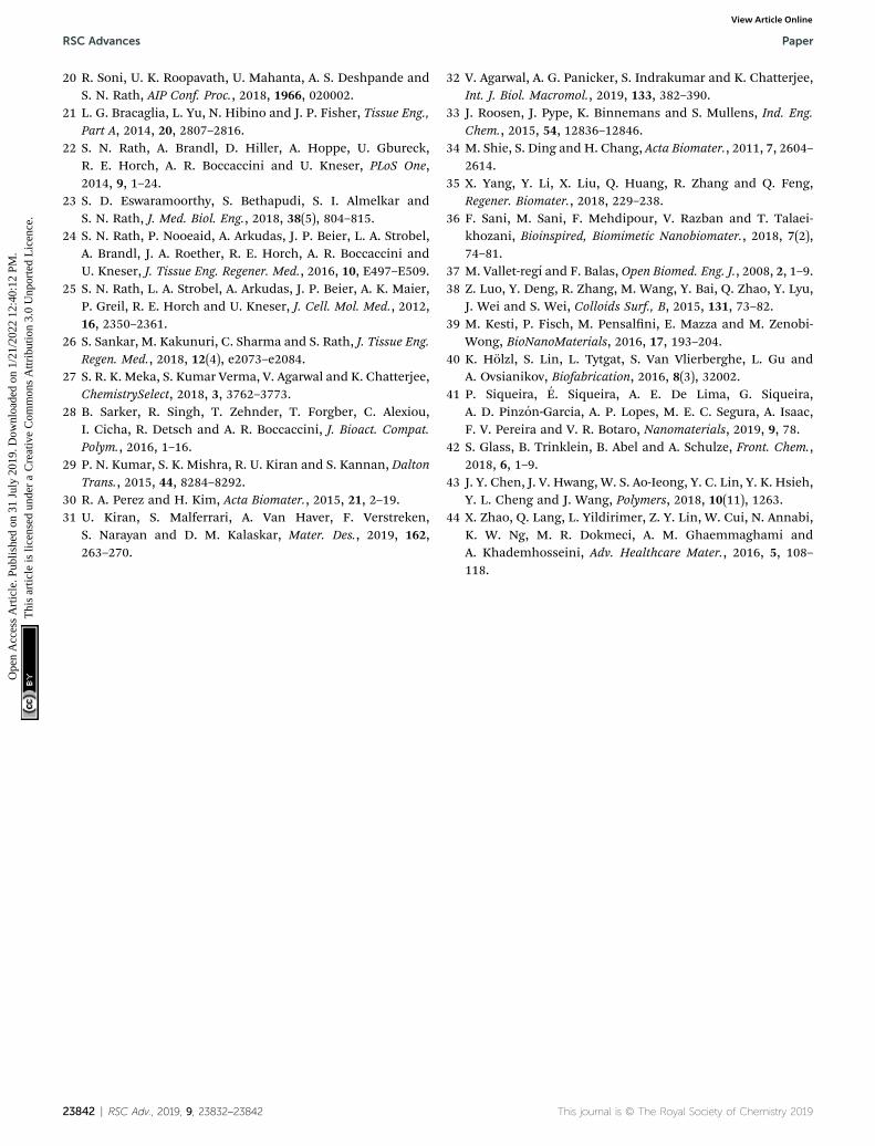
The Alamar blue dye reduction assay performed for days 1,7, 14 and 21 indicate the signicant increase in metabolic
23836 | RSC Adv., 2019, 9, 23832–23842
activity of the cells in all sample groups between all the timepoints from day 1 to day 21. There is no statistical signi-cance in the metabolic activity of the cells seeded on scaffoldsbetween all sample groups aer day 1 indicating the samecell seeding density on the surface of the scaffolds. There isno signicant difference in the metabolic activity of group Aand group B until day 14 but the metabolic activity of group Bis signicantly different (P < 0.05) from group A on day 21.The metabolic activity of group C is signicantly higher whencompared to group A on all time points from day 7 to 21.There is no statistical signicance between group B andgroup C on day 7 but group C shows a signicant increase inmetabolic activity compared group B on day 14 and day 21.Total DNA quantication performed on cell seeded scaffoldsof all sample groups from day 1 to day 21 are in closeagreement with the results obtained from Alamar blue dyereduction assay. There is a signicant increase in the DNAcontent of all sample groups between different time pointsfrom day 1 to day 21. The total DNA content of group Csample from day 7 to day 21 is signicantly higher whencompared to group A. The DNA content of group C asobserved on day 14 and day 21 is statistically signicant whencompared with the DNA content of group B. The alkalinephosphatase activity (Fig. 6C) was analyzed for all the samplegroups to study the differentiation of UMSCs into osteogenic
This journal is © The Royal Society of Chemistry 2019

Fig. 2 Viscosity of the hydrogels used for 3D printing (A), swelling percentage (B), degradation percentage (C) and mass percentage calculatedusing ICP-MS (D) of the lyophilized hydrogels. * indicates a significant difference between sample groups with P < 0.005.
Paper RSC Advances
Ope
n A
cces
s A
rtic
le. P
ublis
hed
on 3
1 Ju
ly 2
019.
Dow
nloa
ded
on 1
/21/
2022
12:
40:1
2 PM
. T
his
artic
le is
lice
nsed
und
er a
Cre
ativ
e C
omm
ons
Attr
ibut
ion
3.0
Unp
orte
d L
icen
ce.
View Article Online
lineage. ALP activity (IU L�1) of group A samples showa signicant difference from day 1 to day 14 but day 14 andday 21 are not signicant. ALP activity of group B samplesshows a signicant increase from day 1 to day 7 and day 7 today 21. Whereas for group C samples the ALP activity isincreasing for all time points from day 1 to day 21 whencompared among them. The statistical signicance betweenthe groups shows that group B shows increased ALP activitythan group A from day 7 to day 21. Group C exhibited
Table 2 Table showing the viscosity (Pa s), swelling% and degradation%with 5 wt% SiO2 nanoparticle concentration)
Company Bioink Materials Viscosity (Pa s)
CELLINK CELLINK 1.36% nanocellulose and 0.5%alginate crosslinked with cationicsolution
11 � 0.7
RegenHU BioInk® Polyethyleneglycol-diacrylate(PEGDA) photo-crosslinked withphotoinitiator
1.05 � 0.09 (100PEGDA)
Biobot BioGel 10% gelatin methacrylate photo-crosslinked with 0.05% IrgacureI2959
65 � 14
Group C Asprepared
Alginate/gelatin/SiO2 nanoparticlebased
13.65 � 3
This journal is © The Royal Society of Chemistry 2019
a signicant increase in ALP activity (IU L�1) when comparedwith the ALP activity of both group A and group B from day 7to day 21. A virtual skull defect as depicted in Fig. 7 wassuccessfully 3D printed using the formulated nanoparticleink and the 3D printed defect was subjected for lyophiliza-tion. The lyophilized structure was similar to that of the 3Dprinted structure and to the designed CAD model of thevirtual defect. No major change in the external structure withrespect to volume was observed aer lyophilization.
of commercially available bioinks and the prepared hydrogel (group C
Swelling% Degradation% Ref.
1145 � 42 70 � 5 39–41
wt% 342 � 3 (100 wt%PEGDA)
53.56 � 6.16 (100 wt%PEGDA)
39, 40, 42 and43
719 � 24 30 � 2 39, 40 and 44
998.27 � 87.54 61.05 � 4.26
RSC Adv., 2019, 9, 23832–23842 | 23837

Fig. 3 SEM images of the lyophilized hydrogels (A), (B) and (C) indicate sample groups A, B and C respectively at scale bar 500 mm and (D), (E) and(F) indicate higher resolution images of sample groups A, B and C respectively at scale bar 200 mm.
Fig. 4 Compressive moduli (MPa) of lyophilized hydrogels. *** indi-cates statistical significance with P < 0.001.
RSC Advances Paper
Ope
n A
cces
s A
rtic
le. P
ublis
hed
on 3
1 Ju
ly 2
019.
Dow
nloa
ded
on 1
/21/
2022
12:
40:1
2 PM
. T
his
artic
le is
lice
nsed
und
er a
Cre
ativ
e C
omm
ons
Attr
ibut
ion
3.0
Unp
orte
d L
icen
ce.
View Article Online
4. Discussions
The major challenge in using hydrogels for tissue fabrication istheir lack of regeneration ability, usage of chemicals like CaCl2for crosslinking and their ability to hold 3D structure on theirown. The regeneration ability of hydrogels can be increased byloading such hydrogels with various regenerative drugs andgrowth factors. But controlling the release of such drugs andgrowth factors is still a challenge and can be rectied by usingcarries such as silica nanoparticles.16,30 Hence validating theincorporation of SiO2 nanoparticles in hydrogel polymernetwork which can be 3D printable according to patient's crit-ical defects has immense scope in bone tissue engineering. Inthis study, we have shown successfully incorporated SiO2
nanoparticles in a 3D printable hydrogel polymer network andvalidated the process for patient specic defect 3D printing. Theviscosity of hydrogels is an important parameter for extrusion-based 3D printing and is expected to increase with the addi-tion of ceramic particles like SiO2 nanoparticles.31 Hence withthe gradual addition of up to 2.5 wt%, the viscosity of thehydrogels increased gradually. But aer 2.5 wt% up to 5 wt%addition, the viscosity remained almost same indicating thesaturation and phase separation hence reduction in resis-tance.32 Thereaer, when the concentration reached to 7.5 wt%the viscosity appeared to be lower than 2.5% (data not shown).Aer a certain addition of silica, they tend to agglomerate andlose their colloidal property hence start to settle at the bottom ofthe hydrogel suspension. Once the homogenous dispersion ofSiO2 nanoparticles in the hydrogel suspension is lost they aredeemed to be not suitable for 3D printing hence 2.5 wt% and
23838 | RSC Adv., 2019, 9, 23832–23842
5 wt% silica incorporated hydrogels were selected for furtheranalysis. ICP-MS analysis also conrms the increase in siliconelemental concentration with addition of SiO2 nanoparticles inthe hydrogel system. As the SiO2 nanoparticle concentration inthe alginate–gelatin hydrogel mixture increases, the free OHgroups present on the surface of SiO2 facilitate more bondingsites for the formation of hydrogen bond between SiO2 andsodium alginate as well as SiO2 and gelatin.
As evident from the FTIR spectrum all the peaks correspondingto SiO2 nanoparticles are present in group B and group C which areclearly absent in group A (Fig. 1B andC). This indicates the presence
This journal is © The Royal Society of Chemistry 2019
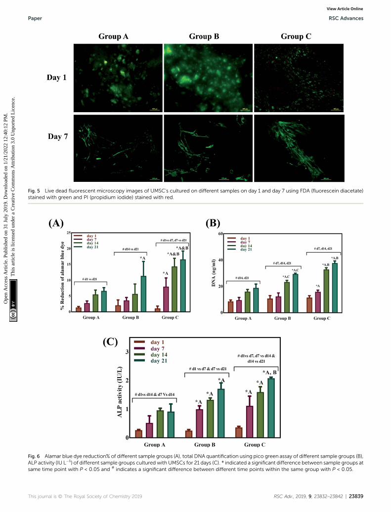
Fig. 5 Live dead fluorescent microscopy images of UMSC's cultured on different samples on day 1 and day 7 using FDA (fluorescein diacetate)stained with green and PI (propidium iodide) stained with red.
Fig. 6 Alamar blue dye reduction% of different sample groups (A), total DNA quantification using pico green assay of different sample groups (B),ALP activity (IU L�1) of different sample groups cultured with UMSCs for 21 days (C). * indicated a significant difference between sample groups atsame time point with P < 0.05 and # indicates a significant difference between different time points within the same group with P < 0.05.
This journal is © The Royal Society of Chemistry 2019 RSC Adv., 2019, 9, 23832–23842 | 23839
Paper RSC Advances
Ope
n A
cces
s A
rtic
le. P
ublis
hed
on 3
1 Ju
ly 2
019.
Dow
nloa
ded
on 1
/21/
2022
12:
40:1
2 PM
. T
his
artic
le is
lice
nsed
und
er a
Cre
ativ
e C
omm
ons
Attr
ibut
ion
3.0
Unp
orte
d L
icen
ce.
View Article Online

RSC Advances Paper
Ope
n A
cces
s A
rtic
le. P
ublis
hed
on 3
1 Ju
ly 2
019.
Dow
nloa
ded
on 1
/21/
2022
12:
40:1
2 PM
. T
his
artic
le is
lice
nsed
und
er a
Cre
ativ
e C
omm
ons
Attr
ibut
ion
3.0
Unp
orte
d L
icen
ce.
View Article Online
of SiO2 nanoparticles in the hydrogel network. Even aer 72 hoursimmersion in PBS, the peaks corresponding to SiO2 are quiteevident. The Si–O group at 1080 cm �1 and C–O–C group at1070 cm�1 are merging together indicating hydrogen bondingbetween silica and alginate–gelatin polymer network. Therefore, theactive sites facilitating the binding of water molecules are alsoreduced due to the hydrogen bonding with SiO2. This may becorrelated to the decrease in swelling percentage of the hydrogelsystem with an increase in the SiO2 concentration. Swelling is alsoused to determine the extent of crosslinking. More degree ofswelling results in less crosslinking and vice versa. In this case,though all the hydrogel groups are crosslinked using CaCl2 solutionfor the same duration, the hydrogels withmore silica concentrationexhibit less swelling. This indicates the additional degree of cross-linking achieved by the hydrogen bonding between silica and algi-nate–gelatin polymer network. The extent of crosslinking alsodetermines the rate of degradation, hence the results of swellingand degradation correlate with each other verifying the interactionof SiO2 and alginate–gelatin polymer network. Swelling and degra-dation properties of hydrogels also have an important role to play intissue engineering the water retaining ability and the degradationare to be controlled to achieve a controlled release of drugs, growthfactors and ions.28,33 Using SiO2 nanoparticles as an additionalcrosslinking agent appears to achieve this objective of controllingthe swelling and degradation properties of the alginate–gelatinhydrogels. With the inclusion of ceramic nanoparticles into thepolymer hydrogel, these hybrid composite materials (group B andC) are expected to show enhanced mechanical properties whencompared with the normal polymer ink (group A) as evident fromFig. 4. The increased compressive modulus (MPa) for group B and
Fig. 7 Process showing the 3D printing of patient-specific virtual bone
23840 | RSC Adv., 2019, 9, 23832–23842
group C samples may also be due to the tight bonding of silica withthe free OH� and COO� functional groups in the alginate andgelatin polymer network. The lyophilized scaffolds exhibit similarmacro porosity across all groups but, appears to be slightly more ingroup B. Whereas, micro porosity is appeared to be signicantlymore in group C as observed from higher magnication SEMimages. This may be due to the pattern of water accumulationduring the process of gelation. As group B has higher viscosity andeven distribution of SiO2 nanoparticles, it shows homogeneousgelation resulting in uniformmacro porosity. In the case of groupC,the hydrogel suspension reaches its maximum capacity to accom-modate SiO2 nanoparticles and water accumulation is minimumenabling the development of micro porosity on the surface duringthe process of lyophilization.
The surface morphology of the scaffolds signicantly affectsthe proliferation and differentiation of MSCs. Hence, group Cwith micro porosity facilitates the adherence of MSCs betterthan group A and group B by facilitating greater surface areaand nutrient inltration.34 The cell viability on the scaffolds isin correlation with the earlier reports suggesting the prolifera-tion of cells as the effect of silica nanoparticles. The signicantincrease in the DNA content of cells seeded on the surface ofgroup C indicates the proliferation of cells with time from day 1to day 21. Themetabolic activity analyzed using Alamar blue dyereduction assay and total DNA quantication using pico greenagree with each other. SiO2 nanoparticles are known to promoteosteogenesis, the release of silicon ions have a direct impact onpromoting osteogenic pathways thereby enhancing osteo-genesis.34,35 As reported by Shie et al.,34 Si ion concentration atan appropriate level helps in the proliferation of osteoblast like
defect.
This journal is © The Royal Society of Chemistry 2019

Paper RSC Advances
Ope
n A
cces
s A
rtic
le. P
ublis
hed
on 3
1 Ju
ly 2
019.
Dow
nloa
ded
on 1
/21/
2022
12:
40:1
2 PM
. T
his
artic
le is
lice
nsed
und
er a
Cre
ativ
e C
omm
ons
Attr
ibut
ion
3.0
Unp
orte
d L
icen
ce.
View Article Online
cells and actively stimulate the production of osteo specicproteins. Hence the ALP activity of group C scaffolds is signi-cantly higher when compared to group A and group B scaffolds.This may be due to the effect of Si ions, as they actively stimulatethe entry of cells into S and G2 phases of cell division. SiO2
nanoparticles have various applications in tissue engineeringand regenerative medicine. The functionalization ability of SiO2
nanoparticles makes them an effective carrier for various drugs,growth factors.30,36–38 3D printing as a bio fabrication techniqueto develop patient-specic bone gras has taken place in recentyears for effective bone regeneration therapies. The SiO2 nano-particles seem to enhance osteogenic ability when incorporatedin the alginate–gelatin hydrogel mixture but the validation ofthe prepared hydrogel ink for 3D printing a patient-specicdefect is crucial for tissue engineering application. The virtualirregular large scale defect created on a skull model usinga patient CT scan was successfully 3D printed using 5 wt% silicaloaded hydrogel ink. This is assumed to show better cellviability and enhanced osteogenic ability as observed from cellproliferation studies and ALP assay. By replacing the SiO2
nanoparticles withmesoporous SiO2 nanoparticles (MSNs) theirability to deliver regenerative drugs and growth factors can beexplored further. Various previous studies have explored theability of MSNs in controlled release of anticancer drugs byfunctionalizing them with various bioactive compounds.37 Thisability of SiO2 nanoparticles in synergy with their osteogenicability offers huge scope for the above validated technique fortheir use in 3D printed models for bone tissue engineering anddrug delivery applications.
5. Conclusion
Addition of SiO2 nanoparticles into the hydrogel system hasincreased the viscosity of the hydrogel ink up to a certainconcentration of 2.5 wt%, which increased printability of thescaffold. Compressive modulus (MPa) has been signicantlyimproved whereas, swelling and degradation properties aresignicantly inhibited. Micro porosity favoring cell attachmentand proliferation can also be enhanced. Biocompatibility andosteogenic ability of the hydrogels are signicantly increasedwith the addition of SiO2. 3D printing of SiO2 nanoparticlehydrogel composite ink to fabricate a bone gra using a patient-specic virtual defect was successfully validated. Hence thistype of hydrogel composite ink has huge potential and scope forits application in tissue engineering and nanomedicine. Thisstudy of validating the 3D printing of SiO2 nanoparticles opensthe possibility of exploring the use of mesoporous SiO2 andfunctionalizing the nanoparticles with desirable growth factorsand drugs. This approach seems to be promising for creating animpact in the health care industry.
Ethical approval
This article does not contain any clinical study with humanparticipants or animals performed by any of the authors.
This journal is © The Royal Society of Chemistry 2019
Conflicts of interest
The authors declare that there is no conict of interest.
Acknowledgements
The authors also acknowledge the Ministry of Human Resourceand Development (MHRD) for nancial assistance to authorRUK and Institutional Central Facilities of Indian Institute ofTechnology, Hyderabad for the characterization of developedbiomaterials.
References
1 K. Tucker, D. Tucker, J. Eastham, E. Gibson, S. Varma andT. Daim, Technol. Invest., 2014, 05, 137–156.
2 Y. He, F. Yang, H. Zhao, Q. Gao, B. Xia and J. Fu, Sci. Rep.,2016, 6, 29977.
3 R. N. Maniar and T. Singhi, Curr. Rev. Musculoskelet. Med.,2014, 7, 125–130.
4 B. Derby, Science, 2012, 338, 921–926.5 U. K. Roopavath and D. M. Kalaskar, in 3D Printing inMedicine, Woodhead Publishing, 2017, pp. 1–20.
6 H. N. Chia and B. M. Wu, J. Biol. Eng., 2015, 9, 4.7 F. You, X. Wu and X. Chen, Int. J. Polym. Mater. Polym.Biomater., 2017, 66, 299–306.
8 H.-W. Kang, S. J. Lee, I. K. Ko, C. Kengla, J. J. Yoo and A. Atala,Nat. Biotechnol., 2016, 34, 312–319.
9 L. Wang, R. M. Shelton, P. R. Cooper, M. Lawson, J. T. Triffittand J. E. Barralet, Biomaterials, 2003, 24, 3475–3481.
10 M. C. Echave, L. Saenz del Burgo, J. L. Pedraz and G. Orive,Curr. Pharm. Des., 2017, 23, 3567–3584.
11 T. Zehnder, B. Sarker, A. R. Boccaccini and R. Detsch,Biofabrication, 2015, 7, 025001.
12 A. G. B. Castro, M. Diba, M. Kersten, J. A. Jansen, J. J. J. P. vanden Beucken and F. Yang, Mater. Sci. Eng., C, 2018, 85, 154–161.
13 W. Aljohani, M. W. Ullah, W. Li, L. Shi, X. Zhang andG. Yang, J. Polym. Res., 2018, 25, 62.
14 J. A. Sowjanya, J. Singh, T. Mohita, S. Sarvanan, A. Moorthi,N. Srinivasan and N. Selvamurugan, Colloids Surf., B, 2013,109, 294–300.
15 R. K. Singh, G. Z. Jin, C. Mahapatra, K. D. Patel,W. Chrzanowski and H. W. Kim, ACS Appl. Mater.Interfaces, 2015, 7, 8088–8098.
16 W. Cui, Q. Liu, L. Yang, K. Wang, T. Sun, Y. Ji, L. Liu, W. Yu,Y. Qu, J. Wang, Z. Zhao, J. Zhu and X. Guo, ACS Biomater. Sci.Eng., 2018, 4, 211–221.
17 S. R. K. Meka, V. Agarwal and K. Chatterjee, Mater. Sci. Eng.,C, 2019, 94, 565–579.
18 X. D. Wang, Z. X. Shen, T. Sang, X. Bin Cheng, M. F. Li,L. Y. Chen and Z. S. Wang, J. Colloid Interface Sci., 2010,341, 23–29.
19 S. L. Greasley, S. J. Page, S. Sirovica, S. Chen, R. A. Martin,A. Riveiro, J. V. Hanna, A. E. Porter and J. R. Jones, J.Colloid Interface Sci., 2016, 469, 213–223.
RSC Adv., 2019, 9, 23832–23842 | 23841
RSC Advances Paper
Ope
n A
cces
s A
rtic
le. P
ublis
hed
on 3
1 Ju
ly 2
019.
Dow
nloa
ded
on 1
/21/
2022
12:
40:1
2 PM
. T
his
artic
le is
lice
nsed
und
er a
Cre
ativ
e C
omm
ons
Attr
ibut
ion
3.0
Unp
orte
d L
icen
ce.
View Article Online
20 R. Soni, U. K. Roopavath, U. Mahanta, A. S. Deshpande andS. N. Rath, AIP Conf. Proc., 2018, 1966, 020002.
21 L. G. Bracaglia, L. Yu, N. Hibino and J. P. Fisher, Tissue Eng.,Part A, 2014, 20, 2807–2816.
22 S. N. Rath, A. Brandl, D. Hiller, A. Hoppe, U. Gbureck,R. E. Horch, A. R. Boccaccini and U. Kneser, PLoS One,2014, 9, 1–24.
23 S. D. Eswaramoorthy, S. Bethapudi, S. I. Almelkar andS. N. Rath, J. Med. Biol. Eng., 2018, 38(5), 804–815.
24 S. N. Rath, P. Nooeaid, A. Arkudas, J. P. Beier, L. A. Strobel,A. Brandl, J. A. Roether, R. E. Horch, A. R. Boccaccini andU. Kneser, J. Tissue Eng. Regener. Med., 2016, 10, E497–E509.
25 S. N. Rath, L. A. Strobel, A. Arkudas, J. P. Beier, A. K. Maier,P. Greil, R. E. Horch and U. Kneser, J. Cell. Mol. Med., 2012,16, 2350–2361.
26 S. Sankar, M. Kakunuri, C. Sharma and S. Rath, J. Tissue Eng.Regen. Med., 2018, 12(4), e2073–e2084.
27 S. R. K. Meka, S. Kumar Verma, V. Agarwal and K. Chatterjee,ChemistrySelect, 2018, 3, 3762–3773.
28 B. Sarker, R. Singh, T. Zehnder, T. Forgber, C. Alexiou,I. Cicha, R. Detsch and A. R. Boccaccini, J. Bioact. Compat.Polym., 2016, 1–16.
29 P. N. Kumar, S. K. Mishra, R. U. Kiran and S. Kannan, DaltonTrans., 2015, 44, 8284–8292.
30 R. A. Perez and H. Kim, Acta Biomater., 2015, 21, 2–19.31 U. Kiran, S. Malferrari, A. Van Haver, F. Verstreken,
S. Narayan and D. M. Kalaskar, Mater. Des., 2019, 162,263–270.
23842 | RSC Adv., 2019, 9, 23832–23842
32 V. Agarwal, A. G. Panicker, S. Indrakumar and K. Chatterjee,Int. J. Biol. Macromol., 2019, 133, 382–390.
33 J. Roosen, J. Pype, K. Binnemans and S. Mullens, Ind. Eng.Chem., 2015, 54, 12836–12846.
34 M. Shie, S. Ding and H. Chang, Acta Biomater., 2011, 7, 2604–2614.
35 X. Yang, Y. Li, X. Liu, Q. Huang, R. Zhang and Q. Feng,Regener. Biomater., 2018, 229–238.
36 F. Sani, M. Sani, F. Mehdipour, V. Razban and T. Talaei-khozani, Bioinspired, Biomimetic Nanobiomater., 2018, 7(2),74–81.
37 M. Vallet-regı and F. Balas, Open Biomed. Eng. J., 2008, 2, 1–9.38 Z. Luo, Y. Deng, R. Zhang, M. Wang, Y. Bai, Q. Zhao, Y. Lyu,
J. Wei and S. Wei, Colloids Surf., B, 2015, 131, 73–82.39 M. Kesti, P. Fisch, M. Pensalni, E. Mazza and M. Zenobi-
Wong, BioNanoMaterials, 2016, 17, 193–204.40 K. Holzl, S. Lin, L. Tytgat, S. Van Vlierberghe, L. Gu and
A. Ovsianikov, Biofabrication, 2016, 8(3), 32002.41 P. Siqueira, E. Siqueira, A. E. De Lima, G. Siqueira,
A. D. Pinzon-Garcia, A. P. Lopes, M. E. C. Segura, A. Isaac,F. V. Pereira and V. R. Botaro, Nanomaterials, 2019, 9, 78.
42 S. Glass, B. Trinklein, B. Abel and A. Schulze, Front. Chem.,2018, 6, 1–9.
43 J. Y. Chen, J. V. Hwang, W. S. Ao-Ieong, Y. C. Lin, Y. K. Hsieh,Y. L. Cheng and J. Wang, Polymers, 2018, 10(11), 1263.
44 X. Zhao, Q. Lang, L. Yildirimer, Z. Y. Lin, W. Cui, N. Annabi,K. W. Ng, M. R. Dokmeci, A. M. Ghaemmaghami andA. Khademhosseini, Adv. Healthcare Mater., 2016, 5, 108–118.
This journal is © The Royal Society of Chemistry 2019


![Ultrafine Nickel‐Nanoparticle‐Enabled SiO2 Hierarchical ...mai.group.whut.edu.cn/chs/lw/slected/201802/P...x/NiSi x with enhanced structural stability and kinetics.[34] However,](https://static.fdocuments.us/doc/165x107/600aa20a049ab32e231cee93/ultrafine-nickelananoparticleaenabled-sio2-hierarchical-maigroupwhuteducnchslwslected201802p.jpg)