3D Collagen Alignment Limits Protrusions to Enhance Breast ...
15
Georgia State University Georgia State University ScholarWorks @ Georgia State University ScholarWorks @ Georgia State University Mathematics and Statistics Faculty Publications Department of Mathematics and Statistics 2014 3D Collagen Alignment Limits Protrusions to Enhance Breast 3D Collagen Alignment Limits Protrusions to Enhance Breast Cancer Cell Persistence Cancer Cell Persistence Kristin M. Riching University of Wisconsin-Madison, [email protected] Benjamin L. Cox University of Wisconsin-Madison, [email protected] Max R. Salick University of Wisconsin-Madison Carolyn Pehleke University of Wisconsin-Madison, [email protected] Andrew S. Riching University of Wisconsin-Madison See next page for additional authors Follow this and additional works at: https://scholarworks.gsu.edu/math_facpub Part of the Mathematics Commons Recommended Citation Recommended Citation Riching, Kristin M.; Cox, Benjamin L.; Salick, Max R.; Pehleke, Carolyn; Riching, Andrew S.; Ponik, Susan M.; Bass, Benjamin R.; Crone, Wendy C.; Jiang, Yi; Weaver, Alissa M.; Eliceiri, Kevin W.; and Keely, Patricia J., "3D Collagen Alignment Limits Protrusions to Enhance Breast Cancer Cell Persistence" (2014). Mathematics and Statistics Faculty Publications. 13. https://scholarworks.gsu.edu/math_facpub/13 This Article is brought to you for free and open access by the Department of Mathematics and Statistics at ScholarWorks @ Georgia State University. It has been accepted for inclusion in Mathematics and Statistics Faculty Publications by an authorized administrator of ScholarWorks @ Georgia State University. For more information, please contact [email protected].
Transcript of 3D Collagen Alignment Limits Protrusions to Enhance Breast ...
Georgia State University Georgia State University
ScholarWorks @ Georgia State University ScholarWorks @ Georgia State University
Mathematics and Statistics Faculty Publications Department of Mathematics and Statistics
2014
3D Collagen Alignment Limits Protrusions to Enhance Breast 3D Collagen Alignment Limits Protrusions to Enhance Breast
Cancer Cell Persistence Cancer Cell Persistence
Kristin M. Riching University of Wisconsin-Madison, [email protected]
Benjamin L. Cox University of Wisconsin-Madison, [email protected]
Max R. Salick University of Wisconsin-Madison
Carolyn Pehleke University of Wisconsin-Madison, [email protected]
Andrew S. Riching University of Wisconsin-Madison
See next page for additional authors
Follow this and additional works at: https://scholarworks.gsu.edu/math_facpub
Part of the Mathematics Commons
Recommended Citation Recommended Citation Riching, Kristin M.; Cox, Benjamin L.; Salick, Max R.; Pehleke, Carolyn; Riching, Andrew S.; Ponik, Susan M.; Bass, Benjamin R.; Crone, Wendy C.; Jiang, Yi; Weaver, Alissa M.; Eliceiri, Kevin W.; and Keely, Patricia J., "3D Collagen Alignment Limits Protrusions to Enhance Breast Cancer Cell Persistence" (2014). Mathematics and Statistics Faculty Publications. 13. https://scholarworks.gsu.edu/math_facpub/13
This Article is brought to you for free and open access by the Department of Mathematics and Statistics at ScholarWorks @ Georgia State University. It has been accepted for inclusion in Mathematics and Statistics Faculty Publications by an authorized administrator of ScholarWorks @ Georgia State University. For more information, please contact [email protected].

Authors Authors Kristin M. Riching, Benjamin L. Cox, Max R. Salick, Carolyn Pehleke, Andrew S. Riching, Susan M. Ponik, Benjamin R. Bass, Wendy C. Crone, Yi Jiang, Alissa M. Weaver, Kevin W. Eliceiri, and Patricia J. Keely
This article is available at ScholarWorks @ Georgia State University: https://scholarworks.gsu.edu/math_facpub/13

Article
3D Collagen Alignment Limits Protrusions to Enhance Breast Cancer CellPersistence
Kristin M. Riching,1,2 Benjamin L. Cox,2,3 Max R. Salick,4,5 Carolyn Pehlke,2 Andrew S. Riching,6
Susan M. Ponik,6 Benjamin R. Bass,8 Wendy C. Crone,1,4,5 Yi Jiang,9 Alissa M. Weaver,10
Kevin W. Eliceiri,1,2,7 and Patricia J. Keely1,2,6,7,*1Biomedical Engineering Program, 2Laboratory for Optical and Computational Instrumentation, 3Department of Medical Physics, 4MaterialsScience Program, 5Department of Engineering Physics, 6Department of Cell and Regenerative Biology, and 7University of Wisconsin Paul P.Carbone Comprehensive Cancer Center, University of Wisconsin-Madison, Madison, Wisconsin; 8Saris Cycling Group, Madison, Wisconsin;9Department of Mathematics and Statistics, Georgia State University, Atlanta, Georgia; and 10Department of Cancer Biology, VanderbiltUniversity Medical Center, Nashville, Tennessee
ABSTRACT Patients with mammographically dense breast tissue have a greatly increased risk of developing breast cancer.Dense breast tissue contains more stromal collagen, which contributes to increased matrix stiffness and alters normal cellularresponses. Stromal collagen within and surrounding mammary tumors is frequently aligned and reoriented perpendicular to thetumor boundary. We have shown that aligned collagen predicts poor outcome in breast cancer patients, and postulate this isbecause it facilitates invasion by providing tracks on which cells migrate out of the tumor. However, the mechanisms by whichalignment may promote migration are not understood. Here, we investigated the contribution of matrix stiffness and alignmentto cell migration speed and persistence. Mechanical measurements of the stiffness of collagen matrices with varying densityand alignment were compared with the results of a 3D microchannel alignment assay to quantify cell migration. We further in-terpreted the experimental results using a computational model of cell migration. We find that collagen alignment confers anincrease in stiffness, but does not increase the speed of migrating cells. Instead, alignment enhances the efficiency of migrationby increasing directional persistence and restricting protrusions along aligned fibers, resulting in a greater distance traveled.These results suggest that matrix topography, rather than stiffness, is the dominant feature by which an aligned matrix canenhance invasion through 3D collagen matrices.
INTRODUCTION
Increased mammographic density is associated with a 4-to 6-fold increased risk of breast cancer (1–3), makingmammographic density one of the greatest independentrisk factors for breast cancer (1,3,4). This increase in densityis correlated with a significantly increased deposition ofextracellular matrix (ECM) proteins, most notably collagenI (5–7), which is in part responsible for the overall increasein stiffness in mammary tumors (8,9). Matrix stiffness hasbeen shown to promote a malignant phenotype in tumorcells (8,10–12), enhance migration and invasion (13–16),and alter cell signaling, leading to increased proliferation(10,17–19). Although it is clear that matrix stiffness playsa profound role in tumor progression, we do not yet fully un-derstand the mechanisms by which cells respond to changesin matrix stiffness.
In addition to the amount of collagen, the orientation ofcollagen fibers appears to play a critical role in tumor pro-gression. Our laboratory previously characterized changesin the alignment and orientation of collagen fibers, and iden-tified tumor-associated collagen signatures (TACS), which
manifest in predictable ways during tumor progression. Inparticular, deposition of aligned collagen that is orientedperpendicular to the tumor boundary (termed TACS-3) cre-ates highways on which tumor cells are observed to migratein vivo (20), and correlates with increased invasion andmetastasis in mouse models (21). More recently, we showedthat TACS-3 alignment is an independent prognostic signa-ture that correlates strongly with poor patient survival (22).These initial findings strongly indicate that matrix stiffnessresulting from increased collagen deposition and matrixalignment contributes to mammary tumor progression.
Although the cellular players andmechanism for alignmentgeneration in vivo remain elusive, in vitro studies have shownthat epithelial cells and fibroblasts are capable of using Rho-and Rho kinase (ROCK)-mediated actin-myosin contractilityto orient collagen fibers (23–26). Additionally, fibroblasts candeposit matrices containing aligned fibronectin or collagenin vitro (27,28). Recently, Yang et al. (28) showed that thisability of fibroblasts to produce aligned matrices is associatedwith expression of the cell-surface proteoglycan syndecan-1.
The high correlation of collagen alignment with breasttumor progression suggests that the mechanisms by whichalignment facilitates cell migration need to be evaluatedmore closely. Studies of cells cultured in matrices with
Submitted May 8, 2014, and accepted for publication October 3, 2014.
*Correspondence: [email protected]
Editor: David Odde.
� 2014 by the Biophysical Society
0006-3495/14/12/2546/13 $2.00 http://dx.doi.org/10.1016/j.bpj.2014.10.035
2546 Biophysical Journal Volume 107 December 2014 2546–2558

aligned fibers have revealed that cells polarize and orientwith respect to the alignment (29–31), and that alignmentis associated with increased migration and directionality(23,28,32). The underlying mechanisms for these responsesto alignment, however, remain unclear. One possibility isthat alignment organizes cell adhesions along fibers, result-ing in more efficient migration from coordinated tractionforces. It has been demonstrated that parallel-oriented fibersmay also afford cells less spatial impedance and therebyenhance migration (33). Additionally, it has been suggestedthat alignment incurs changes in matrix stiffness, and thatenhanced migration along fibers may be due to durotacticguidance. However, the effects of increasing alignment ontensile modulus have been largely assumed, without beingwell documented and quantified. Here, we created novel(to our knowledge) aligned matrices to parse out the con-tributions of 3D matrix alignment and stiffness to the migra-tion of breast cancer cells. We find that alignment facilitatespersistence without affecting migration speed, and that con-tact guidance via aligned fibers, rather than durotaxis dueto stiffness, is the likely mechanism by which migration isenhanced. Moreover, we present a model to describe trendsin cell migration induced by matrix physical and mechanicalproperties. Validating the model’s prediction, we found thatan aligned matrix limits the number of stabilized protrusionsand likely serves as mechanism that leads to more persistentmigration.
RESULTS
The aim of this study was to characterize the effects ofcollagen fiber alignment and matrix stiffness on the 3Dcell migration of invasive ductal breast carcinoma cells. To
study the mechanical properties of aligned collagen andhow cells respond to changing matrix mechanics, we gener-ated 3D collagen gels containing aligned fibers by using adevice to impart mechanical strain. Similar to the methoddescribed by Vader et al. (34), we designed and used a 3Dprinter to create a strain device that uses a micrometer-driven arm and pin assembly to stretch one end of a collagengel (Fig. 1 A). The pins contacted two pieces of polypro-pylene mesh embedded in the gel and had a width of 1 cmto produce a wide aligned region. The device was designedto fit the stage of a multiphoton microscope, and secondharmonic generation (SHG) images of a 2 mg/ml gel werecollected with increasing strain applied (Fig. 1 B). Weused CurveAlign software (http://www.loci.wisc.edu) anal-ysis, which employs a curvelet-based algorithm to measurefiber orientation and assigns a coefficient of alignment fromzero (no alignment of one fiber relative to another) to one(all fibers in perfect alignment). The coefficient of alignmentincreased with strain and appeared to approach a maximumalignment at 30% strain (Fig. 1 C).
To determine whether aligned collagen had a greaterelastic modulus than gels with randomly oriented fibers,we cast 2 mg/ml collagen gels in a stainless-steel, dog-bone-shaped mold that was machined to the dimensionsspecified in Roeder et al. (35). We then tested the tensilemoduli of the gels using an Instron MicroTester 5548.(For an image of a representative collagen gel in the gripsof the Instron, see Fig. S1 B in the Supporting Material.)Samples were either prestrained using the strain deviceshown in Fig. 1 or left unstrained. All samples wereimaged via SHG to observe the fiber alignment and thensubjected to tensile testing at a rate of 1 mm/min until fail-ure occurred. Sample strain was computed by measuring
FIGURE 1 Collagen gels are aligned by me-
chanical strain. (A) A strain device designed and
assembled for use with a multiphoton microscope
is fitted with a micrometer to precisely control
the amount of strain on a collagen gel placed in
the center of the stage. Two pins contact the sample
and the left pin/arm assembly is driven by a micro-
meter. The arrow indicates the direction of strain.
(B) SHG images reveal collagen fiber alignment
with increasing strain; scale bar, 50 mm. (C) Cur-
veAlign software analysis of coefficient of align-
ment from SHG images (n ¼ 3 gels).
Biophysical Journal 107(11) 2546–2558
ECM Alignment Organizes Cell Protrusions 2547
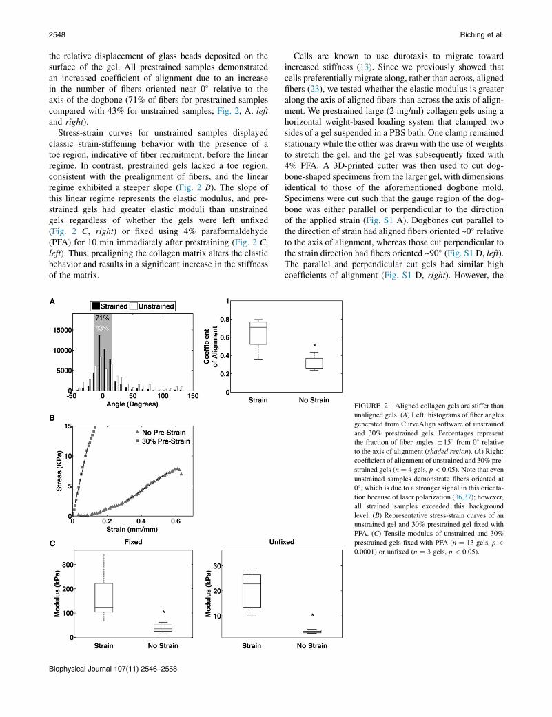
the relative displacement of glass beads deposited on thesurface of the gel. All prestrained samples demonstratedan increased coefficient of alignment due to an increasein the number of fibers oriented near 0� relative to theaxis of the dogbone (71% of fibers for prestrained samplescompared with 43% for unstrained samples; Fig. 2, A, leftand right).
Stress-strain curves for unstrained samples displayedclassic strain-stiffening behavior with the presence of atoe region, indicative of fiber recruitment, before the linearregime. In contrast, prestrained gels lacked a toe region,consistent with the prealignment of fibers, and the linearregime exhibited a steeper slope (Fig. 2 B). The slope ofthis linear regime represents the elastic modulus, and pre-strained gels had greater elastic moduli than unstrainedgels regardless of whether the gels were left unfixed(Fig. 2 C, right) or fixed using 4% paraformaldehyde(PFA) for 10 min immediately after prestraining (Fig. 2 C,left). Thus, prealigning the collagen matrix alters the elasticbehavior and results in a significant increase in the stiffnessof the matrix.
Cells are known to use durotaxis to migrate towardincreased stiffness (13). Since we previously showed thatcells preferentially migrate along, rather than across, alignedfibers (23), we tested whether the elastic modulus is greateralong the axis of aligned fibers than across the axis of align-ment. We prestrained large (2 mg/ml) collagen gels using ahorizontal weight-based loading system that clamped twosides of a gel suspended in a PBS bath. One clamp remainedstationary while the other was drawn with the use of weightsto stretch the gel, and the gel was subsequently fixed with4% PFA. A 3D-printed cutter was then used to cut dog-bone-shaped specimens from the larger gel, with dimensionsidentical to those of the aforementioned dogbone mold.Specimens were cut such that the gauge region of the dog-bone was either parallel or perpendicular to the directionof the applied strain (Fig. S1 A). Dogbones cut parallel tothe direction of strain had aligned fibers oriented ~0� relativeto the axis of alignment, whereas those cut perpendicular tothe strain direction had fibers oriented ~90� (Fig. S1 D, left).The parallel and perpendicular cut gels had similar highcoefficients of alignment (Fig. S1 D, right). However, the
FIGURE 2 Aligned collagen gels are stiffer than
unaligned gels. (A) Left: histograms of fiber angles
generated from CurveAlign software of unstrained
and 30% prestrained gels. Percentages represent
the fraction of fiber angles 515� from 0� relative
to the axis of alignment (shaded region). (A) Right:
coefficient of alignment of unstrained and 30% pre-
strained gels (n ¼ 4 gels, p < 0.05). Note that even
unstrained samples demonstrate fibers oriented at
0�, which is due to a stronger signal in this orienta-
tion because of laser polarization (36,37); however,
all strained samples exceeded this background
level. (B) Representative stress-strain curves of an
unstrained gel and 30% prestrained gel fixed with
PFA. (C) Tensile modulus of unstrained and 30%
prestrained gels fixed with PFA (n ¼ 13 gels, p <
0.0001) or unfixed (n ¼ 3 gels, p < 0.05).
Biophysical Journal 107(11) 2546–2558
2548 Riching et al.
moduli were greatest in the parallel strained gels (Fig. S1 C),indicating that collagen is stiffest along the axis of align-ment. Interestingly, the moduli for perpendicular strainedand unstrained gels were similar, which served as a controlfor other effects of prestraining the collagen gel that werenot related to orientation of the fibers.
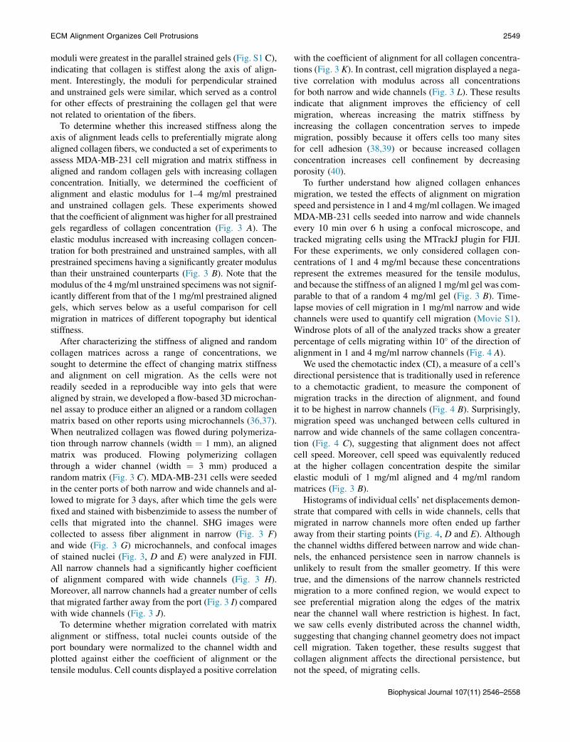
To determine whether this increased stiffness along theaxis of alignment leads cells to preferentially migrate alongaligned collagen fibers, we conducted a set of experiments toassess MDA-MB-231 cell migration and matrix stiffness inaligned and random collagen gels with increasing collagenconcentration. Initially, we determined the coefficient ofalignment and elastic modulus for 1–4 mg/ml prestrainedand unstrained collagen gels. These experiments showedthat the coefficient of alignment was higher for all prestrainedgels regardless of collagen concentration (Fig. 3 A). Theelastic modulus increased with increasing collagen concen-tration for both prestrained and unstrained samples, with allprestrained specimens having a significantly greater modulusthan their unstrained counterparts (Fig. 3 B). Note that themodulus of the 4 mg/ml unstrained specimens was not signif-icantly different from that of the 1 mg/ml prestrained alignedgels, which serves below as a useful comparison for cellmigration in matrices of different topography but identicalstiffness.
After characterizing the stiffness of aligned and randomcollagen matrices across a range of concentrations, wesought to determine the effect of changing matrix stiffnessand alignment on cell migration. As the cells were notreadily seeded in a reproducible way into gels that werealigned by strain, we developed a flow-based 3D microchan-nel assay to produce either an aligned or a random collagenmatrix based on other reports using microchannels (36,37).When neutralized collagen was flowed during polymeriza-tion through narrow channels (width ¼ 1 mm), an alignedmatrix was produced. Flowing polymerizing collagenthrough a wider channel (width ¼ 3 mm) produced arandom matrix (Fig. 3 C). MDA-MB-231 cells were seededin the center ports of both narrow and wide channels and al-lowed to migrate for 3 days, after which time the gels werefixed and stained with bisbenzimide to assess the number ofcells that migrated into the channel. SHG images werecollected to assess fiber alignment in narrow (Fig. 3 F)and wide (Fig. 3 G) microchannels, and confocal imagesof stained nuclei (Fig. 3, D and E) were analyzed in FIJI.All narrow channels had a significantly higher coefficientof alignment compared with wide channels (Fig. 3 H).Moreover, all narrow channels had a greater number of cellsthat migrated farther away from the port (Fig. 3 I) comparedwith wide channels (Fig. 3 J).
To determine whether migration correlated with matrixalignment or stiffness, total nuclei counts outside of theport boundary were normalized to the channel width andplotted against either the coefficient of alignment or thetensile modulus. Cell counts displayed a positive correlation
with the coefficient of alignment for all collagen concentra-tions (Fig. 3 K). In contrast, cell migration displayed a nega-tive correlation with modulus across all concentrationsfor both narrow and wide channels (Fig. 3 L). These resultsindicate that alignment improves the efficiency of cellmigration, whereas increasing the matrix stiffness byincreasing the collagen concentration serves to impedemigration, possibly because it offers cells too many sitesfor cell adhesion (38,39) or because increased collagenconcentration increases cell confinement by decreasingporosity (40).
To further understand how aligned collagen enhancesmigration, we tested the effects of alignment on migrationspeed and persistence in 1 and 4 mg/ml collagen. We imagedMDA-MB-231 cells seeded into narrow and wide channelsevery 10 min over 6 h using a confocal microscope, andtracked migrating cells using the MTrackJ plugin for FIJI.For these experiments, we only considered collagen con-centrations of 1 and 4 mg/ml because these concentrationsrepresent the extremes measured for the tensile modulus,and because the stiffness of an aligned 1mg/ml gel was com-parable to that of a random 4 mg/ml gel (Fig. 3 B). Time-lapse movies of cell migration in 1 mg/ml narrow and widechannels were used to quantify cell migration (Movie S1).Windrose plots of all of the analyzed tracks show a greaterpercentage of cells migrating within 10� of the direction ofalignment in 1 and 4 mg/ml narrow channels (Fig. 4 A).
We used the chemotactic index (CI), a measure of a cell’sdirectional persistence that is traditionally used in referenceto a chemotactic gradient, to measure the component ofmigration tracks in the direction of alignment, and foundit to be highest in narrow channels (Fig. 4 B). Surprisingly,migration speed was unchanged between cells cultured innarrow and wide channels of the same collagen concentra-tion (Fig. 4 C), suggesting that alignment does not affectcell speed. Moreover, cell speed was equivalently reducedat the higher collagen concentration despite the similarelastic moduli of 1 mg/ml aligned and 4 mg/ml randommatrices (Fig. 3 B).
Histograms of individual cells’ net displacements demon-strate that compared with cells in wide channels, cells thatmigrated in narrow channels more often ended up fartheraway from their starting points (Fig. 4, D and E). Althoughthe channel widths differed between narrow and wide chan-nels, the enhanced persistence seen in narrow channels isunlikely to result from the smaller geometry. If this weretrue, and the dimensions of the narrow channels restrictedmigration to a more confined region, we would expect tosee preferential migration along the edges of the matrixnear the channel wall where restriction is highest. In fact,we saw cells evenly distributed across the channel width,suggesting that changing channel geometry does not impactcell migration. Taken together, these results suggest thatcollagen alignment affects the directional persistence, butnot the speed, of migrating cells.
Biophysical Journal 107(11) 2546–2558
ECM Alignment Organizes Cell Protrusions 2549
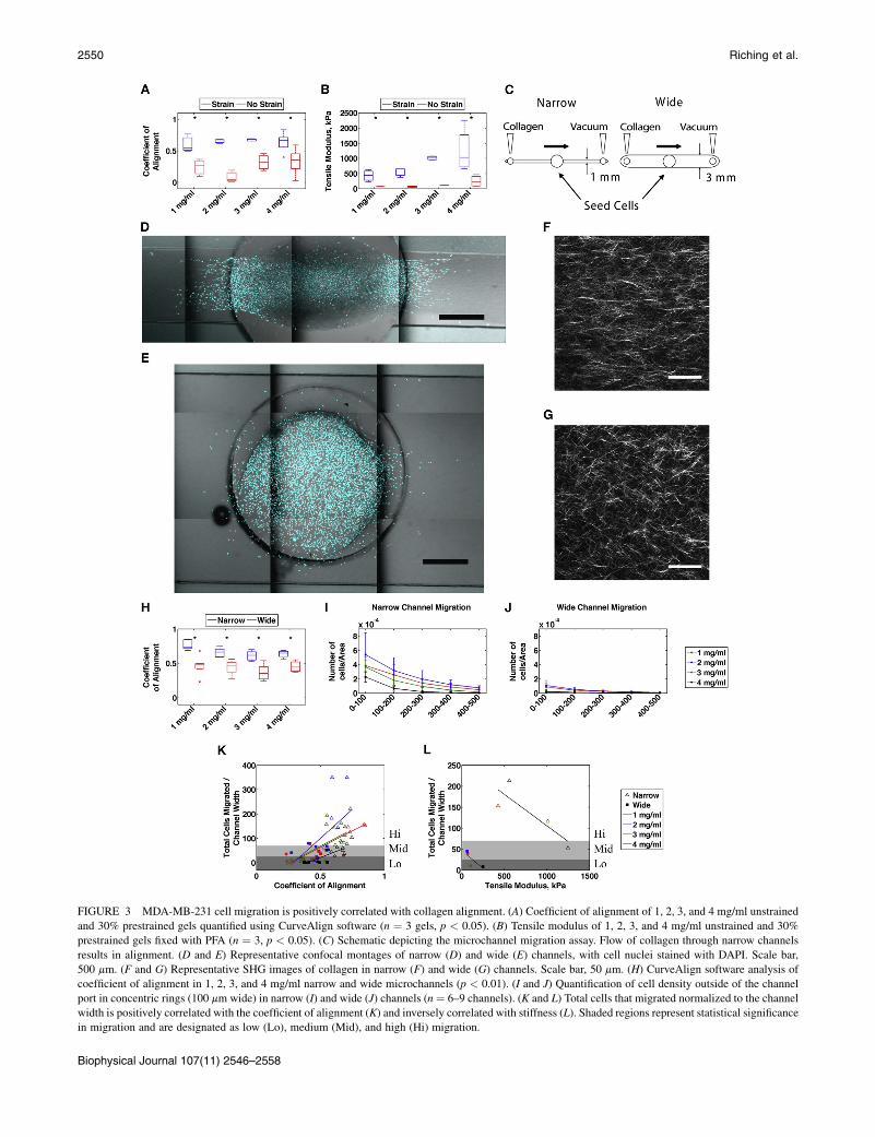
FIGURE 3 MDA-MB-231 cell migration is positively correlated with collagen alignment. (A) Coefficient of alignment of 1, 2, 3, and 4 mg/ml unstrained
and 30% prestrained gels quantified using CurveAlign software (n ¼ 3 gels, p < 0.05). (B) Tensile modulus of 1, 2, 3, and 4 mg/ml unstrained and 30%
prestrained gels fixed with PFA (n ¼ 3, p < 0.05). (C) Schematic depicting the microchannel migration assay. Flow of collagen through narrow channels
results in alignment. (D and E) Representative confocal montages of narrow (D) and wide (E) channels, with cell nuclei stained with DAPI. Scale bar,
500 mm. (F and G) Representative SHG images of collagen in narrow (F) and wide (G) channels. Scale bar, 50 mm. (H) CurveAlign software analysis of
coefficient of alignment in 1, 2, 3, and 4 mg/ml narrow and wide microchannels (p < 0.01). (I and J) Quantification of cell density outside of the channel
port in concentric rings (100 mm wide) in narrow (I) and wide (J) channels (n ¼ 6–9 channels). (K and L) Total cells that migrated normalized to the channel
width is positively correlated with the coefficient of alignment (K) and inversely correlated with stiffness (L). Shaded regions represent statistical significance
in migration and are designated as low (Lo), medium (Mid), and high (Hi) migration.
Biophysical Journal 107(11) 2546–2558
2550 Riching et al.
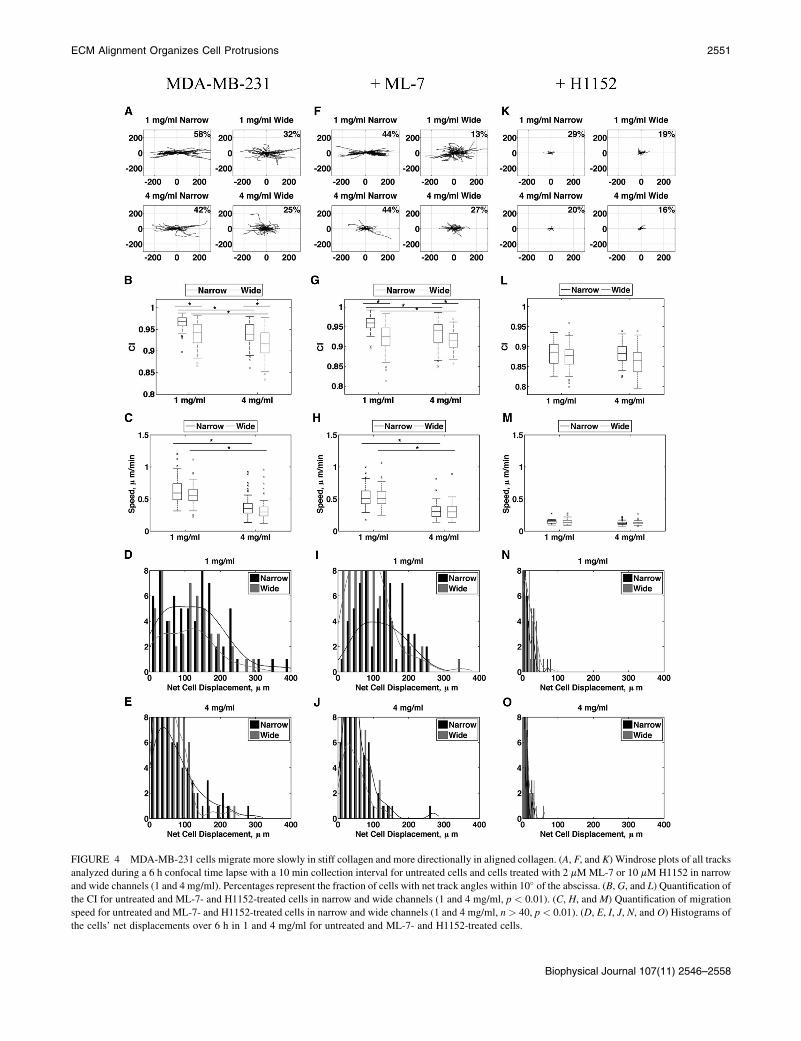
FIGURE 4 MDA-MB-231 cells migrate more slowly in stiff collagen and more directionally in aligned collagen. (A, F, and K) Windrose plots of all tracks
analyzed during a 6 h confocal time lapse with a 10 min collection interval for untreated cells and cells treated with 2 mMML-7 or 10 mM H1152 in narrow
and wide channels (1 and 4 mg/ml). Percentages represent the fraction of cells with net track angles within 10� of the abscissa. (B,G, and L) Quantification ofthe CI for untreated and ML-7- and H1152-treated cells in narrow and wide channels (1 and 4 mg/ml, p < 0.01). (C, H, and M) Quantification of migration
speed for untreated and ML-7- and H1152-treated cells in narrow and wide channels (1 and 4 mg/ml, n > 40, p < 0.01). (D, E, I, J, N, and O) Histograms of
the cells’ net displacements over 6 h in 1 and 4 mg/ml for untreated and ML-7- and H1152-treated cells.
Biophysical Journal 107(11) 2546–2558
ECM Alignment Organizes Cell Protrusions 2551
To deduce a potential mechanism for cells’ enhanceddirectional response to alignment, we asked whether Rho-mediated cell contractility plays a role in sensing alignment.We previously showed that cells require Rho and its effector,ROCK, to align collagen fibers, but once alignment is estab-lished, migration ensues independently of Rho and ROCK(23). To test whether cells require contractility to migratein aligned collagen, we conducted time-lapse experimentssimilar to those shown in Fig. 4, A–E, but added to the cul-ture media either H1152 to inhibit ROCK, or ML-7 toinhibit myosin light chain kinase (MLCK) 30 min beforeimaging. Inhibition of either ROCK or MLCK in MDA-MB-231 cells resulted in reduced levels of phosphorylatedthreonine-18 and serine-19 on MLC (data not shown),whereas ML-7 had no effect on migration speed or persis-tence compared with untreated cells (Fig. 4, F–J). However,H1152 dramatically inhibited cell speed across all condi-tions tested, which further resulted in reduced persistence(Fig. 4, K–O). These results indicate that Rho- andROCK-mediated signaling is required for cell migration in3D collagen matrices, but signaling from MLCK is not.
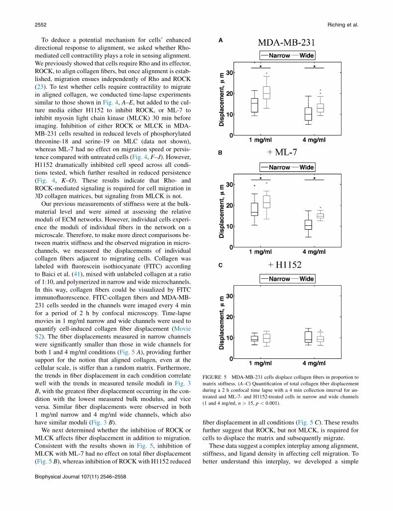
Our previous measurements of stiffness were at the bulk-material level and were aimed at assessing the relativemoduli of ECM networks. However, individual cells experi-ence the moduli of individual fibers in the network on amicroscale. Therefore, to make more direct comparisons be-tween matrix stiffness and the observed migration in micro-channels, we measured the displacements of individualcollagen fibers adjacent to migrating cells. Collagen waslabeled with fluorescein isothiocyanate (FITC) accordingto Baici et al. (41), mixed with unlabeled collagen at a ratioof 1:10, and polymerized in narrow and wide microchannels.In this way, collagen fibers could be visualized by FITCimmunofluorescence. FITC-collagen fibers and MDA-MB-231 cells seeded in the channels were imaged every 4 minfor a period of 2 h by confocal microscopy. Time-lapsemovies in 1 mg/ml narrow and wide channels were used toquantify cell-induced collagen fiber displacement (MovieS2). The fiber displacements measured in narrow channelswere significantly smaller than those in wide channels forboth 1 and 4 mg/ml conditions (Fig. 5 A), providing furthersupport for the notion that aligned collagen, even at thecellular scale, is stiffer than a random matrix. Furthermore,the trends in fiber displacement in each condition correlatewell with the trends in measured tensile moduli in Fig. 3B, with the greatest fiber displacement occurring in the con-dition with the lowest measured bulk modulus, and viceversa. Similar fiber displacements were observed in both1 mg/ml narrow and 4 mg/ml wide channels, which alsohave similar moduli (Fig. 3 B).
We next determined whether the inhibition of ROCK orMLCK affects fiber displacement in addition to migration.Consistent with the results shown in Fig. 5, inhibition ofMLCK with ML-7 had no effect on total fiber displacement(Fig. 5 B), whereas inhibition of ROCK with H1152 reduced
fiber displacement in all conditions (Fig. 5 C). These resultsfurther suggest that ROCK, but not MLCK, is required forcells to displace the matrix and subsequently migrate.
These data suggest a complex interplay among alignment,stiffness, and ligand density in affecting cell migration. Tobetter understand this interplay, we developed a simple
FIGURE 5 MDA-MB-231 cells displace collagen fibers in proportion to
matrix stiffness. (A–C) Quantification of total collagen fiber displacement
during a 2 h confocal time lapse with a 4 min collection interval for un-
treated and ML-7- and H1152-treated cells in narrow and wide channels
(1 and 4 mg/ml, n > 15, p < 0.001).
Biophysical Journal 107(11) 2546–2558
2552 Riching et al.
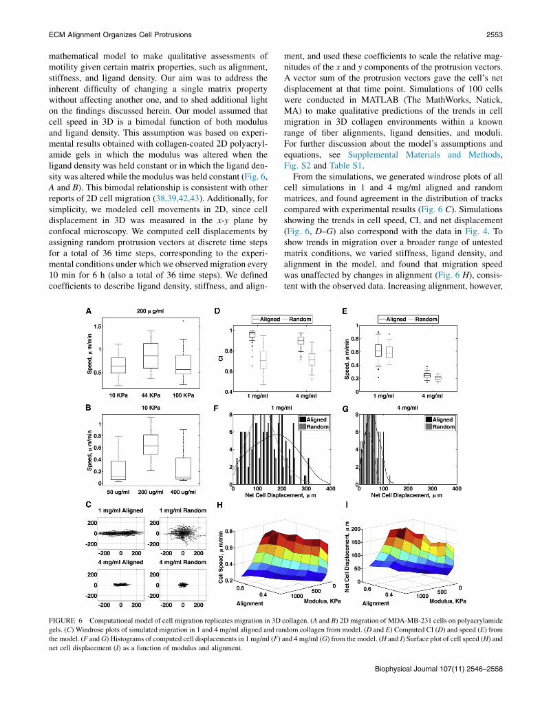
mathematical model to make qualitative assessments ofmotility given certain matrix properties, such as alignment,stiffness, and ligand density. Our aim was to address theinherent difficulty of changing a single matrix propertywithout affecting another one, and to shed additional lighton the findings discussed herein. Our model assumed thatcell speed in 3D is a bimodal function of both modulusand ligand density. This assumption was based on experi-mental results obtained with collagen-coated 2D polyacryl-amide gels in which the modulus was altered when theligand density was held constant or in which the ligand den-sity was altered while the modulus was held constant (Fig. 6,A and B). This bimodal relationship is consistent with otherreports of 2D cell migration (38,39,42,43). Additionally, forsimplicity, we modeled cell movements in 2D, since celldisplacement in 3D was measured in the x-y plane byconfocal microscopy. We computed cell displacements byassigning random protrusion vectors at discrete time stepsfor a total of 36 time steps, corresponding to the experi-mental conditions under which we observed migration every10 min for 6 h (also a total of 36 time steps). We definedcoefficients to describe ligand density, stiffness, and align-
ment, and used these coefficients to scale the relative mag-nitudes of the x and y components of the protrusion vectors.A vector sum of the protrusion vectors gave the cell’s netdisplacement at that time point. Simulations of 100 cellswere conducted in MATLAB (The MathWorks, Natick,MA) to make qualitative predictions of the trends in cellmigration in 3D collagen environments within a knownrange of fiber alignments, ligand densities, and moduli.For further discussion about the model’s assumptions andequations, see Supplemental Materials and Methods,Fig. S2 and Table S1.
From the simulations, we generated windrose plots of allcell simulations in 1 and 4 mg/ml aligned and randommatrices, and found agreement in the distribution of trackscompared with experimental results (Fig. 6 C). Simulationsshowing the trends in cell speed, CI, and net displacement(Fig. 6, D–G) also correspond with the data in Fig. 4. Toshow trends in migration over a broader range of untestedmatrix conditions, we varied stiffness, ligand density, andalignment in the model, and found that migration speedwas unaffected by changes in alignment (Fig. 6 H), consis-tent with the observed data. Increasing alignment, however,
FIGURE 6 Computational model of cell migration replicates migration in 3D collagen. (A and B) 2D migration of MDA-MB-231 cells on polyacrylamide
gels. (C) Windrose plots of simulated migration in 1 and 4 mg/ml aligned and random collagen from model. (D and E) Computed CI (D) and speed (E) from
the model. (F andG) Histograms of computed cell displacements in 1 mg/ml (F) and 4 mg/ml (G) from the model. (H and I) Surface plot of cell speed (H) and
net cell displacement (I) as a function of modulus and alignment.
Biophysical Journal 107(11) 2546–2558
ECM Alignment Organizes Cell Protrusions 2553
increased the net distance cells traveled (Fig. 6 I), suggest-ing that an increase in persistence is the main factor thatdetermines enhanced migration due to aligned collagen ininvasive breast tumors. The model best fits the data whenthe number of protrusions per cell is decreased as alignmentincreases (Fig. S2 E), which leads to the prediction thatalignment enhances persistence by limiting the number ofprotrusions.
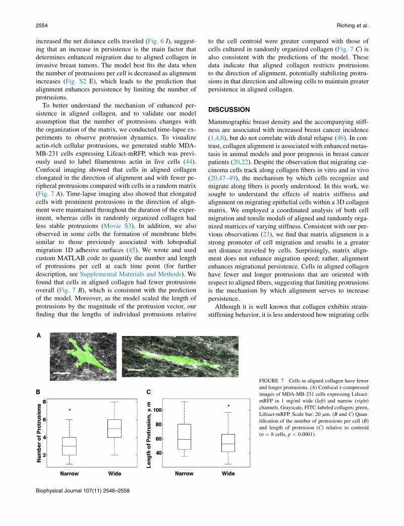
To better understand the mechanism of enhanced per-sistence in aligned collagen, and to validate our modelassumption that the number of protrusions changes withthe organization of the matrix, we conducted time-lapse ex-periments to observe protrusion dynamics. To visualizeactin-rich cellular protrusions, we generated stable MDA-MB-231 cells expressing Lifeact-mRFP, which was previ-ously used to label filamentous actin in live cells (44).Confocal imaging showed that cells in aligned collagenelongated in the direction of alignment and with fewer pe-ripheral protrusions compared with cells in a random matrix(Fig. 7 A). Time-lapse imaging also showed that elongatedcells with prominent protrusions in the direction of align-ment were maintained throughout the duration of the exper-iment, whereas cells in randomly organized collagen hadless stable protrusions (Movie S3). In addition, we alsoobserved in some cells the formation of membrane blebssimilar to those previously associated with lobopodialmigration 1D adhesive surfaces (45). We wrote and usedcustom MATLAB code to quantify the number and lengthof protrusions per cell at each time point (for furtherdescription, see Supplemental Materials and Methods). Wefound that cells in aligned collagen had fewer protrusionsoverall (Fig. 7 B), which is consistent with the predictionof the model. Moreover, as the model scaled the length ofprotrusions by the magnitude of the protrusion vector, ourfinding that the lengths of individual protrusions relative
to the cell centroid were greater compared with those ofcells cultured in randomly organized collagen (Fig. 7 C) isalso consistent with the predictions of the model. Thesedata indicate that aligned collagen restricts protrusionsto the direction of alignment, potentially stabilizing protru-sions in that direction and allowing cells to maintain greaterpersistence in aligned collagen.
DISCUSSION
Mammographic breast density and the accompanying stiff-ness are associated with increased breast cancer incidence(1,4,8), but do not correlate with distal relapse (46). In con-trast, collagen alignment is associated with enhanced metas-tasis in animal models and poor prognosis in breast cancerpatients (20,22). Despite the observation that migrating car-cinoma cells track along collagen fibers in vitro and in vivo(20,47–49), the mechanism by which cells recognize andmigrate along fibers is poorly understood. In this work, wesought to understand the effects of matrix stiffness andalignment on migrating epithelial cells within a 3D collagenmatrix. We employed a coordinated analysis of both cellmigration and tensile moduli of aligned and randomly orga-nized matrices of varying stiffness. Consistent with our pre-vious observations (23), we find that matrix alignment is astrong promoter of cell migration and results in a greaternet distance traveled by cells. Surprisingly, matrix align-ment does not enhance migration speed; rather, alignmentenhances migrational persistence. Cells in aligned collagenhave fewer and longer protrusions that are oriented withrespect to aligned fibers, suggesting that limiting protrusionsis the mechanism by which alignment serves to increasepersistence.
Although it is well known that collagen exhibits strain-stiffening behavior, it is less understood how migrating cells
FIGURE 7 Cells in aligned collagen have fewer
and longer protrusions. (A) Confocal z-compressed
images of MDA-MB-231 cells expressing Lifeact-
mRFP in 1 mg/ml wide (left) and narrow (right)
channels. Grayscale, FITC-labeled collagen; green,
Lifeact-mRFP. Scale bar: 20 mm. (B and C) Quan-
tification of the number of protrusions per cell (B)
and length of protrusion (C) relative to centroid
(n ¼ 8 cells, p < 0.0001).
Biophysical Journal 107(11) 2546–2558
2554 Riching et al.
respond to matrices of different fiber orientations and stiff-ness. Therefore, we characterized the mechanical effects ofprestraining a collagen matrix to induce fiber alignment.Our results show that collagen prealigned by strain is stifferthan a random collagen matrix of the same density and thatthe stiffness is greatest along the axis of alignment. Xu et al.(50) previously conducted biaxial tensile testing of collagenprealigned by flow of magnetic beads and showed that afterfixation, gels aligned parallel to the direction of alignmentwere stiffer than gels aligned orthogonally. Here, we ob-tained similar findings utilizing uniaxial tensile testing anda simpler, more robust approach to induce prealignmentby strain rather than flow of magnetic beads embedded inthe matrix. Moreover, we found that the increased stiffnessof aligned gels is preserved even in unfixed collagen gels.
Our results further suggest that alignment of collagen fi-bers potentially strengthens and stabilizes interactions be-tween neighboring fibers, presumably causing the observedincrease in modulus. The act of stretching a collagen matrixnot only results in increased fiber proximity but can alsoincrease subfibrillar orientation and packing (51). Thesestretch-induced modifications may allow for enhanced mo-lecular interactions between adjacent fibrils (52), thus in-creasing the overall matrix stiffness. Similarly, we expectthat the flow-induced fiber alignment in our microchannelspotentially enhances interactions between adjacent fiberssuch that alignment, rather than the method by which thealignment is induced, is the key feature.
To study cell migration in aligned matrices, we developedan assay that makes use of the observation made by Leeet al. (36) and Sung et al. (37) that collagen alignment canbe controlled by flow through a sufficiently narrow micro-channel. To our knowledge, this assay represents a novelapproach for studying the effects of 3D collagen alignmenton cell migration. Here, we used channels 1 mm and 3 mmwide to produce highly aligned collagen matrices and arandomly organized matrix, respectively, into which weseeded cells and quantified total cell migration, speed, andpersistence. We found that more cells migrated fartherinto the aligned collagen, but migration speed was un-changed relative to migration in the randomly organizedcollagen matrix.
The increase in the total number of migrating cells in thealigned matrix can be accounted for by the marked increasein the persistence of cells in aligned collagen. Furthermore,our results demonstrate that the effects of collagen align-ment on persistence are not due to the increased stiffnessof an aligned matrix, or durotaxis, because cells migratedfaster and were more persistent in 1 mg/ml aligned collagenthan in 4 mg/ml random matrix, even though the two condi-tions had nearly the same stiffness. In contrast, we foundthat when increased matrix stiffness was generated byincreased collagen concentration, which also increased theligand density and reduced the porosity of the matrix, cellspeed was reduced. This result is in contrast to the well-
documented durotactic response of cells cultured on 2D sub-strates (14) and may reflect the greater inherent complexityof 3D matrices. Importantly, the mechanical propertiesextrapolated from measurements of fiber displacementcorrelate well with the measured bulk tensile moduli ofaligned and random gels, suggesting that the differencesin the mechanical properties of aligned and random collagengels at the microscale correlate to the macroscale. More-over, this finding demonstrates that increased stiffness alongthe axis of alignment is observed regardless of the mecha-nism of alignment (flow or strain).
We further show that migration is dependent on Rho/ROCK-generated contractility and independent of MLCK.Previously, we demonstrated that cells first require a con-tractile mechanism involving Rho and ROCK to generatealigned collagen, and that the requirement for Rho/ROCKceases for migration in a prealigned collagen matrix (23).One possible explanation for this discrepancy is the differ-ence in the duration of inhibitor treatment between thetwo studies (8 h in this study compared with every 24 hfor a period of 3 days in the previous study), suggestingthat compensation for Rho inhibition may occur over longertreatment time frames. As in our previous study, we againfind that fiber displacement is also dependent on ROCK,not MLCK, which agrees with other reports of matrix defor-mation by contractile forces (53–55). These results provideadditional support for the idea proposed by Grinnell et al.(56) and Miron-Mendoza et al. (57) that cell migrationand matrix deformation are both consequences of thesame Rho- and ROCK-driven contractile mechanism andcould occur in concert. Matrix stiffness likely determinesthe extent of fiber deformation as well as the amount of trac-tion a migrating cell can generate by utilizing Rho-mediatedcontractility.
From our results, we infer that the collagen fibers that aremost constrained in the matrix move the least when pulledand exhibit the greatest stiffness. This finding is also sup-ported by the observation of Lopez-Garcia et al. (58) thatNMuMG cells produce larger strains in matrices of lowerstiffness. These results lead to the assumption that stifferconditions may provide the most traction and thus allowcells to use their contractile forces to achieve the fastestmigration. Consistent with this idea, previous studiesshowed that stiffness increases the amount of force adhe-sions generate, and force-generating active Rho is upregu-lated in stiff matrices (10,53,59). These findings on theirown would predict that increased stiffness in 3D wouldlead to enhanced migration speeds. However, increasingstiffness in 3D matrices is also typically accompanied byincreased ligand density and decreased matrix porosity,both of which are known to significantly impact migrationspeed (39,40). For this reason, it has often been challengingto develop techniques that allow one to separate stiffnessfrom other factors in a 3D environment to assess changesin migratory behavior. Recently, however, Harley et al.
Biophysical Journal 107(11) 2546–2558
ECM Alignment Organizes Cell Protrusions 2555
(60) were able to alter the individual strut stiffness of acollagen-GAG scaffold, and identified a bimodal relation-ship between stiffness and cell migration speed similar tothat seen on 2D substrates. As new methods for engineeringmatrices with precisely controlled physical and mechanicalproperties become available, we will be able to study addi-tional relationships involving cell speed and persistence.
To address the multifactorial responses to the topograph-ical and mechanical properties of the matrix, we developed amathematical model that incorporates matrix ligand density,stiffness, and alignment. Our model allows us to better un-derstand the trends over the range of experimental condi-tions tested, where it is difficult to deduce effects incurredby isolating a single 3D matrix parameter. Others have pro-posed a model to describe the effect of fibroblast migrationspeed on the alignment of fibrin and collagen in dermalwound healing (61). Our model considers protrusion gener-ation as a stochastic process that leads to adhesion formationwith ECM fibers, whereas other groups have used models tosuggest that in vivo migration in discontinuous environ-ments may not require adhesions (62). Our model predictsthat the number of protrusions diminish with increasedalignment and increase as ligand density increases. Weshow experimentally that cells in aligned collagen indeedhave fewer protrusions, which is likely responsible for thisobserved increase in persistence. To better replicate cellpersistence, the model incorporates a probability for a pro-trusion vector to maintain its orientation across multipletime steps that increases as a function of matrix alignment.Although we are unsure of the exact cellular mechanism ofpersistence, the predictions of our model would be consis-tent with the hypothesis that persistence reflects the presen-tation of ligands along an axis allowing cells to form andstabilize adhesions in a given direction, enabling more effi-cient migration. This notion is consistent with the observa-tion that integrin adhesions are localized along collagenfibers in 3D (63) and on 1D collagen fibers, where cellmigration along 1D fibers recapitulates migration in 3Dmatrices (64). Moreover, we expect that persistence alsoreflects the loss of competing ligand binding choices inother directions. We predict that as ligand concentration in-creases, there will be more opportunities for adhesion-stabi-lized protrusions to occur outside the direction of alignment,consistent with the observation that persistence decreases asligand concentration increases.
A limitation of our model is that, for simplicity, it as-sumes all matrix parameters are held constant over the si-mulated time frame, when in reality cells are constantlymodifying their environment via Rho/ROCK contractility,which causes local changes in alignment, stiffness, andligand density. Moreover, there are additional effects of pro-teolytic modification of collagen that we have not taken intoaccount (63). Furthermore, the model input parameters re-quire tested modulus values for given alignments and liganddensities, which can also change with different matrix com-
ponents and the extent of cross-linking. Once we are able tomeasure the stiffness of a broader range of matrix composi-tions and more reliably establish the relationships amongmatrix composition, alignment, and stiffness, we can revisethe model parameters. Despite these limitations, however,we show that our model can accurately replicate trends inmigration speed and persistence in 1 mg/ml and 4 mg/mlaligned and random collagen matrices. Furthermore, themodel is consistent with the observation that migrationspeed across a range of matrix conditions is unaffected byalignment, but alignment profoundly increases the net dis-tance cells travel. This finding is significant in the contextof breast tumor progression, where the presence of TACS-3 aligned fibers is correlated with a dramatic increase in me-tastases and poor long-term survival (21,22). Our resultssuggest that highly aligned regions in the tumor micro-environment can provide cells that have a high migratory ca-pacity with the most reliable and robust escape route, andprovide additional mechanistic understanding of how thealigned collagen characterized by TACS-3 relates to itsrole as a potential biomarker for breast cancer.
SUPPORTING MATERIAL
Supplemental Materials and Methods, three figures, one table, and three
movies are available at http://www.biophysj.org/biophysj/supplemental/
S0006-3495(14)01118-7.
The authors thank Drs. Kyung Sung and David Beebe for assistance with
microfluidic channels, Dr. Maddy Parsons for generously supplying the
Lifeact-mRFP construct, and Curtis Rueden for assistance with the FIJI
software and analysis.
This work was funded by NIH grants U01CA143069 to A.W., Y.J., and
P.J.K., and R01 CA142833 and R01 CA114462 to P.J.K. and supported
by the University of Wisconsin-Madison Graduate School for M.R.S. and
W.C.C.
SUPPORTING CITATIONS
References (65–74) appear in the Supporting Material.
REFERENCES
1. Boyd, N. F., G. A. Lockwood, ., M. J. Yaffe. 1998. Mammographicdensities and breast cancer risk. Cancer Epidemiol. Biomarkers Prev.7:1133–1144.
2. Boyd, N. F., L. J. Martin, ., M. J. Yaffe. 2001. Mammographic den-sities as a marker of human breast cancer risk and their use in chemo-prevention. Curr. Oncol. Rep. 3:314–321.
3. McCormack, V. A., and I. dos Santos Silva. 2006. Breast density andparenchymal patterns as markers of breast cancer risk: a meta-analysis.Cancer Epidemiol. Biomarkers Prev. 15:1159–1169.
4. Boyd, N. F., G. S. Dite,., J. L. Hopper. 2002. Heritability of mammo-graphic density, a risk factor for breast cancer. N. Engl. J. Med.347:886–894.
5. Alowami, S., S. Troup,., P. H. Watson. 2003. Mammographic densityis related to stroma and stromal proteoglycan expression. Breast Can-cer Res. 5:R129–R135.
Biophysical Journal 107(11) 2546–2558
2556 Riching et al.
6. Li, T., L. Sun, ., N. Boyd. 2005. The association of measuredbreast tissue characteristics with mammographic density and otherrisk factors for breast cancer. Cancer Epidemiol. Biomarkers Prev.14:343–349.
7. Guo, Y. P., L. J. Martin,., N. F. Boyd. 2001. Growth factors and stro-mal matrix proteins associated with mammographic densities. CancerEpidemiol. Biomarkers Prev. 10:243–248.
8. Paszek, M. J., N. Zahir,., V. M. Weaver. 2005. Tensional homeostasisand the malignant phenotype. Cancer Cell. 8:241–254.
9. Lopez, J. I., I. Kang, ., V. M. Weaver. 2011. In situ force mapping ofmammary gland transformation. Integr. Biol. (Camb.). 3:910–921.
10. Provenzano, P. P., D. R. Inman, ., P. J. Keely. 2009. Matrix density-induced mechanoregulation of breast cell phenotype, signaling andgene expression through a FAK-ERK linkage. Oncogene. 28:4326–4343.
11. Tilghman, R. W., C. R. Cowan, ., J. T. Parsons. 2010. Matrix rigidityregulates cancer cell growth and cellular phenotype. PLoS ONE.5:e12905.
12. Alexander, N. R., K. M. Branch,., A. M. Weaver. 2008. Extracellularmatrix rigidity promotes invadopodia activity. Curr. Biol. 18:1295–1299.
13. Lo, C. M., H. B. Wang,., Y. L. Wang. 2000. Cell movement is guidedby the rigidity of the substrate. Biophys. J. 79:144–152.
14. Pelham, Jr., R. J., and Yl. Wang. 1997. Cell locomotion and focaladhesions are regulated by substrate flexibility. Proc. Natl. Acad. Sci.USA. 94:13661–13665.
15. Menon, S., and K. A. Beningo. 2011. Cancer cell invasion is enhancedby applied mechanical stimulation. PLoS ONE. 6:e17277.
16. Kostic, A., C. D. Lynch, and M. P. Sheetz. 2009. Differential matrix ri-gidity response in breast cancer cell lines correlates with the tissuetropism. PLoS ONE. 4:e6361.
17. Wozniak, M. A., K. Modzelewska,., P. J. Keely. 2004. Focal adhesionregulation of cell behavior. Biochim. Biophys. Acta. 1692:103–119.
18. Klein, E. A., L. Yin, ., R. K. Assoian. 2009. Cell-cycle control byphysiological matrix elasticity and in vivo tissue stiffening. Curr.Biol. 19:1511–1518.
19. Ulrich, T. A., E. M. de Juan Pardo, and S. Kumar. 2009. The mechan-ical rigidity of the extracellular matrix regulates the structure, motility,and proliferation of glioma cells. Cancer Res. 69:4167–4174.
20. Provenzano, P. P., K. W. Eliceiri, ., P. J. Keely. 2006. Collagen reor-ganization at the tumor-stromal interface facilitates local invasion.BMC Med. 4:38.
21. Provenzano, P. P., D. R. Inman, ., P. J. Keely. 2008. Collagen densitypromotes mammary tumor initiation and progression. BMC Med. 6:11.
22. Conklin, M. W., J. C. Eickhoff,., P. J. Keely. 2011. Aligned collagenis a prognostic signature for survival in human breast carcinoma. Am. J.Pathol. 178:1221–1232.
23. Provenzano, P. P., D. R. Inman,., P. J. Keely. 2008. Contact guidancemediated three-dimensional cell migration is regulated by Rho/ROCK-dependent matrix reorganization. Biophys. J. 95:5374–5384.
24. Kim, A., N. Lakshman, and W. M. Petroll. 2006. Quantitative assess-ment of local collagen matrix remodeling in 3-D culture: the role ofRho kinase. Exp. Cell Res. 312:3683–3692.
25. Brownfield, D. G., G. Venugopalan, ., M. J. Bissell. 2013. Patternedcollagen fibers orient branching mammary epithelium through distinctsignaling modules. Curr. Biol. 23:703–709.
26. Sander, E. A., V. H. Barocas, and R. T. Tranquillo. 2011. Initial fiberalignment pattern alters extracellular matrix synthesis in fibroblast-populated fibrin gel cruciforms and correlates with predicted tension.Ann. Biomed. Eng. 39:714–729.
27. Wang, J. H., F. Jia, ., S. L. Woo. 2003. Cell orientation determinesthe alignment of cell-produced collagenous matrix. J. Biomech.36:97–102.
28. Yang, N., R. Mosher, ., A. Friedl. 2011. Syndecan-1 in breast cancerstroma fibroblasts regulates extracellular matrix fiber organization andcarcinoma cell motility. Am. J. Pathol. 178:325–335.
29. Gruschwitz, R., J. Friedrichs, ., K. Engelmann. 2010. Alignment andcell-matrix interactions of human corneal endothelial cells on nano-structured collagen type I matrices. Invest. Ophthalmol. Vis. Sci.51:6303–6310.
30. Vernon, R. B., M. D. Gooden, ., T. N. Wight. 2005. Microgroovedfibrillar collagen membranes as scaffolds for cell support and align-ment. Biomaterials. 26:3131–3140.
31. Kim, D. H., P. P. Provenzano, ., A. Levchenko. 2012. Matrix nanoto-pography as a regulator of cell function. J. Cell Biol. 197:351–360.
32. Lai, E. S., N. F. Huang, ., G. G. Fuller. 2012. Aligned nanofibrillarcollagen regulates endothelial organization and migration. Regen.Med. 7:649–661.
33. Friedl, P., and K. Wolf. 2008. Tube travel: the role of proteases in in-dividual and collective cancer cell invasion. Cancer Res. 68:7247–7249.
34. Vader, D., A. Kabla, ., L. Mahadevan. 2009. Strain-induced align-ment in collagen gels. PLoS ONE. 4:e5902.
35. Roeder, B. A., K. Kokini, ., S. L. Voytik-Harbin. 2002. Tensile me-chanical properties of three-dimensional type I collagen extracellularmatrices with varied microstructure. J. Biomech. Eng. 124:214–222.
36. Lee, P., R. Lin,., L. P. Lee. 2006. Microfluidic alignment of collagenfibers for in vitro cell culture. Biomed. Microdevices. 8:35–41.
37. Sung, K. E., G. Su, ., D. J. Beebe. 2009. Control of 3-dimensionalcollagen matrix polymerization for reproducible human mammaryfibroblast cell culture in microfluidic devices. Biomaterials. 30:4833–4841.
38. Palecek, S. P., J. C. Loftus, ., A. F. Horwitz. 1997. Integrin-ligandbinding properties govern cell migration speed through cell-substratumadhesiveness. Nature. 385:537–540.
39. Zaman, M. H., L. M. Trapani,., P. Matsudaira. 2006. Migration of tu-mor cells in 3D matrices is governed by matrix stiffness along withcell-matrix adhesion and proteolysis. Proc. Natl. Acad. Sci. USA.103:10889–10894.
40. Wolf, K., M. Te Lindert, ., P. Friedl. 2013. Physical limits of cellmigration: control by ECM space and nuclear deformation and tuningby proteolysis and traction force. J. Cell Biol. 201:1069–1084.
41. Baici, A., G. Cohen,., A. Boni. 1980. A handy assay for collagenaseusing reconstituted fluorescein-labeled collagen fibrils. Anal. Biochem.108:230–232.
42. Wu, P., J. B. Hoying, ., D. A. Lauffenburger. 1994. Integrin-bindingpeptide in solution inhibits or enhances endothelial cell migration, pre-dictably from cell adhesion. Ann. Biomed. Eng. 22:144–152.
43. Peyton, S. R., and A. J. Putnam. 2005. Extracellular matrix rigiditygoverns smooth muscle cell motility in a biphasic fashion. J. Cell.Physiol. 204:198–209.
44. Riedl, J., A. H. Crevenna,., R. Wedlich-Soldner. 2008. Lifeact: a ver-satile marker to visualize F-actin. Nat. Methods. 5:605–607.
45. Petrie, R. J., N. Gavara, ., K. M. Yamada. 2012. Nonpolarizedsignaling reveals two distinct modes of 3D cell migration. J. CellBiol. 197:439–455.
46. Park, C. C., J. Rembert, ., K. Kerlikowske. 2009. High mammo-graphic breast density is independent predictor of local but not distantrecurrence after lumpectomy and radiotherapy for invasive breast can-cer. Int. J. Radiat. Oncol. Biol. Phys. 73:75–79.
47. Wang,W., J. B. Wyckoff,., J. S. Condeelis. 2002. Single cell behaviorin metastatic primary mammary tumors correlated with gene expres-sion patterns revealed by molecular profiling. Cancer Res. 62:6278–6288.
48. Dickinson, R. B., S. Guido, and R. T. Tranquillo. 1994. Biased cellmigration of fibroblasts exhibiting contact guidance in orientedcollagen gels. Ann. Biomed. Eng. 22:342–356.
Biophysical Journal 107(11) 2546–2558
ECM Alignment Organizes Cell Protrusions 2557
49. Petrie, R. J., A. D. Doyle, and K. M. Yamada. 2009. Random versusdirectionally persistent cell migration. Nat. Rev. Mol. Cell Biol.10:538–549.
50. Xu, B., M. J. Chow, and Y. Zhang. 2011. Experimental and modelingstudy of collagen scaffolds with the effects of crosslinking and fiberalignment. Int. J. Biomater. 2011:172389.
51. Pins, G. D., D. L. Christiansen,., F. H. Silver. 1997. Self-assembly ofcollagen fibers. Influence of fibrillar alignment and decorin on mechan-ical properties. Biophys. J. 73:2164–2172.
52. Freeman, J. W., and F. H. Silver. 2005. The effects of prestrain andcollagen fibril alignment on in vitro mineralization of self-assembledcollagen fibers. Connect. Tissue Res. 46:107–115.
53. Wozniak, M. A., R. Desai, ., P. J. Keely. 2003. ROCK-generatedcontractility regulates breast epithelial cell differentiation in responseto the physical properties of a three-dimensional collagen matrix.J. Cell Biol. 163:583–595.
54. Wyckoff, J. B., S. E. Pinner, S. Gschmeissner, J. S. Condeelis, and E.Sahai. 2006. ROCK- and myosin-dependent matrix deformationenables protease-independent tumor-cell invasion in vivo. Curr. Biol.Aug 8; 16(15):1515–1523.
55. Lakshman, N., A. Kim, ., W. M. Petroll. 2007. Rho plays a centralrole in regulating local cell-matrix mechanical interactions in 3D cul-ture. Cell Motil. Cytoskeleton. 64:434–445.
56. Grinnell, F., L. B. Rocha,., H. Jiang. 2006. Nested collagen matrices:a new model to study migration of human fibroblast populations inthree dimensions. Exp. Cell Res. 312:86–94.
57. Miron-Mendoza, M., J. Seemann, and F. Grinnell. 2008. Collagen fibrilflow and tissue translocation coupled to fibroblast migration in 3Dcollagen matrices. Mol. Biol. Cell. 19:2051–2058.
58. Lopez-Garcia, M. D., D. J. Beebe, and W. C. Crone. 2010. Mechanicalinteractions of mouse mammary gland cells with collagen in a three-dimensional construct. Ann. Biomed. Eng. 38:2485–2498.
59. Bhadriraju, K., M. Yang,., C. S. Chen. 2007. Activation of ROCK byRhoA is regulated by cell adhesion, shape, and cytoskeletal tension.Exp. Cell Res. 313:3616–3623.
60. Harley, B. A., H. D. Kim, ., L. J. Gibson. 2008. Microarchitecture ofthree-dimensional scaffolds influences cell migration behavior viajunction interactions. Biophys. J. 95:4013–4024.
61. Dallon, J. C., J. A. Sherratt, and P. K. Maini. 2001. Modeling the effectsof transforming growth factor-beta on extracellular matrix alignment indermal wound repair. Wound Repair Regen. 9:278–286.
62. Tozluo�glu, M., A. L. Tournier,., E. Sahai. 2013. Matrix geometry de-termines optimal cancer cell migration strategy and modulatesresponse to interventions. Nat. Cell Biol. 15:751–762.
63. Wolf, K., Y. I. Wu, ., P. Friedl. 2007. Multi-step pericellular proteol-ysis controls the transition from individual to collective cancer cell in-vasion. Nat. Cell Biol. 9:893–904.
64. Doyle, A. D., F. W. Wang, ., K. M. Yamada. 2009. One-dimensionaltopography underlies three-dimensional fibrillar cell migration. J. CellBiol. 184:481–490.
65. Heck, J. N., S. M. Ponik, ., P. J. Keely. 2012. Microtubules regulateGEF-H1 in response to extracellular matrix stiffness. Mol. Biol. Cell.23:2583–2592.
66. Chua Chee, K. 1994. Three-dimensional rapid prototyping technolo-gies and key development areas. Comput. Contr. Eng. J. 5:200–206.
67. Wozniak, M. A., and P. J. Keely. 2005. Use of three-dimensionalcollagen gels to study mechanotransduction in T47D breast epithelialcells. Biol. Proced. Online. 7:144–161.
68. Lopez-Garcia, M. D., D. J. Beebe, and W. C. Crone. 2010. Young’smodulus of collagen at slow displacement rates. Biomed. Mater. Eng.20:361–369.
69. Schindelin, J., I. Arganda-Carreras, ., A. Cardona. 2012. Fiji: anopen-source platform for biological-image analysis. Nat. Methods.9:676–682.
70. Yeung, T., P. C. Georges, ., P. A. Janmey. 2005. Effects of substratestiffness on cell morphology, cytoskeletal structure, and adhesion.Cell Motil. Cytoskeleton. 60:24–34.
71. Aratyn-Schaus, Y., P. W. Oakes,., M. L. Gardel. 2010. Preparation ofcomplaint matrices for quantifying cellular contraction. J. Vis. Exp.(46): pii: 2173.
72. Schoen, I., B. L. Pruitt, and V. Vogel. 2013. The yin-yang of rigiditysensing: how forces and mechanical properties regulate the cellularresponse to materials. Annu. Rev. Mater. Res. 43:589–618.
73. Smith, A. S., K. Sengupta, ., E. Sackmann. 2008. Force-inducedgrowth of adhesion domains is controlled by receptor mobility. Proc.Natl. Acad. Sci. USA. 105:6906–6911.
74. Huttenlocher, A., and A. R. Horwitz. 2011. Integrins in cell migration.Cold Spring Harb. Perspect. Biol. 3:a005074.
Biophysical Journal 107(11) 2546–2558
2558 Riching et al.