3D Cell Culturing by Magnetic Levitation, The Next...
81
3D Cell Culturing by Magnetic Levitation, The Next Generation of Cell Culturing? Presented in the Embryo Physics Course February 8, 2012 By Glauco R. Souza [email protected] Nano3D Biosciences TM , Inc. Houston, Texas, USA 1
Transcript of 3D Cell Culturing by Magnetic Levitation, The Next...

3D Cell Culturing by Magnetic
Levitation,
The Next Generation of Cell Culturing?
Presented in the Embryo Physics Course
February 8, 2012
By
Glauco R. Souza
Nano3D BiosciencesTM, Inc.
Houston, Texas, USA 1

3D Cell Culturing by Magnetic Levitation,
The Next Generation of Cell Culturing? Glauco R. Souza, Ph.D.
Chief Scientific Officer Nano3D BiosciencesTM, Inc.

What is Cell Culture?
http://www.sociology.ccsu.edu/images/benton_mural.jpg
http://www.microscopyu.com/staticgallery/phasecontrast/images/chocellspositive.jpg

What is Cell Culture?
http://www.ptei.org/assets/HistoryofTissues.jpg
http://www.freewebs.com/bnip1/segmentation.htm
http://universe-review.ca/I10-35-organs.jpg
in vivo: Inside the body
in vitro: Outside the body

Why 3D Cell Culturing?

http://www.neosurrealismart.com/3d-artist-gallery/3d-artworks/3d-fantasy-art/353d-Final-Frontier-B.jpg

Culturing cells remains
essential to all work in
life sciences.
Now widely recognized
that cells grown in 2D
inaccurately represents
real tissue.
Paradigm Shift
in vivo
2D

Start of the 3D Wave
“Development of complex 3D tissue
models will revolutionize the study of
human responses…” - The National Institute of Health

Today’s 3D Cell Culturing?

Gel, such as Matrigel
Rotary Bioreactor
Polymer Scaffolds
Today’s 3D Cell Culturing

Today’s gold standard
Extensive body of literature
– Often harvested from animals - rats
– Exogenous extracellular matrix proteins –
no translational application
– Batch-to-batch variability
– Laborious
– Poor co-culturing capability
– Difficult to handle cells post-culture
Gel and Matrigel

Cell expansion
Mimics microgravity
In vivo traits
– Difficult to visualize live cultures
– Excess components
– Not compatible with high-throughput
– Poor co-culturing capability
– No spatial control of cells
– Poor co-culturing capability
– Difficult to handle cells post-
culture
Rotary Bioreactors

Compatible with high-throughput
Porous polymers
Diffusion of nutrients
– Poor cell-cell interaction -
artificial cell migration
– Poor co-culturing capability
– Difficult visualization
– Poor translational applications
Polymeric Scaffolds

Next Generation?

Next Generation: Magnetic Levitation?

The Genesis
Multidisciplinary Collaboration
in vitro in vivo
Spin off from Rice and MD Anderson
“Develop the Bio-Assembler™ into the industry leading standard for 3D in vitro cell
culturing and to apply this breakthrough technology in the fields of toxicology
screening, drug discovery, and regenerative medicine.”
n3D – Glauco R. Souza, Ph.D. (Physical Chemist, CSO)
– David Lee (Business, President)
Rice University – Thomas Killian, Ph.D. – Professor of Physics and Astronomy (co-
inventor)
– Robert Raphael, Ph.D. – Professor of Bioengineering (co-inventor)

How?
We “decorate” cells with
magnetic nanoparticles
Levitated 3D
Cell Culture
Magnetic
Drive
1 inch
NS
media
cells and NS
Souza et al. Nature Nanotechnol. April 2010
Nanoparticle Assembly:
Nanoshuttles (NS)
The Bio-AssemblerTM

Magnetic Levitation

3D Structure SEM – 24 hours vs. 8 days GBM Cell Cultures
24 hours 8 days
Souza et al. Nature Nanotechnol. April 2010
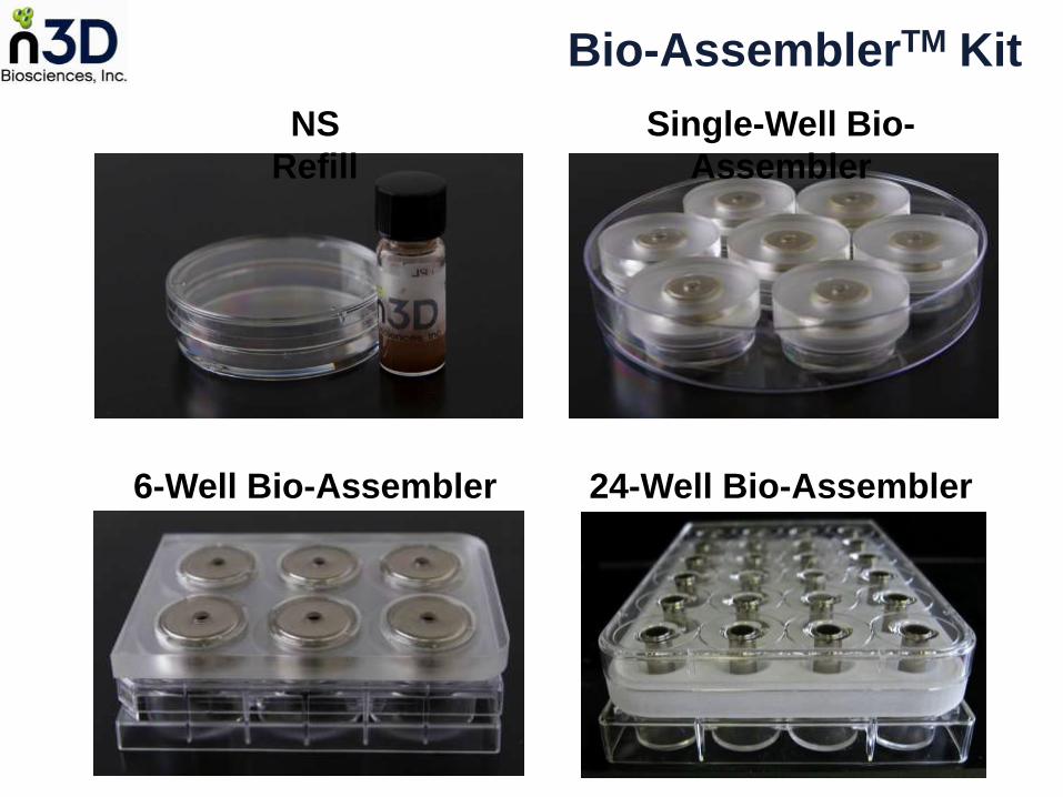
NS
Refill
Single-Well Bio-
Assembler
6-Well Bio-Assembler 24-Well Bio-Assembler
Bio-AssemblerTM Kit

What about in vivo like?

In Vivo like In Vitro Mouse Brain Xenograft Comparison - Glioblastoma
Souza et al. Nature Nanotechnol. April 2010

Shape but Scaffoldless:
The New Paradigm

Levitated Magnetic Pattern
New paradigm
Scaffoldless tissue engineering with shape
So
uza
et a
l. N
atu
re N
an
ote
ch
no
l. A
pril 2
01
0
500 µm

Co-Culture, Spatial Control,
Invasion Assay

Invasion Assay & Co-Culture Magnetic Guidance = Control
Normal Human Astrocyte
mCherry fluorescence
Human Glioblastoma
GFP fluorescence
t = 0
Souza et al. Nature Nanotechnol. April 2010

(Scale bar, 200 µm.)
Invasion Assay & Co-Culture Magnetic Guidance = Control
Souza et al. Nature Nanotechnol. April 2010
Molina et al. Neoplasia, May 2010

What About the Nanoparticles?

SEM & TEM - 1 day = Intracellular NP 8 days = Extracellular NP
24 hours 8 days
SE
M
TE
M

TEM of Cells Closer to the Edge
24 hours 8 days

Nanoshuttles
All components of the reagent mix are individually FDA.
Nanoshuttle was tested in mice and no acute toxicity was found.
Over 20 different cells types have been cultured with the Bio-Assembler, including primary cells
We have not found a cell type that did not culture in the Bio-Assembler.
Healthy cell cultures have been maintained for as long as 2 months. They were terminated at the end of experiment.
Comparative Genome hybridization (CGH) profile was comparable between Nanoshuttle treated and non-treated human primary cells, indicating that the nanoparticles do not cause any genomic instability.
No difference in viability and proliferation between cells in 2D treated and not-treated with Nanoshutlle
Western blotting showed no difference in gene expression between primary cells treated and not-treated with Nanoshutlle cultured in 2D: fibronectin, laminin, N-Cadherin, E-Cadherin, smooth muscle α-actin

Step-by-Step

magnetic drive
lid
petri-dish
As Simple As 2D
Trypsinize
cells
Add Nanoshuttle to
media & cells

Preparing Neural Stem Cells for Magnetic Levitation
Add & Incubate
Neural Stem cells with Nanoshuttle
PBS Wash
Remove Excess Nanoshuttle
Add Nanoshuttle to media & cells
Souza et al. Nature Nanotechnol. April 2010
Trypsinize
cells
First
Step

Levitating Cells
Levitated NSC 3D NSC Culture
15 minutes 12 hours
*NSC = Neural Stem cells

Tuning the Culture

24 hours 48 hours
Ch
on
dro
cyte
s 10 Days
Levitation Time
200k 400k 880k
Hep
ato
ma
Number of Cells – 24 hours

Human Mesenchymal Stem Cells
100k cells 200k cells

Number of Cells – Human Primary
Pulmonary Fibroblast
300k
150k
75k
37k
19k
10k

6-Well Bio-AssemblerTM
*Submitted for Publication

Lung Primary Cells

Human Lung Primary Cells
Fibroblast
Epithelial Smooth Muscle
Endothelial

Bio-AssemblerTM
2D
In Vivo
Primary Pulmonary Fibroblasts

Human Umbilical Vein Cells
HUVEC

HUVEC – Macrostructure
*48 hour culture

HUVEC - Microstructure

Rapid 3D Formation by
Promoting Cell-Cell Interaction

HUVEC 1 & 4 Hours Culture

Fibroblast Epithelial
4 Hours of Levitation – Primary Cells

Extracellular Matrix:Laminin
Immunohistochemistry
Fibroblasts
Smooth Muscle Cells

Stem Cells - Dental Pulp

Day 1 Day 2
Cells from Dental Pulp
In collaboration with Dr. Dozortsev, Director of Advanced Fertility Center of Texas

Stem Cells from Dental Pulp
Immunohistochemistry
*Negative Controls – secondary only
Stro-1 Vimentin
Ste
m C
ells
N
eg
ative
Co
ntr
ols
*
H&E

Stem Cells – Adipose Derived

Co-Culturing
Endothelial (GFP) and Fibroblast

PLN GFP ToPro3
bEND.3-GFP endothelial cells formed microvessels within the adiposphere.
Larger lipid droplet formations are also observed.
Day 14 after induction
of adipogenesis
Adiposphere organoid composed of differentiated
3T3-L1 in co-culture with bEND.3-GFP

Levitated Cell Types
Human Primary Cells
Pulmonary Fibroblast
Pulmonary Endothelial & HUVEC
Small Air Way Epithelial
Tracheal Smooth muscle
Mesenchymal Stem Cells
Dental Pulp Stem Cells
Murine Adipose Tissue
Bone Marrow Endothelial
Heart Valve endothelial
Human Mammary Epithelial - MCF10A
Pre-adipocytes Fibroblasts
Adipocytes
Neural Stem Cells
HEK 293
Melanoma
Astrocytes
Glioblastomas
T-Cells and Antigen Presenting Cells
Chondrocytes

New Tool
3D Wound Healing Assay

2D Scratch Assay
Today’s In Vitro Wound healing Model
2D culture
Poor cell-cell interaction
representation
Interaction of cells with plastic or
coated surface = NOT in vivo like
Difficult to co-culture different cell
types
No “wound” contraction
Scratch
Liang et al., Nature Protocols, 1 March 2007

New Tool
3D Wound Healing Assay

6-Well Bio-AssemblerTM
*Submitted for Publication

B A C D
E F
Puncturing Wound
*Submitted for Publication

Zero closure
0 mM IBU
4 mM IBU
0.1 mM IBU
1 mM IBU
In DMSO
t = 0 h t = 2 h t = 4 h t = 24 h t = 48 h t = 96 h
HEK293 Wound Healing vs. Ibuprofen Concentration
*Subm
itte
d for
Public
ation

3D Wound Healing Assay by
Co-Culturing Primary Cells
100% HPF
50%HPF
&
50% SMC
100% SMC
t = 3 h t = 12 h t = 20h t = 34 h t = 3 h t = 12 h t = 20h t = 34 h
Smooth Muscle Cell (SMC) Media Fibroblast Media
300 μm

Magnetic Printing Method
Average Area of opening = 1.18 ± 0.18 mm2
650 μm
Future Tools
Tissue Layering with
The Magnetic Pen
24- & 96-Well Plates

B
C
D
E
G
F
F
Magnetic Pen

Layered Co-Culture
*In collaboration with Dr. Jane Grande-Allen – Rice Bioengineering


Layered Co-Culture Four Primary Cell Types
*In collaboration with Dr. Jane Grande-Allen – Rice Bioengineering

Immunohistochemistry of Four Cell Types Layered Co-Culture
*In collaboration with
Dr. Jane Grande-Allen
– Rice Bioengineering

“Viability” Assay

Traditional In Vitro Cell Viability Testing
A Challenge in 3D Cell Culturing
Metabolic assays - MTT
– Poor permeability through 3D culture
– underestimate cell viability
DNA content – picoGreen
– Challenging to extract DNA – requires extensive
cell manipulation
– underestimate cell viability
Cell counting
– Requires dispersing cells by enzimatic digestion
which compromises cell viability
– underestimate cell viability
Poor correlation with side-by-side 2D results

*Li et al. J. Pharmacol. Exp. Ther. 2010, 332, 821-8.
Breast Cancer MDA231 10x
Control
0.1 uM DOXO
100 uM DOXO
1 uM DOXO
10 uM DOXO
“Pre-cancer” MCF10A 10x Pre-Cancer more resistant to
Doxorubicin Chemotherapy
Levitated
3 days
Spheroid
9 days
From Literature*:

*Li et al. J. Pharmacol. Exp. Ther. 2010, 332, 821-8.
Control
0.1 uM DOXO
100 uM DOXO
1 uM DOXO
10 uM DOXO
3D Levitated
3 days
2D 3 days
From Literature*:
2D showed opposite response to
Doxorubicin Chemotherapy
NOT PREDICTIVE
Breast Cancer MDA231 10x
“Pre-cancer” MCF10A 10x

Control
0 mM
10 mM
2 mM
4 mM
In DMSO
0.25 mM
1 mM
t = 0 h t = 24 t = 48 t = 168
HEK 293 at 4x Magnification
Ibuprofen
Concentration
Low
High
Time of
Culture
Dynamic Process
4D = time + 3D

Control siRNA Treated
LNCaP – siRNA Treated
Df = 1.7 Df = 1.9

The Next Generation!
Needed Research & Operational Impact
Rapid formation of 3D in vivo-like multicellular structures
Promote cell-cell interaction
Co-Culturing capability
Easy to handle, pre- and post- culture
Compatible with standard diagnostics
Fast set-up time, as simple as 2D
Minimum deviation from general protocols

Publication and Media

Acknowledgements
Rice University Tom Killian Robert Raphael Jane Grande-Allen Rebecca Richards-Kortum Veronica Leautaud Hubert Tseng Tom Kraft n3D Carly Filgueira Dave Lee Christopher Bertucci David Sing
University of Texas Misha Kolonin Richard Clark Jacqueline Hatch Joe Alcorn BMC Robert Moore Jeanne Becker The George Washington University J. Houston Miller MD Anderson Cancer Center Wadih Arap Maria Georgescu Jami Mandelin Jennifer Molina Renata Pasqualini
Funding:
Tom Kraft Deborah Mansfield
Fertility Center of Texas Dmitri Dozortsev

Glauco R. Souza, Ph.D.
Chief Scientific Officer Nano3D BiosciencesTM, Inc.
www.n3Dbio.com