32nd Mycotoxin Workshop; 14th - 16th June, 2010, Lyngby ... · June, 15th‐17th, 2010, Lyngby ......
183
Transcript of 32nd Mycotoxin Workshop; 14th - 16th June, 2010, Lyngby ... · June, 15th‐17th, 2010, Lyngby ......

���� ������ ��������������� ��� ����� ������ ������
������� � ��������
�������� ��

June, 15th‐17th, 2010, Lyngby
A | P a g e
Table of contents Scientific and Organizing Committee B Programme Monday, 14th of June 2010 C‐D Tuesday, 15th of June 2010 E‐F Wednesday, 16th of June 2010 G Table of Presentations Table of Lectures Fungal metabolites and their occurrence H Analysis of fungal metabolites I Toxicology: metabolism and distribution J Toxicology: synergistic effects K Table of Posters Analytical developments L‐N Occurrence of mycotoxins O‐Q Prevention strategies R Toxicology S‐T Abstracts of Lectures 1‐42 Abstracts of Posters 43‐155 Author Index 156‐160
32th Mycotoxin Workshop
B | P a g e
Scientific and Organizing Committee The Society for Mycotoxin Research (Board of Directors) Prof. Dr. Hans‐Ulrich Humpf (President) Westfälische Wilhelms‐Universität Münster, Institut für Lebensmittelchemie 48149 Münster, Germany Prof. Dr. Rolf Geisen (Vice President) Max‐Rubner‐Institut, Bundesforschungsinstitut für Ernährung und Lebensmittel Institut für Sicherheit und Qualität bei Obst und Gemüse, 76131 Karlsruhe, Germany Dir. & Prof. Dr. Manfred Gareis (Secretary) Max‐Rubner‐Institut, Bundesforschungsinstitut für Ernährung und Lebensmittel Institut für Mikrobiologie und Biotechnologie, 95326 Kulmbach, Germany Dr. Uwe Lauber (Treasurer) CVUA Stuttgart, 70736 Fellbach, Germany Technical University of Denmark Dr. Birgitte Andersen CMB, Department for Systems Biology, Technical University of Denmark Dr. Kristian F Nielsen CMB, Department for Systems Biology, Technical University of Denmark Organizing Assistants Homepage: Thomas Kampp, CMB, Department for Systems Biology, Technical University of Denmark Abstract book: Peter Boldsen Knudsen, CMB, Department for Systems Biology, Technical University of Denmark Registration and logistics: Welcome ApS, Billedvej 4, 1. floor, DK‐2100 Copenhagen, Denmark Acknowledgements The organizing committee would like to thank all the master students, PhD. students and faculty at Microbial Biodiversity & Systematic and Analytical Microbial Chemistry at Centre for Microbial Biotechnology, Department of Systems Biology, Technical University of Denmark. Without their help, this workshop would not have been possible. Disclaimer: Abstracts submitted to this workshop are reproduced in the programme with only minor editorial revision. The editors are not responsible for the content of the abstracts.

June, 15th‐17th, 2010, Lyngby
C | P a g e
Programme Monday morning 09:00‐09:30 Opening of the 32nd mycotoxin workshop
Fungal metabolites and their occurrence ‐ Chairman: Manfred Gareis
L01 9:30‐9:45
Peptaibols, first marine mycotoxins?
Yves F. Pouchus L02 9:45‐10:00
Pathogenicity and toxin production of Fusarium proliferatum in onion bulbs
Frank M. Ellner L03 10:00‐10:15
Ecological speciation in Claviceps purpurea vs. ergot alkaloid and indole‐diterpene production
Silvio Uhlig
10:15‐10:45 Coffee break / Exhibition
Fungal metabolites and their occurrence ‐ Chairman: Jens. C. Frisvad
L04 10:45‐11:00
Determination of fumigaclavine A in respiratory system tissue of birds with aspergillosis
Hadri Latif L05 11:00‐11:15
Occurrence of aflatoxins in poultry feeds and residues of aflatoxins on broiler meat in Indonesia
Merry M. D. Utami
L06 11:15‐11:30
Exposure assessment to Ochratoxin A from the consumption of cocoa and chocolate products
Clara Berdini L07 11:30‐11:45
One possible ecological reason for the production of ochratoxin by Penicillium
Rolf Geisen L08 11:45‐12:00
Pre‐and post‐harvest mycotoxins and other secondary metabolites in maize silage detected by LC‐MS/MS
Rie R. Rasmussen
12:00‐12:45 Lunch / Exhibition

32th Mycotoxin Workshop
D | P a g e
Monday afternoon 12:45‐14:00 Poster session
Analysis of fungal metabolites ‐ Chairman: Thomas O. Larsen
L09 14:00‐14:15
A validated multi‐analyte LC‐MS/MS method for the quantification of 25 mycotoxins in cassava flour, peanut cake and maize samples
Emmanuel, N. Ediage L10 14:15‐14:30
Comparison of extraction solvents for Fusarium toxins in wheat, maize and rice
Sasithorn Limsuwan L11 14:30‐14:45
Ochratoxin A is a complete carcinogen: Identification by LC‐MS/MS of C8‐ 2’‐deoxyguanosine OTA‐adduct in vivo
Annie Pfohl‐Leszkowicz L12 14:45‐15:00
Novel high‐throughput non‐destructive sampling and extraction technology for mycotoxins from grains and cereals – development and application
Simone Staiger L13 15:00‐15:15
ISO 17025 accreditation of LC‐MS methods for mycotoxin analyses in cereals and feedstuffs
Barbara Daxner
15:15‐15:45 Coffee break / Exhibition
Analysis of fungal metabolites ‐ Chairman: Ulf Thrane
L14 15:45‐16:00
New lecture: see abstract P‐96
L15 16:00‐16:15
Chemical modification of B‐type fumonisins
Florian Hübner L16 16:15‐16:30
Enzyme immunoassay for tenuazonic acid in apple and tomato products
Madeleine Groß L17 16:30‐16:45
A multiplex flow cytometric immunoassay for mycotoxins in food and feed using paramagnetic microspheres
Jeroen Peters L18 16:45‐17:00
Application of Mass spectrometry in indoor environmental quality (IEQ) assessment
Vinay Vishwanath
19:00‐.... Reception at Copenhagen town hall with light dinner

June, 15th‐17th, 2010, Lyngby
E | P a g e

32th Mycotoxin Workshop
F | P a g e
Tuesday morning Toxicology: Metabolism and distribution‐ Chairman: Ulf Thrane
L19 09:00‐09:15
Fumonisin induced alteration of sphingolipid metabolism in piglets – Analytical and biological aspects
Heidi Schwartz L20 09:15‐09:30
Investigations on the transfer of deoxynivalenol or its metabolite from cows to their offspring
Susanne Döll L21 09:30‐09:45
Deoxynivalenol alters the interactions of Salmonella typhimurium with porcine intestinal epithelial cells and macrophages
Virginie Vandenbroucke L22 09:45‐10:00
Metabolism and distribution of the mycoestrogen zearalenone as a function of physiological status and condition of exposure
Sylvaine Lecoeur L23 10:00‐10:15
The Fusarium toxins β‐zearalenol and zearalenone inhibit Hsp90 ATPase activity and are inactivated by glucosylation and sulfatation
Gerlinde Wiesenberger
10:15‐10:45 Coffee break / Exhibition
Toxicology: Metabolism and distribution ‐ Chairman: Thomas O. Larsen
L24 10:45‐11:00
Toxicity of ergot alkaloids: Effects on human cells in primary culture
Dennis Mulac L25 11:00‐11:15
Alternaria toxins ‐ toxicological potential and mechanistical aspects
Doris Marko L26 11:15‐11:30
Live cell imaging confirms disruption of mitosis as a key event in ochratoxin A toxicity
Angela Mally L27 11:30‐11:45
Ochratoxin A in dairy goats
Ralf Blank L28 11:45‐12:00 Ochratoxin A levels in biological fluids from mother‐child pairs in
Chile: A 6 months follow up study Katherine Muñoz
June, 15th‐17th, 2010, Lyngby
G | P a g e
12:00‐12:45 Lunch / Exhibition
Tuesday afternoon 12:45‐14:00 Poster session Toxicology: synergistic effects ‐ Chairman: Hans‐Ulrich Humpf
L29 14:00‐14:15
Daily Intake of carcinogenic and reprotoxic mycotoxins ‐ Molecular evidence of synergistic effects
Annie Pfohl‐Leszkowicz L30 14:15‐14:30
Effects of low and high dietary deoxynivalenol on growth performance and response to vaccines in broilers
Josef Böhm L31 14:30‐14:45
The interaction effect of two major Fusarium mycotoxins on the pig systemic and immune responses
Bertrand Grenier L32 14:45‐15:00
The effect of mycotoxin binders on the oral bioavailability and tissue residues of doxycycline in pigs
Joline Goossens
15:00‐15:30 Coffee break / Exhibition 15:30‐17:30 General assembly of the Society 19:30‐.... Dinner in Tivoli
32th Mycotoxin Workshop
H | P a g e
Wednesday morning Metabolites in plant/fungi interactionMetabolites in plant/fungi
interaction − Chairman: Rolf Geisen
L33 09:00‐09:15
Formation of DON‐glutathione conjugates: a role for glutathione‐S‐transferases in DON resistance of barley?
Gerhard Adam L34 09:15‐09:30
The role of deoxynivalenol (DON) and nivalenol (NIV) in the infection of roots of wheat and maize with F. graminearum
Awais Ahmed L35 09:30‐09:45
Infection process and mycotoxin production in Fusarium culmorum‐infected maize ears
Elisabeth Oldenburg L36 09:45‐10:00
Effect of mixed inoculation of Fusarium verticillioides and Fusarium graminearum on the dynamic of fungal colonization and mycotoxin production in maize cobs
Sabine Nutz L37 10:00‐10:15
Proteome analysis of Fusarium infected emmer (Triticum dicoccum) and naked barley (Hordeum vulgare subsp. nudum) grains
Kai Eggert
10:15‐11:00 Coffee break / Exhibition
Detoxification – Chairman: Uwe Lauber L38 11:00‐11:15 Isolation and characterization of a novel non‐toxic metabolite of the
Fusarium mycotoxin diacetoxyscirpenol Mehrdad Shams L39 11:15‐11:30
Reduction of aflatoxin B1 contamination in corn with natural zeolite and bentonite
Nuryono L40 11:30‐11:45
New results on the thermal degradation of Alternaria mycotoxins in food matrices
David Siegel
11:45‐12:00 Award for best poster 12:00‐12:15 Closing of the 32nd mycotoxin workshop

June, 15th‐17th, 2010, Lyngby
I | P a g e
Table of Presentations Table of Lectures Fungal Metabolites and their occurrence L01 Peptaibols, first marine mycotoxins? 3
Yves François Pouchus, Nicolas Ruiz, Claire Sallenave‐Sallenave‐Namont , Laurence Poirier , Zouher Amzil and Olivier Grovel
L02 Ecological speciation in Claviceps purpurea vs. ergot alkaloid and indole‐diterpene production
4
Frank M. Ellner and Monika Goßmann L03 Determination of Fumigaclavine A in Respiratory System Tissue of Birds with
Aspergillosis 5
Silvio Uhlig, Elin Rolèn and Trude Vrålstad L04 Occurance of Aflatoxins in Poultry Feeds and Residues of Aflatoxins on Broiler
Meat in Indonesia 6
Hadri Latif, Valeriu Curtui, Yvonne Ackermann, Madeleine Groß, Nadja Lorenz, Mirjam R. Hampel, Michael Lierz and Ewald Usleber
L05 Occurance of Aflatoxins in Poultry Feeds and Residues of Aflatoxins on Broiler Meat in Indonesia
7
Merry Muspita Dyah Utami, Ali Agus, Wihandoyo and Kurniasih L06 Exposure assessment to Ochratoxin A from the consumption of cocoa and
chocolate products. 8
Clara Berdini, Carlo Brera, Elena Pannunzi and Emanuela Iafrate L07 One possible ecological reason for the production of ochratoxin by Penicillium. 9 Rolf Geisen, Julia Batzler and Markus Schmidt‐Heydt L08 Pre‐and post‐harvest mycotoxins and other secondary metabolites in maize
silage detected by LC‐MS/MS 10
Rasmussen RR, Storm IMLD, Rasmussen PH, Smedsgaard J and Nielsen KF

32th Mycotoxin Workshop
J | P a g e
Analysis of fungal metabolites L09 A validated multi‐analyte LC–MS/MS method for the quantification of 25
mycotoxins in cassava flour, peanut cake and maize samples 11
Emmanuel Njumbe Ediage, Jose Diana Di Mavungu, Carlos Van Peteghem and Sarah De Saeger
L10 Comparison of extraction solvents for Fusarium toxins in wheat, maize and rice 12 Sasithorn Limsuwan and Petr Karlovsky L11 Ochratoxin A is a complete carcinogen: Identification by LC ms/ms of C8‐ 2’‐
deoxyguanosine OTA‐adduct in vivo 13
Annie Pfohl‐Leszkowicz, Richard Manderville, Bianca Squilacci and Peter Mantle L12 Novel high‐throughput non‐destructive sampling and extraction technology for
mycotoxins from grains and cereals – development and application 14
Simone Staiger, Ngnintendem Nkengfack, Susanne Rathjen and Scarlett Biselli L13 ISO 17025 accreditation of LC‐MS methods for mycotoxin analyses in cereals and
feedstuffs 15
Barbara Daxner L14 16
L15 Chemical Modification of B‐type Fumonisins. 17 Florian Hübner, Lilia Lohrey and Hans‐Ulrich Humpf L16 Enzyme Immunoassay for Tenuazonic Acid in Apple and Tomato Products 18 Madeleine Groß, Valeriu Curtui, Yvonne Ackermann, Hadri Latif and Ewald Usleber
L17 A multiplex flow cytometric immunoassay for mycotoxins in food and feed using paramagnetic microspheres.
19
Jeroen Peters and Willem Haasnoot L18 Application of Mass spectrometry in indoor environmental quality (IEQ)
assessment 20
Vinay Vishwanath, Michael Sulyok, Stefan Mayer and Rudolf Krska

June, 15th‐17th, 2010, Lyngby
K | P a g e
Toxicology: Metabolism and Distribution L19 Fumonisin induced alteration of sphingolipid metabolism in piglets – Analytical
and biological aspects. 21
Heidi Schwartz, Bertrand Grenier, Irene Schöner, Anita Seeböck, Isabelle P. Oswald, Gerd Schatzmayr and Wulf‐Dieter Moll
L20 Investigations on the transfer of deoxynivalenol or its metabolite from cows to their offspring
22
Susanne Döll, Lydia Renner, Ulrich Meyer, Hana Valenta and Sven Dänicke L21 Deoxynivalenol alters the interactions of Salmonella Typhimurium with porcine
intestinal epithelial cells and macrophages 23
Virginie Vandenbroucke, Frank Pasmans, Elin Verbrugghe, Joline Goossens, Freddy Haesebrouck, Patrick De Backer and Siska Croubels
L22 Metabolism and distribution of the mycoestrogen zearalenone as a function of physiological status and condition of exposure.
24
Sylvaine Lecoeur, Michelle Mazallon and Bernadette Videmann L23 The Fusarium toxins b‐zearalenol and zearalenone inhibit Hsp90 ATPase activity
and are inactivated by glucosylation and sulfatation. 25
Juan Antonio Torres Acosta, Franz Berthiller, Gerlinde Wiesenberger, Rudolf Mitterbauer, Ulrike Werner, David Merz, Marie‐Theres Hauser, Mehrdad Shams, Rudolf Krska and Gerhard Adam
L24 Toxicity of ergot alkaloids: Effects on human cells in primary culture 26 Dennis Mulac and Hans‐Ulrich Humpf L25 Alternaria toxins ‐ toxicological potential and mechanistical aspects 27 Doris Marko, Markus Fehr, Christoph Schwarz and Gudrun Pahlke L26 Live cell imaging confirms disruption of mitosis as a key event in ochratoxin A
toxicity 28
Kristin Czakai, Elisabeth Jeanclos, Alexander Zürn, Antje Gohla and Angela Mally L27 Ochratoxin A in dairy goats 29 Ralf Blank, May Looff, Muhammad Mobashar, Anne Westphal and Karl‐Heinz
Südekum
L28 Ochratoxin A levels in biological fluids from mother‐child pairs in Chile: A 6 months follow up study.
30
Katherine Muñoz, Victor Campos, Meinolf Blaszkewicz, Mario Vega, Alejandro Alvarez, Jorge Neira and Gisela H. Degen

32th Mycotoxin Workshop
L | P a g e
Toxicology: Synergistic Effects L29 Daily Intake of carcinogenic and reprotoxic mycotoxins ‐ Molecular evidence of
synergistic effects 31
Mariana Tozlovanu, Kheira Hadjeba‐Medjdoub, Maja Peraica, Vladisav Stefanovic, Richard Manderville and Annie Pfohl‐Leszkowicz
L30 Effects of low and high dietary deoxynivalenol on growth performance and response to vaccines in broilers
32
Agha W. Yunus, Khaled Ghareeb, Ahmad Abdelfattah, Manfred Hollmann, Jan Grajewski, Twaruzek Magdalena, Anna Blajet‐Kosicka, Robert Kosicki and Josef Böhm
L31 The interaction effect of two major Fusarium mycotoxins on the pig systemic and immune responses
33
B. Grenier, G. Pacheco, D. Moll, A.M. Cossalter, A.P. Loureiro‐Bracarense, G. Schatzmayr and I.P. Oswald
L32 The effect of mycotoxin binders on the oral bioavailability and tissue residues of doxycycline in pigs
34
J. Goossens, F. Pasmans, V. Vandenbroucke, E. Verbrugghe, F. Haesebrouck, P. De Backer and S. Croubels
L33 Formation of DON‐glutathione conjugates: a role for glutathione‐S‐transferases in DON resistance of barley?
35
Gerhard Adam, Franz Berthiller, Christian Hametner, Stephanie A. Gardiner, Jayanand Boddu, Robert Stupar and Gary J. Muehlbauer
L34 The role of deoxynivalenol (DON) and nivalenol (NIV) in the infection of roots of wheat and maize with F. graminearum
36
Awais Ahmed, Stephan Bucher and Petr Karlovsky L35 Infection process and mycotoxin production in Fusarium culmorum‐infected
maize ears 37
Elisabeth Oldenburg and Frank Ellner L36 Effect of mixed inoculation of Fusarium verticillioides and Fusarium graminearum
on the dynamic of fungal colonization and mycotoxin production in maize cobs 38
Sabine Nutz, Katharina Döll, Sasithorn Limsuwan and Petr Karlovsky L37 Proteome analysis of Fusarium infected emmer (Triticum dicoccum) and naked
barley (Hordeum vulgare subsp. nudum) grains 39
Kai Eggert, Christian Zörb and Elke Pawelzik L38 Isolation and characterization of a novel non‐toxic metabolite of the Fusarium
mycotoxin diacetoxyscirpenol 40
Mehrdad Shams, Rudolf Mitterbauer, Roberto Corradini, Gerhard Adam, Rudolf Krska, Rainer Schuhmacher and Franz Berthiller
L39 Reduction of aflatoxin B1 contamination in corn with natural zeolite and bentonite
41
Nuryono1, A. Agus, S. Wedhastri, Y. M. S. Maryudhani, D. Pranowo, Yunianto and E. Razzazi‐Fazeli
L40 New results on the thermal degradation of Alternaria mycotoxins in food matrices
42

June, 15th‐17th, 2010, Lyngby
M | P a g e
David Siegel, Matthias Koch and Irene Nehls Table of Posters Analytical Developments P01 Towards the validation of an LC‐DAD‐MS/MS‐method for the detection of ZON,
ZOLs and a novel non‐estrogenic metabolite (ZOM‐1) 45
Sonja Anderle, Heidi Schwartz, Sebastian Fruhauf and Elisavet Vekiru P02 Simulated digestion assay for hidden fumonisin evaluation. 46 A. Dall’Erta, C. Falavigna, C. Dall'Asta, G. Galaverna, A. Dossena and R. Marchelli. P03 Accurate and precise determination of the Alternaria mycotoxins Alternariol
and Alternariol monomethylether in cereals, fruit and vegetables using stable isotope dilution assays
47
Asam S., Konitzer K. and Rychlik M. P04 Development of a sample preparation method for the determination of T‐2
toxin and metabolites in blood plasma with LC‐MS/MS. 48
Ulrike Brezina, Katrin Wilkerling, Hana Valenta, Susanne Döll and Sven Dänicke P05 Modulation of the early apoptotic potential of DON by MAPK signaling 49 Maximilian Casteel, Carina Nielsen, Richard Dietrich and Erwin Märtlbauer P06 Development of Lateral Flow Devices for the Qualitative and Semi‐Quantitative
Detection of Mycotoxins in Agricultural Commodities 50
Barbara Cvak, Alexandra Molinelli and Rudolf Krska P07 Evaluation of a Proficiency Test on the Determination of Fumonisin B1 and B2
in Maize‐Based Peanut Snack 51
Rudolf Hackenberg and Carolin Stachel P08 Detection of mould and mycotoxine on grain using a gas Sensor Array 52 C. Idler, A. Walte, M. Ditz, A. Plessing‐Menze and K. Briese P09 New Fusarium mycotoxin CRMs – perfect tools for reliable food analysis 53 Matthias Koch, Robert Köppen, Tatjana Rasenko, Karin Klein‐Hartwig, Matthias
Proske, David Siegel, Wolfram Bremser and Irene Nehls
P10 Rapid Determination of Patulin in Fruit and selected Vegetable Products with LC‐MS‐MS
54
Ulrike Kocher, Luzia Liebherr, Markus Martin P11 Synthesis of molecularly imprinted polymers as an alternative clean‐up strategy
for ergot alkaloids 55
Pieterjan Lenain, José Diana Di Mavungu, Peter Dubruel, Johan Robbens, Carlos Van Peteghem and Sarah De Saeger
P12 Untarget screening of fungal metabolites produced by Penicillium by means of LC‐Q‐TOF‐MS.
56
S. Malysheva, V. Polizzi, C. Van Peteghem, N. De Kimpe, A. Moretti, J. Van Bocxlaer and S. De Saeger
P13 Production of isotope labeled mycotoxins. Use and advantages in routine analysis
57
Anna Mitterauer

32th Mycotoxin Workshop
N | P a g e
P14 Development of an immunodiagnostic strip test for the screening of ochratoxin
A in food and feed within 10 min. 58
Alexandra Molinelli, Barbara Cvak,, Georg Häubl and Rudolf Krska P15 Multi‐mycotoxin LC‐MS/MS used to analyse tea, herbal infusions and their
drinkable products. 59
S. Monbaliu, A. Wu, D. Zhang, C. Van Peteghem and S. De Saeger P16 Pitfalls encountered during PAT determination in apple juice and apple puree 60 Alessandra Moseriti and Gerhard Buttinger P17 A meta‐analysis of aflatoxins results in transgenic Bt‐maize vs. non‐Bt Maize
Hybrids. 61
Vladimir Ostry, Jaroslava Ovesna, Jarmila Skarkova, Vladimira Pouchova and Jiri Ruprich
P18 Development of a novel sample preparation technique for the rapid detection of mycotoxins in grain
62
Christoph Portner, Volker Plegge, Werner Kohorst and Thomas Rohe P19 Development of a direct coupled mass spectrometric method for the fast
determination of biological activity in house dust using an enzymatic assay 63
Katja Oeste, Christoph Portner , Thorsten Teutenberg and Thomas Letzel P20 A Rapid Lateral Flow Quantitative Test for Deoxynivalenol in Wheat 64 Stephen P. Powers and Jianmini Liu P21 Two‐photon‐induced fluorescence for the determination of Aflatoxin B1 in
alcoholic beverages 65
C. Rasch, M. Böttcher, M. Kumke and H.‐G. Löhmannsröben P22 ESI‐IT/MS peptaibol fingerprints for identification of toxic marine‐derived
Trichoderma strains 66
Ruiz Nicolas, Robiou du Pont Thibaut, Joubert Yolaine, Poirier Laurence, Sallenave‐Namont Claire, Grovel Olivier, Bissett John and Pouchus Yves François
P23 Aflatoxin M1 in milk and milk products: Development of a new, highly sensitive ELISA test
67
Alois Schiessl, Michael Z. Zheng and Mao J. Hong P24 Novel applications of hydrazine chemistry in the instrumental analysis of food
mycotoxins 68
David Siegel, Matthias Koch and Irene Nehls P25 Comparison of two different analytical methods for determination of
ochratoxin‐A in wine 69
Biljana Stojanovska‐Dimzoska, Zehra Hajrulai‐Musliu, Elizabeta Dimitrieska‐Stojkovic and Pavle Sekulovski
P26 RAPD, RFLP and UPLC‐MS/MS: a multidisciplinary approach to study the diversity of Aspergillus fumigatus isolates from silage
70
Els Van Pamel, Els Daeseleire, Marc Heyndrickx, Annemieke Verbeken and Geertrui Vlaemynck
June, 15th‐17th, 2010, Lyngby
O | P a g e
P27 UPLC‐MS/MS multimycotoxin analysis of the mycotoxin production by pure
fungal cultures from silage 71
Els Van Pamel, Geertrui Vlaemynck, Marc Heyndrickx, Annemieke Verbeken and Els Daeseleire
P28 Glucuronide Conjugates of Deoxynivalenol and Zearalenone in Human Urine: A Novel LC‐MS/MS Approach
72
Benedikt Warth, Philipp Fruhmann, Hannes Mikula, Franz Berthiller, Rainer Schuhmacher, John Gilbert, Christian Hametner, Michael Sulyok, Johannes Fröhlich and Rudolf Krska
P29 An LC‐MS/MS method for analysis of very low stable isotope enrichments of [2H5]‐phenylalanine in physiological samples as a tool to investigate the inhibitory effect of deoxynivalenol (DON) on the protein synthesis
73
K. Wilkerling, H. Valenta, S. Dänicke and S. Döll P30 Post‐Column Derivatization Methods in the HPLC Analysis of Aflatoxins. 74 Henry Joshua P31 Simultaneous HPLC Determination of Aflatoxins, Zearalenone and Ochratoxin
A Without Changing the Excitation Wavelength. 75
Henry Joshua P32 Fungi and their mycotoxins in maize and maize silage 76 Storm IMLD, Sørensen JL, Rasmussen RR, Thrane U

32th Mycotoxin Workshop
P | P a g e
Occurrence of Mycotoxins P33 Comparative Immunochemical Assessment of Alternariol in Food using
Monoclonal and Polyclonal Antibodies 77
Yvonne Ackermann, Valeriu Curtui, Richard Dietrich, Madeleine Groß, Hadri Latif, Erwin Märtlbauer and Ewald Usleber
P34 Effect of calcium propionate, aw and incubation time on mould incidence and aflatoxins production in commercial poultry feed starter ration
78
Sahib Alam, Hamid U Shah and Naresh Magan P35 Interaction field trials between mycotoxin producers F. graminearum and F.
poae strains involved in Fusarium Head Blight of durum wheat 79
B. Amato, A. Prodi, K. Döll , P. Karlovsky , P. Nipoti and A. Pisi P36 Occurrence of type A, B, and D trichothecenes in barley from the Bavarian
market 80
Jörg Barthel, Martin Rapp, Matthias Berger, Christoph Gottschalk, Johann Bauer and Karsten Meyer
P37 Fusarium toxins and fungi in oats and its products from ecological and conventional agriculture practice
81
Anna Blajet‐Kosicka, Jan Grajewski, Magdalena Twaruzek and Robert Kosicki P38 Hidden fumonisins: a study of the masking mechanism in raw maize. 82 Claudia Falavigna, Andrea Dall’Erta, Chiara Dall’Asta, Gianni Galaverna,
Arnaldo Dossena and Rosangela Marchelli
P39 Monitoring of ochratoxin A content of must during fermentation 83 Csaba Csutorás, Péter Fűtő, Péter Forgó, László Rácz and Zsolt Tajcs P40 Seasonal variations in adult serum ochratoxın a and aflatoxin levels
in Central Anatolıa region, Turkey 84
Belma Giray, Gönül Şahin, Sevtap Aydın, Suna Sabuncuoğlu and Pınar Erkekoğlu P41 Ochratoxin A, aflatoxin B1 and aflatoxin M1 levels of breast milk samples
obtained from Ankara, Turkey. 85
Aylin Gürbay, Gözde Girgin, Suna Sabuncuoğlu, Gönül Şahin, Şule Yiğit, Murat Yurdakök and Gülsevin Tekinalp
P42 Mycotoxins from marine‐derived fungi: use of LC‐HRMS/MS for identification of new communesins analogs in Penicillium expansum
86
Olivier Grovel, Isabelle Kerzaon, Fabrice Monteau, Bruno Le Bizec and Yves François Pouchus
P43 Mycotoxins in pet food could reduced fertility and stillbirth and induced genotoxic damages
87
Kheira Hadjeba‐Medjdoub, Mariana Tozlovanu‐Fergane, Ivana Polisenska and Annie Pfohl‐Leszkowicz
P44 Aflatoxins in brazil nuts 88 Beatriz T. Iamanaka, Thaiane O. Calderari, Carolina M. Albers,
Luciana M. R. Esper and Marta H. Taniwaki
P45 Incidence of aflatoxins and ochratoxin A in cocoa and by‐products 89
June, 15th‐17th, 2010, Lyngby
Q | P a g e
Beatriz Thie Iamanaka, Marina Venturini Copetti, Melanie Alexandra Nester, Priscila Efraim and Marta Hiromi Taniwaki
P46 Fumonisins from Aspergillus niger in grapes and derived products 90 Jesper M. Mogensen, Peter B. Knudsen, Jens C. Frisvad, Thomas O. Larsen and
Kristian F. Nielsen
P47 Ergot alkaloids in Finnish grains with sclerotia. 91 Marika N. Jestoi, Elina Sieviläinen and Meri K. Kokkonen P48 Mycotoxin air sampling in indoor environments –
Sentinel Health Investigation utilizing effect based toxicity screening test, an ELISA QuantiTox™ Kit and conventional fungal identification methods
92
Eckardt Johanning, Manfred Gareis and Paul Landsbergis P49 Effect of age and dietary aflatoxin levels upon tissue residue in broiler chicks. 93 Muhammad Z Khan, Zahid Hussain, Ahrar Khan, Muhammad K Saleemi, Ijaz
Javed, Muhammad R Hameed, Zahoor‐Ul‐Hassan and Sultan Mahmood
P50 Fusarin C: isolation, stability and occurrence. 94 Karin Kleigrewe, Patrick Piecuch and Hans‐Ulrich Humpf P51 Correlation between Fusarium graminearum DNA and deoxynivalenol
content in wheat flours and bran 95
Marie Kreuzberger, Sasithorn Limsuwan, Petr Karlovsky and Elke Pawelzik P52 Mycotoxin production in maize inoculated with Fusarium graminearum,
Fusarium verticillioides and Fusarium subglutinans 96
Eva‐Maria Kuhl, C. Göbel, I. Feußner and P. Karlovsky P53 A novel cytotoxic cyclic pentadepsipeptide produced by Fusarium solani,
which is isolated from Fusarium‐contaminated potato in South Korea 97
HS Lee, JW Kang, YS Jung, JM Choi and C Lee P54 Occurrence of Fusarium mycotoxin beauvericin in animal feeds in Korea 98 HS Lee, JW Kang, JM Choi, YS Jung and C Lee P55 Ochratoxin A in human milk, results from the Bavarian Monitoring of Breast
Milk (BAMBI) 99
D. Mann, M. Rapp and M. Berger P56 Researches regarding the nutritive value correlated with myco‐bacteriological
analysis of some pastry products from Romanian commercial units 100
Maria Virginia Morar, Zoe Dancea, Constantin Bele, Adrian M. Macri, Andrei R. Szakacs and Cornel Popa
P57 High levels of aflatoxins found in niche foodstuffs and the use of aflatoxin contaminated ingredients in composite food production
101
Stefanie Marschik, Margit Kettl‐Grömminger and Renate Schnaufer P58 New methods for the synthesis of mycotoxin glucuronides (part B) 102 Dominik Matscheko, Hannes Mikula, Christian Hametner and Johannes Fröhlich P59 Varying the content of T‐2 and HT‐2 toxins in processing of oats 103 Ute Meister P60 Formation of type A trichothecene glucosides by plant cells 104 Karsten Meyer, Julia Matthes and Johann Bauer P61 Migration of mycotoxins produced in sweet pepper inoculated by Fusarium 105

32th Mycotoxin Workshop
R | P a g e
species and analysed using a multi‐mycotoxin lc‐ms/ms method. S. Monbaliu, K. Van Poucke, K. Heungens, C. Van Peteghem and S. De Saeger P62 Exposure of infants to ochratoxin A ‐ New data on OTA levels in breast milk
samples from Germany. 106
Katherine Muñoz, Meinolf Blaszkewicz, Hermann Kalhoff and Gisela H. Degen P63 Ochratoxin A metabolites in human blood and urine samples 107 Katherine Muñoz, Benedikt Cramer , Matthias Kockmann , Meinolf Blaszkewicz ,
Hans‐Ulrich Humpf and Gisela H. Degen
P64 Monitoring of aflatoxin B1 content of must during fermentation 108 László Rácz, Péter Fűtő, Péter Forgó, Csaba Csutorás and Zsolt Tajcs P65 Results of official controls of mycotoxins in food in Lower Saxony in 2009 109 Lilli Reinhold and Katja Reinhardt P66 Occurrence of mycotoxins in feedstuffs and feed in Asian regions in the year
2009. 110
Inês Rodrigues and Karin Griessler P67 A study of mycobiota and mycotoxins in wheat and wheat‐bran from Pakistan 111 M. Kashif Saleemi, M. Zargham Khan, Ahrar Khan, Sohail Hameed, M. Aamer
Mehmood, Ijaz Javed, Zahoor –Ul‐Hasan and M. Raza Hameed
P68 Mycotoxin occurrence in European feed samples 2009. 112 Karin Griessler, Dian Schatzmayr and Inês Rodrigues P69 Presence of Deoxynivalenol in German wheat and rye and its comparison to
Nivalenol and other type B trichothecenes. 113
Christine Schwake‐Anduschus and Meinolf G. Lindhauer P70 New methods for the synthesis of mycotoxin glucuronides (part A) 114 Markus Schwarz, Hannes Mikula, Christian Hametner and Johannes Fröhlich P71 Isolation, purification and characterization of the mycotoxin FS‐4 from
Δtri101 mutants of Fusarium sulphureum. 115
Mehrdad Shams, Rudolf Mitterbauer, Heidemarie Hellmer, Franz Berthiller, Christian Hametner, Gerlinde Wiesenberger and Gerhard Adam
P72 Novel glucosyl donors in the synthesis of masked mycotoxins 116 Dennis Svatunek, Hannes Mikula, Christian Hametner and Johannes Fröhlich P73 Mouldy status characterisation of maize and grass silages: Adequacy between
visual appraisal during collection and categorisation according to ergosterol contents
117
Emmanuel K. Tangni, Luc Pussemier, François van Hove, Jurgen Depoorter, Geert Haesaert and Johan Robbens
P74 Mycotoxins and moulds in silages 118 Magdalena Twaruzek, Anna Blajet‐Kosicka, Robert Kosicki and Jan Grajewski P75 Investigation of mycotoxins produced by marine‐derived fungal strains of
Penicillium sp. 119
Marieke Vansteelandt, Isabelle Kerzaon, Nicolas Ruiz, Jean‐François Biard, Yves‐François Pouchus and Olivier Grovel

June, 15th‐17th, 2010, Lyngby
S | P a g e
Prevention Strategies
P76 Does lycopene play a protective role in low dose aflatoxin B1‐induced hepatotoxicity in rats?
120
Terken Baydar, Gözde Girgin , Güldeniz Selmanoglu , Elif Karacaoglu , Aysun Kilic , Serdar Balci and Gönül Sahin
P77 Silibinin Does Not Prevent OTA‐Mediated Apoptosis in Primary Rat Hepatocytes and HepG2 Cells
121
Ebtisam Essid, Ernst Petzinger P78 The effects of ochratoxin A on oxidative DNA damage and antioxidant
enzymes in rat kidney and protective role of lycopene 122
Sevtap Aydin, Şaziye Sezin Palabiyik, Pınar Erkekoğlu, Gönül SahinNursen Basaran and Belma Giray
P79 Degradation of ochratoxin A by lactobacillus acidophilus K1 strain. 123 Jarosław Mazurkiewicz P80 Effect of Trichoderma harzianum on growth and mycotoxin production of
Fusarium strains. 124
Marina Müller and Susann Siebert P81 Evaluation of Mycofix® Plus supplementation in ochratoxin and zearalenone
contaminated diets in broiler breeders 125
Katia Pedrosa and Chang‐Won Kang P82 The occurrence of mycotoxins in organic winter rye and spring barley
protected by using natural compounds. 126
Eliza Potocka, Ewa Solarska, Adam Kuzdraliński and Marta Muszyńska P83 De‐acetylation of trichothecenes by plant and Fusarium carboxylesterases. 127 Clemens Schmeitzl, Mehrdad Shams, Franz Berthiller, Juan Antonio Torres
Acosta, Gerlinde Wiesenberger, Imer Maloku, Marc Lemmens, and Gerhard Adam
P84 The influence of light on ochratoxin A producing Penicillia 128 Markus Schmidt‐Heydt, Anja Bruchmann, Frank Raupp and Rolf Geisen

32th Mycotoxin Workshop
T | P a g e
Toxicology P85 Effect of zearalenone on ovarian activity in the gilt 129 A. Macri, V. Miclaus, Z. Dancea, L. Bogdan, M.V. Morar, N.Pasca, I. Scurtun and
V. Simion
P86 Production of crude Aflatoxin B1 using different isolates and substrates 130 Ali Agus, Y.B. Maryudhani, Yunianta, Sri Wedhastri and Nuryono P87 On the effects of acute and chronic deoxynivalenol intoxication on gene
expression of glucose transporters in the small intestine of chickens 131
Wageha Awad, Wilfried Vahjen and Jürgen Zentek P88 The mycotoxin deoxynivalenol alters the proliferation and the metabolic
function of porcine intestinal epithelial cells 132
Wageha Awad, Josef Böhm and Jürgen Zentek P89 Effects of enniatin B on viability and cell cycle of different cell types (tumour
and non‐tumour cells) in vitro 133
Claudia Behm, Alena Rasche, Gisela H. Degen and Wolfram Föllmann P90 Effect of individual and combined intravenous infusion of Deoxynivalenol
(DON) and Lipopolysaccharides (LPS) on plasma DON concentrations in the pig
134
Bianca Brosig, Susanne Döll, Patricia Panther, Hana Valenta, H.J. Rothkötter and S. Dänicke
P91 Mutagenicity and cytotoxicity of Alternaria mycotoxins in cultured Chinese hamster V79 cells
135
Britta Burkhardt, Frano B. Šeremet, Sima Payandeh, Erika Pfeiffer and Manfred Metzler
P92 Effects of deoxynivalenol (DON) and related compounds on bovine peripheral blood mononuclear cells (PBMC)
136
Sven Dänicke, Christina Keese, Tanja Goyarts and Susanne Döll P93 Synthesis of Ochratoxin A Derivatives 137 Ines Ferse, Junichiro Yamaguchi, Kenichiro Itami, Benedikt Cramer, Hans‐Ulrich
Humpf*
P94 Brain and hypophyseal total protein concentrations in pubertal boars fed dietary fumonisin B1
138
Francis A. Gbore and Baharuddin Salleh P95 Deoxynivalenol (DON) reduces viability and induces both cell cycle arrest in
the G2/M phase and apoptosis in the intestinal epithelial cell lines IPEC‐1 and IPEC‐J2
139
A.‐K. Hegewald, C. Nossol and H.‐J. Rothkötter P96 Deoxynivalenol (DON) impairs intestinal barrier integrity and cell cycle in vitro 140 A.‐K. Hegewald, C. Nossol, S. Kahlert, P. Panther, J. Kluess, S. Dänicke and H.‐J.
Rothkötter
P97 Oxidative metabolism of zearalenone with rat liver microsomes 141 Andreas Hildebrand*, Erika Pfeiffer, Manfred Metzler

June, 15th‐17th, 2010, Lyngby
U | P a g e
P98 In vitro metabolism of enniatin B in rat liver microsomes 142 Lada Ivanova, Silvio Uhlig and Christiane K. Fæste P99 Fumonisin B1 mediated alterations in the expression of genes associated with
cell growth and invasion in NRK‐52E cells. 143
Stephanie Müller, Fausto Bolzon, Wolfgang Dekant and Angela Mally P100 Deoxynivalenol induced expression of MAPK‐dependent transcription factors
in human cell lines 144
Carina Nielsen, Maximilian Casteel, Andrea Didier, Richard Dietrich and Erwin Märtlbauer
P101 An occurrence of ochratoxin A and aflatoxin M1 biomarkers in human urine. 145
Vladimir Ostry, Jarmila Skarkova, Radek Kavrik and Jiri Ruprich P102 OTA levels in blood and urine and the development of cancer of kidneys and
efferent urinary pathways. 146
Frantisek Malir, Vladimir Ostry, Miroslav Brndiar, and Tomas Roubal P103 Effects of feeding diets containing increasing proportions of bunt infected
wheat (Tilletia caries) on performance and health of piglets 147
Karin Preugschat, Susanne Döll and Sven Dänicke P104 Toxicity guided fractionation of complex Alternaria alternata extract. 148 Christoph Schwarz, Martin Kreutzer, Gudrun Pahlke and Doris Marko P105 Epigenetic and cis‐regulatory factors affecting FUM1 gene expression for
fumonisins biosynthesis in Fusarium verticillioides 149
V. Montis , I. Visentin, F. Cardinale, G. Tamietti and P. Karlovsky P106 Enzymatic Synthesis of Mycotoxin Glucuronides. 150 Tanja Welsch, Julia Jäger, and Hans‐Ulrich Humpf P107 The Dilemma of Using Aliphatic Alcohols as Solvent: Effects on
Electrophysiological Variables of Chicken’s Jejunal Epithelium 151
Agha W. Yunus, Wageha Awad, Susan Kröger, J. Zentek and Josef Böhm P108 In vitro Aflatoxin B1 Exposure Increases Apical Anion Secretion in the Jejunal
Epithelium of Broilers 152
Agha W. Yunus, Wageha Awad, Susan Kröger, J. Zentek and Josef Böhm P109 Kinetics of Deoxynivalenol and its De‐epoxy Metabolite in Blood Plasma of
Broilers after a Single Oral Dose of the Toxin 153
Agha W. Yunus, Hana Valenta, Sherif M. Abdel‐Raheem, Susanne Döll, Sven Dänicke and Josef Böhm
P110 A preliminary survey of Fumonisin B1 contamination in red cargo rice from retail markets in Thailand
154
Natthasit Tansakul and Sasithorn Limsuwan P111 Fusarium ear rot and mycotoxin content in genetically modified maize grown
in Poland 155
Piotr Ochodzki and Roman Warzecha

June, 15th‐17th, 2010, Lyngby
1 | P a g e
Abstracts of Lectures

32th Mycotoxin Workshop
2 | P a g e

June, 15th‐17th, 2010, Lyngby
3 | P a g e
Lecture 1
Peptaibols, first marine mycotoxins?
Yves François Pouchus1*, Nicolas Ruiz1, Claire Sallenave‐Sallenave‐Namont1 , Laurence Poirier1 , Zouher Amzil2 & Olivier Grovel1
1 MMS, University of Nantes, Faculty of Pharmacy, Nantes, France; 2 IFREMER, Nantes, France
In the beginning of the 90’s, several unexplained toxicity episodes of cultured shellfish took place along the French coast (Amzil et al., 1996). Neither known toxins nor chemical pollutants could be found in sufficient amounts to explain observed toxicities. A new hypothesis was then established involving a fungal origin. For that purpose, fungi were isolated from shellfish and sediments, and tested for their toxicities (Sallenave‐Namont et al., 2000). Several strains of the genus Trichoderma exhibited a high level of acute toxicity on Diptera larvae, and accumulation of their toxic metabolites by mussel in laboratory conditions was demonstrated to be possible (Sallenave et al., 1999). Toxic metabolites produced by Trichoderma sp. strains were identified as peptaibols, small linear peptides, with a length ranging from 11 to 20 amino acid residues (Landreau et al., 2002; Ruiz et al., 2007). Peptaibols create ion channels and pores in neuronal membranes which contribute to the intake of Ca2+ into cells leading to different toxicities. Under laboratory conditions, they exhibit a direct toxicity against oyster larvae (Crassostrea gigas) (Poirier et al., 2007b). More recently, peptaibols have been identified in natural sediment and shellfish samples. This constitutes the first observation of contamination of the marine human‐food chain by fungal metabolites and peptaibols can be considered as markers of the development of Trichoderma spp. in the marine environment. (Poirier et al., 2007a). Finally, peptaibols were shown to potentiate toxicity of domoic acid, a neurotoxic phycotoxin (Ruiz et al, 2010); indicating that combined effects of toxic compounds could be a possible explanation for some unexplained toxicities. For all these reasons peptaibols could be considered as the first marine mycotoxins. References: Amzil, Z. et al. 1996. Unexplained toxicity in molluscs gathered during phytoplankton monitoring. In: Yasumoto, T., Oshima, Y., Furuyo, Y. (Eds.), Harmful and Toxic Algal Blooms, Int. Oceanogr. Com. of Unesco, pp. 543‐546.
Landreau, A.et al. 2002. Combined use of LC/MS and biological test during rapid identification of marine mycotoxins produced by Trichoderma koningii. J. Microbiol. Methods 48, 181‐194.
Poirier, L., et al. 2007a. Peptaibols, stable markers of fungal contamination in the marine environment. Chem. Biodiv. 4, 1116‐1128.
Poirier, L., et al. 2007b. Toxicity assessment of peptaibols and contaminated sediments on Crassostrea gigas embryos. Aquat. Toxicol. 83, 254‐262.
Ruiz, N., et al. 2007. New trichobrachins, 11‐residue peptaibols from a marine strain of Trichoderma longibrachiatum. Relation hydrophobicity / cytotoxicity. Peptides 28, 1351‐1358.
Ruiz N., et al. 2010. Enhancement of domoic acid neurotoxicity on Diptera larvae bioassay by marine fungal metabolites. Toxicon. 55, 805–810
Sallenave, C., et al. 1999. Bioaccumulation of mycotoxins by shellfish: contamination of mussels by metabolites of a Trichoderma koningii strain isolated in the marine environment. Toxicon 37, 77‐83.
Sallenave‐Namont, et al. 2000. Toxigenic saprophytic fungi in marine shellfish farming areas. Mycopathologia 149, 21‐25.
* Corresponding author: University of Nantes, MMS, Faculty of Pharmacy, BP 53508, F‐44035 NANTES‐CEDEX 1, France ‐ yves‐francois.pouchus@univ‐nantes.fr
32th Mycotoxin Workshop
4 | P a g e
Lecture 2
Pathogenicity and toxin production of Fusarium proliferatum in onion bulbs
Frank M. Ellner1 & Monika Goßmann2
1 Institute for Ecochemistry Plant Analysis and Stored Product Protection, Julius Kühn‐Institut (JKI), Berlin, Germany 2 Humboldt‐University of Berlin, Faculty of Agriculture and Horticulture, Department of Crop‐ and Animal Sciences,
Division Phytomedicine, Berlin, Germany 300 onion bulbs of the varieties Marimba, Red Baron, Takstar, Centurion und Corraro were collected from 3 different locations in Rhineland‐Palatine und Baden‐Wurttemberg during the growing season in 2008 and analysed for Fusarium contamination. 10% of the bulb halves used for Fusarium determination were Fusarium positive with F. proliferatum being the dominant species followed by F. oxysporum. F. arthrosporioides, F. avenaceum, F. redolens, F. solani, F. sporotrichioides and F. tricinctum could also be determined. Fumonisins could be detected in the majority of the Fusarium infested bulbs but in relatively low concentrations of 2.3 µg/100g bulb tissue for FB1 at the maximum. Therefore the question arose whether or not onion bulbs are a suitable substrate for F. proliferatum and F. oxysporum to grow and to produce mycotoxins. Two varieties of Allium cepa were inoculated with 5 different isolates of F. proliferatum and two isolates of F. oxysporum, selected from the field samples. Onion tissue served as a very good growth substrate for the fungi tested. Spore germination and mycelium growth of the F. proliferatum as well as F. oxysporum isolates on bisected bulbs was comparable to SNA agar, although no visible disease symptoms could be detected within the cultivation period of two weeks. F. oxysporum isolates produced only marginal amounts of Fumonisins in both onion varieties, 5 ‐10µg/100g tissue and 0.2 – 1.3 µg/100g tissue for FB1 and FB2, respectively. In contrast, the variety had a significant influence on the fumonisin concentration in the bulbs inoculated with F. proliferatum. All isolates produced up to 10 times more FB1 in the variety ‘Galatea’ compared to ‘Hykeeper’ with 70.1 µg/100g tissue as the maximum. There were also significant differences in the toxin production between the isolates, varying from 3.9 to 1.4.2 µg/100g and 43.7 to 70.1 µg/100g tissue in Hykeeper and Galatea, respectively. The time course of the toxin production in intact onion bulbs inoculated at the basal plate with the different F. proliferatum isolates is also shown.
June, 15th‐17th, 2010, Lyngby
5 | P a g e
Lecture 3
Ecological speciation in Claviceps purpurea vs. ergot alkaloid and indole‐diterpene production
Silvio Uhlig1*, Elin Rolèn2 & Trude Vrålstad2
1 Chemistry Section, National Veterinary Institute, Oslo, Norway. 2 Mycology Section, National Veterinary Institute,
Oslo, Norway. Recent research has shown that Claviceps purpurea may more accurately be described as a species complex, as three genotypes have been recognised within this species (G1, G2 and G3) with little or no gene flow occurring between the different genotypes. It has been argued that the genetic divergence is driven by adaptations to ecological habitats, and G1 isolates have been associated with terrestrial grasses, G2 isolates with wet and shady environments, while G3 isolates are found in salt marsh habitats. It has further been argued that each genotype produces a different cluster of ergot alkaloids. Sclerotia of G1 strains were reported to contain up to seven different ergot alkaloids, sclerotia of G2 strains contained only ergosine, ergocristine and small amounts of ergocryptine and sclerotia of G3 strains contained mixtures of ergocristine and ergocryptine. In the present study we examined sclerotia from nine grass species from three habitats in Southeast Norway with regard to genotype and alkaloid content. Phylogenetic analyses based on ITS‐ and beta‐tubulin sequences of pure culture isolates obtained from the C. purpurea sclerotia demonstrated the coexistence of G1 and G2 genotypes within the same habitat. The sclerotia from G1 strains contained, in accordance to literature data, several ergot alkaloids of the ergotamine‐ and ergotoxine‐type in varying relative concentrations. The extent of this variation for G1 strains from populations of grasses that grew in close proximity was striking, e.g. the ergot alkaloid pattern of G1‐strain sclerotia from two populations of Festuca rubra that grew within a distance of a few meters was very different. The ratio of lactamic ergopeptines vs. their cyclol‐analogues was also highly variable. The G2‐strain sclerotia contained mainly ergosine, ergocristine and ergocryptine. However, in contrast to literature data, ergocryptine was one of the major ergot alkaloids of G2‐strain sclerotia. Interestingly, the sclerotia from grasses from one locality contained the recently described ergoheline as a major ergot alkaloid, whereas ergoheline was almost absent in the sclerotia from the other locations. Indole‐diterpene production appears to be unreported in C. purpurea. Screening of the G1‐ and G2‐strain sclerotia for indole‐diterpenes demonstrated the presence of indole‐diterpenes in all G2‐strain sclerotia and some G1‐strain sclerotia. Especially the sclerotia from Phalaris arundinacea (G2) contained a variety of indole‐diterpenes, and the indole‐diterpene content of the sclerotia from this grass species was much higher than from the sclerotia of the other grass species. This might be of relevance for the Phalaris staggers syndrome, which is sometimes observed when cattle graze on Phalaris spp. Our data indicate that the habitat specialisation of C. purpurea G1 and G2 genotypes may not be especially pronounced. Instead, both genotypes may thrive side‐by‐side in the same habitat. At the same time, the variation of the ergot alkaloid pattern within strains of one genotype may be as large or larger as it is between the genotypes. The limited data indicate that the production of indole‐diterpenes may be more pronounced among G2 strains. * Corresponding author, e‐mail: [email protected]

32th Mycotoxin Workshop
6 | P a g e
Lecture 4
Determination of Fumigaclavine A in Respiratory System Tissue of Birds with Aspergillosis
Hadri Latif1,2*, Valeriu Curtui2,3, Yvonne Ackermann2, Madeleine Groß2, Nadja Lorenz4, Mirjam R. Hampel4, Michael
Lierz4, Ewald Usleber2
1Department of Animal Diseases and Veterinary Public Health, Faculty of Veterinary Medicine, Bogor Agricultural University, 16680 Bogor, Indonesia. 2Institute of Veterinary Food Science, Chair of Dairy Science, Veterinary
Faculty, Justus Liebig University, 35390 Giessen, Germany. 3European Food Safety Authority, Largo N. Palli 5/A, 43100 Parma, Italy. 4Clinic for Birds, Reptiles, Amphibians and Fish, Veterinary Faculty, Justus Liebig University,
35392 Giessen, Germany
Fumigaclavine A (FuA) is an indole alkaloid belonging to the large group of ergolines (clavine alkaloids). FuA and the closely related compounds FuB and FuC are mainly produced by Aspergillus fumigatus (1,2). This fungus is an opportunistic pathogen in humans and animals, and causes a wide range of severe respiratory diseases including aspergillosis in birds (3). The determination of toxic metabolites such as FuA in Aspergillus‐infected birds could be interesting, both for diagnostic purposes and to study its potential role within this disease complex. Recently we have developed a competitive indirect enzyme immunoassay (EIA) for the specific determination of FuA, with a detection limit of 0.5 ng/ml in buffer solutions (4). Here we report the utilization of this test for the post‐mortem detection of FuA in birds. Respiratory system tissue samples (lung, air sac) of dead birds with confirmed aspergillosis (“cases”, n=9), and of dead birds without pathological signs of aspergillosis (“controls”, n=7) were analyzed. Avian species included falcons (Falco sp.), parrots (Psittacus erithacus and Eclectus roratus), humboldt penguin (Spheniscus humboldti), common redstart (Phoenicurus phoenicurus), diamond dove (Geopelia cuneata), chicken (Gallus domesticus), and turkey (Meleagris sp.). Sample material was homogenized with 10% methanol/PBS (v/v), centrifuged and the supernatant analysed by EIA. The detection limit for FuA in tissue was at 1.5 ng/g, recoveries from artificially contaminated control samples (range: 5‐100 ng/g) were 83‐108%. FuA was found in six (66%) tissue samples of aspergillosis cases, typically at levels of 10‐40 ng/g, while all control samples were negative. In two cases, fungal growth was visibly detectable on tissue samples of aspergillosis cases. Cultivation of these fungi on MEA yielded fungal material which was typical for A. fumigatus and contained high amounts of FuA (up to 8 mg/g). This is the first report demonstrating that FuA is correlated with aspergillosis in birds. However, the role of this mycotoxin in this disease remains to be clarified.
References: 1. Spilsbury JF, Wilkinson S. 1961. The isolation of festuclavine and two new clavine alkaloids from Aspergillus
fumigatus Fres.. J. Chem. Soc. 5: 2085‐2091. 2. Panaccione DG, Coyle CM. 2005. Abundant respirable ergot alkaloids from the common airborne Fungus
Aspergillus fumigatus. Appl. Environ. Microbiol. 71: 3106‐3111. 3. Rementeria A, López‐Molina N, Ludwig A, Vivanco AB, Bikandi J, Pontón J, Garaizar J. 2005. Genes and molecules
involved in Aspergillus fumigatus virulence ‐ revew. Rev. Iberoam. Micol. 22: 1‐23. 4. Latif H, Curtui V, Ackermann Y, Groß M, Usleber E. 2009. Production and characterization of antibodies against
fumigaclavine A. Mycotox. Res. 25: 159‐164. * Institute of Veterinary Food Science, Chair of Dairy Science, Justus Liebig University, Ludwigstr. 21, 35390 Giessen, Germany. [email protected]‐giessen.de

June, 15th‐17th, 2010, Lyngby
7 | P a g e
Lecture 5
Occurance of Aflatoxins in Poultry Feeds and Residues of Aflatoxins on Broiler Meat in Indonesia
Merry Muspita Dyah Utami1, Ali Agus1*, Wihandoyo2, Kurniasih3
1 Polytechnic State of Jember, Indonesia, 2 Faculty of Animal Science, Gadjah Mada University, Jogjakarta, Indonesia,
3 Faculty of Veterinary Medicine, Gadjah Mada University, Jogjakarta, Indonesia
Aflatoxin is great threat to food safety around the world. Humans are unavoidable exposed to carcinogenic agents, include aflatoxins. Although aflatoxins can occur at low levels but the presence of this substance may result in synergistic effects leading to cancer in humans. The effects of aflatoxins are cumulative, can not be detect quickly. It is not easy to indicate the food has been contaminated aflatoxin and difficult to prove the etiology. This paper present information on aflatoxin residues on broiler meat and factors involved in that process in Indonesia. Contamination of aflatoxin in animal feed is one of the problems in the development of poultry industries, particularly in tropical countries. In Indonesia based on research in 1995, from 35 samples, consist of 19 samples of feed, 8 samples of rice bran and 8 samples of feed mixture by farmers. The result reported all of the commercial feed, 75% of rice bran and 12.5% samples of feed mixture by farmers were found contaminated aflatoxin B1 with range between 3‐160 ppb. In 2001, aflatoxin also detected in commercial feed was 27 ppb. Few years later in 2004, aflatoxin detected on corn from 2‐236 ppb. The other research in 2006 conducted on 35 samples tested, all samples contaminated aflatoxin B1, 31% of samples contaminated aflatoxin B2 and level of total aflatoxin 48,10‐213,80 ppb. In 2008, the level of aflatoxin increased become 236 ppb. These results indicate that the contamination aflatoxins on the commercial feeds are quite high. Aflatoxins contaminated feedstuff when consumed enter into the body of broiler and accumulated on meat. Aflatoxins are distributed to tissues (especially liver) and meat, leading to significant concerned about residue. Studied in 1996, from 31 meat of poultry samples analyzed, 45.16% samples containing aflatoxin M1 7.36 ppb and aflatoxicol (Ro) 0.34 ppb, then 31 liver of poultry samples analyzed 77.42% containing aflatoxin M1 12.07 ppb and Ro 1.54 ppb. Residues aflatoxin on meat and liver are the result of aflatoxin contaminated feed. Broiler meat must have a good quality, free of residues aflatoxin so safe for human consumption, and have a high competition in global market. * Corresponding author: Polytechnic State of Jember, Jl.Mastrip PO Box 164 Jember, Indonesia e‐mail: [email protected]

32th Mycotoxin Workshop
8 | P a g e
Lecture 6
Exposure assessment to Ochratoxin A from the consumption of cocoa and chocolate products.
Clara Berdini1, Carlo Brera1*, Elena Pannunzi1 & Emanuela Iafrate1
1 Italian National Institute of Health (ISS), Veterinary Public Health and Food Safety Department, GMO and Mycotoxins Unit, Viale Regina Elena, 299, 00161 Roma, Italy.
Mycotoxins are secondary metabolites of moulds, contaminating a wide range of crop plants and fruits before or after harvest. Ochratoxin A (OTA), B, and C are mycotoxins produced by some Aspergillus and Penicillium species, like A. ochraceus and P. viridicatum, being Ochratoxin A as the most prevalent and relevant fungal toxin of this group. Ochratoxin A is known to occur in commodities like cereals, coffee, cocoa, dried fruit and red wine. It is considered as a potential human carcinogen (Group 2B) by the International Agency for Research of Cancer and it is of special interest as it can be accumulated in animal meat. This study was designed to make an exposure assessment of the Italian population to OTA, as derived from cocoa and chocolate consumption. According to the European Commission Regulation (EC) N.1881/2006 setting maximum limits for mycotoxins in various foods, there is no maximum level for OTA in cocoa and chocolate. At the time of this study, OTA in cocoa and chocolate products was regulated by the Circular N. 6 of 28 November 2003 put in force by the Italian Ministry of Health (2.0 µg/kg and 0.5 µg/kg, respectively). Therefore, the aim of this study was to establish the need to confirm or repeal the national provision. Cocoa and chocolate products have been selected from several large‐scale outlets in Lazio and Lombardy regions. A criterion for guaranteeing the representativeness of the collected samples was followed assuring the collection of samples from as many as possible leaders brands on the market, market stores of national extension and random sampling plans. Samples (N=300) of cocoa and chocolate were analyzed for their OTA levels by high‐performance liquid chromatography (HPLC) method (Brera et al. 2003). GfK‐IHA Italia provided consumption databases differentiated per age groups and ranging from 100 to 500 grams per year. The mean concentration of OTA in chocolate samples (N = 260) was 0.18 μg / kg, resulting far below the national limit set at 0.50 μg/kg. Samples of cocoa powder (N= 40) were found all positive with a mean concentration of OTA of 0.55 μg / kg, again lower than the national limit set at 2.00 μg/kg. The exposure assessment study showed that the consumer is exposed under 0.08% (chocolate) and between 0.01% and 0.02% (cocoa powder) of the current TWI (120 ng/Kg bw/g) as set by the EFSA opinion (Ochratoxin A in food ‐ Question N° EFSA‐Q‐2005‐154). Therefore, on the basis of this study, no risk for human health due to the consumption of cocoa and chocolate occurs even for more susceptible groups like children. The output of this study led the Ministry of Health to put in force in December 2009 a new legislative provision repealing the limits set by the Circular of 2003 and harmonizing the Italian legislation with the EU Regulation. References: Brera C., Grossi S., De Santis B., Miraglia M. 2003. High performance liquid chromatographic method for the determination of Ochratoxin A in cocoa powder. J. Liq. Chromatog. Rel. Tech. 26: 585 – 598. EFSA Opinion of the scientific panel on contaminants in the food chain on a request from the Commission related to Ochratoxin A in food. Adopted on 4 April 2006. Question N° EFSA‐Q‐2005‐154. * Carlo Brera Viale Regina Elena, 299 – 00161 Rome, Italy. E‐mail: [email protected]

June, 15th‐17th, 2010, Lyngby
9 | P a g e
Lecture 7
One possible ecological reason for the production of ochratoxin by Penicillium.
Rolf Geisen*, Julia Batzler, & Markus Schmidt‐Heydt
Max Rubner Institute , Karlsruhe, Germany
Ochratoxin A is a nephrotoxic mycotoxin which is produced by several species of the genus Aspergillus as well as species of the genus Penicillium. Due to their ecological niche the Aspergilli are adapted to warmer temperatures whereas the Penicillia are adapted to a more moderate and humid climate. The most important species from the food safety point of view are Aspergillus carbonarius and A. niger, which are responsible for the occurrence of ochratoxin A on grapes and products thereof, A. westerdikijae, A. steynii or A. ochraceus, which can be isolated from coffee or cocoa samples. P. verrucosum occur predominately in wheat and is solely responsible for the occurrence of ochratoxin A in this commodity, whereas P. nordicum is especially adapted to sodium chloride rich fermented foods. The influence of external parameters on the activation of ochratoxin A biosynthesis genes has been extensively carried out by transcriptional analysis. During that analysis it became obvious, that the concentration of NaCl plays an essential role for the production of ochratoxin A. At a certain concentration of NaCl, which is in the range of the NaCl concentration of the fermented products, a shift in the metabolism of P. nordicum could be observed. This could be correlated at the molecular level to an activation of the hog signal cascade. The hog MAP kinase cascade is responsible for gene regulation in relation to osmotic changes in the environment. Moreover the inactivation of the main ochratoxin A polyketide synthase by gene disruption in P. nordicum revealed a second polyketide synthase which obviously can substitute for the former, but only at low NaCl concentrations. A comparison of the competitiveness of the mutant strain which only produce ochratoxin A at lower NaCl concentrations with the wild type revealed that the growth rate of the wild type at higher NaCl concentrations is much less affected than that of the mutant. Moreover the mutant accumulated chloride salts to a much higher concentration than the wild type. These results suggest that the biosynthesis of ochratoxin A increase the capacity of a strain to be compatible under high NaCl concentrations. *Rolf Geisen, Max Rubner Institute, Haid‐und‐Neu‐Str. 9, 76131 Karlsruhe, [email protected]

32th Mycotoxin Workshop
10 | P a g e
Lecture 8
Pre‐and post‐harvest mycotoxins and other secondary metabolites in maize silage detected by LC‐MS/MS
Rasmussen RR1*, Storm IMLD2, Rasmussen PH1, Smedsgaard J & Nielsen KF2
1 Dep. Food Chemistry, National Food Institute, Technical University of Denmark, DK‐2860 Søborg, Denmark.
2Center for Microbial Biotechnology, Dep. Systems Biology, Technical University of Denmark, DK‐2800 Kgs. Lyngby, Denmark.
Maize silage is contaminated with a wide variety of pre and post‐harvest fungi, which may lead to undesired production of mycotoxins and other secondary metabolites (Storm et al., 2008). With a recently validated LC‐MS‐MS method (Ramussen et al., 2010) samples of visibly un‐mouldy maize silage and hot‐spots with visible fungal growth were analysed for their content of 27 mycotoxins and other secondary fungal metabolites. The limit of detection (LOD) for the quantitatively validated analytes ranged from 1 to 739 μg/kg. About 100 samples collected for monitoring of Danish maize silages were mainly contaminated with pre‐harvest mycotoxins from Fusarium. 61 samples contained one or more of the 27 analytes in detectable concentrations. High levels of several secondary metabolites were present in visible mouldy maize silage infected with post‐harvest fungi such as Penicillium paneum, P. roqueforti and Aspergillus fumigatus. The obtained results will be discussed in relation to the mycotoxin hazard for cattle feeding on maize silage and whether mycotoxins in silage could be implicated in unexplained cases of diseases and death observed at Danish dairy farms. References: Storm IMLD, Sørensen JL, Rasmussen RR, Nielsen KF, Thrane U. 2008. Mycotoxins in silage. Stewart Posthar Rev 4. doi:10.2212/spr.2008.6.4 Rasmussen RR, Storm IMLD, Rasmussen PH, Smedsgaard J, Nielsen KF (2010) Multi‐mycotoxin analysis of maize silage by LC‐MS/MS. Analytical and Bioanalytical Chemistry (in press). doi: 10.1007/s00216‐010‐3545. R. R. Rasmussen National Food Institute, Division of Food Chemistry, Technical University of Denmark, Mørkhøj Bygade 19, DK‐2860 Søborg, Denmark e‐mail: [email protected]
June, 15th‐17th, 2010, Lyngby
11 | P a g e
Lecture 9
A validated multi‐analyte LC–MS/MS method for the quantification of 25 mycotoxins in cassava flour, peanut cake and maize samples
Emmanuel Njumbe Ediage, Jose Diana Di Mavungu, Carlos Van Peteghem, Sarah De Saeger
Laboratory of Food Analysis, Ghent University, Ghent, Belgium
Cassava, peanuts and maize are the three most widely consumed staple foods in tropical and sub‐tropical countries. Cassava (Manihot esculanta Crantz subspecies esculanta) is an important starchy root crop which is used for human consumption and as animal feed in South America, Asia and Africa. It is a staple food for half a billion people and the third largest source of carbohydrate for human food in the world, with Africa as its largest center of production. In most cases, a significant proportion (> 85%) of the populations of most developing countries rely mostly on staple foods as a basic source of nutrients and other essential components. These staple foods have been reported to be contaminated with mycotoxins, the most prevalent being fumonisins and aflatoxins. So far, only single analyte analysis has been performed and investigations generally focused on less advanced techniques such as thin layer chromatography (TLC) and enzyme linked immunosorbent assay (ELISA) which are less accurate and precise. Therefore, highly sensitive multi‐mycotoxin LC‐MS/MS methods are needed for quantitative evaluation of intake (exposure assessment) of multiple mycotoxins in populations in sub‐Saharan Africa. This paper reports the development and validation of a multi‐mycotoxin LC‐MS/MS method for the determination of 25 mycotoxins in cassava flour, peanut cake and maize flour samples. The 25 mycotoxins were extracted in a single step with methanol/ethylacetate/water (70:20:10, v/v/v). Sample extracts were subsequently cleaned‐up on amino propyl SPE cartridges. The method limits of quantification (LOQ) varied from 0.3 μg/kg to 106 μg/kg. The method was used to analyze samples obtained from the republic of Benin. Fifteen peanut cake samples were investigated. All the analyzed peanut cake samples were mycotoxin‐positive. Five mycotoxins were detected in the peanut cake samples with the aflatoxins exceeding acceptable limits. AF‐B1 ranged from < LOQ to 282 μg/kg, AF‐B2 from < LOQ to 31 μg/kg, AF‐G1 from < LOQ to 79 μg/kg and AF‐G2 from 6 to 96 μg/kg. Four cassava samples were analyzed. Co‐existence of AF‐B1 (< LOQ‐9 μg/kg), AF‐B2 (< LOQ‐8 μg/kg) and FB1 (4‐21 μg/kg) was observed in the cassava flour samples. Four maize samples were analyzed. All the maize flour samples analyzed were FB1 positive at concentrations between 13‐51 μg.kg ‐1. BEAU was also detected in two of the four maize samples at concentrations of 25 μg/kg and 35 μg/kg. A probabilistic preliminary exposure assessment was done with AF‐B1 levels in peanut cake samples. The outcome of this preliminary exposure assessment was non complacent as it revealed that the population (mostly children) which rely to this form of peanut cake as a source of nutrient are at high risk of developing liver cancer at one stage of their life. Emmanuel Njumbe Ediage, Laboratory of Food Analysis, Ghent University Harlbekestraat 72, 9000, Ghent, Belgium Tel: +3292648133; Fax: +3292648199. E‐mail [email protected]

32th Mycotoxin Workshop
12 | P a g e
Lecture 10
Comparison of extraction solvents for Fusarium toxins in wheat, maize and rice
Sasithorn Limsuwan, Petr Karlovsky*
Molecular Phytopathology and Mycotoxin Research Unit, Georg‐August‐University,
Goettingen, Germany Fungi of genus Fusarium produce a series of toxic secondary metabolites commonly found in agricultural commodities. Because the polarity of these toxins varies widely, different extraction solvents should be used for the extraction of different mycotoxins in order to maximize recovery. Modern multitoxin methods, however, aim at the quantification of many mycotoxin in a single extract. The choice of the extraction solvent is therefore a compromise between the conditions optimal for different mycotoxins. Commonly extraction solvent for Fusarium mycotoxins is acetonitrile‐water (84/16). This solvent extracts trichothecenes with a high recovery but it is known that it is less suitable for fumonisins. Moreover, when HPLC‐MS is used for quantification, matrix effects occur during ionization, affecting the efficiency of ionization. Matrix effects vary with matrix, extraction solvent and effect different toxins to a different degree because matrix components responsible for these effects are separated on the HPLC column. Consequently, evaluation of extraction efficiencies and matrix effects, which is an important part of method validation, is a complex process. The uses of isotope‐labeled internal standards compensate for both suboptimal recoveries and matrix effects, but its prohibitive costs prevents the use of isotope standards in routine analysis [1]. In this work we compared the efficiency of common extraction solvents acetonitrile‐water and methanol‐water, both with and without acidification, with less common solvents consisting of acetone‐water and methanol‐isopropanol‐water mixtures. Three kinds of matrixes were used: wheat, maize, and rice. In the first set of experiments the matrixes were spiked with deoxynivalenol, zearalenone, fumonisin B1, fumonisin B2 and beauvericin and extracted with 13 solvents mixtures in two replicates. The apparent recovery (RA) was determinated by ESI‐LC‐MS/MS and four most suitable solvents were selected for further investigation. The matrices were spiked with a total of 13 mycotoxins at 6 concentration levels in three replicates and extracted with the selected solvents. Furthermore, extracts of blank matrix were prepared ("matrix‐matched standards") and spiked to the same levels to determine the effect of matrix on ionization. In addition to apparent recovery (RA), the extractability (extraction recovery), RE) and the signal suppression/enhancement (SSE) [2] were compared for the solvents, matrices and mycotoxins. Large differences in the suitability of solvents for different mycotoxins were confirmed, which furthermore varied to a large extent with the matrix. Acidification of solvents with 1% acetic acid generally improved recovery. New solvents methanol‐isopropanol‐water‐acetic acid (79‐15‐5‐1) and acetone‐water‐acetic acid (80‐19‐1) are interesting alternatives to acetonitrile‐water and methanol‐water for certain matrices and Fusarium toxins. References [1] Trebstein A, Lauber U, Humpf H‐U. 2009. Analysis of Fusarium toxins via HPLC‐MS/MS multimethods: matrix effects and strategies for compensation. Mycotoxin Research.25: 201‐213. [2] Sulyok M, Berthiller F, Krska R, Schuhmacher R. 2006. Development and validation of a liquid chromatography/tandem mass spectrometric method for the determination of 39 mycotoxins in wheat and maize. Rapid Communications in Mass Spectrometry.20: 2649‐2659. * University Goettingen, Molecular Phytopathology & Mycotoxin Research, Grisebachstrasse 6, 37077 Goettingen, Germany. email: [email protected], web: www.gwdg.de/~instphyt/karlovsky

June, 15th‐17th, 2010, Lyngby
13 | P a g e
Lecture 11
Ochratoxin A is a complete carcinogen: Identification by LC ms/ms of C8‐ 2’‐deoxyguanosine OTA‐adduct in vivo
Annie Pfohl‐Leszkowicz 1*, Richard Manderville2, Bianca Squilacci3 & Peter Mantle4
1 Department Bioprocess & Microbial System, Laboratory Chemical Engineering, UMR CNRS/INPT/UPS 5503,
Toulouse, France. 2 Department of chemistry, University of Guelph, Guelph, Canada. 3 GlaxoSmith Kline, United Kingdom 4 Center for Environmental policy, Imperial College, London, United Kingdom.
Ochratoxin A is a nephrotoxic and carcinogenic mycotoxin, involved in Balkan endemic nephropathy (BEN) and associated urinary tract cancers. Conflicting results have been obtained regarding the genotoxicity of OTA and its ability to react directly with DNA upon oxidative bioactivation to yield covalent DNA adducts (1, 2). Using high sensitive method of DNA adduct postlabeling, we previously demonstrated by comigration with authentic standard the presence of covalent OTA DNA adducts (3). The aim of this paper was the LC/ms/ms identification of covalent OTA adduct in in vivo samples. For this purpose, the first step was to form OTA guanine adducts by in vitro incubation of DNA in presence of OTA and activation. The purified DNA has been hydrolysed in the conditions used for postlabeling. Large amount of the OTA DNA adducts after phosphorylation have been collected and purified. The diphospho dG OTA has been converted into dG‐OTA nucleoside, and then analysed by LC ms/ms in view to define the sensitivity of the method. In a second time we purified OTA‐DNA adducts from kidney of Lewis and Dark Agouti rat fed OTA (subchronic study and carcinogenic study). One adduct has been identified as C‐C8 dG OTA (4). These DNA adducts are found in kidney tissue from human suffering Balkan endemic nephropathy and urinary tract tumors as well as in rat fed OTA. In another hand, we identified by ms/ms the OTA derivatives in blood and urine. Ochratoxin B (OTB), open‐ring ochratoxin A (OP‐OA), 4hydroxylated OTA, 10 hydroxylated OTA, OTalpha, OTbeta, two different OTHQ metabolites (quinone form) could be identified by nano‐ESI‐IT‐MS. As for DNA adduct, the repartition of the metabolites are dependent of the sex and could be related to genetic polymorphism. Like other chlorinated phenols, OTA undergoes an oxidative dechlorination process to generate a quinone (OTQ)/hydroquinone (OTHQ) redox couple that may play a role in OTA‐mediated genotoxicity. To determine whether the OTQ/OTHQ redox couple of OTA contributes to genotoxicity, the DNA adduction properties, as evidenced by the 32P‐postlabeling technique, of the hydroquinone analog (OTHQ) have been compared to OTA in the absence and presence of metabolic activation (pig kidney microsomes) and within human kidney cells. OTHQ generates DNA adduct in the absence of metabolic activation. While OTA does not interact with DNA in the absence of metabolism but the OTQ‐mediated DNA adduct noted with OTHQ are also observed with OTA following activation with pig kidney microsomes. Comparison of DNA adduction by OTHQ and OTA in human cell lines shows that OTQ‐mediated adduct form in a dose‐ and time‐dependent manner. The adduct form at a faster rate with OTHQ, which is consistent with more facile generation of OTQ from its hydroquinone precursor. References: 1‐Pfohl‐Leszkowicz A, Manderville R. (2007) Mol Nutr Food Res. 51, 61‐99 2‐Pfohl‐Leszkowicz A, Tozlovanu M, Manderville R, Peraica M, Castegnaro M & Stefanovic V (2007) Molecular Nutrition Food Research 51, 131‐1146 3‐Faucet, V., Pfohl‐Leszkowicz, A., Dai, J., Castegnaro, M. & Manderville, R. (2004) Chem. Res in Toxicology, 17, 1289‐1296 4‐ Mantle P, Faucet‐Marquis V, Manderville R, Sciqualli B, Pfohl‐Leszkowicz A (2010) Chem Research in Toxicology, 23, 89‐98 * Corresponding author : Annie Pfohl‐Leszkowicz, ENSAT, Department BioSyM, 1 avenue agrobiopole BP 32607; F‐31320 Auzeville‐Tolosane ; e‐mail : [email protected]

32th Mycotoxin Workshop
14 | P a g e
Lecture 12
Novel high‐throughput non‐destructive sampling and extraction technology for mycotoxins from grains and cereals – development and application
Simone Staiger1*, Ngnintendem Nkengfack1, Susanne Rathjen1 & Scarlett Biselli1
1 Eurofins WEJ Contaminants GmbH, Hamburg, Germany.
Representative analysis of mycotoxins from unprocessed food and feed raw materials is presently very time and labour‐intensive due to the fact that the contamination is commonly very heterogeneously distributed in these materials. To meet legal requirements of the current EU food law routinely several kilograms of food raw materials have to be homogenized before being extracted and cleaned up for chemical analysis. The aim of the current study was to develop a high‐throughput non destructive sampling technology which can be easily coupled with state‐of‐the‐art HPLC methodologies with fluorescence or MS/MS‐detection. Once being fully established this novel method should allow for the determination of all relevant mycotoxins from cereals, grains and potentially other food and feed raw materials with significantly reduced turn‐around times. Based on the hypothesis that the contamination of the overall sample and its dust particles can be correlated dust particles are mobilised and released from the sample by rotating the sample in a drum mixer, evacuated from bulk sample by means of a vacuum stream and segregated and collected using a centrifugal separator. Results from initial sieve analysis of various wheat samples showed that a specific fraction of dust particles is most suitable with regard to their contamination with mycotoxins. The layout of the sampling equipment and the operating conditions were adjusted to collect this respective particle size fraction. Homogeneous distribution of mycotoxins in this particle size fraction was proven. As expected comparative analysis of conventional bulk samples and dust samples show that dust samples are much higher contaminated with mycotoxins than the overall bulk samples. For wheat and malt samples it was shown that concentrations for the main mycotoxins DON and ZON in bulk samples and dust samples each correlate very well over a wide range of concentrations (DON: r2 = 0.948; ZON: r2 = 0,917). Currently further trials are carried out to prove the suitability of the dust sampling procedure on‐site e.g. at grain mills and processing plants and to expand the methodology to additional food matrices. References: Stroka J, Spanjer M, Buechler S, Kos G, Anklam E: Toxicol. Lett. 2004 Oct 10, 153 (1): 99‐107 Biselli S, Persin C, Syben M: Mykotoxin Research, Vol. 24, No 2 (2008), 98‐104 * Simone Staiger, Eurofins WEJ Contaminants GmbH, Neuländer Kamp 1, D‐21079 Hamburg, [email protected].

June, 15th‐17th, 2010, Lyngby
15 | P a g e
Lecture 13
ISO 17025 accreditation of LC‐MS methods for mycotoxin analyses in cereals and feedstuffs
Barbara Daxner1*
1 Romer Labs Diagnostic GmbH, Analytical Service, 3430 Tulln, Austria
In mycotoxin analysis the application of LC‐MS(MS) methods has become more and more common in recent years. Also, an ISO 17025 [1] accreditation of test laboratories is gaining in importance because of a steady tightening of safety values and new regulations by the authorities. Accredited test laboratories have to meet high quality standards for all processes within the accreditation scope. A well functioning quality management system requires good maintenance and constant improvement to allow 100% traceability at any time. LC‐MS methods are more complex compared to other HPLC methods. In a LC‐MS system, more parameters have to be set and adjusted which requires more knowhow, especially concerning chemical structures and behavior of the analyte. Due to this complexity of LC‐MS methods, there are also more parameters which have to be monitored on a regular basis in routine analytics. This control includes, for example, the use of a matrix reference material (control sample) in each sequence as well as the use of an internal, isotope labeled standard or matrix matched calibration. Matrix matched calibration should not be the preferred option as it is very inflexible and more time consuming, because each matrix has to be calibrated. Ideally, an internal 13C‐isotope labeled standard is the first choice in LC‐MS analysis to relatively easily approach problems with matrix effects or ion suppression/enhancement. An extensive validation of the established LC‐MS method is indispensable to produce reliable results. It has to include different matrices and cover a wide range of realistic contamination levels with as many separately processed samples each for good statistical purposes. In such a validation process, the following parameters should be determined: LOD and LOQ, selectivity and sensitivity, recovery for each matrix, linearity, precision and robustness [2]. This presentation will highlight the practical approach to a successful application of LC‐MS methods for mycotoxin analysis in ISO 17025 accredited laboratories on a routine basis. References: [1] General requirements for the competence of testing and calibration laboratories (ISO/IEC 17025:2005), Ed. 2007‐01‐01 [2] Stavros Kromidas, Validierung in der Analytik, Wiley VCH 2005 * Corresponding author: Barbara Daxner, Romer Labs Diagnostic GmbH, Technopark 1, A‐3430 Tulln; [email protected]

32th Mycotoxin Workshop
16 | P a g e
Lecture 14
New lecture: see abstract P‐96
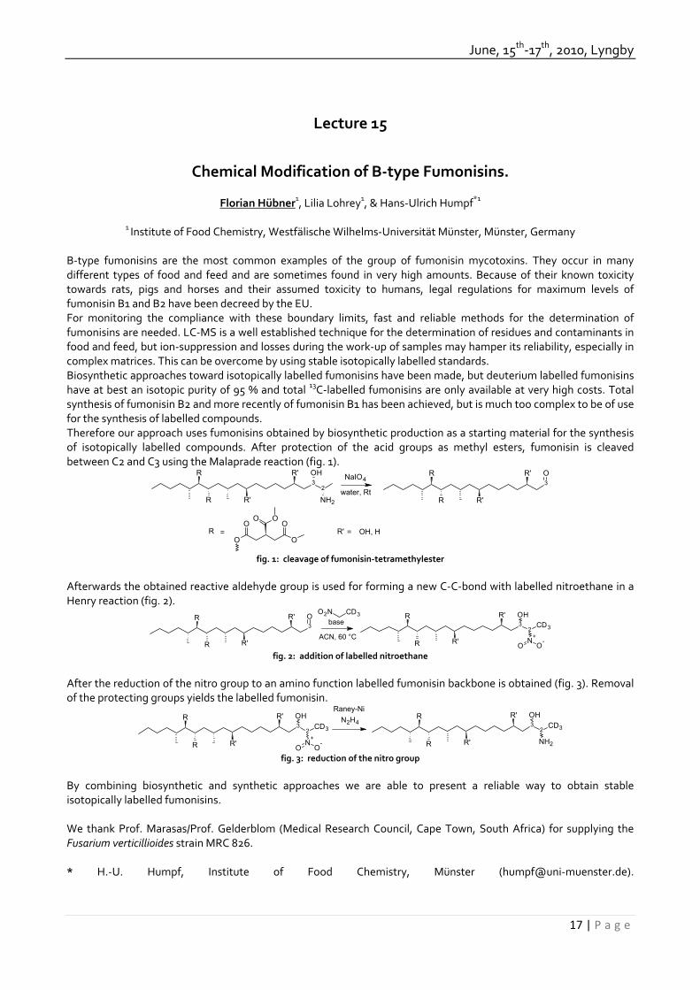
June, 15th‐17th, 2010, Lyngby
17 | P a g e
Lecture 15
Chemical Modification of B‐type Fumonisins.
Florian Hübner1, Lilia Lohrey1, & Hans‐Ulrich Humpf*1
1 Institute of Food Chemistry, Westfälische Wilhelms‐Universität Münster, Münster, Germany
B‐type fumonisins are the most common examples of the group of fumonisin mycotoxins. They occur in many different types of food and feed and are sometimes found in very high amounts. Because of their known toxicity towards rats, pigs and horses and their assumed toxicity to humans, legal regulations for maximum levels of fumonisin B1 and B2 have been decreed by the EU. For monitoring the compliance with these boundary limits, fast and reliable methods for the determination of fumonisins are needed. LC‐MS is a well established technique for the determination of residues and contaminants in food and feed, but ion‐suppression and losses during the work‐up of samples may hamper its reliability, especially in complex matrices. This can be overcome by using stable isotopically labelled standards. Biosynthetic approaches toward isotopically labelled fumonisins have been made, but deuterium labelled fumonisins have at best an isotopic purity of 95 % and total 13C‐labelled fumonisins are only available at very high costs. Total synthesis of fumonisin B2 and more recently of fumonisin B1 has been achieved, but is much too complex to be of use for the synthesis of labelled compounds. Therefore our approach uses fumonisins obtained by biosynthetic production as a starting material for the synthesis of isotopically labelled compounds. After protection of the acid groups as methyl esters, fumonisin is cleaved between C2 and C3 using the Malaprade reaction (fig. 1).
32
NH2
OHR'
R'R
R3OR'
R'R
RNaIO4
water, Rt
R =O
OOO
O
OR' = OH, H
fig. 1: cleavage of fumonisin‐tetramethylester
Afterwards the obtained reactive aldehyde group is used for forming a new C‐C‐bond with labelled nitroethane in a Henry reaction (fig. 2).
3OR'
R'R
R3OHR'
R'R
R
2
N+
CD3
O O-
base
ACN, 60 °C
CD3O2N
fig. 2: addition of labelled nitroethane
After the reduction of the nitro group to an amino function labelled fumonisin backbone is obtained (fig. 3). Removal of the protecting groups yields the labelled fumonisin.
3OHR'
R'R
R
2
N+
CD3
O O-
32
CD3
NH2
OHR'
R'R
RRaney-Ni
N2H4
fig. 3: reduction of the nitro group
By combining biosynthetic and synthetic approaches we are able to present a reliable way to obtain stable isotopically labelled fumonisins. We thank Prof. Marasas/Prof. Gelderblom (Medical Research Council, Cape Town, South Africa) for supplying the Fusarium verticillioides strain MRC 826. * H.‐U. Humpf, Institute of Food Chemistry, Münster (humpf@uni‐muenster.de).

32th Mycotoxin Workshop
18 | P a g e
Lecture 16
Enzyme Immunoassay for Tenuazonic Acid in Apple and Tomato Products
Madeleine Groß1*, Valeriu Curtui1,2, Yvonne Ackermann1, Hadri Latif1,3, and Ewald Usleber1
1Institute of Veterinary Food Science, Dairy Science, Justus Liebig University, Ludwigstrasse 21, D‐35390 Giessen. 2European Food Safety Authority, Largo N. Palli 5/A, 43100 Parma, Italy. 3Department of Animal Diseases and
Veterinary Public Health, Faculty of Veterinary Medicine, Bogor Agricultural University, 16680 Bogor, Indonesia. Tenuazonic acid (TeA, I) is a tetramic acid derivative commonly listed within the group of Alternaria mycotoxins. Production of TeA has been reported for species belonging to several fungal genera, in particular by Alternaria spp., but also by Aspergillus spp., Phoma sorghina and Pyricularia oryzae. Fungi of the genus Alternaria belong to the most cosmopolitan in biological environments and in agricultural commodities. Therefore, TeA may occur in many different products e.g. apples, tomatos, citrus fruits, carrots, and products thereof. Here we describe the development of an enzyme immunoassay (EIA) for the determination of TeA in foods after acetylation (II).
5
43
2
NH
CH3
CH2CH2
CH3
OH CH3
O
O
H5
43
2
NH
CH3
CH2CH2
CH3
O CH3
O
O
H
CH3 O
I II
TeA was derivatized with succinic anhydride; the TeA hemisuccinate was conjugated as its N‐hydroxysuccinimide ester to keyhole limpet hemocyanin (KLH) and to horseradish peroxidase (HRP), respectively. The KLH‐conjugate was used to produce Anti‐TeA antiserum in rabbits. A competitive direct EIA was established with Anti‐TeA antiserum and TeA‐HRP conjugate. The EIA was moderately sensitive for free TeA (IC50 of the TeA standard curve: 200‐400 ng/ml), but was very sensitive for acetyl‐TeA (IC50: 15‐40 ng/ml, detection limit 5±3 ng/ml). Cross‐reactivities with other Alternaria mycotoxins (alternariol, alternariol monomethyl ether, altenuene) were <1%. Therefore a sample preparation protocol was established which includes a 30 min acetylation step of free TeA, and quantification by EIA using an acetyl‐TeA standard curve. The detection limits for TeA in various matrices were as follows: apple juice: 50 ng/ml; tomato juice: 25 ng/ml; tomato ketchup: 30 ng/g. The recoveries were as follows: apple juice: 78‐130 % at 50‐500 ng/ml; tomato juice 61‐113 % at 50‐2000 ng/ml; tomato ketchup 40‐150 % at 50‐2000 ng/g). One of seven analyzed samples of commercial apple juices contained TeA, at a level of 58 ng/ml. Four out of 15 tomato juice samples contained TeA in a concentration range of 25‐200 ng/ml, and 2 out of 18 tomato ketchup samples contained TeA at levels of 55.2 ng/g and 67.3 ng/g, respectively. We thank the Brigitte and Wolfram Gedek‐Stiftung (Ismaning, Germany) for financial support. *Institute of Veterinary Food Science. Chair of Dairy Science, Justus Liebig University, Ludwigstr. 21, 35390 Giessen, Germany. [email protected]‐giessen.de

June, 15th‐17th, 2010, Lyngby
19 | P a g e
Lecture 17
A multiplex flow cytometric immunoassay for mycotoxins in food and feed using paramagnetic microspheres.
Jeroen Peters and Willem Haasnoot.
Department of Veterinary Drugs, RIKILT ‐ Institute of Food Safety, Wageningen UR, Wageningen, The Netherlands. Within RIKILT‐Institute of Food Safety, mycotoxin detection in food and feed is one of the legal tasks. For this, a multi‐mycotoxin method based on LC‐MS is used. Our research is focused on the development of an alternative method using the Luminex xMAP multiplex technology. The new method should be faster and cheaper than LC‐MS while also being robust and complying with legal detection limits. With the method to be developed we aim for a combined competitive inhibition immunoassay for the detection of Aflatoxins(AFB), Deoxynivalenol (DON), Zearalenone (ZEA), T2‐ toxin (T2), Fumonisin (F) and Ochratoxin A (OTA) in food and feed. Luminex Xmap is an emerging technology and is based on unique color coded microspheres in combination with flow cytometry. The paramagnetic microspheres (6.5 μm) used in our research are internally dyed with precise ratios of two fluorochromes to create 65 unique microspheres. Inside the microsphere there is a magnetite core for response to a magnetic field which simplifies the working with sample extracts. Each microsphere can be distinguished based on its internal dye ratio using a red laser for excitation. The specific protein‐mycotoxin conjugates are each coupled to the carboxylated surface of a unique microsphere. These beads are then added to the sample together with all the specific monoclonal mycotoxin antibodies. After a washing step, phycoerythrin labeled anti‐mouse antibodies are added as a reporter molecule. The phycoerythrin reporter molecule is excited by a green laser. This way the signal on each unique bead is quantified. In the initial experiments, available antibodies and mycotoxin‐protein conjugates were tested. A selection of conjugates and antibodies was made based on their sensitivity and cross‐reactions. All combinations showed good calibration curves in buffer and in spiked maize extract. Negative control samples spiked at EU guidance levels all showed satisfying competition when compared to the control sample. The developed assay allows multiplexed detection with high sensitivities in buffer and is now tested for naturally contaminated feed and food samples. [email protected] Jeroen Peters Veterinary drugs (DGM) RIKILT ‐ Institute of Food Safety , Wageningen UR. P.O. Box 230 6700 AE Wageningen The Netherlands

32th Mycotoxin Workshop
20 | P a g e
Lecture 18
Application of Mass spectrometry in indoor environmental quality (IEQ) assessment
Vinay Vishwanath1*, Michael Sulyok1, Stefan Mayer2 and Rudolf Krska2
1 University of Natural Resources and Applied Life Sciences, Vienna, Department for Agrobiotechnology, IFA‐Tulln,
Center for Analytical Chemistry, 3430 Tulln, Austria 2 Berufsgenossenschaft Handel und Warendistribution (BGHW), Direktion Mannheim, M 5, 7. 68161, Mannheim,
Germany Economic development and rising living standards have led to increases in the quantity and complexity of generated waste. The waste recycling such as of plastics has reduced the costs of production and pollution along with creating newer employment. The modern technologies for waste treatment have reduced human exposure and thereby minimising the health risks. However employee’s accidental exposure to waste components that are colonised with microbes cannot be ruled out completely during treatment or disposal processes. A study was conducted concerning occupational health of employees that are working in waste treatment processes due to mycotoxins and microbial bioactive substances. Samples were procured from different working areas of waste treatment plants that are mainly dealing with municipal solid wastes. The settled indoor dust was chosen for IEQ analysis because of its high relevance in the formation of aerosol and human exposure. Since the major fraction of municipal waste was composed of organic matter the analysis and quantification was confined to mycotoxins and microbial bioactive substances. An LC‐MS/MS method validated for 186 relevant fungal and bacterial metabolites was used for the analysis (1, 2). Extraction was done using acetonitrile/water/acetic acid (79:20:1) in a single step without any cleanup procedures. Further fractions of the collected dust samples were subjected to SPME‐GC‐MS analysis without any pre‐treatment. Volatiles were enriched on a PDMS/DVB‐SPME‐fiber and analysed on a GC‐MS instrument (Agilent 6890 GC with 5975B MSD detector) using GC‐columns with opposite polarities (HP‐5 and Optimawax). The LC‐MS/MS analysis resulted in quantification of 34 fungal and 5 bacterial metabolites in varying concentrations. With a few exception most of the analytes were in the limit of quantification range of our LC‐MS/MS method. GC‐MS analysis revealed a probable release of 60 organic compounds which are either reported as volatile organic compounds(VOCs) or Microbial volatile organic compounds (MVOCs) in the context of indoor environment (3,4). References: 1. Vishwanath V, Sulyok M, Labuda R, Bicker W, Krska R. 2009. Simultaneous determination of 186 fungal and bacterial metabolites in indoor matrices by liquid chromatography/tandem mass spectrometry. Anal Bioanal Chem. 2009 Nov;395(5):1355‐72. 2. Peltola J, Andersson MA, Haahtela T, Mussalo‐Rauhamaa H, Rainey FA, Kroppenstedt RM, Samson RA, Salkinoja‐Salonen MS. Toxic‐Metabolite‐Producing Bacteria and Fungus in an Indoor Environment, APPLIED AND ENVIRONMENTAL MICROBIOLOGY, July 2001, p. 3269–3274.
3. Korpi A, Järnberg J, Pasanen AL. 2009. Microbial Volatile Organic Compounds, Crit Rev Toxicol. 2009;39(2):139‐93.
4. Pedersen EK, Bjørseth O, Syversen T, Mathiesen M. 2003. A screening assessment of emissions of volatile organic compounds and particles from heated indoor dust samples, Indoor Air. 2003 Jun; 13(2):106‐17.
* Corresponding author's address and e‐mail. University of Natural Resources and Applied Life Sciences, Vienna, Department for Agrobiotechnology, IFA‐Tulln, Center for Analytical Chemistry, 3430 Tulln, Austria E‐mail:[email protected]

June, 15th‐17th, 2010, Lyngby
21 | P a g e
Lecture 19
Fumonisin induced alteration of sphingolipid metabolism in piglets – Analytical and biological aspects.
Heidi Schwartz1, Bertrand Grenier2, Irene Schöner1, Anita Seeböck1, Isabelle P. Oswald2, Gerd Schatzmayr3 & Wulf‐Dieter Moll3
1: IFA Tulln, University of Natural Resources and Applied Life Sciences of Vienna, 3430 Tulln, Austria.
2: INRA, Laboratoire de Pharmacologie‐Toxicologie, 31027 Toulouse, France. 3: Biomin Research Center, Technopark 1, 3430 Tulln, Austria
Introduction: Increased concentrations of sphingoid bases and elevated ratios of free sphinganine (Sa) to sphingosine (So) in plasma and tissues of animals have been proposed as a biomarker for exposure to fumonisins [1, 2]. The first part of this presentation will point out the pitfalls of sphingolipid analysis in tissues. The second part will describe the alteration of sphingolipid metabolism in kidney, liver, lung and of piglets exposed to fumonisin B1. Materials and Methods: A group of 6 piglets was administered 2 mg of fumonisin B1 per kg body weight by gavage in addition to basal diet for 14 days. A control group of 6 piglets received basal diet. At the end of the experiment, piglets were euthanized, tissue samples were taken, immediately frozen with liquid nitrogen and stored at ‐80°C until analysis. For analysis of tissue samples, aliquots of kidney, lung and liver were homogenized on ice in cold phosphate buffer and aliquots of the homogenates were worked up and analyzed as previously described for plasma samples [3] with minor modifications. Results and discussion: The main source of error upon analysis of tissue samples is an uneven distribution of sphingoid bases in tissues (especially lung), so that pooling of different parts of one tissue, homogenization of the pooled tissue pieces and storage of the homogenate at ‐80°C is advisable. Concentrations of sphingoid bases and Sa to So ratios varied greatly among pigs of one feeding group. Despite that, all tissues from fumonisin treated piglets could be distinguished from tissues of control piglets by their concentrations of So, So and 1‐deoxy‐Sa and by their Sa to So ratios. The concentration ranges (ng/g) of sphingoid bases and the ranges of Sa to So ratios in tissues of the six piglets of each feeding group are given in the table below. c (ng/g) Kidney Liver Lung So Sa So Sa So Sa Control 592‐903 81‐130 502‐1050 59‐86 5940‐11700 697‐1770 FB1 4040‐5660 20100‐31600 3020‐6090 4620‐26200 20400‐32400 15000‐67400 1‐Deoxy‐Sa Sa/So 1‐Deoxy‐Sa Sa/So 1‐Deoxy‐Sa Sa/So Control traces 0.09‐0.18 traces 0.07‐0.14 169‐211 0.12‐0.17 FB1 292‐721 4.95‐5.75 180‐985 1.53‐4.44 840‐3400 0.73‐2.31
Conclusion: Administration of 2 mg of fumonisin B1 per kg body weight significantly altered the sphingoid metabolism of piglets. The analytical method used seems promising for investigating the efficiency of feed additives for detoxification of fumonisins. References 1. Riley, RT, Voss, KA. 2006. Differential sensitivity of rat kidney and liver to fumonisin toxicity: Organ‐specific differences in toxin accumulation and sphingoid base metabolism. Toxicol. Sci. 92: 335‐345. 2. Zitomer, NC, Mitchell, T, Voss, KA et al. 2009. Ceramide synthase inhibition by fumonisin B1 causes accumulation of 1‐deoxysphinganine. J. Biol. Chem. 8: 4786–4795. 3. Schwartz, H, Moll, D, Schatzmayr, G, Krska, R. 2009. An accelerated method for determining sphingoid bases in serum of pigs. 31st Mycotoxin Workshop: 49.
* Dr. Heidi Schwartz, Center for Analytical Chemistry, IFA‐Tulln, Konrad‐Lorenz‐Straße 20, 3430 Tulln, Austria; [email protected]

32th Mycotoxin Workshop
22 | P a g e
Lecture 20
Investigations on the transfer of deoxynivalenol or its metabolite from cows to their offspring
Susanne Döll*, Lydia Renner, Ulrich Meyer, Hana Valenta & Sven Dänicke
Institute of Animal Nutrition, Friedrich‐Loeffler‐Institute, Federal Research Institute for Animal Health,
Braunschweig, Germany. Ruminants are considered to be less susceptible to the Fusarium toxin deoxynivalenol (DON) as monogastric species owing to the ability of ruminal microorganisms to metabolize DON to the less toxic metabolite de‐epoxy‐DON which is almost exclusively detected in systemic circulation. However, a transfer of unmetabolized toxin to the circulation can not be excluded, especially if the integrity of the rumen mucosa is disturbed due to various rumen digestive disorders. Toxins present in the blood of gestating animals may also posses a risk to the developing offspring as transplacental transfer may occur, especially for rather small molecules like DON, which was demonstrated to pass the placental barrier in pigs (Goyarts et al., 2007). So far, there is no data available on the transplacental transfer of DON or its metabolite de‐epoxy‐DON in cows. Hence, the present study aimed to elucidate this issue. Methods: Corresponding blood samples of cows immediately after parturition and the respective calves before colostrum intake, as well as colostrum samples were collected from 16 German Holstein cows fed a roughage mix of 40% grass‐ and 60% maize‐silage in addition to 2kg of a concentrate. A subsequent blood sample of the calves after colostrum intake was taken the day after. After 21 days another set of corresponding samples of blood and milk of the same cows was taken. The concentrations of DON and de‐epoxy‐DON were analyzed in feed, plasma, colostrum and milk by HPLC with UV detection and LC‐ESI‐MS/MS (Valenta et al., 2003) as described elsewhere (Keese et al., 2008). The detection limit of DON in feed was 0.03mg/kg and of DON and de‐epoxy‐DON were 3ng/ml in plasma and 4ng/g freeze dried colostrum or milk with HPLC and 0.3ng DON/ml and 0.9ng de‐epoxy‐DON/ml in plasma and 0.4ng DON/g and 1.1ng de‐epoxy‐DON/g freeze dried colostrum or milk when analysed by LC‐ESI‐MS/MS, respectively. Results: The cows were exposed to DON mainly via the maize‐silage which contained 6.5 mg DON/kg DM. The grass‐silage and the concentrate contained typical background contaminations of 0.07 and 0.17 mg DON/kg DM, respectively. Assuming a dry matter intake of 10 kg/cow/day the contamination would amount to 3.2 mg DON/kg diet. The exposure to DON was reflected by the presence of de‐epoxy‐DON in plasma of cows. The original toxin could neither be detected in plasma of cows nor in plasma of calves. However, the presence of de‐epoxy‐DON in 4 out of 16 plasma samples of calves before colostrum intake indicates a transplacental transfer. The concentrations of de‐epoxy‐DON and DON in colostrum were independent of the concentration in plasma at the same time. Moreover the median transfer ratio from blood plasma to colostrum of 1.5 hints at an accumulation of de‐epoxy‐DON in colostrum. Yet, exposure of calves via colostrum did not result in detectable plasma levels. Conclusions: Transplacental transfer of de‐epoxy‐DON from the cow to the calf may occur. However as the transfer rate was far below 1 an effective barrier seems to protect the calf from uterine exposure. In contrast, the blood milk barrier seems to be less efficient. References: Goyarts, T et al. 2007. Toxicol. Lett. 171: 38‐49. Valenta, H. et al. 2003. Mycotoxin Res. 19: 51‐55. Keese, C. et al. 2008. Mol. Nutr. Food Res. 52: 1514‐1529. * Institute of Animal Nutrition, FLI, Bundesallee 50, 38116 Braunschweig, Germany, e‐mail: [email protected].

June, 15th‐17th, 2010, Lyngby
23 | P a g e
Lecture 21
Deoxynivalenol alters the interactions of Salmonella Typhimurium with porcine intestinal epithelial cells and macrophages
Virginie Vandenbroucke1*, Frank Pasmans2, Elin Verbrugghe2, Joline Goossens1, Freddy Haesebrouck2, Patrick De
Backer1, Siska Croubels1
1 Department of Pharmacology, Toxicology and Biochemistry, 2 Department of Pathology, Bacteriology and Avian
Diseases, Faculty of Veterinary Medicine, Ghent University, Salisburylaan 133, 9820 Merelbeke, Belgium. Introduction: In European countries with temperate climates, the Fusarium toxin deoxynivalenol (DON) is one of the most frequent contaminants of maize and small grain cereals [1]. The toxic effects of DON have been well documented and among farm animals, pigs seem to be particularly sensitive to the dietary intake of DON. Substantial economic losses have been attributed to DON contamination of pig feed [2]. In European countries, pigs infected with Salmonella Typhimurium have become the main source of salmonellosis in humans. Since Fusarium toxins such as DON have been shown to affect among others the immune system of pigs [3], exposure to DON might affect the interactions of Salmonella Typhimurium with the porcine host. The objective of this study is to examine the effect of low level concentrations of DON on the intestinal and the systemic phase of a Salmonella infection in pigs, using in vitro cell models with porcine intestinal epithelial cells (IPEC‐J2 cells) and porcine alveolar macrophages (PAM). Materials and Methods: Invasion and intracellular survival assays with Salmonella Typhimurium were performed at non‐cytotoxic but practically relevant concentrations of DON in IPEC‐J2 cells and PAM. IPEC‐J2 cells grown on Transwell® inserts were incubated with various concentrations of DON to measure the effect of the mycotoxin on the epithelial barrier function and the passage of Salmonella Typhimurium through the IPEC‐J2 cell epithelium. The effect of DON on both the direct IL‐8 secretion by IPEC‐J2 cells as well as on the IL‐8 secretion induced by Salmonella Typhimurium was tested using a commercial ELISA kit (Swine IL‐8 ELISA kit, Invitrogen, Camarillo, CA). The morphological changes in PAM were visualized using diverse staining techniques. Results: Levels of DON plausibly encountered in the gastrointestinal tract of pigs after consumption of contaminated feed lead to a dose dependent increase of invasion in and translocation of Salmonella Typhimurium through the IPEC‐J2 cell monolayer. The results of the effect of DON on IL‐8 secretion will be presented at the congress. Pre‐exposure of PAM to 0.025 µg/mL of DON significantly promotes the uptake of Salmonella Typhimurium through F‐actin reorganization. On average, approximately 1.5 times more intracellular Salmonella bacteria were present in DON treated macrophages. Discussion and Conclusion: These findings suggest that low concentrations of DON severely affect the interactions of Salmonella Typhimurium with porcine host cells. The relevance for the in vivo situation needs further study. References: [1] Bottalico, A & Perrone, G (2002). Eur. J. Plant Pathol. 108, 611‐624. [2] Böhm, J (1992). Arch. Tierernahr. 42, 95‐111. [3] Pestka, J (2004). Toxicol Lett. 153, 61‐73. *Virginie Vandenbroucke, Department of Pharmacology, Toxicology and Biochemistry, Faculty of Veterinary Medicine, Ghent University, Salisburylaan 133, 9820 Merelbeke, Belgium [email protected]

32th Mycotoxin Workshop
24 | P a g e
Lecture 22
Metabolism and distribution of the mycoestrogen zearalenone as a function of physiological status and condition of exposure.
Sylvaine Lecoeur1,2*, Michelle Mazallon1,2 , Bernadette Videmann2
1 National Institute for Agronomic Research, Animal Health Dept, Marcy l’Etoile, France
2 Vetagro Sup, UMR 1233, Marcy l’Etoile, France The mycotoxin zearalenone (ZEA) is produced by a variety of Fusarium fungi and contaminates numerous cereals, fruits and vegetables. A fresh and wet climate facilitating the growth of the mushroom, ZEA is particularly prevailing in Europe (North, East, and West). Interacting with the estrogen receptors ERα and ERß, ZEA causes hormonal effects on animal, including early thelarche, decrease in the rate of ovulation and in the size of the litter, penis abnormalities, and degenerating sperm. The in utero exposure to ZEA provokes abnormalities in the development of the reproductive tract and mammary gland in female rodent offspring. Limited information is available on the pharmacokinetics of ZEA and its metabolites, particularly in case of repeated exposure, which better reflects a real situation. This study was conducted to characterize the disposition, oral bioavailability and tissue distribution of ZEA and its metabolites in rats as a function of physiological status (gender, pregnancy, nursing) and exposure condition (subcutaneous vs oral, single vs repeated exposure to 1 mg/kg ZEA). Placental and mammary respective transfer to the fetus and the neonate was also evaluated. The 24 hours kinetics of ZEA and its main metabolites (α‐zearalenol, ß‐zearalenol, glucuronides of ZEA and α‐zearalenol) were determined by HPLC in plasma, urine, small intestine, colon, kidneys, liver, muscle, fat, uterus, mammary gland, placenta, fetus, fetus liver, ovaries, testes, and prostate. ZEA, α‐zearalenol, and their respective glucuronides were the main molecules encountered into the organs. The mycotoxins were excreted from the body within 4 hours after subcutaneous exposure, and within 24 hours after oral exposure, excepted urine and intestine. Compared to non‐pregnant females, ZEA and metabolites concentration was significantly higher in the genital organs of pregnant females. Compared to single exposure, daily exposure (GD7‐20) to ZEA led to an increased accumulation of ZEA and its metabolites in the liver, placenta, fetus and fetal liver of the pregnant females, and in the mammary gland of pregnant and breasting females. ZEA transfer in the milk was also enhanced by repeated exposure. Fetus and neonate are critical stages of vulnerability to the long‐term effects of hormonomimetic compounds. Higher exposure to ZEA and metabolites during these stages, consecutively to repeated exposure of the mother, could constitute a health risk on the long term. *National Veterinary School of Lyon, 1 avenue Bourgelat, 69280 Marcy l’Etoile, France e‐mail : s.lecoeur@vetagro‐sup.fr

June, 15th‐17th, 2010, Lyngby
25 | P a g e
Lecture 23
The Fusarium toxins β‐zearalenol and zearalenone inhibit Hsp90 ATPase activity and are inactivated by glucosylation and sulfatation.
Juan Antonio Torres Acosta1, Franz Berthiller2, Gerlinde Wiesenberger1, Rudolf Mitterbauer1, Ulrike Werner1, David
Merz1, Marie‐Theres Hauser1, Mehrdad Shams2, Rudolf Krska2, & Gerhard Adam1*
1 Department of Applied Genetics and Cell Biology, University of Natural Resources and Applied Life Sciences
(BOKU), Vienna, Austria. 2 Center for Analytical Chemistry, Department for Agrobiotechnology, IFA Tulln, BOKU, Tulln, Austria.
The Fusarium mycotoxin zearalenone (ZON) is well known for its estrogenic activity in animals. Plants do not have an estrogen receptor, and it is an open question whether the Fusarium metabolite ZON has a biological role in plant‐pathogen interaction. We have identified a prominent target for zearalenone: Hsp90. Zearalenone and more strongly β‐zearalenol (bZOL) inhibit the ATPase activity of purified yeast Hsp90 (ScHsp82p) in vitro. Hsp90 is necessary for the stability of many client proteins such as signal transduction components, and has been shown to be essential for plant defense (1). Microarray experiments of ZON treated Arabidopsis plants showed marked changes in gene expression (Werner et al. in preparation). Many genes encoding proteins with a role in cell wall remodeling and peroxidases were repressed, while several small heat shock proteins and AtHSP90‐1 were upregulated upon ZON treatment. Also candidate detoxification genes (PDR‐type ABC transporter, UDP‐glucosyltransferases, glutathione‐S‐transferases) were induced. ZON was found to be rapidly converted into ZON‐4‐O‐glucoside and ZON‐4‐sulfate in plants. Both conjugates are no longer inhibitors of Hsp90 ATPase in vitro. The finding that ZON and its biosynthetic precursor bZOL are Hsp90 inhibitors also raises the question about the mechanism of self resistance in the toxin producing fungus. We have engineered yeast strains with increased ZON sensitivity (deletion of several ABC transporters) and with deletions of the endogenous yeast HSP90 genes (hsp82 hsc82), that can serve as a host for expression of heterologous genes as sole source of Hsp90. We also have expressed the yeast and Fusarium graminearum Hsp90 proteins with a purification tag in E. coli to allow comparison of the properties in vitro. The Fusarium Hsp90, which has conspicuous amino‐acid changes in the N‐terminus (ATP binding region) compared to the yeast gene product, showed increased ZON resistance but unaltered resistance to radicicol, a resorcylic acid lactone and strong Hsp90 inhibitor from Nectria radicicola. The finding that ZON has a prominent target implicated in plant resistance warrants to reinvestigate whether zearalenone biosynthesis is indeed irrelevant for Fusarium virulence as reported by three different groups (2, 3, 4). We therefore transformed Fusarium graminearum with a construct deleting both PKS4 and PKS13, the polyketide synthase genes required for ZON biosynthesis. The mutants deficient in ZON production in wild‐type and tri5‐background (deficient in DON production) are currently tested for altered virulence. References:
1. Shirasu K. 2009. The HSP90‐SGT1 chaperone complex for NLR immune sensors. Annu. Rev. Plant Biol. 60: 139‐164.
2. Kim YT, Lee YR, Jin J, Han KH, Kim H, Kim JC, Lee T, Yun SH, Lee YW. 2005. Two different polyketide synthase genes are required for synthesis of zearalenone in Gibberella zeae. Mol. Microbiol. 58: 1102‐13.
3. Gaffoor I, Brown DW, Plattner R, Proctor RH, Qi W, Trail F. 2005 Functional analysis of the polyketide synthase genes in the filamentous fungus Gibberella zeae (anamorph Fusarium graminearum). Eukaryot. Cell 4: 1926‐33.
4. Lysøe E, Klemsdal SS, Bone KR, Frandsen RJ, Johansen T, Thrane U, Giese H. 2006. The PKS4 gene of Fusarium graminearum is essential for zearalenone production. Appl. Environ. Microbiol. 72: 3924‐32.
* Muthgasse 18, A‐1190 Vienna, Austria. E‐mail: [email protected]

32th Mycotoxin Workshop
26 | P a g e
Lecture 24
Toxicity of ergot alkaloids: Effects on human cells in primary culture
Dennis Mulac1,2, Hans‐Ulrich Humpf2*
1Graduate School of Chemistry, Westfälische Wilhelms‐Universität Münster, Wilhelm‐Klemm‐Str. 10, 48149 Münster,
Germany. 2Institute of Food Chemistry, Westfälische Wilhelms‐Universität Münster, Münster, Germany
Ergot alkaloids are secondary metabolites produces from fungi of the species Claviceps. The toxic effects of ergots are well known since mediaeval times. They are ascribed to their alkaloids with their most prominent being ergometrine, ergocornine, ergotamine, ergocryptine, ergocristine and ergosine along with their corresponding –inine forms. Nevertheless, at present, there are no legal regulations in respect to the content of ergot alkaloids in food and feed. Since the toxic effects of the whole sclerotia are well known, it is important to complete the data with information about the effects of the single compounds for risk assessment. Therefore the toxic effects of different ergot alkaloids were determined in human cells in primary culture using human renal proximal tubule epithelial cells (RPTEC) and normal human astrocytes (NHA). Besides the general cytotoxicity, the endpoints necrosis (LDH‐release) and apoptosis (caspase‐3 activation) were measured. The first preliminary results identified a correlation between the lipophilic character and the cytotoxicity. Using concentrations in higher micromolar ranges resulted in a necrosis for the studied peptide alkaloids. In contrast ergometrine as a lysergic acid amide is an exception and has shown no cytotoxic effects in the used concentrations. Additionally for the first time an apoptotic effect of the peptide ergot alkaloids could be clearly identified using fluorescence microscopy and flow cytometry. Reference: EFSA 2005, Opinion of the Scientific Panel on Contaminants in Food Chain on a request from the commission related to ergot as undesirable substance in animal feed, EFSA Journal, 225, 1‐27 * H.‐U. Humpf, Institute of Food Chemistry, Münster (humpf@uni‐muenster.de).

June, 15th‐17th, 2010, Lyngby
27 | P a g e
Lecture 25
Alternaria toxins ‐ toxicological potential and mechanistical aspects
Doris Marko*1, Markus Fehr2,Christoph Schwarz1 & Gudrun Pahlke1
1 Department for Food Chemistry and Toxicology, University of Vienna, Vienna, Austria.
2 Institute of Applied Biosciences, Section for Food Chemistry, Karlsruhe Institute for Technology, Karlsruhe, Germany.
Fungi of the genus Alternaria are known for their potential to produce a variety of mycotoxins, with alternariol (AOH), alternariol monomethyl ether (AME), altenuene (ALT) and tenuazonic acid (TA) being the most prominent. Being able to considerable mycotoxin production even under relatively unfavourable conditions (e.g. low temperature, low humidity etc.) Alternaria spp. play an important role for the spoilage of refrigerated food and stored feedstuffs, as well as contamination of the living crop plant in the field. Alternaria toxins have been described as cytotoxic, genotoxic and mutagenic in vitro and in vivo. Still, comparatively little toxicological attention has been paid to Alternaria spp., as their main toxins AOH, AME, ALT and TA exhibit only low acute toxicity. Epidemiological data also indicates a possible correlation between contamination with Alternaria toxins and an increased incidence of esophageal tumours. Due to the ubiquitous occurrence of Alternaria toxins in many foods and the environment, a possible chronic toxicity would be of relevance, making the understanding of the underlying toxicological mechanisms essential. As we previously reported, Alternaria toxins AOH and AME impair the integrity of DNA in cultured human colon carcinoma cells at low micromolar concentrations. In this respect we investigated, to which extent interactions with the topoisomerases I and II contribute to this effect. Being able to stabilize the complex between DNA and topoisomerase II, alternariol acts was found to act as a topoisomerase II poison with a preference for the topo II α isoform (Fehr et al.,2009). These results were supported by the finding that an enzyme specialised on the repair of respective covalent topoisomerase‐DNA intermediates, tyrosyl‐DNA phosphodiesterase 1 (TDP1), represents an important factor for the modulation of AOH‐mediated DNA damage in human cells. Recent studies using the dichlorofluorescein (DCF) assay further indicated the formation of reactive oxygen species (ROS) thus raising the hypothesis that not only the interference with topoisomerases but also the induction of oxidative stress might contribute to the genotoxic effects of AOH. Treatment of HT29 cells (human colon carcinoma) for 1 h with AOH or AME resulted in a concentration‐dependent increase of dichlorofluorescein in the DCF assay, indicative for ROS formation. Concomitantly, the level of total glutathione (GSH) was decreased. However, after 24 h this effect appeared to be compensated. In accordance with the results of the DCF assay, AOH and AME enhanced the proportion of the transcription factor Nrf2 in the nucleus as detected by Western blot analysis. After 3 h of incubation enhanced transcription of Nrf2/ARE‐dependent genes was observed as exemplified for the glutathione‐S‐transferases (GST) A1 and A2. After 24 h of incubation this effect was also reflected on the protein level by an increase of GST‐activity. Taken together, the mycotoxins AOH and AME were found to enhance the level of intracellular ROS in HT29 cells, associated with a transient decrease of the tGSH‐level. Concomitantly, Nrf2‐activation enhanced the expression of GST isoenzymes indicative for the onset of cellular defense systems via the Nrf2/ARE pathway. In summary, interference with topoisomerases and the induction of oxidative stress were identified as targets for the DNA‐damaging properties of AOH. References: Fehr M, Pahlke G, Fritz J, Christensen MO, Boege F, Altemoeller M, Podlech J, Marko D. 2009, Alternariol acts as a topoisomerase poison, preferentially affecting the IIα isoform. Mol. Nutr. Food Res. 53(4), 441‐451. * Prof. Dr. Doris Marko, Institut für Lebensmittelchemie und Toxikologie, Universität Wien, Währingerstr. 38, A‐1010 Wien, Austria
32th Mycotoxin Workshop
28 | P a g e
Lecture 26
Live cell imaging confirms disruption of mitosis as a key event in ochratoxin A toxicity
Kristin Czakai1, Elisabeth Jeanclos2, Alexander Zürn2, Antje Gohla2 & Angela Mally1*
1 Department of Toxicology, University of Würzburg, Würzburg, Germany. 2 Department of Pharmacology, University
of Würzburg, Würzburg, Germany Ochratoxin A (OTA), a mycotoxin and important food contaminant, is one of the most potent rodent renal carcinogens studied to date. Although controversial results regarding OTA genotoxicity have been published, it is now widely accepted that OTA is not a mutagenic, DNA‐reactive carcinogen. Instead, there is increasing evidence from both in vivo and in vitro studies to suggest that OTA may promote genomic instability and tumorigenesis through interference with cell division (Rached et al., 2006; Adler et al., 2009). In this study, we have used live cell imaging to monitor the effects of OTA on cell division in human kidney epithelial cells (IHKE). IHKE cells cultured in 8 well microscopy chamber slides were treated with OTA at 0, 10, 25 and 50 µM and monitored by differential interference contrast (DIC) microscopy overnight. Images were acquired at 15 min intervals for up to 16 h. Image analysis confirmed that OTA perturbs normal mitotic progression. Significant prolongation of mitosis was observed in the presence of OTA as compared to vehicle treated controls and the percentage of cells that completed normal mitoses dropped to < 20 % in cells treated with OTA at ≥ 10 µM. In cells exposed to OTA, the following mitotic abnormalities were observed: (i) nuclear division without cytokinesis, (ii) exit from mitosis without nuclear or cellular division (mitotic slippage), (iii) repeated entry into and exit from mitosis without division, and (iv) asymmetric cell division. Although the precise mechanism as to how OTA functions to perturb the intricate and tightly controlled process of mitosis remains to be elucidated, results from this study provide further support for a mechanism of OTA carcinogenicity involving interference with the mitotic machinery and subsequent chromosome segregation errors, accompanied by an increased risk of aneuploidy acquisition and subsequent tumor formation. References: Rached, E., Pfeiffer, E., Dekant, W. and Mally, A. (2006). Ochratoxin A: apoptosis and aberrant exit from mitosis due to perturbation of microtubule dynamics? Toxicol Sci. 92: 78‐86. Adler, M., Muller, K., Rached, E., Dekant, W. and Mally, A. (2009). Modulation of key regulators of mitosis linked to chromosomal instability is an early event in ochratoxin A carcinogenicity. Carcinogenesis. 30: 711‐9. * Department of Toxicology, University of Würzburg, Würzburg, Germany. [email protected]‐wuerzburg.de
June, 15th‐17th, 2010, Lyngby
29 | P a g e
Lecture 27
Ochratoxin A in dairy goats
Ralf Blank 1, * , May Looff 2, Muhammad Mobashar 2, Anne Westphal 1 ,Karl‐Heinz Südekum 2
1 Institute of Animal Nutrition and Physiology, Christian‐Albrechts‐University Kiel, Germany. 2Institute of Animal
Science, University of Bonn, Germany Introduction: Extensive hydrolysis of ochratoxin A (OTA) to its less toxic metabolite ochratoxin α (OTα) and phenylalanine by rumen microorganism has been postulated to be responsible for the relative resistance of ruminants to acutely toxic effects to OTA and its carry over into milk. Recent studies, however, showed that high concentrate diets fed to ruminants increase the systemic availability of OTA and possibly increase the risk of carry over of OTA into milk. High concentrate diets decrease the ruminal pH, thereby reducing the number of rumen protozoa, which are substantially involved in ruminal degradation of OTA. The aim of the present study was to investigate the effects of chronic feeding of two different doses of OTA on feed intake, milk yield as well as systemic availability, urinary excretion and carry over of OTA into milk in lactating goats fed high concentrate diets. Additionally the effect of sodium bicarbonate supplementation, which has a rumen pH stabilizing effect, has been evaluated. Material Methods: For the experiment, 10 mid‐lactating goats, divided into two groups of 5 animals each, received a diet consisting of 70 % concentrate (1.5 kg/d) and 30 % hay (0.65 kg/d). After feeding of two weeks the OTA free diet, one of the group (5 animals) received a concentrate containing 1.5 mg of OTA per kg concentrate, while the other group received the same concentrate containing 2.8 mg/kg of OTA. After three weeks of feeding the OTA‐concentrate, the animals were switched for a period of two weeks to the same compound feed except that it was supplemented with 15 g/kg sodium bicarbonate. The hay fed was free of OTA. During the entire experiment feed intake and milk yield was recorded. Additionally blood samples from the jugular vein were drawn and the urinary output of OTA was measured. Blood, urine, milk and casein samples were analyzed for OTA and OTα using HPLC with fluorescence detection. Results: During the entire experiment no toxic effects due to OTA could be detected. Feed intake of goats fed the high OTA concentrate was marginally lower compared with the low dosage group. Milk yield decreased slightly during the entire experiment but was unaffected by OTA feeding. Concentration of OTA in blood was significantly lower in the low dose compared to the high dose group. Renal excretion of intact OTA expressed in percent of the OTA dose given ranged from 1.2–1.6 % and was not affected by sodium bicarbonate supplementation. OTA and OTα were detected in all milk samples. Concentration of OTα was always higher than OTA concentration. Concentration of OTA in milk samples ranged from 0.1 to 1 ng/mL. In the casein fraction, being the most relevant for cheese making, only OTα and no OTA could be detected. Conclusion: The present results indicate that a daily intake of 2‐4 mg OTA in lactating goats fed high concentrate diets have no impact on milk performance, but may lead to marginally carry over of OTA into milk. In addition, supplementation of sodium bicarbonate which has been shown to have beneficial effects on the rumen environment had no major impact on the systemic availability and carry over of OTA in ruminants. * Corresponding author: Institute of Animal Nutrition and Physiology, Christian‐Albrechts‐University, 24098 Kiel, Germany; Phone: +49‐431‐880‐2962, Fax: +49‐431‐880‐1528; email: [email protected]‐kiel.de
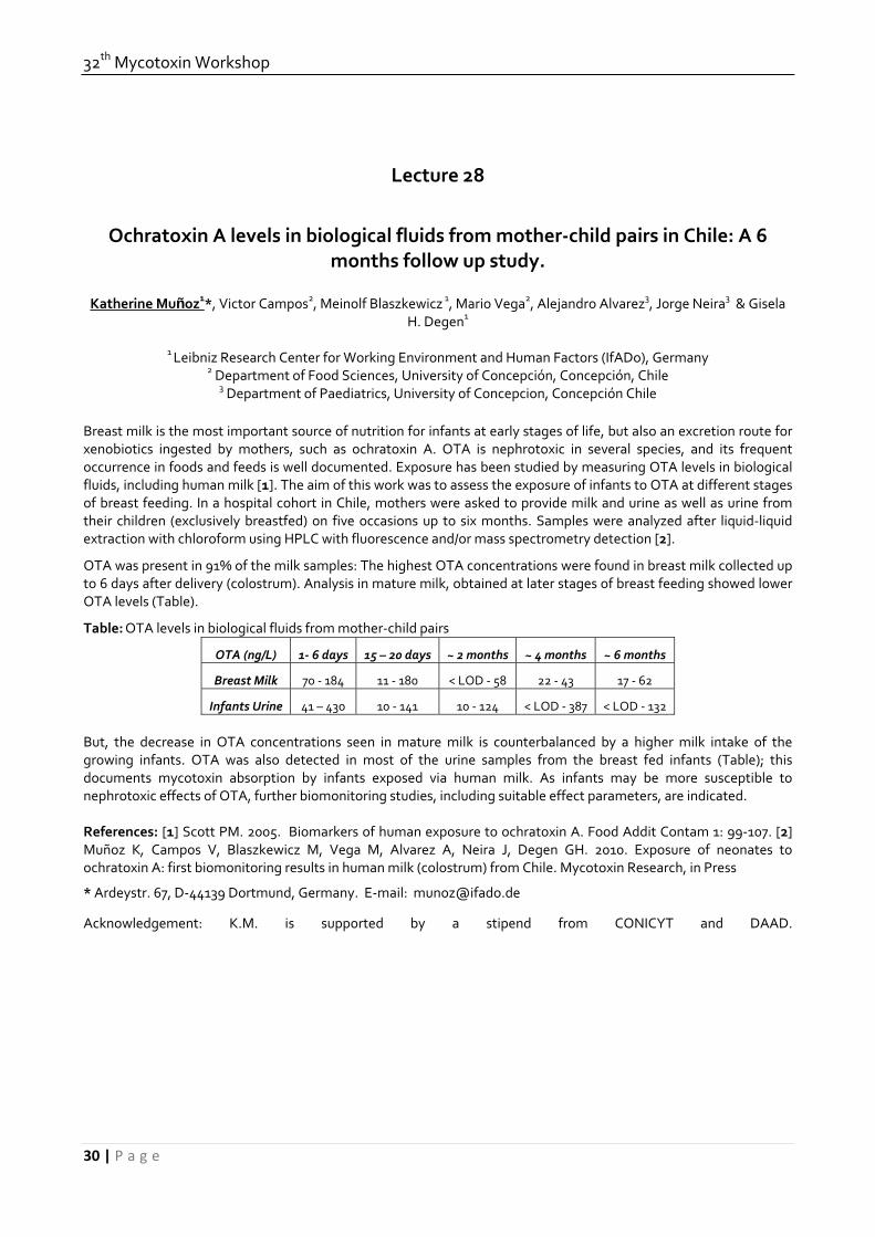
32th Mycotoxin Workshop
30 | P a g e
Lecture 28
Ochratoxin A levels in biological fluids from mother‐child pairs in Chile: A 6 months follow up study.
Katherine Muñoz1*, Victor Campos2, Meinolf Blaszkewicz 1, Mario Vega2, Alejandro Alvarez3, Jorge Neira3 & Gisela
H. Degen1
1 Leibniz Research Center for Working Environment and Human Factors (IfADo), Germany
2 Department of Food Sciences, University of Concepción, Concepción, Chile 3 Department of Paediatrics, University of Concepcion, Concepción Chile
Breast milk is the most important source of nutrition for infants at early stages of life, but also an excretion route for xenobiotics ingested by mothers, such as ochratoxin A. OTA is nephrotoxic in several species, and its frequent occurrence in foods and feeds is well documented. Exposure has been studied by measuring OTA levels in biological fluids, including human milk [1]. The aim of this work was to assess the exposure of infants to OTA at different stages of breast feeding. In a hospital cohort in Chile, mothers were asked to provide milk and urine as well as urine from their children (exclusively breastfed) on five occasions up to six months. Samples were analyzed after liquid‐liquid extraction with chloroform using HPLC with fluorescence and/or mass spectrometry detection [2].
OTA was present in 91% of the milk samples: The highest OTA concentrations were found in breast milk collected up to 6 days after delivery (colostrum). Analysis in mature milk, obtained at later stages of breast feeding showed lower OTA levels (Table).
Table: OTA levels in biological fluids from mother‐child pairs
OTA (ng/L) 1‐ 6 days 15 – 20 days ~ 2 months ~ 4 months ~ 6 months
Breast Milk 70 ‐ 184 11 ‐ 180 < LOD ‐ 58 22 ‐ 43 17 ‐ 62
Infants Urine 41 – 430 10 ‐ 141 10 ‐ 124 < LOD ‐ 387 < LOD ‐ 132 But, the decrease in OTA concentrations seen in mature milk is counterbalanced by a higher milk intake of the growing infants. OTA was also detected in most of the urine samples from the breast fed infants (Table); this documents mycotoxin absorption by infants exposed via human milk. As infants may be more susceptible to nephrotoxic effects of OTA, further biomonitoring studies, including suitable effect parameters, are indicated. References: [1] Scott PM. 2005. Biomarkers of human exposure to ochratoxin A. Food Addit Contam 1: 99‐107. [2] Muñoz K, Campos V, Blaszkewicz M, Vega M, Alvarez A, Neira J, Degen GH. 2010. Exposure of neonates to ochratoxin A: first biomonitoring results in human milk (colostrum) from Chile. Mycotoxin Research, in Press
* Ardeystr. 67, D‐44139 Dortmund, Germany. E‐mail: [email protected]
Acknowledgement: K.M. is supported by a stipend from CONICYT and DAAD.

June, 15th‐17th, 2010, Lyngby
31 | P a g e
Lecture 29
Daily Intake of carcinogenic and reprotoxic mycotoxins ‐ Molecular evidence of synergistic effects
Mariana Tozlovanu 1, Kheira Hadjeba‐Medjdoub1, Maja Peraica2, Vladisav Stefanovic3 , Richard Manderville4 & Annie
Pfohl‐Leszkowicz1*
1Department Bioprocess & Microbial System, Laboratory Chemical Engineering, UMR CNRS/INPT/UPS 5503,
Toulouse, France. 2 Institute for medical research and occupational health, Zagreb, Croatia. 3 Institut of nephrology, Nis, Serbia. 4 Department Chemistry, University of Guelph, Guelph, Ontario, Canada
Several crops are susceptible to fungal attack either in the field or during storage. These fungi may produce mycotoxins. Several of them are very stable and could be found in final products such breakfast cereal or meal. We have analysed several cereals (rice, wheat, maize) from different origin (France, Vietnam, Moldavia, Czech Republic, Morocco) but also olives; coffee; breakfast cereals [1, 2,3]. We also analysed during one month the total diet of three Serbian families [4]. The contamination of rice by these mycotoxins was at alarming rate; especially AFB1. OTA, AFB and FB were also detected in maize and wheat to level over the acceptable limit put by EU. OTA has been detected in 75% of the breakfast cereal ranging from LOQ (<0.2 µg/kg) to 12.7µg/kg. All samples of ground coffee contain OTA ranging from trace (< LOQ, 5 samples) to 11.9 µg/kg. The amount of OTA passing in the beverage ranged between 20‐140%. Based on a typical menu including some of these ingredients and using the average mycotoxin’s amount for calculation, we observed that the tolerable daily intake (TDI) was respectively 39‐fold; 7‐fold and 3 fold higher than the virtual safety dose (VSD) established for AFB1, OTA and FB. In the both families suffering Balkan endemic nephropathy (BEN), the TDI of OTA was regularly over the VSD and their food contains simultaneously Citrinin which is known to enhance carcinogenicity of OTA. In contrast, in the family non‐affected by BEN, the OTA intake is low, and the food is not contaminated by CIT. When OTA and CIT, or OTA and FB or OTA and ZEA are simultaneously present, the decrease of viability of human kidney cells (HK2) induced by OTA is considerably enhanced. The main covalent OTA DNA‐adduct, found in human tumours, identified as C8 dG‐OTA [5,6] is increased by simultaneous presence of CIT and OTA [7]. In the same way, in in vivo studies on rat and pig fed simultaneously by OT and FB in feed we observed increased OTA specific DNA adducts including C‐C8dG OTA adduct and the both OTHQ related adduct. The data indicate clearly that exposure to low concentration of mycotoxin which is considered as safe when they are present together can lead to dramatic effect. Until now, regulation does not take into account co‐contamination. References: 1 Molinié A, Faucet V., Castegnaro, M. & Pfohl‐Leszkowicz, A. (2005) Food Chemistry 92, 391‐400 2Nguyen Minh T, Tozlovanu M, Tran Thi L, Pfohl‐Leszkowicz A (2007) Food Chemistry 105, 42‐47 3El Adlouni Tozlovanu M., Natman, F., Faid, M. & Pfohl‐Leszkowicz, A (2006) Mol. Nutr Food Res, 50, 507‐512 4 Pfohl‐Leszkowicz Tozlovanu M, Manderville R, Peraica M, Castegnaro M & Stefanovic V (2007) Mol Nutr Food Res 51, 131‐146 5 Faucet V, Pfohl‐Leszkowicz, A., Dai, J., Castegnaro, M. & Manderville, R. (2004) Chem. Res in Toxicology, 17, 1289‐1296 6 Mantle P, Faucet‐Marquis V, Manderville R, Sciqualli B, Pfohl‐Leszkowicz A (2010) Chem Research in Toxicology, 23, 89‐98 7 Pfohl‐Leszkowicz A, Molinié, A. Tozlovanu, M., Manderville R.A (2008) ACS Symposium series 1001, pp 56‐80 * Corresponding author Annie Pfohl‐Leszkowicz, ENSAT, Department BioSyM, 1 avenue agrobiopole BP 32607; F‐31320 Auzeville‐Tolosane ; e‐mail : [email protected]

32th Mycotoxin Workshop
32 | P a g e
Lecture 30
Effects of low and high dietary deoxynivalenol on growth performance and response to vaccines in broilers
Agha W. Yunus1, Khaled Ghareeb1, Ahmad Abdelfattah1, Manfred Hollmann1, Jan Grajewski2, Twaruzek Magdalena2,
Anna Blajet‐Kosicka2, Robert Kosicki2 and Josef Böhm1*
1Institute of Animal Nutrition, University of Veterinary Medicine Vienna, Vienna, Austria. 2Kazimierz Wielki University, Bydgoszcz, Poland.
Introduction: Contamination of feed with deoxynivalenol (DON) has been known to negatively affect gut, lymphoid, and haemopoietic tissues in most of the farm animals. Chickens in this regard have been generally considered comparatively resistant as more than 10 ppm dietary levels of DON are required to elicit a toxic response in this species. However, recent reports indicate that dietary levels as low as 1‐3 ppm dietary DON can negatively affect lipid and protein metabolism, and intestinal tissue morphometry in broilers. Present study was therefore planned to investigate the effects of feeding low and high levels of DON on growth performance, response to vaccine, and intestinal health in broilers. Materials and Methods: Deoxynivalenol was produced by inoculating Fusarium graminearum on rice medium and its crude extract was then used to produce broiler starter and grower diets containing low and high DON. Three‐day‐old Ross 308 chicks were brooded under standard husbandry conditions in cages as approved by Austrian Federal Ministry of Science and Research via BMWF‐68.205/0229‐C/GT/2007. At 7 d age, the chicks were graded according to body weight and divided into 3 treatments with 3 replicates and 8 chicks under each replicate. Thereafter the chicks were fed on diets containing no detectable DON (control), 1.8 ppm DON (low DON), and 18 ppm DON (high DON). Chicks were fed ad libitum and feed offered was recorded daily. Feed refusal and body weight of birds were recorded on weekly intervals. Birds were vaccinated against ND, IB, and IBD on 10, 12, and 13 d age, respectively. Blood and organ samples were collected after slaughtering 7 birds under each treatment at 14‐15 and then at 28‐30 d. Further blood samples were collected from 10 birds each at 3 and 7 d age, and then from 6 birds under each treatment at 42 d age. Data regarding bird performance were statistically analysed by using multivariate analysis of variance. Data on antibody titers were analysed by using repeated measures ANOVA. Results and Discussion: Weekly average body weight gain under high DON was lower than that under the control during 2nd (P < 0.001), 3rd (P < 0.05), and 4th (P < 0.01) week of age. For low DON, the weight gain in the 4th week was lower (P < 0.05) than the control. Overall weight gain at 28 d age was lower (P < 0.01) under high DON diets as compared to the control. Overall feed consumption was negatively affected (P < 0.01) only by the high DON diets. No significant effects of the treatments could be noted for feed conversion ratio at any stage of rearing. At 15 d age, weight of liver (per unit body weight) was increased by the low DON diet (P < 0.05) while the tissue weight of duodenum (g/cm) was decreased (P < 0.05) by the high DON diet as compared to the control. At 29 d age, weight of spleen (per unit body weight) was increased (P < 0.05) by the low DON diet. However, reduction in the weight (g/cm) of duodenum (P < 0.001) and jejunum (P < 0.01) were noted under both the low and high DON treatments as compared to the control. Antibody titers against ND and IB tended to be higher at 15 and 29 d age, while tended to be lower at 42 d age under both the low and high DON diets as compared to the control. However, statistical significance (P < 0.05) could be achieved only for the titers against ND at 15 d, IBD at 29 d, and ND and IB at 42 d under high DON treatment as compared to the control. Overall these data indicate that while the high levels of DON can affect response to vaccines, both the low and high levels of DON can affect bird performance and intestinal tissue weights to various degrees. * Corresponding author: Institute of Animal Nutrition, University of Veterinary Medicine Vienna, Veterinärplatz 1, 1210 Vienna, Austria. Email: [email protected]

June, 15th‐17th, 2010, Lyngby
33 | P a g e
Lecture 31
The interaction effect of two major Fusarium mycotoxins on the pig systemic and immune responses
B. Grenier1*, G. Pacheco2, D. Moll3, A.M. Cossalter1, A.P. Loureiro‐Bracarense2, G. Schatzmayr3 & I.P. Oswald1
1 INRA‐UR66, Pharmacologie – Toxicologie, Toulouse, France. 2 Universidade Estadual de Londrina, Lab. Patologia
Animal, Londrina, Brazil. 3 BIOMIN Research Center, Technopark 1, Tulln, Austria. Mycotoxins are secondary metabolites produced by fungi and considered as an important risk factor for both human and animal health. Consumption of fungal toxins may result in impaired immunity and decreased resistance to infectious disease. Due to the worldwide occurrence of deoxynivalenol (DON) and fumonisin B1 (FB1) in food/feed, and the limited data on mycotoxins combination, impact of their interaction on the immune and systemic responses was investigated during a chronic exposure in swine.
The feeding trial consisted of 24 piglets receiving 4 different diets for 5 weeks : a control diet, a diet contaminated with either DON (3mg/kg) or FB1 (4.5mg/kg), a diet contaminated with both toxins (DON 3mg/kg and FB1 4.5mg/kg). In order to reproduce an antigenic challenge and to assess the specific response, animals were vaccinated subcutaneously at days 4 and 16 with 2 and 1 mg ovalbumin (OVA) respectively, diluted with Freund’s incomplete adjuvant.
The low doses of mycotoxins used in the experiment didn’t affect the weight gain of piglets. However, histological analysis on target organs such as liver, lung and kidney showed mild to moderate lesions. Occurrence of these lesions was more pronounced after consumption of both mycotoxins, especially on liver. The FB1 transit across the intestinal barrier, which is a mycotoxin known to be poorly absorbed all along the gastrointestinal tract, might have been enhanced by the DON presence in the diet. Indeed, the DON trichothecene has been described to act directly on the intestinal epithelial cells by increasing the permeability and therefore, could promote the gut absorption of feed contaminants.
Analysis on the immune parameters clearly showed that piglets developed an inappropriate response toward the OVA immunization, especially after ingestion of the multi‐contaminated diet. Results suggest that the mechanism of antigen presentation and cells activation may have been disrupted following mycotoxins exposure. Indeed, in our proliferation assay, lymphocytes from piglets fed with contaminated diets, were unable to recognize and to be activated upon OVA stimulation. The cytokines expression in spleen showed that levels of IL‐8, IL‐1β, IL‐6 and to a lesser extent MIP‐1β, decreased significantly, especially for combined mycotoxins. Such cytokines play a key role in the recruitment of antigen‐presenting cells, T‐cell proliferation and B‐cell activation in response to antigenic challenge. As a result of this disruption, concentration of specific anti‐ovalbumin antibodies (subset G) was significantly decreased in the serum of animals fed with both DON and FB1. Our observations are supported by individual mycotoxin data available in the literature, where some inhibitory effects on lymphocytes activation, cytokines secretion and expression of surface markers were reported either for DON or FB1. On contrary, no or limited effect has been observed for the total immune response.
In conclusion, exposure to DON or FB1 contaminated feed induces histopathological lesions in the liver, the kidney and the lung. It also impairs the immune response of piglets, especially during a vaccination protocol. Ingestion of multi‐contaminated diet induces greater histopathological lesions and higher immune suppression than ingestion of mono‐contamination diet. These results may have some impacts on the current regulation/recommendation that only take into account individual mycotoxins and not the co‐contamination.
*INRA‐UR66, Pharmacologie‐Toxicologie, 180 chemin de Tournefeuille, 31300 Toulouse. E‐mail : [email protected]

32th Mycotoxin Workshop
34 | P a g e
Lecture 32
The effect of mycotoxin binders on the oral bioavailability and tissue residues of doxycycline in pigs
J. Goossens1*, F. Pasmans2, V. Vandenbroucke1, E. Verbrugghe2, F. Haesebrouck2, P. De Backer1 & S. Croubels1
1Department of Pharmacology, Toxicology and Biochemistry. 2 Department of Pathology, Bacteriology and Avian Diseases, Faculty of Veterinary Medicine, Ghent University, Salisburylaan 133, 9820 Merelbeke, Belgium
Introduction Mycotoxins are fungal secondary metabolites that, if ingested, can cause a variety of adverse effects on both humans and animals. Especially pigs appear to be one of the most sensitive species [1]. Therefore, exposure to mycotoxins should be minimized. A variety of physical, chemical and biological methods have been developed for decontamination of mycotoxins from contaminated foods and feeds [2]. The use of mycotoxin adsorbents as feed additives is one of the most prominent approaches to reduce the risk for mycotoxicoses in farm animals. The purpose of this study was to test whether a commonly used mycotoxin binder affects the oral bioavailability of frequently used antibiotics, like d0xycycline, in pigs. Materials and Methods After an acclimatization period of one week, six pigs were given for 7 days feed contaminated with 100 µg T‐2/kg feed supplemented with a modified glucomannan mycotoxin binder. Six other pigs received blank feed combined with the mycotoxin binder and a third group, also consisting of six pigs, received only blank feed and served as the control group. Next, all the animals received an oral bolus of doxycycline at 10 mg/kg BW (Soludox 50%, Eurovet). Blood samples were taken at 30’, 1h, 2h, 3h, 4h, 6h, 8h, 12h and 24h after intake and the plasma concentration of doxycycline was determined using a validated HPLC‐UV method. Five days after administration of the antibiotic, the pigs were euthanized and the organs were collected to determine tissue residues with a validated LC‐MS/MS method. Results A significantly higher area under the plasma concentration‐time curve (AUC) of doxycycline was found in the group which received feed contaminated with 100 µg T‐2/kg feed supplemented with the binder (40 879 ng/ml*h), compared to the control group (14 303 ng/ml*h) (P<0.05). The group which received blank feed supplemented with a binder has not yet been analysed. When looking at the residues of doxycycline in tissues and after comparison with the control group, a significantly higher concentration of doxycycline was found in the kidneys and the liver of those pigs which received the T‐2 contaminated feed supplemented with a binder. Analysis of the results is still ongoing, further results will be presented at the congress. Discussion and Conclusion These results suggest that mycotoxin binders may significantly affect the oral bioavailability and residues of doxycycline in edible tissues. This might have consequences for the withdrawal time of frequently used drugs, and consequently for public health. References [1] Hussein et al. (2001). Toxicity, metabolism, and impact of mycotoxins on humans and animals. Toxicol. 167(2):101‐34. [2] Kabak et al. (2009). Biological strategies to counteract the effects of mycotoxins. J Food Prot. 72(9):2006‐16. Acknowledgements This work was supported by the Institute for the Promotion of Innovation by Science and Technology in Flanders (IWT‐Vlaanderen), Brussels, Belgium (N° 070574). * Corresponding author: Joline Goossens, Department of Pharmacology, Toxicology and Biochemistry, Faculty of Veterinary Medicine, Ghent University, Salisburylaan 133, 9820 Merelbeke, Belgium. E‐mail: [email protected]

June, 15th‐17th, 2010, Lyngby
35 | P a g e
Lecture 33
Formation of DON‐glutathione conjugates: a role for glutathione‐S‐transferases in DON resistance of barley?
Gerhard Adam1*, Franz Berthiller2, Christian Hametner3, Stephanie A. Gardiner4, Jayanand Boddu4, Robert Stupar4 &
Gary J. Muehlbauer4
1 Department of Applied Genetics and Cell Biology, University of Natural Resources and Applied Life Sciences
(BOKU), Vienna, Austria. 2 Department for Agrobiotechnology, IFA Tulln, BOKU, Tulln, Austria. 3 Institute for Applied Synthetic Chemistry, Vienna University of Technology, Vienna, Austria. 4 Department of Agronomy and Plant
Genetics, University of Minnesota, St. Paul, MN, USA.
Trichothecenes such as the Fusarium mycotoxin deoxynivalenol (DON) inhibit protein biosynthesis in eukaryotic cells and promote plant disease. The role of host responses to toxin accumulation is considered an important aspect of plant defense and resistance to fungal infection. RNA profiling of DON‐treated barley spikes revealed strong upregulation of gene transcripts encoding ABC transporters and UDP‐glucosyltransferases, two known resistance mechanisms which lead to toxin efflux and formation of the non‐toxic conjugate DON‐3‐O‐glucoside. We also noted that transcripts encoding several glutathione‐S‐transferases (GSTs) and cysteine biosynthetic genes were dramatically induced by DON. In an unrelated functional approach we selected yeast cDNAs which were able to confer DON resistance when overexpressed in yeast and also identified a cysteine biosynthetic gene. Increasing the cysteine or glutathione content of the medium increased DON‐resistance of yeast. We therefore speculate that preventing glutathione depletion by increasing the cysteine supply could play a role in ameliorating the impact of DON. Evidence for non‐enzymatic formation of unstable DON‐glutathione adducts in vitro was found using both LC‐MS and NMR analysis. Enzyme mediated formation of DON‐glutathione conjugates by GSTs in planta may be relevant for DON resistance (Gardiner et al. 2010). References: Gardiner SA, Boddu J, Berthiller F, Hametner C, Stupar R, Adam G, Muehlbauer GJ. 2010. Transcriptome analysis of the barley‐deoxynivalenol interaction: evidence for a role of glutathione in deoxynivalenol detoxification. Mol. Plant Microbe Interact. (in press) * Muthgasse 18, A‐1190 Vienna, Austria. E‐mail: [email protected]

32th Mycotoxin Workshop
36 | P a g e
Lecture 34
The role of deoxynivalenol (DON) and nivalenol (NIV) in the infection of roots of wheat and maize with F. graminearum
Awais Ahmed, Stephan Bucher & Petr Karlovsky*
Molecular Phytopathology and Mycotoxin Research Unit, Georg‐August‐University,
Goettingen, Germany
F. graminearum is an important pathogen of several crops including wheat (head and seedling blight) and maize (ear and root rot). F. graminearum produces a range of mycotoxins. Trichothecenes type B deoxynivalenol (DON), nivalenol (NIV) and their acetylated derivatives as well as estrogenic polyketide zearalenone (ZEN) are commonly found in wheat and maize grain infected with F. graminearum (Desjardins and Hohn, 1997). Mutants of F. graminearum with inactivated DON biosynthesis were shown to be impaired in the ability to grow from spikelets infected via flowers into the rachis and spread along the ear (Bai at al. 2001). The result has been confirmed in several laboratories with independently generated mutants. A recent publication (Maier et al. 2006) indicated that NIV production has little or no effect on the infection of wheat ears, but it appeared to be a virulence factor in the infection of maize cobs. To our knowledge, no solid data are available regarding the role of trichothecenes of F. graminearum in the infection of roots of wheat and maize. We investigated this issue using DON and NIV chemotypes of F. graminearum strains carrying disruptions of Tri5 gene generated in the cited work (Maier at al. 2006). Additionally, we compared the aggressiveness of 20 wildtype F. graminearum strains (one NIV producer and 19 DON producers) regarding the infection of wheat ears and maize cobs with seedling infection via roots. The experiments were conducted in the field and in greenhouse. HPLC‐MS/MS was used to determine the accumulation of mycotoxins in infected tissues; fungal biomass was quantified using species‐specific real‐time PCR. We also recorded disease rating for the infected spikes/cobs and total disease index and survival rates for seedling infection. We observed large differences in the aggressiveness of NIV‐producing strains and NIV‐nonproducing mutants in wheat seedling infection while no significant differences were observed between DON wild type strains and their Tri5 mutants. These results were confirmed by the determination of fungal biomass. Mycotoxin analysis of wheat ears and maize cobs infected with wildtype F. graminearum strain revealed large amount of trichothecenes but only traces of zearalenone. There was a tight positive correlation between mycotoxin content and fungal biomass in wheat ears. We conclude that NIV but not DON is a virulence factor for the infection of wheat root with F. graminearum, which is an opposite situation as compared to the infection of wheat ears. References [1] Bai GH, Desjardins AE & Plattner RD. 2001. Deoxynivalenol‐nonproducing Fusarium graminearum causes initial infection, but does not cause disease spread in wheat spikes. Mycopathologia 153: 91–98. [2] Desjardins AE. & Hohn TM 1997. Mycotoxins in plant pathogenesis. Mol. Plant‐Microbe Interact. 10: 147–152. [3] Maier FJ, Miedaner T, Hadeler B, Felk A, Salomon S, et al. 2006. Involvement of trichothecenes in fusarioses of wheat, barley and maize evaluated by gene disruption of the trichodiene synthase (Tri5) gene in three field isolates of different chemotype and virulence. Mol. Plant Path. 7:449–61. * University Goettingen, Molecular Phytopathology & Mycotoxin Research, Grisebachstrasse 6, 37077 Goettingen, Germany. email: [email protected], web: www.gwdg.de/~instphyt/karlovsky

June, 15th‐17th, 2010, Lyngby
37 | P a g e
Lecture 35
Infection process and mycotoxin production in Fusarium culmorum‐infected maize ears
Elisabeth Oldenburg1 & Frank Ellner2
1 Institute for Plant Protection in Field Crops and Grassland, Julius Kühn‐Institut (JKI), Braunschweig, Germany. 2
Institute for Ecochemistry Plant Analysis and Stored Product Protection, Julius Kühn‐Institut (JKI), Berlin, Germany Red ear rot of maize caused by Fusarium graminearum and F. culmorum usually starts at the ear tip after female flowering and moves towards the base. However, characteristic symptoms of red ear rot often develop late in cultivation, and the weakness of external signs leads to difficulties in disease evaluation. To get more detailed information concerning the infection process and the spectrum of Fusarium mycotoxins produced in the ears, a field study was conducted with maize artificially infected by F. culmorum to induce red ear rot. Two maize varieties were cultivated near Braunschweig in field plots of 100 m2 each during the season of 2009. Approximately 240 plants per variety were inoculated with 0.5 ml of a water suspension containing 1 million of F. culmorum conidia, which were placed directly into the silk channel of each ear at the stage of female flowering in late July. Ten infected ears of both varieties were removed in two replications every fortnight after inoculation until the end of cultivation in mid‐October. Harvested ears were dehusked and visually examined for Fusarium disease symptoms. To follow the infection path of the fungus, each ear was chopped to obtain four slices of approximately equal size. The corresponding quarters of each 10 ears representing one replicate were pooled and freeze‐dried for at least 3 days. After the kernels were manually separated from the rachis, the fractions were ground and analysed for Fusarium mycotoxins by HPLC. First infection symptoms were visible after 4 weeks of inoculation showing desiccated and lightly brown‐coloured spikelets at the ear tips. Within the following 2 weeks, the brownish colouration became more intensive and co‐infection with black fungi was observed. During the successive 4 weeks, small kernels situated at the tip area of the rachis began to pale or showed beige brown‐marbled spots or white smears. Simultaneously, the piths of the rachis tip turned from whitish‐yellowish to greyish‐brownish colouration. At the last harvest (81‐83 days from inoculation) the rachillae of infected kernels situated at the ear tip coloured dark brown and the subjacent part of the rachis showed intensively grey or pink colouration. A few cases saw white coloured mycelium on the outside of an ear tip. The black spore layers on the ear tips deriving from black fungi infection were most intense at the latest harvest. Basically, these infection symptoms developed similarly in both maize varieties. Mycotoxin production started after 4 weeks after inoculation beginning at the rachis tip showing traces of acetyl‐DON and DON. Successively, contamination of the rachis increased, but was mainly restricted to the tip in one variety reaching very high amounts of acetyl‐DON, DON and zearalenone (mean 75 , 67 and 3.4 mg kg‐1) at the last harvest. In contrast the kernels connected with these contaminated parts of the rachis showed considerably lower amounts of DON, acetyl‐DON and zearalenone (mean 3.1, 1.4 and 0.03 mg kg‐1, respectively). In the other variety, toxin production was even more severe showing contamination of DON, acetyl‐DON and zearalenone within the whole rachis being highest at the tip (109, 39 and 24 mg kg‐1, respectively) and lowest at the bottom. Toxin production in the corresponding kernels was mainly detected in the upper half of the ear, showing highest amounts of DON, acetyl‐DON and zearalenone (mean 4.9, 0.4 and 0.08 mgkg‐1, respectively) at the tip. Nivalenol was only occasionally observed either in the rachis (max. 3.1 mg kg‐1) or in the kernels (max 0.08 mg kg‐1) of both varieties. This suggests that F. culmorum first infects the rachis from the tip towards the bottom, as it subsequently does the kernels via the rachillae connected to the rachis. As infection symptoms and mycotoxin production were much more pronounced in the rachis than in kernels, red ear rot evaluation should focus on disease signs of the former rather than the later.
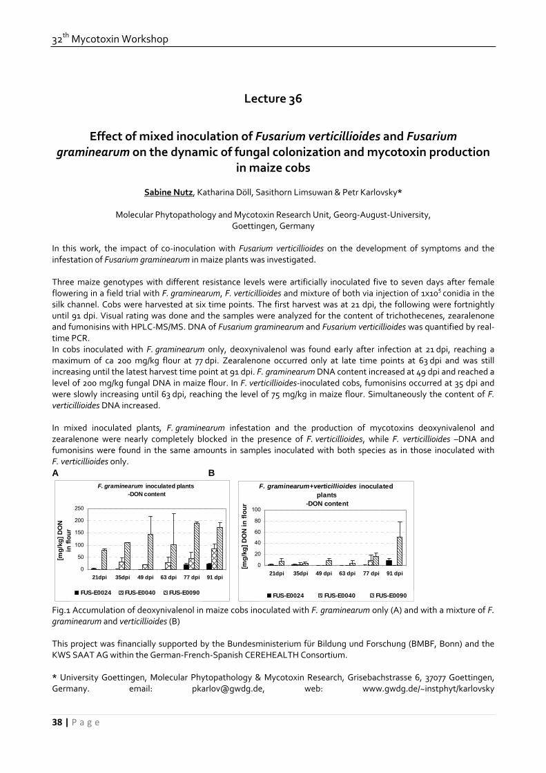
32th Mycotoxin Workshop
38 | P a g e
Lecture 36
Effect of mixed inoculation of Fusarium verticillioides and Fusarium graminearum on the dynamic of fungal colonization and mycotoxin production
in maize cobs
Sabine Nutz, Katharina Döll, Sasithorn Limsuwan & Petr Karlovsky*
Molecular Phytopathology and Mycotoxin Research Unit, Georg‐August‐University,
Goettingen, Germany In this work, the impact of co‐inoculation with Fusarium verticillioides on the development of symptoms and the infestation of Fusarium graminearum in maize plants was investigated. Three maize genotypes with different resistance levels were artificially inoculated five to seven days after female flowering in a field trial with F. graminearum, F. verticillioides and mixture of both via injection of 1x10⁵ conidia in the silk channel. Cobs were harvested at six time points. The first harvest was at 21 dpi, the following were fortnightly until 91 dpi. Visual rating was done and the samples were analyzed for the content of trichothecenes, zearalenone and fumonisins with HPLC‐MS/MS. DNA of Fusarium graminearum and Fusarium verticillioides was quantified by real‐time PCR. In cobs inoculated with F. graminearum only, deoxynivalenol was found early after infection at 21 dpi, reaching a maximum of ca 200 mg/kg flour at 77 dpi. Zearalenone occurred only at late time points at 63 dpi and was still increasing until the latest harvest time point at 91 dpi. F. graminearum DNA content increased at 49 dpi and reached a level of 200 mg/kg fungal DNA in maize flour. In F. verticillioides‐inoculated cobs, fumonisins occurred at 35 dpi and were slowly increasing until 63 dpi, reaching the level of 75 mg/kg in maize flour. Simultaneously the content of F. verticillioides DNA increased. In mixed inoculated plants, F. graminearum infestation and the production of mycotoxins deoxynivalenol and zearalenone were nearly completely blocked in the presence of F. verticillioides, while F. verticillioides –DNA and fumonisins were found in the same amounts in samples inoculated with both species as in those inoculated with F. verticillioides only. A B
F. graminearum inoculated plants -DON content
0
50
100
150
200
250
21dpi 35dpi 49 dpi 63 dpi 77 dpi 91 dpi
[mg/
kg] D
ON
in fl
our
FUS-E0024 FUS-E0040 FUS-E0090
F. graminearum+verticillioides inoculated plants
-DON content
0
20
40
60
80
100
21dpi 35dpi 49 dpi 63 dpi 77 dpi 91 dpi
[mg/
kg] D
ON
in fl
our
FUS-E0024 FUS-E0040 FUS-E0090
Fig.1 Accumulation of deoxynivalenol in maize cobs inoculated with F. graminearum only (A) and with a mixture of F. graminearum and verticillioides (B) This project was financially supported by the Bundesministerium für Bildung und Forschung (BMBF, Bonn) and the KWS SAAT AG within the German‐French‐Spanish CEREHEALTH Consortium. * University Goettingen, Molecular Phytopathology & Mycotoxin Research, Grisebachstrasse 6, 37077 Goettingen, Germany. email: [email protected], web: www.gwdg.de/~instphyt/karlovsky

June, 15th‐17th, 2010, Lyngby
39 | P a g e
Lecture 37
Proteome analysis of Fusarium infected emmer (Triticum dicoccum) and naked barley (Hordeum vulgare subsp. nudum) grains
*Kai Eggert 1, Christian Zörb 2& Elke Pawelzik 1
1 Department of Crop Sciences, Section Quality of Plant Products, Göttingen, Germany. 2 Institute of Plant Nutrition
and Soil Science, Kiel, Germany. Göttingen, Germany.
Introduction: Fungal infection on plants results in numerous host‐specific biochemical responses which show strategies for the ability of the plant to withstand disease [1‐5]. For that reason pathogen exposure leads to changes in host proteins correlated to defense strategies of the plant at different functional levels like cell signaling pathway, protein degradation, hormone production, apoptosis and rearrangement of the cytoskeleton and be reported as changes in secondary metabolism or induction of proteins such as enzymes and enzyme inhibitors [1, 6, 7]. Therefore, the effect of artificial Fusarium graminearum and Fusarium culmorum infection at the level of the proteome in comparison to natural infection of emmer and naked barley grains was investigated. Material and Methods: ‐LC‐MS/MS for Toxin toxin (Deoxynivalenol) analysis ‐MALDI‐Tof‐MS and nanoLC‐MS/MS for proteome analysis Results and Discussion: Artificial Fusarium infection leads in both investigated species emmer and naked barley to up‐ or down regulation of specific proteins in comparison to the natural infection. For host specific response of the plant we found a group of serine protease inhibitors, the serpins, up‐regulated in artificial infected samples. These inhibitors of protease may prevent the digestion of seed proteins after fungal infection to a certain degree in late development stage [7, 8]. Peroxidases known to play important role in combating the reactive oxygen species exposed to biotic stress in early infection stages were in emmer down regulated in late stages of development [2, 9]. In Emmer, a thaumatin‐like protein that inhibits hyphal growth and sporulation was found to be induced and considered as pathogenesis‐related protein (PR protein) [10]. In naked barley a NBS‐LRR disease resistance protein was induced that interacts direct or indirect with pathogen molecules and is involved in host‐protein and hostpathogen recognition [11]. References: [1] Dixon R. A., 2001. Natural products and plant disease resistance. Nature, 411, 843‐847. [2] Geddes J., Eudes F., Laroche A., Selinger L. B., 2008. Differential expression of proteins in response to the interaction between the pathogen Fusarium graminearum and its host, Hordeum vulgare. Proteomics, 8, 545‐554. [3] Wang Y., Yang L. M., Xu H. B., Li Q. F., et al., 2005. Differential proteomic analysis of proteins in wheat spikes induced by Fusarium graminearum. Proteomics, 5, 4496‐4503. [4] Zhou W. C., Eudes F., Laroche A., 2006. Identification of differentially regulated proteins in response to a compatible interaction between the pathogen Fusarium graminearum and its host, Triticum aestivum. Proteomics, 6, 4599‐4609. [5] Zhou W. C., Kolb F. L., Riechers D. E., 2005. Identification of proteins induced or upregulated by Fusarium head blight infection in the spikes of hexaploid wheat (Triticum aestivum). Genome, 48, 770‐780. [6] Coiras M., Camafeita E., Lopez‐Huertas M. R., Calvo E., et al., 2008. Application of proteomics technology for analyzing the interactions between host cells and intracellular infectious agents. Proteomics, 8, 852‐873. [7] Pekkarinen A. I., Longstaff C., Jones B. L., 2007. Kinetics of the inhibition of Fusarium serine proteinases by barley (Hordeum vulgare L.) inhibitors. J. Agric. Food Chem., 55, 2736‐2742. [8] Phalip V., Delalande F., Carapito C., Goubet F., et al., 2005. Diversity of the exoproteome of Fusarium graminearum grown on plant cell wall. Curr. Genet., 48, 366‐379. [9] Campo S., Carrascal M., Coca M., Abian J., San Segundo B., 2004. The defense response of germinating maize embryos against fungal infection: A proteomics approach. Proteomics, 4, 383‐396. [10] Kuwabara C., Takezawa D., Shimada T., Hamada T., et al., 2002. Abscisic acid‐ and cold‐induced thaumatin‐like protein in winter wheat has an antifungal activity against snow mould, Microdochium nivale. Physiol. Plant., 115, 101‐110. [11] Feng D. S., Li Y., Wang H. G., Li X. F., Gao J. R., 2009. Isolation and Evolution Mode Analysis of NBS‐LRR Resistance Gene Analogs from Hexaploid Wheat. Plant Mol. Biol. Rep., 27, 266‐274. * Carl‐Sprengel‐Weg 1, Faculty of Agricultural Sciences, Georg‐August University of Göttingen, D‐37075 Göttingen, Germany, e‐mail: [email protected]

32th Mycotoxin Workshop
40 | P a g e
Lecture 38
Isolation and characterization of a novel non‐toxic metabolite of the Fusarium mycotoxin diacetoxyscirpenol
Mehrdad Shams1, Rudolf Mitterbauer2†, Roberto Corradini3, Gerhard Adam2, Rudolf Krska1, Rainer Schuhmacher1,
Franz Berthiller1*
1 Center for Analytical Chemistry, Department for Agrobiotechnology (IFA‐Tulln), University of Natural Resources and Applied Life Sciences Vienna (BOKU), Tulln, Austria. 2 Department of Applied Genetics and Cell Biology, BOKU,
Vienna, Austria. 3 Dipartimento di Chimica Organica e Industriale, Parma, Italy. Trichothecenes are an important class of mycotoxins which act as potent protein synthesis inhibitors in eukaryotic organisms. Scirpentriol and its derivatives are members of the A‐type trichothecenes. The most prominent member of the scirpentriol derivatives ‐ 4,15‐diacetoxyscirpenol (DAS, figure 1 left) ‐ is highly toxic against plants and animals. Especially oats or potatoes are prone to be contaminated with DAS after infection with e.g. Fusarium sambucinum. In the current study, we monitored the reduction of DAS due to thermal treatment of infected potatoes and detected a new major metabolite (figure 1 right). After isolation, the structure of this novel metabolite was elucidated with LC‐MS and NMR.
Whole potatoes were infected with F. sambucinum strain MRC514 for 14 days at 20°C. The infected potatoes were cooked at 100°C or at 121°C (in a pressure cooker) for 1, 4 or 8 hours respectively. A drastic reduction of DAS from about 49% (1h at 100°C) to 99% (4h at 121°C) has been monitored by LC‐MS analysis. The toxicity of the metabolite has been examined in vitro by inhibition of protein synthesis compared to DAS using a wheat germ extract transcription/translation system. In vivo toxicity on Saccharomyces cerevisiae was also evaluated. The results show that the inhibitory influence of DAS is significantly higher (more than 100 times) than the same molar amount of the novel metabolite, obtained after thermal treatment. * Konrad Lorenz Straße 20, 3430 Tulln, Austria. E‐mail: [email protected]
Δ
OO
CH3
CH3
HH
H
OH OH
H
HCH3
OHH
HH H
H
CH3O
O HO
O
CH3
CH3
HH
H
OH
OH
H
HCH3
HH H
H
CH3O
O H
HH
OH
OH

June, 15th‐17th, 2010, Lyngby
41 | P a g e
Lecture 39
Reduction of aflatoxin B1 contamination in corn with natural zeolite and bentonite
Nuryono1*, A. Agus2, S. Wedhastri3, Y. M. S. Maryudhani 3, D. Pranowo1, Yunianto4, E. Razzazi‐Fazeli5
1 Department of Chemistry, Universitas Gadjah Mada, Yogyakarta, Indonesia. 2Faculty of Animal Sciences,
Universitas Gadjah Mada, Yogyakarta, Indonesia. 3Faculty of Agriculture, Universitas Gadjah Mada, Yogyakarta, Indonesia. 4Animal Husbandry Academy Brahma Putra, Yogyakarta, Indonesia. 5Institute of Nutrition, University of
Veterinary Medicine, Vienna, Austria
In tropical countries like Indonesia with high humidity and temperature it is very conducive for growing various toxigenic fungi This cause economic losses in agriculture commodities such as corn being contaminated with mycotoxins. Aflatoxin B1 (AFB1) is one of highly toxic and carcinogenic compounds that has been implicated as causative agent in human hepatic carcinogenesis. The mycotoxins have been found in locally produced agricultural crops and their food and feed products. Many strategies for reduction of the AFB1 contamination in feed and foods have been developed. In this research, a study of reduction of AFB1 in corn (kernel and grounded) using adsorption technique with natural zeolite and bentonite as adsorbents was investigated. In preliminary studies adsorption in a batch system of standard AFB1 solution on adsorbents were investigated. Some factors such as contact time, concentration of AFB1 and particle size of adsorbent were evaluated. The amount of adsorbed AFB1 was calculated based on the difference of AFB1 concentration before and after adsorption determined by high performance liquid chromatography (HPLC) method. Reduction of AFB1 in corn sample was emphasized by mixing corn sample aqueous suspension with adsorbent. Concentration of AFB1 in suspension was analyzed by enzyme‐linked immuno‐sorbent assay (ELISA) method. Result shows that adsorption of AFB1 on adsorbents of natural zeolite and bentonite is very fast. Within 15 minutes 99 % of AFB1 (200 ng/mL) has been adsorbed by 25 mg of bentonite and 96 % by zeolite. The particle size higher than 200 mesh did not show significant effect on the AFB1 adsorption capability. In comparison, effectiveness of zeolite in adsorbing AFB1 is lower than that of bentonite. The difference is probable caused by the structure rather than the presence of functional groups of the adsorbents. It was supported by data of infrared spectroscopy indicating similarity of spectra and x‐ray diffraction (XRD) data showing different pattern. Capability of both adsorbents in reducing AFB1 contamination in corn samples (kernel and grain) was lower than those in standard solution. Six hundred milligram of zeolite and bentonite reduce 17 and 37 %, respectively, of AFB1 (107 ng/mL) in kernel corn, and 78 and 67 %, of AFB1 (124 ng/mL) in grain corn suspension. * Sekip Utara, Yogyakarta 55281, Indonesia. E‐mail: nur [email protected]
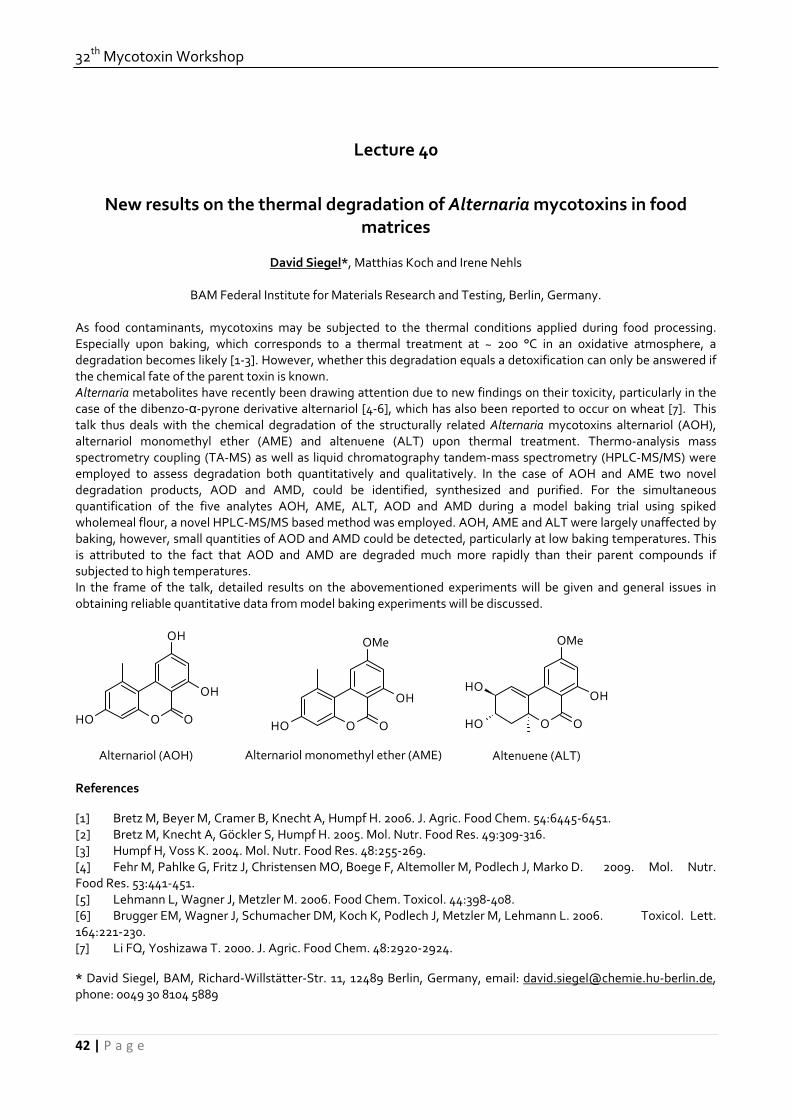
32th Mycotoxin Workshop
42 | P a g e
Lecture 40
New results on the thermal degradation of Alternaria mycotoxins in food matrices
David Siegel*, Matthias Koch and Irene Nehls
BAM Federal Institute for Materials Research and Testing, Berlin, Germany. As food contaminants, mycotoxins may be subjected to the thermal conditions applied during food processing. Especially upon baking, which corresponds to a thermal treatment at ~ 200 °C in an oxidative atmosphere, a degradation becomes likely [1‐3]. However, whether this degradation equals a detoxification can only be answered if the chemical fate of the parent toxin is known. Alternaria metabolites have recently been drawing attention due to new findings on their toxicity, particularly in the case of the dibenzo‐α‐pyrone derivative alternariol [4‐6], which has also been reported to occur on wheat [7]. This talk thus deals with the chemical degradation of the structurally related Alternaria mycotoxins alternariol (AOH), alternariol monomethyl ether (AME) and altenuene (ALT) upon thermal treatment. Thermo‐analysis mass spectrometry coupling (TA‐MS) as well as liquid chromatography tandem‐mass spectrometry (HPLC‐MS/MS) were employed to assess degradation both quantitatively and qualitatively. In the case of AOH and AME two novel degradation products, AOD and AMD, could be identified, synthesized and purified. For the simultaneous quantification of the five analytes AOH, AME, ALT, AOD and AMD during a model baking trial using spiked wholemeal flour, a novel HPLC‐MS/MS based method was employed. AOH, AME and ALT were largely unaffected by baking, however, small quantities of AOD and AMD could be detected, particularly at low baking temperatures. This is attributed to the fact that AOD and AMD are degraded much more rapidly than their parent compounds if subjected to high temperatures. In the frame of the talk, detailed results on the abovementioned experiments will be given and general issues in obtaining reliable quantitative data from model baking experiments will be discussed.
OH O O
OH
OH
OH O O
OH
OMe
OH O O
OH
OMe
OH
Alternariol (AOH) Alternariol monomethyl ether (AME) Altenuene (ALT) References [1] Bretz M, Beyer M, Cramer B, Knecht A, Humpf H. 2006. J. Agric. Food Chem. 54:6445‐6451. [2] Bretz M, Knecht A, Göckler S, Humpf H. 2005. Mol. Nutr. Food Res. 49:309‐316. [3] Humpf H, Voss K. 2004. Mol. Nutr. Food Res. 48:255‐269. [4] Fehr M, Pahlke G, Fritz J, Christensen MO, Boege F, Altemoller M, Podlech J, Marko D. 2009. Mol. Nutr. Food Res. 53:441‐451. [5] Lehmann L, Wagner J, Metzler M. 2006. Food Chem. Toxicol. 44:398‐408. [6] Brugger EM, Wagner J, Schumacher DM, Koch K, Podlech J, Metzler M, Lehmann L. 2006. Toxicol. Lett. 164:221‐230. [7] Li FQ, Yoshizawa T. 2000. J. Agric. Food Chem. 48:2920‐2924. * David Siegel, BAM, Richard‐Willstätter‐Str. 11, 12489 Berlin, Germany, email: [email protected]‐berlin.de, phone: 0049 30 8104 5889

June, 15th‐17th, 2010, Lyngby
43 | P a g e
Abstracts of Posters

32th Mycotoxin Workshop
44 | P a g e
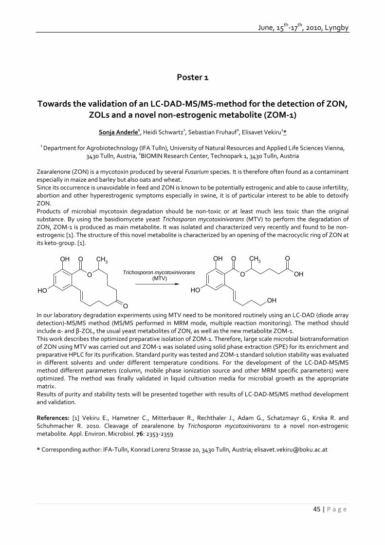
June, 15th‐17th, 2010, Lyngby
45 | P a g e
Poster 1
Towards the validation of an LC‐DAD‐MS/MS‐method for the detection of ZON, ZOLs and a novel non‐estrogenic metabolite (ZOM‐1)
Sonja Anderle1, Heidi Schwartz1, Sebastian Fruhauf2, Elisavet Vekiru1*
1 Department for Agrobiotechnology (IFA Tulln), University of Natural Resources and Applied Life Sciences Vienna,
3430 Tulln, Austria, 2BIOMIN Research Center, Technopark 1, 3430 Tulln, Austria Zearalenone (ZON) is a mycotoxin produced by several Fusarium species. It is therefore often found as a contaminant especially in maize and barley but also oats and wheat. Since its occurrence is unavoidable in feed and ZON is known to be potentially estrogenic and able to cause infertility, abortion and other hyperestrogenic symptoms especially in swine, it is of particular interest to be able to detoxify ZON. Products of microbial mycotoxin degradation should be non‐toxic or at least much less toxic than the original substance. By using the basidiomycete yeast Trichosporon mycotoxinivorans (MTV) to perform the degradation of ZON, ZOM‐1 is produced as main metabolite. It was isolated and characterized very recently and found to be non‐estrogenic [1]. The structure of this novel metabolite is characterized by an opening of the macrocyclic ring of ZON at its keto‐group. [1].
O
O
OOH
OH
CH3
O
OOH
OH
CH3
OH
O
OH
Trichosporon mycotoxinivorans (MTV)
In our laboratory degradation experiments using MTV need to be monitored routinely using an LC‐DAD (diode array detection)‐MS/MS method (MS/MS performed in MRM mode, multiple reaction monitoring). The method should include α‐ and β‐ZOL, the usual yeast metabolites of ZON, as well as the new metabolite ZOM‐1. This work describes the optimized preparative isolation of ZOM‐1. Therefore, large scale microbial biotransformation of ZON using MTV was carried out and ZOM‐1 was isolated using solid phase extraction (SPE) for its enrichment and preparative HPLC for its purification. Standard purity was tested and ZOM‐1 standard solution stability was evaluated in different solvents and under different temperature conditions. For the development of the LC‐DAD‐MS/MS method different parameters (column, mobile phase ionization source and other MRM specific parameters) were optimized. The method was finally validated in liquid cultivation media for microbial growth as the appropriate matrix. Results of purity and stability tests will be presented together with results of LC‐DAD‐MS/MS method development and validation. References: [1] Vekiru E., Hametner C., Mitterbauer R., Rechthaler J., Adam G., Schatzmayr G., Krska R. and Schuhmacher R. 2010. Cleavage of zearalenone by Trichosporon mycotoxinivorans to a novel non‐estrogenic metabolite. Appl. Environ. Microbiol. 76: 2353‐2359 * Corresponding author: IFA‐Tulln, Konrad Lorenz Strasse 20, 3430 Tulln, Austria; [email protected]
32th Mycotoxin Workshop
46 | P a g e
Poster 2
Simulated digestion assay for hidden fumonisin evaluation.
A. Dall’Erta , C. Falavigna, C. Dall'Asta*, G. Galaverna, A. Dossena, R. Marchelli.
Department of Organic and Industrial Chemistry, University of Parma, Parma, Italy. Masked mycotoxins are forms that easily escape conventional techniques of analysis because they are conjugated with more polar compounds or bounded with matrix constituents. Among them, hidden fumonisins play a role of concern due to their possible toxicity. Very recently, a masking mechanism through complexation with food macroconstituents was proposed (Dall’Asta et al., 2009). In particular, the nature of masking is assign to a formation of covalents or non covalents bonds between fumonisins and starch or proteins, that are the major constituents of maize. Such compounds are detectable through the application of a hydrolysis step and could be released from the matrix during technological treatments or digestion, causing an effective higher exposure than cannot be estimated by routine methods. In this work a validated in vitro digestion model has been applied on cereals and cereal‐based products to simulate the digestive conditions of the human gastrointestinal tract. This experiment allowed to evaluate the real presence of hidden fumonisins in the considered samples, estimating their bioaccessibility after food consumption. The proposed digestion model, thus, allowed the study of hidden fumonisin behaviour when physiological conditions are applied to a contaminate food and the evaluation the of the theoretical fumonisin amount which may be available for intestinal absorption. Reference: C. Dall’Asta, M. Mangia, F. Berthiller, A. Molinelli, M. Sulyok, R. Schuhmacher, R. Krska, G. Galaverna, A. Dossena, R. Marchelli. 2009. Difficulties in fumonisin determination: the issue of hidden fumonisins. Anal Bioanal Chem 395. * Chiara Dall’Asta: Department of Organic and Industrial Chemistry University of Parma, v.le Usberti 17/A, I‐43124, Parma, Italy; e‐mail: [email protected]

June, 15th‐17th, 2010, Lyngby
47 | P a g e
Poster 3
Accurate and precise determination of the Alternaria mycotoxins Alternariol and Alternariol monomethylether in cereals, fruit and vegetables using stable
isotope dilution assays
Asam S.*, Konitzer K. & Rychlik M.
Technische Universität München, Lehrstuhl für Analytische Lebensmittelchemie, Freising
Molds of the genus Alternaria are virulent plant pathogens and are found on a variety of possible substrates, especially on foods. Alternaria is characterized by the formation of dark, almost black colonies and is reckoned among the heterogeneous group of “black molds”, therefore. Most species of Alternaria are known to produce one ore more mycotoxins. Consequently, the occurrence of the mycotoxins Alternariol (AOH), Alternariolmonomethylether (AME), Altenuen and Tenuazonic acid in various foods has been reported [1]. For the analysis of AOH and AME in beverages like fruit juice and wine, stable isotope dilution assays have been developed [2]. These were based on the use of [2H4]‐AOH and [2H4]‐AME as internal standards that were synthesized by palladium‐catalyzed H/D exchange of the unlabeled toxins. The detection and quantitative determination of AOH and AME was performed by LC‐MS/MS. The aim of this study was to extend the existing stable isotope dilution assay for AOH and AME to other foods, particularly to grain, fruit and vegetables and their associated processing products. The contamination of cereals and cereal products with AOH and AME was low. From a total of 15 samples, including wheat, breakfast cereals and various flours, AOH or AME were detected only in one spelt flour (4.1 ppb AOH) and in two samples of oat flakes (0.6 and 0.2 ppb AME). In feed grain, destined for animal feeding exclusively, AOH and AME could be quantified in all 6 samples with maximum values up to 250 ppb AOH and 100 ppb AME, however. Investigation of fruit and fruit products confirmed the findings of earlier studies [2]. While fruit juice was contaminated with AOH and AME only in moderate values, wine contained both toxins in higher levels. In 6 samples of wine that was marketed under the label “Spätlese” or “Trockenbeerenauslese” values of AOH and AME ranged from 0 – 5 ppb and from 0 – 0.3 ppb, respectively. Despite the production process of these specialty wines that bears the possibility of mold growth no divergence in the AOH and AME content in comparison to wine in general was seen. The content of AOH and AME in vegetables and vegetable products was low, mostly. An exception, however, was found in products based on tomatoes. In these products, such as tomato ketchup, tomato juice or tomato puree, AOH and AME were detected in almost every sample. In particular, high contents of AOH and AME were quantified in tomato concentrates, while the contents increased with higher concentration grade and reached 25 ppb AOH and 5.3 ppb AME in one case. References: [1] Scott P.M., J. AOAC Int. 2001, 84 (6), 1809‐1817 [2] Asam S. et al., J. Agric. Food Chem 2009, 57 (12), 5152‐5160 * Dr. Stefan Asam, Technische Universität München, Lehrstuhl für Analytische Lebensmittelchemie, Alte Akademie 10, 85350 Freising; [email protected]
32th Mycotoxin Workshop
48 | P a g e
Poster 4
Development of a sample preparation method for the determination of T‐2 toxin and metabolites in blood plasma with LC‐MS/MS.
Ulrike Brezina*, Katrin Wilkerling, Hana Valenta, Susanne Döll and Sven Dänicke
Institute of Animal Nutrition, Friedrich‐Loeffler‐Institut (FLI), Federal Research Institute for Animal Health,
Braunschweig, Germany.
Introduction: T‐2 toxin (T‐2) and its metabolites HT‐2 toxin (HT‐2), neosolaniol (NEO), T‐2 triol and T‐2 tetraol belong to the type A trichothecenes produced by several Fusarium species, which are commonly found in various cereal crops and processed grains. T‐2 is readily metabolised by the intestinal flora to HT‐2, which is the major metabolite, but in tissues also metabolising reactions like hydrolysis, oxidation, reduction and conjugation proceed. Both toxins are among the most toxic trichothecenes and have adverse effects on animals, humans and plants [1]. For studies on kinetics and carry‐over of T‐2 in farm animals methods for the determination of T‐2 and metabolites in physiological samples are needed. Therefore the aim of this work was to develop a sample preparation method for determination of T‐2 and metabolites in blood plasma with LC‐MS/MS. Methods: HybridSPE™‐PPT cartridges (Supelco) and Chem Elut™ cartridges (Varian) were tested for their suitability for sample preparation for measuring T‐2, HT‐2, NEO, T‐2 triol and T‐2 tetraol in blood plasma. HybridSPE™‐PPT cartridges are – according to an information of the manufacturer ‐ particularly suitable as sample preparation for LC‐MS/MS because they remove proteins and phospholipids from blood plasma/serum. These cartridges were first tested according to manufacturer's prescription and then with slight modifications. Chem Elut™ cartridges are packed with diatomaceous earth and work on the principle of solid‐supported‐liquid‐liquid‐extraction (SSLLE). For method development elution with ethyl acetate and with mixtures of ethyl acetate and methanol (98:2, 95:5, 92:8, 90:10; v/v) was tested. The determination of T‐2 and the four metabolites was carried out by LC‐ESI‐MS/MS in positive mode. Results: Due to low recoveries (40‐50 %), the HybridSPE™‐PPT cartridges were unsuitable. However, the Chem Elut™ cartridges showed good recoveries in preliminary tests and were finally used for method development. The optimized method includes elution of the toxins with ethyl acetate/methanol (95:5 v/v) as this mixture gave the best results. For method validation, recoveries, precision in terms of repeatability (intra‐day precision) and reproducibility (inter‐day precision) and limits of detection were evaluated. The mean recoveries (n=8) of the five toxins were in the range of 53 % ‐ 85 %, the limits of detection (S/N ≥ 3) were 0.1, 0.1, 0.1, 0.25 and 2 ng/ml for T‐2, HT‐2, NEO, T‐2 triol and T‐2 tetraol, respectively. Conclusions: In this study a new, sensitive, rapid, and cost efficient preparation method for determination of T‐2 and metabolites in blood plasma using LC‐MS/MS detection was developed. This method is suitable for studies on kinetics and carry‐over of T‐2 in farm animals. References: [1] European Commission, Scientific Committee on Food 2001. Opinion on Fusarium toxins, part 5: T‐2 toxin and HT‐2 toxin (adopted by the SCF on 30 May 2001) *Institute of Animal Nutrition, Friedrich‐Loeffler‐Institut (FLI), Federal Research Institute for Animal Health, Bundesallee 50, 38116 Braunschweig, Germany email: [email protected]
June, 15th‐17th, 2010, Lyngby
49 | P a g e
Poster 5
Modulation of the early apoptotic potential of DON by MAPK signaling
Maximilian Casteel*, Carina Nielsen, Richard Dietrich & Erwin Märtlbauer
Department of Veterinary Sciences, Ludwig Maximilians‐Universität München, Schönleutnerstraße 8, 85748 Oberschleissheim, Germany.
Introduction: The trichothecene deoxynivalenol (DON) is the most common mycotoxin contaminant of cereal‐based food products. Due to this situation, the toxic effects of DON were the subject of various in vivo and in vitro studies during the last years. The main toxic effect of DON is the inhibition of protein synthesis via binding to the ribosome. A further capability of DON is the disruption of normal cell function by the activation of cellular kinases involved in proliferation, differentiation and apoptosis. However, little is known about cell type‐specific toxic mechanisms especially of the mitogen activated protein kinases (MAPKs) pathway and its influence on the regulation of apoptosis. Materials and Methods: DON was purchased from Sigma Chemical Company. MAPK inhibitors were purchased from Calbiochem. HepG2 cells were grown in RPMI‐1640 medium supplemented with 10% fetal bovine serum. Apoptosis were measured by Western blotting and flow cytometry. Phosphorylation of MAPKs and their targets were assessed by Western blotting and immunofluorescence. Results: Following 1h‐12h incubation, DON (1‐10 µM) markedly induced the phosphorylation of MAPKs as well as the expression of negative regulators of the MAPK activity. Treatment with high concentrations of DON induced the activation of caspase‐3 and translocation of phosphatidylserine, whereas low concentrations of DON didn't influence these hallmarks of early‐stage apoptosis. Under condition in which the DON‐mediated induction of negative regulation of the MAPK pathway was inhibited, a considerable increase of the apoptotic potential of DON was observed. Discussion and Conclusion: Taken together, these data suggest that DON treatment of epithelial cells results in an increased expression of negative regulators of the MAPK pathway which may explain the comparative low apoptotic potential of DON. * Corresponding author E‐mail: [email protected]‐muenchen.de
32th Mycotoxin Workshop
50 | P a g e
Poster 6
Development of Lateral Flow Devices for the Qualitative and Semi‐Quantitative Detection of Mycotoxins in Agricultural Commodities
Barbara Cvak1,2, Alexandra Molinelli1, Rudolf Krska2*
1Romer Labs Division Holding GmbH, Tulln, Austria. 2Center for Analytical Chemistry, Department for
Agrobiotechnology (IFA‐Tulln), University of Natural Resources and Applied Life Sciences, Vienna, Austria
Mycotoxins are toxic natural secondary metabolites produced by several species of fungi on agricultural commodities in the field or during storage. Besides standard analytical methods, screening methods are increasingly needed. Lateral flow devices (LFDs), also called strip tests, are immunoassay‐based methods which allow the qualitative or semi‐quantitative determination of mycotoxins within a few minutes. The development of this rapid immunochemical test system for the screening of different mycotoxins such as zearalenone (ZON), Aflatoxin B1 (Afla), Ochratoxin A (OTA), and T‐2/HT‐2 toxin, will be the main focus of the presented work. Furthermore, latest results towards the development of multi‐mycotoxin strip tests will be presented. Colloidal gold as well as latex particles were used as color reagents for labeling mycotoxin‐specific antibodies. The antibody‐colloidal gold conjugate is mixed with sample extract in case of solid samples such as wheat and maize or directly with liquid samples such as wine. An LFD is then inserted into the mixture and the liquid migrates onto the membrane of the strip, which contains a test zone, where a mycotoxin‐protein complex has been coated and a control zone, which confirms correct test performance. The immobilized mycotoxin‐protein conjugate (test zone) captures free mycotoxin‐antibody/colloidal gold particles, allowing a concentration of them in the test zone, and leads to the formation of a colored line, which is measured with a photometric reader. All presented LFDs were tested with spiked extracts or liquid samples as well as naturally contaminated samples and calibrated with quality control reference material if available. By relative reflectance reading of the test lines the presence and amount of different mycotoxins in agricultural commodities could be determined within the linear working ranges of the corresponding strips. References: R. Krska, A. Molinelli. 2009. Rapid test strips for analysis of mycotoxins in food and feed. Anal. Bioanal. Chem. 393, 67‐71. * Konrad‐Lorenz‐Straße 20, 3430 Tulln, Austria; [email protected]
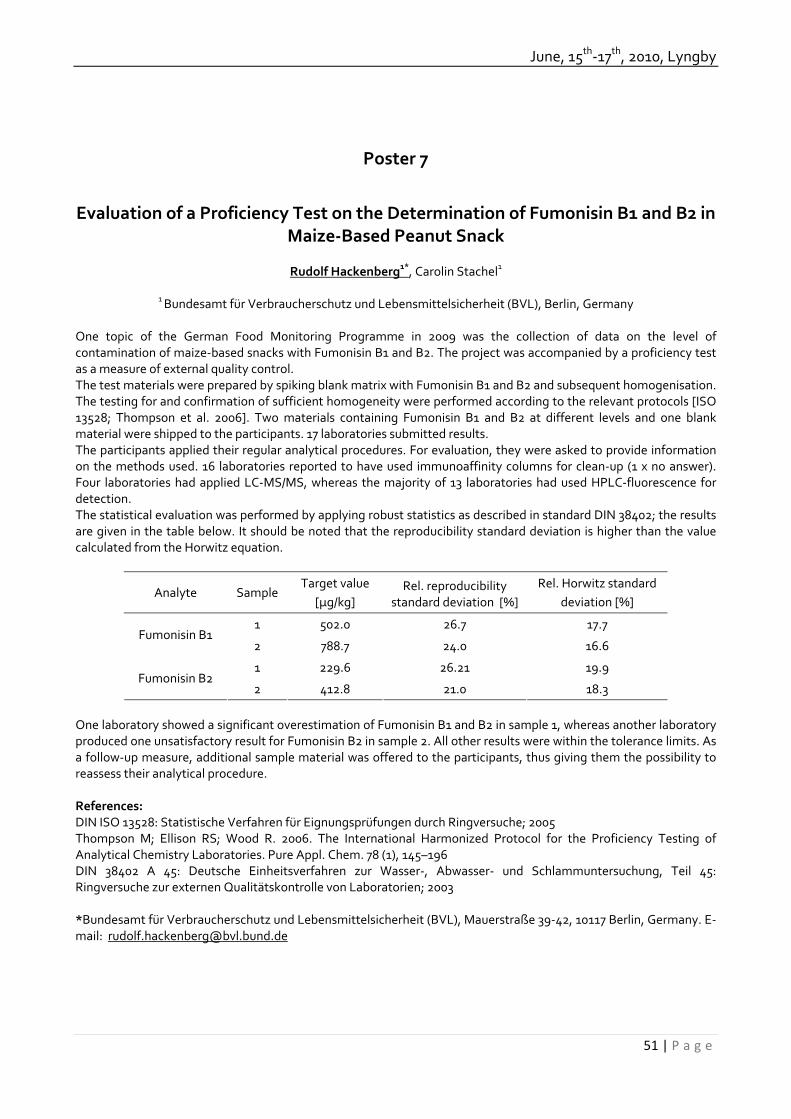
June, 15th‐17th, 2010, Lyngby
51 | P a g e
Poster 7
Evaluation of a Proficiency Test on the Determination of Fumonisin B1 and B2 in Maize‐Based Peanut Snack
Rudolf Hackenberg1*, Carolin Stachel1
1 Bundesamt für Verbraucherschutz und Lebensmittelsicherheit (BVL), Berlin, Germany
One topic of the German Food Monitoring Programme in 2009 was the collection of data on the level of contamination of maize‐based snacks with Fumonisin B1 and B2. The project was accompanied by a proficiency test as a measure of external quality control. The test materials were prepared by spiking blank matrix with Fumonisin B1 and B2 and subsequent homogenisation. The testing for and confirmation of sufficient homogeneity were performed according to the relevant protocols [ISO 13528; Thompson et al. 2006]. Two materials containing Fumonisin B1 and B2 at different levels and one blank material were shipped to the participants. 17 laboratories submitted results. The participants applied their regular analytical procedures. For evaluation, they were asked to provide information on the methods used. 16 laboratories reported to have used immunoaffinity columns for clean‐up (1 x no answer). Four laboratories had applied LC‐MS/MS, whereas the majority of 13 laboratories had used HPLC‐fluorescence for detection. The statistical evaluation was performed by applying robust statistics as described in standard DIN 38402; the results are given in the table below. It should be noted that the reproducibility standard deviation is higher than the value calculated from the Horwitz equation.
Analyte Sample Target value
[µg/kg] Rel. reproducibility
standard deviation [%] Rel. Horwitz standard
deviation [%]
1 502.0 26.7 17.7 Fumonisin B1
2 788.7 24.0 16.6
1 229.6 26.21 19.9 Fumonisin B2
2 412.8 21.0 18.3 One laboratory showed a significant overestimation of Fumonisin B1 and B2 in sample 1, whereas another laboratory produced one unsatisfactory result for Fumonisin B2 in sample 2. All other results were within the tolerance limits. As a follow‐up measure, additional sample material was offered to the participants, thus giving them the possibility to reassess their analytical procedure. References: DIN ISO 13528: Statistische Verfahren für Eignungsprüfungen durch Ringversuche; 2005 Thompson M; Ellison RS; Wood R. 2006. The International Harmonized Protocol for the Proficiency Testing of Analytical Chemistry Laboratories. Pure Appl. Chem. 78 (1), 145–196 DIN 38402 A 45: Deutsche Einheitsverfahren zur Wasser‐, Abwasser‐ und Schlammuntersuchung, Teil 45: Ringversuche zur externen Qualitätskontrolle von Laboratorien; 2003 *Bundesamt für Verbraucherschutz und Lebensmittelsicherheit (BVL), Mauerstraße 39‐42, 10117 Berlin, Germany. E‐mail: [email protected]

32th Mycotoxin Workshop
52 | P a g e
Poster 8
Detection of mould and mycotoxine on grain using a gas Sensor Array
C. Idler1*, A. Walte2, M. Ditz1, A. Plessing‐Menze2, K. Briese2
1 Leibniz‐Institute for Agricultural Engineering Potsdam‐Bornim., Dept. Bioengineering, Potsdam, Germany. 2
Airsense Analytics GmbH, Schwerin, Germany. Within the joint research project “ProSenso.net2” a sensor technology for the identification of mycotoxine producing fungi in the processing of grain is developed. In a joint collaborative effort between the Leibniz‐Institute and the Airsense Analytics GmbH different gas sensor arrays (Electronic Nose) for the identification of mycotoxine and mycotoxin producing fungi on grain are tested. Food is often contaminated with toxic compounds which are not manmade. Some of the toxic compounds, the mycotoxine, are synthesized by moulds. Mycotoxine are compounds with nearly no vapor pressure and cannot be measured directly in the vapor phase. Grain with and without contaminations with various moulds and mycotoxine were investigated by measuring the VOCs with an array of different chemical sensors. Electronic noses usually consist of an array of simple sensors, such as metal oxide semiconductors, combined with the required electronics and chemometrical software. The portable electronic nose PEN3 is a stand‐alone system, consisting of a special sampling system with integrated dilution system, an array of ten metal oxide semiconductors, the required electronics and an integrated micro‐controller. The different sensors based on different metal oxides are positioned in a very small sensor chamber. With an internal pump the gaseous compounds are sucked through the sensor array. With a second pump filtered reference air can be transported to the sensor array. Wheat and rye samples were contaminated with various moulds and stored in vials. After incubation time of one week the headspace of the vials was measured with the PEN3 system. It could shown from the raw sensor data that the sensor signals of the infected samples were more intensive compared to the sensors signals of the not infected grain samples. This can be also shown by using statistical methods such as the principal component analysis PCA. With the data transformation performed with the PCA a dependency on the moisture content of the samples is also displayed. It was also clear shown differences between sterile and inoculated samples. The results show, that a direct detection of mycotoxine is not possible with a gas sensor array system. However, an indirect detection of mycotoxine could be possible by the detection of VOCs from the moulds. The Project was supported by the Federal Ministry of Education and Research. References: Ditz M, Idler C, Rasch C, Kumke M, Walte A. Exposure of corn with mould and mycotoxine – detection by sensor technology. Lecture 8th International Conference “Mycotoxine and moulds”. 25‐27 Juni, 2008, Bydgoszcz, Polen, Proceedings pp. 28 Idler C, Rasch C, Ditz M, Kumke M, Walte A, Briese K. Sensor technology for the identification of mykotoxine and fungi in the processing of grain‐first results. Poster auf dem 30. Mykotoxin‐Workshop vom 28. bis 30. April 2008 in Utrecht, Niederlande. Proceedings pp. 92 * Leibniz‐Institute for Agricultural Engineering Potsdam‐Bornim, Dept. Bioengineering, Max‐Eyth‐Allee 100, 14469 Potsdam, Germany. E‐Mail: cidler@atb‐potsdam.de

June, 15th‐17th, 2010, Lyngby
53 | P a g e
Poster 9
New Fusarium mycotoxin CRMs – perfect tools for reliable food analysis
Matthias Koch*, Robert Köppen, Tatjana Rasenko, Karin Klein‐Hartwig, Matthias Proske, David Siegel, Wolfram Bremser and Irene Nehls
BAM Federal Institute for Materials Research and Testing, Berlin, Germany.
Food safety and consumer protection have increasingly gained importance during the last decade. Due to serious toxic effects caused by mycotoxins, the surveillance, determination and reduction of these compounds in food and feed is subject to the work of regulators, food business operators and researchers. In the frame of the ERM® (European Reference Materials) initiative [1] BAM has initialized two new certified reference materials (CRM) projects for Fusarium mycotoxins in food. Several Fusarium strains are able to produce a large number of structurally different mycotoxins, e.g. fumonisins, zearalenone (ZON) and trichothecenes. The latter group is comprised of type A toxins (e.g. T‐2, HT‐2) and type B toxins like nivalenol (NIV) and deoxynivalenol (DON). Fusarium toxins occur worldwide in a large variety of products, particularly in those, which are cereal based. While for DON and ZON EU legal limits are already in effect [2, 3], new limits for T‐2 and HT‐2 toxins are currently being discussed. The new candidate reference materials ERM®‐BC600 “DON, NIV and ZON in wheat” and ERM®‐BC720 “T‐2 and HT‐2 in oat flakes” intend to close the current gap of available CRM for mycotoxins in food. They contribute to laboratory internal quality control and facilitate method development as well as validation. The preparation and characterization of the wheat material including homogeneity and stability studies based on ISO Guide 35 [4] will be outlined. Results of the interlaboratory comparison study for certification of the mass fractions of DON, NIV and ZON are presented. Furthermore, different approaches in obtaining a suitable T‐2/HT‐2 starting material for the production of ERM®‐BC720 will be discussed. References: [1]. www.erm‐crm.org [2]. Commission Regulation (EC) No 1881/2006 setting maximum levels for certain contaminants in foodstuffs, 19 December 2006. [3]. Commission Regulation (EC) No 1126/2007 amending Regulation (EC) No 1881/2006 setting maximum levels for certain contaminants in foodstuffs as regards Fusarium toxins in maize and maize products, 28 September 2007. [4]. ISO Guide 35 Reference materials: General and statistical principles for certification; ISO/REMCO, 2006. * Matthias Koch, BAM, Richard‐Willstätter‐Str. 11, 12489 Berlin, Germany, email: [email protected], phone: 0049 30 8104 1123

32th Mycotoxin Workshop
54 | P a g e
Poster 10
Rapid Determination of Patulin in Fruit and selected Vegetable Products with LC‐MS‐MS
Ulrike Kocher*, Luzia Liebherr, Markus Martin
Chemisches und Veterinäruntersuchungsamt Sigmaringen, Baden‐Württemberg, Germany
Patulin is a secondary metabolite produced by a number of fungal species in the genera Penicillium, Aspergillus and Byssochlamys. Patulin has be found as a contaminant in many mouldy fruits and vegetables, the major sources of contamination are apples and apple products. In the Commission regulation (EC) No 1881/2006 are for example the following maximum levels for patulin established: 50 µg/kg for fruit juices, fruit nectars and fermented drinks derived from apples or containing apple juice, 25 µg/kg for solid apple products including apple puree and apple compote, 10 µg/kg for apple juice and solid apple products for infants and young children.
There exist already different methods for the analyses of patulin in food. The most frequently used methods imply the use of two critical clean‐up steps: First of all the use of an alkaline wash (1) or extraction (2) step, although it is well known that patulin is not stable under alkaline conditions. Secondly the extract is evaporated to dryness (1) (2), although it is well known that this step can lead to remarkable recovery losses. In addition both methods consist of numerous steps, which aren’t automatable. Though the SPE‐clean‐up step in the method of Wendt (3) is automated, the steps before imply also critical clean‐up steps which can lead to recovery losses. For quantification of patulin all mentioned methods use a liquid chromatography method with a meanwhile rather expensive acetonitrile/water eluent and UV detection. Over the past years we used for the food inspection the method of Arranz (2) and we received in the case of apple juice normally interference‐free chromatograms, however in the case of carrot juice for example it was difficult to quantify patulin, because there were interfering peaks.
Considering all the mentioned crtical steps and difficulties with the existing methods we developed a new easy practicable patulin method. Here a short description: The juice is weighed in a closable 50‐ml‐Centrifuge tube. With a predefined volume ‐ dependent of the kind of matrix like apple, carrot, grape or for example tomato juice ‐ of a phosphate buffer solution and mostly a sodium bicarbonate solution the pH is adjusted to approximately pH 7. The extraction is carried out with ethyl acetate. After short centrifugation an aliquot of the upper layer is transferred to a graduated 15‐ml‐Centrifuge tube which contains 2 ml water with acetic acid and the internal standard Patulin 13C5‐7. The ethyl acetate is simultaneously evaporated for up to 50 ‐ 100 samples with an infrared vortex evaporator (CombiDancer, Hettlab AG). After about 1 hour the evaporation of the ethyl acetate is finished. If reqired the volume is adjusted to 2 ml and after a short mixing a portion of the remaining water layer is transferred into a microvial. For chromatography a gradient of water and methanol without any additives is used. Patulin as well as the internal standard Patulin 13C5‐7 are detected with two product ions (MRM negative) with electrospray tandem mass spectrometry (API3000, Applied Biosystems). The poster will present more details of the method and the validation data for several matrices including also products for infants and young children.
References: (1) Brause A. R, Trucksess M. W, Thomas F. S, Page S. W. 1996 . Determination of Patulin in Apple Juice by Liquid
Chromatography: Collaborative Study. J. AOAC Int. 79: 451‐455 (2) Arranz I, Derbyshire M, Kroeger K et al. 2005. Liquid Chromatographic Method for Quantitation of Patulin at 10
ng/ml in Apple‐Based Products Intended for Infants: Interlaboratory Study. J. AOAC Int. 88: 518‐525. (3) Wendt A.‐S, Raezke K.‐P, Winterhalter P. 2006. Determination of Patulin by Online‐SPE‐LC. Mycotoxin Research
2006: 222‐229 * Corresponding Ulrike Kocher, Fidelis‐Graf‐Str. 1, 72488 Sigmaringen, Germany, [email protected]

June, 15th‐17th, 2010, Lyngby
55 | P a g e
Poster 11
Synthesis of molecularly imprinted polymers as an alternative clean‐up strategy for ergot alkaloids
Pieterjan Lenain1, José Diana Di Mavungu1, Peter Dubruel2, Johan Robbens3, Carlos Van Peteghem1
and Sarah De Saeger1
1Laboratory of Food Analysis, Ghent University, Ghent, Belgium
2Polymer Chemistry and Biomaterials Research Group, Ghent University, Ghent, Belgium 3 ILVO Fisheries, Ostend, Belgium
Ergot alkaloids are mycotoxins produced by Claviceps species, especially C. purpurea, which can be found on cereals. These fungi produce sclerotia during the growth of cereals and these sclerotia vary in composition and concentration of ergot alkaloids. However, they are harvested together with the grains and thus pose a potential threat toward human and animal health. The mechanical separation techniques currently available can only remove up to 80% of the sclerotia, and in periods of drought, they become unreliable. This because smaller sclerotia, similar in size as the grains, are produced or because sclerotia are broken during transport. An excess in ergot alkaloids uptake can cause nausea, convulsions, hallucinations and even gangrenous symptoms in humans. Weight loss, raised death ratios and other adverse effects have been reported in livestock leading to substantial economic losses. Few adequate analytical methods exist which allow the analysis of food and feed samples for the ergot alkaloids. Up to now, no thorough investigation has been performed to map the ergot issue in Europe or other parts of the world[2]. An often reoccurring problem in analytical analysis when LC‐MS|MS is applied, are matrix interferences. An LC‐MS|MS method is a powerful tool for the determination and quantification of substances, however clean‐up procedures which allow removal of the matrix interferences in complex food samples are still required. Different clean‐up methods are described with Molecular Imprinted Polymers[1] (MIPs) as one of the promising developments. MIPs are synthetic antibodies which are selective for a chosen target molecule. These cross‐linked polymers are synthesized in the presence of a template which is the target molecule or a structural analogue. Removal of the template from the formed polymer thus generates a structure which shows a high affinity for the target molecule. MIPs obtain a specificity and recovery similar to the results obtained by immunoaffinity columns, which contain antibodies from animal origin. The MIPs can be reused several times and once an MIP is optimized, they are produced in a short period. Another advantage is the fact that MIPs are robust and thermally stable which allows them to be used in a broad range of applications. The MIPs synthesized in this project are produced through suspension polymerization in order to obtain uniform sized beads and are applied as sorbent in solid phase extraction columns. The used template molecules are ergotamine and ergotaminine. Methacrylic acid (MAA) and ethylene glycol dimethacrylate (EGDMA) are used as monomers in the polymerization reaction. The selectivity and recovery of the MIP is compared to a non‐imprinted polymer (NIP). An NIP is prepared the same way as an MIP except that it does not contain the template. This way, no selective cavities are created in the NIP. Barley and rye samples are used as matrix in the validation procedure. Acknowledgements: This study was funded by the Federal Public Service of Health, Food Chain Safety and Environment (contract RF 6204). References: [1] Sellergren B. 2001. Molecular Imprinted Polymers. Techniques and Instrumentation in Analytical Chemistry, Vol: 23, Elsevier, Amsterdam. [2 ]European Food Safety Authority (2005). Opinion of the Scientific Panel on Contaminants in Food Chain on a request from the Commission related to ergot as undesirable substance in animal feed. In: The EFSA Journal, pp. 1‐27.
32th Mycotoxin Workshop
56 | P a g e
Poster 12
Untarget screening of fungal metabolites produced by Penicillium by means of LC‐Q‐TOF‐MS.
S. Malysheva 1*, V. Polizzi 2, C. Van Peteghem 1, N. De Kimpe 2, A. Moretti 4, J. Van Bocxlaer 3 & S. De Saeger 1
1 Laboratory of Food Analysis, Faculty of Pharmaceutical Sciences, Ghent University, Ghent, Belgium. 2 Department of Organic Chemistry, Faculty of Bioscience Engineering, Ghent University, Ghent, Belgium. 3 Laboratory of Medical Biochemistry and Clinical Analysis, Faculty of Pharmaceutical Sciences, Ghent University, Ghent, Belgium. 4 National
Research Council, Institute of Sciences of Food Production, Bari, Italy. Recent studies show that people living and working in damp, mouldy buildings suffer from similar health problems. This phenomenon was called Sick Building Syndrome 1. Under certain conditions some moulds may produce secondary metabolites (e.g., mycotoxins) 2 that can cause a danger for inhabitant’s health by ingestion or inhalation. There are LC‐MS/MS methods developed for mycotoxin detection in environmental samples 3. But when working only in target MS/MS mode, the instrument is tuned to detect a limited set of the most relevant target compounds while other possibly present secondary metabolites are not visible. A LC‐Q‐TOF‐MS method was developed to screen untarget metabolites produced by fungi grown on different media such as malt extract agar (MEA) and one of the common building materials – wallpaper (WP). LC‐Q‐TOF‐MS is a powerful technique being used by many laboratories. High sensitivity and accurate mass measurement in full scan acquisition mode enable to use that instrumentation for qualitative purposes. The possibility of recording accurate mass spectra and MS/MS spectra allows application of Q‐TOF for identification of untarget compounds. The method has been successfully applied for profiling untarget metabolites produced by Penicillium species. The culture was inoculated on MEA in Petri dishes and WP and incubated at 250C for 7‐21 days. Non‐inoculated samples were also analyzed to consider matrix. Prepared samples were solvent extracted and analyzed by LC‐Q‐TOF‐MS. A statistical approach as an additional tool for marker’s discovery was applied to process raw LC‐MS data. Metabolites were identified by searching in available databases (PubChem, SciFinder, NIST). Finally, three metabolites ― mycophenolic acid, roquefortine C, brevianamide A, ― were found to be produced by Penicillium grown on MEA and WP. The identification of mycophenolic acid and roquefortine C was additionally confirmed by comparison with commercially available standards. Thus, untarget screening can give new insights into relevant fungal metabolites that should be further investigated in target analysis of indoor environments. Results can reveal a complete picture of mould threat and help to understand the problems associated with mould growth and Sick Building Syndrome. Acknowledgements: This work was financially supported by the Research Foundation‐Flanders (FWO‐Vlaanderen) (research project G.0034.07). References: 1 Redlich KA, Sparer G, Cullen MR. 1997. Sick‐building syndrome. Lancet. 349: 1013‐1016. 2 Nielsen KF, Holm G, Uttrup LP, Nielsen PA. 2004. Mould growth on building materials under low water activities. Influence of humidity and temperature on fungal growth and secondary metabolism. Internat.Biodeterioration and Biodegradation. 54: 325‐336. 3 Polizzi V, Delmulle B, Adams A, Moretti A, Susca A, Picco AM, Rosseel Y, ‘t Kindt R, Van Bocxlaer J, De Kimpe N, Van Peteghem C, De Saeger S. 2009. Fungi, mycotoxins and microbial volatile organic compounds in mouldy interiors from water‐damaged buildings. J. Environ. Monit. 11: 1849‐1858. * Corresponding author. Address: Harelbekestraat 72, B‐9000, Ghent, Belgium. E‐mail: [email protected]

June, 15th‐17th, 2010, Lyngby
57 | P a g e
Poster 13
Production of isotope labeled mycotoxins. Use and advantages in routine analysis
Anna Mitterauer1
1 Romer Labs Diagnostic GmbH, Analytical Service and Production, Tulln, Austria
The analysis of mycotoxins demonstrates a topic of worldwide importance. As the regulations for mycotoxin‐levels in food and feed have been tightened more and more in the recent years, improvement of methods, measurement accuracy and avoidance of matrix effects should be provided. LC‐MS/MS Systems are becoming the preferred techniques for determining mycotoxins in routine analysis in food and feed samples. Using Isotope labeled mycotoxins enables the compensation of severe matrix effects when using LC‐MS/MS which furthermore lead to very weak recoveries and high measurement uncertainties. Stable isotope labeled mycotoxins are characterized by the same chemical and chromatographical properties as the unlabeled ones. A simple HPLC‐analysis wouldn’t be able to distinguish between labeled and unlabeled toxin. Spectra and retention times are the same. The main difference between the labeled and unlabeled mycotoxin is found in a different molecular weight, which can be utilized in mass spectrometric applications. Romer Labs Diagnostic GmbH produces a variety of isotopic labeled mycotoxins in a high quality (>98 % purity). In the forthcoming speech an overview should be given how such isotopic labeled mycotoxins are produced in our laboratory. Diverse methods exist to make a conversion of 12C to 13C feasible. One possibility is to convert a purified non‐labeled toxin to the labeled one by various chemical syntheses. Romer Labs Diagnostic on the other hand specialized itself on the production via the biosynthetical pathway of a specific fungus. Therefore the source of glucose a fungal strain needs to produce its’ characteristic toxin is replaced by 13C glucose. During the fermentation the fungus builds in the 13C‐source in the course of the biosynthesis of the toxin. These implemented 13C‐Atoms can be very well distinguished from non‐labeled 12C atoms in internal standards using HPLC‐MS systems. Isotope labeled mycotoxin‐standards demonstrate a very fascinating topic in the routine analysis for laboratories to ensure safe products for the food and feed industry. Anna Mitterauer Romer Labs Diagnostic GmbH Technopark 1 3430 Tulln e‐mail: [email protected]
32th Mycotoxin Workshop
58 | P a g e
Poster 14
Development of an immunodiagnostic strip test for the screening of ochratoxin A in food and feed within 10 min.
Alexandra Molinelli1*, Barbara Cvak1,2, Georg Häubl1 & Rudolf Krska2
1 Romer Labs Division Holding GmbH, Tulln, Austria. 2 Center for Analytical Chemistry, Department for
Agrobiotechnology (IFA Tulln), University of Natural Resources and Applied Life Sciences, Vienna, Austria. The European Community has set regulatory limits for the occurrence of ochratoxin A (OTA) in unprocessed cereals (5 ppb), products derived from cereals (3 ppb), dried vine fruit (10 ppb), roasted coffee (5 ppb), soluble coffee (10 ppb), wine and grape juice (2 ppb), and processed cereal‐based foods and baby foods for infants and young children (0.5 ppb) [1]. Furthermore, guidance values are available for products intended for animal feed [2]. Besides conventional analytical techniques, fast screening methods based on immunochemical assays such as enzyme linked immuno‐sorbent assays (ELISAs) are nowadays commonly used for the monitoring of mycotoxin contamination in food and feed [3]. Besides ELISA tests, the faster strip tests or lateral flow devices (LFDs) are gaining importance. LFDs for mycotoxins are based on a competitive immunoassay format where an antibody‐ coloured bead particle complex is mixed with sample extract in a microwell and used as signal reagent. The strip test is inserted into the well and the mixed content then migrates onto a nitrocellulose membrane, which contains a test zone and a control zone. Mycotoxin‐protein conjugate coated on the test zone captures free antibody‐particle complex, allowing colour particles to concentrate and form a visible line. The intensity of the test line is dependent on the sample mycotoxin concentration and is measured with a photometric strip reader. The control line will always be visible regardless of the presence of contaminant confirming correct test development. A one‐step lateral flow test was developed for the determination of ochratoxin A in food and feed commodities such as wheat. The aim of the presented work was to develop qualitative as well as semi‐quantitative strip tests for ochratoxin A with a working range including the limits defined in EC 1881/2006. Polyclonal antibodies were developed and tested, and coupling procedures were performed with colloidal gold particles and with latex particles. The influence of selected antibody clean‐up procedure, and of the particle type and size as well as the type of coating of the coloured particles on conjugate quality and strip test result was investigated. First results will be presented and showed that the strip test gives a result within 5 min including 3 min test and a 1 min strip drying and does not require any sample clean‐up steps. 50 µL of sample extract were needed for performing the test. Measurements were performed with a compact photometric strip reader (XReader, Romer Labs) which allowed measuring up to four strips simultaneously. Different extraction solvents and ratios were tested with spiked samples. Matrix effects required matrix‐matched calibrations for each analyzed matrix. The accuracy of the strip tests was determined testing quality control samples. Challenges in ochratoxin A strip test development will be discussed. References: [1] EC 2006. Commission Regulation No 1881/2006 of 19 December 2006 setting maximum levels for certain contaminants in foodstuffs. Official Journal of the European Union. L 364:5‐24. [2] EC 2006. Commission Recommendation No 2006/576/EC of 17 August 2006 on the presence of deoxynivalenol, zearalenone, ochratoxin A, T‐2 and HT‐2 and fumonisins in products intended for animal feeding. Official Journal of the European Union. L 229:7‐9. [3] Krska R, Schubert‐Ullrich P, Molinelli A, Sulyok M, Macdonald S, Crews C. 2008. Mycotoxin analysis: An update. Food Addit. Contam. 25(2): 152‐163. * Technopark 1, 3430 Tulln; [email protected]

June, 15th‐17th, 2010, Lyngby
59 | P a g e
Poster 15
Multi‐mycotoxin LC‐MS/MS used to analyse tea, herbal infusions and their drinkable products.
S. Monbaliu1*, A. Wu2, D. Zhang2, C. Van Peteghem1 & S. De Saeger1
1 Laboratory of Food Analysis, Ghent University, Ghent, Belgium 2 College of Life Science and Biotechnology, Shanghai Jiao Tong University, Shanghai, China
In recent years the consumption of natural products has increased. This is also the case for the consumption of tea and herbal infusions. These hot drinks are consumed as daily drink as well as for medical purposes. All tea varieties (white, yellow, green, oolong, black and puerh) originate from the leaves of the tea plant, Camellia sinensis, and are subjected to different processes. All extracts made of plant or herbal materials which do not contain Camellia sinensis are referred as herbal infusions or tisanes. During processing and manufacturing fungal contamination of the plant materials is possible. Different studies in which black tea, herbal tea en puerh tea were investigated for fungal contamination resulted in the detection of Aspergillus, Penicillium, Pacelomyces, Cladosporium, Alternaria, Mucor, Fusarium, Rhizopus, Absidia and Trichoderma species. (1‐4) Aspergillus, Penicillium, Fusarium and Alternaria species are known mycotoxin producing fungi which make tea and infusions possibly mycotoxin contaminated products. The detection of ochratoxin A and Fumonisin B1 has already been published. (2,5,6) But in the previously executed investigations only single mycotoxin analysis was performed. In this study a multi‐mycotoxin LC‐MS/MS method was developed and validated for the analysis of the raw tea and herbal infusion materials as well as for their drinkable products. In total 90 different tea and herbal infusion samples were analysed. By analysing the drinkable product of mycotoxin contaminated plant materials it will be possible to study the mycotoxin migration from the plant material into a cup of tea. Validation data of the analytical methods and the results of the analysis of the 90 samples will be presented. Acknowledgement: This research was partly financially supported by the Belgian Federal Science Policy Office (BELSPO), Joint Belgian‐Chinese project BL/02/C46 References: 1) Elshafie AE, Al‐Lawatia T, Al‐Bahry S. 1999. Fungi associated with black tea and tea quality in the Sultanate of Oman. Mycopathologia.145:89‐93. 2) Halt M. 1998. Moulds and mycotoxins in herb tea and medicinal plants. Eur. J. Epidemiol. 14:269‐274. 3) Martins HM, Martins ML, Dias MI, Bernardo F. 2001. Evaluation of microbiological quality of medicinal plants used in natural infusions. Int. J. Food Microbiol. 65: 149‐153. 4) MogensonJM, Varga J, Thrane U, Frisvad JC. 2009. Aspergillus acidus from puerh tea and black tea does not produce ochratoxin A and fumonisin B2. Int. J. Food Microbiol. 132: 141‐144. 5) Martins ML, Martins HM, Bernardo F. 2001. Fumonisins B1 and B2 in black tea and medicinal plants. J. Food Prot. 64: 1268‐1270. 6) Omurtag GZ, Yazicioglu D. 2004. Determination of fumonisins B1 and B2 in herbal tea and medicinal plants in Turkey by high‐performance liquid chromatography. J. Food Prot 67: 1782‐1786 *Corresponding author: Sofie Monbaliu, Harelbekestraat 72, 9000 Gent, Belgium, [email protected]
32th Mycotoxin Workshop
60 | P a g e
Poster 16
Pitfalls encountered during PAT determination in apple juice and apple puree
Alessandra Moseriti1*, Gerhard Buttinger1
1 Institute for Reference Materials and Measurements ‐ Joint Research Centre European Commission, Geel, Belgium.
Patulin (PAT) is a mycotoxin produced by certain Penicillium, Aspergillus, and Byssochlamys species, on fruits, such as apples [1]. PAT carryover during factory processing into commercial apple by‐products is a cause of food safety concern. Although several researchers reported the genotoxicity, embryotoxicity, neurotoxicity, immunotoxicity and mutagenicity of PAT, it has been classified in Group 3 by International Agency for Research on Cancer (IARC) because of lacking evidences for its carcinogenicity in animal experiments. In the European Union the Commission Regulation have established allowable limits of PAT content in apple juice and apple juice ingredients in other beverages of 50 μg/kg, in solid apple products of 25 μg/kg and in baby food of 10 μg/kg [2]. PAT {4‐hydroxy‐4H‐furo[3,2‐c]pyran‐2(6H)‐one} is an unsaturated heterocyclic lactone. It is stable in acid solutions and susceptible to alkaline hydrolysis. It is reduced by SO2 and is broken down by ascorbic acid and fermentation [3]. PAT is capable of binding to proteins. Due to its reactivity, the number of possible pre‐treatment operations is limited. Immunoaffinity columns (IAC), typically used for the extraction and purification of mycotoxins, are not commercially available for PAT. In fact, the small size (154 daltons) of the PAT molecule has hampered the raising of specific antibodies for PAT [4]. Therefore, the effectiveness of the clean up procedure is a very critical point in PAT analysis. Due to its polarity and its strong absorption of UV light, analytical methods are mostly based on Reversed Phase (RP) High Performance Liquid Chromatography (HPLC) and UV detection [5]. Even with well established methods occasionally interferences are observed causing difficulties in quantifying PAT even in apparently well understood matrices. This work describes pitfalls encountered during the optimisation of our method for PAT analysis in apple juice and apple puree. Different purification procedures and several HPLC columns were tested. The outcome of our method validation is presented. References: [1] Shephard G.S.; Leggott N. L. (2000). Chromatographic determination of the mycotoxin patulin in fruit and fruit juices. Journal of Chromatography A, v.882, n.1‐2, pp.17‐22. [2] Commission Regulation (EC) N. 1881/2006 – of 19 December 2006 setting maximum levels for certain contaminants in foodstuffs. [3] Harrison A.M. (1989). Presence and stability of patulin in apple products: a review. Journal of Food Safety v. 9, n. 3, pp. 147 ‐ 153 [4] McElroy L. J., Weiss C. M. (1993). The production of polyclonal antibodies against the mycotoxin derivative patulin hemiglutarate. Canadian Journal of Microbiology, v. 39, pp. 861–863. [5] Arranz I., Derbyshire M., Kroeger K., Mischke C., Stroka J., Anklam E. (2005). Liquid chromatographic method for the quantification of patulin at 10 ng/ml in apple‐based products intended for infants: Interlaboratory study, Journal of the Association of Official Analytical Chemists International, v. 88, pp. 518–525. * EC – JRC – IRMM – Reference Materials Unit, Retieseweg 111, 2440 Geel (Belgium); Tel +32 (0) 14 571 607; E‐mail: [email protected].

June, 15th‐17th, 2010, Lyngby
61 | P a g e
Poster 17
A meta‐analysis of aflatoxins results in transgenic Bt‐maize vs. non‐Bt Maize Hybrids.
Vladimir Ostry1,2,3*, Jaroslava Ovesna3, Jarmila Skarkova2, Vladimira Pouchova3 & Jiri Ruprich1
1 Department for Food Safety and Nutrition, National Institute of Public Health, Brno, Czech Republic.
2 Laboratory department for Food Safety and Nutrition, National Institute of Public Health, Brno, Czech Republic. 3 Scientific Committee on Genetically Modified Food and Feed, Crop Research Institute, Prague, Czech Republic.
Maize (Zea mays L.) is cultivated on large areas world‐wide and serves as a staple food for millons of inhabitants. Maize is, however, a particularly problematic commodity in the context of aflatoxins (AFs) and fumonisins. The global climate change is predicted to make more commonplace, already occurs in the tropics and there is no doubt at all that many people are exposed to cocktails of mycotoxins from maize with adverse health consequences that we probably underestimate (Strosnider et al. 2006). Bt maize is one of the most commonly grown genetically modified crops in the world today. It contains a gene from the soil bacterium Bacillus thuringiensis Berl., which encodes for formation of a crystal (Cry) protein that is toxic to certain members of the orders Lepidoptera or/and Coleoptera. Bt maize production has expanded to more than 35 million hectares worldwide. Now, about 80 % of the maize produced in the US is genetically modified. Many countries in North and South America, Africa, and Asia grow GM maize. Maize is the only GM crop that is grown commercially in the European Union. It is planted in four countries: the Spain, Portugal, the Czech Republic and Slovakia (GMO Compass 2009). A review on the relationship between Bt maize and AFs levels in harvest produce have been published by Wu (2007). The present review will focus on current information about Bt maize and the reduction of AFs. Meta‐analysis was used to the evaluation of the studies dealing with the reduction of AFs in Bt maize (e.g. MON 810, Bt 11). Meta‐analysis can be used as a guide to answer the question „Is a difference in the reduction of aflatoxins concentrations in Bt maize hybrids compared to the correspondent isogenic plants?“. The quality criteria for meta‐analysis of individual studies were field trial design and analytical results of AFs determination. Thirteen relevant studies about AFs contamination in isogenic Bt maize and non‐Bt maize hybrids grown in USA and Italy were analysed. Bt maize had significantly lower levels of AFs than non‐Bt maize hybrids in four studies. Other studies show no significant effect of Bt maize (two studies), or mixed results (seven studies). AFs reduction have important health as well as economic impacts in less developed countries. References: Strosnider H, Azziz‐Baumgartner E, Banziger M, Bhat RV, Breiman R, Brune MN, DeCock K, Dilley A, Groopman J, et al. 2006. Workgroup report: public health strategies for reducing aflatoxin exposure in developing countries. Environ. Health Perspect. 114:1898‐1903.
GMO Compass (2009) GM maize in the EU (http://www.gmo‐compass.org)
Wu F. 2007. Bt corn and mycotoxin reduction. CAB Reviews: Perspectives in Agriculture, Veterinary Science, Nutrition and Natural Resources 2(60), 8 pp.
This study was supported by the Czech Scientific Committee on Genetically modified Food and Feed * Palackeho 3a, CZ‐612 42 Brno, Czech Republic, [email protected].

32th Mycotoxin Workshop
62 | P a g e
Poster 18
Development of a novel sample preparation technique for the rapid detection of mycotoxins in grain
Christoph Portner1*, Volker Plegge1, Werner Kohorst2, Thomas Rohe2
1 Institute of Energy and Environmental Technology e. V. (IUTA), Duisburg, Germany
2 German Institute of Food Technologies e. V. (DIL), Quakenbrueck, Germany Mycotoxin contaminated cereals constitute a significant health and economic risk for food and animal feed. Sophisticated laboratory facilities are usually required for the analysis of mycotoxins in aliments in order to control guideline or boundary values regulated by the federal and European authorities. However, to avoid extensive and time consuming laboratory analysis of mycotoxins, rapid test methods for on the spot analysis are needed especially for critical points such as grain mills. In case of a contamination, a reliable on the spot assessment of mycotoxins allows to take action without significant time delay. In order to fulfil the requirements regarding the guideline or boundary values, fast and reliable automated extraction procedures are needed for the use in combination with commercially available immunochemical rapid test methods. The aim of this work was the development and optimization of a rapid and efficient extraction procedure which is able to generate extracts from grain in a few minutes without using toxic solvents. Therefore, the influences of temperature and pH for aqueous extraction of mycotoxins from cereals with commercial laboratory equipment (Accelerated Solvent Extraction) were investigated. Extracts obtained under optimized extraction conditions were directly analyzed with commercial immunochemical rapid tests. In addition, the technical requirements for milling and extract production were determined. The direct impact of crushing in a pressure vessel was identified as a favourable system for extract production. This experimental arrangement points to a good feasible design for a future device. The scientific and economical requirements for a novel sample preparation technique for the rapid detection of mycotoxins in grain without using toxic solvents could be appointed. The achieved time savings of one to three days compared to the laboratory methodology provides the basis for an improved quality of food and feed and the compliance with legal requirements, such as the dilution ban. * Institute of Energy and Environmental Technology e. V. (IUTA), Department Environmental Hygiene, Bliersheimer Str. 60, 47229 Duisburg, Germany. Email: [email protected]

June, 15th‐17th, 2010, Lyngby
63 | P a g e
Poster 19
Development of a direct coupled mass spectrometric method for the fast determination of biological activity in house dust using an enzymatic assay
Katja Oeste1,2 , Christoph Portner1 , Thorsten Teutenberg1*, Thomas Letzel2
1 Institute of Energy and Environmental Technology e. V. (IUTA), Duisburg, Germany
2 TU Munich, Freising, Germany An increasing amount of people suffer from partially diffuse disease patterns like Sick Building Syndrome (SBS) or Multiple Chemical Sensitivity (MCS). Indoor pollutants are supposed to be one reason why people suffer from such diseases. The presented project is focused on the development of a fast and cost‐efficient analytical method to detect biological activity of indoor contaminants like biocides or mycotoxins in house dust. House dust acts like a diffusive sampler for spores, particles and semi or low volatile organic compounds. Therefore, prevalent substances in house dust were used for method development. To detect such regulatory active substances the method combines separation by HPLC with a continuous‐flow enzymatic assay and mass spectrometry [1]. In order to maintain the enzymatic activity of the assay, a reduction of organic solvent in the mobile phase is necessary. Hence, high‐temperature HPLC (HTHPLC) is used to improve the chromatographic separation and to permit the biological function of the assay [2]. This setup also provides the opportunity to determine biological active substances in food and feed. References: [1] A.R. de Boer et al. 2005. Anal. Chem. 77 (24), 7894–7900. [2] T. Teutenberg 2010. High Temperature Liquid Chromatography: A User's Guide for Method Development. RSC Chromatography Monographs. *Correspondence: Thorsten Teutenberg, Institute of Energy and Environmental Technology e. V.
(IUTA), Bliersheimer Str. 60, 47229 Duisburg, Germany. Email: [email protected]

32th Mycotoxin Workshop
64 | P a g e
Poster 20
A Rapid Lateral Flow Quantitative Test for Deoxynivalenol in Wheat
Stephen P. Powers & Jianmini Liu
VICAM A Waters Business, Milford, USA.
Deoxynivalenol (DON) also known as vomitoxin is produced by a number of species of Fusarium and some other fungi. DON is one of the common mycotoxins contaminating cereals such as wheat, barley and corn. To detect DON in cereals, VICAM has developed a rapid, simple, and quantitative test for measuring total DON in wheat. Wheat samples are simply extracted in water. Extract is filtered and mixed with diluent (1:1) and then 100ul of diluted solution is applied to the testing cassette. The result can be read in 4min. The results show that VICAM DON‐V has a very high degree of linearity (r2 = 0.996) with a limit of detection about 0.1ppm, and a testing range of 0.1 to 5ppm. Comparative study indicates that the results generated from the VICAM DON‐V testing procedure correlate very well with the results from HPLC. The time required for test is less than 5 min with a less than 5 min sample preparation. Corresponding author: Stephen P. Powers, Ph.D. VICAM A Waters Business 34 Maple St. Milford, MA 01757 USA
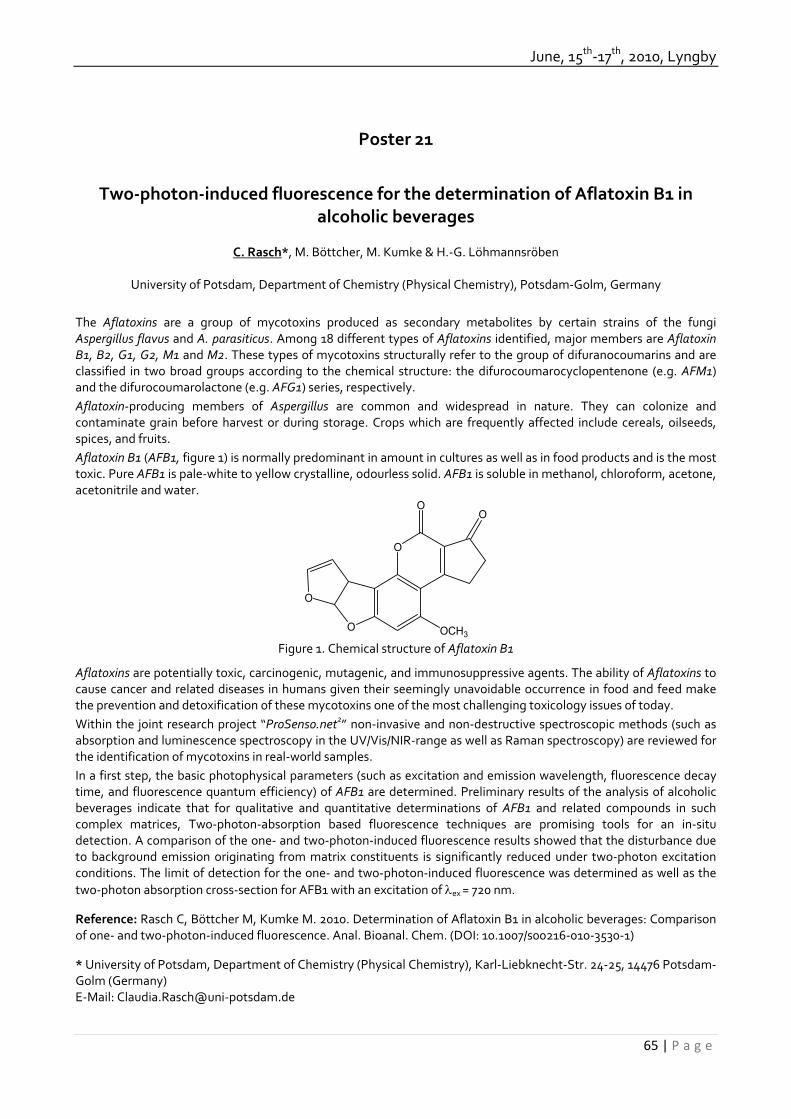
June, 15th‐17th, 2010, Lyngby
65 | P a g e
Poster 21
Two‐photon‐induced fluorescence for the determination of Aflatoxin B1 in alcoholic beverages
C. Rasch*, M. Böttcher, M. Kumke & H.‐G. Löhmannsröben
University of Potsdam, Department of Chemistry (Physical Chemistry), Potsdam‐Golm, Germany
The Aflatoxins are a group of mycotoxins produced as secondary metabolites by certain strains of the fungi Aspergillus flavus and A. parasiticus. Among 18 different types of Aflatoxins identified, major members are Aflatoxin B1, B2, G1, G2, M1 and M2. These types of mycotoxins structurally refer to the group of difuranocoumarins and are classified in two broad groups according to the chemical structure: the difurocoumarocyclopentenone (e.g. AFM1) and the difurocoumarolactone (e.g. AFG1) series, respectively. Aflatoxin‐producing members of Aspergillus are common and widespread in nature. They can colonize and contaminate grain before harvest or during storage. Crops which are frequently affected include cereals, oilseeds, spices, and fruits. Aflatoxin B1 (AFB1, figure 1) is normally predominant in amount in cultures as well as in food products and is the most toxic. Pure AFB1 is pale‐white to yellow crystalline, odourless solid. AFB1 is soluble in methanol, chloroform, acetone, acetonitrile and water.
O
OO
O
O
OCH3 Figure 1. Chemical structure of Aflatoxin B1
Aflatoxins are potentially toxic, carcinogenic, mutagenic, and immunosuppressive agents. The ability of Aflatoxins to cause cancer and related diseases in humans given their seemingly unavoidable occurrence in food and feed make the prevention and detoxification of these mycotoxins one of the most challenging toxicology issues of today. Within the joint research project “ProSenso.net2” non‐invasive and non‐destructive spectroscopic methods (such as absorption and luminescence spectroscopy in the UV/Vis/NIR‐range as well as Raman spectroscopy) are reviewed for the identification of mycotoxins in real‐world samples. In a first step, the basic photophysical parameters (such as excitation and emission wavelength, fluorescence decay time, and fluorescence quantum efficiency) of AFB1 are determined. Preliminary results of the analysis of alcoholic beverages indicate that for qualitative and quantitative determinations of AFB1 and related compounds in such complex matrices, Two‐photon‐absorption based fluorescence techniques are promising tools for an in‐situ detection. A comparison of the one‐ and two‐photon‐induced fluorescence results showed that the disturbance due to background emission originating from matrix constituents is significantly reduced under two‐photon excitation conditions. The limit of detection for the one‐ and two‐photon‐induced fluorescence was determined as well as the two‐photon absorption cross‐section for AFB1 with an excitation of λex = 720 nm. Reference: Rasch C, Böttcher M, Kumke M. 2010. Determination of Aflatoxin B1 in alcoholic beverages: Comparison of one‐ and two‐photon‐induced fluorescence. Anal. Bioanal. Chem. (DOI: 10.1007/s00216‐010‐3530‐1) * University of Potsdam, Department of Chemistry (Physical Chemistry), Karl‐Liebknecht‐Str. 24‐25, 14476 Potsdam‐Golm (Germany) E‐Mail: Claudia.Rasch@uni‐potsdam.de
32th Mycotoxin Workshop
66 | P a g e
Poster 22
ESI‐IT/MS peptaibol fingerprints for identification of toxic marine‐derived Trichoderma strains
Ruiz Nicolas1*, Robiou du Pont Thibaut1, Joubert Yolaine1, Poirier Laurence1, Sallenave‐Namont Claire1, Grovel
Olivier1, Bissett John2 & Pouchus Yves François1
1 MMS, University of Nantes, Faculty of Pharmacy, Nantes, France ; 2 Agriculture and Agri‐Food Canada, ECORC, Ottawa, Ontario, Canada
Peptaibols can be considered as the first mycotoxins observed in the marine environment (see communication of Pouchus et. al.). They correspond to a family of peptides produced principally by fungi belonging to Trichoderma/Hypocrea genera. They are characterized by short chains ranging from 11 to 20 amino‐acid residues, a N‐terminal acetylated residue, a C‐terminal amino‐alcohol residue and high levels of non‐proteinogenic amino acids such as ‐aminoisobutyric acid (Aib). Peptaibols are produced as complex microheterogeneous mixtures of isoforms due to their non‐ribosomal biosynthesis involving large multidomain enzymes, the Non‐Ribosomal Peptide Synthetase (NRPS). Considering complexity of the genus‐Trichoderma taxonomy, the aim of this study is to evaluate the relevance of the peptaibol‐production profiles as a chemotaxonomic tool, and their possible use for the identification of marine Trichoderma strains. Strains of Trichoderma isolated from shellfish farming areas were identified by the amplification and direct sequencing of the ITS region of the ribosomal DNA. They were then screened for their peptaibol‐production profile by ESI‐IT/MS allowing elaboration of peptaibol fingerprints. Characteristic profiles were established for each of the 3 sections of the genus Trichoderma observed in the marine environment (Pachybasium, Longibrachiatum and Trichoderma) and for each Trichoderma species observed (T. harzianum, T. pleuroticola, T. virens, T. longibrachiatum, T. reesei, T. citrinoviride, T. ghanense, T. sp. MA3642, T. atroviride). These profiles were then used for section and species identification of marine‐derived Trichoderma strains. Identifications were then compared to those obtained using molecular biology. Sections were always correctly identified and correct species identification was obtained for 90 % samples, indicating the relevance of peptaibol fingerprinting for Trichoderma chemotaxonomy. * Corresponding author: University of Nantes, MMS, Faculty of Pharmacy, BP 53508, F‐44035 NANTES‐CEDEX 1, France ‐ nicolas.ruiz@univ‐nantes.fr

June, 15th‐17th, 2010, Lyngby
67 | P a g e
Poster 23
Aflatoxin M1 in milk and milk products: Development of a new, highly sensitive ELISA test
Alois Schiessl1*, Michael Z. Zheng2 & Mao J. Hong2
1 Romer Labs Division Holding GmbH, Tulln, Austria. 2 Romer Labs Singapore Pte. Ltd., Singapore, Singapore.
Aflatoxins are toxic and carcinogenic secondary metabolites of Apergillus flavus and Aspergillus parasiticus. There are four major forms of aflatoxins: B1, B2, G1 and G2. Aflatoxin B1 is the most frequently encountered of the group and the most toxic. Aflatoxin B1, taken up by cattle with their feed, is metabolized and converted into a hydrolyzed form called Aflatoxin M1. It will be secreted in cow’s milk and can be found in milk and all products made from it. Processing of milk and dairy products do not lead to a significant degradation of aflatoxin M1 and therefore can occur in such products as milk powder and cheese. Young and developing individuals of any species are more susceptible to aflatoxin than adults, and as milk is part of baby and childrens food in many cultures, aflatoxin M1 becomes of concern. A direct competitive ELISA test kit was developed for the quantitative detection of aflatoxin M1 in milk and milk products. With a sensitive and a rapid kit version, concentrations from 25 ppt up to 2000 ppt aflatoxin M1 in milk can be determined. Total incubation times are set to a total of 80 minutes for the sensitive and 30 minutes for the rapid assay format. A cleanup after extraction is not needed. The procedure asks for 100 µl of the extract to be mixed with 200 µl of the antigen‐enzyme conjugate. Then 100 µl of this mix is transferred into a corresponding Antibody Coated Microwell. After a 60 respectively 20 minutes incubation the microwell is washed and once more incubated for 20 respectively 10 minutes with an enzyme substrate. Finally, the reaction is stopped and OD is read using a 450 nm filter with a reference wavelength of 630 nm. The OD is indirect proportional to the aflatoxin M1 concentration. For quantification, a linear calibration curve is created with a set of six aflatoxin M1 standards at different concentration levels. Validation studies on several aflatoxin M1 contamination levels in milk, milk powder and cheese were performed to guarantee the good performance of the presented test kit. * Alois Schiessl, Romer Labs Division Holding GmbH, Technopark 1, 3430 Tulln, Austria [email protected]
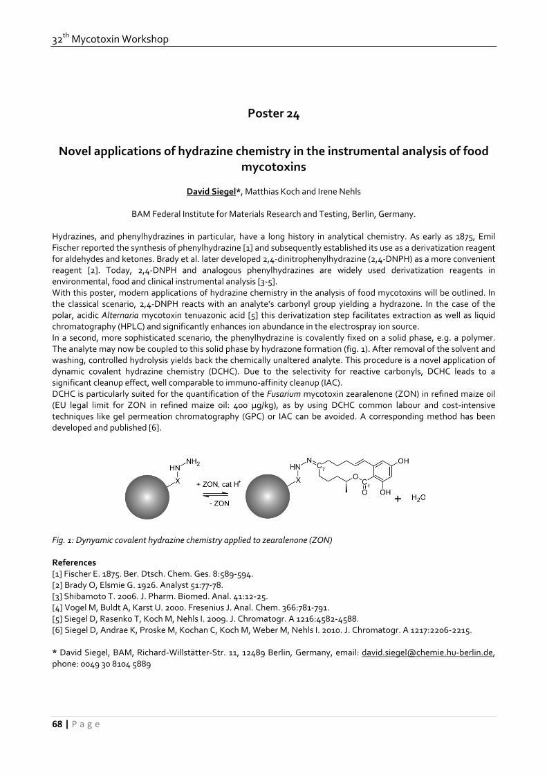
32th Mycotoxin Workshop
68 | P a g e
Poster 24
Novel applications of hydrazine chemistry in the instrumental analysis of food mycotoxins
David Siegel*, Matthias Koch and Irene Nehls
BAM Federal Institute for Materials Research and Testing, Berlin, Germany. Hydrazines, and phenylhydrazines in particular, have a long history in analytical chemistry. As early as 1875, Emil Fischer reported the synthesis of phenylhydrazine [1] and subsequently established its use as a derivatization reagent for aldehydes and ketones. Brady et al. later developed 2,4‐dinitrophenylhydrazine (2,4‐DNPH) as a more convenient reagent [2]. Today, 2,4‐DNPH and analogous phenylhydrazines are widely used derivatization reagents in environmental, food and clinical instrumental analysis [3‐5]. With this poster, modern applications of hydrazine chemistry in the analysis of food mycotoxins will be outlined. In the classical scenario, 2,4‐DNPH reacts with an analyte’s carbonyl group yielding a hydrazone. In the case of the polar, acidic Alternaria mycotoxin tenuazonic acid [5] this derivatization step facilitates extraction as well as liquid chromatography (HPLC) and significantly enhances ion abundance in the electrospray ion source. In a second, more sophisticated scenario, the phenylhydrazine is covalently fixed on a solid phase, e.g. a polymer. The analyte may now be coupled to this solid phase by hydrazone formation (fig. 1). After removal of the solvent and washing, controlled hydrolysis yields back the chemically unaltered analyte. This procedure is a novel application of dynamic covalent hydrazine chemistry (DCHC). Due to the selectivity for reactive carbonyls, DCHC leads to a significant cleanup effect, well comparable to immuno‐affinity cleanup (IAC). DCHC is particularly suited for the quantification of the Fusarium mycotoxin zearalenone (ZON) in refined maize oil (EU legal limit for ZON in refined maize oil: 400 μg/kg), as by using DCHC common labour and cost‐intensive techniques like gel permeation chromatography (GPC) or IAC can be avoided. A corresponding method has been developed and published [6]. Fig. 1: Dynyamic covalent hydrazine chemistry applied to zearalenone (ZON) References [1] Fischer E. 1875. Ber. Dtsch. Chem. Ges. 8:589‐594. [2] Brady O, Elsmie G. 1926. Analyst 51:77‐78. [3] Shibamoto T. 2006. J. Pharm. Biomed. Anal. 41:12‐25. [4] Vogel M, Buldt A, Karst U. 2000. Fresenius J. Anal. Chem. 366:781‐791. [5] Siegel D, Rasenko T, Koch M, Nehls I. 2009. J. Chromatogr. A 1216:4582‐4588. [6] Siegel D, Andrae K, Proske M, Kochan C, Koch M, Weber M, Nehls I. 2010. J. Chromatogr. A 1217:2206‐2215. * David Siegel, BAM, Richard‐Willstätter‐Str. 11, 12489 Berlin, Germany, email: [email protected]‐berlin.de, phone: 0049 30 8104 5889

June, 15th‐17th, 2010, Lyngby
69 | P a g e
Poster 25
Comparison of two different analytical methods for determination of ochratoxin‐A in wine
Biljana Stojanovska‐Dimzoska1, Zehra Hajrulai‐Musliu1, Elizabeta Dimitrieska‐Stojkovic1, Pavle Sekulovski1
1Food Institute, Faculty of Veterinary Medicine, University “St. Cyril and Methodius”, Skopje, R. Macedonia Abstract Ochratoxin A (OTA) is a mycotoxin, among the others which might contaminate agriculture commodities. It is nephrotoxin, a liver toxin, an immune suppressant, a potent teratogen and a carcinogen. OTA may also be involved in the etiology of Balkan endemic nephropathy. Wine is considered to be the second major source of OTA intake (13 %), and therefore is important to assure that it is free of harmful contaminants. It is a product significantly important for the European economy and population and therefore it requires develop of a reliable, highly sensitive and accurate method for OTA determination. Liquid chromatography‐fluorescence detector (LC‐FD) and fluorometry with previous immunoaffinity column clean‐up, are two methods that are compared in order to evaluate their performances for determination of ochratoxin A in wine. The use of immunoaffinity columns (IAC) for the clean‐up procedure is the most important analytical step and its use is highly recommended, allowing the isolation of the analyte from matrix interferences, due to its selectivity. The linearity of the methods was checked, and a good coefficient of correlation was found for bought methods (0.9965 for LC‐FD and 0.9814 for fluorometry). The limits of detection were 0.043 ng/ml and 0.199 ng/ml, respectively. The precision, estimated as RSD values were satisfactory, 8.61% and 4.54% for LC‐FD method in the concentration level of 0.1 ng/ml and 1.0 ng/ml, respectively. Fluorometry showed acceptable RSD value only for the concentration level of 0.1 ng/ml (6.47%), but too high for concentration level of 1.0 ng/ml (17.86%). Eighty six (86) wine samples, originated from wine producing regions in Macedonia, were analyzed parallel and the results showed differences (high readouts when fluorometry was applied). However, due to the factors that influence fluorometric analysis (especially red color), it was found that it does not present an accurate and selective quantitative method in the field of determination of OTA in wine. Fluorometry can be applicable only as a screening method for the prediction of ochratoxin A contamination, especially in the laboratories who are dealing with a big number of samples for mycotoxin analysis. Having in mind the good performances of LC‐FD method with immunoaffinity column clean‐up, it should be method of choice for the determination of ochratoxin A in wine. Keywords: ochratoxin A, immunoaffinity columns, LC‐FD, fluorometry, wine, method validation References:
1. Creppy E.E. (2002). Update of survey, regulation and toxic effects of mycotoxins in Europe. Toxicology Letters 127, 19‐28.
2. Opinion of the scientific panel on contaminants in the food chain on a request from the commission related to ochratoxin A in food. (2006). The EFSA Journal 365, 1‐56.
3. Sáez, J.M., Medina, A., Gimeno‐Adelantado, J.V. Mateo, R., and Jiménez, M. (2004). Comparison of different sample treatments for the analysis of ochratoxin A in must, wine and beer by liquid chromatography. Journal of Chromatography A, 1029, 125‐133.
4. Ratola N., Barros P., Simões T., Cerdeira A, Venâncio A., Aves A. (2006). Worldwide interlaboratory study on the determination of ochratoxin A in different wine type samples. Talanta 70, 720‐731.
5. Serra, R., Mendonca, C., Abrunhosa, L., Pietri, A., and Venâncio, A. (2004). Determination of ochratoxin A in wine grapes: comparison of extraction procedures and method validation. Analytica Chimica Acta 513, 41‐47.
6. Visconti A., Pascale M., Centonze G. (1999). Determination of ochratoxin A in wine by means of immunoaffinity column clean‐up and high‐performance liquid chromatography. Journal of Chromatography A 864, 89‐101.
Faculty of Veterinary Medicine, Lazar Pop Trajkov 5‐7, Skopje, R. Macedonia Corresponding author’s e‐mail: [email protected]

32th Mycotoxin Workshop
70 | P a g e
Poster 26
Abstract removed by request of the authors

June, 15th‐17th, 2010, Lyngby
71 | P a g e
Poster 27
Abstract removed by request of the authors

32th Mycotoxin Workshop
72 | P a g e
Poster 28
Glucuronide Conjugates of Deoxynivalenol and Zearalenone in Human Urine: A Novel LC‐MS/MS Approach
Benedikt Warth1*, Philipp Fruhmann2, Hannes Mikula2, Franz Berthiller1, Rainer Schuhmacher1,
John Gilbert1, Christian Hametner2, Michael Sulyok1, Johannes Fröhlich2 & Rudolf Krska1
1 Center for Analytical Chemistry, IFA Tulln, University of Natural Resources and Applied Life Sciences, Vienna,
Austria. 2Institute of Applied Synthetic Chemistry, Vienna University of Technology, Vienna, Austria.
Deoxynivalenol (DON) and zearalenone (ZON) are mycotoxins produced by several Fusarium species in temperate and warm climate regions. They occur frequently in commodities like cereals or maize and after processing along the whole food and feed chain. Therefore they are of major concern for food safety. In man and animals one of the main metabolism mechanisms of those toxins is conjugation with glucuronic acid. To estimate ones exposure to a mycotoxin in a biological fluid, it is crucial to consider not only the parent toxin itself but also all main metabolic products. Due to a lack of standards for DON and ZON glucuronides and sulfates, to date the exposure to humans or animals in urine is determined after enzymatic cleavage of the conjugates by β‐glucuronidase and sulfatase over night (Meky et al 2003, Jodlbauer et al 2000). This has the advantage of resulting in a sum parameter and the option of using immunoaffinity clean up after the enzymatic conversion to the parent toxin. The drawbacks are time‐consuming sample preparation, doubts about the efficiency of the enzymatic hydrolysis and the fact that these approaches can not determine levels and changes in individual conjugate patterns. We developed an LC‐MS/MS method for the simultaneous determination of DON, ZON and their glucuronide conjugates (DON‐3‐glucuronide, ZON‐4‐glucuronide) in human urine. Other metabolites included in the method are α‐ and β‐zearalenol, ZON‐4‐sulfate and the microbial conversion product deepoxy‐DON. Since urine contains various other glucuronide conjugates at high concentration levels, an appropriate separation of the two target conjugates from matrix constituents is hard to achieve. To overcome the severe matrix effects several clean up procedures, including solid phase extraction on reversed phase, mixed‐mode anion exchange and hydrophilic interaction materials were evaluated with spiked blank urine. In addition the feasibility of a “dilute and shoot” approach was tested. References: Jodlbauer J, Zöllner P, Lindner W. 2000. Determination of Zeranol, Taleranol, Zearalenone, α‐ and β‐ Zearalenol in Urine and Tissue by High‐Perfomance Liquid Chromatogrphy‐Tandem Mass Spectrometry. Chromatographia Vol. 51: 681‐687. Meky F A, Turner P C, Ashcroft A E, Miller J D, Qiao Y‐L, Roth M J, Wild C P. 2003. Development of a urinary biomarker of human exposure to deoxynivalenol. Food and Chem. Toxicology 41: 265‐273. * Benedikt Warth, Center for Analytical Chemistry, Department of Agrobiotechnology IFA Tulln (BOKU), Konrad Lorenz Straße 20, A‐3430 Tulln, Austria. [email protected]
June, 15th‐17th, 2010, Lyngby
73 | P a g e
Poster 29
An LC‐MS/MS method for analysis of very low stable isotope enrichments of [2H5]‐phenylalanine in physiological samples as a tool to investigate the
inhibitory effect of deoxynivalenol (DON) on the protein synthesis
K. Wilkerling*, H. Valenta, S. Dänicke, S. Döll
Institute of Animal Nutrition, Friedrich‐Loeffler‐Institut, Federal Research Institute for Animal Health, Braunschweig, Germany.
Introduction: The Fusarium mycotoxin deoxynivalenol (DON) is able to affect the performance and health of farm animals primarily by inhibition of the protein synthesis. Frequently stable isotope labeled amino acids are used as tracers to examine nutritive effects on protein synthesis by tracing the incorporation of e.g. [2H5]‐phenylalanine ([2H5]‐phe) into newly synthesized proteins. Predominantly GC‐MS methods exist for determination of the enrichment of [2H5]‐phe [1]. In the present investigation, an LC‐MS/MS method was developed, which overcomes some drawbacks of GC‐MS. This method will be used for determination of inhibitory effects of DON on the protein synthesis in farm animals. Material and methods: The LC‐MS/MS measurements were carried out on a 4000 Qtrap (Applied Biosystems) in positive APCI (atomic pressure chemical ionization) mode, coupled with an HPLC (1200 Series, Agilent). The separation was carried out on a phenyl‐hexyl column using 0.1% formic acid as eluent A and methanol as eluent B. Protein extracts of 26 liver samples from an experiment with one‐day‐old broiler chicks (Lohman Meat), fed ad libitum with DON contaminated feed (15 mg/kg) for 42 days [2], were isolated and measured by GC‐MS one year ago according to a method described at Dänicke et al. (2001). These samples were re‐analysed using the developed LC‐MS/MS method. Results: For the LC‐MS/MS method two mass transitions each for [2H5]‐phe (171.1/125.1 and 171.1/106.1) and phe (1661.1/91.1 and 166.1/93.1) were chosen for quantification and qualification. The method was calibrated with calibration standard solutions in the range of 0.01‐0.5 mole percent excess (MPE) for very low enrichments (= protein bound [2H5]‐phe). Linear regression analysis led to coefficient of determination (r2) greater than 0.995. Precision was determined with calibration‐standard solutions and liver samples, respectively, whereby results showed that the method leads to reproducible analytical results over at least one week (coefficient of variation 0.4‐7.1%). First measurements of liver samples showed that the presented method is suitable for measuring physiological samples, as the chromatogram of the liver samples exhibit no interfering peaks. The comparison of results for MPE in liver samples, which were measured with LC‐MS/MS as well as one year ago with GC‐MS showed that the LC‐MS/MS determination provided approximately 38% higher MPE for the same liver samples while the correlation amounted to 0.77. Reasons for these deviations might be the conversion and derivatisation associated biases inherent in the GC‐MS methods, which can be excluded for LC‐MS/MS method. Conclusion: In this study a sensitive and selective LC‐MS/MS method for the investigation of the inhibitory effect of DON on the protein synthesis was developed, which makes it possible to measure very low enrichments of [2H5]‐phe (0.01‐0.5 MPE) in proteins, beside high excess of unlabeled phe. Compared to GC‐MS, the main advantage of the LC‐MS/MS method is that there is no need to convert and derivatise the samples before measuring whereby confounding effects are avoided. References: [1] Dänicke S, Schippel K and Halle I. 2001. Landbauforschung Völkenrode Vol: 51. [2] Dänicke S, Pappritz J, Goyarts T, Xu B, Rautenschlein S: Effects of feeding a Fusarium toxin contaminated diet to infectious bursal disease virus‐infected broilers on the protein turnover of the bursa of Fabricius and spleen. Submitted *Institute of Animal Nutrition, Friedrich‐Loeffler‐Institut, Federal Research Institute for Animal Health, Bundesallee 50, 38116 Braunschweig, [email protected]

32th Mycotoxin Workshop
74 | P a g e
Poster 30
Post‐Column Derivatization Methods in the HPLC Analysis of Aflatoxins.
Henry Joshua*, Aura Industries, Inc., New York, NY, USA
The quantitative, sensitive and specific analysis of mycotoxins in general and of aflatoxins in particular is of great importance in keeping our food chain safe and within the regulatory requirements. The procedures generally followed consist of sampling the material to be tested (peanuts, corn, barley, nuts, spices, beer, etc.), homogenizing, extracting mycotoxins from the homogenate, purifying the extract with immuno‐affinity columns, and the quantitative determination of the mycotoxins in the purified extract by HPLC. The HPLC detector of choice for the detection of aflatoxins is the fluorescence detector due to its sensitivity and specificity. However, of the four naturally occurring aflatoxins, G2, G1, B2, and B1, only the G2 and B2 are naturally fluorescing compounds. Compounds G1 and B1 can be made into fluorescing compounds by derivatizations. The derivatizations are made in‐line after separation of the analytes on the column and before entry into the detector. Each of the three following derivatization methods add either water, bromine or iodine across the leftmost double bond on the B1 and G1 molecules as shown for the photochemical derivatization of B1 in Figure 1. The resultant chromatograms for all three methods are essentially identical and are illustrated for the photochemical hydration method in Figure 2.
1. Hydration. Water is added to the double bond photochemically by uv irradiation using a photochemical reactor “PHRED”.
2. Bromination. Electrochemically generated bromine adds across the double bond using a “Coring Cell”. 3. Iodination. A solution containing iodine is added to and mixed with the post column eluent using a second
pump. These methods, their requirements, advantages and limitations will be discussed in the body of this presentation. *Aura Industries, Inc., 545 8th Ave, Suite 5W, New York, NY 10018, USA, Tel.: 212‐290‐9190, Fax: 212‐290‐9191, E‐mail: Joshua@aura‐inc.com
2.0
1.5
1.0
0.5
1.0
1.5
0.0
0.5
15.0 5.0
5.0
10.0
10.0 0.0
20.0
25.0
25.0
15.0 20.0
min.
min.
mv
PHRED light off
PHRED light on
G1 B2 B1 G2
12.0
1
14.7
7
18.2
2 22.9
7
12.2
6
18.2
1 2.0
G2 B2 O
O
O
O O
OCH3
O
O
O
O
O
O
H
OCH3
O
O
O
O
O
O
H
OCH3
B1
B2a
hv/H2O
Figure 1. Photochemical derivatization of aflatoxin B1 to aflatoxin B2a.
Figure 2. Chromatograms of aflatoxins G1, G2, B1 and B2 pre and post derivatization with uv irradiation.

June, 15th‐17th, 2010, Lyngby
75 | P a g e
Poster 31
Simultaneous HPLC Determination of Aflatoxins, Zearalenone and Ochratoxin A Without Changing the Excitation Wavelength.
Henry Joshua* Aura Industries, Inc., New York, NY, USA
The initial publications for the simultaneous HPLC analysis of aflatoxins, zearalenone and ochratoxin A using photochemical derivatization required a change in the fluorescence excitation wavelength from 365 nm for aflatoxins to 315 nm for zearalonone and ochratoxin A. The emission wavelength >415 nm on the other hand was kept constant. The change of the excitation wavelength was accomplished using a programmable fluorescence detector or by the manual adjustment of the detector during each run. In the current analysis, we determined that the aflatoxins as well as zearalonone and ochratoxin A give excellent fluorescence response with a constant excitation wavelength of 315 nm for all analytes. This simplification allows the use of more economical non‐programmable fluorescence detectors and does not require the manual wavelength change during each run. Figure shows chromatograms using fluorescent detector with 315 nm excitation wavelength and >415 nm emission wavelength. In Figure 1a the uv light of the photochemical derivatization unit (“PHRED”) was off, while it was on in Figure 1b. Figure 1. a) Chromatogram of Aflatoxins G2, G1, B2, B1, Zearalenone and Ochratoxin A with uv light off. Aflatoxins G1 and B1 were not detected. b) Chromatograms of Aflatoxins G2, G1, B2, B1, Zearalenone and Ochratoxin A with uv light on. References: Joshua, H. 1999. The simultaneous HPLC determination of six aflatoxins, zearalenone and ochratoxin A using gradient elution, post‐column photochemical derivatization and fluorescence detection. presented at The 90th AOAC Meeting and Exposition. Orlando, Fl. *Aura Industries, Inc., 545 8th Ave, Suite 5W, New York, NY 10018, USA, Tel.: 212‐290‐9190, Fax: 212‐290‐9191, E‐mail: Joshua@aura‐inc.com
a.
b.

32th Mycotoxin Workshop
76 | P a g e
Poster 32
Fungi and their mycotoxins in maize and maize silage
Storm IMLD1*, Sørensen JL12, Rasmussen RR3, Thrane U1
1Cent. for Microbial Biotech., Dep. Systems Bio., Technical University of Denmark, DK‐2800 Kgs. Lyngby, Denmark.
2Presently: Dep. of Anim. Health and Biosc., Faculty of Agri. Sci., Aarhus University, DK‐8830 Tjele, Denmark. 3Dep. Food Chemistry, National Food Institute, Technical University of Denmark, DK‐2860 Søborg, Denmark.
Maize silage is contaminated with a wide variety of pre‐ and post‐harvest fungi, which may lead to undesired production of mycotoxins and other bioactive secondary metabolites (Storm et al., 2008). Problems with ill‐thrift, disease and death in Danish dairy herds have been suspected of being related to mycotoxins in maize silage. To asses the risks of fungal spoilage and mycotoxin contamination of maize silage a cooperative research project has been conducted, covering microbial, chemical and toxicological aspects of fungal contamination of maize silage from field to cow. The primary species of Fusarium infecting maize in Denmark pre‐harvest was F. avenaceum, which produces moniliformin and enniatins. Methods were developed and these compounds were detected in maize samples but in concentrations to low to be of concern for acute intoxication. Other fungi detected were Alternaria infectoria, A. tenuissima, and Phoma pomorum. A survey of the post‐harvest contaminants of maize silage over a whole storage season (Storm et al, in press) revealed Penicillium roqueforti/P. paneum, Zygomycetes (primarily Mucor spp.) and Aspergillus fumigatus to be the most common contaminants. Less common fungi were Byssochlamys nivea/Paecilomyces niveus, Geotrichum candidum and Monascus ruber. Significant differences in the numbers of filamentous fungi, yeasts, and lactic acid bacteria were detected over a whole season with the highest levels 5‐7 months after ensiling. A multi‐mycotoxin method for the detection of 27 mycotoxins and other fungal secondary metabolites was developed and validated for maize silage (Rasmussen et al. 2010). A survey of 99 maize silage samples detected 13 of these analytes and 31% of samples contained more than one analyte. Fusarium toxins were most frequently detected but none of the regulated toxins found were above EC commission guideline values for cattle feed. The occurrence of mycotoxins from Alternaria, Phoma, Penicillium, Aspergillus, Monascus and Byssochlamys suggest that they are most likely not the cause of general problems observed in dairy cattle farms. References: Storm IMLD, Sørensen JL, Rasmussen RR, Nielsen KF, Thrane U. 2008. Mycotoxins in silage. Stewart Posthar Rev 4. doi:10.2212/spr.2008.6.4
Storm IMLD, Kristensen NB, Raun BML, Smedsgaard J, Thrane U. In press. Dynamics in the microbiology of maize silage during whole‐season storage. Journal of Applied Microbiology. doi: 10.1111/j.1365‐2672.2010.04729.x
Rasmussen RR, Storm IMLD, Rasmussen PH, Smedsgaard J, Nielsen KF. 2010. Multi‐mycotoxin analysis of maize silage by LC‐MS/MS. Analytical and Bioanalytical Chemistry. doi: 10.1007/s00216‐010‐3545‐7
Ida ML Drejer Storm Center for Microbial Biotechnology, Department of Systems Biology Technical University of Denmark Søltofts Plads, building 221 DK‐2800 Kgs. Lyngby Denmark e‐mail: [email protected]

June, 15th‐17th, 2010, Lyngby
77 | P a g e
Poster 33
Comparative Immunochemical Assessment of Alternariol in Food using Monoclonal and Polyclonal Antibodies
Yvonne Ackermann1*, Valeriu Curtui1,2, Richard Dietrich3, Madeleine Groß1, Hadri Latif1,4, Erwin Märtlbauer3 & Ewald
Usleber1
1Institute of Veterinary Food Science, Chair of Dairy Science, Veterinary Faculty, Justus‐Liebig‐University, 35390
Giessen, Germany. 2European Food Safety Authority, Largo N. Palli 5 /A, 43100 Parma, Italy. 3Chair of Hygiene and Technology of Milk, Veterinary Faculty, Ludwig‐Maximilians‐University of Munich, 85764 Oberschleissheim,
Germany. 4Department of Animal Diseases and Veterinary Public Health, Faculty of Veterinary Medicine, Bogor Agriculture University, 16680 Bogor, Indonesia.
The Alternaria toxin alternariol (AOH) has been found in cereals, fruits, vegetables, and products thereof. However, the amount of data concerning the occurrence of AOH (and other alternaria toxins) in foods and feeds is relatively small. This may be due to the fact that cheap and simple analytical screening methods for AOH are not available. Here we report the application of two competitive indirect enzyme immunoassay (EIA) systems, utilizing either monoclonal (mAb) or polyclonal antibodies (pAb), for the determination of AOH in various matrices. Both tests are highly sensitive for AOH, with mean detection limits of the AOH standard curves of 44±7.6 pg/ml (mAb EIA) and 59±16 pg/ml (pAb EIA). Both EIAs are specific for AOH, showing cross‐reactivities lower than 0.5% with other Alternaria toxins, namely altenuene, alternariol monomethylether, and tenuazonic acid. Samples of apple juice (n=44), applesauce (n=10), ketchup (n=18), tomato paste (n=10), tomato juice (n=16), and white wine (n=11) were analyzed in both EIAs. Sample materials were either analysed directly after dilution with phosphate buffered saline (apple juice, wine), or purified by liquid‐liquid‐extraction from diluted aqueous samples into ethyl acetate, which was then dried and the residue redissolved with buffer solution. Detection limit for AOH in tomato paste was 2 µg/kg, in all other matrices a detection limit of about 1 µg/kg was achieved. Mean recoveries of AOH at spiking levels of 1–10 µg/kg were 43‐78% in the mAb‐EIA, and 60–98% in the pAb‐EIA, respectively. A high percentage (79% in mAb‐EIA, 50% in pAb‐EIA) of all samples was positive for AOH, although toxin concentrations were typically low, at levels of 1–3 µg/kg. Only tomato paste had a slightly higher mean AOH value of 6.7 µg/kg, the maximum value was 13 µg/kg. In general, the agreement between both immunoassays was acceptable. Both EIAs show similar sensitivity and specificity, and have therefore the potential to enhance mycotoxin screening strategies. At present they are used to search various commodities for potential AOH contamination. We thank the Brigitte und Wolfram Gedek‐Stiftung (Ismaning, Germany) for financial support. *Institute of Veterinary Food Science, Chair of Dairy Science, Justus Liebig University, Ludwigstr. 21, 35390 Giessen, Germany. [email protected]‐giessen.de

32th Mycotoxin Workshop
78 | P a g e
Poster 34
Effect of calcium propionate, aw and incubation time on mould incidence and aflatoxins production in commercial poultry feed starter ration
Sahib Alam1, 2*, Hamid U Shah1 and Naresh Magan2
1 Department of Agricultural Chemistry, NWFP Agricultural University Peshawar, Pakistan 2 Institute of BioScience and Technology, Cranfield University, UK
Aflatoxins are a family of highly toxic fungal metabolites that cause developmental and immune system suppression, cancer, and death in both human and livestock. Poultry feed starter ration samples added with 0 (control) and 0.5% Calcium Propionate (preserved) at water activity levels of 0.85, 0.90 and 0.95 aw
were investigated for total fungal viable counts and aflatoxins B1 (AFB1), B2 (AFB2), G1 (AFG1) and G2 (AFG2) contents at 7 days intervals for 28 days. All the factors alone and in combination significantly (P≤0.05) affected both the total fungal viable counts and aflatoxins production. After 28 days of incubation, total fungal viable counts in control samples were examined to be 2.43 x 10⁸, 50 x 108, 29.9 x 10⁸ CFUs/g whereas that in preserved feed as 18.33 x 10⁷, 75.33 x 10⁷ and 84.67 x 10⁷ CFUs/g at 0.85, 0.90 and 0.95aw, respectively. Regarding aflatoxins production, minimum average AFB1 (45.66ng/g), AFB2 (0.22 ng/g), AFG1 (9.36 ng/g) and AFG2 (0.07 ng/g) content were produced in calcium propionate added feed at 0.85 aw after 28 days of incubation. While, maximum average AFB1 (91.14 ng/g), AFB2 (2.13 ng/g), AFG1 (58.29 ng/g) and AFG2 (2.70 ng/g) content were produced in control samples at 0.95 aw after an incubation period of 28 days. It was concluded that preservative addition along with aw amelioration are effective tools for controlling molds incidence and aflatoxins contamination in poultry feed. * Corresponding author's address and e‐mail. Department of Agricultural Chemistry, NWFP Agricultural University Peshawar, Pakistan Email: [email protected] Tel (Off): +92 91 9216903 Cell: +92 333 9386835
June, 15th‐17th, 2010, Lyngby
79 | P a g e
Poster 35
Interaction field trials between mycotoxin producers F. graminearum and F. poae strains involved in Fusarium Head Blight of durum wheat
B. Amato1, A. Prodi1*, K. Döll2 , P. Karlovsky2 , P. Nipoti1, A. Pisi1
1 Department of Agroenvironmental Science and Technology, Alma Mater Studiorum University of Bologna, viale Fanin 40, 40127 Bologna, Italy. 2 Department of Crop Science, Molecular Phytophatology and Mycotoxin Research
unit, Georg‐August‐Universität of Göttingen, Grisebachstraße 6, 37077 Göttingen, Germany.
Fusarium Head Blight (FHB) is an important cereals disease caused by several fungal species belonging mainly to the Fusarium genus. In Italy the most frequent species of Fusarium associated to the disease in wheat are F. graminearum, F. culmorum and recently F. poae. These species are responsible of yield losses and decreasing grain quality characteristics, including the accumulation of mycotoxins, that are capable of harming the health of humans and animals. The most widespread F. graminearum mycotoxins are trichothecenes, in particular deoxynivalenol (DON) and its acetylated forms (3‐ADON and 15‐ADON) and nivalenol (NIV). F. poae is able to produce a wide range of mycotoxins, in particular T‐2 and its derivative HT‐2. The T‐2 toxin is one of the most dangerous among all the trichothecene produced by Fusarium spp. The aim of this work is to investigate on the “in vivo” interaction between F. graminearum and F. poae, which may contribute to the dynamics populations of these species in the field. F. graminearum and F. poae strains producing different mycotoxins were tested for interaction field trials for two years (2008‐2009). The strains were single and mixed inoculated on a durum wheat cultivar sensitive to the FHB and the incidence and severity of the disease were assessed. The flour obtained from the grain was analysed by Real‐Time PCR to assess the biomass of F. graminearum and F. poae and by HPLC‐MS/MS to determine the total amount of mycotoxins. F. poae strains, both producers and non producers of the mycotoxins T‐2 HT‐2 influence in a variable way the parameters estimated of FHB and the DNA quantity of F. graminearum recovered in the flour. The variability recordered seems to be influenced by the environmental conditions. The attention of the scientific community on F. poae in the disease is increasing, due to its capacity to produce dangerous mycotoxins; in order to control Fusarium Head Blight, we need to understand its role in the disease, including interactions with other pathogens that colonize durum wheat. * Antonio Prodi Viale Fanin 40 40127 Bologna, Italy E‐mail: [email protected]

32th Mycotoxin Workshop
80 | P a g e
Poster 36
Occurrence of type A, B, and D trichothecenes in barley from the Bavarian market
Jörg Barthel1*, Martin Rapp1, Matthias Berger1, Christoph Gottschalk2,
Johann Bauer3, Karsten Meyer3
1 Bavarian Health and Food Safety Authority, Veterinärstraße 2, 85764 Oberschleißheim, Germany. 2Federal Institute for Risk Assessment, National Reference Laboratory for Mycotoxins, Thielallee 88‐92, 14195 Berlin, Germany.
3Technische Universität München, Institute of Animal Hygiene, Weihenstephaner Berg 3, 85354 Freising, Germany. Type A, B, and D trichothecenes are known as cytotoxic and immunosuppressive mycotoxins produced by various species of Fusarium, Myrothecium or Stachybotrys. Among cereals oats, maize, and wheat are contaminated with type A trichothecenes like T‐2 and HT‐2 toxin and type B trichothecenes. The contamination of barley with type B trichothecenes like deoxynivalenol (DON) is well known, but only few data exist concerning a contamination of barley with T‐2/HT‐2 and other type A trichothecenes. Representative samples of barley (n=59) have been drawn according to Commission Regulation (EC) No 401/2006 at mills, malt houses, wholesale, and at retail stage in Bavaria between February and September 2009. The samples were analyzed for 18 type A, B, and D trichothecenes by a sensitive LC‐MS/MS method after a clean‐up‐procedure with MycoSep®‐226‐columns. For all samples, the mean levels of T‐2 and HT‐2 were 3.0 µg/kg and 6.8 µg/kg with rates of contamination of 63 % and 71 %, respectively. The maximum values were 41 µg/kg for T‐2 and 47 µg/kg for HT‐2. For T‐2 tetraol a mean level of 9.2 µg/kg and a maximum level of 51 µg/kg with a rate of contamination of 71 % were determined. As expected, the contamination of DON was high (95 % positive samples) with a low mean level of 23 µg/kg in the range of 3.4 to 420 µg/kg. At this, winter barley and malting barley were the most contaminated groups of barley. Type D trichothecenes were not detected in any sample. The results for the contamination of barley from the Bavarian market will be presented and discussed with respect to possible health risk for the consumer. References: Commission Regulation (EC) No 401/2006 (23 February 2006) laying down the methods of sampling and analysis for the official control of the levels of mycotoxins in foodstuffs * Corresponding author: Jörg Barthel Bavarian Health and Food Safety Authority Veterinärstraße 2 85764 Oberschleißheim, Germany [email protected]

June, 15th‐17th, 2010, Lyngby
81 | P a g e
Poster 37
Fusarium toxins and fungi in oats and its products from ecological and conventional agriculture practice
Anna Blajet‐Kosicka *, Jan Grajewski, Magdalena Twaruzek, Robert Kosicki
Kazimierz Wielki University, Institute of Experimental Biology, Division of Physiology and Toxicology, Bydgoszcz,
Poland Oats (Avena sativa) cultivation is of great significance for Polish farming not only due to its high nutritious values but also because of its importance in crops changing. Oats is the only cereal which is not infected by the ear base diseases and is not present in the food chain of the fungi and therefore does not pass them on other plants. Nonetheless, the specific structure of the kernel seems favourable to fungi growth. According to the EU regulations it is necessary to carry out research on T‐2 and HT‐2 toxins presence in cereal and in oats and its by‐products in particular. The aim of the study was determination of trichothecenes and zearalenone, as well as moulds and yeast in the oats grains and their products, coming from ecological and conventional agriculture practice. The results presented are a part of the project studying the influence of the mycotoxins contamination of cereal products on public health. The analyzed material included 31 samples (18 – conventional and 13 – ecological) of the grains, flakes, bran, flour and groats. Mycotoxins (T‐2, HT‐2, deoxynivalenol – DON, nivalenol – NIV, 3‐acetyldeoxynivalenol – 3‐ADON, monoacetoxyscirpenol – MAS, diacetoxyscirpenol – DAS, and zearalenone – ZEA) were determined using the HPLC‐MS/MS method (3200 QTRAP). During sample preparation procedures Bond Elut® Mycotoxin columns (Varian) were used. The following internal standards were applied: 13C‐DON, 13C‐T‐2, 13C‐HT‐2, ZAN. The analysis was performed in triplicate. The moulds and yeast were determined according to PN ISO 7954: 1999 – with own alterations (inoculation on YGC medium, chloramphenicol – 100ppm, incubation for 5‐7 days in 25°C). The highest concentration among all the toxins was evaluated for NIV, which was present only in grains samples ‐ both conventional and ecological, at maximum levels of 444 ± 12 ppb and 247 ± 12 ppb, respectively. The most commonly detected toxins were T‐2 (65% of positive samples, at maximum level of 44,7 ± 11,8 ppb – conventional grains) and HT‐2 (84% of positive samples, at maximum level of 98,0 ± 12,8 ppb – conventional grains). DON was not found in any of the examined food samples and its highest concentration in conventional grains was 45,6 ± 9,7 ppb. Similar situation was noticed in case of MAS and DAS, which were not detected in oats products. However, the concentrations of these toxins were close to the quantification limit, mainly in conventional grains (LOQ = 3ppb – in both cases). The only product which contained ZEA was oats groats (c = 0,67 ± 0,12 ppb), whereas in the grains the concentration of this toxin was rather low (~1,5 ppb) – but for one sample of conventional grains (c = 43,2 ± 11,0 ppb). 3‐ADON was not detected in any of the examined samples (LOD = 5ppb). The mean values of the total number of fungi in ecological samples were: 4,1*104 cfu/g (grains) and 1,1*104 cfu/g (products), and the domineering moulds turned out to be: Cladosporium and Aspergillus, respectively. In conventional products dominated Penicillium, while in conventional grains ‐ Cladosporium, and the mean total number of fungi was: 5,0*102 cfu /g i 5,6*105 cfu /g, respectively. The total number of fungi in ecological products was over twenty times higher than in conventional products, and in ecological grains this value was ten times lower when compared to the conventional samples. In the samples coming from conventional farming practice the mean concentrations of all the detected toxins were higher when compared to the ecological material. Higher mycotoxins contamination of the grains may mean that some impurities are removed during the processing procedures used in food production. Financial support: Ministry of Science and Higher Education Project no. NN 305 122136. *Corresponding author: Chodkiewicza Street 30, 85064 Bydgoszcz, Poland; [email protected]

32th Mycotoxin Workshop
82 | P a g e
Poster 38
Hidden fumonisins: a study of the masking mechanism in raw maize. Claudia Falavigna, Andrea Dall’Erta, Chiara Dall’Asta*, Gianni Galaverna, Arnaldo Dossena, Rosangela Marchelli.
Department of Organic and Industrial Chemistry, University of Parma, V.le G. P. Usberti 17/a, I‐43124 Parma, Italy.
Hidden fumonisins are fumonisins that easily escape conventional techniques of analysis since they are covalently or not‐covalently linked with various food macroconstituents (Galaverna et al. 2009). Such compounds are detectable only through the application of a hydrolysis step and could be released from food matrix during technological treatments or digestion, thus exerting a toxic effect. In a recent work from our group an in vitro digestion model (Versantvoort et al. 2005) has been applied to raw maize and maize‐based products in order to evaluate the possible release of masked fumonisins under gastrointestinal conditions (Dall’Asta et al. 2009). Comparing the data with those obtained by routine analyses, a significative increase of fumonisin levels after digestion has been observed. This means that enzymes present in the gastrointestinal tract are able to destroy the matrix‐fumonisins interaction, thus releasing the masked forms. In this study, the model previously utilized has been modified in several experiments, in order to study the masking phenomenon. In particular, the role of each enzyme and its contribution to the release of hidden fumonisins has been investigated, for understanding the fraction mainly responsible for the masking of fumonisins in raw maize. Moreover, recovery experiments from blank raw maize, corn starch and corn zeins spiked with a fumonisin B1 standard solution have been performed using both the routine extraction method and the digestion assay to evaluate the strength of fumonisin‐macroconstituent interaction. The experiments performed using the digestion model allowed the study of masking phenomenon, making clearly which are the raw maize macroconstituents mainly involved in the interaction and which is the nature of such bond. References: Galaverna G., Dall’Asta C., Mangia M., Dossena A.,Marchelli R. 2009. Masked Mycotoxins: an Emerging Issue for Food Safety. Czech J. Food Sci. 27, S90‐S92. Dall’Asta C., Mangia M., Berthiller F., Molinelli A., et al. 2009. Difficulties in fumonisin determination: the issue of hidden fumonisins. Anal. and Bioanal. Chem. 395, 1335–1345. Versantvoort C.H.M., Oomen, A.G., Van de Kamp, E., Rompelberg, C.J.M., Sips, A.J.A.M. 2005. Applicability of an in vitro digestion model in assessing the bioaccessibility of mycotoxins from food. Food Chem. Toxicol. 43, 31–40. * Chiara Dall’Asta, Department of Organic and Industrial Chemistry, University of Parma, V.le G. P. Usberti 17/a, I‐43124 Parma, Italy, [email protected]

June, 15th‐17th, 2010, Lyngby
83 | P a g e
Poster 39
Monitoring of ochratoxin A content of must during fermentation
Csaba Csutorás1*, Péter Fűtő1, Péter Forgó1, László Rácz1 and Zsolt Tajcs2
1 Eszterházy Károly College , Eger, Hungary.
2Eger Crown Winehouse LtD., Kerecsend, Hungary. Ochratoxin A (OTA) is the most abundant and the most toxic among ochratoxins. The International Agency for Research on Cancer has classified ochratoxin A as a possible human carcinogen (category 2B). The occurrence of OTA in wine samples has been reported in various studies predominantly dealing with European wines but also with wines of other regions. Generally red wines seem to contain a higher amount of OTA than white or rosé wines, and some results suggest, that at least for European and North African cultivation areas, southern regions are more affected by the contamination problem. The aim of the present work was to investigate the change of OTA content during must fermentation processes, using artificial contamination of must samples with OTA of higher than 98% purity. Different concentrations of OTA combined with different sort of wines (white, rosé, red) were applied. Our experiments were carried out in laboratorial as well as in industrial scale, and demonstrated the level of OTA content in must and wine during fermentation. Sample collections and preparations were carried out systematically, the prepared samples were analyzed by HPLC‐MS and by HPLC. This work was supported by a GOP‐1.1.1 grant of the Hungarian National Development Agency. References: Etzel RA. 2002. Mycotoxins. J. Am. Med. Assoc. 287‐425. Zöllner P, Mayer‐Helm B. 2006. Trace mycotoxin analysis in complex biological and food matrices by liquid chromatography–atmospheric pressure ionisation mass spectrometry . J. Chrom. A 1136‐123. * H‐3300 Eger, Eszterhazy Sqr. 1., Hungary, [email protected]

32th Mycotoxin Workshop
84 | P a g e
Poster 40
Seasonal variations in adult serum ochratoxın a and aflatoxin levels ın Central Anatolıa region, Turkey
Belma Giray, Gönül Şahin, Sevtap Aydın, Suna Sabuncuoğlu, Pınar Erkekoğlu*
Hacettepe University Faculty of Pharmacy Department of Toxicology, Sihhiye Ankara Turkey
Aflatoxins (AFs) are naturally occurring mycotoxins that are produced by many species of Aspergillus, a common and widespread fungus, most notably by Aspergillus flavus and Aspergillus parasiticus. AFs are among the most toxic and carcinogenic substances known. This study has been undertaken to investigate the seasonal differences in serum OTA and AF (AFB1, ABG1, AFB2, AFG2) levels in healthy adult subjects living in Central Anatolia region of Turkey. A total of 120 samples were collected in winter and summer of 2007. Serum OTA concentrations were measured by ELISA and serum aflatoxin concentrations were determined by HPLC. The OTA levels were found to be 0.314 ± 0.038 ng/ml in summer and 0.499 ± 0.035 ng/ml in winter. The difference among the seasons is statistically significant. The AFB1 levels were found to be 27.08±1.99, AFG1 as 58.73±5.87, AFB2 as 20.06±1.35, and AFG2 as 23.69±1.85 ng/ml in summer. The winter AFB1 levels was found to be 21.89±2.10, AFG1 as 58.90±5.66, AFB2 as 14.53±1.39 and AFG2 as 20.98±2.01 ng/ml. These results suggest that AFB2 levels are significantly higher in summer than in winter while AFB1 and AFG1 are higher in summer samples insignificantly. The high OTA levels in winter samples might be due to high consumption of bread and leguminosae in this part of Turkey especially in cold weather. On the other hand, high AF levels in summer serum samples may be due to some environmental conditions such as high ambient temperature and humidity and unscientific storage practices, which favor high production of mycotoxins. As exposure to AF exposure is highly hepatocarcinogenic and OTA exposure is a cause of renal acute and chronic toxicity as well as renal carcinomas, surveillance in each step of processed and unprocessed food must be continuous, widespread and must be conducted by both local authorities and government. It is a fact that mycotoxin control programs will result in economic gains as well as health improvement in the region and scientific research is being conducted to enable the reduction of mycotoxin contamination throughout Turkey.
*Hacettepe University, Faculty of Pharmacy, Department of Toxicology, Sihhiye, Ankara Turkey e‐mail: [email protected]

June, 15th‐17th, 2010, Lyngby
85 | P a g e
Poster 41
Ochratoxin A, aflatoxin B1 and aflatoxin M1 levels of breast milk samples obtained from Ankara, Turkey.
Aylin Gürbay1*, Gözde Girgin1, Suna Sabuncuoğlu1, Gönül Şahin1, Şule Yiğit2, Murat Yurdakök2
& Gülsevin Tekinalp2
1 Toxicology Department, Faculty of Pharmacy, Hacettepe University, Ankara, Turkey. 2 Neonatology Section,
Paediatrics Department, Faculty of Medicine, Hacettepe University, Ankara, Turkey. Breast feeding is very important in the first year of life. Since it is a major nutrient for infants, feeding infants with safe milk is essential. Breast milk may be contaminated with many residues of xenobiotics and naturally occuring toxins such as mycotoxins. Ochratoxin A (OTA), aflatoxin (AF) B1 and AFM1 are some of the important mycotoxins that may have roles in different types of diseases observed in both adults and neonates. OTA is a mycotoxin produced by a number of Aspergillus and Penicillium species. It can be found in cereals, oleaginous seeds, green coffee, pulses and poultry meat. OTA may exert nephrotoxic, carcinogenic, immunosuppressive and teratogenic effects and it is also suspected of being the main etiological agent responsible for Balkan endemic nephropathy. OTA was classified by the International Agency for Research on Cancer (IARC) as a possible carcinogen to humans (Group 2B). Aflatoxins are produced by moulds such as Aspergillus flavus and Aspergillus parasitucus. They are common and unavoidable contaminants of various types of foods. Aflatoxins are among the major etiological factors in the hepatocellular carcinoma development. AFB1 is the most prevalent and the most potent form of aflatoxins. In humans, AFB1 can be hydroxylated by cytochrome P450 1A2 to AFM1 metabolite and this metabolite can be excreted into milk. AFs are classified as carcinogenic to humans (Group 1) by IARC. Therefore, the objective of this study was to determine these three mycotoxin levels in breast milk samples of 46 mothers, whose babies were in‐patients in the Department of Pediatrics, Section of Neonatology, Hacettepe University, Faculty of Medicine, in Ankara, Turkey. The volunteers (aged between 20 and 37) have filled standart questionnaire forms including educational and social information. Breast milk samples were kept at ‐20°C until analysis. Following an extraction procedure, mycotoxin levels were detected by high performance liquid chromatography (HPLC). OTA levels in breast milk samples were found to be between 575,01 and 5065,14 ng/L while AFB1 and AFM1 levels were observed to range between 101,05–460,06 ng/L and 60,15–247,27 ng/L, respectively. In conclusion, detecting mycotoxin levels in breast milk may provide important information about the degree of mother’s and baby’s exposure. This information is also used in estimation of the overall risk characterisation. This study was supported by Hacettepe University Research Foundation (0401301003). References: Chang HL, DeVries JW. 1983. Rapid high pressure liquid chromatographic determination of aflatoxin M1 in milk and nonfat dry milk. J. Assoc. Off. Anal. Chem. 66: 913‐917. Gürbay A, Girgin G, Atasayar Sabuncuoglu S, Sahin G, Yurdakök M, Yigit S, Tekinalp G. 2009. Ochratoxin A: is it present in breast milk samples obtained from mothers from Ankara, Turkey? J. Appl. Toxicol. Doi: 10.1002/jat.1499. Gürbay A, Atasayar Sabuncuoglu S, Girgin G, Sahin G, Yigit S, Yurdakök M, Tekinalp G. 2010. Exposure of newborns to Aflatoxin M1 and B1 from mothers’ breast milk in Ankara, Turkey. Food Chem. Toxicol. 48: 314‐319. Jonsyn FE, Maxwell SM, Hendrickse RG. 1995. Ochratoxin A and aflatoxins in breast milk samples from Sierra Leone. Mycopathologia. 131: 121‐126. * Corresponding author: Aylin Gürbay, Ph.D. Hacettepe University, Faculty of Pharmacy, Toxicology Department 06100 Sihhiye Ankara/TURKEY e‐mail: [email protected]

32th Mycotoxin Workshop
86 | P a g e
Poster 42
Mycotoxins from marine‐derived fungi: use of LC‐HRMS/MS for identification of new communesins analogs in Penicillium expansum
Olivier Grovel1*, Isabelle Kerzaon1, Fabrice Monteau2, Bruno Le Bizec2 & Yves François Pouchus1
1 MMS, University of Nantes, Faculty of Pharmacy, Nantes, France. 2 LABERCA, National Veterinary School, Nantes,
France. Since the observation in the 90’s of unexplained neurotoxic phenomena linked to shellfish contamination in France, the hypothesis of a mycotoxinogenic role for marine‐derived fungi was investigated. In this way, a marine‐derived Penicillium expansum strain was studied on various culture media for its production of compounds exhibiting neurotoxicity on a blowfly larvae assay. Using bio‐guided fractionation, communesin B, an indole alkaloid described for cytotoxicity and acute toxicity on brine shrimps larvae, was identified by 1H and 13C‐NMR as the main active compound of the neurotoxic extract. As other neurotoxic compounds were present in some fractions of the culture extracts, other communesin‐related compounds were searched by LC‐ESI‐MS/MS and LC‐HRMS/MS dereplication using a LCQ ion trap mass spectrometer and an Orbitrap mass spectrometer respectively. The four known communesins A, D, E and F together with their putative biosynthetic precursor, the ergot alkaloid aurantioclavine, were identified by their retention time index (RI, according to (1)), their accurate mass and the observation of common characteristic fragmentation patterns. An exhaustive investigation of their high‐resolution MS/MS spectra allowed the development of a predictive model for identification of communesins and structural elucidation of their variable substituents. Using this model, ten new dimethyl‐epoxy and dimethyl‐vinyl communesins derivatives were identified among the minor molecules of the crude extracts and structural hypotheses were proposed (2). Investigations of biosynthetic pathways and structure‐activity relationships were performed correlating their amounts produced and with the neurotoxicity of the culture extracts. The use of LC‐HRMS/MS appears to be a powerful tool for the identification and first‐step structural elucidation of new analogs of bioactive fungal metabolites belonging to a chemical series, in a single analysis of complex matrices. Furthermore the detection of ten new communesins in a marine‐derived strain belonging to a widely studied terrestrial species exemplifies the chemical diversity of marine‐derived fungi. References: 1: Nielsen KF, Smedsgaard J. 2003. Fungal metabolite screening: database of 474 mycotoxins and fungal metabolites for dereplication by standardised liquid chromatography‐UV‐mass spectrometry methodology. J. Chromatogr. A. 1002 (1‐2): 111‐136. 2: Kerzaon I, Pouchus YF, Monteau F, Le Bizec B, Nourrisson MR, Biard JF, Grovel O, 2009. Structural investigation and elucidation of new communesins from a marine‐derived Penicillium expansum Link by liquid chromatography/electrospray ionisation ion trap tandem mass spectrometry. Rapid Commun. Mass Spectrom., 23, 3928–3938. * Corresponding author: Olivier Grovel, MMS, University of Nantes, Faculty of Pharmacy, BP 53508, F‐44035 NANTES‐CEDEX 1, FRANCE – olivier.grovel@univ‐nantes.fr

June, 15th‐17th, 2010, Lyngby
87 | P a g e
Poster 43
Mycotoxins in pet food could reduced fertility and stillbirth and induced genotoxic damages
Kheira Hadjeba‐Medjdoub1, Mariana Tozlovanu‐Fergane1, Ivana Polisenska2 & Annie Pfohl‐Leszkowicz1*
1Department Bioprocess & Microbial System, Laboratory Chemical Engineering, UMR CNRS/INPT/UPS 5503,
Toulouse, France. 2 Agrotest Fyto, Ltd., Havlickova 2787/121, 76701 Kromeriz, Czech Republic. As cereal grain and nuts are often used as ingredients in industrial pet food, companion animals such cat and dog are exposed to mycotoxins. Some owners and breeder of cats and dogs observed a decrease of fertility and stillbirth. As all other causes (virus…) have been ruled out, it has been hypothesised that feed contaminants should be involved especially when replacement of this feed by another without cereals avoid the problems. The aim of this study was to analyse the occurrence of mycotoxins in feed and in tissues (blood of mother, liver, kidney, intestine, brain of freak). We analysed the pet food given to the mothers who gave birth to monstrous cats: intestines were outside the abdomen, face are malformed, and some were without fur, wasted legs… The feed without cereals was also analysed. Ochratoxin A (OTA), Aflatoxins (AF), fumonisins (FB), zearalenone (ZEA), citrinin (CIT) and deoxynivalenol (DON) have been analysed after purification, by specific HPLC method with fluorimetric detection. All the pet food leading to reproductive problems are contaminated by several mycotoxins : OTA (1 µg/kg), AFB1 (1.5 µg/kg + other metabolites AFB2, AG1, AFG2), CIT (0.5µg/kg), ZEA (50µg/kg ‐ 170µg/kg), FB1 (ranged 90 – 290 µg/kg, often presence in addition of FB 2), DON (ranged 200‐320 µg/kg). The pet food without cereal contains much less mycotoxins. Large amount of OTA, ZEA, AF and their metabolites were found in blood, kidney, liver, intestine, brain and placenta. FBs are also found in these tissues. Specific DNA adducts (related to OTA and/or ZEA) were detected in the tissue of little cat. Although the amounts of individual mycotoxins seem to be low, the simultaneous occurrence of these mycotoxins in pet food due to a synergistic effect seems to be responsible of the reduce fertility and still birth. * Corresponding author : Annie Pfohl‐Leszkowicz, Ecole Nationale Supérieure d’Agronomie, Department BioSyM, 1 avenue agrobiopole BP 32607; F‐31320 Auzeville‐Tolosane ; e‐mail : [email protected]

32th Mycotoxin Workshop
88 | P a g e
Poster 44
Aflatoxins in brazil nuts
Beatriz T. Iamanaka1, Thaiane O. Calderari1, Carolina M. Albers1, Luciana M. R. Esper1, Marta H. Taniwaki1
1Food Technology Institute – ITAL, Campinas, SP, Brazil
Brazil nuts (Bertholletia excelsa) are one of the most important products extracted from Amazon forest region. Brazil is the second largest producer after Bolivia, with 38% of the total. One of the major challenges for brazil nut production is to control the high contamination of aflatoxigenic fungi and aflatoxin. The objectives of this study were to evaluate the ability of fungi to produce aflatoxins and to evaluate the levels of aflatoxins in brazil nut kernels. Up to now, 84 samples of brazil nuts (kernel and shell) from different cities of the Amazon region as well as São Paulo markets have been analyzed. The aflatoxin methodology for brazil nut kernels was validated using immunoaffinity column and HPLC with fluorescence detector. The recovery values for total aflatoxins obtained were: 83.9%, 85.3% and 85.0% for 0.50; 5.0 and 14.6µg/Kg levels, respectively. The detection limit found was 0.03µg/Kg and the quantification limit was 0.2µg/Kg. From the total fungi isolates 16.7% (1,273 isolates) belonged to section Flavi, including Aspergillus flavus (49.6%), Aspergillus nomius (17.7%) and 32.7% (6 different groups) that are being identified. Regarding A. flavus and A nomius, 27.4% and 100%, respectively were aflatoxin producers. Over a half (51%) of the kernel samples showed aflatoxin levels higher than the detection limit. The highest level was found in samples from markets in Para with an average of 7.9μg/Kg.
June, 15th‐17th, 2010, Lyngby
89 | P a g e
Poster 45
Incidence of aflatoxins and ochratoxin A in cocoa and by‐products
Beatriz Thie Iamanaka1, Marina Venturini Copetti1, Melanie Alexandra Nester1, Priscila Efraim1, Marta Hiromi Taniwaki1
1 Microbiology Laboratory, Food Technology Institute, Campinas, Brazil
The objectives of the present work were to verify the ochratoxin A and aflatoxins in cocoa and by‐products using immunoaffinity column and HPLC and to verify the influence of the processing on the reduction of ochratoxin A in these products. A total of 440 cocoa samples were analyzed: 230 collected on the farm, 122 collected at different steps of cocoa seed industrialized processing, all from Ilhéus‐BA and 88 chocolate samples from supermarkets. Recovery and detection limit values of ochratoxin A were 97.45% and 0.01μg/Kg respectively, and for aflatoxins 96.4% and 0.2μg/Kg respectively. The highest average of ochratoxin A contamination in the processing of the by‐products was obtained from cake (1.32μg/Kg), followed by cocoa powder (1.13μg/Kg), liquor (0.32μg/Kg), shell (0.28μg/Kg), nibs (0.09μg/Kg) and cocoa butter (0.03μg/Kg). In the chocolate analyzed from the market, the contamination of OTA was low and the highest amount was found in the powdered chocolate (0.49μg/Kg), dark chocolate (0.34μg/Kg), milk chocolate (0.15μg/Kg) and white chocolate (0.03μg/Kg). In relation to the aflatoxin contamination in the processed products, the highest amount occurred in the nibs (0.83μg/Kg), cake (0,39μg/Kg), cocoa powder (0.33μg/Kg), shell (0.26μg/Kg), liquor (0.09μg/Kg) and cocoa butter (<detection limit). Concerning the chocolate, the contamination was also low, with the highest amount in the powdered chocolate (0.69μg/Kg), dark chocolate (0.43μg/Kg), milk chocolate (0.08μg/Kg) and white chocolate (<detection limit). In relation to the influence of the processing on the decrease in ochratoxin A, a reduction of 86.6% in the ochratoxin A level in the nibs after 150ºC for 40 minutes was observed.

32th Mycotoxin Workshop
90 | P a g e
Poster 46
Fumonisins from Aspergillus niger in grapes and derived products
Jesper M. Mogensen*, Peter B. Knudsen, Jens C. Frisvad, Thomas O. Larsen & Kristian F. Nielsen
Center for Microbial Biotechnology, Department of Systems Biology, Technical University of Denmark, Soeltofts
Plads 221, DK‐2800 Kgs. Lyngby, Aspergillus species in section Nigri have been reported to cause bunch rot and berry rot of grapes, with A. niger and A. carbonarius being the most important due to their production of ochratoxin A. Since A. carbonarius is a frequent and better producer of ochratoxin A, the main focus concerning grapes and black Aspergilli has been on A. carbonarius and ochratoxin A. The combination of the new discovery that Aspergillus niger produce fumonisins, and that the presence of A. niger on grapes during the growing season, are generally higher than that of other black Aspergilli, indicates that fumonisin is an overlooked problem in grapes and derived products. In the present study, we have investigated the potential contamination of fumonisin in grapes as well as the two derived products raisins and wine. By performing worst case scenarios, we determined that production of fumonisins B2 and B4 by A. niger could occur on grapes and raisins in the range of 0.2‐9 µg/g. Subsequently we developed two simple and quantitative cation exchange‐based purification methods for the subsequent quantification based on isotope dilution and liquid chromatography‐tandem mass spectrometry (LC‐MS/MS) for determination of fumonisins in wine and raisins. A total of 77 wine samples from 13 countries were tested, and surprisingly, 18 (23%) were found to contain fumonisin B2 in the range of 1‐25 μg/L. Investigation of the raisins are currently going on and preliminary results indicates that the fumonisin level is the same as in wine, but with large package variations. *Technical University of Denmark, Soeltofts Plads 221, DK‐2800 Kgs. Lyngby. E‐mail: [email protected]

June, 15th‐17th, 2010, Lyngby
91 | P a g e
Poster 47
Ergot alkaloids in Finnish grains with sclerotia.
Marika N. Jestoi1*, Elina Sieviläinen2 & Meri K. Kokkonen1
1 Chemistry and Toxicology Unit, Finnish Food Safety Authority (Evira), Helsinki, Finland.
2Plant Analysis Laboratory, Finnish Food Safety Authority (Evira), Helsinki, Finland. Ergot alkaloids are a group of bioactive compounds produced by the fungal genus of Clavicipitaceae. The most relevant ergot alkaloid producer regarding food safety is Claviceps purpurea occurring e.g. on rye, triticale, wheat and barley. Claviceps spp. replace kernels on grain ears which become discoloured sclerotia containing ergot alkaloids. The main ergot alkaloids produced by C. purpurea include ergometrine, ergosine, ergotamine, ergocornine, ergocristine, ergocryptine and their respective (S)‐diastereomers (–inines) produced through epimerization at C8‐position. Up to now, only a limited number of studies have reported the natural occurrence of ergot alkaloids in grains. A review of the published data was recently conducted by Battilani et al. (1). Ergot alkaloids have been detected e.g. in Danish, German, Canadian and Swiss grains, the highest level reaching 3280 µg/kg, although the mean levels were usually substantially lower. So far, no regulatory limits for ergot alkaloids (or sclerotia) have been set for grains intended for human consumption. For intervention grain and feed, maximum values for ergot bodies exist (2). However, the relation of sclerotia and individual ergot alkaloids is still unclear. In this study, the presence and concentration levels of six major C. purpurea ergot alkaloids and four of their respective (S)‐isomers was determined in Finnish grain samples visually determined positive for sclerotia. Ergot alkaloids were analysed by using a push‐through solid‐phase extraction –column purification followed by quantitation using an ultra‐performance liquid chromatography‐tandem mass spectrometry (UPLC‐MS/MS) with an in‐house validated method (3). In total 24 samples (17 rye and 7 barley) were analysed. Additionally, 18 samples (8 rye, 5 barley and 5 wheat) with no observed sclerotia were analysed for ergot alkaloids. In the poster presentation, the amount of sclerotia determined as well as the levels of ergot alkaloids in the respective grain samples will be presented. Furthermore, their possible relationship will be reported. To our knowledge, this is the first report to study the natural occurrence of ergot alkaloids in Finnish grains. References: 1) Battilani P, Costa LG, Dossena A, Gullino ML, Marchelli R, Galaverna G, Pietri A, Dall’asta C, Giorni P, Gualla A. 2009. Scientific information on mycotoxins and natural plant toxicants. Scientific/technical report. CFP/EFSA/CONTAM/2008/01. 2) European Union. 2002. Official journal of the European Communities. 3) Kokkonen M, Jestoi M. Submitted. Determination of ergot alkaloids from grains with UPLC‐MS/MS. J. Sep. Sci. * Mustialankatu 3, FI‐00790 Helsinki, FINLAND. [email protected]

32th Mycotoxin Workshop
92 | P a g e
Poster 48
Mycotoxin air sampling in indoor environments – Sentinel Health Investigation utilizing effect based toxicity screening test, an ELISA QuantiTox™ Kit and
conventional fungal identification methods
Eckardt Johanning1, Manfred Gareis2, Paul Landsbergis3
1Fungal Research Group Foundation, Albany, New York, USA
2 Max Rubner‐Institut, Federal Research Institute of Nutrition and Food, Kulmbach, Germany 3 State University of New York‐Downstate, School of Public Health, New York, USA
There is a paucity of information regarding airborne mycotoxin exposure in indoor environments of patients with a history of moisture problems and fungal exposures. Traditional risk assessment is typically based on fungal identification and quantification, but appears often poorly correlated with specific health outcomes in epidemiological health studies. Indeed, many currently conducted indoor environmental investigations only use non‐viable fungal air sampling methods (for example "Allergenco" spore traps) and possible mycotoxin exposure is inferred without specific testing. Many commercial laboratories do not employ toxic effect based diagnostic tests utilizing cell culture methodology. In this study we compared results of sentinel health event investigations of patients with primarily non‐allergic health problems (immunological, endocrine and neurological disorders) that included such methods such as the effect based MTT‐screening assay in combination with an ELISA test (QuantiTox™ Kit) for quantification of trichothecenes (Roridin A, E, and H, Satratoxin G and H, Isosatratoxin F, Verrucarin A and J, and Verrucarol) and mycological identification. The detection limit of the kit is listed as 0.14 ppb RoA. High volume air sampling for 24h with micropore filter papers (n=200) was conducted in homes (n=80) of patients with primarily non‐allergic symptomatology and visible indoor fungal growth problems. The association between the viable and non‐viable fungi and trichothecenes was statistically assessed by the Spearman rank order correlation coefficient. Approximately two‐thirds of the 24h indoor air samples showed mild to high (+ to +++) toxicity in the MTT screening test and 8.7 % of these samples had RoA results of >50 ng/g. Among all the fungi identified, there was only a weak association of viable Stachybotrys fungi and RoA (Spearman rank order, p=0.009), but not with other toxigenic fungi or any of the non‐viable fungal identification methods. Traditional fungal sampling and identification methods appear to be insufficient predictors of mycotoxin (trichothecene) toxicity results and will bias the health risk assessment related to “toxic fungi”. Non‐allergic health effects in patients with prior indoor atypical fungal exposure may be related to toxic fungal metabolites typically not sampled in routine industrial and non‐industrial hygiene surveys or epidemiological studies. References: Mullan RJ, Murthy LI (1991).Occupational Sentinel Health Events: An Updated List for Physician Recognition and Public Health Surveillance. Am J Ind Med 19:775‐799, E. Johanning et al. (1999) Clinical experience and results of a Sentinel Health Investigation related to indoor fungal exposure, 107 Suppl 3 ENVIRON.HEALTH PERSPECT.489, 489‐494 E. JOHANNING et al. (2002) Airborne Mycotoxin sampling and screening analysis,(International Academy of Indoor Air Sciences, Indoor Air, Monterey, CA, USA. T. O. Larsen et al. (2002).Cell cytotoxicity and mycotoxin and secondary metabolite production by common penicillia on cheese agar, 50 J.AGRIC.FOOD CHEM.6148, 6148‐6152. Author address: Dr. med. Eckardt Johanning M.D., M.Sc., 4 Executive Park Drive, Albany, N.Y. 12203 [email protected]
June, 15th‐17th, 2010, Lyngby
93 | P a g e
Poster 49
Effect of age and dietary aflatoxin levels upon tissue residue in broiler chicks.
Muhammad Z Khan1, Zahid Hussain1, Ahrar Khan1, Muhammad K Saleemi1, Ijaz Javed2, Muhammad R Hameed1, Zahoor‐Ul‐Hassan1 & Sultan Mahmood3
1Department of Pathology, University of Agriculture, Faisalabad 38040, Pakistan.2Department of Physiology &
Pharmacology, University of Agriculture, Faisalabad 38040, Pakistan. 3Department of Poultry Science, University of Agriculture, Faisalabad 38040, Pakistan
It was hypothesized that that aflatoxin B1 (AFB1) residues levels of liver and muscles are influenced by mycotoxin concentration of the feed and age of the birds. To confirm this hypothesis, three experiments were conducted. Broiler chicks originating from same hatch of a breeding stock were grown to days 7 (experiment 1), 14 (experiment 2) and 28 (experiment 3) and given aflatoxin contaminated feeds at concentration of 0, 1.6, 3.2 and 6.4 µg/kg AFB1 for 7 days. AFB1 concentration of liver and muscles was determined periodically in each experiment. Birds of all the three experiments given 1.6 µg/kg AFB1 showed presence of AFB1 in liver tissues on day 2, 3 and 5, respectively. Those fed 3.2 µg/kg AFB1 had detectable levels of AFB1 in liver as early as day 3 in all experiments. In birds fed 6.4 µg/kg AFB1 the residues were detected on day 2 in experiment 1 where as in experiment 2 and 3, AFB1 residues could be detected on day 3 of experiment. The concentration of AFB1 observed on last day (day 7) of AFB1 feeding in experiment 1 was 3.51±0.08, 3.74+0.09 and 6.97±0.08 ng/g in 1.6, 3.2 and 6.4 µg/kg groups, respectively. Broiler chicks fed AFB1 contaminated feeds in experiment 3 had lower AFB1 residue levels of liver than those of experiment 1. Following cessation of AFB1 feeding the residues from liver gradually decreased and could not be detected on day 15 in all groups of experiment 1 and 2, however, residues were present in livers of birds in experiment 1. In muscles the trends of AFB1 detection and deposition from the muscle tissues were similar to those of liver. The concentration of AFB1 in muscles observed on last day (day 7) of AFB1 feeding in experiment 1 was 1.63±0.04, 1.90±0.05 and 3.27±0.05 ng/g, in birds fed 1.6, 3.2 and 6.4 µg/kg AFB1, respectively. Broiler chicks fed AFB1 contaminated feeds in experiment 2 and 3 had lower AFB1 residue levels of muscles than those of experiment 1.Upon cessation of AFB1 feeding its residues in the muscles gradually decreased and on day 10 were present in all experiments at all AFB1 level with the exception of 6.4 µg/kg AFB1 group in experiment 3. It was concluded that tissue residue levels increase with increase in dietary concentration of AFB1 but decrease with increase in age of the broiler chicks. Also the elimination of aflatoxin B1 from the tissues was rapid in older birds than younger ones. * Corresponding author's address and e‐mail. Department of Pathology, University of Agriculture, Faisalabad 38040, Pakistan. [email protected]

32th Mycotoxin Workshop
94 | P a g e
Poster 50
Fusarin C: isolation, stability and occurrence.
Karin Kleigrewe1, Patrick Piecuch1 & Hans‐Ulrich Humpf1*
1Institute of Food Chemistry, University of Münster, Münster, Germany
Several Fusarium species produce the light‐sensitive mycotoxin fusarin C. In the eighties the occurrence of fusarin C was shown in moldy corn and healthy corn kernels form the Transkei region in South Africa and in corn screenings in Pennsylvania (Thiel et al. 1986). But nowadays there is little known about the occurrence of fusarin C in food and feed. Because of the high incidence of esophageal cancer and the occurrence of fusarin C in the Transkei region and in Linxian country, china, there might be a positive causal correlation. Furthermore fusarin C is known to be genotoxic and immunosuppressive (Lu et al. 1988). Former studies showed also a mutagenic effect of fusarin C in the Ames test after S‐9 liver activation and an inhibitory effect on the production and function of macrophages (Gelderblom et al. 1983). Nevertheless fusarin C does not cause the formation of DNA adducts as Fusarium culture extracts do. Besides fusarin C, which is the most common fusarin, there are several structural analogs known (figure 1). All fusarins consist of a 2‐pyrrolidone moiety substituted with a pentaene chain and an epoxide group, which can be rearranged by an intramolecular ring formation. This ring closure leads to the fusarins D and A which lack the mutagenic effect. Furthermore the pentaene chain can easily rearrange to form several stereo‐isomers like 10z‐, 8z‐ and 6z‐fusarin C. Fusarin F differs in the position of the epoxide group and the hydroxyl group attached to the pyrrolidone which can rearrange to fusarin C during chromatography using reversed phase conditions. Rearrangement of fusarin C also takes place under UV‐light, therefore all laboratory work needs to be carried out under goldfluorescent light or in amber glassware. During this work a rapid method for the isolation of fusarins was developed. Therefore F. verticillioides MRC 712 was cultivated in submerged culture and the culture filtrate was extracted by using solid phase extraction with C18‐cartridges. Final separation and purification of the fusarins was achieved using preparative HPLC under isocratic conditions. For a better understanding and handling of fusarin C and the other fusarins the stability of these mycotoxins was tested. Therefore several storage conditions were examined as for example the influence of light, temperature and solvents. In addition to that the controlled transformation of the fusarins under UV‐light leads to the formation of further fusarins with unknown structures. Samples of all fusarins are necessary for the development of an analytical method for the determination of fusarins in food and feed. References: Thiel PG, Gelderblom WCA, Marasas WFO, Nelson PE, Wilson TM. 1986. Natural occurrence of moniliformin and fusarin C in corn screenings known to be hepatocarcinogenic in rats. J Agri Food Chem. Vol 34: 773‐775 Lu SJ, Ronai ZA, Li MH, Jeffrey AM. 1988. Fusarium moniliforme metabolites: genotoxicity of culture extracts. Carcinogenesis. Vol 9: 1523‐1527 Gelderblom WCA, Thiel PG, Van der Merwe KJ, Marasas WFO, Spies HSC. 1983. A mutagen produced by Fusarium moniliforme. Toxicon. Vol 21: 467‐473. We thank Prof. Gelderblom (Medical Research Council, Cape Town, South Africa) for supplying the Fusarium verticillioides strain MRC 712. * H.‐U. Humpf, Institute of Food Chemistry, Münster (humpf@uni‐muenster.de).
NH
O
HOOH
O
OO
O
Figure 1: fusarin C

June, 15th‐17th, 2010, Lyngby
95 | P a g e
Poster 51
Correlation between Fusarium graminearum DNA and deoxynivalenol content in wheat flours and bran
Marie Kreuzberger1*, Sasithorn Limsuwan2, Petr Karlovsky2, Elke Pawelzik1
1 Quality of Plant Products, Department of Crop Sciences, Faculty of Agricultural Sciences, Georg‐August University
Göttingen, Göttingen, Germany, 2 Molecular Phytopathology and Mycotoxin Research, Department of Crop Sciences, Faculty of Agricultural Sciences,
Georg‐August University Göttingen, Göttingen, Germany Introduction: Infection of ears with Fusarium ssp . results in the reduction of yield and quality losses in wheat and its processed products [1]. In Europe the main species causing the disease known as Fusarium head blight (FHB) are Fusarium graminearum and Fusarium culmorum. These Fusarium spp. produce toxins such as deoxynivalenol (DON) and zearalenon which pose a threat to animal and human health [2]. This study investigates the correlation between the amount of Fusarium graminearum DNA and DON content in wheat flours and bran. Material and Methods: White flours (ash 0.55 % DM, protein 12 % DM), whole grain flours and bran where gained from two wheat cultivars (Centrum, Ritmo) differing in their susceptibility against FHB. The wheat cultivars were grown in 2007 on two different locations north of Göttingen within a field trial which investigates factors supporting natural Fusarium infection in wheat. DON was quantified by HPLC‐MS/MS [3] and Fusarium graminearum DNA by real‐time PCR [4]. Results: DON content in whole grain flours from the highly susceptible cultivar ranged from 0.88‐10.2 mg/kg DM, DON content in flours from the less susceptible cultivar was significantly lower and ranged from 0.2‐2.75 mg/kg DM. The amount of DNA and the DON content in whole grain flours and bran were highly correlated in both cultivars, Fusarium DNA and DON content in white flours gave good correlations. The ratio of DON amount to DNA of F. graminearum in white flours was in average 4‐fold higher than in whole grain flour and bran. Discussion and Conclusion: The results support the findings of other studies which have found a very close correlation between Fusarium DNA and content of DON in wheat kernels, whole grain flour, white flour and bread [5, 6]. New in this study is the consideration of the specific DON concentration in the milling products. The significantly higher DON content per unit Fusarium graminearum DNA in white flours compared to bran indicates a translocation of DON from the outer parts of the grain into the endosperm. DON is well soluble in water; the translocation may occur in the field while the water content of grains is still high, and/or during storage or tempering before milling. References: [1] Häller Gärtner, B., Munich, M., Kleijer, G., Mascher, F. 2008. Characterisation of kernel resistance against Fusarium infection in spring wheat by baking quality and mycotoxin assessments. Eur J Plant Pathol. 120:61‐68. [2] Botallico, A., Perrone, G. 2002. Toxigenic Fusarium species and mycotoxins associated with head blight in small‐grain cereals in Europe. Eur J Plant Pathol. 108:611‐624. [3] Hettwer, U., Karlovsky, P. 2007. Multimethode zur Bestimmung von Fusarientoxinen. LaborPraxis. Mai:1‐3.[4] Brandfass, C., Karlovsky, P. 2008. Upscaled CTAB‐Based DNA Extraction and Real‐Time PCR Assays for Fusarium culmorum and F. graminearum DNA in Plant Material with Reduced Sampling error. Int J Mol Sci. 9:2306‐2321. [5] Terzi, V. , Morcia, C., Faccioli, P., Faccini, N., Rossi, V., Cigolini, M., Corbellini, M., Scudellari, D., Delogu, G. 2007. Fusarium DNA traceability along the bred production chain. Int J Food Sci Technol. 42:1390‐1396. [6] Schnerr, H., Vogel, R.F., Niessen, L. 2002. Correlation between DNA of tricothecene‐producing Fusarium species and doexynivalenol concentrations in wheat samples. Lett Appl Microbiol. 35:121‐125. * Carl‐Sprengel‐Weg 1, D‐37075 Göttingen, Email: [email protected].
32th Mycotoxin Workshop
96 | P a g e
Poster 52
Mycotoxin production in maize inoculated with Fusarium graminearum, Fusarium verticillioides and Fusarium subglutinans
Eva‐Maria Kuhl¹, C. Göbel², I. Feußner² and P. Karlovsky¹*
1 Molecular Phytopathology and Mycotoxin Research Unit, Georg‐August‐University,
Goettingen, Germany 2 Department for Plant Biochemistry, Albrecht‐von‐Haller Institute for Plant Sciences,
Georg‐August‐University, Goettingen, Germany Fusarium verticillioides and Fusarium graminearum are the most important pathogens in the context of ear rot disease on maize plants. Their occurrence on corn cobs is mostly accompanied by the contamination with mycotoxins like zearalenone, fumonisins and trichothecenes. The production of mycotoxins in plant tissue is affected by plant metabolites, among which oxylipins are known to play an important role. Oxylipins are oxidated derivatives of polyunsaturated fatty acids produced by non‐enzymatic or enzymatic reactions. Maize plants (n=56) were artificially inoculated under greenhouse conditions (24°C, 16h day/8h night cycle) using the silk channel infection method published by Chungu et al. (1997) and Reid et al. (1995). One ml of spore suspension was injected into the silk channel on the fifth day after flowering. As pathogens we used two strains of F. graminearum, two strains of F. verticillioides, two mixed inoculations (F. graminearum with F. verticillioides) and one strain of F. subglutinans. Cobs were harvested in the fourth week after inoculation (BBCH 75). The individual level of infection was evaluated visually after the husk was removed. Kernels were separated from the rachis and frozen immediately at ‐20°C, freeze‐dried and milled. To determine the mycotoxin content of the infected cobs, toxins were extracted and analysed with HPLC‐MS. Species‐specific real‐time PCR was used to determine the fungal biomass in the cob material. An aliquot of milled material corresponding to 5 g fresh weight was used for oxylipin profiling with GC‐MS. The F. graminearum strains caused visible symptoms on a large scale (33% and 34% of cob surface area). The area with visible symptoms caused by F. verticillioides strains was considerably lower (18% and 21%) and there were hardly any visible symptoms within the F. subglutinans treatments (5% of cob surface area). The mycotoxin analysis with HPLC‐MS shows that the optical evaluation of infestation level is not sufficient for the prediction of mycotoxin contamination. The oxylipin profiles differ according to the fungal treatments. References [1] Chungu C, Mather DE, Reid LM, Hamilton RI. 1997. Assessment of ear rot symptom development in maize hybrids inoculated with Fusarium graminearum. Can J Plant Pathol. 19, 4, 390‐396. [2] Reid LM, Hamilton RI, Mather DE. 1995. Effect of macroconidial suspension volume and concentration on expression of resistance to Fusarium graminearum in maize. Plant Dis. 79, 5, 461‐466. * University Goettingen, Molecular Phytopathology & Mycotoxin Research, Grisebachstrasse 6, 37077 Goettingen, Germany. email: [email protected], web: www.gwdg.de/~instphyt/karlovsky

June, 15th‐17th, 2010, Lyngby
97 | P a g e
Poster 53
A novel cytotoxic cyclic pentadepsipeptide produced by Fusarium solani, which is isolated from Fusarium‐contaminated potato in South Korea
HS Lee, JW Kang, YS Jung, JM Choi & C Lee*
Department of Food Science and Technology, Chung‐Ang University, Anseong, South Korea
A novel cyclic pentadepsipeptides have been isolated from organic extracts of the mycelium of Fusarium strain collected from the surface of Fusarium‐contaminated potato in South Korea. In this study, the chemical structure of this new cyclic peptide and its cytotoxic effect on various human carcinoma cell lines will be discussed. A novel cyclic pentadepsipeptide was extracted from the mycelium of Fusarium solani KCCM90040 cultured on cereal substrate with solvent of acetonitrile/methanol/water (16:3:1) and it was purified with reverse‐phase HPLC system (Shiseido pack C18 column). This compound was elucidated for C33H52N4O6 by electrospray ionization mass spectrometry (ESI‐MS) and combined structural analysis (infrared (IR), one and two‐dimensional nuclear magnetic resonance (NMR)). The IR spectrum of this compound shown amide (1654.42 cm‐1) and ester (1745.52 cm‐1) bond. Furthermore, NMR analysis (1H, 13C, DEPT, HMQC, HMBC, COSY, and NOESY; in CDCl3) revealed that a purified cyclic pentadepsipeptide was consisted of four amino acids and one hydroxy acid. The in vitro cytotoxic effects of this compound was evaluated against four human carcinoma cell lines (human non‐small‐cell lung cancer cell line, A549; ovarian cancer cell line, SK‐OV‐3; skin melanoma SK‐MEL‐2; uterine sarcoma, MES‐SA) using the sulforhodamine B bioassay to establish the possibility that it would be a biohazard if present in foods. The concentrations of a purified cyclic pentadepsipeptide required to inhibit cell growth in vitro by 50% for A549, SK‐OV‐3, SK‐MEL‐2, and MES‐SA cell lines were 10.73±0.15, 11.24±1.23, 10.02±0.53, and 13.96±0.74 μM, respectively References Bugni TS, Ireland CM. 2004. Marine‐derived fungi: a chemically and biologically diverse group of microorganisms. Nat. Prod. Rep. 21: 143. Cueto M, Jensen PR, Fenical W. 2000. N‐Methylsansalvamide, a cytotoxic cyclic depsipeptide from a marine fungus of the genus Fusarium. Phytochemistry. 55: 223‐226. * Corresponding author Department of Food Science and Technology, Chung‐Ang University Laboratory Building 4203 Anseong, South Korea 456‐756 E‐mail: [email protected]

32th Mycotoxin Workshop
98 | P a g e
Poster 54
Occurrence of Fusarium mycotoxin beauvericin in animal feeds in Korea
HS Lee, JW Kang, JM Choi, YS Jung & C Lee*
Department of Food Science and Technology, Chung‐Ang University, Anseong, South Korea The aim of this study was to determine the occurrence of Fusarium mycotoxin beauvericin in animal feeds and the levels of that contamination. Levels of beauvericin were analyzed in 52 samples of feed ingredients and 143 samples of compound feeds that were distributed throughout Korea during 2006 and 2007, using high‐performance liquid chromatography. It was found that 27% of feed ingredients were contaminated with beauvericin at levels of 0.01–1.80 μg/g (mean, 0.48 μg/g). The mean concentration of beauvericin was highest in bran feeds (0.76 μg/g), followed by vegetable protein feeds (0.32 μg/g) and grain feeds (0.016 μg/g). In compound feeds, 33% of samples were contaminated with beauvericin at levels of 0.01–4.66 μg/g (mean, 0.58 μg/g). Proportion of contaminated feeds was higher in those for beef cattle (48%) than in those for dairy cattle (28%), swine (33%), chickens (35%), and aquaculture (no contamination). The mean beauvericin concentration was higher in swine (0.74 μg/g) and dairy cattle (0.72 μg/g) feeds than in beef cattle (0.43 μg/g) and chicken (0.37 μg/g) feeds. This is the first scientific report on the presence of beauvericin in animal feeds from East Asian countries, including Korea. The reported results provide analytical data for possible national regulations for the toxin. References Driehuis F, Spanjer MC, Scholten JM, Giffel MCT. 2008. Occurrence of mycotoxins in feedstuffs of dairy cows and estimation of total dietary intakes. J. Dairy Sci. 91: 4261‐4271. Pereyra MLG, Pereyr, CM, Ramirez ML, Rosa CAR, Dalcero AM, Cavaglieri LR. 2008. Determination of mycobiota and mycotoxins in pig feed in central Argentina. Lett. Appl. Microbiol. 46: 555‐561. Schollenberger M, Muller HM, Rufle M, Drochner W. 2006. Natural occurrence of 16 Fusarium toxins in grains and feedstuffs of plant origin from Germany. Mycopathologia 161: 219 43‐52. * Corresponding author Department of Food Science and Technology, Chung‐Ang University Laboratory Building 4203 Anseong, South Korea 456‐756 E‐mail: [email protected]

June, 15th‐17th, 2010, Lyngby
99 | P a g e
Poster 55
Ochratoxin A in human milk, results from the Bavarian Monitoring of Breast Milk (BAMBI)
D. Mann, M. Rapp, M. Berger*
Bavarian Health and Food Safety Authority, Veterinärstraße 2, 85764 Oberschleißheim, Germany.
The poster presents the results of a study of the occurrence of Ochratoxin A in 125 human milk samples, collected randomly in Bavaria. For this a new method was established which consisted of a centrifugation step of the milk, an extraction with a salt mixture (15 % sodium chloride and 2 % sodium bicarbonate) and methanol (10/30; v/v), a cleaning step by immuno affinity columns, a pre‐concentration step and an analysis by HPLC‐FLD (quantification by external standard). Different extraction procedures were compared. It was shown that a slightly basic extraction with a saline solution gave better recoveries than an acidic extraction and that the suitability of the immuno affinity column for extraction of OTA was different for several brands. After measurements, ten of 125 samples gave a signal in the HPLC chromatogram. In nine of these samples OTA varied from traces to around the limit of detection (0.01 ng/ml). One milk sample was contaminated with OTA above the limit of quantification (0.025 ng/ml) in an amount of 0.051 ng/ml. In view of the toxicological impact for the highest OTA‐contaminated milk, a calculation with a tolerable weekly intake of 120 ng/kg body weight (established for adults), a body weight of 5 kg and a daily intake of 200 ml milk resulted in an equivalent of 12 % of the proposed tolerable weekly intake. The results were interpreted by an evaluation of the questionnaires filled in by the mothers. The available data revealed neither a special influence of the dietary habits nor of the age or the residence of the mothers. But it is mentionable that two of the contaminated milk samples corresponded to mothers with an ethnic background belonging to countries in central Asia. References: Bascarán, V., Hernández de Rojas, A., Chouciño, P., Delgado, T.: Analysis of ochratoxin A in milk after direct immunoaffinity column clean‐up by high‐performance liquid chromatography with fluorescence detection. Journal of Chromatography A (2007). H. 1167. S. 95–101. Muñoz, K., Campos, V., Blaszkewicz, M., Vega M., Alvarez A., Neira, J., Degen, G. H.: Exposure of neonates to ochratoxin A: first biomonitoring results in human milk (colostrum) from Chile. Mycotoxin Res. (2010). H. 1. published online. Navas, S. A., Sabino, M., Rodriguez‐Amaya, D. B.: Aflatoxin M1 and ochratoxin A in human milk bank in the city of São Paulo, Brazil. Food Additives and Contaminants (2005), H. 22. S. 457–462. * Corresponding author's address and e‐mail: Bavarian Health and Food Safety Authority, Veterinärstraße 2, 85764 Oberschleißheim, Germany, [email protected].

32th Mycotoxin Workshop
100 | P a g e
Poster 56
Researches regarding the nutritive value correlated with myco‐bacteriological analysis of some pastry products from Romanian commercial units
Maria Virginia Morar1*, Zoe Dancea2, Constantin Bele3, Adrian M. Macri2, Andrei R. Szakacs2 & Cornel Popa2
1 Department of Ecotoxicology, 2Department of Nutrition and Control of Feed Quality, Department of Chemistry3,
University of Agricultural Science and Veterinary Medicine, Cluj‐Napoca The purpose of our researches was the qualitative appreciations of some pastry products consumed mostly by pupils and students. There were studied 20 assortments of pastry products, commercialized in alimentary units of Cluj city. The ingredients used in the products composition were: wheat flour, dairy cheese, milk, margarine, vegetal oil, yeasts, salt, vinegar, ham, tomato, poppy, oregano, different flavours, in different number and variable proportions according to the assortment recipe. For the evaluation of the products quality there were made the determinations: the appreciations of the organoleptic characteristics (in natural light, directly and with the stereoloupe), the raw chemical composition (after Weende methodology, myco‐bacteriological analysis (TGN/g on gelosis culture and interpretation after incubation at 37 °C for 24 h and the CFU/g on solid Sabouraud agar culture and interpretation after 3, respectively 5 days of incubation at 24 °C). The identification of the dominant mycets species was made on the basis of the cultural and morphological characteristics (stereo‐microscopic, on preparations slides colored with bleu cotton). Concomitantly, it was estimated the energetic value by calculation (based on the raw chemical composition) and of the average coefficient of digestive use. The results emphasized that 80 % of the products had an energetic value far greater than the maxim limits foreseen by the dietetic norms (over 300 kcal/100 g product). These correlated with a high raw fat content (values between 9,14 % product, respectively 12,48 % DM and 32,71 % product, respectively 50,87 % DM). For 20 % of the products there were ascertained considerable mycotic charge (0,1 to 1,605 mill.CFU/g). The highest value was determined for the roll filled with poppy. For one of the products with ham there was ascertained next to the mycotic charge of CFU/g=1.03 mill. an increased TGN/g value (78 thousands). The dominant mycets species were of the genus Penicillium (especially, Penicillium crustosum). The conclusions of the obtained data pointed out that the studied assortments of pastry products presented a very high energetic value and in the fifth part of these there were found potentially mycotoxigenic fungi. There were made recommendations for the changing of the preparation recipes and especially for reducing of the fat content. There were also made proposals for more rigorous control measures of the hygienic state on the processing chain of the different components used as ingredients for the obtaining of alimentary products, as well as for the mycotoxins determination, basically for the assortments for which is ascertained mycotic pollution. References (selection): Drochner W., Dancea Z., Baba A.I., Macri A., Morar M.V., Catoi C., 2007, Mykotoxin Research in a Research Team, Bulletin USAMV, Cluj‐Napoca, Veterinary Medicine, Volume 64, p. 1‐8 Dancea Z., Drochner W., Morar M.V., Catoi C., Macri A., Szakacs A., 2007, Approach regarding the contamination with potential mycotoxigenic mycets of some vegetable product from the area of Transylvania, Bulletin USAMV, Cluj‐Napoca, Veterinary Medicine, Volume 63, p.565 Morar M.V., Dancea Z., Bele C., Kleinhaus I.Z., Schlezak V., 2007, An approach upon the mycotoxicological charge degree of products of vegetal origin from the chains of cluj, correlated with their fats quality contents, 29th Mycotoxin Workshop, Fellbach, Conference Abstracts, Germany Spring‐Digital‐Druck GmbH, Stuttgart, p. 102 * Universitatea de Stiinte Agricole si Medicina Veterinara, Cluj-Napoca, Calea Manastur Nr. 3-5, 400372 Cluj-Napoca, [email protected].

June, 15th‐17th, 2010, Lyngby
101 | P a g e
Poster 57
High levels of aflatoxins found in niche foodstuffs and the use of aflatoxin contaminated ingredients in composite food production
Stefanie Marschik1*, Margit Kettl‐Grömminger1, Renate Schnaufer1
1 Chemisches und Veterinäruntersuchungsamt Stuttgart, Section Mycotoxins,
Schaflandstraße 3/2, D ‐ 70736 Fellbach, Germany
The occurrence of aflatoxins B1, B2, G1 and G2 in melon seeds and apricot kernels, as well as in composite foods containing nuts, was analyzed using a validated HPLC‐FLD method1 with postcolumn derivatization between 2006 and 2010. In 28 of 32 (87 %) tested samples of melon seeds and in 4 of 14 (28 %) samples of apricot kernels, as well as in 17 of 20 (85 %) samples of composite foods aflatoxins were detected above the limits of quantification (LOQ) of 0,2 µg/kg. In 21 % of the tested apricot kernels and in 65 % of the melon seeds, the level of 2 µg/kg aflatoxin B1 and/or 4 µg/kg for the sum of aflatoxins B1, B2, G1 and G2 was exceeded. Most of these apricot kernels and melon seeds had their origin in middle eastern countries. Till march 2010, the mentioned levels of 2 µg/kg aflatoxin B1 and 4 µg/kg sum of aflatoxins for oilseeds, as melon seeds, were only regulated by a national German regulation (“Mykotoxinhöchstmengenverordnung”, replaced 2010 by “Verordnung zur Begrenzung von Kontaminanten in Lebensmitteln”2). But since more and more high aflatoxin levels in melon seeds were reported (RASFF), the EU fixed a maximum level for aflatoxins in oilseeds (2 µg/kg and 4 µg/kg, respectively). In 65 % of the composite foods containing nuts the european maximum level for aflatoxin B1 or the sum of aflatoxins in nuts was exceeded, if the “dilution factor” due to other ingredients was considered and the level was extrapolated to the amount of the potential aflatoxin containing ingredient. According to the Commission Regulation (EC) No. 1881/20063 foodstuffs, which are not complying to the maximum levels set out in the Annex of the regulation, shall not be used as food ingredients. This study demonstrates that EU member states should include supposedly niche products such as melon seeds or apricot kernels in their food monitoring. Due to the high levels of aflatoxins, some of these foodstuffs, e.g. melon seeds, can be a significant source of aflatoxin exposure for humans in the EU. Furthermore, this work should draw the attention to composite products. Instead of destroying aflatoxin contaminated foods, we observed a trend that these contaminated foods are used as ingredients in composite foods to obtain a “dilution effect” and thus to make high profits for substandard foodstuffs. Besides the toxicological risk due to aflatoxins, the consumer should also be protected of fraud by substandard ingredients in composite foods. References: 1 BVL, Amtliche Sammlung von Untersuchungsverfahren nach § 64 Lebensmittel‐ und Futtermittelgesetzbuch (LFGB), L 15.00‐2. Publication date July 2001. 2 Verordnung zur Begrenzung von Kontaminanten in Lebensmitteln vom 19. März 2010. BGBl I 2010, 287. 3 European Commission, 2006. Commission Regulation (EC) No 1881/2006. Official Journal of the European Union, L 364, 5‐24. * Chemisches und Veterinäruntersuchungsamt Stuttgart, Section Mycotoxins, Schaflandstraße 3/2, D ‐ 70736 Fellbach, Germany. Email: [email protected]
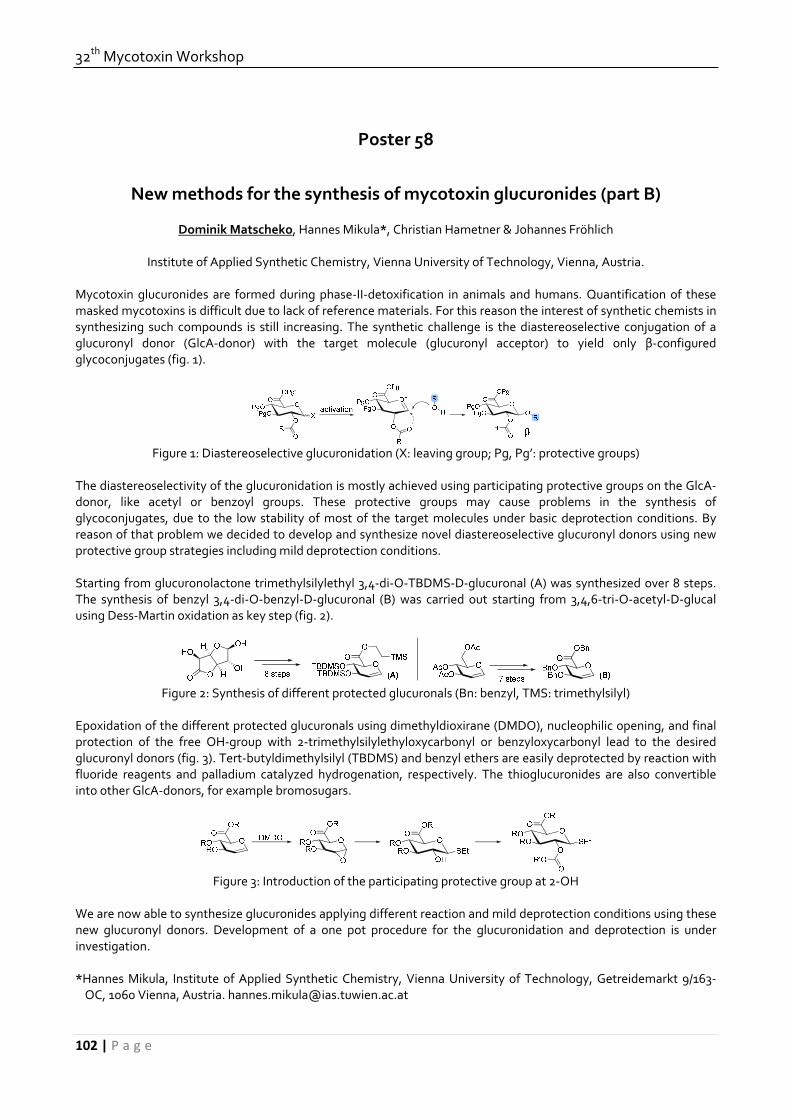
32th Mycotoxin Workshop
102 | P a g e
Poster 58
New methods for the synthesis of mycotoxin glucuronides (part B)
Dominik Matscheko, Hannes Mikula*, Christian Hametner & Johannes Fröhlich
Institute of Applied Synthetic Chemistry, Vienna University of Technology, Vienna, Austria.
Mycotoxin glucuronides are formed during phase‐II‐detoxification in animals and humans. Quantification of these masked mycotoxins is difficult due to lack of reference materials. For this reason the interest of synthetic chemists in synthesizing such compounds is still increasing. The synthetic challenge is the diastereoselective conjugation of a glucuronyl donor (GlcA‐donor) with the target molecule (glucuronyl acceptor) to yield only β‐configured glycoconjugates (fig. 1).
Figure 1: Diastereoselective glucuronidation (X: leaving group; Pg, Pg’: protective groups)
The diastereoselectivity of the glucuronidation is mostly achieved using participating protective groups on the GlcA‐donor, like acetyl or benzoyl groups. These protective groups may cause problems in the synthesis of glycoconjugates, due to the low stability of most of the target molecules under basic deprotection conditions. By reason of that problem we decided to develop and synthesize novel diastereoselective glucuronyl donors using new protective group strategies including mild deprotection conditions. Starting from glucuronolactone trimethylsilylethyl 3,4‐di‐O‐TBDMS‐D‐glucuronal (A) was synthesized over 8 steps. The synthesis of benzyl 3,4‐di‐O‐benzyl‐D‐glucuronal (B) was carried out starting from 3,4,6‐tri‐O‐acetyl‐D‐glucal using Dess‐Martin oxidation as key step (fig. 2).
Figure 2: Synthesis of different protected glucuronals (Bn: benzyl, TMS: trimethylsilyl)
Epoxidation of the different protected glucuronals using dimethyldioxirane (DMDO), nucleophilic opening, and final protection of the free OH‐group with 2‐trimethylsilylethyloxycarbonyl or benzyloxycarbonyl lead to the desired glucuronyl donors (fig. 3). Tert‐butyldimethylsilyl (TBDMS) and benzyl ethers are easily deprotected by reaction with fluoride reagents and palladium catalyzed hydrogenation, respectively. The thioglucuronides are also convertible into other GlcA‐donors, for example bromosugars.
Figure 3: Introduction of the participating protective group at 2‐OH
We are now able to synthesize glucuronides applying different reaction and mild deprotection conditions using these new glucuronyl donors. Development of a one pot procedure for the glucuronidation and deprotection is under investigation. *Hannes Mikula, Institute of Applied Synthetic Chemistry, Vienna University of Technology, Getreidemarkt 9/163‐OC, 1060 Vienna, Austria. [email protected]

June, 15th‐17th, 2010, Lyngby
103 | P a g e
Poster 59
Varying the content of T‐2 and HT‐2 toxins in processing of oats
Ute Meister Institut für Lebensmittel‐ und Umweltforschung e.V., Arthur‐Scheunert‐Allee 40‐41, D‐14558 Nuthetal OT Bergholz‐
Rehbrücke, Germany
Introduction: The Fusarium toxins T‐2 and HT‐2 toxins occur mainly in oats, which always is considered as especially healthy grain variety. The toxicity of T‐2 and HT‐2 toxins was evaluated by the Scientific Committee for Food, in 2001. The general toxicity of T‐2 toxin, the hematotoxicity and the immunotoxicity were considered to be highly critical. Although the European Community is planning maximum levels for T‐2 and HT‐2 toxins since a long time there are no maximum levels because of some still unexplained questions. Materials and Methods: Samples: Oats from food industry (raw oats, cleaned oats with husks, dehulled oat kernels, dark and bright coloured kernels, husks, dust), extrudates and oat flakes. Technological trials: Extrusion: APV double screw extruder (MPF 50), 23 experimental design under variation of the parameters extrusion temperature (150/180 °C), extrusion moisture (26/30 %), rotation speed (400/500 min‐1). Because of the low toxin content of the oat kernels the extrusion trials were done with ground raw oats (content of T‐2 / HT‐2 toxins: 22 / 61 µg/kg). Trials to make oat flakes: kiln‐drying of dehulled and un‐dehulled oats under variation of kiln‐drying temperature and duration; Steaming under variation of length; flaking. All samples were analysed to T‐2 and HT‐2 toxins according to a HPLC method published by Meister. Results: Already with the clean‐up of the raw oats in mill 40‐50 % of the toxin content can be removed. In the process of dehulling of cleaned oats approx. 90 % of the content of T‐2 and HT‐2 toxins were removed. In the dehulled oat kernels only approx. 10 % of the toxin content remained. By sorting the highly contaminated dark kernels from the bright coloured kernels in an especially sorting machine the content of T‐2 and HT‐2 toxins could be further reduced if the part of the dark kernels is high enough. The selecting of approx. 5 % of dark kernels with relatively high toxin content (sum of T‐2 and HT‐2 toxins: 251 µg/kg) from a batch with a content of T‐2 and HT‐2 toxins of 19 µg/kg (sum) did not reduce the toxin content considerably (toxin content after removal of the dark oat kernels: 12 µg/kg). In case of extrusion the content of the T‐2 and HT‐2 toxins was decreased relatively irrespective of the extrusion parameters by approx. 50‐60 %. The lowest toxin contents were achieved with the combination of high moisture (30 %), low temperature (150 °C) and low rotation speed (400 min‐1) and in the mean test of the experimental design (moisture 28 %, temperature 165 °C, rotation speed 450 min‐1). The combination of low moisture (26 %), high temperature (180 °C) and high rotation speed (500 min‐1) reduced the toxin content slightly. In the laboratory tests to produce oat flakes there was reached a reduction of the toxin content of 70‐80 % by kiln‐drying of raw oats, dehulling, steaming and flaking of the oat kernels. The achieved reduction probably is based in the main on the dehulling of the raw oats. The kiln‐drying, steaming and flaking of dehulled oat kernels did not reduce furthermore the very low toxin content. Conclusion: The results of the investigations of the variation of the two toxins in the processing of oats (dehulling, extrusion, oat flakes production) consolidate the knowledge in this area of research. Only few results are described in literature until now. The knowledge can be used to a systematical control of the processes in food industry in terms of an improvement of the consumer protection.

32th Mycotoxin Workshop
104 | P a g e
Poster 60
Formation of type A trichothecene glucosides by plant cells
Karsten Meyer*, Julia Matthes & Johann Bauer
Technische Universität München, Lehrstuhl für Tierhygiene, Freising, Germany.
Type A trichothecenes are toxigenic secondary metabolites of several Fusarium spp. and are often found in food and feedstuff of plant origin. In living plants conjugation is regarded as a general principle to detoxify endogenic and exogenic compounds. The formation of conjugates has been approved for many substances, for example mycotoxins. Those conjugated substances can be cleaved in the gastrointestinal tract by enzymatic hydrolysis. Because of this the aglucon can regain its toxicity. In this study we used plant cell suspension cultures as a model system to observe the metabolism of diacetoxyscirpenol and T‐2 toxin in plants. We confirmed the uptake of those mycotoxins by the presence of T‐2 toxin, HT‐2 toxin, diacetoxyscirpenol, monoacetoxyscirpenol and scirpentriol in extracts of contaminated plant cell cultures. Furthermore we detected some possible glucoside metabolites for HT‐2 toxin, monoacetoxyscirpenol and scirpentriol. We confirmed their glucoside structures by digestion experiments and characteristic fragmentation patterns built by MS/MS analysis. These results support the assumption that type A trichothecen glucosides were initially formed by plant cells. * Weihenstephaner Berg 3, 85354 Freising, E‐mail: [email protected]

June, 15th‐17th, 2010, Lyngby
105 | P a g e
Poster 61
Migration of mycotoxins produced in sweet pepper inoculated by Fusarium species and analysed using a multi‐mycotoxin lc‐ms/ms method.
S. Monbaliu1*, K. Van Poucke2, K. Heungens2, C. Van Peteghem1 & S. De Saeger1
1 Laboratory of Food Analysis, Ghent University, Ghent, Belgium 2 Institute for Agricultural and Fisheries Research, Merelbeke, Belgium
The occurrence of mycotoxins in foodstuffs such as maize, grain, nuts and different kind of seeds have been extensively investigated. Data describing the occurrence of mycotoxins in vegetables is rare. Internal fruit rot of sweet peppers is a fungal disease that causes considerable yield losses in commercial greenhouses in British Columbia and Alberta (Canada) and several European countries since 2003. In the early stages of infection, there are no symptoms visible on the outside of the fruit. External soft rot symptoms may appear following further mycelial colonization of the internal parts, but frequently these symptoms only become visible after the fruits have been harvested or even commercialized. The aetiology and epidemiology of this disease is still under investigation. This study investigated the migration behaviour of mycotoxins formed in sweet pepper, inoculated by Fusarium species involved with internal fruit rot. Two different sweet pepper cultivars were inoculated with two different Fusarium species. After one week of incubation at 20°C in a closed container, the lesion that was caused by the fungal infection was dissected from the pepper. Around the lesion, up to four concentric rings of pepper fruit tissue with a width of 5 mm were cut out and analyzed using a multi‐mycotoxin LC‐MS/MS method. The analyses resulted in the detection of beauvericin (BEAU), fumonisin B1, B2 and B3 (FB). BEAU was only detected in the lesions (95%) and the detected concentrations varied between 67 and 73837 µg.kg‐1. FB1, B2 and B3 were detected in the lesions, but also in the surrounding tissue, showing possible migration of these toxins into healthy parts of the sweet pepper. In the lesion the FB1 concentration varied between 690 and 104410 µg.kg‐1. Even in the outer ring FB1 was still present mostly at a lower level than in the lesion with a maximum concentration of 556 µg.kg‐1. Similar results were obtained for FB2 and FB3. *Corresponding author: Sofie Monbaliu, Harelbekestraat 72, 9000 Gent, Belgium, [email protected]
32th Mycotoxin Workshop
106 | P a g e
Poster 62
Exposure of infants to ochratoxin A ‐ New data on OTA levels in breast milk samples from Germany.
Katherine Muñoz1, Meinolf Blaszkewicz1, Hermann Kalhoff2 & Gisela H. Degen1*
1 Leibniz Research Center for Working Environment and Human Factors (IfADo), Germany 2 Klinikum Dortmund, Dortmund, Germany.
Breast milk is the ideal form of nutrition for the first 6 months of infant life, but also an excretion route for numerous xenobiotics. Reported ochratoxin A levels in human milk indicate quite variable exposure of breastfed infants to the mycotoxin ochratoxin A (OTA) in Europe (Tab. 1 in [1]). The aim of this work was to assess levels of OTA and its metabolites, ochratoxin alpha (OTα) and ochratoxin B (OTB), in mothers’ milk, and estimate current exposure of German infants to this mycotoxin.
In a German hospital cohort, lactating women were asked in 2009 to provide breast milk samples. Samples were collected at different stages of breast feeding (up to 60 days after delivery) from 27 mothers who had signed an informed consent sheet. The analytical method involved liquid–liquid extraction with chloroform after enzymatic hydrolysis, and extract analysis was performed by liquid chromatography with mass spectrometry (detection limit of 10 ng/L for OTA, OTα and OTB). 74% of the milk samples were tested positive for OTA; the mean OTA levels (31 ± 19 ng/L) were similar to those reported previously in Germany [2]. The nontoxic form, OTα, was found in most of the samples at a mean level of 44 ± 48 ng/L, whilst the non chlorinated form OTB was less frequently detected in human milk with an average level just above the detection limit.
To estimate the magnitude of the exposure, daily intake of OTA was calculated based on body weight and daily amount of breast milk consumed by the infants. Less than 10% of the samples did lead to infant exposures exceeding a tolerable daily intake (TDI) of 5 ng/kg‐bw/day proposed for adults by the Nordic working group. The new results indicate that even at low OTA‐levels in breast milk, for some babies this TDI can be exceeded. Since infants may be more susceptible than adults to nephrotoxic effects of OTA, further biological monitoring is advised. References: [1] Muñoz K, Campos V, Blaszkewicz M, Vega M, Alvarez A, Neira J, Degen GH. 2010. Exposure of neonates to ochratoxin A: first biomonitoring results in human milk (colostrum) from Chile. Mycotoxin Research, in press. [2] Gareis M, Märtlbauer E, Bauer J, Gedek B. 1988. Bestimmung von Ochratoxin A in Muttermilch. Z Lebensm Unters Forsch 186: 114‐117. * Ardeystr. 67, D‐44139‐Dortmund, Germany. E‐mail: [email protected] Acknowledgement: K.M. is supported by a stipend from CONICYT and DAAD.

June, 15th‐17th, 2010, Lyngby
107 | P a g e
Poster 63
Ochratoxin A metabolites in human blood and urine samples
Katherine Muñoz 1*, Benedikt Cramer 2, Matthias Kockmann 2, Meinolf Blaszkewicz 1, Hans‐Ulrich Humpf 2 & Gisela H. Degen 1
1 Leibniz Research Centre for Working Environment and Human Factors at the TU Dortmund (IfADo), Dortmund,
Germany 2 Institute of Food Chemistry – Westfälische Wilhelms Universität, Münster, Germany
Ochratoxin A (OTA) is one of the most widespread mycotoxins, and frequently found in various food and feed commodities. Human mycotoxin exposure with contaminated food is reflected by OTA levels found in biological samples such as blood, urine or breast milk. In plasma or serum, OTA is easily detected, due its long high half‐life. OTA levels in urine are about 10 fold lower which requires a more sophisticated cleanup for detection of the parent compound and/or of relevant metabolites. Since information on the occurrence of OTA‐metabolites in humans is scarce, the aim of this study is to generate a complete profile of OTA‐metabolites in human biological fluids, and to find additional biomarkers of OTA‐exposure in these matrices. Therefore, a broad spectrum of potential OTA‐metabolites was studied quantitatively or qualitatively in plasma and urine with or without enzymatic hydrolysis for phase II metabolites (glucuronides /sulfates). The method involved a liquid‐liquid extraction with chloroform, direct analysis and cleanup using solid phase extraction. Based on the respective metabolite, different procedures were optimized and applied for analysis by HPLC coupled with high resolution mass spectrometry and tandem mass spectrometry. Samples from a small cohort who provided urine and plasma at the same day, were analyzed allowing a direct comparison of the profiles found in urine and plasma (Munoz et al. 2010). In plasma, OTA was the major form and OTα aglycone was also detected. After enzymatic hydrolysis, the non toxic form OTα became the major metabolite present. The 14R‐OTA, a known thermal degradation product, was also detected in plasma but at very low levels. In urine, OTα levels were clearly higher than the parent compound OTA. An increase of OTα levels after enzymatic hydrolysis clearly showed the occurrence of OTα‐conjugates, which was further corroborated by mass spectrometric analysis. Additional analysis of minor OTA metabolites is currently underway. References: Muñoz K, Blaszkewicz M, Degen GH. 2010. Simultaneous analysis of ochratoxin A and its major metabolite ochratoxin alpha in plasma and urine for an advanced biomonitoring of the mycotoxin. J Chromatogr B, in Press (doi: 10.1016/j.chromb.2009.11.044) * Ardeystr. 67, D‐44139‐Dortmund, Germany. Email: [email protected] Acknowledgement: K.M. is supported by a stipend from CONICYT and DAAD.

32th Mycotoxin Workshop
108 | P a g e
Poster 64
Monitoring of aflatoxin B1 content of must during fermentation
László Rácz1*, Péter Fűtő1, Péter Forgó1, Csaba Csutorás1 and Zsolt Tajcs2
1 Eszterházy Károly College , Eger, Hungary.
2Eger Crown Winehouse LtD., Kerecsend, Hungary. Aflatoxins (AFs) are a group of highly toxic secondary metabolites produced by the filamentous fungi A. flavus and A. parasiticus. Under favorable conditions of temperature and humidity, these fungi grow on certain foods and feeds, resulting in the production of these metabolites. Aflatoxins are considered as natural contaminants of a variety of agricultural products. The most pronounced contamination has been encountered in corn, peanuts, cottonseed, and other grain crops being most frequently contaminated. The major AFs of concern are designated as B1, B2, G1 and G2. However, Aflatoxin B1 (AFB1) is the most predominant and the most toxic aflatoxin with a toxicity demonstrated in all species of animals tested, resulting in LD50 values ranging from 0.3 to 9.0 mg/kg body weight. AFB1 is also known as being one of the most potent genotoxic agent and hepatocarcinogen identified. The European Commission set the maximum level for AFB1 in foods to 2 ng/g, although new limits are likely to be established at 1 ng/g. Production of Aflatoxin B1 by ochratoxygenic fungi are still not common in grapes.[1] In Tunisia, strains isolated from grapes, potentially AFB1 producers were A. flavus responsible of 39% of AFB1 production. The quantification of this mycotoxin shows concentrations between 0.021 and 0.054 mg/g.[1] Bouraima et al (1993) have reported the competition between A. flavus and A. ochraceus. The occurrence of these two species has led essentially to the production of AFB1. This phenomenon was explicated by the fact that in the biogenesis of the Aflatoxins, A. flavus uses the phenylalanine from the support on which it develops. When this species is abundant, it would divert with its profit all the phenylalanine. The OTA, an analogue of phenylalanine could not then beproduced. Concerning the AFB1, the occurrence of this mycotoxin in must seems to be linked to the presence of the aflatoxigenic fungus, the A. flavus on grapes in the vineyards. This is related to the fact that the A. flavus is the only known AFB1‐producing species among all fungi isolates and since that 43.4 % of its isolates have produced the mycotoxin up to a concentration of 22.6 lg/g CYA. The aim of the present work was to investigate the change of Aflatoxin B1 content during must fermentation processes, using artificial contamination of must samples with Aflatoxin B1 of higher than 95% purity. Different concentrations of Aflatoxin B1 combined with different sort of wines (white, rosé, red) were applied. Our experiments were carried out in laboratorial as well as in industrial scale, and demonstrated the level of Aflatoxin B1 content in must and wine during fermentation. Sample collections and preparations were carried out systematically, the prepared samples were analyzed by HPLC‐MS and by HPLC. This work was supported by a GOP‐1.1.1 grant of the Hungarian National Development Agency. References: [1] S. Melki Ben Fredj, S. Chebil and A. Mliki. 2009. Isolation and characterization of ochratoxin A and aflatoxin B1 producing fungi infecting grapevines cultivated in Tunisia. Afr. J. of Microbiology Res. 3(9), 523. [2] André EL Khoury, Toufic Rizk, Roger Lteif, Hayat Azouri, Marie‐Line Delia, Ahmed Lebrihi. 2008. Fungal contamination and Aflatoxin B1 and Ochratoxin A in Lebanese wine–grapes and musts. Food Chem. Toxicology. 46, 2244. * H‐3300 Eger, Eszterhazy Sqr. 1., Hungary, [email protected]

June, 15th‐17th, 2010, Lyngby
109 | P a g e
Poster 65
Results of official controls of mycotoxins in food in Lower Saxony in 2009
Lilli Reinhold1*, Katja Reinhardt1
1 Niedersächsisches Landesamt für Verbraucherschutz und Lebensmittelsicherheit, Lebensmittelinstitut Braunschweig, Braunschweig, Germany.
Scientific mycotoxin research in food started in 1960, when toxic metabolites of mould fungi were found to be the cause of death of thousands of turkeys (“turkey‐x‐disease”). The first official regulation concerning mycotoxins in Germany was legislated in 1976 – the Aflatoxin‐Verordnung [1]. Since then a lot of new regulations came into force. The basic principles of EU legislation on contaminants in food are laid down in Council Regulation 315/93 [2] and maximum levels for certain contaminants in food, like mycotoxins are set in Commission Regulation 1881/2006 [3]. In addition to these EU regulations, in Germany, the Kontaminanten‐Verordnung [4] gives rules concerning mycotoxins and other contaminants. For consumer protection it is necessary to analyse a large number of food samples. In the following the results of our investigation as regards mycotoxins in food are presented. Aflatoxin and/or ochratoxin A contents were determined in dried fruits, spices, cereals and tree nuts. Regarding the analysed dried fruits ochratoxin A only was detected in dried vine fruits, but here in all samples with contents up to 8.1 µg/kg (mean 3.0 µg/kg). Among the analysed spices aflatoxins and ochratoxin A were mainly found in nutmeg and curry powder, also in all samples. The maximum levels for aflatoxin B1 and/or sum of aflatoxins were exceeded in 25 % of the nutmeg samples. Basmati rice showed aflatoxin contents in nearly all samples (mean of total aflatoxins: 1.1 µg/kg, one exceeding the maximum level (5.1 µg/kg)). Aflatoxins were detectable in about half of the hazelnut samples (maximum of total aflatoxin content 23.2 µg/kg), in almost 20 % of the hazelnut samples maximum levels for aflatoxin B1 and/or sum of aflatoxins were transcended. In contrast in pistachios aflatoxins contents were very low. Fusarium toxins were analysed in cereals and cereal products like flour, bread and pasta. Among these mycotoxins deoxynivalenol was predominantly present and found in about 40 % of the samples with contents not exceeding the maximum levels (up to 418 µg/kg). Fumonisines and zearalenone were investigated especially in maize products, e.g. snacks, flour and oil. Most of these sampIes (about 80 %) were positive with contents lower than the maximum values (zearalenone up to 98 µg/kg, sum of fumonisine B1 and B2 up to 577 µg/kg). Ergot alkaloids were determined in rye flour and were detected in about half of the samples. The values measured for the sum of the six predominantly present alkaloids ranged up to 1063 µg/kg, whereas highest contents were found for ergometrine and ergotamine. The results of mycotoxin analysis of about 500 samples of different food commodities are evaluated in detail and discussed e.g. regarding previous results and latest amendments of regulation no. 1881/2006. References: [1] Aflatoxin‐Verordnung vom 30.11.1976 (BGBl. I S. 3313), [2] Council Regulation (EEC) No 315/93 of 8 February 1993 laying down Community procedures for contaminants in food: OJ L 37, 13.2.1993, p.1, [3] Commission Regulation (EC) No 1881/2006 of 19 December 2006 setting maximum levels for certain contaminants in foodstuffs: OJ L 364, 20.12.2006, p.5, [4] Kontaminanten‐Verordnung vom 19.03.2010 (BGBl. I S. 287) * Niedersächsisches Landesamt für Verbraucherschutz und Lebensmittelsicherheit, Lebensmittelinstitut Braunschweig, Dresdenstr. 2, 38124 Braunschweig, Germany, [email protected].

32th Mycotoxin Workshop
110 | P a g e
Poster 66
Occurrence of mycotoxins in feedstuffs and feed in Asian regions in the year 2009.
Inês Rodrigues1* and Karin Griessler1
1 BIOMIN Holding GmbH, Herzogenburg, Austria.
From the cereal trade companies up until feedmillers and animal producers, all those involved in the long chain of food production are at risk of being impacted by the effects of some invisible, odourless and tasteless substances called mycotoxins. Mycotoxin contamination often begins in the field and continues throughout harvest, transportation and storage, depending on the activity and colonization levels of fungi which are in turn determined by the prevailing environmental conditions and the nutritional components of the food matrix. In general, fungi are divided in two main groups, field fungi and storage fungi, depending if they occur more frequently on the field or after harvest, respectively. However, even if this terminology has been commonly used, conditions for growth of a specific organism can occur in either the field or during storage, especially because even the same fungal genus contains species that differ greatly in their optimum temperature for growth and for their parasitic abilities. Nonetheless, what is of interest for all participants in the “farm to fork” chain is the prevalence and concentration of these substances in feed intended for animal consumption. The objective of this work is to show the size and importance of the mycotoxin problem to all players of this industry. From January 2009 until December 2009, a total of 4 615 analyses were carried out on samples sourced in Asian countries for the most important mycotoxins in terms of agriculture and animal production – aflatoxins (Afla), zearalenone (ZON), deoxynivalenol (DON), fumonisins (FUM) and ochratoxin A (OTA). Samples tested were diverse, ranging from cereals such as corn, wheat and rice to processing by‐products, namely soybean meal, corn gluten meal, dried distillers grains with solubles (DDGS) and other fodder such as straw, silage and finished feed. High Performance Liquid Chromatography (HPLC) methodology was used for the analysis of all mycotoxins. For the purpose of data analysis, non‐detection levels are based on the detection limits of the test method for each mycotoxin: Total aflatoxins < 4 μg/kg (ppb); zearalenone < 32 μg/kg (ppb); deoxynivalenol < 50 μg/kg (ppb); fumonisin B1 < 100 μg/kg (ppb) and ochratoxin A < 2μg/kg (ppb). 17% of the samples analysed in this survey were sourced in North Asia, and within North Asia, the majority was originated in China. DON, ZON, FUM, OTA and Afla were found in 68%, 59%, 40%, 16% and 11% of the analyzed samples, respectively. FUM was the most abundant mycotoxin in the South‐Eastern area of Asia, present in 64% of the analyzed samples at average levels of 1688 ppb. Afla, ZON, OTA and DON were present in 55%, 44%, 33% and 30% of the analyzed samples, respectively. Aflatoxin is a great concern in the Southern Asia region, as 79% of the samples tested positive for this mycotoxin. FUM, OTA, ZON and DON were found in 66%, 63%, 40% and 18% of analyzed samples. In Oceania, 19%, 17%, 11%, 7% and 6% of the samples were contaminated with DON, ZON, OTA, Afla and FUM, respectively. From the whole samples sourced in Asian countries, only 18% tested negative for the presence of the analysed mycotoxins, 26% presented one mycotoxin only and 56% were contaminated with more than one mycotoxin. This fact raises the attention to the possibility of interaction between mycotoxins, leading to additive and/or synergistic effects. On a daily basis animal producers are confronted with the fact that even low contamination levels mycotoxins have negative impacts on animal health and performance, enhanced by production stress animals are submitted to. On the other hand, animals often undergo extreme performance losses in the field, even at low contamination levels, potentialized by the co‐occurrence of more than one mycotoxin in the feed. * BIOMIN Holding GmbH, Industriestrasse 21, 3130 Herzogenburg, Austria. Email: [email protected].
June, 15th‐17th, 2010, Lyngby
111 | P a g e
Poster 67
A study of mycobiota and mycotoxins in wheat and wheat‐bran from Pakistan
M. Kashif Saleemi1*, M. Zargham Khan1, Ahrar Khan1, Sohail Hameed3, M. Aamer Mehmood3, Ijaz Javed2,
Zahoor –Ul‐Hasan1 and M. Raza Hameed1
1 Department of Pathology, University of Agriculture, Faisalabad‐38040, Pakistan. 2Department of Physiology and Pharmacology, University of Agriculture, Faisalabad‐38040, Pakistan.
3 National Institute for Biotechnology and Genetic Engineering, Faisalabad Pakistan.
A total of 67 samples of wheat and 17 samples of wheat bran were collected from different grain markets, ingredient suppliers, poultry feed manufacturing units and home mix feed manufacturing poultry farms over a period of two years from august 2005 to august 2007. These samples were mixed thoroughly and kept in polythene bags prior to inoculation onto culture media weekly. The samples were inoculated in petri plates containing different culturing media for fungi, including potato dextrose agar (PDA), Czapek dox solution agar, Czapek yeast autolysate agar (CYA) and yeast extract sucrose agar (YES). These plates were incubated at 27 oC in dark for 7‐10 days. For mycotoxins analysis samples were processed through micro‐scale extraction method and analyzed on HPLC with florescent detector. Forty five (67. 16 %) out of 67 samples yielded fungi, while 22 (32.84 %) samples yielded no fungus. Based upon total samples, frequency distribution of Aspergillus, Alternaria, Fusarium and Penicillium was 34.88, 5.97, 7.46 and 26.74 percent, respectively. P. verrucosum was the most frequently isolated species in total samples followed by A. niger aggregates, and other species. Among Aspergilli A. niger aggregates (42.42%) was most frequently isolated species followed by A. flavus (18.18%), A. ochraceous (18.18%), , A. parasiticus (15.15%), A. carbonarius (3.03%) and A. fumigatus (3.03%). Out of 30 Aspergillus isolates of wheat, 17 (56.67%) were found toxigenic. Aflatoxigenic isolates included A. flavus (3/6, 50%), A. parasiticus (4/5, 80%). Ochratoxigenic isolates included A. niger aggregates (6/14, 42.86%) and A. ochraceous (3/3, 100%). Their AFB1production potential ranged from 1.44 to 836.53 ng/g and maximum ochratoxin A (OTA) production was up to 15045 ng/g. In wheat‐bran out of the total 17 samples, 10 (58.82 %) samples yielded fungi, while 7 (41.18 %) samples yielded no fungus. Based upon total samples, frequency distribution of Aspergillus, Fusarium and Penicillium was 35.28, 11.76 and 29.40 percent, respectively. A. niger aggregates & P. verrucosum were predominant species in wheat bran. Among Aspergilli, A. niger aggregates (50 %) was the most frequently isolated specie followed by A. flavus (16.67 %), A. fumigates (16.67 %) and A. ochraceous, (16.67 %). Out of 6 Aspergillus isolates of Wheat‐bran, 1 (16.67 %) was found toxigenic. No aflatoxigenic isolates was found toxigenic. Ochratoxigenic isolates included A. ochraceous (1/1, 100 %). Key words: Fungi, Mycobiota, Aspergillus, Penicillium, Alternaria, Fusarium, Aflatoxin B1 and Ochratoxin A. *Department of Pathology, University of Agriculture, Faisalabad PAKISTAN. E‐mail: [email protected]

32th Mycotoxin Workshop
112 | P a g e
Poster 68
Mycotoxin occurrence in European feed samples 2009.
Karin Griessler1*, Dian Schatzmayr2 and Inês Rodrigues1
1BIOMIN Holding GmbH, Industriestrasse 21, 3130 Herzogenburg, Austria . 2 BIOMIN Holding, Technopark 1, 3430
Tulln, Austria. The presence of mycotoxins in animal feed samples is an omnipresent problem. A survey about the most important mycotoxins in feedstuffs from Europe was initiated with the objective to identify major mycotoxin occurrence in these different samples. From January to December 2009, a total of 1339 raw material and finished feed samples from different European regions (Northern Europe, Central Europe and Southern Europe) were analysed for the most important mycotoxins in terms of agriculture and animal production – aflatoxins (Afla), zearalenone (ZON), deoxynivalenol and acetyl‐deoxynivalenol (DON), fumonisins (FUM) and ochratoxin A (OTA). All analyses were performed either by HPLC (high performance liquid chromatography) or ELISA (enzyme‐linked immunosorbent assay) according to the standard procedures. For the purpose of data analysis, non‐detect levels are based on the quantification limits (LOQ) of the test method for each toxin. An overview of the survey results are shown in table 1. In 60% of all survey samples at least one mycotoxin was present. 10%, 17%, 56%, 45% and 41% of the samples tested positive for contamination with Afla, ZON, DON, FUM and OTA, respectively. Table 1: Overview of the European survey results Afla ZON DON FUM OTA Sample number 125 758 933 44 102 Positive [%] 10 17 56 45 41 Average of positive µg/kg] 3 94 725 2930 4 Maximum Level [µg/kg] 6 973 6000 11050 21 In all samples analysed from Europe a high prevalence of the Fusarium mycotoxin DON was detected with an average contamination of 725 µg/kg. Aflatoxins represent a minor problem in Europe as only 12 out 125 samples were contaminated with this mycotoxin with a mean contamination of 3 µg/kg. The mycotoxin occurrence in the different analysed feed samples confirmed the necessity of monitoring mycotoxins. * Karin Griessler, BIOMIN Holding GmbH, Industriestrasse 21, 3130 Herzogenburg, Austria, E‐Mail: [email protected]

June, 15th‐17th, 2010, Lyngby
113 | P a g e
Poster 69
Presence of Deoxynivalenol in German wheat and rye and its comparison to Nivalenol and other type B trichothecenes.
Christine Schwake‐Anduschus*, Meinolf G. Lindhauer
Department of Safety and Quality of Cereals, Max Rubner‐Institut MRI, Detmold, Germany
For the Fusarium trichothecene mycotoxin Deoxynivalenol (DON) the European Commission has established maximum levels in different cereals and products thereof (EU 1881/2006). The mycotoxin Nivalenol, which has a lower temporary TDI of 0.7 µg/kg bw/d than DON (TDI = 1 µg/kg bw/d), is said to partially co‐occur with DON; therefore separate maximum levels have not been implemented (EU 1881/2006). The Max Rubner‐Institut ‐Federal Research Institute of Nutrition and Food‐ (MRI) has analysed every year statistically taken samples from 13 or 12 different Federal States in Germany. The occurrence data of Deoxynivalenol in German wheat and rye from the year 2009 will be presented. The presentation will include comparison of DON levels to Nivalenol‐concentration and other trichothecenes, which were also analysed in 2009. Co‐occurrence of these mycotoxins will be discussed. References: European Commission. 2006. Commission Regulation (EC) No 1881/2006 of 19 December 2006 setting maximum levels for certain contaminants in foodstuffs. Off J Eur Union. L364:5‐24.
* Schützenberg 12, 32756 Detmold, Germany; christine.schwake‐[email protected]

32th Mycotoxin Workshop
114 | P a g e
Poster 70
New methods for the synthesis of mycotoxin glucuronides (part A)
Markus Schwarz, Hannes Mikula*, Christian Hametner & Johannes Fröhlich
Institute of Applied Synthetic Chemistry, Vienna University of Technology, Vienna, Austria.
Mycotoxin glucuronides are formed during phase‐II‐detoxification in animals and humans. Quantification of these masked mycotoxins is difficult due to lack of reference materials. For this reason the interest of synthetic chemists in synthesizing such compounds is still increasing. The synthetic challenge is the diastereoselective conjugation of a glucuronyl donor (GlcA‐donor) with the target molecule (glucuronyl acceptor) to yield only β‐configured glycoconjugates (fig. 1).
Figure 1: Diastereoselective glucuronidation (X: leaving group; Pg, Pg’: protective groups)
The diastereoselectivity of the glucuronidation is achieved using participating protective groups on the GlcA‐donor, mostly acyl groups (esters), but this may cause problems in the synthesis of glycoconjugates, due to the low stability of most of the target molecules under basic conditions, which are usually applied to deprotect typical acyl protective groups (acetyl, benzoyl, pivaloyl). By reason of that problem we decided to introduce different acyl protective groups, which can be removed selectively, for the development of new glucuronyl donors. Starting from D‐glucose the 6‐OH‐position was trityl protected and the remaining OH groups were acylated using chloroacetic and 2‐nitrophenylacetic acid. After deprotection, oxidation in 2 steps, and esterification with diphenylmethanol and 2,2,2‐trichlorethanol, respectively, 2 products (A and B) were obtained as intermediates for the divergent synthesis of different GlcA‐donors. Using more expensive glucuronic acid as starting material and allyl protection of the carboxyl group, product B was obtained via a second route as well as phthalimidomethyl 1,2,3,4‐tetra‐levulinoyl‐α,β‐D‐glucuronate (C) (fig. 2).
Figure 2: Different protective group patterns on glucuronic acid for the synthesis
of new glucuronyl donors These compounds (A, B, and C) were converted into the corresponding bromosugars, which are now suitable for Königs‐Knorr glucuronidation. Transformation to other GlcA‐donors like fluorosugars as well as trichloroacetimidates is also possible and still under investigation. The selective and orthogonal deprotection conditions (A: thiourea, B: zinc/acetic acid, C: hydrazine) allow us to select the appropriate protective group pattern in relation to the chemical structure of the target molecule to avoid undesirable reactions. * Hannes Mikula, Institute of Applied Synthetic Chemistry, Vienna University of Technology, Getreidemarkt 9/163‐OC, 1060 Vienna, Austria. [email protected].

June, 15th‐17th, 2010, Lyngby
115 | P a g e
Poster 71
Isolation, purification and characterization of the mycotoxin FS‐4 from Δtri101 mutants of Fusarium sulphureum.
Mehrdad Shams1,2, Rudolf Mitterbauer1, Heidemarie Hellmer1, Franz Berthiller2, Christian Hametner3, Gerlinde
Wiesenberger1 & Gerhard Adam1*
1 Department of Applied Genetics and Cell Biology, University of Natural Resources and Applied Life Sciences
(BOKU), Vienna, Austria. 2 Center for Analytical Chemistry, Department for Agrobiotechnology, IFA Tulln, BOKU, Tulln, Austria. 3 Institute for Applied Synthetic Chemistry, Vienna University of Technology, Vienna, Austria.
Fungi of the genus Fusarium can produce different mycotoxins of the class of trichothecenes, and in addition usually low amounts of apotrichothecenes, sambucinol and less well studied compounds such as the FS‐1 to FS‐4 [1] series from Fusarium sambucinum (see [2] for review). The structure of FS‐4 is related to trichothecenes but unusual because it lacks the epoxy group. In our study the Fusarium sulphureum (syn. F. sambucinum) strain MRC514 was used for the generation of tri101Δ knockout mutants. TRI101 encodes an acetyltransferase implicated in the self protection mechanism of the toxin producer. Inactivation of TRI101 in F. sporotrichoides led to a block in T‐2 biosynthesis and accumulation of isotrichodermol and other precursors, which were not observed in cultures of the parent strain [3]. The wild‐type strain MRC514 used in our study is a prolific producer of diacetoxyscirpenol (DAS) in liquid Czapek‐Dox medium enriched with 2% peptone. The tri101Δ knockout strains produced very low amounts of DAS but accumulated earlier trichothecene intermediates as determined by LC‐MS/MS. One major peak was identified to correspond to FS‐4. Ethyl acetate extracts were used for a silica gel normal phase chromatography and FS‐4 containing fractions were collected. They were further purified with reversed phase preparative chromatography and analyzed by LC‐MS using a heated nebulizer (APCI) source in positive ion mode. The structure of purified FS‐4 was confirmed by NMR spectroscopy. Testing of the ability to inhibit eukaryotic protein biosynthesis using a wheat germ in vitro translation system revealed that FS‐4 had negligible activity. The compound without epoxide accumulates when trichothecene biosynthesis is blocked at the acetylation step important for self protection. The generated tri101Δ mutants of Fusarium sulphureum are a good source for isolating FS‐4 as an analytical standard. References:
1. Sanson D, Corley D, Barnes C, Searles S, Schlemper E, Tempesta M. 1989. New mycotoxins from Fusarium sambucinum. J. Org. Chem. 54: 4313‐4318.
2. Grove JF. 2007. The trichothecenes and their biosynthesis. In: (Herz W, Falk H., Kirby GW, eds.). Progress in the Chemistry of Organic Natural Products 88: 63‐130. Springer Vienna
3. McCormick SP, Alexander NJ, Trapp SE, Hohn TM. 1999. Disruption of TRI101, the gene encoding trichothecene 3‐O‐acetyltransferase, from Fusarium sporotrichioides. Appl Environ Microbiol. 65:5252‐6.
* Muthgasse 18, A‐1190 Vienna, Austria. E‐mail: [email protected]

32th Mycotoxin Workshop
116 | P a g e
Poster 72
Novel glucosyl donors in the synthesis of masked mycotoxins
Dennis Svatunek, Hannes Mikula*, Christian Hametner & Johannes Fröhlich
Institute of Applied Synthetic Chemistry, Vienna University of Technology, Vienna, Austria.
During phase‐II‐detoxification in plants, animals or humans xenobiotic substances like mycotoxins are conjugated to a hydrophilic moiety, for example glucose or glucuronic acid. These conjugates, the so called masked mycotoxins, are usually not detected using standard protocols and quantification is not possible due to lack of reference materials. Therefore this class of substances is getting more and more in the focus of synthetic chemists. The synthetic challenge is the diastereoselective conjugation of a synthetic glycosyl donor (G‐donor) with the target molecule (glycosyl acceptor) to yield only β‐configured glycoconjugates (fig. 1).
Figure 1: Diastereoselective glucosylation (X: leaving group, Pg: protective group)
The diastereoselectivity of the glycosylation is mostly achieved using participating protective groups, like acetyl or benzoyl groups. These protective groups are most commonly used in carbohydrate chemistry but may cause problems in the synthesis of glycoconjugates, due to the lower stability of most of the target molecules compared to saccharides under basic deprotection conditions. By reason of that problem we decided to develop and synthesize novel diastereoselective glucosyl donors using new protective group strategies including mild and orthogonal deprotection conditions. 3,4,6‐Tri‐O‐TBDMS‐D‐glucal and 3,4,6‐Tri‐O‐benzyl‐D‐glucal were used as starting materials for the introduction of different protective group patterns. Tert‐butyldimethylsilyl (TBDMS) and benzyl ethers are also widely used protective groups, easily deprotected by reaction with fluoride reagents and palladium catalyzed hydrogenation, respectively. After epoxidation using dimethyldioxirane (DMDO) and nucleophilic opening, a participating group, also cleaved under conditions used for silyl or benzyl ethers (corresponding alkyloxycarbonyl protective group), was introduced on C2 of the glucosyl donor (fig. 2). It is also possible to convert the resulting thioglucosides to the corresponding bromosugar or trichloroacetimidate as further donors.
Figure 2: Synthesis of novel G‐donors (R = benzyl or TBDMS, R’= benzyl or 2‐trimethylsilylethyl)
Using these novel glucosyl donors we are now able to glucosylate target molecules applying different reaction conditions (dependent on the leaving group) and carry out the final deprotection under mild conditions without any use of acids, bases, and even water. Reference: Berthiller F, Schuhmacher R, Adam G, Krska R. Formation, determination and significance of masked and other conjugated mycotoxins. Anal. Bioanal. Chem. 395:1243‐1252. *Hannes Mikula, Institute of Applied Synthetic Chemistry, Vienna University of Technology, Getreidemarkt 9/163‐OC, 1060 Vienna, Austria. [email protected]

June, 15th‐17th, 2010, Lyngby
117 | P a g e
Poster 73
Mouldy status characterisation of maize and grass silages: Adequacy between visual appraisal during collection and categorisation according to ergosterol
contents
Emmanuel K. Tangni1,*, Luc Pussemier1, François van Hove2, Jurgen Depoorter3, Geert Haesaert4 & Johan Robbens5
1: Veterinary and Agrochemical Research Centre (CODA‐CERVA), 2: Mycothèque de l'Université catholique de Louvain (BCCMTM/MUCL), Louvain‐la‐Neuve (Belgium) 3: CIPF, Louvain‐la‐Neuve 4: Hogeschool Gent, Gent (Belgium)
5: University of Antwerp, Antwerp (Belgium).
Introduction: Fungal contamination and mycotoxin production are problems in silages and are frequently suspected to be responsible of animal health damage or death. Mouldy materials were prone to be highly contaminated. Based on the mouldy status of maize and grass silages, feed quality and safety were assessed by means of the analysis of ergosterol (ERG), roquefortine C (ROC), dry matter (DM) and chemico‐fermentative parameters. In the present study, adequacy between visual appraisals during sampling was checked against quantification of ERG in order to ascertain the useful chemical index for characterizing the forage quality.
Materials and Methods: Maize (n = 42) and grass (n = 40) silages differing in farming systems and geographic locations in Belgium were examined in 2006 and 2007. Visually mouldy and non‐mouldy parts of each silo were taken during 4 periods (with 3‐6 weeks intervals) during the life‐time of silages, after its opening. The chemico‐fermentative characteristics of the forage samples (pH, DM, ammoniac nitrogen/total nitrogen ratio, fermentative acids composition) and ERG were measured. A marker mycotoxin, ROC, was determined using an existing analytical method (Auerbach et al. 1998).
Results: The pH of the mouldy parts was significantly higher compared to the non‐mouldy parts of maize and grass silages. The higher pH value for mouldy parts indicates a disturbed fermentation process with low lactic acid production found in the mouldy parts. Butyric acid content was clearly higher for the mouldy parts, only for maize silages. As for DM, acetic acid and amoniac nitrogen contents, no significant differences were observed between non‐mouldy and mouldy parts of maize and grass silages. Looking to different sampling periods for the same silos no significant evolution was noted for the chemical composition of the silos. ERG was quantified in all examined silages at the levels ranged between 2.7–158.5 µg/g of fresh matter (FM) for maize silages and 2.4–185.5 µg/g FM for grass silages. ERG contents were significantly higher in maize than in grass silages. This is due to the fact that there is a significant difference (p=0.036) in ERG loads of visually mouldy maize and grass silages. Based on the DM, a threshold of 30 µg ERG/g DM was derived as chemical index to classify silage samples in either non‐mouldy (<30 µg/g DM) or mouldy (>30 µg/g DM) category. This threshold level was 40 µg ERG/g DM for grass silages. Overall good adequacy was observed for the outlook of the above chemical parameters using visual and ERG classifications. ROC contents ranged <0.1‐15.1 µg/g DM for maize silages and <0.1–7.2 µg/g DM for grass silages. Mean ROC content was higher in mouldy than in non‐mouldy maize silages (p = 0.015). In contrast, mean ROC content in mouldy grass silage was not different from that obtained in non‐mouldy grass sub‐samples. Mean ROC contents were higher in mouldy maize than in mouldy grass samples (p = 0.013). A significant correlation coefficient (r=+0.334, p < 0.01) was derived between ERG and ROC contents in maize and grass silages.
Discussion and Conclusion: Feeds with no visible mould are contaminated with ROC. The visible non‐mouldy aspect of silage is not necessary a good indicator of the forage acceptability as mycotoxin free feed. Reliable chemical index based on ERG determination is useful for characterizing the forage mouldy status.
Reference: Auerbach H, Oldenburg EL & Weissbach F. 1998. Incidence of Penicillium roqueforti and roquefortine C in silages. J. Sci. Food & Agric 76:565‐572.
* Corresponding author's address: Leuvensesteenweg 17, B‐3080 Tervuren (Belgium), Email: [email protected]

32th Mycotoxin Workshop
118 | P a g e
Poster 74
Mycotoxins and moulds in silages
Magdalena Twaruzek *, Anna Blajet‐Kosicka, Robert Kosicki, Jan Grajewski
1 Kazimierz Wielki University, Institute of Experimental Biology, Division of Physiology and Toxicology, Bydgoszcz,
Poland Very often cereal and green plants (maize, lucerne and grass) used for silages are contaminated with fungi and their metabolites. In this case the most predominant are toxins produced on the plant by Fusarium species (zearalenon, deoxynivalenol, T‐2 toxin) during vegetation process. Whereas silages are mainly contaminated with moulds produced by Penicillium, Aspergillus and Monascus, as well as by Mucoraceae. Fungal metabolites can get to the silages together with the ensiled product or can be produced due to the mould growth, as a result of improper ensiling procedures or storage conditions. The aim of this study was to investigate the occurrence of moulds and mycotoxins: aflatoxins (AF), ochratoxin A (OTA), deoxynivalenol (DON), nivalenol (NIV), T‐2 toxin, HT‐2 toxin, zearalenone (ZEA) and fumonisins (FUM) in silages and TMR. The material used in the study included 107 silage samples provided by farmers in the years 2008‐2010: 67 of maize, 12 of sorghum, 11 of grass, 6 of lucerne and 11 TMR. The following numbers of mycotoxins analysis were performed: aflatoxins – 5, ochratoxin A – 20, zearalenone – 106, deoxynivalenol – 107, fumonisins – 22, T‐2 toxin – 103, HT‐2 toxin – 95, and nivalenol – 96. The general number of fungi was determined in 58 samples. Quality and quantity identification of moulds and yeast was done in accordance with PN ISO 7954: 1999 – with own alternations (inoculation on YGC medium – yeast extract, glucose, 100 ppm chloramphenicol, incubation 5 – 7 days in 250C). The extraction of mycotoxins was done on the immunoaffinity columns: AflaTest and FumoniTest by Vicam for AF and FUM, Ochraprep by R‐Biopharm Rhône Ltd for OTA, for DON, NIV, T‐2 toxin, HT‐2 toxin and zearalenone Bond Elut® Mycotoxin columns were used. Mycotoxins were evaluated with the HPLC method (Merck‐Hitachi, Agilent) with fluorescent and MS/MS (Applied Biosystems) detection. Cobra Cell was used for aflatoxins derivatization. DON was detected in all the samples of maize silages and TMR and its average concentration was 441,6 ng/g and 371,3 ng/g, respectively. The highest concentration of DON was 2050 ng/g in maize silages. ZEA was found in all the samples of sorghum silages and TMR, its average concentration was 1,86 ng/g and 22,91 ng/g, respectively. However, the highest detected concentration of ZEA was found in maize silages (202 ng/g).The highest concentration of T‐2 toxin was detected in TMR (22,1 ng/g), HT‐2 toxin in maize silage (149 ng/g). NIV was detected only in maize silages (95%) and TMR (90,9%), and its average concentration was 100,8 ng/g and 46,4 ng/g, respectively. OTA was found in all sorghum silages and TMR, its average concentration was 1,7 ng/g and 0,49 ng/g, respectively. However higher OTA content was detected in grass silages 11,3 ng/g. Aflatoxins were not detected in any of the analyzed samples. FUM was detected only in maize silages and the highest concentration of fumonisins was 82,4 ng/g, with medium concentration only 18,3 ng/g . The highest level of moulds was identified in maize silages 3,63 x 10⁶, and the dominating species turned out to be Penicillium and Aspergillus. Summarizing, worse parameters were found in maize silages and TMR, where DON, NIV and ZEA dominated. The by‐products of moulds metabolism detected in silages may cause a number of diseases and/or impair the animals inborn immunity as well as have harmful effect on the microflora of their digestive tract, thus leading to decreased efficiency. If silage shows spoilage symptoms, it should not be fed to animals to prevent the occurrence of mycotoxicoses, as there is a high probability of mycotoxin contamination in these cases. Financial support: Ministry of Science and Higher Education Project no. NN 311 122334. * Corresponding author's Chodkiewicza Street 30, 85‐064 Bydgoszcz, Poland, [email protected].
June, 15th‐17th, 2010, Lyngby
119 | P a g e
Poster 75
Investigation of mycotoxins produced by marine‐derived fungal strains of Penicillium sp.
Marieke Vansteelandt1, Isabelle Kerzaon1, Nicolas Ruiz1, Jean‐François Biard1, Yves‐François Pouchus1 & Olivier
Grovel1*
1 MMS, University of Nantes, Faculty of Pharmacy, Nantes, France.
Marine‐derived fungi are a rich source of bioactive secondary metabolites. Some of them exhibit toxic effects on animal models and can be considered as mycotoxins if they are found to be produced in marine environment and bioaccumulated by organisms such as shellfish. Previous studies have shown that toxic fungal metabolites can be produced in situ in the marine environment and bioaccumulated by shellfish (1), such as the lipophilic peptides peptaibols which have been detected in marine natural samples of sediments and fresh mussels, constituting the first observation of contamination of the marine‐human‐food chain by fungal metabolites (2). Marine mycotoxins could then be one of the explanations for the episodes of unexplained shellfish toxicities observed on the Atlantic French coast in the 90’s (3). In this way, marine fungi have been routinely isolated from natural samples of shellfish, sediment and seawater gathered in shellfish farming areas along the French western coast to search for mycotoxins. This previous study has highlighted the predominance of the genus Penicillium with 47% of the strains isolated belonging to this genus (4). In the present work we investigated marine‐derived strains of Penicillium for their production of mycotoxins as this genus is known to be the producer of the widest range of mycotoxins, and is involved in many mold associated diseases. Cultures on different seawater media were extracted with ethyl acetate or dichloromethane/ethyl acetate 50:50 (v/v) and crude extracts were directly dereplicated by LC‐DAD/MSn according to Nielsen and Smedsgaard (5). Metabolic profiles analyses led to the detection of mycotoxins known to be produced by the studied species e.g. citrinin, penicillic acid, roquefortine C or the ergot alkaloid aurantioclavine in P. expansum, P. canescens, P. atramentosum or P. marinum. However, some relevant mycotoxins were not detected in the marine‐derived Penicillium strains whereas they are known to be produced by these species such as patulin by P. expansum and P. marinum, or ochratoxin A by P. chrysogenum and P. expansum. Several mycotoxins were identified for the first time in different marine‐derived Penicillium strains isolated from natural samples, such as verruculotoxin in P. chrysogenum or penitremone A in P. canescens. The next step will consist in looking for these mycotoxins in environmental toxic samples. References: 1: Sallenave C., Pouchus Y.‐F., Badouil M., Lassus P., Roquebert M.‐F., Verbist J.‐F. 1999. Bioaccumulation of mycotoxins by shellfish: contamination of mussels by metabolites of a Trichoderma koningii strain isolated from the marine environment. Toxicon 37: 77‐83. 2: Poirier L., et al. 2007. Peptaibols: stable markers of fungal development in the marine environment. Chem. Biodivers. 4: 1116‐1128. 3: Marcaillou‐LeBaut C., Amzil Z. 1995. Unexplained toxicity in molluscs found in the course of phytoplankton monitoring. Toxicon 33 (9), 1127. 4: Sallenave‐Namont, C., Pouchus, Y.F., Robiou du Pont, T., Lassus, P., Verbist, J.F. 2000. Toxigenic saprophytic fungi in marine shellfish farming areas. Mycopathologia 149: 21‐25. 5: Nielsen K. F., Smedsgaard J.2003. Fungal metabolite screening: database of 474 mycotoxins and fungal metabolites for dereplication by standardised liquid chromatography‐UV‐mass spectrometry methodology. J. Chromatogr. A. 1002 (1‐2): 111‐136. * Corresponding author: Olivier Grovel, MMS, University of Nantes, Faculty of Pharmacy, BP 53508, F‐44035 NANTES‐CEDEX 1, FRANCE – olivier.grovel@univ‐nantes.fr
32th Mycotoxin Workshop
120 | P a g e
Poster 76
Does lycopene play a protective role in low dose aflatoxin B1‐induced hepatotoxicity in rats?
Terken Baydar1*, Gözde Girgin 1, Güldeniz Selmanoglu 2, Elif Karacaoglu 2, Aysun Kilic 2, Serdar Balci 3 & Gönül Sahin1
1 Department of Toxicology, Faculty of Pharmacy, Sihhiye Campus; 2 Department of Biology, Faculty of Science,
Beytepe Campus; 3 Department of Pathology, Faculty of Medicine, Sihhiye Campus; Hacettepe University, Ankara, Turkey
As it is known, aflatoxin B1 (AFB1) is a potent hepatocarcinogen and aflatoxins are assigned to group I carcinogens in International Agency for Research on Cancer (IARC) classification. Consecutive oxidative damage caused by aflatoxins is considered to be the main mechanism leading to their hepatotoxicity. Lycopene, which can be found in high amounts in tomatoes and tomato‐derived foods, is an acyclic carotenoid. It presents one of the highest antioxidant activities among the carotenoids. The dietary intake of lycopene has been suggested to be associated with decreased risk of chronic diseases such as cancer and cardiovascular diseases. This study has aimed to evaluate possible protective effect of lycopene on AFB1 ‐induced hepatotoxicity. Therefore, 20 female Wistar rats were divided to 4 groups. Group I was used as a control group and treated with corn oil which was used to dissolve lycopene, for 7 days. Group II were given AFB1 (0.1 µg/kg, bw) as a single oral administration on the 8th day of the experimental period while Group III and IV were treated with lycopene at 100 mg/kg, bw first 7 days by gavage. Group IV rats received lycopene followed by AFB1 administration at 0.1 µg/kg dose on day 8. At the end of the 10‐day experimental period, the rats were sacrificed, the livers were removed and bloods were collected; the samples were stored at ‐80 °C until analysis. Histopathological evaluation was done. In order to evaluate liver functions, alanine aminotransferase (ALT), aspartate aminotransferase (AST), alkaline phosphatase (ALP), total cholesterol and urea levels were determined in the blood samples. Specific activities of superoxide dismutase (SOD) and catalase (CAT) as antioxidant enzymes were measured in both of blood and liver samples while malondialdehyde (MDA) levels, the end product of lipid peroxidation, were detected in liver homogenates. According to the results, the very low single dose of AFB1 (in Group II) caused decreases in blood urea levels and liver CAT activities and an elevation in liver SOD activities compared with the control group (Group I). Significant increases in ALT, AST, and ALP levels and at some extent, a decrease in urea levels were observed in lycopene treated rats (Group III) compared to control group (all, p < 0.05). When compared with Group III, an elevation was observed in liver MDA levels of rats in Group IV, which were exposed to AFB1 following pretreatment with lycopene (p < 0.05). Additionally, Group IV has presented lower erythrocyte CAT activity than the Group III (p < 0.05). There were significant differences between Group II and IV in the levels of AST and ALT, and liver CAT activities (all, p < 0.05) In conclusion, the results pointed out that AFB1 at this dose causes minor alteration in hepatic functions and it might be suggested that, at this dose of consumption, lycopene does not provide an important protective effect against the hepatotoxic effects of AFB1 in rat population. References: 1. Tang L, Hongxia G, Ding X, Wang JS. 2007. Modulation of aflatoxin toxicity and biomarkers by lycopene in F344 rats. Toxicol. Appl. Pharmacol. 219: 10‐17. 2. Reddy L, Odhav B, Bhoola K. 2006. Aflatoxin B1‐induced toxicity in HepG2 cells inhibited by carotenoids: morphology, apoptosis and DNA damage. Biol. Chem. 387: 87‐93. 3. Gradelet S, Le Bon A‐M, Berges R, Suschetet M, Astorg P. 1998. Dietary carotenoids inhibit aflatoxin B1‐induced liver preneoplastic foci and DNA damage in the rat: Role of the modulation of aflatoxin B1 metabolism. Carcinogenesis. 19: 403‐411.
* Corresponding Author: Prof. Terken Baydar, Ph.D., Toxicology Department, Faculty of Pharmacy, Hacettepe University, 90‐06100, Sihhiye Ankara/TURKEY e‐mail: [email protected]

June, 15th‐17th, 2010, Lyngby
121 | P a g e
Poster 77
Silibinin Does Not Prevent OTA‐Mediated Apoptosis in Primary Rat Hepatocytes and HepG2 Cells
Ebtisam Essid1, Ernst Petzinger1*
1Institute of Pharmacology and Toxicology, Faculty of Veterinary Medicine, Justus‐Liebig‐University Giessen,
Frankfurter Str. 107, D‐35392 Giessen, Germany.
Silibinin is the major pharmacologically active compound extracted from the seeds of the milk thistle Silybum marianum. It is a widely used traditional herbal/dietary supplement and exhibits strong hepatoprotective activity against several liver toxins. Protective effects of silibinin on OTA‐ and LPS‐mediated cytotoxicity in the liver have been described in perfused rat livers and isolated Kupffer cells and were related to inhibition of TNF alpha release [1]. Also antiapoptotic effects were described with silibinin on a human melanoma cell line [2]. Since OTA induces apoptosis in isolated rat primary hepatocytes [3], we investigated protective effects by silibinin on OTA‐ and LPS‐mediated apoptosis and cytotoxicity in primary hepatocytes and HepG2 cells. We used two different preparation methods for hepatocyte isolation and found the EDTA‐perfusion method by [4] better suited for apoptosis research than the traditional isolation method by collagenase perfusion [5]. The EDTA‐perfusion method yielded hepatocytes without DNA‐fragmentation for up to 96 hrs. By treatment with 12.5µMol/L OTA isolated hepatocyte and HepG2 cells developed DNA‐laddering after 24 to 36 hrs. LPS at 0.1 µg/ ml had no apoptotic DNA‐effects under these conditions in both types of liver‐derived cells. Silibinin up to 130µMol/L showed no inhibition of OTA‐mediated DNA‐laddering after 24 and 36 hrs. To evaluate viability of OTA/LPS‐treated hepatocytes and HepG2 cells the MTT‐test and Live/Dead kit was applied. The results demonstrate that the used OTA concentration (12.5 µMol/L OTA) only moderately decreased viability for up to 24 hrs but showed significant cytotoxic effects depending on longer incubation times (≥36 hrs). The late‐term cytotoxicity of OTA was not prevented by silibinin. LPS (0.1 µg/ml) exhibited no cytotoxicity on hepatocytes nor on HepG2 cells up to 36 hrs. In summary, our results show differential effects on apoptosis in liver‐derived cells by OTA (produces apoptosis) versus LPS (produces no apoptosis). Silibinin shows no hepatoprotective effects against OTA‐mediated apoptosis nor does it reduce the cytotoxicity of OTA on cultured hepatocytes and HepG2 cells. References: [1] Al Anati L, Essid E, Reinehr R, Petzinger E. Mol. Nutr. Food Res. 53: 460‐466 (2009) [2] Jiang Y, Wang H, Wang J, Tashiro A, Onodera S, Ikejima T. J. Pharmacol. Sci. 111:137‐146 (2009) [3] Chopra M, Link P, Michels C, Schrenk D. Cell Biol. Toxicol. DOI 10.1007/s10565‐009‐9131‐0 (2009); [4] Meredith MJ. Cell Biol. Toxicol. 4: 405 (1988); [5] Petzinger E, Frimmer M. Biochim. Biophys. Acta 778, 539‐548 (1984) * Corresponding author. Prof. Dr. E. Petzinger Tel.: +49 641 9938400; fax: +49 641 9938409. [email protected]‐giessen.de
32th Mycotoxin Workshop
122 | P a g e
Poster 78
The effects of ochratoxin A on oxidative DNA damage and antioxidant enzymes in rat kidney and protective role of lycopene
Sevtap Aydin1, Şaziye Sezin Palabiyik2, Pınar Erkekoğlu1, Gönül Sahin1Nursen Basaran1,
Belma Giray1,*
1Hacettepe University Faculty of Pharmacy Department of Toxicology, Sihhiye Ankara Turkey
2Ataturk University Faculty of Pharmacy Department of Toxicology, Erzurum, Turkey Ochratoxin A (OTA) is a widespread mycotoxin produced by several species of fungi. OTA is nephrotoxic and it has been implicated as main etiological agents responsible for Balkan endemic nephropathy and associated urinary tract tumours. OTA‐induced oxidative stress may be one of the mechanisms underlying the nephrotoxicity connected to the long‐term exposure to this food contaminant. The present study was designed to investigate the possible protective effect of lycopene against the oxidative damage in kidney of OTA‐treated male Sprague‐Dawley rats. OTA exposure (0.5 mg/kg/day) was introduced by oral gavage and lycopene was applied as 5 mg/kg/day for 7 days. DNA damage was assessed using Comet assay and the change in the oxidant/antioxidant balance was assessed by measuring the activity of antioxidant enzymes including catalase (CAT), total superoxide dismutase (SOD), glutathione peroxidase (GPx1) and thioredoxin reductase (TrxR) in kidney. Enhanced DNA damage as evidenced by significant increases in tail intensity, tail moment and tail length in the Comet assay. Lycopene was found to be protective against DNA damage caused by OTA exposure almost taking these measures to control levels. However, there were not any significant changes in the activities of GPx1 and TrxR by OTA. SOD and CAT activities decreased by exposure of OTA insignificantly. These results indicate that OTA can cause oxidative DNA damage which may be rescued by antioxidants, particularly by lycopene. This protection might be though different mechanisms some of which are not still fully understood in detail. The results presented herein emphasize once more the importance of antioxidant status as a public health concern, and its importance with respect to the high probability of OTA exposures and their adverse effects. * Hacettepe University, Faculty of Pharmacy, Department of Toxicology, Sihhiye Ankara Turkey e‐mail: [email protected]

June, 15th‐17th, 2010, Lyngby
123 | P a g e
Poster 79
Degradation of ochratoxin A by lactobacillus acidophilus K1 strain.
Jarosław Mazurkiewicz*
Deptartment of Biotechnology, Human Nutrition and Science of Food Commodietes, University of Life Sciences in
Lublin, 20‐704 Lublin, Poland
There are many well known methods which are applied to make detoxification of mycotoxins. They can be classified as: physical, chemical and biological [Park and all 2007]. Many accessible physical and chemical methods are limited from the regard on: the problem of the assurance of safety products exposed to these interventions, the possible losses of the nutritional quality of food products, limited efficiency, the costs of the use. This leads to searching for the safe ways of the protection of food products before mycotoxins. Biological methods, like by using of microorganisms to the decay of mycotoxins, can be the solution [Kabak and all 2006]. The bacteria of lactic fermentation can be effectively used to degrading mycotoxins [Niderkorn and all 2006]. The aim of investigations was evaluation of the degree of the biodegradation ochratoxin A by the alive and dead cells of lactic fermentation bacteria ‐ Lactobacillus acidophilus K1 strain . Degradation of ochratoxin A was performed in 16 and 24 hour long culture in PBS as the medium and various pH (from 4 to 7). Standard ochratoxin A was dissolved in acetonitrile and added to PBS. The concentration of mycotoxin was marked with use of the direct competence ELISA tests ‐ AgraQuant Test Kit (Romer Labs). Dead cells of bacteria were obtained by thermal inactivation in PBS. The highest ability to degradation of ochratoxin A after 24 hours incubation on medium in pH5 had the dead cell of Lactobacillus acidophilus K1 strain. In these conditions ochratoxin A was degraded in 80%. Low pH better influenced on the degree of degradation. After 16 hours of incubation in pH 4 the residue of mycotoxin amounted 25%. The longer time of incubation influenced on lower concentration mycotoxin in medium. In the initial stage of incubation, alive cells showed the larger ability to eliminating ochratoxin A, however after the longer time of incubation, dead cells were characterized of higher degree of the biodegradation. The highest residue of the mycotoxin was detected after 16 hours of incubation in pH6 medium with alive cells, and after 24 hours of incubation in pH7 medium with thermally inactivated cells. References: Niderkorn V., Boudra H., Morgavi D.P.: Binding of fusarium mycotoxins by fermentative bacteria in vitro., Jurnal of Applied Microbiology, 2006; 101:849‐856.
Park B.J. et all., 2007. Degradation of mycotoxins using microwave‐induced argon plasma at atmospheric pressure., Surface & Coatings Technology, 210:5733‐5737.
Nielsen KF, Andersen B. 2010. How to write the abstract. Abbrev. J. Vol: X‐Y.
Kabak B., Dobson A.D.W., Var I. 2006. Strategies to prevent mycotoxin contamination of food and animal feed., Critical Reviews in Food Science and Nutrition, 46:8.
*Jarosław Mazurkiewicz, Katedra Biotechnologii, Żywienia Człowieka i Towaroznawstwa Żywności ul. Skromna 8, 20‐704 Lublin. E‐mail: [email protected]

32th Mycotoxin Workshop
124 | P a g e
Poster 80
Effect of Trichoderma harzianum on growth and mycotoxin production of Fusarium strains.
Marina Müller1 & Susann Siebert2
1 Leibniz‐Centre for Agricultural Landscape Research, Institute of Landscape Matter Dynamics, Müncheberg,
Germany*. 2 Freie Universität Berlin, Department of Biology, Chemistry, and Pharmacy, Institute of Biology, Berlin, Germany.
Trichoderma (T.) species are known as potential biocontrol agent against several phytopathogens. Several studies showed the inhibitory impacts of T. viride and T. harzianum on growth and on the fumonisin accumulation of Fusarium (F.) proliferatum and F. verticillioides isolated from maize (Yates et al. 1999). We investigated the influence of mycorrhiza on wheat plant development and plant vitality in the presence of pathogenic F. strains in a climate chamber experiment. A Trichoderma strain was detected in the inoculated mycorrhiza‐soil‐mixture and identified as T. harzianum. The aim of our in vitro study was to evaluate the effect of this Trichoderma strain on growth and mycotoxin production of the phytopathogenic F. culmorum strain ZALF13 and F. graminearum strain ZALF23 isolated from wheat plants. Both F. isolates were characterised as aggressive pathogenic F. strains with a high potential of mycotoxin production. To examine the impacts of T. harzianum on F. growth a Petri dish assay with Sabouraud‐, potato dextrose‐, rice extract‐ or czid‐agar as nutrient medium were used. A standardized conidia suspension was placed in holes located at the outer edges of the agar, two strains on the opposite side of the same Petri dish. The area of hyphal growth of each strain (5 replicates) was measured daily. Macroscopic characteristics of the interacting cultures and microscopic observations (hyphal morphology, conidia form) were monitored after 14 days. The mycotoxin accumulation in autoclaved rehydrated rice after inoculation of the three strains alone or in combination was measured by HPLC (Jasco) with UV and/or fluorescence detection. The inoculation procedures were as follows:
1 T. harzianum during 12 days 2 F. culmorum ZALF13 during 6 and 12 days 3 F. graminearum ZALF23 during 6 and 12 days 4 F. culmorum ZALF 13 and T. harzianum during 6 days 5 F. graminearum ZALF23 and T. harzianum during 6 days 6 F. culmorum ZALF13, 6 days before T. harzianum (a total of 12 days) 7 F. graminearum ZALF23, 6 days before T. harzianum (a total of 12 days) 8 T. harzianum, 6 days before F. culmorum ZALF13 (a total of 12 days) 9 T. harzianum, 6 days before F. graminearum ZALF23 (a total of 12 days)
The experiments were done in fivefold replication for each combination. The inhibition of growth was greatly influenced by the nutrient medium used. On Sabouraud‐ and potato dextrose‐agar T. harzianum overgrew the F. strains. In some cases, the interacting cultures showed a ring of purple pigmentation, but never an inhibition zone. When T. harzianum was growing 6 days prior to the addition of both F. strains, the production of deoxynivalenol (DON), nivalenol (NIV) and zearalenon (ZEA) was nearly fully repressed. A total inhibition of DON and NIV accumulation appeared in the co‐incubation of T. harzianum with F. culmorum and F. graminearum. In contrast, the ZEA production of F. graminearum was not inhibited by Trichoderma, while ZEA was produced by F. culmorum only in a small extend. References: Yates, Meredith, Smart, Bacon, Jaworsk (1999): J. Food Protect 62, 1326‐32. * Dr. Marina Müller, ZALF Müncheberg, Eberswalder Str. 84, D‐15374 Müncheberg, [email protected]

June, 15th‐17th, 2010, Lyngby
125 | P a g e
Poster 81
Evaluation of Mycofix® Plus supplementation in ochratoxin and zearalenone contaminated diets in broiler breeders
Katia Pedrosa1 and Chang‐Won Kang2
2Animal Resources Research Center, College of Animal Bioscience and Technology, Konkuk University, Seoul, 143‐
701 Korea. The study aimed to evaluate the effect of Mycofix® Plus incorporation in ochratoxin (OTA) and zearalenone (ZON) contaminated diets on reproductive performances, egg quality and physiological response in broiler breeders (ROSS 308). One hundred broiler breeders were stocked into 20 experimental groups. The trial design comprised 5 treatments and 4 replicates per treatment. Five different diets were provided ad libitum during 8 weeks, with four of them containing either mycotoxins or mycotoxins and a feed additive (Mycofix® Plus) determined to deactivate mycotoxin‐derived effects on animal performance. Group 1 (T0) was the control group (without mycotoxins and Mycofix® Plus); group 2 (T1) received OTA (2 mg.kg‐1) and ZON (5.0 mg.kg‐1) without Mycofix® Plus; group 3 (T2) received OTA (2 mg.kg‐1) and ZON (5.0 mg.kg‐1) with addition of Mycofix® Plus at 0.25 % in the diet; group 4 (T3) received ZON at 10 mg.kg‐1 without Mycofix® Plus; and group 5 (T4) received ZON at 10 mg.kg‐1with Mycofix® Plus at 0.25 % in the diet. The parameters measured were feed consumption; egg production (egg production rate, daily egg weight and egg mass); egg and eggshell quality (egg shell strength, egg shell thickness, haugh units and yolk color); fertility and hatchability (hatch of egg set, hatch of fertile egg); organ weights (relative weights of liver and immune organs). Results of egg production, egg weight and egg mass of groups fed mycotoxins contaminated diets (T1 and T3) were significantly (P<0.05) reduced when compared to that of the control group; groups receiving Mycofix® Plus (2.5 kg/ton) supplementation alleviated those performance parameters. Egg weight in T1 and T2 were significantly (P<0.05) lower (60.88 g/egg and 61.89 g/egg, respectively) than egg weight in the control group (63.23 g/egg), however, this parameter was significantly (P<0.05) improved in T2 in comparison with T1 due to Mycofix® Plus inclusion. Eggshell strength, eggshell color, egg yolk color and haugh units were not significantly influenced by the intake of mycotoxins. Eggshell thickness of groups fed only mycotoxins but not deactivator (T1 and T3) was significantly (P<0.05) decreased when compared to the control group (32.40 mm/100 and 32.46 mm/100 vs. 33.59 mm/100) but improved by the supplementation with Mycofix® Plus (T2: 33.53 mm/100 and T4: 32.87 mm/100 respectively). The fertility and hatchability of groups fed diets containing mycotoxins tended to decrease when compared to the control group (90,03% and 89.93% for fertility and 80.33% and 77.47% for hatchability) but was improved by the combined feeding Mycofix® Plus (92.1% and 91.03% for fertility and 83.4% and 79.07% for hatchability). Broilers fed ZON and OTA contaminated diets (T1) as well as animals fed ZON contaminated feeds (T3) presented significantly (P<0.05) heavier livers (2.42 g/100g BW and 2.31 g/100g BW, respectively). Supplementation with Mycofix® Plus improved this parameter in both T2 and T4 groups (2.22 g/100g BW and 2.15 g/100g BW respectively). As a conclusion, the addition of 2.5 kg Mycofix® Plus to broiler breeders diets contaminated with a mixture of 2 000 ppb (2 ppm) OTA and 5 000 ppb (5 ppm) ZON and to diets contaminated with 10 000 ppb (10ppm) ZON had a positive impact in performance parameters. 1 Biomin Holding GmbH, Industriestrasse 21, 3130 Herzogenburg, Austria. Email: [email protected].

32th Mycotoxin Workshop
126 | P a g e
Poster 82
The occurrence of mycotoxins in organic winter rye and spring barley protected by using natural compounds.
Eliza Potocka, Ewa Solarska, Adam Kuzdraliński, Marta Muszyńska
Department of Biotechnology, Human Nutrition and Food Commodities,
University of Life Sciences in Lublin, Skromna 8, 20‐704 Lublin
The content of aflatoxins, ochratoxin A, T‐2 toxin, deoxynivalenol and zearalenon was determined with enzyme‐linked immunosorbent assay (ELISA) in grain of different cultivars of winter rye and spring barley protected during vegetation season by compounds on the ground of chitosan. Different reaction cultivars of the cereals was observed in aspect of mycotoxin contamination after application two different natural compounds. Among 13 of the examined winter rye cultivars and 15 of spring barley cultivars zearalenon (up to <40 µg/kg) was indetified in 22 samples and deoxynivalenol (up to <0,25 mg/kg) was indentified in 9 samples of grain. Aflatoxins were not found, while oxratoxin A and T‐2 toxin were found only occasionelly. Among all studied cultivars mycotoxins‐free were found in results of protection and their percentage share was 31% in case of winter rye and 27% in case of spring barley. There are also cultivars with higher content of mycotoxins in comparison with not treated control. * Corresponding author's address and e‐mail: Eliza Potocka Katedra Biotechnologii, Żywienia Człowieka I Towaroznawstwa Żywności Skromna 8, 20‐ 704 Lublin [email protected]

June, 15th‐17th, 2010, Lyngby
127 | P a g e
Poster 83
De‐acetylation of trichothecenes by plant and Fusarium carboxylesterases.
Clemens Schmeitzl1, Mehrdad Shams2, Franz Berthiller2, Juan Antonio Torres Acosta1, Gerlinde Wiesenberger1, Imer Maloku3, Marc Lemmens3, & Gerhard Adam3*
1 Department of Applied Genetics and Cell Biology, University of Natural Resources and Applied Life Sciences
(BOKU), Vienna, Austria. 2 Center for Analytical Chemistry, Department for Agrobiotechnology, IFA Tulln, BOKU, Tulln, Austria. 3 Biotechnology in Plant Production, Department for Agrobiotechnology, IFA Tulln, BOKU, Tulln,
Austria Fusarium graminearum and F. asiaticum strains can be grouped into chemotypes, based on whether they produce deoxynivalenol or nivalenol. The members of the DON chemotype can be subdivided into strains that preferentially produce 3‐Acetyl‐DON (3‐ADON) or 15‐Acetyl‐DON (15‐ADON) in vitro. Most likely both types of strains initially accumulate 3,15‐Diacetyl‐DON, and the rates of the deacetylation reactions determine which intermediate is formed. Recently massive chemotype shifts have been observed in North American F. graminearum populations (1). Acetylation can dramatically change the water solubility and membrane permeability of trichothecenes and the interaction with targets and detoxficiation enzymes. Using a wheat germ coupled in vitro transcription and translation system we have determined that the order of toxicity is 15‐ADON ⟩ DON ⟩⟩ 3‐ADON. Acetylation of the C3‐OH is an important mechanism of self protection during toxin biosynthesis. The product of the Fusarium graminearum TRI8 gene is an esterase (prediced secretory lipase) that removes the acetyl group from C3‐OH and converts 3‐ADON into the more toxic DON (2). Yeast transformants of a host strain with a deletion of the yeast acetyltransferase gene AYT1 increased 3‐ADON sensitivity upon expression of FgTRI8 compared to transformants containing the empty vector. We have also expressed a predicted TRI8 paralog (FGSG_03846) in yeast, but did not observe esterase activity with 15‐ or 3‐ADON in the transformants. It is currently unknown which enzyme removes the acetyl‐group of 15‐ADON, which is a detoxification reaction leading to formation of DON. The deacetylation reactions are most likely highly relevant in the early stage of the plant pathogen interaction and are not only catalyzed by fungal enzymes. We started to characterize this reaction in different tissues of Arabidopsis thaliana, the monocot model system Brachypodium distachyon, and crop plants like wheat and maize. Especially wheat ears can efficiently deacetylate 3‐ADON into the far more toxic DON. Arabidopsis seedlings grown in liquid culture rapidly metabolize added 3‐ADON into DON (and subsequently other metabolites such as DON‐3‐O‐glucoside). We have started to characterize members of the Arabidopsis carboxylesterase family (AtCXE, 3), which consists of 20 genes. Similar candidate genes which encode mostly predicted cytosolic enzymes also exist in grass genomes (e.g. 34 genes in the sequenced Brachypodium genome). We currently try to identify the relevant members, which could compensate for low Tri8 esterase activity of Fusarium (in 3‐ADON producers) and play a role in the recent chemotype shift. References: (1) Ward TJ, Clear RM, Rooney AP, O'Donnell K, Gaba D, Patrick S, Starkey DE, Gilbert J, Geiser DM, Nowicki TW. 2008. An adaptive evolutionary shift in Fusarium head blight pathogen populations is driving the rapid spread of more toxigenic Fusarium graminearum in North America. Fungal Genet. Biol. 45: 473‐84. (2) Marshall SD, Putterill JJ, Plummer KM, Newcomb RD. 2003. The carboxylesterase gene family from Arabidopsis thaliana. J. Mol. Evol. 57: 487‐500. (3) McCormick SP, Alexander NJ. 2002. Fusarium TRI8 encodes a trichothecene C‐3 esterase. Appl. Environ. Microbiol. 68:2959‐64. * Muthgasse 18, A‐1190 Vienna, Austria. E‐mail: [email protected]
32th Mycotoxin Workshop
128 | P a g e
Poster 84
The influence of light on ochratoxin A producing Penicillia
Markus Schmidt‐Heydt*, Anja Bruchmann, Frank Raupp and Rolf Geisen
Max Rubner Institute , Karlsruhe, Germany Ochratoxin A is a nephrotoxic mycotoxin produced predominantly by several Aspergillus species, like A. ochraceus, A. carbonarius and A. niger and two Penicillium species, in particular P. verrucosum and P. nordicum. It is known that the production of mycotoxins is strongly dependent on environmental parameters like temperature, water activity or pH. The influence of these parameters on the regulation of the ochratoxin A biosynthesis has been well analysed. Much less information exist about the influence of the physical parameter LIGHT on mycotoxin biosynthesis in fungi. Growth and ochratoxin biosynthesis in P. nordicum and various other mycotoxin producing fungi could be influenced with light of different wavelengths. Interestingly wavelengths from both sides of the spectrum, e. g. red (long wavelength) and blue (short wavelength) had the strongest inhibitory effects on growth and ochratoxin A biosynthesis. Blue light generally had a stronger effect. Light of moderate wavelength, (yellow to green) had no negative but rather a positive influence on growth and induces ochratoxin A biosynthesis compared to incubation in the dark or under white light. The light effect on growth and ochratoxin A biosynthesis was also dependent on the growth medium. In contrast to malt extract medium (MEA), YES medium as an especially rich nutrient medium, had an attenuating effect on the reactivity against light. However the tendency of the response in both media was the same. Moreover the light intensity is an important factor for the reactive action of the fungus which either results in complete cessation of growth and/or inhibition of ochratoxin A biosynthesis. Light has a growth stalling but not inactivating effect on aerial mycelia. If a non‐growing colony under light is shifted to the dark it immediately grows normally. However on spores blue light has a deactivating effect. After incubation of spores of P. nordicum for 24 h under blue light up to 73 % of the spores were no longer able to germinate. *Markus Schmidt‐Heydt, Max Rubner Institute, Haid‐und‐Neu‐Str. 9, 76131 Karlsruhe, markus.schmidt‐[email protected]
June, 15th‐17th, 2010, Lyngby
129 | P a g e
Poster 85
Effect of zearalenone on ovarian activity in the gilt
A. Macri1*, V. Miclaus1, Z. Dancea1, L. Bogdan1, M.V. Morar1, N.Pasca1, I. Scurtu1, V. Simion2
1 University of Agricultural Sciences and Veterinary Medicine, Cluj‐Napoca, Romania, 2 University of Agronomic
Sciences and Veterinary Medicine of Bucharest, Romania Biological material in the present study has been represented by 60 young sows aged 4 month. Of these, well over 80% have displayed obvious estrogenic signs.The combined fodders fed were mainly made up of maize. The mycological examinations of maize samples were carried out by inseminating on solid Sabouraud medium, incubation at 240C, reading of results at 3‐ and 5 days from incubation. Expression of the result: CFU/g product. Identification of prevailing fungi carried out for the stereomicroscopic examination of the cultural characters and microscopic of the preparations on slides, stained with Bleu Cotton. The quantitative identification of the zearalenone in the feed was performed by means of ELISA, a directly competitive immunoenzymatic test. To carry out histopathological exams, there were drawn the ovaries of 5 young sows by means of lateral abdominal laparoscopic ovariectomy. The maize samples analyzed displayed a heavy mycotic load, the UFC/g varied between 2,800 thousand and 3500 thousand. The highest mycetes occurrences have belonged to Fusarium genus. Zearalenone content of analyzed samples reach values up to 550 μg/kg. Clinically, one can notice distended abdomen, genital infantilism, and tumefaction of vulva, an exaggerated development in the mammary chain, mammilla swelling, as well as the presence of mammary nodules of several size (rice/millet kernel), nodules and mammary cysts. . The histological exam of ovaries revealed a generalized heavy congestion, of even congestive‐hemorrhagic aspect, in certain areas. Ovarian activity shows much more intensely than normally as there exist countless ovarian follicles in varying phases of evolution or involution, of majority being the cavitary follicles. The aspects observed suggest the existence of an exaggerated and persistent estrogenic stimulus which initiates, maintains and accelerates the processes of follicular evolution. The persistency of a high estrogenic level continuously drives other follicles which develop to a certain stage, being in the end eliminated by apoptosis. Concomitantly, the keeping high of the estrogenic level decides that no ovarian follicle reach the final, ovulating stage. The total absence of ovulation is illustrated by the fact that neither the functional‐ nor the involuting yellow bodies were revealed. The rapid depletion through atresia of the ovarian follicles will cause the follicular reserve in the ovaries to drop alarmingly, within a very short space of time. The drastic reduction in the follicular reserve will end in compromising the females from the point of view of reproduction, at an early age. * Calea Manastur 3‐5, Cluj‐Napoca, Romania, e‐mail: [email protected]

32th Mycotoxin Workshop
130 | P a g e
Poster 86
Production of crude Aflatoxin B1 using different isolates and substrates
Ali Agus*1, Y.B. Maryudhani2, Yunianta1, Sri Wedhastri2, and Nuryono3
1Faculty of Animal Science, 2Faculty of Agriculture, 3Faculty of Mathematic and Natural Sciences Universitas Gadjah
Mada, Yogyakarta, Indonesia
Utilization of pure Aflatoxin B1 (AFB1) for the feeding trial in livestock is relatively expensive. To reduce the cost, crude AFB1 self‐produced can be used as the alternatives. However, there is no report on the procedure to produce the crude‐AFB1 by using the isolates and substrates available locally. The trial was done to investigate the used of different Aspergillus flavus isolates and substrates on the crude AFB1 production. Three different isolates respectively FNCC 6122, FNCC 6147, and FNCC 6184 were used in the trial to evaluate the best producer of AFB1 using YES medium and incubated for 15 days in room temperature (28 ○C). Each isolate was made in 3 replicates. Isolates were purchased from culture collection of PAU Pangan dan Gizi, Universitas Gadjah Mada, Yogyakarta. The best isolate was then used and cultured in different substrates (corn or combined with peanut) to evaluate the most suitable substrates in producing the highest crude AFB1. AFB1 concentration was determined by ELISA methods. Based on the ELISA analysis, the highest production of crude AFB1 was showed by isolates FNCC 6122 (128.8 ppb), followed by FNCC 6147 (30.5 ppb) and FNCC 6184 (10.3 ppb). Isolates of FNCC 6122 was then used and cultured in the different substrates, namely: 1) 100% corn (100g), 2) 90% corn+10% peanut (50g), 3) 90% corn+10% peanut (100 g), and 4) without any substrate as control. Either corn or peanut was ground and screened to have a particle size of around 0.6 mm. After culturing, substrate was kept in the plastic bag and incubated for 15 days in room temperature and it was done with 3 replicates each. Average crude AFB1 production was of respectively 5,746 ppb, 2,462 ppb, 15,501 ppb and 336 ppb. It can be concluded that using Aspergillus flavus FNCC 612 isolate cultured in the substrate combined between 10% peanut and 90% corn resulted better and highest crude AFB1 production compared to 100% corn (15.5 vs 5.7 ppm). References: Ali Agus, Y.B. Maryudhani, Yunianta, Sri Wedhastri, and Nuryono. 2010. Production of crude Aflatoxin B1 using different isolates and substrates. J. Vol: ‐ * Faculty of Animal Science, Universitas Gadjah Mada, Bulaksumur Yogyakarta, 55281 Indonesia Email : [email protected]

June, 15th‐17th, 2010, Lyngby
131 | P a g e
Poster 87
On the effects of acute and chronic deoxynivalenol intoxication on gene expression of glucose transporters in the small intestine of chickens
Wageha Awad1,2*, Wilfried Vahjen1 & Jürgen Zentek1
1Institut für Tierernährung, Fachbereich Veterinärmedizin, Freie Universität Berlin, Brümmerstr. 34,
D‐14195 Berlin, Germany. 2Institut Institut für Physiologie, Veterinärmedizinische Universität, Veterinärplatz 1, A‐1210 Vienna, Austria.
Nutrient uptake in the small intestine is mediated by transporter proteins that bind and relocate nutrients from the lumen to the inside of the cell across the brush border membrane. The regulation of nutrient transporters has become a hot area of research due to the potential to be reduced by some mycotoxins. Most studies indicate that deoxynivalenol (DON) has negative effects on the active transport of some nutrients in the small intestine of chickens after short‐term or long‐term of exposure. Up to now the biological mode of action of the inhibition of transport processes is not clear. Therefore, the objective of the present experiments was to determine in the small intestine of broilers the influence of deoxynivalenol on gene expression of glucose transporters (SGLT‐1 and GLUT‐2) after acute and chronic exposure. Methods: In the first set of experiments we measured the acute effects of DON on glucose transport under controlled conditions in vitro using conventional Ussing chamber technique after preincubation of tissues with DON for 1 hr. At the end of the experiment, the tissue were collected and placed in 600 µl RNA‐later. In the second experiments, broiler chickens were fed a diet containing 0 (control), 1 or 5 mg/kg DON. Immediately after slaughter at day 35, intestinal pieces of the mid‐duodenum and mid‐jejunum were washed four times in sterile, ice‐cold phosphate‐buffered saline and immersed in the RNA stabilizing agent, RNA‐later. In both experiments, approximately 35 mg tissue was used to extract total RNA with a commercial extraction kit (Rneasy Protect Mini Kit, Qiagen, Hilden, Germany). The RNA amounts in extracts were determined with a RiboGreen RNA determination kit. Extracts were diluted to 100 ng/µl in sterile water and used as template in the RT‐PCR. A commercial mastermix (One Step RT‐PCR Kit, Qiagen, Hilden, Germany) was used for amplification of β‐actin, GAPDH, SGLT‐1 and GLUT‐2. The software REST2005 was used to calculate expression of SGLT‐1 and GLUT‐2 relative to GAPDH or β‐actin. Results: In the first set of experiments, results revealed no significant effect of the acute in vitro DON exposure for 1 hr on both SGLT‐1 and GLUT‐2 expression. Although, there is a tendency for down‐regulation of GLUT2 expression in DON treated tissue. However, in the second set of experiments after feeding with DON at different levels (1 and 5 mg /kg diets), the results indicated that SGLT‐1 was down‐regulated in duodenal and jejunal tissues of both trial groups (P < 0.01). GLUT‐2 was down‐regulated only in duodenal tissues for both DON groups. Conclusion: The results indicated that the short time of DON exposure may be not long enough for getting a clear effect on mRNA expression. However, after chronic DON exposure SGLT‐1 and GLUT‐2 was down‐regulated in the small intestine. The previous results showed that DON after acute or chronic exposure altered the glucose absorption, which suggests that the decrease in glucose absorption observed is a consequence of a general impairment of epithelial protein synthesis and function and the surface expression of the glucose cotransporters. *Corresponding author's: Wageha Awad, Veterinärplatz 1, A‐1210 Vienna, Austria. E‐mail address; [email protected]
32th Mycotoxin Workshop
132 | P a g e
Poster 88
The mycotoxin deoxynivalenol alters the proliferation and the metabolic function of porcine intestinal epithelial cells
Wageha Awad1,2*, Josef Böhm2 & Jürgen Zentek1
1Institut für Tierernährung, Fachbereich Veterinärmedizin, Freie Universität Berlin, Brümmerstr. 34,
D‐14195 Berlin, Germany. 2Institut für Ehrnährung und Institut für Physiologie, Veterinärmedizinische Universität, Veterinärplatz 1, A‐1210 Vienna, Austria.
The gastrointestinal mucosa serves as a dynamic barrier regulating uptake of nutrient and water, while excluding potential pathogens and toxicants. The digestive tract is a target for the Fusarium toxin deoxynivalenol (DON), a major cereals grain contaminant of public health concern in Europe and world wide. Following ingestion of contaminated food or feed, intestinal epithelial cells could be exposed to a high concentration of toxicants, potentially affecting intestinal functions. Pig, the most sensitive species to DON toxicity, can be regarded as the most relevant animal model for studying the intestinal effects of DON. Porcine cell culture was developed to assess short‐term effects of DON on structural and functional characteristics of intestinal epithelium. Therefore, the purpose of this study was to investigate the effects of DON on IPEC‐J2, a porcine intestinal epithelial cell line. Methods: These undifferentiated porcine intestinal epithelial cell lines were derived from jejunum and small intestine respectively, from unsuckled 1‐day old piglets. Neither of these cell lines was immortalized and therefore they represent a better model of normal porcine intestinal epithelium than transformed cell lines. Both IPEC‐J2 cells were seeded on plastic cell culture flasks (25 cm2). The culture was adapted from the method of Maresca et al. (2002). Before toxin exposure, the cells were washed three times with serum‐free medium and the incubations with DON at different concentrations (0, 0.5, 2.5, 10, µmol/L) for 24 and 48 hr were performed in serum‐free culture medium. All culture wells received the same ethanol volume, i.e. 0.5% of final volume. To study the membrane integrity of cells exposed to the toxin, we measured the release of the cytosolic enzyme, lactate dehyrogenase (LDH), lactate levels and intracellular ATP concentration. Results: We first verified that low concentrations of DON (0.5 µmol/L) did not exert any cytotoxic effect on IPEC‐J2. Indeed, significant LDH release was only observed for DON concentrations greater than 2.5 µmol/L on proliferating cells. We then demonstrated that DON inhibits proliferation of IPEC‐J2 in a time and dose dependent manner and significant cell loss was observed because the optical density of DON‐treated cells after 24 hr and 48 hr of incubation was less than the initial value. DON also decreased the lactate concentration of differentiated IPEC J2 cells at 2.5 µmol/L (P < 0.05) and a maximal effect was at 10 µmol/L of DON. In addition, a significant change in ATP levels was seen at 48 hr in a dose dependent manner.
Conclusion: In conclusion, our results indicate that DON has a toxic effect on intestinal epithelial cells. DON decreased the growth of proliferating epithelial cells, decreased intracellular ATP levels and lactate content. Taken together, our data indicate that DON alters the proliferation and the metabolic function of intestinal cells and can induce intestinal damage which may have consequences for animal and human health consuming DON contaminated food or feed. References: Maresca M, Mahfoud R, Garmy N, Fantini J. 2002. The mycotoxin deoxynivalenol affects nutrient absorption in human intestinal epithelial cells. J. Nutr. 132: 2723–31. *Corresponding author's: Wageha Awad, Veterinärplatz 1, A‐1210 Vienna, Austria. E‐mail address; [email protected]

June, 15th‐17th, 2010, Lyngby
133 | P a g e
Poster 89
Effects of enniatin B on viability and cell cycle of different cell types (tumour and non‐tumour cells) in vitro
Claudia Behm*, Alena Rasche, Gisela H. Degen, Wolfram Föllmann
Leibniz Research Centre for Working Environment and Human Factors at the
TU Dortmund (IfADo), Ardeystr. 67, D‐44139 Dortmund, Germany The Fusarium metabolite Enniatin B (ENNB) is now recognized as a frequent contaminant of grains (Jestoi 2008, Crit Rev Food Sci Nutr 48: 21‐49). Recently, it was shown that the mycotoxin lacks genotoxic properties (Behm et al. 2009, Mol Nutr Food Res 53: 423‐430), but ENNB exerts pronounced cytotoxicity at low micromolar concentrations in V79 cells (Föllmann et al. 2009, Mycotoxin Research 25: 11‐19). Now, we have studied whether there are differences in the toxic effects of ENNB depending on the cell type exposed, using cultures of non‐tumour cells (V79 hamster lung fibroblasts; NHDF normal human dermal fibroblasts; HMEpic human mammary epithelial cells; UROTsa immortalized human urothelial cells) and human tumour cell lines (MCF‐7 human breast cancer cells; 5637 human urothelial carcinoma cells; HeLa cells). When the effects of ENNB were assessed at standard conditions for cytotoxicity testing, i.e. with subconfluent cultures using neutral red uptake assay, no significant difference was observed for its cytotoxic potency in non‐tumour versus tumour cells, with IC50‐values ranging between 2.2 to 2.9 µM after a 48h treatment. But, in confluent cell cultures, toxicity of ENNB differed between non‐tumour and tumour cells, with non‐tumour cells being clearly less sensitive. Moreover, FACS analysis of effects on the cell cycle showed an ENNB‐induced arrest in G0/G1 for non‐tumour cells, and induction of apoptosis in tumour cells. The results indicate that ENNB exerts predominantly a cytostatic effect on normal cells, whilst tumour cells are apparently driven into apoptotic cell death by the mycotoxin. The different modes of action for ENNB toxicity, depending on the treated cell type, is of interest regarding its potential use in chemotherapy, and deserves further investigations. The study was supported by a grant from the Land North‐Rhine Westphalia within EU Ziel 2 Programm NRW 2007 – 2013 (EFRE) to the project “Mykotoxine in Lebens‐ und Futtermitteln – ein ungelöstes Problem in der Qualitätssicherung” *Corresponding author: Wolfram Föllmann, Leibniz Research Centre for Working Environment and Human Factors at the TU Dortmund (IfADo), Ardeystr. 67, D‐44139 Dortmund, Germany; [email protected]

32th Mycotoxin Workshop
134 | P a g e
Poster 90
Effect of individual and combined intravenous infusion of Deoxynivalenol (DON) and Lipopolysaccharides (LPS) on plasma DON concentrations in the pig
Bianca Brosig1*, Susanne Döll1, Patricia Panther2, Hana Valenta1, H.J. Rothkötter2 & S. Dänicke1
1 Institute of Animal Nutrition, Friedrich‐Loeffler‐Institute, Federal Research Institute of Animal Health,
Braunschweig, Germany. 2Institute of Anatomy, Otto‐von‐Guericke University of Magdeburg, Magdeburg, Germany. Introduction: The mycotoxin Deoxynivalenol (DON) is a protein synthesis inhibitor with immunomodulatory properties (EFSA, 2004). Studies with mice demonstrated that lipopolysaccharides (LPS) immuno‐challenged animals show an increased susceptibility towards DON. Little is known about the effects of DON‐LPS co‐exposures in pigs, which react most sensitive to DON. In addition, the mode of action of such interactions is still unclear. Therefore, the aim of the present study was to investigate the influence of a simultaneous LPS exposure ‐ as a model for an immune‐challenge ‐ on the kinetics of DON in plasma of pigs. Materials and Methods: A total of 12 male castrated pigs (40.7 ± 4.4 kg life weight [LW]) equipped with two jugular vein catheters and fed an uncontaminated control feed were used for this experiment. Pigs were allocated to two treatments (n=6) and infused intravenously for 1h. Group 1 received DON (100µg/kg LW/h) and group 2 received an infusion of DON (100 µg/kg LW/h) together with LPS (7.5µg/kg LW/h). Blood samples were collected before (0 h) at 10, 20, 30, 40, 50, 60, 90 min, and 2, 4, 6, 12, 24 h after beginning of the infusion. DON in plasma was analyzed by HPLC/DAD after clean up with IAC (immunoaffinity column) (Valenta et al., 2003). Results: DON levels increased during the infusion and reached the maximum at 1 h (Figure 1) when the infusion period was terminated. The area under the curve (AUC) for animals infused with DON+LPS was significantly increased by approx. 100 % as compared to animals infused with DON only while the elimination half‐life (t1/2) was increased by the same order of magnitude. DON levels of all animals returned to baseline 12 h after infusion.
Figure 1: Mean plasma DON concentration (n=6) in dependence on time
Conclusions: The significant increase of the AUC seems to be related to the higher t1/2. Therefore interactions between DON and LPS on animal health may be, at least in part, be attributed to increased tissue concentrations of DON. References: EFSA 2004. Opinion of the Scientific Panel on contaminants in the food chain on a request from the Commission related to Deoxynivalenol (DON) as undesirable substances in animal feed. EFSA J 73, 1‐41 Valenta H, Dänicke S, Döll S. 2003. Analysis of deoxynivalenol and de‐epoxy‐deoxynivalenol in animal tissues by liquid chromatography after clean‐up with an immunoaffinity column. Mycotoxin Res 19, 51‐55 *Bianca Brosig, Institute of Animal Nutrition, Friedrich‐Loeffler‐Institute, Bundesallee 50, 38116 Braunschweig, Germany, [email protected]
0
20
40
60
80
100
0 4 8 12 16 20 24Time [h]
Plas
ma DON con
cent
ration
[ng/mL]
DON (100 µg/kg LW)
DON (100 µg/kg LW) + LPS (7.5 µg/kg LW)
Time of Infusion
June, 15th‐17th, 2010, Lyngby
135 | P a g e
Poster 91
Mutagenicity and cytotoxicity of Alternaria mycotoxins in cultured Chinese hamster V79 cells
Britta Burkhardt*, Frano B. Šeremet, Sima Payandeh, Erika Pfeiffer & Manfred Metzler
Karlsruhe Institute of Technology (KIT), Institute of Applied Biosciences, Chair of Food Chemistry, Karlsruhe,
Germany. Fungi of the genus Alternaria are ubiquitous in regions with moderate climate, infesting grain, fruits and vegetables. Alternaria alternata is the most abundant of more than 40 species, and several mycotoxins are produced by this fungus, e.g. alternariol (AOH), alternariol‐9‐methyl ether (AME) and altenuene (ALT). These toxins have been found in various fruits, wheat and other grains, sunflower seeds and in processed apple and tomato products. In epidemiological studies, a high incidence of esophageal cancer was correlated with Alternaria contamination of food products, and the associated mycotoxins were proposed to be responsible. Our laboratory has recently reported that AOH and AME cause DNA strand breaks in different cultured human cells (Pfeiffer et al., 2007). Moreover, AOH induces micronuclei containing acentric chromosome fragments and is mutagenic at the hprt gene locus in V79 cells (Brugger et al., 2006). However, ALT so far is considered a non‐toxic compound. As Alternaria toxins usually occur together, the aim of the present study was to investigate the mutagenic potencies of AME and ALT compared to AOH at the hprt gene locus in V79 cells. Incubation with AME (10 to 40 µM) for 24 h caused a concentration‐dependent increase of the mutant frequency. AOH (10 and 20 µM) exhibited a somewhat higher mutagenic potency than AME. Although both AOH and AME caused gene mutations at the hprt gene locus, several striking differences could be observed. AME (30 and 40 µM) and AOH (20 µM) led to comparable numbers of viable cells, but the ability of V79 cells to form colonies after incubation was remarkably lower with AOH. The cell cycle distribution showed a significant increase of cells in G2/M phase with both AOH and AME, but again the effect was much more pronounced with AOH (20 µM) than with AME (20‐40 µM). In contrast, ALT was not mutagenic at the hprt gene locus in V79 cells and did not affect the cell cycle up to 100 µM. Besides mutagenicity and cell viability, uptake of AOH and AME (20 µM) in V79 cells was studied. For this purpose, concentrations of the mycotoxins in medium and cells were determined by HPLC analysis. Both compounds were absorbed within a few minutes, and the cellular concentrations were constant during 24 h of incubation. However, the intracellular amount was markedly higher with AME than with AOH. Our investigations have shown that ALT is not mutagenic at the hprt gene locus in V79 cells and that the mutagenic potency of AME is comparable to that of AOH. Therefore, both AOH and AME should be of interest regarding risk assessment of Alternaria toxins. This study was supported by “Food and Health” of KIT. References: Pfeiffer E, Eschbach S, Metzler M. 2007. Alternaria toxins: DNA strand‐breaking activity in mammalian cells in vitro. Mycotox. Res. 23: 152‐157. Brugger EM, Wagner J, Schumacher DM, Koch K, Podlech J, Metzler M, Lehmann L. 2006. Mutagenicity of the mycotoxin alternariol in cultured mammalian cells. Toxicol. Lett. 164: 221‐230. * Karlsruhe Institute of Technology (KIT), Kaiserstr. 12, PO Box 6980, 76128 Karlsruhe, Germany, e‐mail: [email protected].

32th Mycotoxin Workshop
136 | P a g e
Poster 92
Effects of deoxynivalenol (DON) and related compounds on bovine peripheral blood mononuclear cells (PBMC)
Sven Dänicke*, Christina Keese, Tanja Goyarts & Susanne Döll
Institute of Animal Nutrition, Friedrich‐Loeffler‐Institute, Federal Research Institute for Animal Health,
Braunschweig, Germany. Ruminants are regarded as quite resistant to DON because of its rapid ruminal conversion to the less toxic de‐epoxy‐DON. Moreover, the acetylated derivatives of DON (3‐acetyl‐DON, 15‐acetyl‐DON) which also undergo ruminal metabolism might co‐occur in feed along with DON. Although among these substances de‐epoxy‐DON is detectable in the systemic circulation nearly exclusively, a transfer of intact toxins across a damaged rumen mucosa can not completely be excluded. As cytotoxicity assays are regarded as sensitive indicators for toxic substances in vitro and ex vivo studies were carried out to quantify the effects of the toxins on bovine PBMC. Methods: Dose response studies were performed to determine the 50 % inhibitory toxin concentrations (IC50) of DON, 3‐acetyl‐DON and 15‐acetyl‐DON in vitro while an experiment with dairy cows (Keese et al., 2008a, b) fed a total mixed ration (TMR, 60 % concentrate) containing either clean triticale (CON) or with Fusarium toxins contaminated triticale (FUS) served for the ex vivo assay. Procedures for in vitro and ex vivo proliferation assays are described elsewhere (Goyarts et al., 2006) and the MTT (3‐(4,5‐dimethylthiazol‐2‐yl)‐2,5‐diphenyl‐tetrazolium bromide) test was used as cytotoxicity indicator. Results: Among the DON related compounds DON and 15‐acetyl‐DON resulted in similar IC50 of 0.5 µM while 3‐acetyl‐DON was less toxic (IC50=2.6 µM) while actually no IC50 could be estimated for de‐epoxy‐DON which was characterized by a maximum inhibition of approximately 24 % at the highest tested concentration in vitro of 17 µM. DON exposure of dairy cows resulted in maximum serum de‐epoxy‐DON levels of 52 ng/mL (0.19 µM). Despite this low in vivo de‐epoxy‐DON level the PBMC of these cows were slightly less viable by approximately 18 %. However stimulation capability was not decreased at the same time. Table. Mycotoxin levels in the TMR, in serum of cows and results of the MTT‐test expressed as absorbance relative to the control group and as ratio of the absorbance between concanavaline A‐stimulated and unstimulated PBMC (n=7) Group DON
(mg/kg DM)
DON (ng/mL serum)1
de‐epoxy‐DON (ng/mL serum) 1
Relative absor‐bance (%)2
Stimulation index 2
CON 0.4 0 (0‐0) 5 (4‐5) 100.0±17.5 7.1±1.6 FUS 4.6 0 (0‐9) 39 (17‐52) 81.7±15.0 7.2±1.5 P‐value 0.3293 0.0163 0.0574 0.8714
1 ‐ Medians (minimum‐maximum); 2 ‐ Means ± standard deviation; 3 ‐ Mann‐Whitney U‐Test; 4 ‐ t‐test Conclusions: Although de‐epoxy‐DON was characterized by the lowest in vitro toxicity among the tested DON related compounds there appeared to be a lower viability of the PBMC of de‐epoxy‐DON positive cows (FUS group). As the de‐epoxy‐DON concentrations in serum of these cows can not explain this result other factors or compounds associated with the Fusarium infection of the triticale could be responsible. These factors need to be clarified in order to determine the consequences for the immune system of the cow. References: Keese, C. et al. (2008): On the effects of the concentrate proportion of dairy cow rations in the presence and absence of a Fusarium toxin‐contaminated triticale on cow performance. Arch Anim Nutr. 62, 241‐262. Keese, C. et al. (2009): No carry over of unmetabolised deoxynivalenol in milk of dairy cows fed high concentrate proportions. Mol. Nutr. Food Res. 52, 1514‐1529. Goyarts, T. et al. (2006): Effect of the Fusarium toxin deoxynivalenol (DON) on IgA, IgM and IgG concentrations and proliferation of porcine blood lymphocytes. Toxicol. in vitro 20, 858‐867. * Institute of Animal Nutrition, FLI, Bundesallee 50, 38116 Braunschweig, Germany, e‐mail: [email protected].
June, 15th‐17th, 2010, Lyngby
137 | P a g e
Poster 93
Synthesis of Ochratoxin A Derivatives
Ines Ferse1, Junichiro Yamaguchi2, Kenichiro Itami2, Benedikt Cramer1, Hans‐Ulrich Humpf1*
1 Institute of Food Chemistry, University of Münster, Germany. 2 Department of Chemistry, Graduate School of Science, Nagoya University, Japan.
Ochratoxin A (OTA) is a toxic fungal secondary metabolite produced by moulds of the genera Penicillium and Aspergillus found in various food and feed samples throughout the world. It is known that this mycotoxin possesses nephrotoxic, teratogenic and immunotoxic properties whereas the genotoxicity of the compound is still a matter of controversy. In vitro studies using human kidney cell lines showed that the inhibition of the protein biosynthesis is the major toxic principle of the compound. The high efficiency of ochratoxin A has been discussed to be caused by a selective inhibition of the phenylalanyl‐tRNA‐synthetase, a key enzyme in the protein biosynthesis in human as well as in bacteria [1]. While early publications report a strong affinity of OTA to the enzyme, more recent experiments with purified phenylalanyl‐tRNA‐synthetase as well as computer simulations measured only moderate or low binding affinities [2,3]. Based on these new findings, it seems to be apparent that the mode of action and target of OTA in human cells and bacteria is still unclear. In order to elucidate the detailed mechanism of the toxicity and exact OTA target, synthetic ochratoxin A analogues were synthesized for structure‐activity studies. Modifications were introduced in the halogen and amino acid moieties of OTA. The synthesis of ochratoxin A derivatives is based on the total synthesis and the intermediate ochratoxin beta methyl ester was used as a target for halogenation. Via different reactions, we were able to introduce bromine, iodine and fluorine beside the naturally occurring chlorine into the molecule in satisfying yields. In addition, several amino acids as their methyl ester forms were linked to the molecule using the coupling reagent O‐(7‐Azabenzotriazol‐1‐yl)‐N,N,N',N'‐tetramethyluronium hexafluorophosphate (HATU). Subsequently, the amino acid moieties were deprotected and the obtained diastereomers were separated using preparative high performance liquid chromatography. The new synthesized derivatives will be applied in cell culture systems to identify the exact target of OTA in vitro and the binding affinities towards phenylalanyl‐tRNA‐synthetase. References: [1] Konrad I, Röschenthaler R. 1977. Inhibition of phenylalanine tRNA synthetase from Bacillus subtilis by Ochratoxin A. FEBS Lett. 83: 341‐347. [2] Roth A, Eriani G, Dirheimer G, Gangloff J. 1993. Kinetic properties of pure overproduced Bacillus subtilis phenylalanyl‐tRNA synthetase do not favour its in vivo inhibition by ochratoxin A. FEBS Lett. 326: 87‐91. [3] McMasters DR, Vedani A. 1999. Ochratoxin binding to phenylalanyl‐tRNA synthetase: Computational approach to the mechanism of ochratoxicosis and its antagonism. J. Med. Chem. 42: 3075‐3086. *Prof. Dr. H.‐U. Humpf Institut für Lebensmittelchemie, Westfälische Wilhelms‐Universität Münster Corrensstr. 45, D‐48149 Münster, Germany E‐mail: humpf@uni‐muenster.de
32th Mycotoxin Workshop
138 | P a g e
Poster 94
Brain and hypophyseal total protein concentrations in pubertal boars fed dietary fumonisin B1
Francis A. Gbore* & Baharuddin Salleh
School of Biological Sciences, Universiti Sains Malaysia, 11800 USM, Penang, Malaysia.
Abstract: Fumonisin B1 (FB1), a toxin produced by Fusarium verticillioides (= F. moniliforme) and other Fusarium species that grow on maize worldwide, has been documented to cause various physiological responses in animals. In this present study, the effects of dietary FB1 on regional brain and hypophyseal total protein (TP) concentrations were studied using 24 male Large White weanling pigs randomly divided into four groups, with six animals per group. Each group received one of the four diets containing 0.2, 5.0, 10.0 and 15.0 mg FB1/kg in a 6‐month feeding trial. All animals were slaughtered at the end of the feeding trial; the brains and the hypophyses obtained were carefully dissected out. Total protein concentrations were determined immediately the brain and hypophyseal samples from each animal was homogenized (1%, w/v) with a Potter‐Elvehjem homogenizer in 0.1M ice‐cold phosphate buffer containing 0.1% Triton X‐100. Significant (P < 0.05) decline in TP concentrations in the cerebellum, hypothalamus and the medulla oblongata with increase in dietary FB1 were observed. The TP concentrations in the pons, hippocampus, midbrain and the hypophyses tended to decline with increased dietary FB1 concentrations. The TP concentrations in the brain regions and hypophyses of the animals on diet 3 ranged from 26.3 – 92.3 % of the controls. Dietary exposure to FB1 at concentrations of about 5.0 mg/kg or more for a 6‐month period is a potential health risk that interferes with protein metabolism which may result in hypofunctions of the brain regions in hormones production and secretion in growing animals.
References: Adejumo DO, Egbunike GN. 2002. Regional variation in acetylcholinesterase activity and total protein in the brain
and hypophyses of Large White boars managed under a hot humid environment. ASSET A. 2: 49‐53. Dänicke S, Goyards T, Döll S, Grove M, Spolder M, Flachowsky G. 2006. Effects of Fusarium toxin deoxynivalenol on
tissue protein synthesis in pigs. Toxicol. Lett. 165: 297‐311. Egbunike GN. 1981. Regional distribution of acetylcholinesterase activity in the brain and hypophyses of cross‐bred
European boars reared in the humid tropics. Acta Anat. 110: 248‐252. Gbore FA. 2010. Brain and hypophyseal acetylcholinesterase activity of pubertal boars fed dietary fumonisin B1. J.
Anim. Physiol. Anim. Nutr. DOI: 10.1111/j.1439‐0396.2010.00992.x. House JD, Abramson D, Crow GH, Nyachoti CM. 2002. Feed intake, growth and carcass parameters of swine
consuming diets containing low levels of deoxynivalenol from naturally contaminated barley. Can. J. Anim. Sci. 82: 559‐565.
Nelson PE, Plattner RD, Shackelford DD, Desjardins AE. 1991. Production of fumonisins by Fusarium moniliforme strains from various substrates and geographic areas. Appl. Environ. Microbiol. 57: 2410‐2412.
Nelson PE, Juba JH, Ross PF, Rice LG. 1994. Fumonisin production by Fusarium species on solid substrates. J. Assoc. Off. Anal. Chem. Int. 77: 522‐524.
Reinhold JG. 1953. Standard Methods of Clinical Chemistry. Academic Press, New York. SAS 1999. SAS/STAT User’s Guide. Version 8 for windows. SAS Institute Inc., SAS Campus Drive, Cary, North
Carolina, USA.
*Corresponding author’s address and e‐mail: School of Biological Sciences, Universiti Sains Malaysia, 11800 USM, Penang, Malaysia. E‐mail: [email protected].

June, 15th‐17th, 2010, Lyngby
139 | P a g e
Poster 95
Deoxynivalenol (DON) reduces viability and induces both cell cycle arrest in the G2/M phase and apoptosis in the intestinal epithelial cell lines IPEC‐1 and IPEC‐
J2
A.‐K. Hegewald1*, C. Nossol1, H.‐J. Rothkötter1
1Otto‐von‐Guericke University of Magdeburg, Institute of Anatomy, 39120 Magdeburg, Germany Introduction Epithelial cells of the intestine are exposed to nutrients, microorganisms and toxins. The Fusarium derived mycotoxin deoxynivalenol (DON) is frequently found in cereals used for animal nutrition. Chronic uptake of DON causes economical loss due to low feed intake and low weight gain in pig farming. Cell physiological and toxicological effects of the mycotoxin were analysed in the intestinal porcine epithelial cell lines IPEC‐1 and IPEC‐J2, representing an in vitro model for the organism’s first barrier to the toxin. Thus, we studied the impact of low and high DON doses on cellular viability, cytotoxicity, apoptosis and cell cycle phases. Methods To investigate the cellular reaction, low (200 ng/ml) and high (2000 ng/ml) DON concentrations were applied to confluent cell layers of IPEC‐1 (derived from ileum and jejunum) and IPEC‐J2 (jejunal origin) for 6 h, 8 h, 24 h, 48 h and 72 h. Effects of DON were measured with the commercial apotox glo assay (Roche). Viability and cytotoxicity were measured by fluorescence, caspase 3 activity by luminescence. Furthermore, apoptosis was analysed by DNA fragmentation staining and cell cycle by flow cytometry. Results Application of low DON doses (200 ng/ml) on confluent layers of both cell lines showed no significant effects in all tested assays. In contrast, incubation of high DON doses (2000 ng/ml) disturbed cellular integrity dramatically. Cellular viability decreased significantly after 48 h in both cell lines. Cytotoxicity was extremely triggered by DON in IPEC‐1 already after 8 h, whereas IPEC‐J2 showed no significant effect. DON caused a significant activation of caspase 3 after 6 and 8 h short incubation in both cell lines. Furthermore, a significant increase of DNA fragmentation was observed in IPEC‐1 after 48 h, which indicates apoptotic cells. Cell cycle analysis revealed a significant decrease in G0/G1 phase as well as an arrest in G2/M phase after 48 h in both cell lines. Conclusion High DON concentrations (2000 ng/ml) reduce viability, cause an early‐phase activation of caspase 3 (6 and 8 h) and a cell cycle arrest in G2/M‐phase in both cell lines. Noticeable is the significant increase in PreG1‐phase and therewith the DNA fragmentation in IPEC‐1 cells indicating apoptotic pathways. * Corresponding author: anne‐[email protected]
32th Mycotoxin Workshop
140 | P a g e
Poster 96
Deoxynivalenol (DON) impairs intestinal barrier integrity and cell cycle in vitro
A.‐K. Hegewald1*, C. Nossol1, S. Kahlert1, P. Panther1, J. Kluess1, S. Dänicke2, H.‐J. Rothkötter1
1Otto‐von‐Guericke University of Magdeburg, Institute of Anatomy, 39120 Magdeburg, Germany
2Friedrich‐Loeffler‐Institute, Federal Research Institute of Animal Health, Institute of Animal Nutrition, 38116 Braunschweig, Germany
Introduction Deoxynivalenol (DON), a mycotoxin originating from Fusarium graminearum, is frequently found in cereals used for animal nutrition. Acute DON intoxication causes primarily vomiting whereas chronic uptake results in anorexia and reduced weight gain in fattening pigs. The intestinal epithelium represents the organism’s first barrier to nutrients and other substances such as mycotoxins. We investigated the impact of DON on intestinal barrier integrity, cell cycle and apoptosis using the intestinal porcine epithelial cell lines IPEC‐1 and IPEC‐J2. Methods Low (200 ng/ml) and high (2000 ng/ml) DON concentrations were applied to confluent cell layers of IPEC‐1 (derived from ileum and jejunum) and IPEC‐J2 (jejunal origin) for 24 h, 48 h and 72 h. Effects of DON on tight junctions were characterised by immunfluorescence of ZO‐1 as well as western blotting. Apoptosis was measured by caspase 3 activity in a luminescence assay and shown by western blot, including earlier events (6 and 8 h) in the time panel. Cell cycle phases were analysed by FACS using propidium iodide staining. Results DON exhibited distinct effects on IPEC1 and IPEC‐J2 cell lines. Both were found to be insensitive to low DON concentration of 200 ng/ml for all measured points in time. Application of 2000 ng/ml DON for 48 h showed a dramatic disintegration (immunfluorescence) of tight junction protein ZO1 and a reduction in its expression (western blot). Furthermore high DON doses triggered caspase 3 activation with an initial maximum at 6 and 8 h in both cell lines as shown by western blot and luminescence assay. Moreover, cell cycle analysis revealed a significant increase in G2/M phase and significant decrease in G0/G1 after 48 h incubation with 2000 ng/ml DON in both cell lines. Interestingly, IPEC‐1 cells exhibited a significant increase in PreG1 phase under the same conditions. Conclusion DON compromises intestinal barrier integrity as illustrated by tight junction protein ZO1 in a dose‐ and time‐dependent manner. It causes an early‐phase activation of caspase 3 (6 and 8 h). Furthermore, DON appears to have a different impact on the cell cycle depending on IPEC‐1 or IPEC‐J2: Our data point out a cell cycle arrest in G2/M‐phase in IPEC‐J2, whereas the increase in PreG1‐phase in IPEC1 favours the apoptotic pathway in this cell line. * Corresponding author: anne‐[email protected]
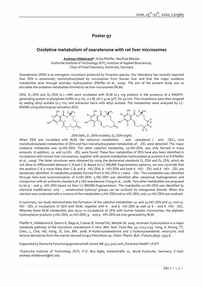
June, 15th‐17th, 2010, Lyngby
141 | P a g e
Poster 97
Oxidative metabolism of zearalenone with rat liver microsomes
Andreas Hildebrand*, Erika Pfeiffer, Manfred Metzler
Karlsruhe Institute of Technology (KIT), Institute of Applied Biosciences, Chair of Food Chemistry, Karlsruhe, Germany
Zearalenone (ZEN) is an estrogenic mycotoxin produced by Fusarium species. Our laboratory has recently reported that ZEN is extensively monohydroxylated by microsomes from human liver and that the major oxidative metabolites arise through aromatic hydroxylation (Pfeiffer et al., 2009). The aim of the present study was to elucidate the oxidative metabolites formed by rat liver microsomes (RLM). ZEN, D2‐ZEN and D6‐ZEN (0.1 mM) were incubated with RLM (0.5 mg protein) in the presence of a NADPH‐generating system in phosphate buffer (0.5 mL, 0.1 M, pH 7.4) at 37°C for 40 min. The incubations were then stopped by adding ethyl acetate (0.5 mL) and extracted twice with ethyl acetate. The metabolites were analyzed by LC‐MS/MS using electrospray ionization (ESI).
OH O
O
CH3
OD
D
HO
H
DD
D D
OH O
O
CH3
OD
D
HO
HOH O
O
CH3
O
HO
H
15
6
8
5
4
10
911
1213
ZEN (left), D2‐ZEN (middle), D6‐ZEN (right)
When ZEN was incubated with RLM, the reductive metabolites �‐ and �‐zearalenol (�‐ and �‐ZEL), nine monohydroxylated metabolites of ZEN and four monohydroxylated metabolites of �‐ZEL were detected. The major oxidative metabolite was 15‐HO‐ZEN. The other catechol metabolite, 13‐HO‐ZEN, was only formed in trace amounts. In addition, 15‐ and 13‐HO‐�‐ZEL were found. These four metabolites of ZEN have also been identified in incubations with human liver microsomes, together with several metabolites hydroxylated at position 6 or 8 (Pfeiffer et al., 2009). The latter structures were obtained by using the deuterated standards D2‐ZEN and D6‐ZEN, which do not allow to differentiate between C‐6 and C‐8. Based on LC‐MS/MS fragmentation patterns, we now conclude that the position C‐6 is more likely than C‐8, and 6�‐HO‐ZEN, 6�‐HO‐ZEN and both 6�‐HO‐�‐ZEL and 6�‐HO‐�‐ZEL are tentatively identified. A metabolite probably formed from 6‐HO‐ZEN is 7‐keto‐�‐ZEL. This metabolite was identified through keto‐enol tautomerization of 6‐HO‐ZEN. 5‐HO‐ZEN was identified after catalytical hydrogenation and comparison with an authentic standard of 5‐HO‐zearalanone (Yang et al., 2008). Two other metabolites are proposed to be 9�‐ and 9�‐HO‐ZEN based on their LC‐MS/MS‐fragmentation. The metabolite 10‐HO‐ZEN was identified by chemical modification: only �,�‐unsaturated hydroxyl groups can be oxidized by manganese dioxide. When this reaction was conducted with a mixture of the metabolites 5‐HO‐ZEN and 10‐HO‐ZEN, only 10‐HO‐ZEN was oxidized. In summary, our study demonstrates the formation of the catechol metabolites 13‐ and 15‐HO‐ZEN and 13‐ and 15‐HO‐�‐ZEL in incubations of ZEN with RLM, together with 6�‐ and 6�‐HO‐ZEN as well as 6�‐ and 6�‐HO‐�‐ZEL. Whereas these RLM metabolites also occur in incubations of ZEN with human hepatic microsomes, the aliphatic hydroxylation products 5‐HO‐ZEN, 10‐HO‐ZEN, 9�‐ and 9�‐HO‐ZEN are only generated by RLM. Pfeiffer E, Hildebrand A, Damm G, Rapp A, Cramer B, Humpf HU, Metzler M. 2009. Aromatic hydroxylation is a major metabolic pathway of the mycotoxin zearalenone in vitro. Mol. Nutr. Food Res. 53: 1123‐1133. Yang, X, Khong, TT, Chen, L, Choi, HD, Kang, JS, Son, BW. 2008. 8'‐Hydroxyzearalanone and 2'‐hydroxyzearalanol: resorcyclic acid lactone derivatives from the marine‐derived fungus Penicillium sp. Chem. Pharm. Bull. (Tokyo) 56(9): 1355‐6. Supported by Deutsche Forschungsgemeinschaft (Grant ME 574‐32/1) and „Food and Health“ of KIT. *Karlsruhe Institute of Technology (KIT), P.O. Box 6980, Kaiserstraße 12, 76128 Karlsruhe, Germany; E‐mail: [email protected]
32th Mycotoxin Workshop
142 | P a g e
Poster 98
In vitro metabolism of enniatin B in rat liver microsomes
Lada Ivanova*, Silvio Uhlig & Christiane K. Fæste
Chemistry Section, National Veterinary Institute, Oslo, Norway
Enniatins are cyclic hexadepsipeptides produced by various fungi, known to have ionophoric, antibiotic and insecticidal activity. They are also known as phytotoxins and are associated with plant diseases characterised by wilt and necrosis. The biological activity of these peptides may in part be explained by their ability to selectively increase the alkali metal ion flux through biological membranes. Mixtures of enniatins or individual enniatin compounds have been shown to possess substantial cytotoxicity. At the same time, cereals in Northern Europe have been reported to be contaminated with high levels of enniatins in recent years. The purpose of this study was to profile and identify the phase I metabolites of enniatin B produced in a rat liver microsome model in the presence of a NADPH‐generating system. Seven major metabolites were tentatively identified using LC‐MS and LC‐MSn. Individual metabolites were also purified in small amounts and studied by high‐resolution MS (HRMS). Metabolites M1‐M5 appeared all to have a molecular mass that was 16 Da higher than that of enniatin B. HRMS unequivocally identified M1‐M5 as monooxygenated species. Fragmentation of the sodiated molecular ions traced the oxidation to either the hydroxyisovaleric or amino acid moieties. Metabolites M6 and M7 appeared to have a molecular mass that was 14 Da and 28 Da lower than that of enniatin B, respectively. These metabolites were tentatively identified to be the result of N‐demethylation of one or two amino acid moieties, respectively. Our findings indicate that oxidation and N‐demethylation are principal metabolic parthways in enniatin B phase I metabolism. * Corresponding author, e‐mail: [email protected]

June, 15th‐17th, 2010, Lyngby
143 | P a g e
Poster 99
Fumonisin B1 mediated alterations in the expression of genes associated with cell growth and invasion in NRK‐52E cells.
Stephanie Müller1, Fausto Bolzon1, Wolfgang Dekant1 & Angela Mally1*
1 Department of Toxicology, University of Würzburg, Würzburg, Germany.
Fumonisin B1 (FB1) is a mycotoxin produced by Fusarium verticillioides, a fungus that grows on crops such as wheat, corn and barley worldwide. In rodents, FB1 is both hepatotoxic and nephrotoxic, and has been shown to induce renal tumors in male F344 rats following chronic exposure via feed (NTP, 2001). Morphologically, the renal tumors induced by FB1 present a rare and highly malignant, anaplastic phenotype, characterized by marked cellular pleomorphism, a high mitotic rate and invasive growth potential. While FB1 does not induce DNA adducts and has been shown to be negative in standard genotoxicity tests indicating that FB1 may operate by a non‐genotoxic mechanism, a comprehensive understanding of the molecular events leading to FB1 induced tumor formation and, particularly, the highly aggressive nature of the carcinomas is still lacking. Several studies have demonstrated that FB1, which is structurally related to the long‐chain sphingoid bases, interferes with sphingolipid metabolism by inhibiting sphinganine N‐acetyltransferase. Considering the importance of sphingolipids in growth control, apoptosis, cell adhesion, migration, and angiogenesis, we speculated that FB1 mediated disruption of sphingolipid metabolism may cause perturbation of signaling pathways which govern cell growth and motility, vascular barrier integrity and angiogenesis, thereby driving tumor formation and invasion. Rat kidney proximal tubular epithelial cells (NRK‐52E) were treated with FB1 at 0, 0.5, 5 and 50 µM FB1 for 24 h and gene expression analysis was performed using a quantitative real‐time PCR array covering pathways critically involved in tumor formation and progression. Of the 84 genes analyzed, 74 genes were significantly altered in response to 0.5 and 5 µM FB1. Consistent with the ability of FB1 to induce cell death by apoptosis, several genes involved in the initiation and execution of apoptosis (Fas, TNFR1, Casp8, Bax) were significantly upregulated in response to FB1. At the same time, however, genes which function to suppress apoptosis and promote growth, including angiogenic growth factors (e.g. Vegfs and Pdgfs), the MAP kinase pathway (e.g. Grb2 and Raf1) and oncogenic transcription factors like c‐Jun, c‐Fos and E2f1, were significantly induced by FB1. Importantly, genes implicated in tumor invasion and metastasis, e.g. matrix metalloproteinases (e.g. Mmp2 and Mmp9), and components of the plasminogen activator system (e.g. uPA, uPAR, Serpine1 and Serpine2), which regulate cell adhesion and facilitate migration by proteolytic remodeling of the extracellular matrix, were markedly upregulated. In summary, our data demonstrate that FB1 causes early changes in the expression of genes which promote growth and invasion, and preliminary results suggest that this is associated with enhanced cell invasion into gelatin matrix. References: NTP (2001). Toxicology and carcinogenesis studies of fumonisin B1 (cas no. 116355‐83‐0) in F344/N rats and B6C3F1 mice (feed studies). Natl Toxicol Program Tech Rep Ser. 1‐352. * Department of Toxicology, University of Würzburg, Würzburg, Germany. [email protected]‐wuerzburg.de

32th Mycotoxin Workshop
144 | P a g e
Poster 100
Deoxynivalenol induced expression of MAPK‐dependent transcription factors in human cell lines
Carina Nielsen*, Maximilian Casteel, Andrea Didier, Richard Dietrich & Erwin Märtlbauer
Department of Veterinary Sciences, Ludwig Maximilians‐Universität München,
Schönleutnerstraße 8, 85748 Oberschleissheim, Germany. Introduction: Contamination of cereals with the trichothecene deoxynivalenol (DON) is a global problem in agriculture. DON‐related cytotoxicity results from inhibition of translation preceded by a ribotoxic stress response, which is characterized by phosphorylation of the mitogen‐activated protein kinases and activation of downstream transcription factors. Considering the chronic dietary exposure of humans, broader investigations on DON influence on cell signalling pathways are required to understand the impact of this mycotoxin on human health. In this study, the expression of downstream genes such as JUN, JUND, FOS, FOSL2, ATF3 and EGR1 in six permanent human cell lines (Hep‐G2, A549, U937, A204, Jurkat and CaCo‐2) of different histological origin was studied after DON treatment. Material and Methods: DON was purchased from Sigma Chemical Company. The mRNA expression profile of AP‐1 associated transcription factors, potentially influenced by the trichothecenes was assayed by the means of real‐time RT‐PCR followed by relative quantification. Time and concentration dependent induction of the transcription factors was studied in detail for Hep‐G2, A549, A204, and U937 cells. Results: We found significant cell‐specific differences in mRNA levels for the transcription factors JUN, JUND, FOS, FOSL2, ATF3, and EGR1. After exposure to 1 µmol/l DON for 3 h, the induction of the transcription factor JUN was highest in the Jurkat (342‐fold) and Hep‐G2 (84‐fold) cell lines. JUND expression was mainly affected in the immunocompetent cell‐lines U937 (11‐fold) and Jurkat (12‐fold), whereas significant FOSL2 induction (5‐fold) was only found in Jurkat cells. DON‐induced expression of FOS was mainly observed in Hep‐G2 (96‐fold) and U937 cells (59‐fold). In contrast, response of A549 cells to DON was characterized by a distinct induction of ATF3 (44‐fold) and EGR1 (92‐fold). Discussion and Conclusion: Taken together the present study underlines the importance of investigation of different cell lines. Time and concentration dependent AP‐1 transcription factor expression seems to be related to overall susceptibility towards DON. Especially the kinetics of survival to death associated transcription factors (JUN/JUND and FOS/FOSL2) may be the underlying mechanism. As human exposure to DON is assumed to be systemic, and clinical symptoms are manifold, the present focus of toxicological studies on lymphoid cell lines may, therefore, underestimate the potential hazard which this mycotoxin may pose on human health. * Corresponding author e‐mail: [email protected]‐muenchen.de

June, 15th‐17th, 2010, Lyngby
145 | P a g e
Poster 101
An occurrence of ochratoxin A and aflatoxin M1 biomarkers in human urine.
Vladimir Ostry1,2*, Jarmila Skarkova2, Radek Kavrik2 & Jiri Ruprich1
1 Department for Food Safety and Nutrition, National Institute of Public Health, Brno, Czech Republic.
2 Laboratory department for Food Safety and Nutrition, National Institute of Public Health, Brno, Czech Republic.
The evaluation of the possible risk of ochratoxin A (OTA) and aflatoxin B1 (AFB1) to human health requires to estimate their daily intake by the population or to determine their biomarkers in biological materials, e.g. blood serum, urine (Malir et al. 2001). The presence of OTA and AFB1 biomarkers has been systematically monitored in biological materials (e.g. human blood serum, human urine) in the Czech Republic (Ostry et al. 2005). The aim of this study was a determination of biomarkers (OTA and AFM1) in human urine. The urine samples were collected in 2007/2008 within EFCOVAL project. Collection of 236 samples of 24‐h urine (male/female, 45‐60 years old, two samples per person from non‐consequative days at least 14 days time difference) was used for the OTA and AFM1 determination. The urine samples were supplied frozen in plastic containers and stored in the laboratory at ‐28 oC. The cleaning of AFM1 and OTA in urine was carried out using immunoaffinity columns AFLAPREP® M and OCHRAPREP® (R‐Biopharm Rhone). The HPLC‐FD method was employed for purposes of the OTA and AFM1
detection and quantification. The limits of quantification (LOQ) of the methods were 2.0 ng/L for OTA, and 0.5 ng/L for AFM1 respectively. The results with high concentration of biomarkers were confirmed by LC‐MS/MS method. The results of total daily urinary OTA and AFM1 biomarkers excretion can be seen in Table 1 and 2. Tab. 1 The results of total daily urinary OTA excretion Sex Male Female n total/n positive 116/107 120/78 OTA
(ng/day) OTA
(ng/kg bw/day) OTA
(ng/day) OTA
(ng/kg bw/day) Mean a 25.4 0.30 9.27 0.15 Median a 14.7 0.17 7.62 0.12 90% perc. a 32.5 0.43 20.2 0.30
a the concentration of OTA <2.0 ng/L considered as ½ LOQ = 1.0 ng/L Tab. 2 The results of total daily urinary AFM1 excretion Sex Male Female n total/n positive 116/10 116/8 AFM1
(ng/day) AFM1
(ng/kg bw/day) AFM1
(ng/day) AFM1
(ng/kg bw/day) Mean b 0.79 0.009 0.77 0.012 Median b 0.61 0.007 0.012 0.010 90% perc. b 1.05 0.014 1.08 0.017
b the concentration of AFM1 <0.5 ng/L considered as ½ LOQ = 0.25 ng/L
References: Malir F, Roubal T, Brndiar M, Osterreicher J, Severa J, Knizek J, Tmejova M, Betbeder AM, Baudrimont I, Creppy EE. 2001. Ochratoxin A in the Czech Republic. J.Toxicol.‐Toxin Reviews 20:261‐274. Ostry V, Malir F, Roubal T, Skarkova J, Ruprich J, Cerna M, Creppy EE, 2005. Monitoring of mycotoxins biomarkers in the Czech Republic. Myc. Res. 21:49‐52.
The urine samples have been collected by the EFCOVAL project (Contract FOOD‐CT‐2006‐022895) investigators and have been made available for this study by the EFCOVAL Consortium. Views expressed in this paper are those of the authors and may not reflect those of EFCOVAL Participants. The content of this presentation reflects only the authors’ views and the Community is not liable for any use that may be made of the information contained therein. * Palackeho 3a, CZ‐612 42 Brno, Czech Republic, [email protected]

32th Mycotoxin Workshop
146 | P a g e
Poster 102
OTA levels in blood and urine and the development of cancer of kidneys and efferent urinary pathways.
Frantisek Malir1,2,3*, Vladimir Ostry4, Miroslav Brndiar5,6 & Tomas Roubal3
1 Department of Biological and Biochemical Sciences, Faculty of Chemical Technology, University Pardubice, Czech
Republic. 2 Department of Biology, Pedagogic Faculty, University Hradec Kralove, Czech Republic.
3 Department of National Reference Laboratory for Biomarkers of Mycotoxins, Institute of Public Health, Hradec Kralove, Czech Republic.
4 Department for Food Safety and Nutrition, National Institute of Public Health, Brno, Czech Republic. 5 Department of II. Internal Medicine – Nephrology, Charles University Medical School Teaching Hospital, Hradec
Kralove, Czech Republic. 6 Department of War Internal Medicine, The Faculty of War Medicine, University of Defence, Hradec Kralove, Czech
Republic.
The Ochratoxin A (OTA) has genotoxic, carcinogenic, teratogenic and immunosuppressive effects in experimental animals. In men it is suspected of being one of the aetiological agents for the Balkan endemic nephropathy and of being associated with urinary tract tumours (Phohl‐Leskowitz A and Manderville RA. 2007. Mol. Nutr. Food Res. 51:61–99). Studies from last 25 years have demonstrated that man is primarily exposed to OTA through his food chain, either directly, e.g. through the consumption of contaminated cereals or oil plants, or indirectly, e.g. through the consumption of contaminated meat or milk. In this regard, specific problems and risks result from the globalization of the food market. OTA means an increased risk for the organism and it affects organism´s physiological degradation and detoxicative potential (Malir F et al. 2001. J.Toxicol.‐Toxin Reviews. 20:261–274,). Exposure to OTA may also result in late toxic effects (e.g. carcinogenic risk) following a single or repeated ingestion of low OTA doses. The dietary exposure of man to OTA can be proved either by analyzing OTA presence in food (average dietary exposure/kg bw/day) or by analyzing OTA biomarkers in a biological material (serum or urine). The later method seems to offer many advantages because it reflects the real exposure of individuals to OTA (Malir F et al. 2006. Mol. Nutr. Food Res. 50:513–518). Even though mycotoxins ‐ including OTA – have been proven to be one of the causal agents in various human and animal diseases, many issues remain unresolved in this regard, in particular the exact mechanisms through which mycotoxins influence human pathologies (in case of OTA, especially the impact of OTA on the development of kidney cancer). Therefore, we prepared the clinical study with an aim to shed more light on the relationship between OTA levels in blood and urine of the patients with kidney cancer (in majority, with diagnosis of Grawitz tumour) and to compare their values with the those of the healthy population. The validated and accredited (CSN EN ISO/IEC 17025) method of HPLC‐FD was employed for purposes of the OTA detection and quantification. The limit of quantification was 0.1 μg l‐1 (Malir F et al. 2001. Myc. Res. 17:39‐44). Validations of this methods and biomarkers confirmations were performed in accordance with the protocol approved by the AOAC. Samples of blood serum were obtained from 40 patients (28 men, 12 women) whose average age was 56.9 years and who suffered from demonstrated carcinoma of kidney without complicated reaction at the time of operation and with the absence of metastases or of total metabolic disruption. Their average mean of OTA serum levels was 0.12 μg l‐1 in comparison with the average mean of OTA serum levels from blood donors which was 0.28 μg l‐1. As the next step, samples of the kidneys should be analyzed with regard to the presence of OTA and their metabolites (Ringot D et al. 2006. Chem. Biol. Interact. 159:18–46).
The study was supported by the System of Monitoring of the Environmental Impact on the Population Health in the CR and by research grant of the CEZ Company.
Dedicated to the memory of Prof. Palle Krogh (*1933 ‐+1990), who substantially contributed to our understanding OTA and its impact on health. *Studentská 573, 532 10 Pardubice, Czech Republic, [email protected]

June, 15th‐17th, 2010, Lyngby
147 | P a g e
Poster 103
Effects of feeding diets containing increasing proportions of bunt infected wheat (Tilletia caries) on performance and health of piglets
Karin Preugschat*, Susanne Döll & Sven Dänicke
Institute of Animal Nutrition, Friedrich‐Loeffler‐Institut (FLI), Federal Research Institute for Animal Health,
Braunschweig, Germany Introduction: Tilletia caries is a fungus that mainly infects wheat causing the disease bunt. Wheat bunt was one of the most dangerous plant diseases, before seed dressing have been a common practice. However, it still is of importance in organic farming, since seed dressing is not allowed. Infected plants are stunted and grain is replaced by bunt balls containing masses of black spores. Trimethylamine (TMA) is a metabolic product of T. caries known to cause a fishy odour of highly contaminated wheat. Therefore, contamination of feed for farm animals can lead to decreased feed intake. However, systematic studies on the effect of practically relevant proportions of bunt infected wheat on performance and health of pigs are not available. It is recommended that the amount of bunt balls in diets for fattening pigs should not exceed 1% (Westermann & al, 1988). Therefore the aim of the present study was to investigate the effects of increasing proportions of bunt infected wheat on performance and health of piglets. Material and methods: Diets containing 0, 10, 20 or 30% bunt infected wheat were produced by stepwise inclusion of infected wheat (TMA concentration below detection limit, 1.86% bunt balls) at the expense of control wheat. The resulting diets were tested on a total of 80 male castrated piglets distributed to 20 boxes (20 piglets/5 boxes per diet). Feed and water were offered for ad libitum consumption for the duration of the trial (5 weeks) and life weights (individual) and feed intake (per box) were recorded weekly. Results: Feeding a diet containing up to 30 % bunt infected wheat resulted in a significant linear decrease in weight gain of the piglets while groups did not differ significantly. The feed intake was numerically but not significantly decreased in groups receiving 20 or 30% infected wheat in the diet while the feed to gain ratio was not affected. Table 1: Effects of feeding diets containing increasing proportions of bunt infected wheat Bunt infected wheat (% in
diet) Feed intake
(g/d) Weight gain
(g/d) Feed to gain ratio
(kg/kg) 0 572 394 1.45 10 581 375 1.55 20 553 351 1.53 30 529 352 1.50
ANOVA (probability) Group 0.539 0.106 0.772 Linear 0.213 0.021 0.707 Quadratic 0.793 0.482 0.372
PSEM1 0.030 0.027 0.068 1PSEM – Pooled standard error of means Conclusions: Under the conditions of the present investigations the linear decrease in weight gain of the piglets hints at adverse effects of the inclusion of bunt infected wheat. Further investigations are needed to clarify the feed safety of bunt infected wheat in diets for piglets. References: Westermann H.‐D. & al. 1988. Gesundheitliche Risiken bei der Verfütterung von Brandweizen. Landwirtschaftliche Forschung 41. 1988. 169‐176 * Karin Preugschat, Institute of Animal Nutrition, Friedrich‐Loeffler‐Institut, Federal Research for Animal Health, Bundesallee 50, 38116 Braunschweig, Germany, [email protected]

32th Mycotoxin Workshop
148 | P a g e
Poster 104
Toxicity guided fractionation of complex Alternaria alternata extract.
Christoph Schwarz1, Martin Kreutzer2, Gudrun Pahlke1, Doris Marko*1
1 Department of Food Chemistry and Toxicology, University of Vienna, Vienna, Austria.
2 Institute of Applied Biosciences, Section for Food Toxicology, Karlsruhe Institute of Technology, Karlsruhe, Germany.
Fungi of the genus Alternaria are relevant to the spoilage of food and feedstuffs, due to their ubiquitous occurrence and their ability to grow even under relatively unfavourable conditions. Though over 40 Alternaria toxins have been described, comparatively little effort has been dedicated to assess their toxicological relevance, as the main toxins alternariol (AOH), alternariol monomethyl ether (AME), altenuene (ALT) and tenuazonic acid (TA) only possess relatively low acute toxicity. Still, epidemiological data indicate a possible correlation between Alternaria contamination of food crops and an increased incidence of esophageal cancer. Moreover, the available toxicological data are often inconclusive, especially when crude extracts of Alternaria spp., wild type isolates were used and the culture conditions were not standardised. We therefore cultivated four strains of A. alternata from the German Collection of Cell Culture Lines and Microorganisms (DSMZ, Braunschweig, Germany) under strictly controlled conditions (autoclaved long grain rice, 25°C, 39% absolute humidity, dark). The resulting crude ethyl acetate extracts showed specific, reproducible toxin profiles and were tested in parallel to their respective main toxin in human colon carcinoma cells (HT29). As the DNA damaging properties of the extracts could not be allocated to their major toxins (AOH, AME, TA), toxicity‐guided fractionation of the most potent extracts was performed, using single cell gel electrophoresis as toxicological endpoint. The DNA damaging effects of the extracts could be narrowed down to three fractions, each containing one UV active peak. In order to obtain sufficient amounts of these substances for identification or structure elucidation, semipreparative HPLC was used (C18 Microsorb column, Varian; H2O(1% FAc) – methanol gradient). Preliminary cleaning of the crude extract was performed using SPE (C18‐E, 10 g, 60 ml) with increasing amounts of methanol in water as eluent. All three relevant peaks were comprised in the 100 % methanol fraction. So far, one potent DNA‐strand breaking peak could be identified as altertoxin II (ATX‐II) by 1H‐ and 13C‐ NMR. The isolation, purification and identification of the remaining substances is object of our ongoing work, as well of the toxicological evaluation of the isolated toxins. * Prof. Dr. Doris Marko, Institut für Lebensmittelchemie und Toxikologie, Universität Wien, Währingerstr. 38, A‐1010 Wien, Austria

June, 15th‐17th, 2010, Lyngby
149 | P a g e
Poster 105
Epigenetic and cis‐regulatory factors affecting FUM1 gene expression for fumonisins biosynthesis in Fusarium verticillioides
V. Montis 1, I. Visentin1, F. Cardinale2*, G. Tamietti1, P. Karlovsky3
1,2Università degli Studi di Torino, Via Leonardo da Vinci,44 – 10095 Grugliasco (TO) Italy
1DiVaPRA – Plant Pathology 2Dip. Colture Arboree – Plant Physiology 3University of Göttingen Grisebachstrasse, 6 ‐ 37077 Göttingen, Germany – Department of Crop Science, Molecular
Phytopathology and Mycotoxin Research
Fumonisins are mycotoxins produced mainly by the pathogenic fungus Fusarium verticillioides (Fv; teleomorph Gibberella moniliformis) that contaminates maize‐based food and feed, causing great concern for human and animal health.
Genes in the fumonisin biosynthetic pathway are included in the so‐called FUM gene cluster. Among them, FUM1 is key to fumonisin production, since strains of F. verticillioides with a disrupted FUM1 fail to produce detectable levels of fumonisins. With this work, we want to explore the possibility that both epigenetic mechanisms and pathway‐specific transcription factors contribute to the observed transcriptional co‐regulation of the genes in the FUM cluster. Namely, the tested hypotheses were: 1) the existence, and relevance for transcription, of cluster‐specific cis‐regulatory motifs on the FUM1 promoter region (pFUM1); and/or 2) the importance of epigenetic mechanisms (histone acetylation/deacetylation, DNA methylation) for FUM1 expression and therefore for fumonisin production.
Results of an in silico analysis suggest the presence of a 6‐bp sequence statistically over‐represented in the FUM gene promoters, compared to the rest of the genome. This motif could cis‐act on the regulation of FUM1, since it is repeated twice in pFUM1. To validate the in silico prediction, a targeted mutant version of it (pFUM1mut) was obtained. Both wt pFUM1 and pFUM1mut sequences were used to drive GFP expression in transgenic Fv strains. Molecular characterization of the Fv transformants is under way, including quantification of endogenous FUM1 and GFP transcripts by qRT‐PCR; this will allow us to validate the role of the identified motif in FUM1 expression.
In parallel, we followed FUM1 expression after treatment with a histone deacetylase inhibitor and investigated the pFUM1 methylation pattern both under fumonisin‐inducing and non‐inducing conditions. Preliminary results suggest that both histone acetylation and pFUM1 methylation may contribute to FUM1 expression under inducing conditions in vitro. * Francesca Cardinale Via L. da Vinci, 44 10095 – Grugliasco (TO), Italy E‐mail: [email protected]

32th Mycotoxin Workshop
150 | P a g e
Poster 106
Enzymatic Synthesis of Mycotoxin Glucuronides.
Tanja Welsch1, Julia Jäger1, & Hans‐Ulrich Humpf1*
1 Institute of Food Chemistry, Westfälische Wilhelms‐Universität Münster, Münster, Germany
The trichothecenes T‐2 toxin and HT‐2 toxin are produced by several fungi of the Fusarium genus. They are important contaminants of food and feed and can be found in oat, wheat and corn. In vitro and in vivo studies showed that T‐2 toxin is metabolized to a variety of substances by typical phase I reactions like hydrolysis, hydroxylation or deepoxidation, with HT‐2 toxin being one of the major metabolites. In phase II reactions these metabolites may also be conjugated with polar moieties like glucuronic acid (Yagen and Bialer 1993). Glucuronidation is catalyzed by UDP‐glucuronosyltransferases (UGTs), which transfer the glucuronide moiety from uridine 5’‐diphosphoglucuronic acid (UDPGA) to the xenobiotic substance, yielding a compound which is excreted more easily. This reaction is widely used to synthesize glucuronides. Since UGTs are located in the membranes of the endoplasmic reticulum, liver microsomes often serve as an enzyme source. For the enzymatic synthesis of T‐2 and HT‐2 glucuronides we used liver microsomes from rodents and pigs. Depending on the animal species different products were formed. To further increase the yield of the corresponding glucuronides the incubation conditions, e.g. incubation time and additives, were optimized. References: Yagen B, Bialer M. 1993.metabolism and pharmacokinetics of T‐2 toxin and related trichothecenes. Drug Metab Rev. 25: 281‐323. *H.‐U. Humpf, Institute of Food Chemistry, Münster (humpf@uni‐muenster.de)
June, 15th‐17th, 2010, Lyngby
151 | P a g e
Poster 107
The Dilemma of Using Aliphatic Alcohols as Solvent: Effects on Electrophysiological Variables of Chicken’s Jejunal Epithelium
Agha W. Yunus1, Wageha Awad2, Susan Kröger2, J. Zentek2 & Josef Böhm1*
1 Institute of Animal Nutrition, University of Veterinary Medicine Vienna, Veterinärplatz 1, A‐1210 Vienna, Austria. 2
Institute of Animal Nutrition, Freie Universität Berlin, Brümmerstrasse 34, D‐14169 Berlin, Germany.
Aliphatic alcohols are commonly used as solvents during in vitro investigations on effects of various drugs/toxins on ion channels in the intestinal epithelial membranes. One feature associated with use of alcohols in such type of studies is their own ability to reduce active absorption of glucose and amino acids, and to induce apoptosis in epithelia. These effects are also observed with exposure of epithelial membranes to various toxins usually dissolved in alcohols. Thus, there are possibilities of synergistic toxicity of solvent and the solute as in case of methanol and T‐2 toxin (A. W. Yunus, unpublished data). It is therefore important to know the borderline dose of alcohols while attempting to study in vitro effects of the toxins/drugs dissolved in these solvents. Such data on aliphatic alcohols in case of chickens and on low levels of aliphatic alcohols in general, are scanty. Present study was therefore executed to investigate the effects of low levels of ethanol and methanol on electrophysiological variables of the jejunal epithelium of chicken. Three separate experiments were conducted to test the effects of ethanol and methanol on electrophysiological variables of chicken’s jejunum. In the first experiment, segments (serosa‐less) from middle jejunum of broilers (4 to 5 wk old) were incubated in Ussing chambers in the presence of zero (control) or 0.1% ethanol. After 40 and 60 min incubation, D‐glucose and carbamoylcholine (analogue of acetylcholine) were respectively added to the incubation medium. The absolute and percent increase in short circuit current (Isc in μA/cm
2) and transmural potential difference (Vt in mV) induced by glucose under 0.1% ethanol treatment were 90% higher (P < 0.01 to 0.04 for different variables) than that under the control. There was no statistically significant effect of 0.1% ethanol on carbamoylcholine‐induced electrophysiological variables. To investigate if higher levels of ethanol have similar effects, we tested the effects of zero (control), 0.3, and 0.6% ethanol under similar experimental conditions until the glucose step. Contrary to 0.1% ethanol in the first experiment, both the 0.3 and 0.6% ethanol levels significantly decreased the basal and glucose‐induced Isc and Vt (P < 0.001 to 0.04 for different variables) as compared to the control. No statistically significant effect of ethanol could be noted on tissue conductivity (Gt in mS/cm2) in these 2 experiments. The third experiment was conducted to investigate if low levels of methanol also act similarly to ethanol. In this case, jejunal segments were incubated in the presence of zero (control) or 0.2% methanol. After 40 and 60 min incubation, glucose and ouabain (specific blocker of Na+K+‐ATPase) were respectively added to the incubation medium. After 40 min incubation with methanol, the basal Isc was 40% higher (P < 0 .05) as compared to that under control. Other basal, and glucose‐induced electrophysiological variables also tended to be high under methanol treatment but no statistical significance could be noted. The ouabain‐induced drop in Isc and Gt in case of methanol treatment were higher (P < 0.01 and 0.05, respectively) than the control. This response from ouabain under 0.2% methanol treatment suggests that the noted effects of ethanol on glucose absorption (in the first experiment) may be due the altered cation gradient across the jejunal epithelium. This is the first report indicating increased active glucose absorption and cation gradient in epithelia (from any specie) in the presence of aliphatic alcohols. Chicken in this regard may be more sensitive to the effects of alcohols. However, a caution is indicated while attempting studies on epithelia from other species. * Corresponding author: Institute of Animal Nutrition, University of Veterinary Medicine Vienna, Veterinärplatz 1, A‐1210 Vienna, Austria. Email: [email protected]

32th Mycotoxin Workshop
152 | P a g e
Poster 108
In vitro Aflatoxin B1 Exposure Increases Apical Anion Secretion in the Jejunal Epithelium of Broilers
Agha W. Yunus1, Wageha Awad2, Susan Kröger2, J. Zentek2 & Josef Böhm1*
1 Institute of Animal Nutrition, University of Veterinary Medicine Vienna, Veterinärplatz 1, A‐1210 Vienna, Austria. 2
Institute of Animal Nutrition, Freie Universität Berlin, Brümmerstrasse 34, D‐14169 Berlin, Germany.
Introdution: Much work has been done until date on the hepatotoxic, carcinogenic, and immunosuppressive effects of aflatoxin B1 (AfB1). However, the literature available on the effects of AfB1 on gastrointestinal tract remains scanty. It has been recently reported that acute AfB1 exposure can evoke acetylcholine sensitive contractions in rat ileum. One effect of acetylcholine and other cholinergic secretagogues is to increase basolateral K+ efflux and apical Cl‐ secretion in epithelia. This increased apical anion secretion is known to reduce Na+ uptake in jejunal epithelium, which in turn can indirectly reduce active glucose absorption. However, these aspects have not been addressed until now. Present study was therefore conducted to test if acute AfB1 exposure can affect ion channel activity in the jejunal epithelia of chicken, and if such an effect is also reflected in terms of reduced active glucose absorption. Materials and Methods: Intestinal segments from middle jejunum of broilers (4 to 5 wk old) were incubated in Ussing chambers. AfB1 solution (250 μg/mL of ethanol) was added to the apical side at the levels of 0, (negative control), 0 (vehicle control), 5, 10, and 15 μL, giving final concentration of AfB1 in the respective chambers as 0, 0, 1.3, 2.5, and 3.7 μg/mL of buffer. Ethanol was added to the respective chambers at the rate of 0, 15, 10, 5, and 0 μL to ensure equal level of solvent in each chamber. After addition of AfB1, tissues were monitored for 40 min to note any change in electrophysiological variables including transmural potential difference (Vt; mV), short circuit current (Isc; μA/cm
2), and tissue conductivity (Gt; mS/cm2). After 40 and 60 minutes of incubation with the tested coumpounds, D‐glucose (20 mmol/L) and carbamoylcholine (200 μmol/L; carbachol; an analogue of acetylcholine and inducer of apical Cl‐ secretion) were respectively added to the incubation medium. Repeated measures ANOVA was used to analyse data statistically.
Vehicle AfB1.3
1
μgAfB 2.5
1
μgAfB 3.7
1
μg
0
1
2
3
*
*Vt
Vehicle AfB1.3
1
μgAfB 2.5
1
μgAfB 3.7
1
μg
0
1
2
3
4
5 *
*
Isc
Figure 1. Average Vt and Isc under vehicle and AfB1 treatments at 12 to 27 min post exposure. Bars bearing * are
different (P < 0.04). Results and Discussion: Addition of AfB1 caused a rise in Vt and Isc between 12 to 27 min post‐exposure as compared to vehicle (figure 1) and negative control, indicating increased cation absorption or anion secretion. As compared to the negative control, the glucose‐induced ΔIsc, and ΔVt were higher under the vehicle control. However this increase over negative control was not seen under any of the AfB1 treatments (all having equal level of vehicle). As compared to the vehicle control, glucose‐induced ΔIsc, and %ΔVt were reduced (P < 0.04) at 2.5, and 3.75 μg AfB1/mL, respectively. The carbamoylcholine‐induced ΔIsc, and ΔVt were both lower (P < 0.05) at 3.75 μg AfB1/mL as compared to the vehicle control. These observations indicate that acute exposure to AfB1 may increase cholinergic and/or Ca++ induced apical anion secretion in the jejunal epithelium of chicken. The negative impact of this increased anion secretion on active glucose uptake was however not prominent and may be considered as moderate to progressive in nature. * Corresponding author: Institute of Animal Nutrition, University of Veterinary Medicine Vienna, Veterinärplatz 1, A‐1210 Vienna, Austria. Email: [email protected]

June, 15th‐17th, 2010, Lyngby
153 | P a g e
Poster 109
Kinetics of Deoxynivalenol and its De‐epoxy Metabolite in Blood Plasma of Broilers after a Single Oral Dose of the Toxin.
Agha W. Yunus1, Hana Valenta2, Sherif M. Abdel‐Raheem3, Susanne Döll2, Sven Dänicke2 & Josef Böhm1*
1 Institute of Animal Nutrition, University of Veterinary Medicine Vienna, Vienna, Austria. 2 Institute of Animal
Nutrition, Friedrich‐Loeffler‐Institut, Braunschweig, Germany. 3 Animal and Clincal Nutrition Department, Faculty of Veterinary Medicine, Assiut Universty, Assiut, Egypt.
Introduction: Chickens are usually considered resistant to the effects of deoxynivalenol (DON). This resistance may result from comparatively lower absorption of DON in chickens or due to microbial transformation of the toxin to unknown compounds in their gut. Various studies documenting metabolism of DON indicate de‐epoxidation to be an important detoxification phenomenon in rats, pigs, cattle, and sheep. However, de‐epoxy‐deoxynivalenol (de‐epoxy‐DON) has not been detected in plasma or tissues of chicken. Furthermore, there is only one study available which documents the plasma levels of DON in chicken just at 2.5 hr post‐exposure (Prelusky et al., 1986). Present study was therefore conducted to investigate the kinetics of DON in plasma of broilers, at 1 to 6 hr post‐exposure, by using liquid chromatography‐tandem mass spectrometry (LC‐MS/MS). Possibility of de‐epoxidation in broilers was also investigated by examining plasma levels of de‐epoxy‐DON. Materials and Methods: One week old Cobb broiler chicks were brooded and reared under standard husbandry conditions. At 5 weeks of age (2±0.2 kg body weight), 4 birds were fasted for 4 hours and then were fed with 20 g of oats naturally contaminated with 9.5 mg DON/kg. Blood samples were collected from 2 birds at 1, 3, and 5 hr, while from the other 2 birds at 2, 4, and 6 hr post‐feeding. Analysis of DON and de‐epoxy‐DON in blood plasma was carried out by using LC‐ESI‐MS/MS (liquid chromatography – electrospray ionization tandem mass spectrometry) after clean‐up with immune affinity columns. The sample preparation, and analysis were according to the methods described by Valenta et al. (2003), and Keese et al. (2008), respectively. The limit of detection for DON and de‐epoxy‐DON in plasma was 0.1 and 0.2 ng/ml, respectively. Results and Discussion: At 1, 3, and 5 hr post‐feeding, the average values of plasma DON were 0.35, 0.20, and 0.15 ng/ml, respectively. The corresponding average values of de‐epoxy‐DON at these time points were 0.70, 0.80, and 0.25 ng/ml, respectively. The sum of DON and de‐epoxy‐DON at 1 hr post‐feeding in these birds was estimated to be 0.044% of the DON fed. At 2, 4, and 6 hr post feeding, the average values of plasma DON were 0.85, 0.45, and 0.30 ng/ml. The total DON in the plasma at 2 hr post‐exposure was 0.036% of the total amount fed. De‐epoxy‐DON could not be detected in the birds sampled at 2, 4, and 6 hr post feeding. This indicates that either the levels of de‐epoxy‐DON in the birds sampled at 2, 4, and 6 hr were lower than the detection limit or there was individual variation in the bird’s capacity to de‐epoxidise DON. Present data indicate very low absorption of DON coupled with a rapid de‐epoxydisation and clearance in broilers. Individual variability in the de‐epoxydation capacity of DON is also indicated. References: Keese C, Meyer U, Valenta H, Schollenberger M, Starke A, Weber I‐A, Rehage J, Breves G, Dänicke S. 2008. No carry over of unmetabolised deoxynivalenol in milk of dairy cows fed high concentrate proportions. Mol. Nutr. Food Res. 52:1514‐1529. Prelusky DB, Hamilton RMG, Trenholm HL, Miller JD. 1986. Tissue distribution and excretion of radioactivity following administration of 14C‐labeled deoxynivalenol to White Leghorn hens. Fundam. Appl. Toxicol. 7:635‐645. Valenta H, Dänicke S, Döll S. 2003. Analysis of deoxynivalenol and de‐epoxy‐deoxynivalenol in animal tissues by liquid chromatography after clean‐up with an immunoaffinity column. Mycotoxin Res. 19:51‐55. * Corresponding author: Institute of Animal Nutrition, University of Veterinary Medicine Vienna, Veterinärplatz 1, 1210 Vienna, Austria. Email: [email protected]

32th Mycotoxin Workshop
154 | P a g e
Poster 110
A preliminary survey of Fumonisin B1 contamination in red cargo rice from retail markets in Thailand
Natthasit Tansakul1 and Sasithorn Limsuwan2,*
1 Department of Pharmacology, Faculty of Veterinary Medicine, Kasetsart University, Bangkok, Thailand. 2 Molecular Phytopathology and Mycotoxin Research Unit, Georg‐August‐University,
Goettingen, Germany
Introduction: Rice is one of the most important foods for human and animals. Red Cargo rice is kind of unpolished rice which has become a popular dish as a nourishing food in Asia, particularly in Thailand. Rice has reportedly been infected by Fusarium spp., a mycotoxigenic fungal [Pitt et al., 1994]. Hence, fumonisins contamination in rice may occur. Fumonisins, a potentially affect sphingolipid biosynthesis which have hepatotoxicity, nephrotoxicity and possibly carcinogenic effects, are of interest to the consumer. In addition, fuminisins cause pathologic effect in animals such as Leukoencephalomalacia in horses and Porcine Pulmonary Edema (PPE) in swine. Information of fumonisins contamination in rice is scarce. Therefore, a preliminary survey of fumonisin B1, the major toxicants of fumonisins, in red cargo rice from retail market in Thailand was studied. Materials and Methods: Thirty eight red cargo rice samples were purchased from different markets in central region of Thailand. 10 g of each sample was soaked under 5 mL of water for 30 min before adding 15 mL of methanol and followed by clean up using strong anion exchange (SAX) cartridges (500 mg sorbent, Varian, CA, USA) column. In short, washing the column with 10 mL of MeOH and precondition it with 10 mL of 75% MeOH. Then, 2 mL of extracted sample were put into the column and followed by 2 steps washing of 75% MeOH (8 mL) and 4 ml MeOH, respectively. Slowly passing of 10 mL 1% acetic acid MeOH (v/v) was applied as elution step. The eluated were analysed with the LC‐ESI‐MS/MS using mobile phase consist of solvent A (0.05% acetic acid in water; v/v) and solvent B (0.05% acetic acid in methanol; v/v). The selected protonate parent ion and daughter ion of FB1 (m/z) were 722.0>686.5. Results and conclusion: The validated method showed a sensitive detection limit (LOD) at 1 µg/kg and limit of quantification (LOQ) at 5 µg/kg. Following the fortification at 50, 100 and 500 µg/kg (n= 5), the mean recoveries were 110.1 ± 13.3 %, 89.3 ± 11.1 % and 91.9 ± 4.6 %, respectively. As a result, two out of thirty eight samples were contaminated with FB1 at very low level (between the LOD and LOQ). Thus, it might deem that fumonisins contamination in Thai red cargo rice is negligible. However, further study of co‐occurrence of mycotoxins and diversity of mycotoxigenic fungi in rice mass production should not be ignored. Reference [1] Pitt J.I., Hocking A. D. , Bhudhasamai K, Miscamble B. F., Wheeler K. A., Tanboon‐Ek P. 1994. The normal mycoflora of commodities from Thailand. 2. Beans, rice, small grains and other commodities. Int. J. Food Microbiol. 23: 35‐53. * University Goettingen, Molecular Phytopathology & Mycotoxin Research, Grisebachstrasse 6, 37077 Goettingen, Germany. email: [email protected]

June, 15th‐17th, 2010, Lyngby
155 | P a g e
Poster 111
Fusarium ear rot and mycotoxin content in genetically modified maize grown in Poland
Piotr Ochodzki and Roman Warzecha
Plant Breeding and Acclimatization Institute, Radzikow, Poland
Introduction: The only genetically modified (GM) maize grown in European Union is Bt modification, protecting plants against European cornborer (Ostrinia nubilalis). Wounded kernels are predisposed to mycotoxin contamination both in the field and during storage. The reduction of mycotoxins content in Bt maize compared to conventional one in some circumstances can be high enough to influence the economical results of breeding. GM maize may also improve both human and animal health. The aim of this studies was comparison of incidence and severity of Fusarium ear rot in both Bt and non‐Bt near‐isogenic maize varieties and comparison of mycotoxins concentration in kernels of transgenic (Bt) and non‐transgenic maize grown is different locations in Poland. Material and methods: Experiments were conducted in 2008 in two locations: in South‐Western and Eastern Poland (Fig.1) where infestation with European cornborer is a serious problem. In each experiment three 1‐hectare plots were sown. Hybrid variety DKC 3420 ( chemically protected against O. nubilalis, and control without protection ) and isogenic Bt‐ containing variety DKC 3421 were sown. Cobs‐ and kernel samples were collected from the field experiments in Autumn. Cobs after collection were evaluated for Fusarium ear rot severity using 0‐5 rating scale. Representative samples of grain were grinded in laboratory mill and mycotoxins: deoxynivalenol (DON) and fumonisins (FB1, FB2, FB3) were determined after extraction with proper solvent (acetonitrile:water (84:16) or methanol) and purification on SPE columns. Analyses were conducted on HPLC system equipped with UV‐VIS (DON) and fluorescence detector (fumonisins) and RP C18 HPLC column. The content of ergosterol as a marker of presence of mycelia was analyzed on HPLC after microwave‐assisted hydrolysis and extraction with n‐pentane. Results: Evaluation of ear rot symptoms and yield of grain showed similar results in both locations. Yield of grain of DKC 3421 YG and chemically protected DKC 3420 was higher than in not protected DKC 3420 (control), but only in location A the difference was statistically significant. Mean grain yield in location A was significantly lower (72,3 g/cob) than in location B (112,9 g/cob). Fusarium ear rot severity was also significantly higher in control field without protection compared to chemically protected or Bt variety. Mycotoxin analysis revealed presence of deoxynivalenol and lack of fumonisins. DON concentration didn’t exceeded limit (1,75 mg/kg grain) in DKC 3421 YG and in chemically protected DKC 3420. The mycotoxin content was exceeded only in control sample from location B in Eastern Poland. Ergosterol content supported data from mycotoxin analysis, especially at higher DON concentrations. Email: [email protected]

32th Mycotoxin Workshop
156 | P a g e
Author Index Abdelfattah, A. 32 Abdel‐Raheem, S. M. 153 Ackermann, Y. 6, 18, 77 Acosta, J. A. T. 25, 127 Adam, G. 25, 35, 40, 115, 127
Agus, A. 7, 41, 130 Ahmed, A. 36 Alam, S. 78 Albers, C. M. 88 Alvarez, A. 30 Amato, B. 79 Amzil, Z. 3 Anderle, S. 45 Asam S. 47 Awad, W. 131, 132, 151, 152 Aydın, S. 84, 122 Balci, S. 120 Barthel, J. 80 Basaran, N. 122 Batzler, J. 9 Bauer, J. 80, 104 Baydarm, T. 120 Behm, C. 133 Bele, C. 100 Berdini, C. 8 Berger, M. 80, 99 Berthiller, F. 25, 35, 40, 72, 115, 127 Biard, J‐F. 119 Biselli, S. 14
Bissett, J. 66 Blajet‐Kosicka, A. 32, 81, 118 Blank, R. 29 Blaszkewicz, M. 30, 106, 107 Boddu, J. 35 Bogdan, L. 129 Böhm, J 32, 132, 151, 152, 153 Bolzon, F. 143 Böttcher, M. 65 Bremser , W. 53 Brera, C. 8 Brezina, U. 48 Briese, K. 52 Brndiar, M 146 Brosig, B. 134 Bruchmann, A. 128 Bucher, S. 36 Burkhardt, B. 135 Buttinger, G. 60
Calderari, T. O. 88 Campos, V. 30 Cardinale, F. 149 Casteel, M. 49, 144 Choi, J. M. 97, 98 Copetti, M. V. 89 Corradini, R. 40 Cossalter, A.M. 33 Cramer, B. 107, 137 Croubels, S. 23, 34
Csutorás, C. 83, 108 Curtui, V. 6, 18, 77 Cvak, B. 50, 58 Czakai, K. 28 Daeseleire, E. 70, 71 Dall’Erta, A. 46, 82 Dall'Asta, C. 46, 82 Dancea, Z. 100, 129 Dänicke, S. 22, 48, 73, 134, 136, 140, 147, 153 Daxner, B. 15 De Backer, P. 23, 34 De Kimpe, N. 56 De Saeger, S. 11, 55, 56, 59, 105 Degen, G. H. 30, 106, 107, 133 Dekant, W. 143 Depoorter, J. 117 Didier, A. 144 Dietrich, R. 49, 77, 144 Dimitrieska‐Stojkovic, E. 69 Ditz, M. 16, 52 Döll, K. 38, 79 Döll, S. 22, 48, 73, 134, 136, 147, 153 Dossena, A. 46, 82 Dubruel, P. 55 Ediage, E. N. 11 Efraim, P. 89 Eggert, K. 39 Ellner, F. K. 4, 37 Erkekoğlu, P. 84, 122 Esper, L. M. R. 88 Essid, E. 121 Fæste, C. K. 142 Falavigna C. 46, 82 Fehr, M. 27, 148 Ferse, I. 137 Feußner, I. 96 Föllmann, W. 133

June, 15th‐17th, 2010, Lyngby
157 | P a g e
Forgó, P. 83, 108 Frisvad, J. C. 90 Fröhlich , J. 72, 102, 114, 116 Joshua, H. 74, 75 Fruhauf, S. 45 Fruhmann, P. 72 Fűtő, P. 83, 108 Galaverna, G. 46, 82 Gardiner, S. A. 35 Gareis, M. 92 Gbore, F. A. 138 Geisen, R. 9, 128 Ghareeb, K. 32 Gilbert, J. 72 Giray, B. 84, 122 Girgin, G. 85, 120 Göbel, C. 96 Gohla, A. 28 Goossen, J. 23, 34 Goßmann, M. 4 Gottschalk, C. 80 Goyarts, T. 136 Grajewski, J. 32, 81, 118 Grenier, B. 21, 33 Griessler, K. 110, 112 Groß, M. 6, 18, 77 Grovel, O. 3, 66, 86, 119 Gürbay, A. 85 Haasnoot, W. 19 Hackenberg, R. 51 Hadjeba‐Medjdoub, K. 31, 87 Haesaert, G. 117 Haesebrouck, F. 23, 34 Hajrulai‐Musliu, Z. 69 Hameed, M. R. 93, 111 Hametner, C. 35, 72, 102, 114, 115, 116 Hampel, M. R. 6 Häubl, G. 58 Hauser, M‐T. 25 Hegewald, A.‐K. 139, 140 Hellmer, H. 115 Heungens, K. 105 Heyndrickx, M. 70, 71 Hildebrand, A. 141 Hollmann, M. 32 Hong, M. J. 67 Hübner, F. 17 Humpf, H‐U. 17, 26, 94, 107, 137, 150 Hussain, Z. 93 Iafrate, E. 8 Idler, C. 16, 52 Itami, K. 137
Ivanova, I. 142 Jäger, J. 150 Javed, I. 93, 111 Jeanclos, E. 28 Jestoi, M. N. 91 Johanning, E. 92 Joshua, H. 74, 75 Joubert, Y. 66 Jung, Y. S. 97, 98 Kahlert, S 140
Kalhoff, H. 106 Kang, C‐W. 125 Kang, J. W. 97, 98 Karacaoglu, E. 120 Karlovsky, P. 12, 36, 38, 79, 95, 96, 149 Kavrik, R. 145 Keese, C. 136 Kerzaon, I. 86, 119 Kettl‐Grömminger, M. 101 Khan, A. 93, 111 Khan, M. Z. 93, 111 Kilic, A. 120 Kleigrewe, K. 94 Klein‐Hartwig, K. 53 Kluess, J. 140 Knudsen, P. B. 90 Koch, M. 42, 53, 68 Kocher, U. 54 Kockmann, M. 107, 137 Kohorst, W. 62 Kokkonen, M. K. 91 Konitzer, K 47 Köppen, R. 53 Kosicki, R. 32, 81, 118 Kreutzer, M. 148 Kreuzberger, M. 95 Kröger, S. 151, 152 Krska, R. 20, 25, 40, 50, 58, 72 Kuhl, E‐M. 96 Kumke, M. 16, 65 Kurniasih 7 Kuzdraliński, A. 126 Lamanaka, B. T. 88, 89 Landsbergis, P. 92 Larsen, T. O. 90 Latif, H. 6, 18, 77 Le Bizec, B. 86 Lecoeur, S. 24 Lee, C. 97, 98 Lee, H. S. 97, 98 Lemmens, M. 127 Lenain, P. 55

32th Mycotoxin Workshop
158 | P a g e
Letzel, T. 63 Liebherr, L. 54 Lierz, M. 6 Limsuwan, S. 12, 38, 95, 154 Lindhauer, M. G. 113 Liu, J. 64 Löhmannsröben, H.‐G. 16, 65 Lohrey, L. 17 Looff, M. 29 Lorenz, N. 6 Loureiro‐Bracarense, A.P. 33 Macri, A. M. 100, 129 Magan, N. 78 Mahmood, S. 93 Malir, F. 146 Mally, A. 28, 143
Maloku, I. 127 Malysheva, S. 56 Manderville, R. 13, 31 Mann, D. 99 Mantle, P. 13 Marchelli, R. 46, 82 Marko, D. 27, 148 Marschik, S. 101 Martin, M. 54 Märtlbauer, E. 49, 77, 144 Maryudhani, Y. M. S. 41, 130 Matscheko, D. 102 Matthes, J. 104 Mavungu, J. D. D. 11, 55 Mayer, S. 20 Mazallon, M. 24 Mazurkiewicz, J. 123 Mehmood, M. A. 111 Meister, U. 103 Merz, D. 25 Metzler, M. 135, 141 Meyer, K. 80, 104 Meyer, U. 22 Miclaus, V. 129 Mikula, H. 72, 102, 114, 116 Mitterauer, A. 57 Mitterbauer, R. 25, 40, 115 Mobashar, M. 29 Mogensen, J. M. 90 Molinelli, A. 50, 58 Moll, W‐D. 21, 33 Monbaliu, S. 59, 105 Monteau, F. 86 Montis, V. 149 Morar, M. V. 100, 129 Moretti, A. 56 Moseriti, A. 60 Muehlbauer, G. J. 35
Mulac, D. 26 Müller, M. 124 Müller, S. 143 Muñoz, K. 30, 106, 107 Muszyńska, M. 126 Nehls, I. 42, 53, 68 Neira, J 30 Nester, M. A. 89 Nielsen, C. 49, 144 Nielsen, K. F. 10, 90 Nipoti, P. 79 Nkengfack, N. 14 Nossol, C. 139, 140 Nuryono 41, 130 Nutz, S. 38 Ochodzki, P. 155 Oeste, K. 63 Oldenburg, E. 37 Ostry, V. 61, 145, 146 Oswald, I. P. 21, 33 Ovesna, J. 61 Pacheco, G. 33 Pahlke, G. 27, 148
Palabiyik, Ş. S. 122
Pannunzi, E. 8 Panther, P. 134, 140 Pasca, N. 129 Pasmans, F. 23, 34 Pawelzik, E. 39, 95 Payandeh, S. 135 Pedrosa, K. 125 Peraica, M. 31 Peters, J. 19 Petzinger, E. 121 Pfeiffer, E. 135, 141 Pfohl‐Leszkowicz, A. 13, 31 Piecuch, P. 94 Pisi, A. 79
Plegge, V. 62 Plessing‐Menze, A. 52 Poirier, L. 3, 66 Polisenska, I. 87
Polizzi, V. 56 Popa, C. 100 Portner, C. 62, 63 Potocka, E. 126 Pouchova, V. 61 Pouchus, Y. F. 3, 66, 86, 119 Powers, S. P. 64 Pranowo, D. 41 Preugschat, K. 147 Prodi, A. 79
June, 15th‐17th, 2010, Lyngby
159 | P a g e
Proske, M. 53 Pussemier, L. 117 Rácz, L. 83, 108 Rapp, M. 80, 99 Rasch, C. 16, 65 Rasche, A. 133 Rasenko, T. 53 Rasmussen, P. H. 10 Rasmussen, R. R. 10 Rathjen, S. 14 Raupp, F. 128 Razzazi‐Fazeli, E. 41 Reinhardt, K. 109 Reinhold, L. 109
Renner, L. 22 Robbens, J. 55, 117 Robiou du Pont, T. 66 Rodrigues, I. 110, 112 Rohe, T. 62 Rolèn, E. 5 Rothkötter, H.J. 134, 139, 140 Roubal, T. 146 Ruiz, N. 3, 66, 119 Ruprich, J. 61, 145 Rychlik, M. 47 Sabuncuoğlu, S. 84, 85 Şahin, G. 84, 85, 120, 122 Saleemi, M. K. 93, 111 Salleh, B. 138 Sallenave‐Namont, C. 3, 66 Schatzmayr, G. 21, 33 Schiessl, A. 67 Schmeitzl, C. 127 Schmidt‐Heydt, M. 9, 128 Schnaufer, R. 101 Schöner, I. 21 Schuhmacher, R. 40, 72 Schwake‐Anduschus, C. 113 Schwartz, H. 21, 45 Schwarz, C. 27. 148 Schwarz, M. 114
Scurtu, I. 129 Seeböck, A. 21, 33 Sekulovski, P. 69 Selmanoglu, G. 120 Šeremet, F. B. 135 Shah, H. U. 78 Shams, M. 25, 40, 115, 127 Siebert, S. 124 Siegel, D. 42, 53, 68 Sieviläinen, E. 91 Simion, V. 129
Skarkova, J. 61, 145
Smedsgaard, J. 10 Solarska, E. 126 Sørensen, J. L. 76
Squilacci, B. 13 Stachel, C. 51 Staiger, S. 14 Stefanovic, V. 31 Stojanovska‐Dimzoska, B. 69 Storm, I. M. L. D. 10 Stupar, R. 35 Südekum, K‐H. 29 Sulyok, M. 20, 72 Svatunek, D. 116 Szakacs, A. R. 100 Tajcs, Z. 83, 108 Tamietti, G. 149
Tangni, E. K. 117 Taniwaki, M. H. 88, 89 Tansakul, N. 154 Tekinalp, G. 85 Teutenberg, T. 63 Thrane, U. 76 Tozlovanu, M. 31, 87 Tozlovanu‐Fergane, M. 87 Twaruzek, M. 32, 81, 118 Uhlig, S. 5, 142 Usleber, E. 6, 18, 77 Utami, M. M. D. 7 Vahjen, W. 131 Valenta, H. 22, 48, 73, 134, 153 Van Bocxlaer, J. 56
van Hove, F. 117 Van Pamel, E. 70, 71 Van Peteghem, C. 11, 55, 56, 59, 105 Van Poucke, K. 105 Vandenbroucke, V. 23, 34 Vansteelandt, M. 119 Vega, M. 30 Vekiru, E. 45
Verbeken, A. 70, 71 Verbrugghe, E. 23, 34 Videmann, B. 24 Visentin, I. 149 Vishwanath, V. 20 Vlaemynck, G. 70, 71 Vrålstad, T. 5 Walte, A. 52 Warth, B. 72 Warzecha, R 155 Wedhastri, S. 41, 130 Welsch, T. 150

32th Mycotoxin Workshop
160 | P a g e
Werner, U. 25 Westphal, A. 29 Wiesenberger, G. 25, 115, 127 Wihandoyo 7 Wilkerling, K. 48, 73 Wu, A. 59 Yamaguchi, J. 137 Yiğit, Ş. 85 Yunianta 130
Yunianto 41 Yunus, A. W. 32, 151, 152, 153 Yurdakök, M. 85
Zahoor‐Ul‐Hassan 93, 111 Zentek, J. 131, 132, 151 Zhang, D. 59 Zheng, M. Z. 67 Zörb, C. 39 Zürn, A. 28

Smart Solutions for Mycotoxins from A to Z From simple screening to simultane-ously determining multiple mycotoxins,our Mycotoxin Test System covers your testing needs from the farm to the supermarket shelf.
• Qualitative test strips for rapid screening – Screen at two differentcutoffs in as little as 3 minutes with no special training or equipment.
• Quantitative immunoassays forFluorometer or HPLC – Get precise fluorometric readings in as little as 10 minutes. Or optimize sample cleanupfor HPLC with your choice of Narrow-Bore, Wide-Bore or Combination-Columns.
• Multi-Analyte-Columns for LC/MS/MS– Extract more than 12 species from asingle sample: determine all analytes inone test run.
Klaus Ruttmann GmbH, Sales and MarketingSchnackenburgallee 179, D-22525 Hamburg [email protected], www.ruttmann.eu
Ruttmann sales and marketing
• precise • accurate • safe • rapid • reliable • easy
Excellent Perspectives: Mycotoxin Test KitsA complete Mycotoxin Test System: For End-to-End Quality Assurance and Compliance
Complete line of Mycotoxin Test Kits• Afla BTM
• AflaCHECKTM
• Afla M1 HPLC• Afla M1 FL• AflaOchra HPLC• AflaTest®
• AflaTest® WB• AOZ HPLC• CitriTest HPLC• DONCHECKTM
• DONtestTM WB• DONtest HPLC• FumoniTestTM
• FumoniTest WB• MYCO6in1TM
(LC/MS/MS)• OchraTestTM
• OchraTest WB• T-2test HPLC• T-2/HT-2 HPLC• ZearalaTestTM
• ZearalaTest WB