Acknowledgments Britt Durham M.D. Chat Dang, M.D. Eugene Hardin, M.D.
Upload
meryl-lydia-perkinsCategory
view
213download
0
3rd Annual Friedman Fellows SymposiumNovember 13th 2010
Pauline Suwandhi, M.D., Amit Seth, M.D., Ashutosh Pareek, D.O., Vanessa Sy, M.D., Leonid Poretsky, M.D., and Donna Seto-Young Ph.
D.
Thiazolidinediones and Bone Metabolism
Beth Israel Medical CenterBeth Israel Medical CenterDivision of Endocrinology and Division of Endocrinology and MetabolismMetabolismAlbert Einstein College of Albert Einstein College of MedicineMedicineNew York, U.S.A.New York, U.S.A.

Thiazolidinediones (TZDs)TZDs are insulin-sensitizing
agents that are widely prescribed in the management of type 2 diabetes mellitus.
TZDs activate the nuclear receptor super family peroxisome-proliferator activator receptor- (PPAR- by binding to the peroxisome proliferator response element (PPRE) and turning on gene transcription. The activated genes include those involved in glucose and lipid metabolism.

TZDs reduce androgen levels and restore ovulation in patients with polycystic ovary syndrome (PCOS)
TZDs also directly reduce estrogen and enhance progesterone production in human ovarian cell culture
Seto-Young et al. 2005, J Clin Endocrinol Metab 90:6099-6105.
TZD effect on Ovarian Steroidogenesis

4
TZDs’ Interaction with Insulin Signaling Pathways and Effect on Steroidogenic
Regulation
Seto-Young et al., 2007, J Clin Endocrinol Metab 92: 2232-2239

5
TZDs’ Interaction with Insulin Signaling Pathways and Effect on Steroidogenic
Regulation Insulin binds to insulin receptor, activates the tyrosine kinase
and stimulates insulin receptor substrate-1 (IRS-1) expression Insulin also activates steroidogenic acute regulatory (StAR)
protein expression which leads to increased progesterone, testosterone and estrogen synthesis
TZDs interact with PPAR- which in turn affect components of insulin signaling pathways.
TZDs indirectly activate insulin receptor, IRS-1 and StAR expression
TZDs increase progesterone production and inhibit testosterone and estrogen synthesis.
TZDs inhibit aromatase activity.
Seto-Young et al., 2007, J Clin Endocrinol Metab 92: 2232-2239

6
TZDs’ effect on Aromatase Activity
TZDs inhibit estrogen synthesisTZDs have no effect on aromatase mRNA or
protein expression, suggesting no effect on gene transcription or protein translation.
In the enzyme kinetics study, TZDs inhibit Vmax and Km of aromatase, acting as un-competitive inhibitors.
Seto-Young et al. 2010 Manuscript submitted to Hormone and Metabolic Research.

7
TZDs, Estrogen and Bone Fragility
Menopause, an estrogen-deficient state, is known to be the cause of osteoporosis; estrogen and its receptor play a major role in bone metabolism
Studies of aromatase inhibitors for the treatment of breast cancer show that letrozole, exemestane and anastrazole induce a decline of bone mineral density (BMD) and increase risk of fracture.
Khosla S, 2010, J Clin Endocrinol Metab 95:356-3577

8
Literature on the effects of TZDs on Bone Metabolism
Clinical trialsClinical trials• Treatment with troglitazone decreases bone turnover in Treatment with troglitazone decreases bone turnover in
patients with DM (Okazaki, et al, 1999)patients with DM (Okazaki, et al, 1999)• TZDs induce bone loss in older DM women (Schwartz et TZDs induce bone loss in older DM women (Schwartz et
al., 2006)al., 2006)• ADOPT trial reported a higher risk of fractures in DM ADOPT trial reported a higher risk of fractures in DM
women treated with rosiglitazone (Kahn et al., 2006)women treated with rosiglitazone (Kahn et al., 2006)• Treatment with rosiglitazone decreases alkaline Treatment with rosiglitazone decreases alkaline
phosphatase (AP) and osteocalcin but has no effect on phosphatase (AP) and osteocalcin but has no effect on bone resorption markers in DM women (Berberoglu et. al, bone resorption markers in DM women (Berberoglu et. al, 2007) 2007)
• ADOPT trial reported that treatment with rosiglitazone ADOPT trial reported that treatment with rosiglitazone increases AP and C-terminal telopeptide (CTX) and increases AP and C-terminal telopeptide (CTX) and reduces procollagen type 1 amino terminal-propeptide reduces procollagen type 1 amino terminal-propeptide (P1NP) (Zinman et al, 2010)(P1NP) (Zinman et al, 2010)

9
In vitro studiesIn vitro studies• TZDs mediate gene transcription and differentiation TZDs mediate gene transcription and differentiation
in mesenchymal progenitor cells to adipocytes and in mesenchymal progenitor cells to adipocytes and increase fat accumulation (Johnson et al., 1999)increase fat accumulation (Johnson et al., 1999)
• TZD inhibits the formation of osteoclast-like cell TZD inhibits the formation of osteoclast-like cell (Okazaki et al., 1999)(Okazaki et al., 1999)
• Rosiglitazone increases apoptosis of osteoblasts Rosiglitazone increases apoptosis of osteoblasts without any change in biomarkers of osteocalcin without any change in biomarkers of osteocalcin and alkaline phosphatase (AP) (Soroceanu et al., and alkaline phosphatase (AP) (Soroceanu et al., 2004)2004)
• Rosiglitazone decreases osteoblast formation Rosiglitazone decreases osteoblast formation markers pro-collagen type-1 N-terminal pro-peptide markers pro-collagen type-1 N-terminal pro-peptide (P1NP) & osteocalcin, but has no effect on (P1NP) & osteocalcin, but has no effect on resorption marker type 1 collagen N-telopeptide resorption marker type 1 collagen N-telopeptide (NTX) (Grey et al., 2007)(NTX) (Grey et al., 2007)
• TZDs inhibit TNF-α-mediated osteoclast-like cells TZDs inhibit TNF-α-mediated osteoclast-like cells differentiation (Yang et al, 2010)differentiation (Yang et al, 2010)
Literature on the effect of TZDs on Bone Metabolism - continued

Bone Turnover Metabolism
Bone has to undergo modeling and remodeling to maintain its structure and function.

11
Modeling and Remodeling
Modeling/construction : bone formation carried out by osteoblasts.
Remodeling/reconstruction : bone resorption carried out by osteoclasts.
Both processes influenced by systemic factors : endocrine (including estrogen level), metabolic and nutritional.

Hypothesis
TZDs inhibit bone metabolism through:aromatase enzyme inhibitiondirect effect on osteoblast/osteoclast

Objective To examine the effects of TZDs on mouse
osteoblast cells : cell growth, bone turnover markers, pro-collagen expression, cell differentiation
To examine whether aromatase inhibition plays a role in any of the TZD effects on mouse osteoblast cells

Methods – Culture System A commercially available mouse osteoblast cell (MOC)
line, 7F2, from American Type Culture Collection (ATCC), was co-cultured with or without human granulosa cells (HGC)
The cells were then incubated with:pioglitazone 25Mrosiglitazone 25MTestosterone 1Mtestosterone 1M+ pioglitazone 25Mtestosterone 1M+ rosiglitazone 25M

TZDs Inhibit Estradiol Synthesis
Pioglitazone inhibited estradiol synthesis in the MOC and HGC co-culture
p <0.001
p <0.001

TZD Effect on MOC-HGC Cell Growth(Optical Density)
TZDs inhibit cell growth. Testosterone can ameliorate the cell growth inhibition caused by TZDs.
Incubation time (day)
0 2 4 6 8
10
100
ControlTestosteroneRosiglitazoneTestosterone + Rosiglitazone
Incubation time (day)
0 2 4 6 8
Op
tic
al
De
ns
ity
(Co
mp
are
d t
o D
ay
7 C
on
tro
l)
10
100
ControlTestosteronePioglitazoneTestosterone + Pioglitazone
p<0.02

TZD effect on MOC Cell Growth (Optical Density)
Pioglitazone & Rosiglitazone are associated with decreased MOC growth as measured by optical density.
Incubation Time (Days)
2 3 4 5 6 7
Op
tica
l Den
sity
(C
om
par
ed t
o D
ay 7
Co
ntr
ol)
10
100
ControlTestosteronePioglitazoneTestosterone + PioglitazoneRosiglitazoneRosiglitazone + Testosterone

TZDs Affect Cell Growth in a Dose-Dependent Manner
Op
tica
l Den
sity
(%
Co
ntr
ol
0
20
40
60
80
100
120

TZD Effect on Osteoblast Growth/Differentiation
Thiazolinediones inhibit cell growth and increase fat accumulation.MOC cultures were stained with Oil Red O to highlight the presence of adipocytes.

TZD Effect on Alkaline Phosphatase (AP) Activity
Pioglitazone and Rosiglitazone are associated with decreased AP activity levels. Addition of testosterone to MOC+HGC co-culture “protects” AP activity levels from effects of Thiazolidinedione.
M.O.C. + H.G.C.
Alkaline Phosphatase Specific Activity
Sp
ec
ific
Ac
tivi
ty
Cell Sample Lysate
C P T T + P0
20
40
60
80
100
ControlPioglitazoneTestosteroneTestosterone + Pioglitazone
C P R T T + P T + R0
20
40
60
80
100
120
140
ControlPioglitazoneRosiglitazoneTestosteroneTestosterone+PioglitazoneTestosterone+Rosiglitazone
M.O.C.
Alkaline Phosphatase Specific ActivityS
pe
cif
ic A
cti
vity
Cell Sample Lysate
p<0.001 p<0.001
p<0.001p<0.001
p<0.025
p<0.029

TZD Effect on Osteocalcin Synthesis
Pioglitazone reduces osteocalcin production in MOC-HGC co-culture and MOC culture.
M.O.C. + H.G.C. Osteocalcin Specific Activity
Sp
ecif
ic A
ctiv
ity
Cell Sample Lysate
C P T T + P0
20
40
60
80
100
120
ControlPioglitazoneTestosteroneTestosterone + Pioglitazone
C P R T T + P T + R0
20
40
60
80
100
120
140
160
180
ControlPioglitazoneRosiglitazoneTestosteroneTestosterone+PioglitazoneTestosterone+Rosiglitazone
M.O.C. Osteocalcin Specific Activity
Sp
ecif
ic A
ctiv
ity
Cell Sample Lysate
p<0.002 p<0.005
p<0.037
p<0.009
p<0.03
p<0.04

Mouse Pro-collagen mRNA expression
Pioglitazone and rosiglitazone inhibit mouse pro-collagen mRNA expression

Conclusions Pioglitazone and rosiglitazonePioglitazone and rosiglitazone
inhibit osteoblast cell growthinhibit osteoblast cell growth decrease bone turnover biomarkers (AP and osteocalcin decrease bone turnover biomarkers (AP and osteocalcin
levels)levels) decrease mouse pro-collagen mRNA expressiondecrease mouse pro-collagen mRNA expression increase differentiation to adipocytesincrease differentiation to adipocytes
Inhibition of aromatase by TZDs does not play a role in the osteoblast cell growth or in the effects of TZDs on bone turnover markers, since inhibition of cell growth and the effects on bone turnover markers were observed in MOC culture which did not contain granulosa cells.
Results are consistent with clinical studies showing Results are consistent with clinical studies showing increased fracture risk and bone loss in patients with increased fracture risk and bone loss in patients with diabetes treated with TZDs.diabetes treated with TZDs.
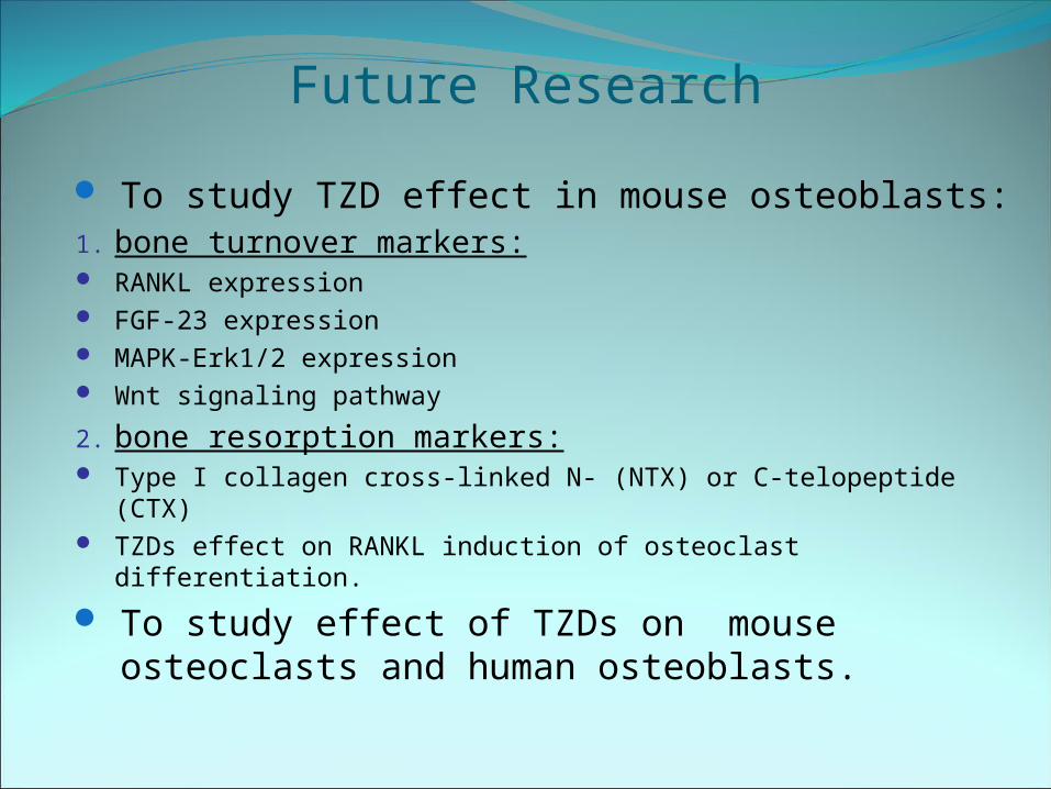
Future Research
To study TZD effect in mouse osteoblasts:1. bone turnover markers: RANKL expression FGF-23 expression MAPK-Erk1/2 expression Wnt signaling pathway
2. bone resorption markers: Type I collagen cross-linked N- (NTX) or C-telopeptide (CTX) TZDs effect on RANKL induction of osteoclast differentiation.
To study effect of TZDs on mouse osteoclasts and human osteoblasts.

Thank You!