![Chiropractic treatment of idiopathic scoliosis with the ......Idiopathic scoliosis (IS) is the most common spinal deformity seen in school-age children [1]. According to the National](https://static.fdocuments.us/doc/165x107/6021a0e84b312545bc186f20/chiropractic-treatment-of-idiopathic-scoliosis-with-the-idiopathic-scoliosis.jpg)
3 Pathogenesis of Idiopathic Scoliosis · Pathogenesis of Idiopathic Scoliosis 3 Robert A. Dickson...
23
Pathogenesis of Idiopathic Scoliosis Robert A. Dickson 3 28 A clear understanding of the pathogenesis of idiopathic sco- liosis is essential to appreciating its clinical behavior. Although epidemiological surveys have shown an enormous number of children with minor coronal-plane deformities of the spine, only a relatively small number show evidence of progressive idiopathic scoliosis, which means that other fac- tors must be superimposed to make a deformity idiopathic and progressive. The environment of growth is clearly important, as with other progressive skeletal deformities. The prevalence rate of minor curves in boys is about half that for each age in girls, and the difference is progressively more obvious the greater the size of the curve. Boys are therefore in some way protected from idiopathic scoliosis, but their spines are going to be subjected to much the same neuromuscular, metabolic, and endocrine processes during growth as those of girls. On a commonsense basis it would seem unlikely that pointing the finger of suspicion, for in- stance, at the paravertebral musculature, brain, eyes, ears, spinal cord, nerve roots, muscles, collagen, and even platelets would not be a profitable line of research into the cause of idiopathic scoliosis. Notwithstanding, this path has largely been the focus of research activity in the etiology of idiopathic scoliosis over the past half century. ■ Muscular Theories Most hypotheses and speculations put forward about the cause of idiopathic scoliosis concern neuromuscular theo- ries, although idiopathic scoliosis is defined as a spinal cur- vature in the absence of any associated musculoskeletal condition. Since Lerique and Le Coeur first demonstated electromyographic asymmetry in the paraspinal muscles of patients with the condition, 1 much work on the paraspinal musculature has been performed, including studies of mus- cle fiber type and ultrastructural differences, 2–11 although Zetterberg and colleagues showed that these abnormalities resembled the sort of changes encountered after endurance training, indicating a secondary or adaptive process (i.e., secondary to the presence of a spinal curvature) in their occurrence. 12 Saartok et al went even further, stating that “a neuromuscular imbalance has not been shown to be an eti- ological factor for the idiopathic form of scoliosis.” 13 The histological specimens were obtained during scoliosis sur- gery from a highly selected group of patients with larger curves, focusing on the difference between the right and left sides, when the geometric problem is instead a front-to- back buckling lordosis (vide infra). Platelets contain actin and myosin, and because of their resemblance to those in skeletal muscle, these structures have also been examined in idiopathic scoliosis. Early stud- ies reporting abnormal morphology and function of platelets in patients with idiopathic scoliosis 14–16 were not confirmed in subsequent studies, which showed no differ- ence between patients with idiopathic scoliosis and con- trols. 17,18 Platelet aggregation abnormalities were shown to be more prevalent in those with larger scoliotic curves, again indicating a secondary effect. 19,20 Calmodulin regu- lates the contractile properties of muscles and platelets through changes in calcium concentration, and higher platelet calmodulin levels have been demonstrated in patients with progressive curves, 21 as have lower levels of melatonin, a calmodulin antagonist. 22 It is unclear what these changes reflect other than the biochemistry of growth in general. ■ Neurological Theories Because scoliosis is associated with many neurological dis- eases, from those affecting the brain to those of peripheral nerves, several neurological abnormalities have been described in association with idiopathic scoliosis. Electroencephalography, proprioception, and vibration sense have been examined in idiopathic scoliosis, as well as balance and electronystagmography. 23–26 Recently, abnor- mal somatosensory function has been demonstrated in patients with adolescent idiopathic scoliosis, 27,28 but its relationship to curve severity again suggests changes sec- ondary to the presence of a deformity. Forty years ago it was thought that idiopathic scoliosis could be caused by a short spinal cord, 29 and more recently Porter has shown that in patients with the condition the spinal canal is shorter than the spine itself. 30–32 This theory has been fan- cifully called “uncoupled neuro-osseous growth,” 32 and the concept of this difference in length has also been supported through screening with magnetic resonance imaging (MRI). 33 Thirty years ago in Oxford, cadaver spines with idiopathic scoliosis were measured and it was shown that the spinal canal takes the shortest route down the spine. 34
Transcript of 3 Pathogenesis of Idiopathic Scoliosis · Pathogenesis of Idiopathic Scoliosis 3 Robert A. Dickson...

Pathogenesis of Idiopathic ScoliosisRobert A. Dickson3
28
A clear understanding of the pathogenesis of idiopathic sco-
liosis is essential to appreciating its clinical behavior.
Although epidemiological surveys have shown an enormous
number of children with minor coronal-plane deformities of
the spine, only a relatively small number show evidence of
progressive idiopathic scoliosis, which means that other fac-
tors must be superimposed to make a deformity idiopathic
and progressive. The environment of growth is clearly
important, as with other progressive skeletal deformities.
The prevalence rate of minor curves in boys is about half
that for each age in girls, and the difference is progressively
more obvious the greater the size of the curve. Boys are
therefore in some way protected from idiopathic scoliosis,
but their spines are going to be subjected to much the same
neuromuscular, metabolic, and endocrine processes during
growth as those of girls. On a commonsense basis it would
seem unlikely that pointing the finger of suspicion, for in-
stance, at the paravertebral musculature, brain, eyes, ears,
spinal cord, nerve roots, muscles, collagen, and even
platelets would not be a profitable line of research into the
cause of idiopathic scoliosis. Notwithstanding, this path has
largely been the focus of research activity in the etiology of
idiopathic scoliosis over the past half century.
■ Muscular TheoriesMost hypotheses and speculations put forward about the
cause of idiopathic scoliosis concern neuromuscular theo-
ries, although idiopathic scoliosis is defined as a spinal cur-
vature in the absence of any associated musculoskeletal
condition. Since Lerique and Le Coeur first demonstated
electromyographic asymmetry in the paraspinal muscles of
patients with the condition,1 much work on the paraspinal
musculature has been performed, including studies of mus-
cle fiber type and ultrastructural differences,2–11 although
Zetterberg and colleagues showed that these abnormalities
resembled the sort of changes encountered after endurance
training, indicating a secondary or adaptive process (i.e.,
secondary to the presence of a spinal curvature) in their
occurrence.12 Saartok et al went even further, stating that “a
neuromuscular imbalance has not been shown to be an eti-
ological factor for the idiopathic form of scoliosis.”13 The
histological specimens were obtained during scoliosis sur-
gery from a highly selected group of patients with larger
curves, focusing on the difference between the right and left
sides, when the geometric problem is instead a front-to-
back buckling lordosis (vide infra).
Platelets contain actin and myosin, and because of their
resemblance to those in skeletal muscle, these structures
have also been examined in idiopathic scoliosis. Early stud-
ies reporting abnormal morphology and function of
platelets in patients with idiopathic scoliosis14–16 were not
confirmed in subsequent studies, which showed no differ-
ence between patients with idiopathic scoliosis and con-
trols.17,18 Platelet aggregation abnormalities were shown to
be more prevalent in those with larger scoliotic curves,
again indicating a secondary effect.19,20 Calmodulin regu-
lates the contractile properties of muscles and platelets
through changes in calcium concentration, and higher
platelet calmodulin levels have been demonstrated in
patients with progressive curves,21 as have lower levels of
melatonin, a calmodulin antagonist.22 It is unclear what
these changes reflect other than the biochemistry of growth
in general.
■ Neurological TheoriesBecause scoliosis is associated with many neurological dis-
eases, from those affecting the brain to those of peripheral
nerves, several neurological abnormalities have been
described in association with idiopathic scoliosis.
Electroencephalography, proprioception, and vibration
sense have been examined in idiopathic scoliosis, as well as
balance and electronystagmography.23–26 Recently, abnor-
mal somatosensory function has been demonstrated in
patients with adolescent idiopathic scoliosis,27,28 but its
relationship to curve severity again suggests changes sec-
ondary to the presence of a deformity. Forty years ago it
was thought that idiopathic scoliosis could be caused by a
short spinal cord,29 and more recently Porter has shown
that in patients with the condition the spinal canal is
shorter than the spine itself.30–32 This theory has been fan-
cifully called “uncoupled neuro-osseous growth,”32 and the
concept of this difference in length has also been supported
through screening with magnetic resonance imaging
(MRI).33 Thirty years ago in Oxford, cadaver spines with
idiopathic scoliosis were measured and it was shown that
the spinal canal takes the shortest route down the spine.34
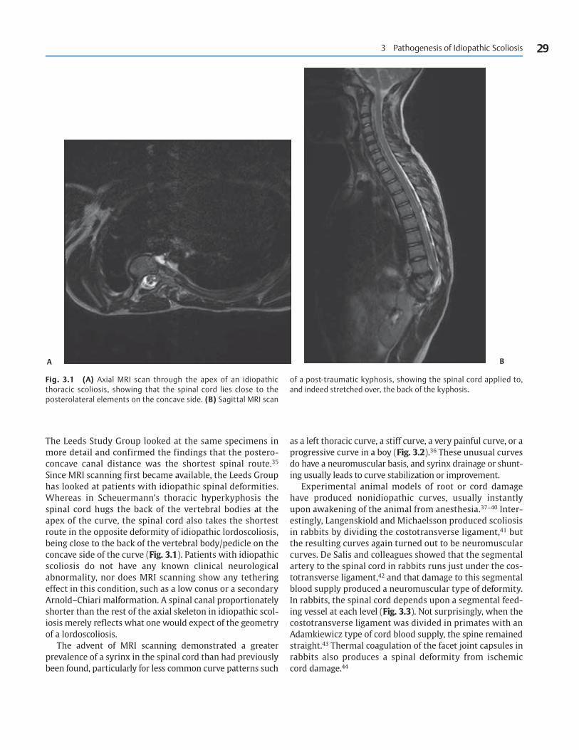
The Leeds Study Group looked at the same specimens in
more detail and confirmed the findings that the postero-
concave canal distance was the shortest spinal route.35
Since MRI scanning first became available, the Leeds Group
has looked at patients with idiopathic spinal deformities.
Whereas in Scheuermann’s thoracic hyperkyphosis the
spinal cord hugs the back of the vertebral bodies at the
apex of the curve, the spinal cord also takes the shortest
route in the opposite deformity of idiopathic lordoscoliosis,
being close to the back of the vertebral body/pedicle on the
concave side of the curve (Fig. 3.1). Patients with idiopathic
scoliosis do not have any known clinical neurological
abnormality, nor does MRI scanning show any tethering
effect in this condition, such as a low conus or a secondary
Arnold–Chiari malformation. A spinal canal proportionately
shorter than the rest of the axial skeleton in idiopathic scol-
iosis merely reflects what one would expect of the geometry
of a lordoscoliosis.
The advent of MRI scanning demonstrated a greater
prevalence of a syrinx in the spinal cord than had previously
been found, particularly for less common curve patterns such
as a left thoracic curve, a stiff curve, a very painful curve, or a
progressive curve in a boy (Fig. 3.2).36 These unusual curves
do have a neuromuscular basis, and syrinx drainage or shunt-
ing usually leads to curve stabilization or improvement.
Experimental animal models of root or cord damage
have produced nonidiopathic curves, usually instantly
upon awakening of the animal from anesthesia.37–40 Inter-
estingly, Langenskiold and Michaelsson produced scoliosis
in rabbits by dividing the costotransverse ligament,41 but
the resulting curves again turned out to be neuromuscular
curves. De Salis and colleagues showed that the segmental
artery to the spinal cord in rabbits runs just under the cos-
totransverse ligament,42 and that damage to this segmental
blood supply produced a neuromuscular type of deformity.
In rabbits, the spinal cord depends upon a segmental feed-
ing vessel at each level (Fig. 3.3). Not surprisingly, when the
costotransverse ligament was divided in primates with an
Adamkiewicz type of cord blood supply, the spine remained
straight.43 Thermal coagulation of the facet joint capsules in
rabbits also produces a spinal deformity from ischemic
cord damage.44
3 Pathogenesis of Idiopathic Scoliosis 29
A B
Fig. 3.1 (A) Axial MRI scan through the apex of an idiopathicthoracic scoliosis, showing that the spinal cord lies close to theposterolateral elements on the concave side. (B) Sagittal MRI scan
of a post-traumatic kyphosis, showing the spinal cord applied to,and indeed stretched over, the back of the kyphosis.

30 Idiopathic Scoliosis
A B
Fig. 3.2 (A) PA view of the lower cervical/upper thoracic spine of a boy with a painful thoracic “idiopathic” scoliosis, showing the typicalgross interpedicular widening of a syrinx. (B) Sagittal MRI scan of the same region, showing a very large syrinx.
Fig. 3.3 Dissection of the segmental blood supply to the spinalcord in the rabbit. The blood supply depends upon a feeding vesselat each level.
■ Connective-tissue AbnormalitiesBecause connective tissue disorders such as Marfan or
Ehlers–Danlos syndrome are associated with an increased
prevalence of spinal deformity collagen structure and me-
tabolism have been extensively investigated both in the skin
and in the intervertebral discs in idiopathic scoliosis.45–50
Again the findings were thought to be secondary to the
presence of a spinal deformity, and indeed, recent research
has excluded collagen abnormalities as potential genetic
causes of idiopathic scoliosis.51,52
■ Genetic TheoriesIn the late 1960s and early 1970s, the familial nature of
idiopathic scoliosis was clearly demonstrated in both Scot-
land53 and the United States.54 It was thought that idiopathic
scoliosis might be inherited in a sex-linked dominant
mode, but with variable expressivity and incomplete pene-
trance. Genomic screening and chromosome studies have
suggested chromosome 19 as a possible candidate for a
genetic source of the disorder,55,56 but idiopathic scoliosis is
so multifactorial that it is extremely unlikely that only a
single gene is responsible for it.
Longitudinal studies of growth in relation to idiopathic
scoliosis show that early reports of children with the con-
dition having been taller and having advanced earlier in

adolescent growth, while later experiencing growth re-
tardation, were not strictly correct because they relied
upon historical controls already shown to be unreliable.57
When compared with contemporaneous controls, these
children showed no differences in comparison with
straight-backed counterparts. However, children with
bigger curves are significantly taller at each age, but do
not grow faster, indicating that a genetically tall stature
may be related to the progression potential of idiopathic
scoliosis.57 In such families one would expect to find a
high prevalence rate of idiopathic scoliosis, and the con-
cept of a gene for idiopathic scoliosis therefore loses cred-
ibility when the familial nature of the disorder can be
explained in part on the basis of stature. Moreover, the
whole pattern of growth during adolescence is strongly
familial,58 with, for instance, girls and their mothers
having their menarches at similar chronological ages.
If idiopathic scoliosis is a matter of abnormal spinal
shape that runs in families, then how that shape is achieved
must also be genetically determined. Delmas59 and Stag-
nara et al60 both put forward the notion that children have
a spinal physiognomy just as they have, for instance, a facial
one, and suggested that lateral profile may be governed
genetically just as are many other aspects of body shape.
Recently, the familial nature of sagittal spinal shape has
been investigated in schools, using the Quantec surface-shape
scanning technique, which can noninvasively register the lat-
eral spinal profile.61 We were particularly interested in the
mid-lower thoracic spine, where idiopathic thoracic scoliosis
is apical. We compared unrelated children of the same age
and sex, opposite-sex siblings, and same-sex siblings, and
then went to the Society of Twins meeting in the United
Kingdom (Twins and Multiple Births Association) and exam-
ined both nonidentical and identical twins. With progression
up the hierarchy from unrelated children to identical twins,
the lateral profile of the lower thoracic spine steadily increas-
ingly correlated with kinship, with identical twins having the
same thoracic spinal shape (Fig. 3.4).
■ How the Three-DimensionalDeformity Develops
In trying to understand the pathogenesis of idiopathic scol-
iosis, it is useful to consider how the deformity develops
and to start with some basic clinical and radiological obser-
vations. Considering thoracic scoliosis, in which the
changes in spinal shape are most obvious, the deformity
looks much less impressive in the erect position than on
forward bending, when the rib hump is maximized
(Fig. 3.5). This was observed by Adams 160 years ago,62 but
its importance was not appreciated by others for many
years. Clearly, something mechanical is happening to the
spinal column from the erect to the forward-bend position.
When posteroanterior (PA) X-ray films of idiopathic sco-
liosis are inspected, it can be observed that the direction of
rotation of the spine is constant, with the posterior ele-
ments turning toward the curve concavity, and with this
rotation being maximal at the curve apex. (Fig. 3.6; see also
Fig. 2.4) The posterior elements of the spine are therefore
running, as it were, the shorter, inside lane of the “running
track,” as this appearance clearly indicates that the back of
the spine must be shorter than the front. The PA X-ray view
of the patient’s spine is, however, a PA view of everything
except the structural curve, because from the neutral
vertebra above down to the apex of the scoliotic curve, each
vertebra is progressively more rotated out of the frontal
plane before recovering from the apical vertebra to the
lower neutral vertebra. If the apical vertebra is, for instance,
rotated by 30 degrees, then to make a true anteroposterior
(AP) film, either the patient or the X-ray beam has to be rotated
by 30 degrees from the frontal plane, in which case the size of
the deformity is maximized. Stagnara devised this AP view
and termed it the plan d’election view (see Figs. 2.9 and
2.10).63 If a true lateral X-ray film of the curve apex is to be
made, the X-ray beam has to be rotated 90 degrees with
reference to the AP plan d’election view (Fig. 3.7). When this
is done, the essential lordosis is visualized.
The Leeds Group studied articulated skeletons with idio-
pathic scoliosis at the Royal College of Surgeons of Edinburgh
Museum, which helped to visualize the lordosis and the
nature of the seemingly complex three-dimensional
deformity in the disorder.35,64 Figure 3.8 shows one such
specimen. The PA view shows a significant deformity, with
3 Pathogenesis of Idiopathic Scoliosis 31
Fig. 3.4 Lateral spinal profile measured in children in a schoolscreening program with a surface-shape computer. This histogram ofcorrelation coefficients demonstrates that as one passes from mixed-sex siblings through same-sex, mixed-sex dizygotic, and same-sexdizygotic to monozyotic siblings (from left to right), the correlationcoefficients steadily increase in magnitude, indicating ever closer cor-respondence between lateral spinal profiles until with identical twinsthe lateral spinal profiles are virtually the same. This is a very impor-tant genetic element in the pathogenesis of idiopathic scoliosis.
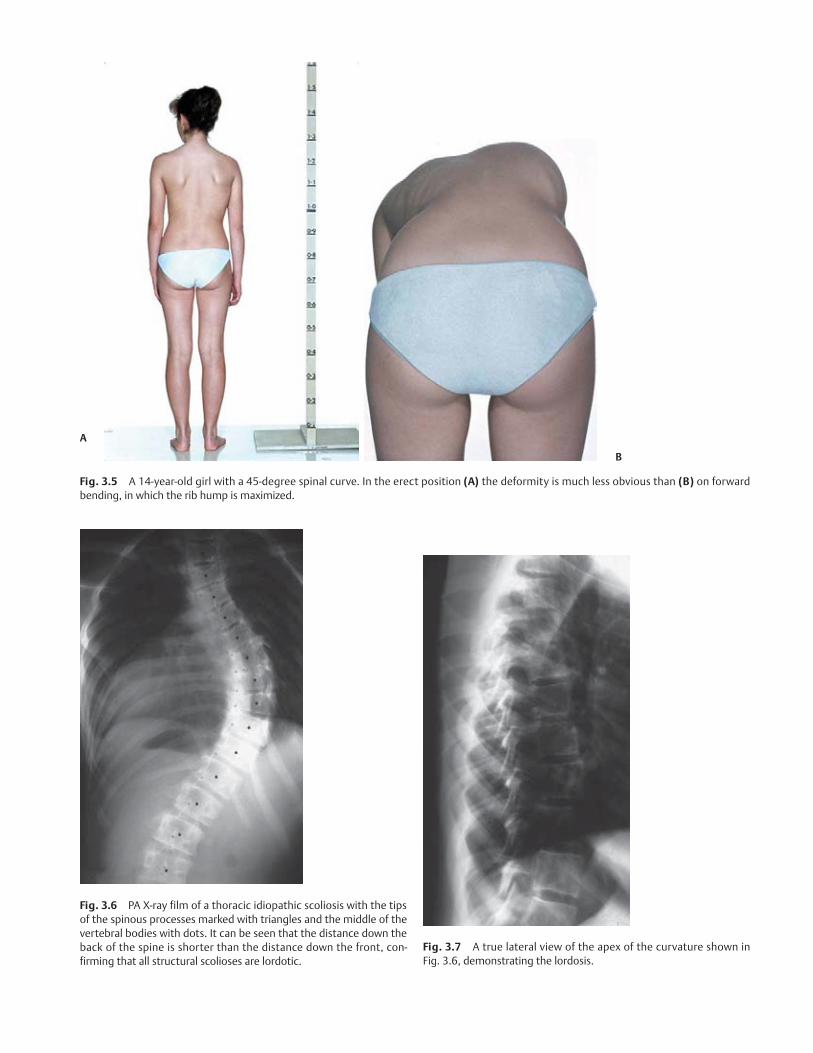
A
B
Fig. 3.5 A 14-year-old girl with a 45-degree spinal curve. In the erect position (A) the deformity is much less obvious than (B) on forwardbending, in which the rib hump is maximized.
Fig. 3.6 PA X-ray film of a thoracic idiopathic scoliosis with the tipsof the spinous processes marked with triangles and the middle of thevertebral bodies with dots. It can be seen that the distance down theback of the spine is shorter than the distance down the front, con-firming that all structural scolioses are lordotic.
Fig. 3.7 A true lateral view of the apex of the curvature shown inFig. 3.6, demonstrating the lordosis.
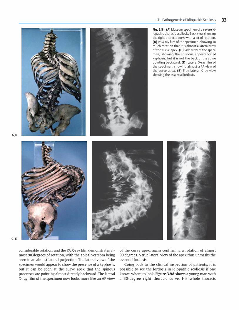
considerable rotation, and the PA X-ray film demonstrates al-
most 90 degrees of rotation, with the apical vertebra being
seen in an almost lateral projection. The lateral view of the
specimen would appear to show the presence of a kyphosis,
but it can be seen at the curve apex that the spinous
processes are pointing almost directly backward. The lateral
X-ray film of the specimen now looks more like an AP view
of the curve apex, again confirming a rotation of almost
90 degrees. A true lateral view of the apex thus unmasks the
essential lordosis.
Going back to the clinical inspection of patients, it is
possible to see the lordosis in idiopathic scoliosis if one
knows where to look. Figure 3.9A shows a young man with
a 30-degree right thoracic curve. His whole thoracic
3 Pathogenesis of Idiopathic Scoliosis 33
A,B
C–E
Fig. 3.8 (A) Museum specimen of a severe id-iopathic thoracic scoliosis. Back view showingthe right thoracic curve with a lot of rotation.(B) PA X-ray film of the specimen, showing somuch rotation that it is almost a lateral viewof the curve apex. (C) Side view of the speci-men, showing the spurious appearance ofkyphosis, but it is not the back of the spinepointing backward. (D) Lateral X-ray film ofthe specimen, showing almost a PA view ofthe curve apex. (E) True lateral X-ray viewshowing the essential lordosis.
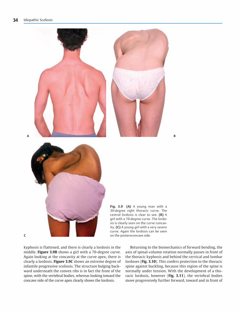
kyphosis is flattened, and there is clearly a lordosis in the
middle. Figure 3.9B shows a girl with a 70-degree curve.
Again looking at the concavity at the curve apex, there is
clearly a lordosis. Figure 3.9C shows an extreme degree of
infantile progressive scoliosis. The structure bulging back-
ward underneath the convex ribs is in fact the front of the
spine, with the vertebral bodies, whereas looking toward the
concave side of the curve apex clearly shows the lordosis.
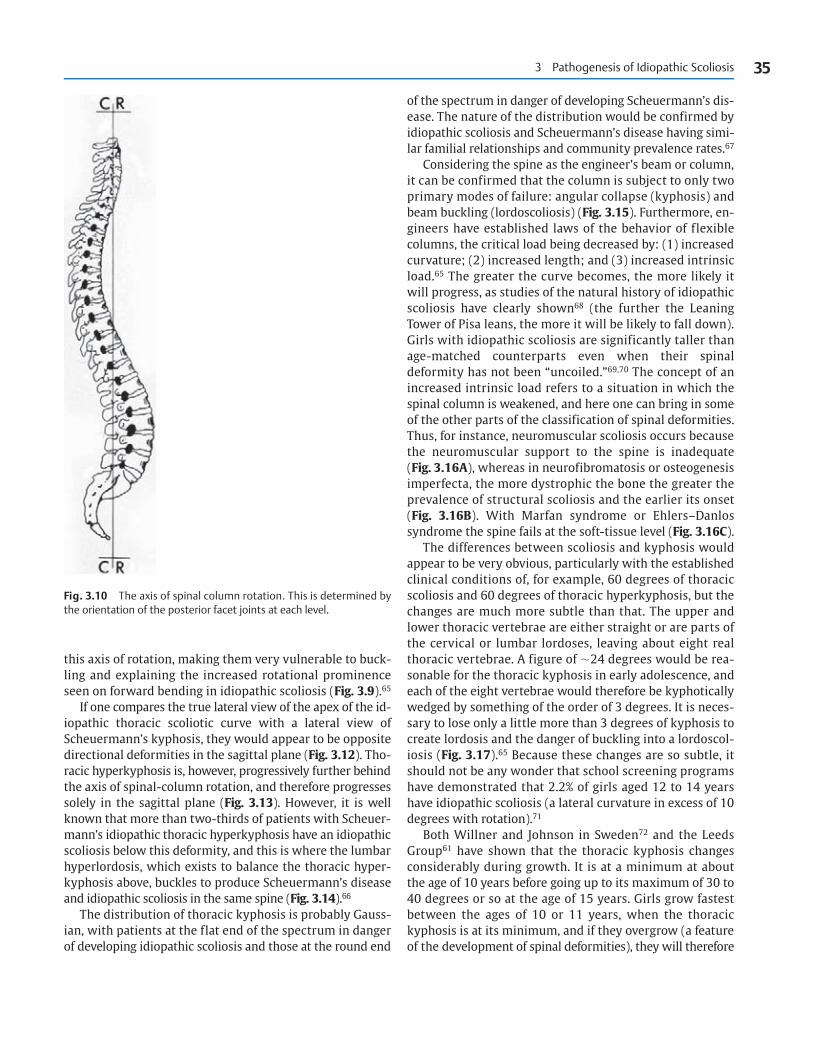
Returning to the biomechanics of forward bending, the
axis of spinal-column rotation normally passes in front of
the thoracic kyphosis and behind the cervical and lumbar
lordoses (Fig. 3.10). This confers protection to the thoracic
spine against buckling, because this region of the spine is
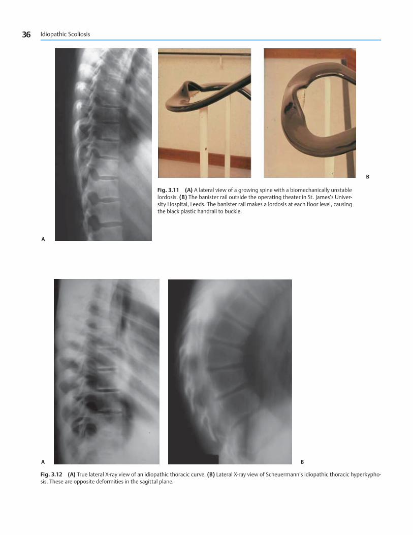
normally under tension. With the development of a tho-
racic lordosis, however (Fig. 3.11), the vertebral bodies
move progressively further forward, toward and in front of
34 Idiopathic Scoliosis
A B
C
Fig. 3.9 (A) A young man with a 30-degree right thoracic curve. Thecentral lordosis is clear to see. (B) Agirl with a 70-degree curve. The lordo-sis is clearly seen on the curve concav-ity. (C) A young girl with a very severecurve. Again the lordosis can be seenon the posteroconcave side.
this axis of rotation, making them very vulnerable to buck-
ling and explaining the increased rotational prominence
seen on forward bending in idiopathic scoliosis (Fig. 3.9).65
If one compares the true lateral view of the apex of the id-
iopathic thoracic scoliotic curve with a lateral view of
Scheuermann’s kyphosis, they would appear to be opposite
directional deformities in the sagittal plane (Fig. 3.12). Tho-
racic hyperkyphosis is, however, progressively further behind
the axis of spinal-column rotation, and therefore progresses
solely in the sagittal plane (Fig. 3.13). However, it is well
known that more than two-thirds of patients with Scheuer-
mann’s idiopathic thoracic hyperkyphosis have an idiopathic
scoliosis below this deformity, and this is where the lumbar
hyperlordosis, which exists to balance the thoracic hyper-
kyphosis above, buckles to produce Scheuermann’s disease
and idiopathic scoliosis in the same spine (Fig. 3.14).66
The distribution of thoracic kyphosis is probably Gauss-
ian, with patients at the flat end of the spectrum in danger
of developing idiopathic scoliosis and those at the round end
of the spectrum in danger of developing Scheuermann’s dis-
ease. The nature of the distribution would be confirmed by
idiopathic scoliosis and Scheuermann’s disease having simi-
lar familial relationships and community prevalence rates.67
Considering the spine as the engineer’s beam or column,
it can be confirmed that the column is subject to only two
primary modes of failure: angular collapse (kyphosis) and
beam buckling (lordoscoliosis) (Fig. 3.15). Furthermore, en-
gineers have established laws of the behavior of flexible
columns, the critical load being decreased by: (1) increased
curvature; (2) increased length; and (3) increased intrinsic
load.65 The greater the curve becomes, the more likely it
will progress, as studies of the natural history of idiopathic
scoliosis have clearly shown68 (the further the Leaning
Tower of Pisa leans, the more it will be likely to fall down).
Girls with idiopathic scoliosis are significantly taller than
age-matched counterparts even when their spinal
deformity has not been “uncoiled.”69,70 The concept of an
increased intrinsic load refers to a situation in which the
spinal column is weakened, and here one can bring in some
of the other parts of the classification of spinal deformities.
Thus, for instance, neuromuscular scoliosis occurs because
the neuromuscular support to the spine is inadequate
(Fig. 3.16A), whereas in neurofibromatosis or osteogenesis
imperfecta, the more dystrophic the bone the greater the
prevalence of structural scoliosis and the earlier its onset
(Fig. 3.16B). With Marfan syndrome or Ehlers–Danlos
syndrome the spine fails at the soft-tissue level (Fig. 3.16C).
The differences between scoliosis and kyphosis would
appear to be very obvious, particularly with the established
clinical conditions of, for example, 60 degrees of thoracic
scoliosis and 60 degrees of thoracic hyperkyphosis, but the
changes are much more subtle than that. The upper and
lower thoracic vertebrae are either straight or are parts of
the cervical or lumbar lordoses, leaving about eight real
thoracic vertebrae. A figure of �24 degrees would be rea-
sonable for the thoracic kyphosis in early adolescence, and
each of the eight vertebrae would therefore be kyphotically
wedged by something of the order of 3 degrees. It is neces-
sary to lose only a little more than 3 degrees of kyphosis to
create lordosis and the danger of buckling into a lordoscol-
iosis (Fig. 3.17).65 Because these changes are so subtle, it
should not be any wonder that school screening programs
have demonstrated that 2.2% of girls aged 12 to 14 years
have idiopathic scoliosis (a lateral curvature in excess of 10
degrees with rotation).71
Both Willner and Johnson in Sweden72 and the Leeds
Group61 have shown that the thoracic kyphosis changes
considerably during growth. It is at a minimum at about
the age of 10 years before going up to its maximum of 30 to
40 degrees or so at the age of 15 years. Girls grow fastest
between the ages of 10 or 11 years, when the thoracic
kyphosis is at its minimum, and if they overgrow (a feature
of the development of spinal deformities), they will therefore
3 Pathogenesis of Idiopathic Scoliosis 35
Fig. 3.10 The axis of spinal column rotation. This is determined bythe orientation of the posterior facet joints at each level.
36 Idiopathic Scoliosis
A
A
B
B
Fig. 3.11 (A) A lateral view of a growing spine with a biomechanically unstablelordosis. (B) The banister rail outside the operating theater in St. James’s Univer-sity Hospital, Leeds. The banister rail makes a lordosis at each floor level, causingthe black plastic handrail to buckle.
Fig. 3.12 (A) True lateral X-ray view of an idiopathic thoracic curve. (B) Lateral X-ray view of Scheuermann’s idiopathic thoracic hyperkypho-sis. These are opposite deformities in the sagittal plane.

3 Pathogenesis of Idiopathic Scoliosis 37
A B
A B
Fig. 3.13 (A) The center of gravity of the body lies just in front of the lum-bar spine, and with hyperkyphosis the thoracic spine is therefore progres-sively behind the axis of spinal column rotation. (B) Consequently, thedeformity progresses solely in the sagittal plane, with no buckling potential.
Fig. 3.14 (A) Lateral X-ray film of a boy with Scheuermann’s disease. (B) PA X-ray film showing that the compensatory lumbar hyperlordosishas buckled to produce idiopathic scoliosis below the area of Scheuermann’s disease.

Fig. 3.15 A column or beam can fail in only two ways: an-gular collapse (kyphosis) or beam buckling (lordoscoliosis).
Fig. 3.16 (A) Scoliosis in association with poliomyelitis. The spinehas failed at the neuromuscular level. (B) Scoliosis in associationwith osteogenesis imperfecta. The spine has failed at the bone level.
(C) Scoliosis in association with Marfan syndrome. The spine hasfailed at the soft-tissue level.
A–C

3 Pathogenesis of Idiopathic Scoliosis 39
Fig. 3.17 Sagittal vertebral body shape is a delicate matter. (Top)Three degrees of kyphosis would be about normal. (Middle) An in-crease by 2 degrees over three levels is Sorenson’s definition ofScheuermann’s disease. (Bottom) Loss of just over 3 degrees ofkyphosis renders the spinal column vulnerable to buckling.
Fig. 3.18 The thoracic kyphosis is at its minimumwhen girls grow fastest.
be vulnerable to the development of idiopathic scoliosis
(Fig. 3.18). Boys do not go through their growth spurt until
much later, when the thoracic kyphosis is maximizing,
which is why boys are more vulnerable to the opposite
condition of idiopathic thoracic hyperkyphosis (Scheuer-
mann’s disease) (Fig. 3.19).
That a thoracic lordosis is the primary event in the gen-
eration of idiopathic thoracic scoliosis was conclusively
shown in the Leeds epidemiological survey.71 A sensitive
positive test of an angle of trunk inclination of 5 degrees or
more was the criterion for admission to the study, and of
the 16,000 Leeds schoolchildren surveyed, 1000 were har-
vested and subsequently radiographed on an annual basis
for 6 years with AP and lateral low dose films. With such a
sensitive entry criterion many children had straight backs
to begin with, but some developed true idiopathic scoliosis
during the course of the study. This afforded the opportu-
nity of going back to look at the lateral profile when the
spine was straight in the frontal plane, and children who
developed idiopathic scoliosis already had a flat thoracic
spine with an apical lordosis (Fig. 3.20).
Transverse plane geometry is also important in the
normal as well as the scoliotic spine. This became apparent
when the specimens of idiopathic scoliosis in the Royal
College of Surgeons of Edinburgh Museum were first exam-
ined.34 More detailed studies of the same specimens con-
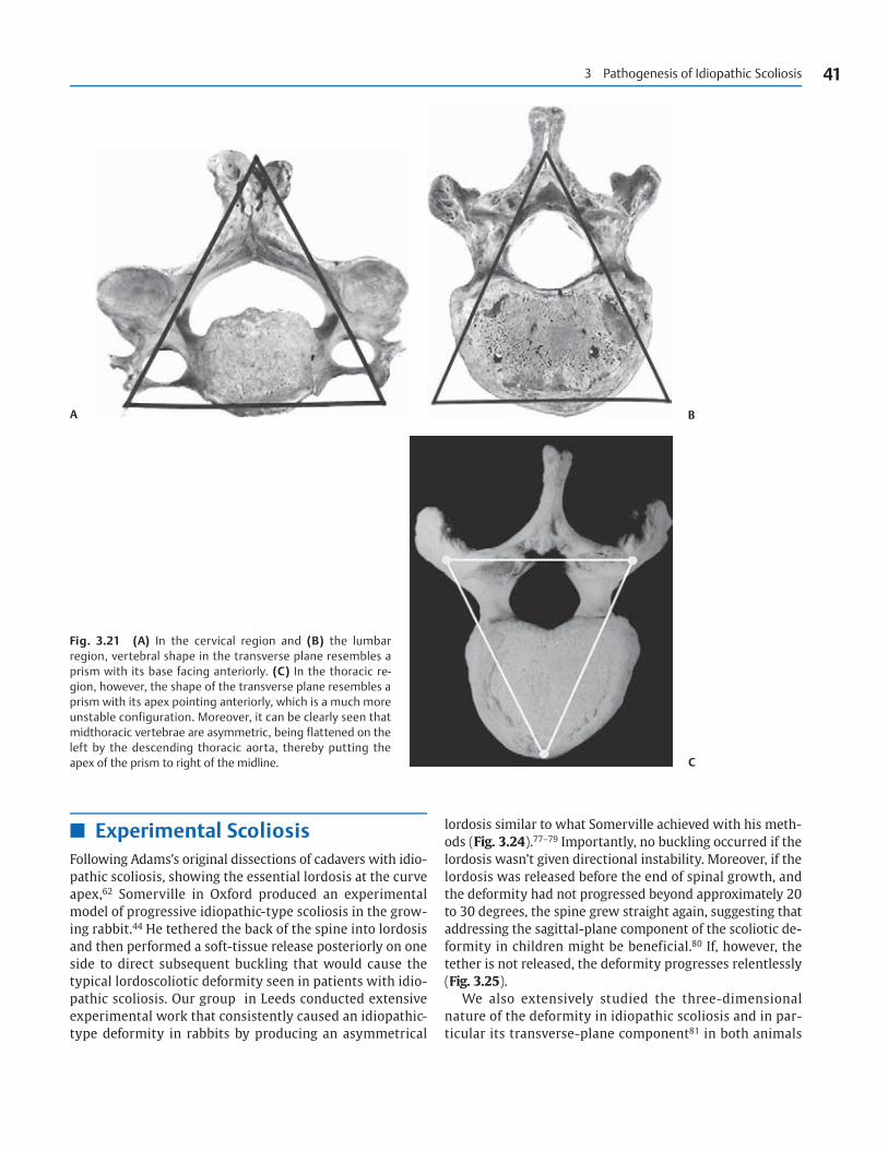
firmed this.35,64 In the cervical and lumbar regions, where
the spine is naturally lordotic, the cross-sectional vertebral
shape is prismatic, with the base pointing anteriorly. This is
most obvious in the lumbar region where the vertebrae in
cross-section are typically described as broad and kidney-
shaped (Fig. 3.21). When prisms are flexed toward their
bases, they are much more stable because of the second
moment of area, and the potentially vulnerable cervical and
lumbar lordoses are therefore countered by having a stable
transverse-planar shape. By contrast, vertebrae in the
thoracic region are typically heart-shaped in the transverse
plane, with the apex of the prism pointing anteriorly. This is

a dangerous configuration, favoring buckling with flexion,
and the thoracic spine is therefore protected by having a safe
kyphosis in the sagittal plane.
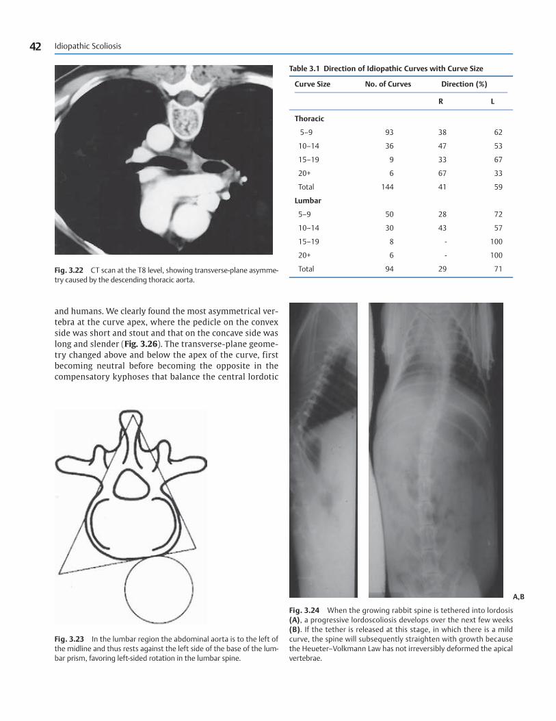
However, more thoracic curves are convex to the right
and more lumbar curves convex to the left, and this is be-
cause of a pre-existing asymmetry of vertebral shape in the
transverse plane. Anatomists have shown that in the tho-
racic spine, the T4 to T9 vertebrae are constantly deformed
on the left side by the descending thoracic aorta73 (Figs.
3.21C and 3.22), whereas a dynamic form of transverse-
planar asymmetry exists in the lumbar region, where the
left-sided abdominal aorta provides a restraint on curves
tending to go to the right (Fig. 3.23).65 This has been con-
firmed more recently with axial computed tomography (CT)
scans of normal human spines.74 Not surprisingly, the situa-
tion was opposite this in individuals with situs inversus.75
Clearly, however, the preponderance of right-sided tho-
racic curves and left-sided lumbar curves does not equate
with the prevalence rate of situs inversus. This is because
among small thoracic curves (�20 degrees), more are left-
sided than right-sided, and because lumbar curves do not
really have a left predominance until they reach or exceed
15 degrees, according to data of the Oxford School Screen-
ing Study (Table 3.1).76
40 Idiopathic Scoliosis
Fig. 3.19 Boys don’t grow fastest until toward theend of growth, when the thoracic kyphosis is maximal.
Fig. 3.20 (A) PA X-ray film of a 14-year-old girl with a mild right tho-racic idiopathic scoliosis. She was part of the Leeds longitudinal epi-demiological survey. (B) PA X-ray film made years earlier, when the
patient’s spine was straight. (C) Lateral X-ray film made years earlier,showing the dangerous lateral profile that preceded the develop-ment of the patient’s curve.
A–C
■ Experimental ScoliosisFollowing Adams’s original dissections of cadavers with idio-
pathic scoliosis, showing the essential lordosis at the curve
apex,62 Somerville in Oxford produced an experimental
model of progressive idiopathic-type scoliosis in the grow-
ing rabbit.44 He tethered the back of the spine into lordosis
and then performed a soft-tissue release posteriorly on one
side to direct subsequent buckling that would cause the
typical lordoscoliotic deformity seen in patients with idio-
pathic scoliosis. Our group in Leeds conducted extensive
experimental work that consistently caused an idiopathic-
type deformity in rabbits by producing an asymmetrical
lordosis similar to what Somerville achieved with his meth-
ods (Fig. 3.24).77–79 Importantly, no buckling occurred if the
lordosis wasn’t given directional instability. Moreover, if the
lordosis was released before the end of spinal growth, and
the deformity had not progressed beyond approximately 20
to 30 degrees, the spine grew straight again, suggesting that
addressing the sagittal-plane component of the scoliotic de-
formity in children might be beneficial.80 If, however, the
tether is not released, the deformity progresses relentlessly
(Fig. 3.25).
We also extensively studied the three-dimensional
nature of the deformity in idiopathic scoliosis and in par-
ticular its transverse-plane component81 in both animals
3 Pathogenesis of Idiopathic Scoliosis 41
Fig. 3.21 (A) In the cervical region and (B) the lumbar region, vertebral shape in the transverse plane resembles aprism with its base facing anteriorly. (C) In the thoracic re-gion, however, the shape of the transverse plane resembles aprism with its apex pointing anteriorly, which is a much moreunstable configuration. Moreover, it can be clearly seen thatmidthoracic vertebrae are asymmetric, being flattened on theleft by the descending thoracic aorta, thereby putting theapex of the prism to right of the midline.
A B
C
42 Idiopathic Scoliosis
Table 3.1 Direction of Idiopathic Curves with Curve Size
Curve Size No. of Curves Direction (%)
R L
Thoracic
5–9 93 38 62
10–14 36 47 53
15–19 9 33 67
20+ 6 67 33
Total 144 41 59
Lumbar
5–9 50 28 72
10–14 30 43 57
15–19 8 - 100
20+ 6 - 100
Total 94 29 71Fig. 3.22 CT scan at the T8 level, showing transverse-plane asymme-try caused by the descending thoracic aorta.
Fig. 3.23 In the lumbar region the abdominal aorta is to the left ofthe midline and thus rests against the left side of the base of the lum-bar prism, favoring left-sided rotation in the lumbar spine.
Fig. 3.24 When the growing rabbit spine is tethered into lordosis(A), a progressive lordoscoliosis develops over the next few weeks(B). If the tether is released at this stage, in which there is a mildcurve, the spine will subsequently straighten with growth becausethe Heueter–Volkmann Law has not irreversibly deformed the apicalvertebrae.
and humans. We clearly found the most asymmetrical ver-
tebra at the curve apex, where the pedicle on the convex
side was short and stout and that on the concave side was
long and slender (Fig. 3.26). The transverse-plane geome-
try changed above and below the apex of the curve, first
becoming neutral before becoming the opposite in the
compensatory kyphoses that balance the central lordotic
A,B

3 Pathogenesis of Idiopathic Scoliosis 43
Fig. 3.25 With further growth the scolio-sis progresses (A) at 6 weeks and (B) at 12 weeks. (C) Axial CT scan of the spine inB, showing significant rotation at the curveapex. (D) Looking inside the chest of thisspecimen shows exactly the same changesas during anterior thoracic surgery for idio-pathic scoliosis. There is no way in whichthis deformity can be created experimen-tally other than through rotation of a pri-mary lordosis.
A,B
C D
area. In these regions the pedicle on the convex side was
long and slender and that on the concave side short and
stout.
The same pattern of apical vertebral-body deformation
was seen in the rabbit as in the human, and by labeling ar-
eas of active vertebral growth with a dye similar to tetracy-
cline, we observed bone drift toward the curve concavity,
indicating that the spine was trying to correct the defor-
mity imposed upon it (Fig. 3.27).81
When the segmental blood supply to the spinal cord was
occluded at the curve apex, a cord infarct was produced, and
this led to a significant deformity, in excess of 40 degrees,
as soon as the procedure was completed, resembling what
was observed in experimental neuromuscular scoliosis

44 Idiopathic Scoliosis
Fig. 3.26 Transverse-plane asymmetry at the curve apex, with a short, stout pedicle on the convex side and a longer, thinner pedicle on theconcave side. (A) Human, (B) rabbit.
A B
A
Fig. 3.27 (A) The diagram in the center shows that growth of a normal vertebra in terms of the spinal canal and the vertebral body is out-ward. Consequently, the orange-stained growth area in the canal above and the vertebral body below is facing outward.

(Fig. 3.28).82 This is how Langenskiold and Michelsson41
accidentally produced scoliosis by dividing the costotrans-
verse ligament, because they damaged the segmental blood
supply to the spinal cord, as De Salis and colleagues42
demonstrated. Interestingly, growth and pulmonary func-
tion were considerably impaired with a rapidly progressive
thoracic deformity, rather like the situation in progressive
infantile idiopathic thoracic scoliosis (Fig. 3.29).82
Much interest in experimental scoliosis was rekindled
by observations of pinealectomized chickens and rats pop-
ularized by Dubousset et al83 and Machida et al.84 The
pineal gland produces melatonin from tryptophan through
a series of enzyme reactions, and serotonin is intermediary
in this pathway. In 1959 Thillard first produced scoliosis in
pinealectomized chickens to assess the role of melatonin
and its associated compounds in the disorder.85 If chickens
are pinealectomized shortly after they hatch, a scoliosis
similar to human idiopathic scoliosis is consistently pro-
duced. If melatonin supplements are given after pinealec-
tomy, a scoliosis does not develop.84 The precise reason
why pinealectomy produces this deformity is uncertain,
and research translated to the human situation has shown
conflicting results with regard to melatonin levels in
patients with idiopathic scoliosis and those in controls,
with some careful studies involving diurnal variation
showing no differences in the two groups.86,87 It is thought
that melatonin activity may be mediated by growth hor-
mone as the common denominator.88
However, the biomechanics of this experimental model
are also interesting. Even with the pinealectomized animal
model it is accepted that the “primary abnormality is a
lordosis,” which subsequently buckles to produce the
typical three-dimensional lordoscoliotic deformity, as
confirmed by Machida.88 This does not occur spontaneously
in quadrupeds, and chickens are bipedal. Consequently,
Dubousset and Machida and colleagues went on to investi-
gate the effects of pinealectomy in rats.89 If rats were initially
rendered bipedal and then pinealectomiaed, they developed
a scoliosis comparable to that in chickens, whereas the spine
remained straight in rats that underwent a sham operation
after being rendered bipedal (Fig. 3.30). Quadrupedal rats
when pinealectomized did not develop a spinal deformity.
3 Pathogenesis of Idiopathic Scoliosis 45
B
Fig. 3.27 (Continued) (B) With the apical scoliotic vertebra, thespinal canal and the vertebral body grow toward the concavity, as theorange-stained growth zones indicate. Thus, the transverse plane is
trying to correct and not cause the deformity. The transverse plane istherefore not an etiological factor in idiopathic scoliosis.

Fig. 3.28 (A) Experimental scoliosis. PA radiograph of a rabbitspine. The spine has been tethered into lordosis and the spinalcord damaged by thermal ablation of the facet joints. There was acurvature of 70 degrees immediately after the animal awakenedfrom anesthesia. (B) Within a couple of weeks the deformity wasgross, as in progressive infantile malignant idiopathic scoliosis.(C) Transverse section of the spinal cord at this level showing adorso-lateral infarct.
A B
C
A B
Fig. 3.29 Two rabbits, one with experimental idiopathic scoliosis (normal eye) (A) and the other (B) with experimental neuromuscular scol-iosis, resembling progressive infantile idiopathic malignant scoliosis with respiratory malfunction and cyanosis (cyanotic eye).
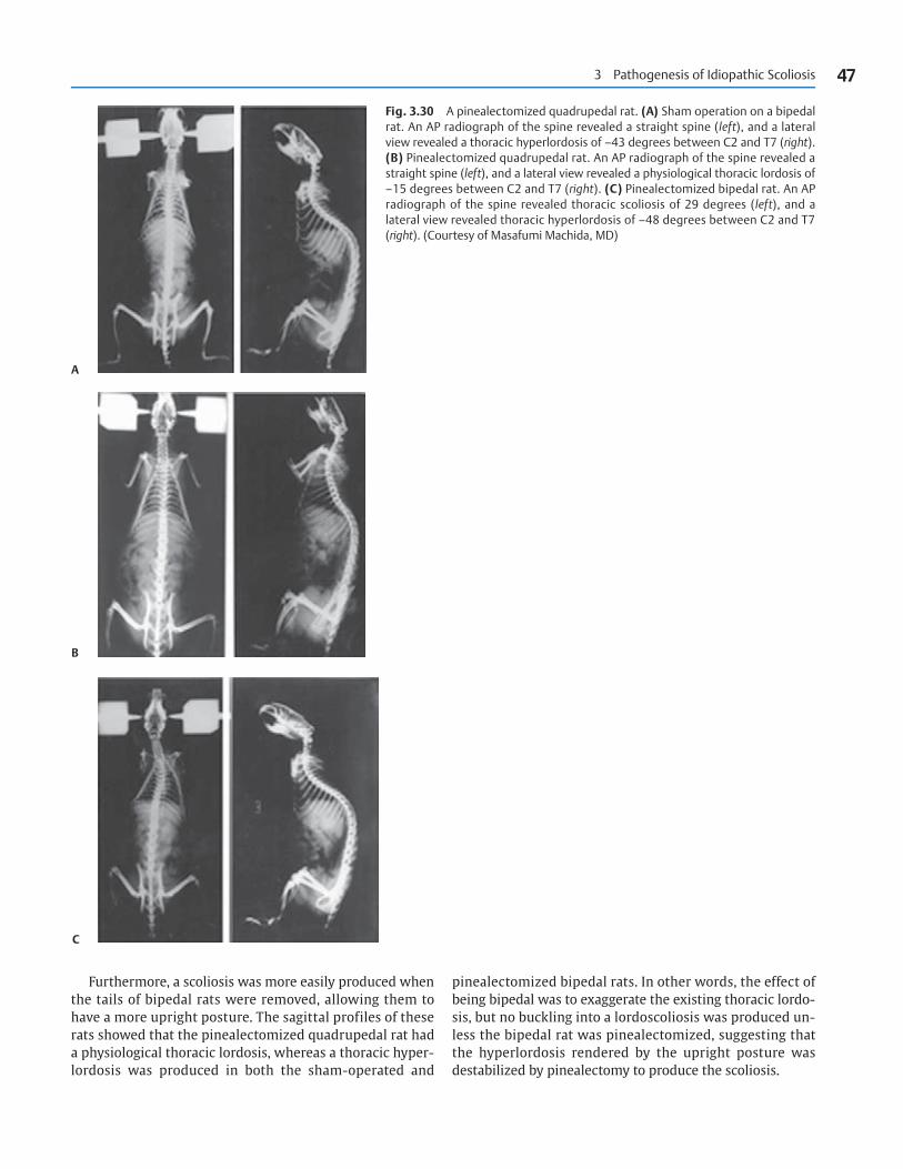
pinealectomized bipedal rats. In other words, the effect of
being bipedal was to exaggerate the existing thoracic lordo-
sis, but no buckling into a lordoscoliosis was produced un-
less the bipedal rat was pinealectomized, suggesting that
the hyperlordosis rendered by the upright posture was
destabilized by pinealectomy to produce the scoliosis.
3 Pathogenesis of Idiopathic Scoliosis 47
Fig. 3.30 A pinealectomized quadrupedal rat. (A) Sham operation on a bipedalrat. An AP radiograph of the spine revealed a straight spine (left), and a lateralview revealed a thoracic hyperlordosis of –43 degrees between C2 and T7 (right).(B) Pinealectomized quadrupedal rat. An AP radiograph of the spine revealed astraight spine (left), and a lateral view revealed a physiological thoracic lordosis of–15 degrees between C2 and T7 (right). (C) Pinealectomized bipedal rat. An APradiograph of the spine revealed thoracic scoliosis of 29 degrees (left), and alateral view revealed thoracic hyperlordosis of –48 degrees between C2 and T7(right). (Courtesy of Masafumi Machida, MD)
Furthermore, a scoliosis was more easily produced when
the tails of bipedal rats were removed, allowing them to
have a more upright posture. The sagittal profiles of these
rats showed that the pinealectomized quadrupedal rat had
a physiological thoracic lordosis, whereas a thoracic hyper-
lordosis was produced in both the sham-operated and
A
B
C

Interestingly, having tethered rabbits’ spines into a lordo-
sis, neither Somerville77 nor the Leeds Group78–80 could make
it buckle unless the lordosis was rendered asymmetrical by
producing a few degrees of scoliosis as well. Perhaps pinealec-
tomy would have done the same. In the bipedal chickens and
rats that developed scoliosis, there was no preferentiality in
its developing either on the right or the left side.
There doesn’t seem to be any other explanation for the
effect of the pinealectomy performed on rats, because the rats
were of much the same weight at the end of the experiment
and were not constitutionally disadvantaged.89 However,
scoliosis was also noted in the thoracolumbar region, and
this is where the lateral radiographs clearly showed a
kyphosis of the order of 40 or 50 degrees. Perhaps these
were the slightly asymmetrical kyphoses seen with severe
Scheuermann’s disease, in which there is a concomitant mild
coronal-plane deformity with the opposite direction of rota-
tion to that in idiopathic scoliosis, with the vertebral bodies
turning into the curve concavity.66 This is simple right–left
growth asymmetry rather than mechanical buckling.
48 Idiopathic Scoliosis
References
1. Le Febvre J, Triboulet-Chassevant A, Missirliu MF. Electromyographic
data in idiopathic scoliosis. Arch Phys Med Rehabil 1961;42:710–711
2. Gorynski T, Bojkowa M. [Histological changes of spinal muscles in
dystonic scoliosis.]. Chir Narzadow Ruchu Ortop Pol 1957;22:
139–142
3. Kaneko T. [Histopathological and histochemical studies on the back
muscles in scoliosis]. Nippon Seikeigeka Gakkai Zasshi 1968;42:13–28
4. Hirano S. Electron microscopic studies on back muscles in scoliosis.
Nippon Seikeigeka Gakkai Zasshi 1972;46:47–62
5. Fidler MW, Jowett RL, Troup JDG. Histochemical study of the func-
tion of multifidus in scoliosis. In: Zorab PA, ed. Scoliosis and Muscle.
London: William Heinemann Medical Books; 1974:184–192
6. Fidler MW, Jowett RL. Muscle imbalance in the aetiology of scoliosis.
J Bone Joint Surg Br 1976;58:200–201
7. Spencer GSG, Zorab PA. Spinal muscle in scoliosis. Comparison of
normal and scoliotic rabbits. J Neurol Sci 1976;30:405–410
8. Spencer GSG, Eccles MJ. Spinal muscle in scoliosis. Part 2. The pro-
portion and size of type 1 and type 2 skeletal muscle fibres meas-
ured using a computer-controlled microscope. J Neurol Sci 1976;
30:143–154
9. Wong YC, Yau ACMC, Low WD, et al. Ultrastructural changes of the
back muscles of idiopathic scoliosis. Spine 1977;2:251–260
10. Yarom R, Robin GC. Studies on spinal and peripheral muscles from
patients with scoliosis. Spine 1979;4:12–21
11. Khosla S, Tredwell SJ, Day B, Shinn SL, Ovalle WK Jr. An ultrastruc-
tural study of multifidus muscle in progressive idiopathic scoliosis.
Changes resulting from a sarcolemmal defect at the myotendinous
junction. J Neurol Sci 1980;46:13–31
12. Zetterberg C, Aniansson A, Grimby G. Morphology of the paraver-
tebral muscles in adolescent idiopathic scoliosis. Spine 1983;8:
457–462
13. Saatok T, Dahlberg E, Bylund P, Eriksson E, Gustafsson JA. Steroid
hormone receptors, protein, and DNA in erector spinae muscle
from scoliotic patients. Clin Orthop Relat Res 1984;183:197–207
14. Liebergall M, Floman Y, Eldor A. Functional, biochemical, and struc-
tural anomalies in platelets of patients with idiopathic scoliosis. J
Spinal Disord 1989;2:126–130
15. Yarom R, Muhlrad A, Hodges S, Robin GC. Platelet pathology in
patients with idiopathic scoliosis: Ultrastructural morphometry,
agrregations, X-ray spectrometry, and biochemical analysis. Lab
Invest 1980;43:208–216
16. Yarom R, Blatt J, Gorodetsky R, Robin GC. Microanalysis and X-ray
fluorescence spectrometry of platelets in diseases with elevated
muscle calcium. Eur J Clin Invest 1980;10(2 Pt 1):143–147
17. Kahmann RD, Donohue JM, Bradford DS, White JG, Rao GH. Platelet
function in adolescent idiopathic scoliosis. Spine 1992;17:145–148
18. Suk SI, Kim IK, Lee CK, Koh YD, Yeom JS. A study on platelet function
in idiopathic scoliosis. Orthopedics 1991;14:1079–1083
19. Sabato S, Rotman A, Robin GC, Floman Y. Platelet aggregation abnor-
malities in idiopathic scoliosis. J Pediatr Orthop 1985;5:558–563
20. Floman Y, Liebergall M, Robin GC, Eldor A. Abnormalities of
aggregation, thromboxane A2 synthesis, and 14C serotonin
release in platelets of patients with idiopathic scoliosis. Spine
1983;8: 236–241
21. Kindsfater K, Lowe T, Lawellin D, Weinstein D, Akmakjian J. Levels
of platelet calmodulin for the prediction of progression and sever-
ity of adolescent idiopathic scoliosis. J Bone Joint Surg Am 1994;
76:1186–1192
22. Machida M, Dubousset J, Imamura Y, Miyashita Y, Yamada T,
Kimura J. Melatonin. A possible role in pathogenesis of adolescent
idiopathic scoliosis. Spine 1996;21:1147–1152
23. Petersén I, Sahlstrand T, Selldén U. Electroencephalographic in-
vestigation of patients with adolescent idiopathic scoliosis. Acta
Orthop Scand 1979;50:283–293
24. Sahlstrand T, Ortengren R, Nachemson A. Postural equilibrium in
adolescent idiopathic scoliosis. Acta Orthop Scand 1978;49:354–365
25. Sahlstrand T, Petruson B. A study of labyrinthine function in pa-
tients with adolescent idiopathic scoliosis. I. An electro-nystagmo-
graphic study. Acta Orthop Scand 1979;50(6 Pt 2):759–769
26. Sahlstrand T, Petruson B, Ortengren R. Vestibulospinal reflex activ-
ity in patients with adolescent idiopathic scoliosis. Postural effects
during caloric labyrinthine stimulation recorded by stabilometry.
Acta Orthop Scand 1979;50:275–281
27. Cheng JC, Guo X, Sher AH, Chan YL, Metreweli C. Correlation be-
tween curve severity, somatosensory evoked potentials, and mag-
netic resonance imaging in adolescent idiopathic scoliosis. Spine
1999;24:1679–1684
28. Guo X, Chau WW, Hui-Chan CW, Cheung CS, Tsang WW, Cheng JC.
Balance control in adolescents with idiopathic scoliosis and disturbed
somatosensory function. Spine 2006;31:E437–E440
29. Roth M. Idiopathic scoliosis caused by a short spinal cord. Acta
Radiol Diagn (Stockh) 1968;7:257–271
30. Porter RW. Idiopathic scoliosis: The relation between the vertebral
canal and the vertebral bodies. Spine 2000;25:1360–1366
31. Porter RW. Can a short spinal cord produce scoliosis? Eur Spine J
2001;10:2–9
32. Porter RW. The pathogenesis of idiopathic scoliosis: Uncoupled
neuro-osseous growth? Eur Spine J 2001;10:473–481
33. Chu WC, Lam WW, Chan YL, et al. Relative shortening and func-
tional tethering of spinal cord in adolescent idiopathic scoliosis?:
Study with multiplanar reformat magnetic resonance imaging and
somatosensory evoked potential. Spine 2006;31:E19–E25
34. Deane G, Duthie RB. A new projectional look at articulated scoliotic
spines. Acta Orthop Scand 1973;44:351–365
35. Deacon P, Flood BM, Dickson RA. Idiopathic scoliosis in three di-
mensions. A radiographic and morphometric analysis. J Bone Joint
Surg Br 1984;66:509–512
36. Inoue M, Minami S, Nakata Y, et al. Preoperative MRI analysis of
patients with idiopathic scoliosis: A prospective study. Spine 2005;
30:108–114
37. Bisgard JD. Experimental thoracogenic scoliosis. J Thorac Surg
1934;4:435–442
38. Schwartzmann JR, Miles M. Experimental production of scoliosis in
rats and mice. J Bone Joint Surg 1945;27:59–69
39. Liszka O. Spinal cord mechanisms leading to scoliosis in animal
experiments. Acta Med Pol 1961;2:45–63
40. MacEwen GD. Experimental scoliosis. In: Zorab PA, ed. Proceedings
of a Second Symposium on Scoliosis: Causation. National Fund for
Research into Crippling Diseases Monograph. Edinburgh: Living-
stone;1968:18–20
41. Langenskiold A, Michelsson JE. Experimental progressive scoliosis
in the rabbit. J Bone Joint Surg Br 1961;43B:116–120
42. De Salis J, Beguiristain JL, Cañadell J. The production of experimental
scoliosis by selective arterial ablation. Int Orthop 1980;3:311–315
43. Robin GC, Stein H. Experimental scoliosis in primates. Failure of a
technique. J Bone Joint Surg Br 1975;57:142–145
44. Somerville EW. Rotational lordosis; the development of single
curve. J Bone Joint Surg Br 1952;34B:421–427
45. Francis MJO, Sanderson MC, Smith R. Skin collagen in idiopathic
adolescent scoliosis and Marfan’s syndrome. Clin Sci Mol Med
1976;51:467–474
46. Bushell GR, Ghosh P, Taylor TKF. Collagen defect in idiopathic scol-
iosis. Lancet 1978;2:94–95
47. Scapinelli R, Little K. Observations on the mechanically induced
differentiation of cartilage from fibrous connective tissue. J Pathol
1970;101:85–91
48. Pedrini VA, Ponseti IV, Dohrman SC. Glycosaminoglycans of interver-
tebral disc in idiopathic scoliosis. J Lab Clin Med 1973;82:938–950
49. Ghosh P, Bushell GR, Taylor TKF, Pearce RH, Grimmer BJ. Distribu-
tion of glycosaminoglycans across the normal and the scoliotic
disc. Spine 1980;5:310–317
50. Taylor TKF, Ghosh P, Bushell GR. The contribution of the intervertebral
disk to the scoliotic deformity. Clin Orthop Relat Res 1981;156:79–90
51. Miller NH, Mims B, Child A, Milewicz DM, Sponseller P, Blanton SH.
Genetic analysis of structural elastic fiber and collagen genes in
familial adolescent idiopathic scoliosis. J Orthop Res 1996;14:994–999
52. Marosy B, Justice CM, Nzegwu N, Kumar G, Wilson AF, Miller NH.
Lack of association between the aggrecan gene and familial idio-
pathic scoliosis. Spine 2006;31:1420–1425
53. Wynne-Davies R. Familial (idiopathic) scoliosis. A family survey. J
Bone Joint Surg Br 1968;50:24–30
54. Cowell HR, Hall JN, MacEwen GD. Genetic aspects of idiopathic sco-
liosis. A Nicholas Andry Award Essay, 1970. Clin Orthop Relat Res
1972;86:121–131
55. Alden KJ, Marosy B, Nzegwu N, Justice CM, Wilson AF, Miller NH.
Idiopathic scoliosis: identification of candidate regions on chromo-
some 19p13. Spine 2006;31:1815–1819
56. Chan V, Fong GC, Luk KD, et al. A genetic locus for adolescent idio-
pathic scoliosis linked to chromosome 19p13.3. Am J Hum Genet
2002;71:401–406
57. Archer IA, Dickson RA. Stature and idiopathic scoliosis. A prospec-
tive study. J Bone Joint Surg Br 1985;67:185–188
58. Tanner JM. Growth at Adolescence, ed. 2. Oxford: Blackwell Scien-
tific; 1962
59. Delmas A. Types rachidiens de statique corporelle. Rev Morpho-
physiol Hum 1951;4:26–32
60. Stagnara P, De Mauroy JC, Dran G, et al. Reciprocal angulation of
vertebral bodies in a sagittal plane: approach to references for the
evaluation of kyphosis and lordosis. Spine 1982; 7:335–342
61. Oxborrow N, Gopal S, Walder A, et al. A new surface topographical
measure of spinal shape in scoliosis. J Bone Joint Surg Br 1998;
80(Supp III):276–277
62. Adams W. Lectures on the Pathology and Treatment of Lateral and
other Forms of Curvature of the Spine. London: Churchill and
Sons; 1865
63. Stagnara P. Le plan d’election pour I’examen radiologique des
cyphoscolioses. Rev Chir Orthop Reparatrice Appar Mot 1965;51:
517–524
64. Deacon P, Archer IA, Dickson RA. The anatomy of spinal deformity:
A biomechanical analysis. Orthopedics 1987;10:897–903
65. Millner PA, Dickson RA. Idiopathic scoliosis: Biomechanics and
biology. Eur Spine J 1996;5:362–373
66. Deacon P, Berkin CR, Dickson RA. Combined idiopathic kyphosis
and scoliosis. An analysis of the lateral spinal curvatures associ-
ated with Scheuermann’s disease. J Bone Joint Surg Br 1985;67:
189–192
67. Sorenson KH. Scheuermann’s Kyphosis. Clinical Appearances. Radi-
ography, Aetiology and Prognosis. Copenhagen: Munksgaard; 1964
68. Weinstein SL. Natural history. Spine 1999;24:2592–2600
69. Archer IA, Dickson RA. Stature and idiopathic scoliosis. A prospec-
tive study. J Bone Joint Surg Br 1985;67:185–188
70. Willner S. A study of height, weight and menarche in girls with
idiopathic structural scoliosis. Acta Orthop Scand 1975;46:71–83
71. Stirling AJ, Howel D, Millner PA, Sadiq S, Sharples D, Dickson RA.
Late-onset idiopathic scoliosis in children six to fourteen years old.
A cross-sectional prevalence study. J Bone Joint Surg Am 1996;78:
1330–1336
72. Willner S, Johnson B. Thoracic kyphosis and lumbar lordosis during
the growth period in children. Acta Paediatr Scand 1983;72:
873–878
73. Farkas A. Physiological scoliosis. J Bone Joint Surg 1941;23:
607–627
74. Kouwenhoven JWM, Vincken KL, Bartels LW, Castelein RM. Analysis
of preexistent vertebral rotation in the normal spine. Spine 2006;
31:1467–1472
75. Kouwenhoven JWM, Bartels LW, Vincken KL, et al. The relation
between organ anatomy and pre-existent vertebral rotation in the
normal spine: Magnetic resonance imaging study in humans with
situs inversus totalis. Spine 2007;32:1123–1128
76. Dickson RA. Scoliosis in the community. Br Med J (Clin Res Ed)
1983;286:615–618
77. Dickson RA, Lawton JO, Archer IA, Butt WP. The pathogenesis of
idiopathic scoliosis. Biplanar spinal asymmetry. J Bone Joint Surg
Br 1984;66:8–15
78. Smith RM, Dickson RA. Experimental structural scoliosis. J Bone
Joint Surg Br 1987;69:576–581
3 Pathogenesis of Idiopathic Scoliosis 49
79. Lawton JO, Dickson RA. The experimental basis of idiopathic scol-
iosis. Clin Orthop Relat Res 1986;210:9–17
80. Dickson RA. Idiopathic scoliosis: Foundation for physiological
treatment. Ann R Coll Surg Engl 1987;69:89–96
81. Smith RM, Pool RD, Butt WP, Dickson RA. The transverse plane
deformity of structural scoliosis. Spine 1991;16:1126–1129
82. Smith RM, Hamlin GW, Dickson RA. Respiratory deficiency in exper-
imental idiopathic scoliosis. Spine 1991;16:94–99
83. Dubousset J, Queneau P, Thillard MJ. Experimental scoliosis induced
by pineal and diencephalic lesions in young chickens: Its relation
with clinical findings. Orthop Trans 1983;7:7
84. Machida M, Dubousset J, Imamura Y, Iwaya T, Yamada T, Kimura J.
An experimental study in chickens for the pathogenesis of idio-
pathic scoliosis. Spine 1993;18:1609–1615
85. Thillard MJ. Deformations de la colonne vertebrale consecutives a
l’epiphysectomie chez le possin. Extrat des comptes Rendus de l’As-
sociation Anatomistes; 1959:751–758
86. Hilibrand AS, Blakemore LC, Loder RT, et al. The role of melatonin
in the pathogenesis of adolescent idiopathic scoliosis. Spine
1996;21:1140–1146
87. Fagan AB, Kennaway DJ, Sutherland AD. Total 24-hour melatonin
secretion in adolescent idiopathic scoliosis. A case-control study.
Spine 1998;23:41–46
88. Machida M. Cause of idiopathic scoliosis. Spine 1999;24:2576–2583
89. Machida M, Saito M, Dubousset J, Yamada T, Kimura J, Shibasaki K.
Pathological mechanism of idiopathic scoliosis: Experimental
scoliosis in pinealectomized rats. Eur Spine J 2005;14:843–848
50 Idiopathic Scoliosis


![Exercises for adolescent idiopathic scoliosis - …tees.openrepository.com/tees/bitstream/10149/249111/2/249111.pdf[Intervention Review] Exercises for adolescent idiopathic scoliosis](https://static.fdocuments.us/doc/165x107/5aa5e2337f8b9ae7438e1827/exercises-for-adolescent-idiopathic-scoliosis-tees-intervention-review-exercises.jpg)


