2nd BSEL Group Meeting Presentation
32
“ Development of ex-vivo three-dimensional model Biological Systems Engineering Laboratory (BSEL) of chronic lymphocytic leukemia (CLL)” SAIFUL IRWAN ZUBAIRI SUPERVISOR: Dr. Sakis Mantalaris CO-SUPERVISOR: Dr. Nicki Panoskaltsis
-
Upload
saiful-irwan-zubairi -
Category
Documents
-
view
429 -
download
6
description
Transcript of 2nd BSEL Group Meeting Presentation

“Development of ex-vivo three-dimensional model
Biological Systems Engineering Laboratory (BSEL)
“Development of ex-vivo three-dimensional model
of chronic lymphocytic leukemia (CLL)”
SAIFUL IRWAN ZUBAIRI
SUPERVISOR: Dr. Sakis Mantalaris
CO-SUPERVISOR: Dr. Nicki Panoskaltsis

Outlines
Introduction
An ideal scaffold?
Aims & objectives
Experimental setupExperimental setup
Results
Future works
Conclusion
Biological Systems Engineering Laboratory (BSEL)

Introduction Introduction Introduction Introduction Polyhydroxyalkanoates (PHAs) →→→→ a family of biopolyesters →→→→ bacteria →→→→intracellular carbon & energy-storage compounds.
Tissue engineering materials →→→→ GOOD →→→→ physical properties, biodegradability & biocompatibility.
Poly(3-hydroxybutyrate) (PHB) & poly(3-hydroxybutyrate-co-3-hydroxyvalerate) →→→→ →→→→
Poly(3-hydroxybutyrate) (PHB) & poly(3-hydroxybutyrate-co-3-hydroxyvalerate) (PHBV) →→→→ biomaterials →→→→ in vitro & in vivo studies
> 150 types →→→→ PHAs →→→→ various monomers
Types of bacterium & growth conditions →→→→ chemical composition →→→→ PHAs & Mw →→→→ 2××××105 to 3××××106 Da.
3 classes →→→→ (sclPHA, C3 - C5) →→→→ (mclPHA, C6 - C14) →→→→ (lclPHA, >C14).
Biological Systems Engineering Laboratory (BSEL)

Molecular structure of PHB and PHBV
31
2
Source: http://biopol.free.fr
m = STRUCTURE BACKBONE = 1, 2, 3, etc. m = 1 is the most common
n = 100 - 30,000 monomers.
R is a variable: Types of homo-polymers in the PHAs family.
m = 1, R = CH3, →→→→ 3-hydroxybutyrate (3-HB)
m = 1, R = C2H5, →→→→ 3-hydroxyvalerate (3-HV)
3-HB + 3-HV
3-HB
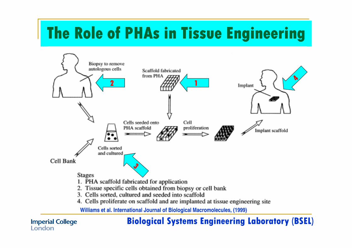
The Role of PHAs in Tissue EngineeringThe Role of PHAs in Tissue EngineeringThe Role of PHAs in Tissue EngineeringThe Role of PHAs in Tissue Engineering
12
Williams et al. International Journal of Biological Macromolecules, (1999)
Biological Systems Engineering Laboratory (BSEL)
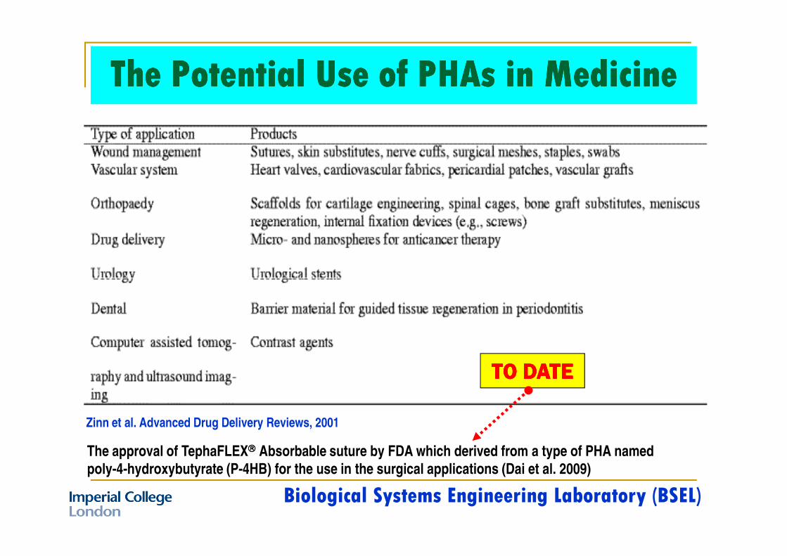
The Potential Use of PHAs in MedicineThe Potential Use of PHAs in MedicineThe Potential Use of PHAs in MedicineThe Potential Use of PHAs in Medicine
Zinn et al. Advanced Drug Delivery Reviews, 2001
The approval of TephaFLEX Absorbable suture by FDA which derived from a type of PHA named
poly-4-hydroxybutyrate (P-4HB) for the use in the surgical applications (Dai et al. 2009)
TO DATETO DATETO DATETO DATE
Biological Systems Engineering Laboratory (BSEL)

An Ideal Scaffold for
the T.E.R.M.?
The scaffold →→→→ inter-connecting pores →→→→ tissue integration &
An ideal scaffold should possess the following characteristics to bring about the desired biological response (Liu, W. & Y. Cao, 2007):
The scaffold →→→→ inter-connecting pores →→→→ tissue integration &
vascularisation process.
Material →→→→ biodegradability/bio-resorbability.
Surface chemistry →→→→ cellular attachment, differentiation & proliferation.
Mechanical properties →→→→ intended site of implantation & handling.
Be easily fabricated into a variety of shapes & sizes.
Tubes derived from PHOH film (left) and porous PHOH
(right) - Williams et al. (1999)
Biological Systems Engineering Laboratory (BSEL)

“To fabricate a novel porous 3-D scaffolds with an improved thickness (more than 2 mm) using the Solvent-Casting Particulate-Leaching (SCPL) technique”
(1) Polymer concentrations with respect to homogenization time
↓↓↓↓(2) Polymer concentrations with respect to polymeric porous 3-D scaffolds
Aim 1Aim 1Aim 1Aim 1
Objectives Objectives Objectives Objectives
(2) Polymer concentrations with respect to polymeric porous 3-D scaffolds thickness
↓↓↓↓(3) Efficacy of Solvent-Casting Particulate-Leaching (SCPL) via conductivity
(mS/cm) measurement
↓↓↓↓(4) Effect of sodium chloride (Sigma-Aldrich) residual in polymeric porous 3-D
scaffolds on the cell growth media
Biological Systems Engineering Laboratory (BSEL)

“To characterize the physico-chemical of polymeric porous 3-D scaffolds
with an improved thickness (more than 2 mm)”
(1) Analysis of porosity/surface area/PSD/void volume/roughness
↓↓↓↓
Aim 2Aim 2Aim 2Aim 2
Objectives Objectives Objectives Objectives
↓↓↓↓
(2) Analysis of pores size and interconnectivity using
scanning electron microscopy (SEM)
↓↓↓↓
(3) Contact angle and surface free energy of dry PHB and PHBV
porous 3-D scaffolds
Biological Systems Engineering Laboratory (BSEL)

Experimental Setup
Polymer solution in
organic solvent
Polymer solution
+ Porogen
Solvent evaporation
(Complied with UK-SED,
2002: <20 mg/m3)
Polymer +
Porous 3-D
scaffolds
Porogen-DIW
leaching
Polymer concentration vs. time
Polymer concentration vs. thickness
FABRICATION
Efficacy of SCPL
Porogen residual effect Vs. growth media
12
34
A
The solvent-casting and particulate-leaching (SCPL)
Porogen (i.e., NaCl,
sucrose etc.)
Polymer +
Porogen cast
Contact angle and surface free energy
PHYSICO-CHEMICAL
Porosity analysisRoughness analysis
Pores size and interconnectivity using SEM
Polymer +
Solvent +
Porogen cast B
Advantages: Simple →→→→ fairly reproducible method →→→→no sophisticated apparatus →→→→ controlled porosity & interconnectivity.
Disadvantages: Thickness limitations →→→→ structures generally isotropic & angular →→→→ hazardous solvent →→→→lack of pores interconnectivity →→→→ limited mechanical properties →→→→ residual of porogen and solvent
Biological Systems Engineering Laboratory (BSEL)

“RESULTS:
PART A” PART A”
Biological Systems Engineering Laboratory (BSEL)

Polymer concentrations with respect to homogenization time
Biological Systems Engineering Laboratory (BSEL)

Polymer concentrations with respect to polymeric 3-D scaffolds thickness

Polymer concentrations with respect to polymer 3-D scaffolds thickness

Polymer concentrations with respect to polymer 3-D scaffolds thickness

Polymer concentrations with respect to polymer 3-D scaffolds thickness
PHBV 4% (w/v)PHB 4% (w/v)
∼∼∼∼10 mm∼∼∼∼10 mm
∼∼∼∼5 mm
PHBV 4% (w/v)
PHB 4% (w/v)

Efficacy of Solvent-Casting Particulate-Leaching (SCPL) via conductivity (mS/cm) measurement
90
100 Source: http://www.4oakton.com
“Mass balance of sodium chloride were calculated
after the leaching and lyophilization process”
y = 2.8475x + 8.5027
R2 = 0.9999
0
10
20
30
40
50
60
70
80
90
0 5 10 15 20 25 30 35
Concentration of NaCl (mg/ml)
Co
nd
uc
tivity
(m
S/c
m)
Biological Systems Engineering Laboratory (BSEL)

Effect of sodium chloride (Sigma-Aldrich) residual in polymeric porous 3-D scaffolds on cell growth media
The effect of sodium chloride residual inside PHB and PHBV porous 3-D scaffolds on the cell growth media
measured by pH changes. The polymeric porous 3-D scaffolds were submerged in the cell growth media (90%
IMDM+10% FBS+1% PS) and incubated at 37 oC, and 5% CO2 (n = 3) for 7 days. NS indicates no significant
differences as compared to control.
Biological Systems Engineering Laboratory (BSEL)

“RESULTS:
PART B” PART B”
Biological Systems Engineering Laboratory (BSEL)

Physical properties of polymeric porous 3-D scaffolds

Pores size and interconnectivity analysis using scanning electron microscopy (SEM)
PHB 4% (w/v) PHB 4% (w/v) - enlarged
PHBV 4% (w/v) PHBV 4% (w/v) - enlarged

Wettability and surface energy of polymeric porous 3-D scaffolds
(a, b) Schematic of a simple derivation of Young’s equation using surface tension vectors for a
liquid on a solid substances (ideal solid surfaces). (c) Wenzel’s model of non-ideal solid surfaces

“CONCLUSIONS”“CONCLUSIONS”
Biological Systems Engineering Laboratory (BSEL)

Polymer concentration of 4% (w/v) for PHB and PHBV →→→→ ideal concentration →→→→thickness of porous 3-D scaffolds →→→→ more than 2 mm.
The insignificant →→→→ pH values →→→→ cell growth media Vs. control →→→→ insignificant amount of porogen residual remained.
No contaminants/residual →→→→ No effect on the in vitro cell proliferation studies.
Both polymeric porous 3-D scaffolds →→→→ highly hydrophobic materials.
Lack of pores interconnectivity and highly hydrophobicity of the surfaces
→→→→ EXPECTED →→→→ low degree of cell attachment and proliferation.
Modifying its surface chemistries →→→→ polymer surface becomes chemically more homogeneous (smoothing effect) →→→→ physically more pores interconnectivity were created →→→→ functionalization with oxygen-containing groups into hydrophilic surfaces →→→→ allow better cell attachment and proliferation
Biological Systems Engineering Laboratory (BSEL)
“FUTURE WORKS”“FUTURE WORKS”
Biological Systems Engineering Laboratory (BSEL)
Biological Systems Engineering Laboratory (BSEL)
“THANK YOU FOR
YOUR KIND
ATTENTION”ATTENTION”
Biological Systems Engineering Laboratory (BSEL)

Question:
1. Why the thickness of 5-mm? ANS: (1) Previous studies show that the thickness of 5-
mm was the optimum level for the cell-depth penetration to be occurred - Problem could
occurred if >5-mm e.g.: no nutrient, oxygen and waste could be transported across the
scaffolds – this could trigger apostosis (programmed cell death) due to the starvation. (2)
Since our aim to mimic the BM micro-environment for transplanting HSC into the leukemia
BM, we’re aiming to mimic the thickness as similar to the human BM. Thickness of human
BM in reticular (resembling a net in form; netlike) connective tissue area which consist of BM in reticular (resembling a net in form; netlike) connective tissue area which consist of
a complex sinusoidal system (arterial vascular system) + hematopoietic cells + stroma
(non-hemato).
2. Novelty of your research? – Can fabricate 3-D scaffolds with an improved thickness of
more than 2 mm – Up the extent of our knowledge - none of the studies produce 3-D
scaffolds with thickness than 2 mm with this particular type of biopolyesters – most of
them are at the µm size.
3. Why PHB and PHBV? Why not PU, PP and others? – This polymers can be
synthesized – waste/renewable sources – to become as added value product – for the
application of leukemia treatment

OMIT SLIDESOMIT SLIDES

Weight fraction and ratio of materials and chemicals
in fabricating polymeric porous 3-D scaffolds of
Solvent-Casting Particulate-Leaching (SCPL)
As the salt weight fraction increased from 60% to 90% (w/w), the porosities increased
gradually from 0.69 to 0.90 and porosities are homogenous with interconnected pores -
[Lu et al. (2000) & Mikos (1994)]
Biological Systems Engineering Laboratory (BSEL)

World’s Manufacturer of PHB & PHBV - May 2010
Gurieff, N. and P. Lant, Comparative life cycle
assessment and financial analysis of mixed culture
polyhydroxyalkanoate production. Bioresource Technology, 2007. 98(17): p. 3393-3403.

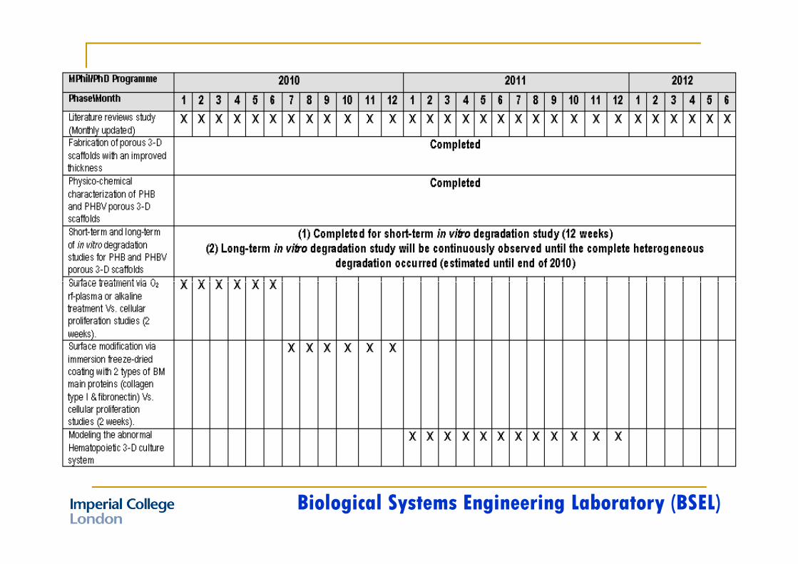
In vitro degradation studies for PHB and PHBV porous 3-D scaffolds - PBS & cell growth media.
↓↓↓↓Mechanical testing (compressive moduli): Untreated & immersion porous 3-D
scaffolds with cell growth media (4 wks & 8 wks)
↓↓↓↓Surface treatment via O2 rf-plasma or alkaline treatment Vs. cellular proliferation
studies (2 weeks).studies (2 weeks).
↓↓↓↓Surface modification via immersion freeze-dried coating with 2 types of BM main
proteins (collagen type I & fibronectin) Vs. cellular proliferation studies (2 weeks).
↓↓↓↓Modeling the abnormal hematopoietic 3-D culture system for short- and long-term
of 4 and 8 weeks respectively.
Biological Systems Engineering Laboratory (BSEL)