2.Mechanism of drug actons
28
Compiled by: Prof.Mirza Anwar Baig Chapter :2 Chapter :2 Mechanisms of drug action Mechanisms of drug action Presented by: Prof.Mirza Anwar Baig Presented by: Prof.Mirza Anwar Baig Anjuman-I-Islam's Kalsekar Technical Campus Anjuman-I-Islam's Kalsekar Technical Campus School of Pharmacy,New Pavel,Navi School of Pharmacy,New Pavel,Navi Mumbai,Maharashtra Mumbai,Maharashtra 1
-
Upload
mirza-anwar-baig -
Category
Education
-
view
71 -
download
0
Transcript of 2.Mechanism of drug actons
Compiled by: Prof.Mirza Anwar Baig
Chapter :2Chapter :2
Mechanisms of drug actionMechanisms of drug action
Presented by: Prof.Mirza Anwar BaigPresented by: Prof.Mirza Anwar Baig
Anjuman-I-Islam's Kalsekar Technical CampusAnjuman-I-Islam's Kalsekar Technical CampusSchool of Pharmacy,New Pavel,Navi School of Pharmacy,New Pavel,Navi
Mumbai,MaharashtraMumbai,Maharashtra
11

Compiled by: Prof.Mirza Anwar Baig
Outlines:
• Introduction to physiological receptors• Structural and functional families of receptors• Mechanisms of drug action:
-Drug receptor interaction-Dose response curve (DRC)-Drug antagonism
Compiled by: Prof.Mirza Anwar Baig
At the end of topic you should be able to....
• Explain the pharmacological basis of drug action.
• Define the scientific terms mentioned in this section.
• Identify type of antagonism
• Rearrange the drugs in ascending or descending order for their efficacy and potency.
Compiled by: Prof.Mirza Anwar Baig
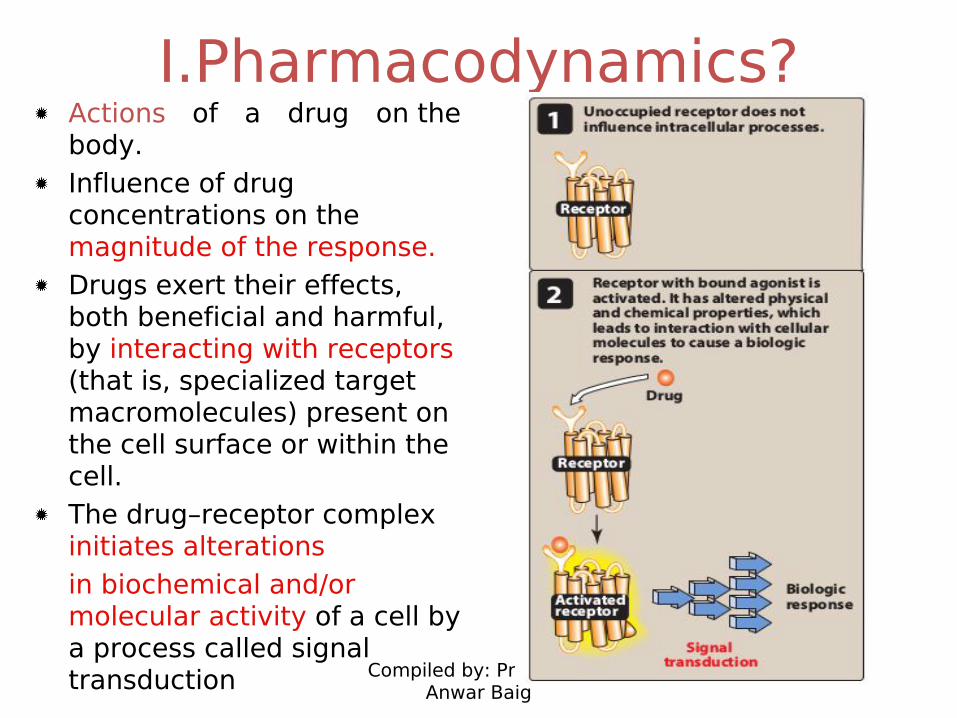
I.Pharmacodynamics?Actions of a drug on the body.Influence of drug concentrations on the magnitude of the response. Drugs exert their effects, both beneficial and harmful, by interacting with receptors (that is, specialized target macromolecules) present on the cell surface or within the cell. The drug–receptor complex initiates alterationsin biochemical and/or molecular activity of a cell by a process called signal transduction
Compiled by: Prof.Mirza Anwar Baig
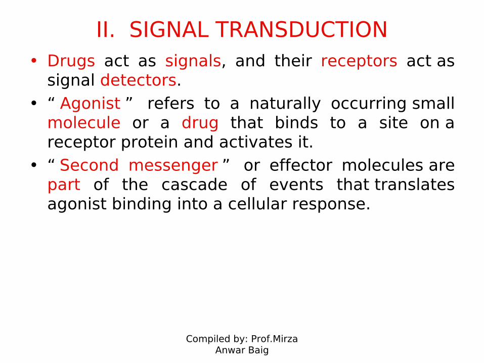
II. SIGNAL TRANSDUCTION• Drugs act as signals, and their receptors act as
signal detectors. • “ Agonist ” refers to a naturally occurring small
molecule or a drug that binds to a site on a receptor protein and activates it.
• “ Second messenger ” or effector molecules are part of the cascade of events that translates agonist binding into a cellular response.
Compiled by: Prof.Mirza Anwar Baig
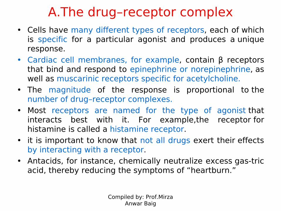
A.The drug–receptor complex• Cells have many different types of receptors, each of which
is specific for a particular agonist and produces a unique response.
• Cardiac cell membranes, for example, contain β receptors that bind and respond to epinephrine or norepinephrine, as well as muscarinic receptors specific for acetylcholine.
• The magnitude of the response is proportional to the number of drug–receptor complexes.
• Most receptors are named for the type of agonist that interacts best with it. For example,the receptor for histamine is called a histamine receptor.
• it is important to know that not all drugs exert their effects by interacting with a receptor.
• Antacids, for instance, chemically neutralize excess gas-tric acid, thereby reducing the symptoms of “heartburn.”
Compiled by: Prof.Mirza Anwar Baig
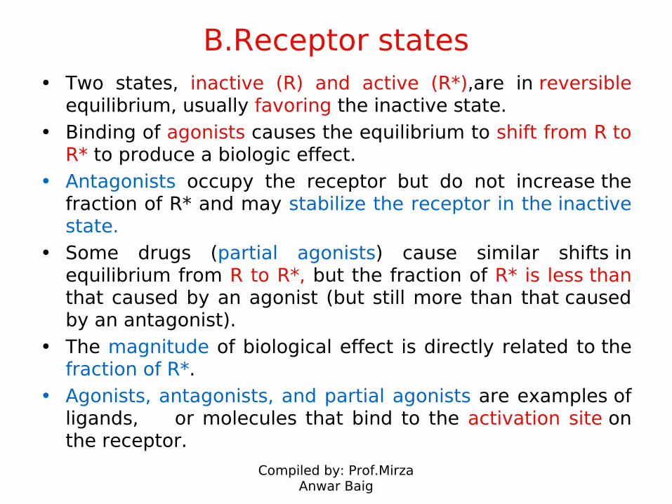
B.Receptor states• Two states, inactive (R) and active (R*),are in reversible
equilibrium, usually favoring the inactive state. • Binding of agonists causes the equilibrium to shift from R to
R* to produce a biologic effect. • Antagonists occupy the receptor but do not increase the
fraction of R* and may stabilize the receptor in the inactive state.
• Some drugs (partial agonists) cause similar shifts in equilibrium from R to R*, but the fraction of R* is less than that caused by an agonist (but still more than that caused by an antagonist).
• The magnitude of biological effect is directly related to the fraction of R*.
• Agonists, antagonists, and partial agonists are examples of ligands, or molecules that bind to the activation site on the receptor.
Compiled by: Prof.Mirza Anwar Baig
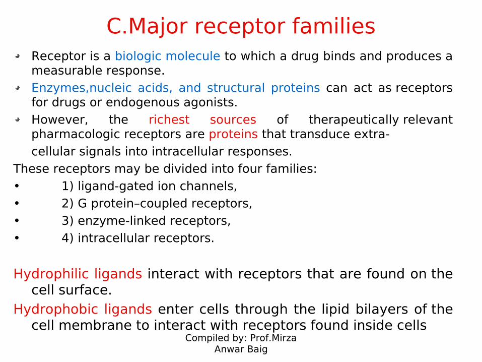
C.Major receptor familiesReceptor is a biologic molecule to which a drug binds and produces a measurable response. Enzymes,nucleic acids, and structural proteins can act as receptors for drugs or endogenous agonists.However, the richest sources of therapeutically relevant pharmacologic receptors are proteins that transduce extra-cellular signals into intracellular responses.
These receptors may be divided into four families: • 1) ligand-gated ion channels, • 2) G protein–coupled receptors, • 3) enzyme-linked receptors,• 4) intracellular receptors.
Hydrophilic ligands interact with receptors that are found on the cell surface.
Hydrophobic ligands enter cells through the lipid bilayers of the cell membrane to interact with receptors found inside cells
Compiled by: Prof.Mirza Anwar Baig
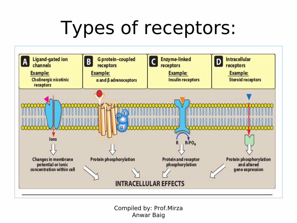
Types of receptors:
Compiled by: Prof.Mirza Anwar Baig
1. Transmembrane ligand-gated ion channels:
• Contains the ligand-binding site regulates the shape of the pore through which ions can flow across cell membranes.
• Agonist opens the channel briefly for a few milliseconds. • Depending on the ion conducted through these channels,
these receptors medi-ate diverse functions• For example, stimulation of the nicotinic receptor by
acetylcholine results in sodium influx and potassium outflux (action potential in a neuron or contraction in skeletal muscle).
• Agonist stimulation of the γ -aminobutyric acid (GABA) receptor increases chloride influx and hyperpolarization of neurons.
• Local anesthetics bind to the voltage-gated sodium channel, inhibiting sodium influx and decreasing neuronal conduction.
Compiled by: Prof.Mirza Anwar Baig
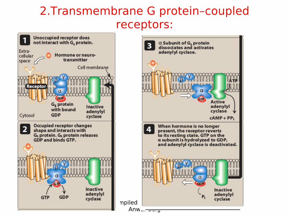
2.Transmembrane G protein–coupled receptors:
• Receptor contains the ligand-binding area, and the intracellular domain interacts (when activated) with a G protein or effector molecule.
• There are many kinds of G proteins (for example, Gs , Gi , and Gq ), but they all are composed of three pro-tein subunits.
• The α subunit binds guanosine triphosphate (GTP).• β and γ subunits anchor the G protein in the cell membrane. • Binding of an agonist to the receptor increases GTP binding to
the α subunit, causing dissociation of the α-GTP complex from the βγ complex.
• These two complexes can then interact with other cellular effectors, usually an enzyme, a protein,or an ion channel, that are responsible for further actions within the cell. These responses usually last several seconds to minutes.
Compiled by: Prof.Mirza Anwar Baig
A common effector, activated by Gs and inhibited by Gi , is adenylyl cyclase, which produces the second messenger cyclic adenosine monophosphate (cAMP).
Gq activates phospholipase C, generating two other second messengers: inositol 1,4,5/ trisphosphate (IP 3 ) and diacylglycerol (DAG).
DAG and cAMP activate different protein kinases within the cell, leading to a myriad of physiological effects.
IP 3 regulates intracellular free calcium concentrations, as
well as some protein kinases.
Compiled by: Prof.Mirza Anwar Baig
2.Transmembrane G protein–coupled receptors:
Compiled by: Prof.Mirza Anwar Baig
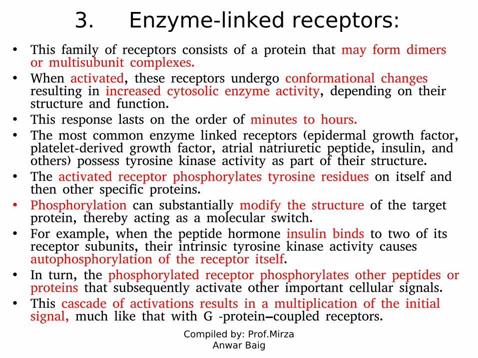
3. Enzyme-linked receptors:• This family of receptors consists of a protein that may form dimers
or multisubunit complexes.• When activated, these receptors undergo conformational changes
resulting in increased cytosolic enzyme activity, depending on their structure and function.
• This response lasts on the order of minutes to hours. • The most common enzyme linked receptors (epidermal growth factor,
platelet-derived growth factor, atrial natriuretic peptide, insulin, and others) possess tyrosine kinase activity as part of their structure.
• The activated receptor phosphorylates tyrosine residues on itself and then other specific proteins.
• Phosphorylation can substantially modify the structure of the target protein, thereby acting as a molecular switch.
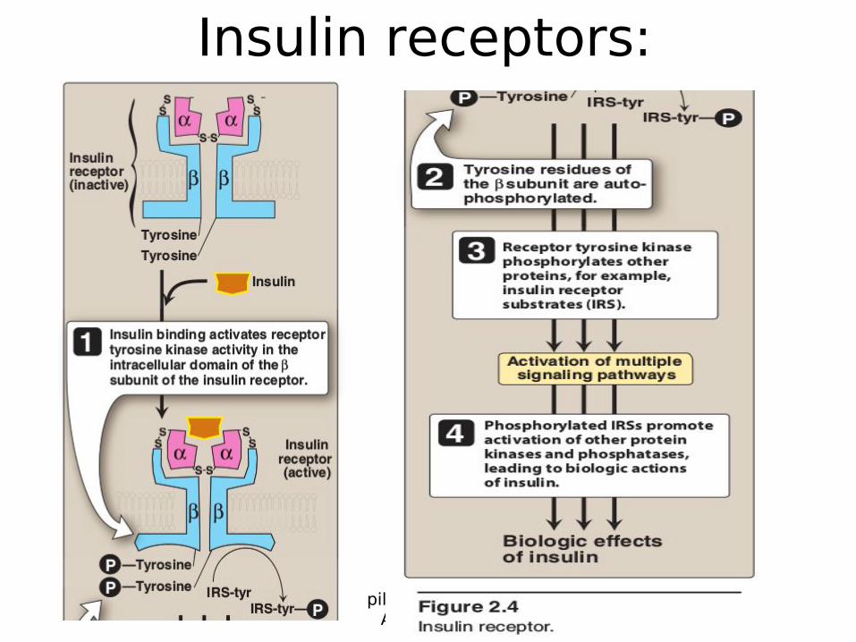
• For example, when the peptide hormone insulin binds to two of its receptor subunits, their intrinsic tyrosine kinase activity causes autophosphorylation of the receptor itself.
• In turn, the phosphorylated receptor phosphorylates other peptides or proteins that subsequently activate other important cellular signals.
• This cascade of activations results in a multiplication of the initial signal, much like that with G protein–coupled receptors.
Compiled by: Prof.Mirza Anwar Baig
Insulin receptors:
Compiled by: Prof.Mirza Anwar Baig
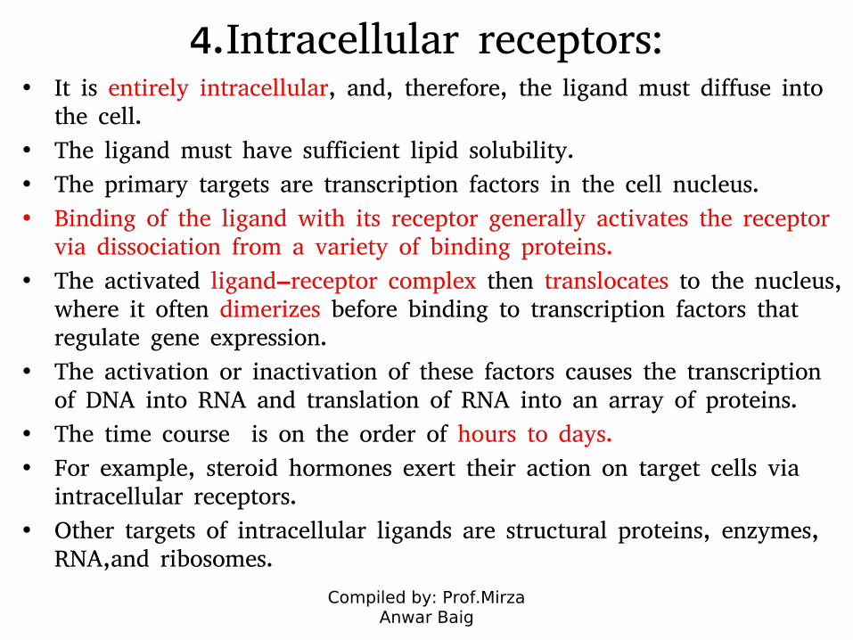
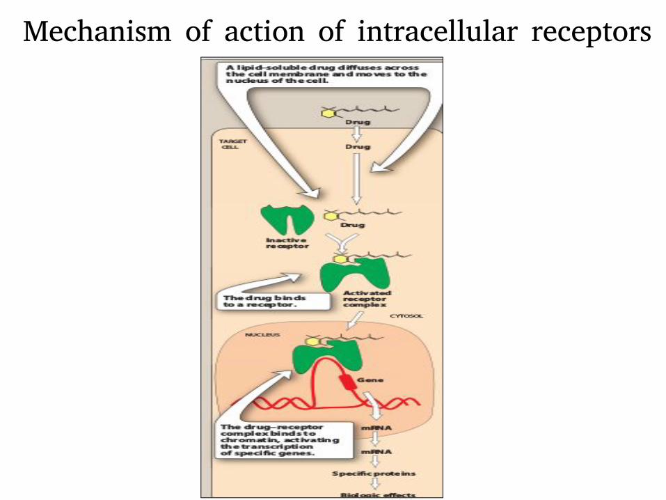
4.Intracellular receptors: • It is entirely intracellular, and, therefore, the ligand must diffuse into
the cell.
• The ligand must have sufficient lipid solubility.
• The primary targets are transcription factors in the cell nucleus.
• Binding of the ligand with its receptor generally activates the receptor via dissociation from a variety of binding proteins.
• The activated ligand–receptor complex then translocates to the nucleus, where it often dimerizes before binding to transcription factors that regulate gene expression.
• The activation or inactivation of these factors causes the transcription of DNA into RNA and translation of RNA into an array of proteins.
• The time course is on the order of hours to days.
• For example, steroid hormones exert their action on target cells via intracellular receptors.
• Other targets of intracellular ligands are structural proteins, enzymes, RNA,and ribosomes.
Compiled by: Prof.Mirza Anwar Baig
Mechanism of action of intracellular receptors
Compiled by: Prof.Mirza Anwar Baig
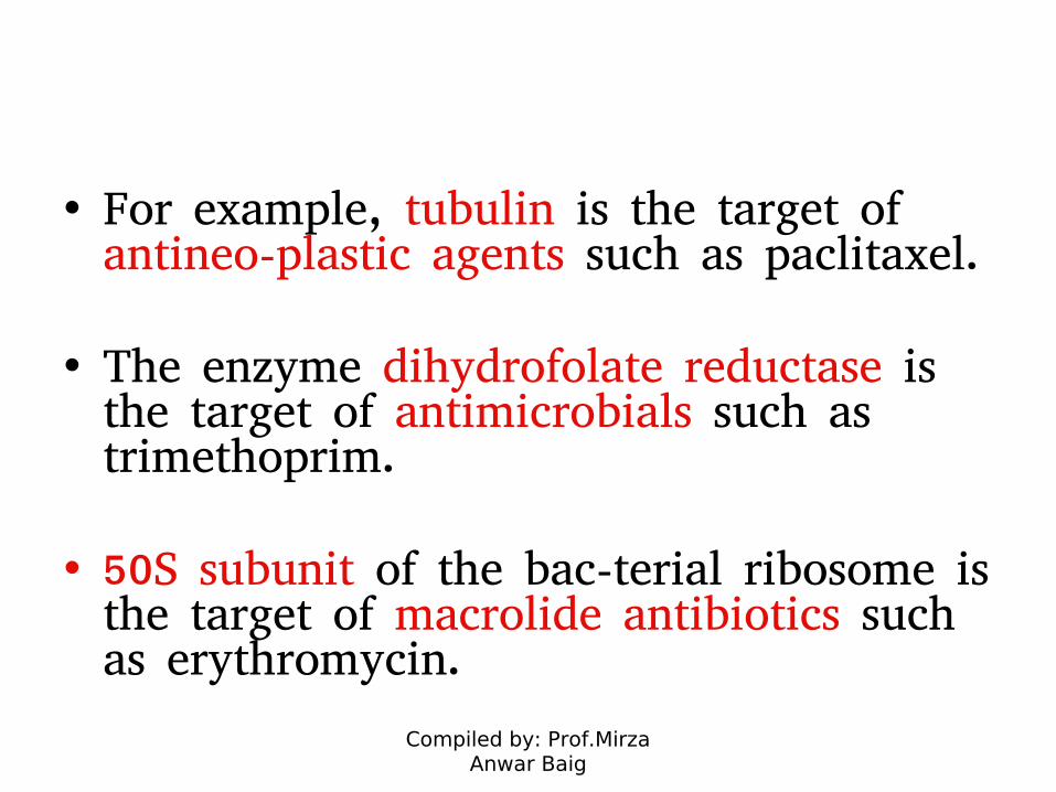
• For example, tubulin is the target of antineo-plastic agents such as paclitaxel.
• The enzyme dihydrofolate reductase is the target of antimicrobials such as trimethoprim.
• 50S subunit of the bac-terial ribosome is the target of macrolide antibiotics such as erythromycin.
Compiled by: Prof.Mirza Anwar Baig
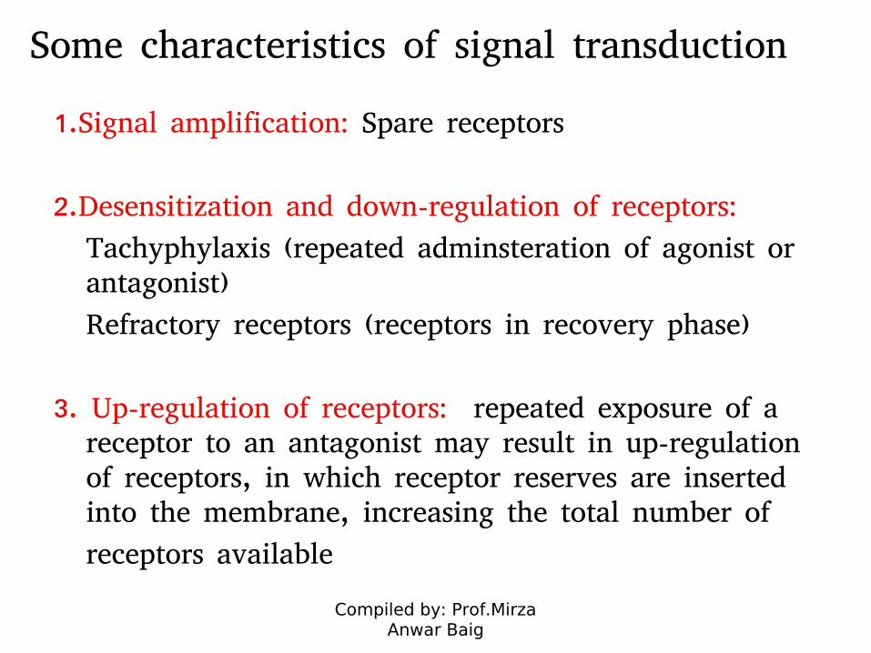
Some characteristics of signal transduction
1.Signal amplification: Spare receptors
2.Desensitization and down-regulation of receptors:
Tachyphylaxis (repeated adminsteration of agonist or antagonist)
Refractory receptors (receptors in recovery phase)
3. Up-regulation of receptors: repeated exposure of a receptor to an antagonist may result in up-regulation of receptors, in which receptor reserves are inserted into the membrane, increasing the total number of
receptors available
Compiled by: Prof.Mirza Anwar Baig
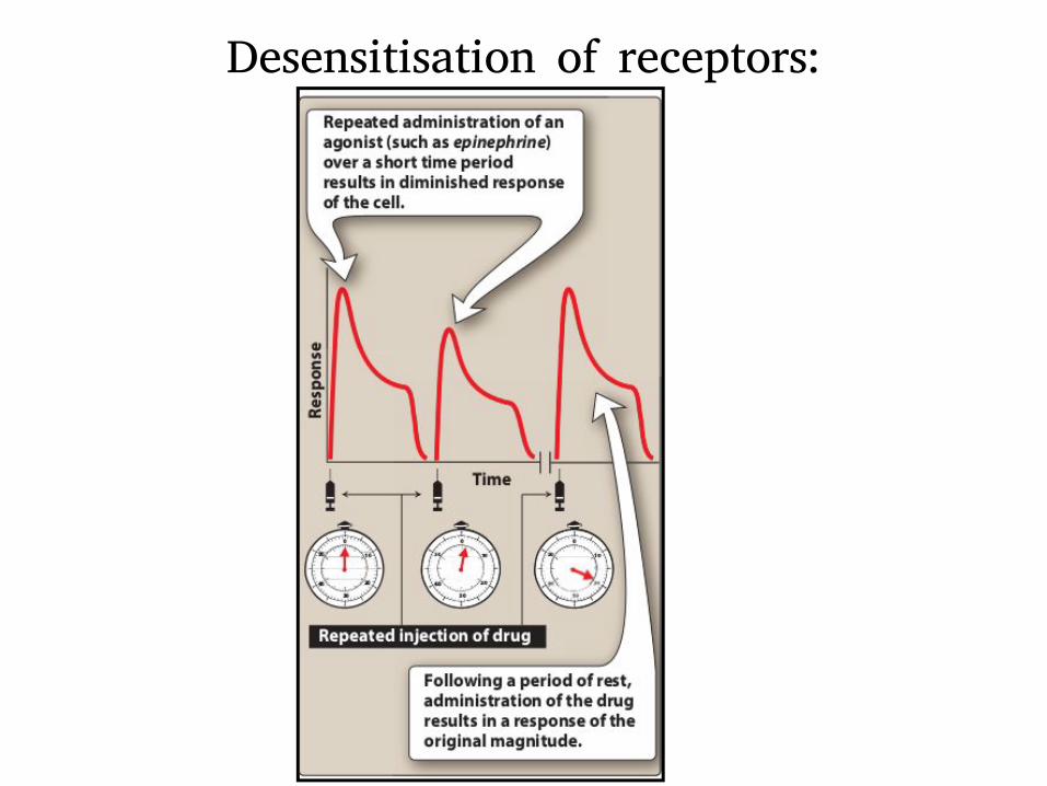
Desensitisation of receptors:
Compiled by: Prof.Mirza Anwar Baig
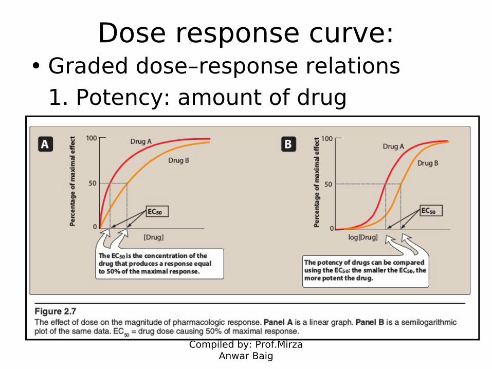
Dose response curve:• Graded dose–response relations
1. Potency: amount of drug
Compiled by: Prof.Mirza Anwar Baig
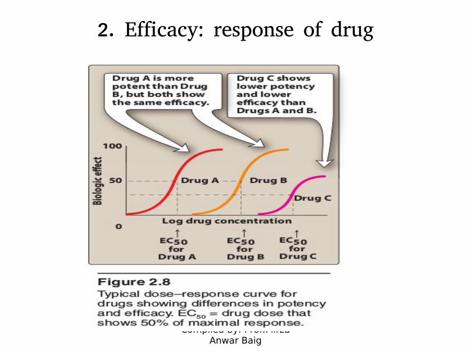
2. Efficacy: response of drug
Compiled by: Prof.Mirza Anwar Baig
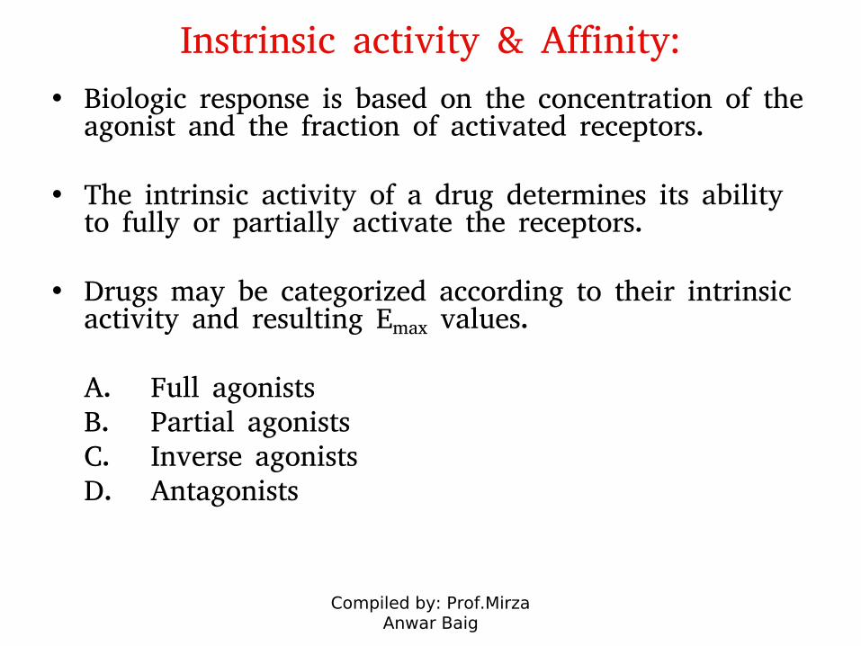
Instrinsic activity & Affinity:
• Biologic response is based on the concentration of the agonist and the fraction of activated receptors.
• The intrinsic activity of a drug determines its ability to fully or partially activate the receptors.
• Drugs may be categorized according to their intrinsic activity and resulting Emax values.
A. Full agonistsB. Partial agonistsC. Inverse agonistsD. Antagonists
Compiled by: Prof.Mirza Anwar Baig
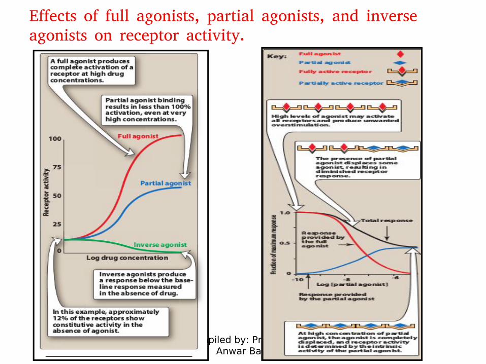
Effects of full agonists, partial agonists, and inverse agonists on receptor activity.
Compiled by: Prof.Mirza Anwar Baig
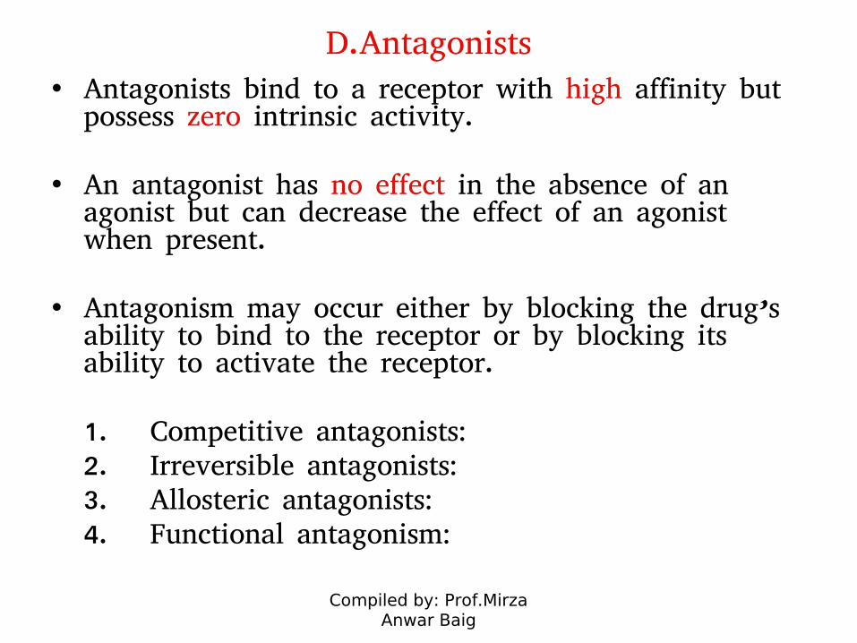
D.Antagonists• Antagonists bind to a receptor with high affinity but
possess zero intrinsic activity.
• An antagonist has no effect in the absence of an agonist but can decrease the effect of an agonist when present.
• Antagonism may occur either by blocking the drug’s ability to bind to the receptor or by blocking its ability to activate the receptor.
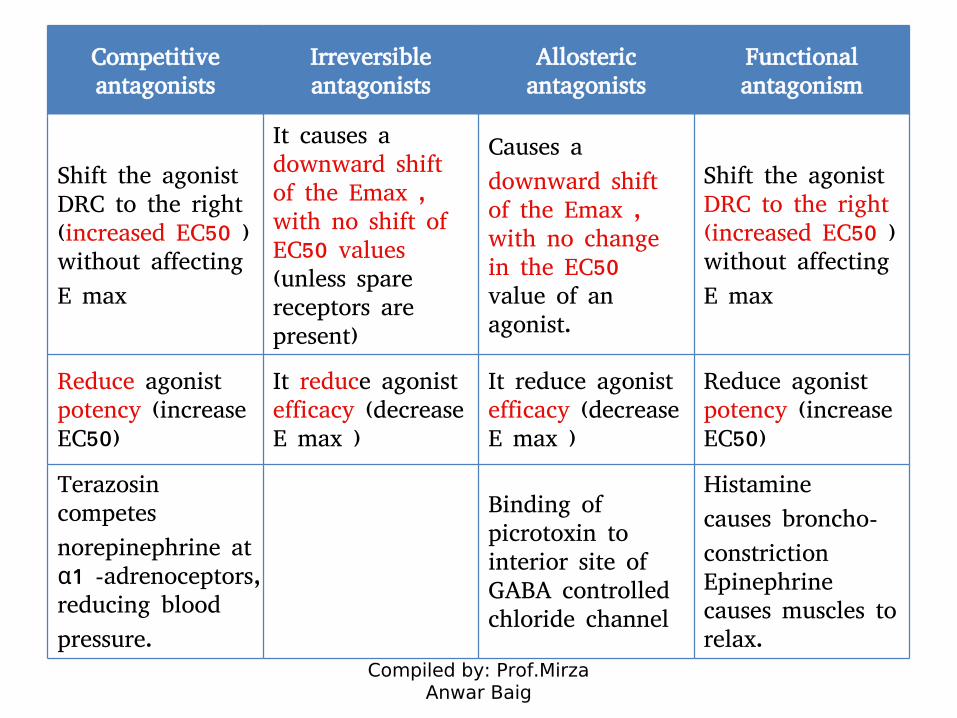
1. Competitive antagonists:2. Irreversible antagonists:3. Allosteric antagonists:4. Functional antagonism:

Compiled by: Prof.Mirza Anwar Baig
Competitive antagonists
Irreversible antagonists
Allosteric antagonists
Functional antagonism
Antagonist and the agonist bind to the same site on the receptor in a reversible manner
Irreversible antagonists bind covalently to the active site of the receptor, thereby reducing the number of receptors available to the agonist
Antagonist binds to a site (“allosteric site”) other than the agonist-binding site and prevents the receptor from
being activated by the agonist.
An antagonist may act at a completely
separate receptor, initiating effects that are functionally opposite those of the agonist
Inhibition can be overcome by increasing the concentration of agonist relative to antagonist.
The effect of irreversible antagonists cannot be overcome by adding more agonist
The effect of irreversible antagonists cannot be overcome by adding more agonist
Inhibition can be overcome by increasing the concentration of agonist relative to antagonist.
Compiled by: Prof.Mirza Anwar Baig
Competitive antagonists
Irreversible antagonists
Allosteric antagonists
Functional antagonism
Shift the agonist DRC to the right (increased EC50 ) without affecting
E max
It causes a downward shift of the Emax , with no shift of EC50 values (unless spare receptors are present)
Causes a
downward shift of the Emax , with no change in the EC50 value of an agonist.
Shift the agonist DRC to the right (increased EC50 ) without affecting
E max
Reduce agonist potency (increase EC50)
It reduce agonist efficacy (decrease E max )
It reduce agonist efficacy (decrease E max )
Reduce agonist potency (increase EC50)
Terazosin competes
norepinephrine at α1 -adrenoceptors, reducing blood
pressure.
Binding of picrotoxin to interior site of GABA controlled chloride channel
Histamine
causes broncho-
constriction Epinephrine causes muscles to relax.
Compiled by: Prof.Mirza Anwar Baig
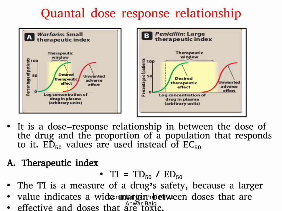
Quantal dose response relationship
• It is a dose–response relationship in between the dose of the drug and the proportion of a population that responds to it. ED50 values are used instead of EC50
A. Therapeutic index• TI = TD50 / ED50
• The TI is a measure of a drug’s safety, because a larger• value indicates a wide margin between doses that are • effective and doses that are toxic.