
26|FLUID, ELECTROLYTE, AND ACID-BASE BALANCE26|FLUID, ELECTROLYTE, AND ACID-BASE BALANCE...
32
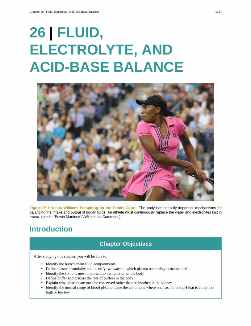
26 | FLUID, ELECTROLYTE, AND ACID-BASE BALANCE Figure 26.1 Venus Williams Perspiring on the Tennis Court The body has critically important mechanisms for balancing the intake and output of bodily fluids. An athlete must continuously replace the water and electrolytes lost in sweat. (credit: “Edwin Martinez1”/Wikimedia Commons) Introduction Chapter Objectives After studying this chapter, you will be able to: • Identify the body’s main fluid compartments • Define plasma osmolality and identify two ways in which plasma osmolality is maintained • Identify the six ions most important to the function of the body • Define buffer and discuss the role of buffers in the body • Explain why bicarbonate must be conserved rather than reabsorbed in the kidney • Identify the normal range of blood pH and name the conditions where one has a blood pH that is either too high or too low Chapter 26 | Fluid, Electrolyte, and Acid-Base Balance 1247
Transcript of 26|FLUID, ELECTROLYTE, AND ACID-BASE BALANCE26|FLUID, ELECTROLYTE, AND ACID-BASE BALANCE...
26 | FLUID,ELECTROLYTE, ANDACID-BASE BALANCE
Figure 26.1 Venus Williams Perspiring on the Tennis Court The body has critically important mechanisms forbalancing the intake and output of bodily fluids. An athlete must continuously replace the water and electrolytes lost insweat. (credit: “Edwin Martinez1”/Wikimedia Commons)
Introduction
Chapter Objectives
After studying this chapter, you will be able to:
• Identify the body’s main fluid compartments• Define plasma osmolality and identify two ways in which plasma osmolality is maintained• Identify the six ions most important to the function of the body• Define buffer and discuss the role of buffers in the body• Explain why bicarbonate must be conserved rather than reabsorbed in the kidney• Identify the normal range of blood pH and name the conditions where one has a blood pH that is either too
high or too low
Chapter 26 | Fluid, Electrolyte, and Acid-Base Balance 1247

Homeostasis, or the maintenance of constant conditions in the body, is a fundamental property of all living things. In thehuman body, the substances that participate in chemical reactions must remain within narrows ranges of concentration. Toomuch or too little of a single substance can disrupt your bodily functions. Because metabolism relies on reactions that are allinterconnected, any disruption might affect multiple organs or even organ systems. Water is the most ubiquitous substancein the chemical reactions of life. The interactions of various aqueous solutions—solutions in which water is the solvent—arecontinuously monitored and adjusted by a large suite of interconnected feedback systems in your body. Understanding theways in which the body maintains these critical balances is key to understanding good health.
26.1 | Body Fluids and Fluid Compartments
By the end of this section, you will be able to:
• Explain the importance of water in the body
• Contrast the composition of the intracellular fluid with that of the extracellular fluid
• Explain the importance of protein channels in the movement of solutes
• Identify the causes and symptoms of edema
The chemical reactions of life take place in aqueous solutions. The dissolved substances in a solution are called solutes.In the human body, solutes vary in different parts of the body, but may include proteins—including those that transportlipids, carbohydrates, and, very importantly, electrolytes. Often in medicine, a mineral dissociated from a salt that carries an
electrical charge (an ion) is called and electrolyte. For instance, sodium ions (Na+) and chloride ions (Cl-) are often referredto as electrolytes.
In the body, water moves through semi-permeable membranes of cells and from one compartment of the body to anotherby a process called osmosis. Osmosis is basically the diffusion of water from regions of higher concentration to regionsof lower concentration, along an osmotic gradient across a semi-permeable membrane. As a result, water will move intoand out of cells and tissues, depending on the relative concentrations of the water and solutes found there. An appropriatebalance of solutes inside and outside of cells must be maintained to ensure normal function.
Body Water Content
Human beings are mostly water, ranging from about 75 percent of body mass in infants to about 50–60 percent in adultmen and women, to as low as 45 percent in old age. The percent of body water changes with development, because theproportions of the body given over to each organ and to muscles, fat, bone, and other tissues change from infancy toadulthood (Figure 26.2). Your brain and kidneys have the highest proportions of water, which composes 80–85 percent oftheir masses. In contrast, teeth have the lowest proportion of water, at 8–10 percent.
1248 Chapter 26 | Fluid, Electrolyte, and Acid-Base Balance
This OpenStax book is available for free at http://cnx.org/content/col11496/1.8

Figure 26.2 Water Content of the Body’s Organs and Tissues Water content varies in different body organs andtissues, from as little as 8 percent in the teeth to as much as 85 percent in the brain.
Fluid Compartments
Body fluids can be discussed in terms of their specific fluid compartment, a location that is largely separate from anothercompartment by some form of a physical barrier. The intracellular fluid (ICF) compartment is the system that includes allfluid enclosed in cells by their plasma membranes. Extracellular fluid (ECF) surrounds all cells in the body. Extracellularfluid has two primary constituents: the fluid component of the blood (called plasma) and the interstitial fluid (IF) thatsurrounds all cells not in the blood (Figure 26.3).
Chapter 26 | Fluid, Electrolyte, and Acid-Base Balance 1249
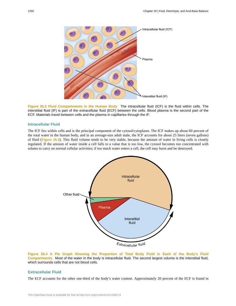
Figure 26.3 Fluid Compartments in the Human Body The intracellular fluid (ICF) is the fluid within cells. Theinterstitial fluid (IF) is part of the extracellular fluid (ECF) between the cells. Blood plasma is the second part of theECF. Materials travel between cells and the plasma in capillaries through the IF.
Intracellular Fluid
The ICF lies within cells and is the principal component of the cytosol/cytoplasm. The ICF makes up about 60 percent ofthe total water in the human body, and in an average-size adult male, the ICF accounts for about 25 liters (seven gallons)of fluid (Figure 26.4). This fluid volume tends to be very stable, because the amount of water in living cells is closelyregulated. If the amount of water inside a cell falls to a value that is too low, the cytosol becomes too concentrated withsolutes to carry on normal cellular activities; if too much water enters a cell, the cell may burst and be destroyed.
Figure 26.4 A Pie Graph Showing the Proportion of Total Body Fluid in Each of the Body’s FluidCompartments Most of the water in the body is intracellular fluid. The second largest volume is the interstitial fluid,which surrounds cells that are not blood cells.
Extracellular Fluid
The ECF accounts for the other one-third of the body’s water content. Approximately 20 percent of the ECF is found in
1250 Chapter 26 | Fluid, Electrolyte, and Acid-Base Balance
This OpenStax book is available for free at http://cnx.org/content/col11496/1.8
plasma. Plasma travels through the body in blood vessels and transports a range of materials, including blood cells, proteins(including clotting factors and antibodies), electrolytes, nutrients, gases, and wastes. Gases, nutrients, and waste materialstravel between capillaries and cells through the IF. Cells are separated from the IF by a selectively permeable cell membranethat helps regulate the passage of materials between the IF and the interior of the cell.
The body has other water-based ECF. These include the cerebrospinal fluid that bathes the brain and spinal cord, lymph, thesynovial fluid in joints, the pleural fluid in the pleural cavities, the pericardial fluid in the cardiac sac, the peritoneal fluidin the peritoneal cavity, and the aqueous humor of the eye. Because these fluids are outside of cells, these fluids are alsoconsidered components of the ECF compartment.
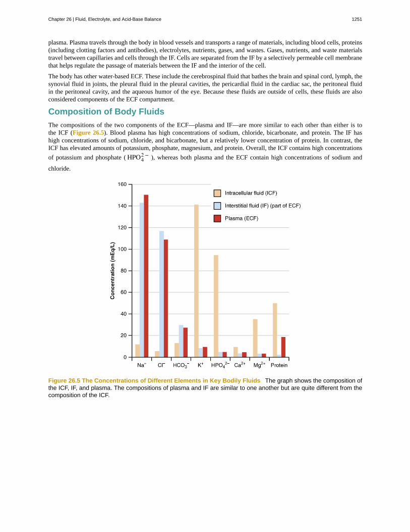
Composition of Body Fluids
The compositions of the two components of the ECF—plasma and IF—are more similar to each other than either is tothe ICF (Figure 26.5). Blood plasma has high concentrations of sodium, chloride, bicarbonate, and protein. The IF hashigh concentrations of sodium, chloride, and bicarbonate, but a relatively lower concentration of protein. In contrast, theICF has elevated amounts of potassium, phosphate, magnesium, and protein. Overall, the ICF contains high concentrations
of potassium and phosphate ( HPO42 − ), whereas both plasma and the ECF contain high concentrations of sodium and
chloride.
Figure 26.5 The Concentrations of Different Elements in Key Bodily Fluids The graph shows the composition ofthe ICF, IF, and plasma. The compositions of plasma and IF are similar to one another but are quite different from thecomposition of the ICF.
Chapter 26 | Fluid, Electrolyte, and Acid-Base Balance 1251

Watch this video (http://openstaxcollege.org/l/bodyfluids) to learn more about body fluids, fluid compartments, andelectrolytes. When blood volume decreases due to sweating, from what source is water taken in by the blood?
Most body fluids are neutral in charge. Thus, cations, or positively charged ions, and anions, or negatively charged ions, are
balanced in fluids. As seen in the previous graph, sodium (Na+) ions and chloride (Cl-) ions are concentrated in the ECF of
the body, whereas potassium (K+) ions are concentrated inside cells. Although sodium and potassium can “leak” through“pores” into and out of cells, respectively, the high levels of potassium and low levels of sodium in the ICF are maintainedby sodium-potassium pumps in the cell membranes. These pumps use the energy supplied by ATP to pump sodium out ofthe cell and potassium into the cell (Figure 26.6).
Figure 26.6 The Sodium-Potassium Pump The sodium-potassium pump is powered by ATP to transfer sodium outof the cytoplasm and into the ECF. The pump also transfers potassium out of the ECF and into the cytoplasm. (credit:modification of work by Mariana Ruiz Villarreal)
Fluid Movement between Compartments
Hydrostatic pressure, the force exerted by a fluid against a wall, causes movement of fluid between compartments. Thehydrostatic pressure of blood is the pressure exerted by blood against the walls of the blood vessels by the pumping actionof the heart. In capillaries, hydrostatic pressure (also known as capillary blood pressure) is higher than the opposing “colloidosmotic pressure” in blood—a “constant” pressure primarily produced by circulating albumin—at the arteriolar end of thecapillary (Figure 26.7). This pressure forces plasma and nutrients out of the capillaries and into surrounding tissues. Fluidand the cellular wastes in the tissues enter the capillaries at the venule end, where the hydrostatic pressure is less than theosmotic pressure in the vessel. Filtration pressure squeezes fluid from the plasma in the blood to the IF surrounding thetissue cells. The surplus fluid in the interstitial space that is not returned directly back to the capillaries is drained fromtissues by the lymphatic system, and then re-enters the vascular system at the subclavian veins.
1252 Chapter 26 | Fluid, Electrolyte, and Acid-Base Balance
This OpenStax book is available for free at http://cnx.org/content/col11496/1.8

Figure 26.7 Capillary Exchange Net filtration occurs near the arterial end of the capillary since capillary hydrostaticpressure (CHP) is greater than blood colloidal osmotic pressure (BCOP). There is no net movement of fluid near themidpoint of the capillary since CHP = BCOP. Net reabsorption occurs near the venous end of the capillary since BCOPis greater than CHP.
Watch this video (http://openstaxcollege.org/l/dynamicfluid) to see an explanation of the dynamics of fluid in thebody’s compartments. What happens in the tissue when capillary blood pressure is less than osmotic pressure?
Hydrostatic pressure is especially important in governing the movement of water in the nephrons of the kidneys to ensureproper filtering of the blood to form urine. As hydrostatic pressure in the kidneys increases, the amount of water leavingthe capillaries also increases, and more urine filtrate is formed. If hydrostatic pressure in the kidneys drops too low, as canhappen in dehydration, the functions of the kidneys will be impaired, and less nitrogenous wastes will be removed from thebloodstream. Extreme dehydration can result in kidney failure.
Fluid also moves between compartments along an osmotic gradient. Recall that an osmotic gradient is produced by thedifference in concentration of all solutes on either side of a semi-permeable membrane. The magnitude of the osmoticgradient is proportional to the difference in the concentration of solutes on one side of the cell membrane to that on the otherside. Water will move by osmosis from the side where its concentration is high (and the concentration of solute is low) tothe side of the membrane where its concentration is low (and the concentration of solute is high). In the body, water movesby osmosis from plasma to the IF (and the reverse) and from the IF to the ICF (and the reverse). In the body, water movesconstantly into and out of fluid compartments as conditions change in different parts of the body.
For example, if you are sweating, you will lose water through your skin. Sweating depletes your tissues of water andincreases the solute concentration in those tissues. As this happens, water diffuses from your blood into sweat glands andsurrounding skin tissues that have become dehydrated because of the osmotic gradient. Additionally, as water leaves theblood, it is replaced by the water in other tissues throughout your body that are not dehydrated. If this continues, dehydrationspreads throughout the body. When a dehydrated person drinks water and rehydrates, the water is redistributed by the samegradient, but in the opposite direction, replenishing water in all of the tissues.
Chapter 26 | Fluid, Electrolyte, and Acid-Base Balance 1253
Solute Movement between Compartments
The movement of some solutes between compartments is active, which consumes energy and is an active transport process,whereas the movement of other solutes is passive, which does not require energy. Active transport allows cells to move aspecific substance against its concentration gradient through a membrane protein, requiring energy in the form of ATP. Forexample, the sodium-potassium pump employs active transport to pump sodium out of cells and potassium into cells, withboth substances moving against their concentration gradients.
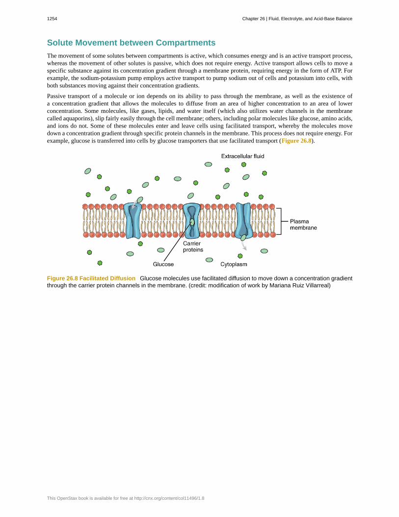
Passive transport of a molecule or ion depends on its ability to pass through the membrane, as well as the existence ofa concentration gradient that allows the molecules to diffuse from an area of higher concentration to an area of lowerconcentration. Some molecules, like gases, lipids, and water itself (which also utilizes water channels in the membranecalled aquaporins), slip fairly easily through the cell membrane; others, including polar molecules like glucose, amino acids,and ions do not. Some of these molecules enter and leave cells using facilitated transport, whereby the molecules movedown a concentration gradient through specific protein channels in the membrane. This process does not require energy. Forexample, glucose is transferred into cells by glucose transporters that use facilitated transport (Figure 26.8).
Figure 26.8 Facilitated Diffusion Glucose molecules use facilitated diffusion to move down a concentration gradientthrough the carrier protein channels in the membrane. (credit: modification of work by Mariana Ruiz Villarreal)
1254 Chapter 26 | Fluid, Electrolyte, and Acid-Base Balance
This OpenStax book is available for free at http://cnx.org/content/col11496/1.8

Fluid Balance: EdemaEdema is the accumulation of excess water in the tissues. It is most common in the soft tissues of the extremities.The physiological causes of edema include water leakage from blood capillaries. Edema is almost always caused byan underlying medical condition, by the use of certain therapeutic drugs, by pregnancy, by localized injury, or by anallergic reaction. In the limbs, the symptoms of edema include swelling of the subcutaneous tissues, an increase in thenormal size of the limb, and stretched, tight skin. One quick way to check for subcutaneous edema localized in a limbis to press a finger into the suspected area. Edema is likely if the depression persists for several seconds after the fingeris removed (which is called “pitting”).
Pulmonary edema is excess fluid in the air sacs of the lungs, a common symptom of heart and/or kidney failure. Peoplewith pulmonary edema likely will experience difficulty breathing, and they may experience chest pain. Pulmonaryedema can be life threatening, because it compromises gas exchange in the lungs, and anyone having symptoms shouldimmediately seek medical care.
In pulmonary edema resulting from heart failure, excessive leakage of water occurs because fluids get “backed up” inthe pulmonary capillaries of the lungs, when the left ventricle of the heart is unable to pump sufficient blood into thesystemic circulation. Because the left side of the heart is unable to pump out its normal volume of blood, the bloodin the pulmonary circulation gets “backed up,” starting with the left atrium, then into the pulmonary veins, and theninto pulmonary capillaries. The resulting increased hydrostatic pressure within pulmonary capillaries, as blood is stillcoming in from the pulmonary arteries, causes fluid to be pushed out of them and into lung tissues.
Other causes of edema include damage to blood vessels and/or lymphatic vessels, or a decrease in osmotic pressure inchronic and severe liver disease, where the liver is unable to manufacture plasma proteins (Figure 26.9). A decreasein the normal levels of plasma proteins results in a decrease of colloid osmotic pressure (which counterbalances thehydrostatic pressure) in the capillaries. This process causes loss of water from the blood to the surrounding tissues,resulting in edema.
Figure 26.9 Edema An allergic reaction can cause capillaries in the hand to leak excess fluid that accumulates inthe tissues. (credit: Jane Whitney)
Mild, transient edema of the feet and legs may be caused by sitting or standing in the same position for long periodsof time, as in the work of a toll collector or a supermarket cashier. This is because deep veins in the lower limbs relyon skeletal muscle contractions to push on the veins and thus “pump” blood back to the heart. Otherwise, the venousblood pools in the lower limbs and can leak into surrounding tissues.
Medications that can result in edema include vasodilators, calcium channel blockers used to treat hypertension, non-
Chapter 26 | Fluid, Electrolyte, and Acid-Base Balance 1255
steroidal anti-inflammatory drugs, estrogen therapies, and some diabetes medications. Underlying medical conditionsthat can contribute to edema include congestive heart failure, kidney damage and kidney disease, disorders that affectthe veins of the legs, and cirrhosis and other liver disorders.
Therapy for edema usually focuses on elimination of the cause. Activities that can reduce the effects of the conditioninclude appropriate exercises to keep the blood and lymph flowing through the affected areas. Other therapies includeelevation of the affected part to assist drainage, massage and compression of the areas to move the fluid out of thetissues, and decreased salt intake to decrease sodium and water retention.
26.2 | Water Balance
By the end of this section, you will be able to:
• Explain how water levels in the body influence the thirst cycle
• Identify the main route by which water leaves the body
• Describe the role of ADH and its effect on body water levels
• Define dehydration and identify common causes of dehydration
On a typical day, the average adult will take in about 2500 mL (almost 3 quarts) of aqueous fluids. Although most of theintake comes through the digestive tract, about 230 mL (8 ounces) per day is generated metabolically, in the last stepsof aerobic respiration. Additionally, each day about the same volume (2500 mL) of water leaves the body by differentroutes; most of this lost water is removed as urine. The kidneys also can adjust blood volume though mechanisms thatdraw water out of the filtrate and urine. The kidneys can regulate water levels in the body; they conserve water if you aredehydrated, and they can make urine more dilute to expel excess water if necessary. Water is lost through the skin throughevaporation from the skin surface without overt sweating and from air expelled from the lungs. This type of water loss iscalled insensible water loss because a person is usually unaware of it.
Regulation of Water Intake
Osmolality is the ratio of solutes in a solution to a volume of solvent in a solution. Plasma osmolality is thus the ratio ofsolutes to water in blood plasma. A person’s plasma osmolality value reflects his or her state of hydration. A healthy bodymaintains plasma osmolality within a narrow range, by employing several mechanisms that regulate both water intake andoutput.
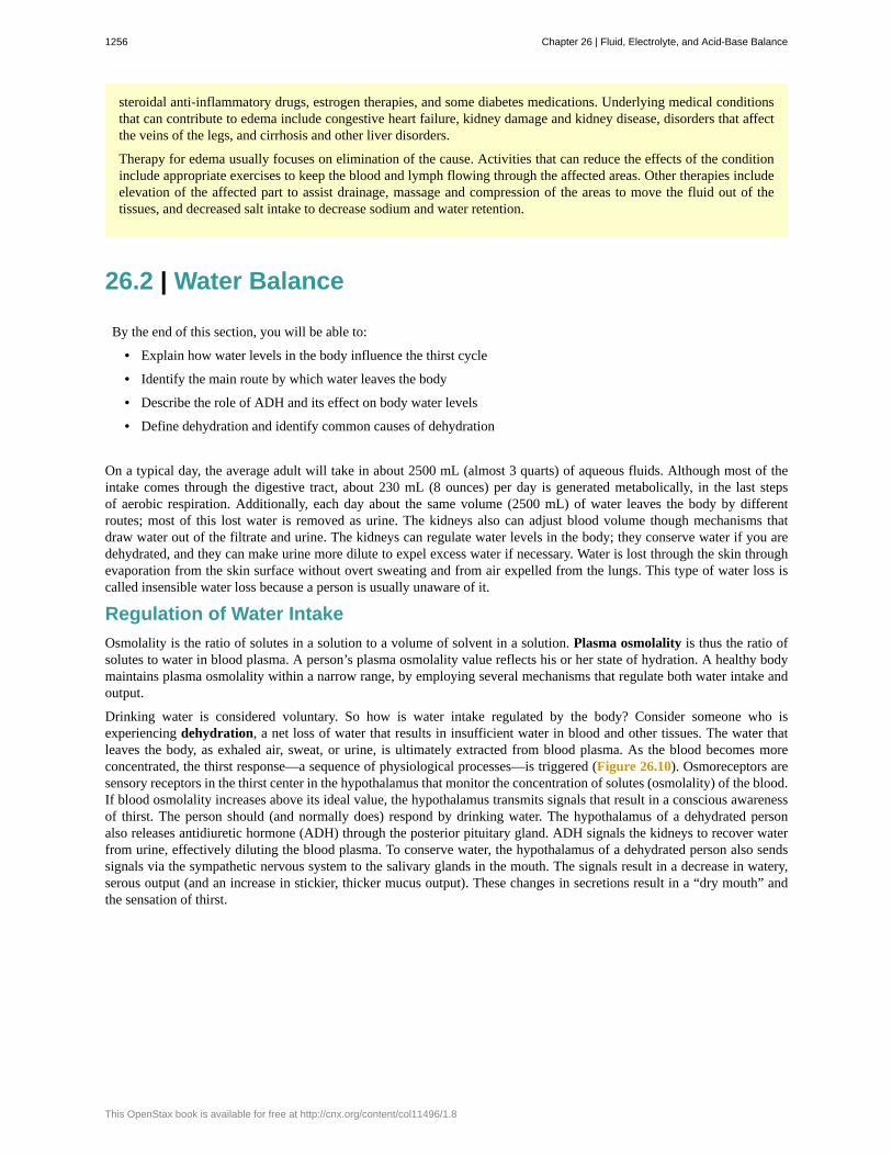
Drinking water is considered voluntary. So how is water intake regulated by the body? Consider someone who isexperiencing dehydration, a net loss of water that results in insufficient water in blood and other tissues. The water thatleaves the body, as exhaled air, sweat, or urine, is ultimately extracted from blood plasma. As the blood becomes moreconcentrated, the thirst response—a sequence of physiological processes—is triggered (Figure 26.10). Osmoreceptors aresensory receptors in the thirst center in the hypothalamus that monitor the concentration of solutes (osmolality) of the blood.If blood osmolality increases above its ideal value, the hypothalamus transmits signals that result in a conscious awarenessof thirst. The person should (and normally does) respond by drinking water. The hypothalamus of a dehydrated personalso releases antidiuretic hormone (ADH) through the posterior pituitary gland. ADH signals the kidneys to recover waterfrom urine, effectively diluting the blood plasma. To conserve water, the hypothalamus of a dehydrated person also sendssignals via the sympathetic nervous system to the salivary glands in the mouth. The signals result in a decrease in watery,serous output (and an increase in stickier, thicker mucus output). These changes in secretions result in a “dry mouth” andthe sensation of thirst.
1256 Chapter 26 | Fluid, Electrolyte, and Acid-Base Balance
This OpenStax book is available for free at http://cnx.org/content/col11496/1.8

Figure 26.10 A Flowchart Showing the Thirst Response The thirst response begins when osmoreceptors detect adecrease in water levels in the blood.
Decreased blood volume resulting from water loss has two additional effects. First, baroreceptors, blood-pressure receptorsin the arch of the aorta and the carotid arteries in the neck, detect a decrease in blood pressure that results from decreasedblood volume. The heart is ultimately signaled to increase its rate and/or strength of contractions to compensate for thelowered blood pressure.
Second, the kidneys have a renin-angiotensin hormonal system that increases the production of the active form of thehormone angiotensin II, which helps stimulate thirst, but also stimulates the release of the hormone aldosterone from theadrenal glands. Aldosterone increases the reabsorption of sodium in the distal tubules of the nephrons in the kidneys, andwater follows this reabsorbed sodium back into the blood.
If adequate fluids are not consumed, dehydration results and a person’s body contains too little water to function correctly. Aperson who repeatedly vomits or who has diarrhea may become dehydrated, and infants, because their body mass is so low,can become dangerously dehydrated very quickly. Endurance athletes such as distance runners often become dehydratedduring long races. Dehydration can be a medical emergency, and a dehydrated person may lose consciousness, become
Chapter 26 | Fluid, Electrolyte, and Acid-Base Balance 1257
comatose, or die, if his or her body is not rehydrated quickly.
Regulation of Water Output
Water loss from the body occurs predominantly through the renal system. A person produces an average of 1.5 liters (1.6quarts) of urine per day. Although the volume of urine varies in response to hydration levels, there is a minimum volumeof urine production required for proper bodily functions. The kidney excretes 100 to 1200 milliosmoles of solutes per dayto rid the body of a variety of excess salts and other water-soluble chemical wastes, most notably creatinine, urea, and uricacid. Failure to produce the minimum volume of urine means that metabolic wastes cannot be effectively removed fromthe body, a situation that can impair organ function. The minimum level of urine production necessary to maintain normalfunction is about 0.47 liters (0.5 quarts) per day.
The kidneys also must make adjustments in the event of ingestion of too much fluid. Diuresis, which is the production ofurine in excess of normal levels, begins about 30 minutes after drinking a large quantity of fluid. Diuresis reaches a peakafter about 1 hour, and normal urine production is reestablished after about 3 hours.
Role of ADH
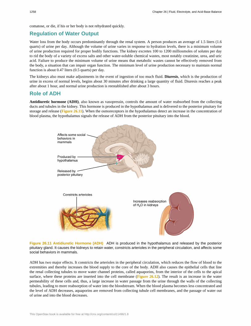
Antidiuretic hormone (ADH), also known as vasopressin, controls the amount of water reabsorbed from the collectingducts and tubules in the kidney. This hormone is produced in the hypothalamus and is delivered to the posterior pituitary forstorage and release (Figure 26.11). When the osmoreceptors in the hypothalamus detect an increase in the concentration ofblood plasma, the hypothalamus signals the release of ADH from the posterior pituitary into the blood.
Figure 26.11 Antidiuretic Hormone (ADH) ADH is produced in the hypothalamus and released by the posteriorpituitary gland. It causes the kidneys to retain water, constricts arterioles in the peripheral circulation, and affects somesocial behaviors in mammals.
ADH has two major effects. It constricts the arterioles in the peripheral circulation, which reduces the flow of blood to theextremities and thereby increases the blood supply to the core of the body. ADH also causes the epithelial cells that linethe renal collecting tubules to move water channel proteins, called aquaporins, from the interior of the cells to the apicalsurface, where these proteins are inserted into the cell membrane (Figure 26.12). The result is an increase in the waterpermeability of these cells and, thus, a large increase in water passage from the urine through the walls of the collectingtubules, leading to more reabsorption of water into the bloodstream. When the blood plasma becomes less concentrated andthe level of ADH decreases, aquaporins are removed from collecting tubule cell membranes, and the passage of water outof urine and into the blood decreases.
1258 Chapter 26 | Fluid, Electrolyte, and Acid-Base Balance
This OpenStax book is available for free at http://cnx.org/content/col11496/1.8
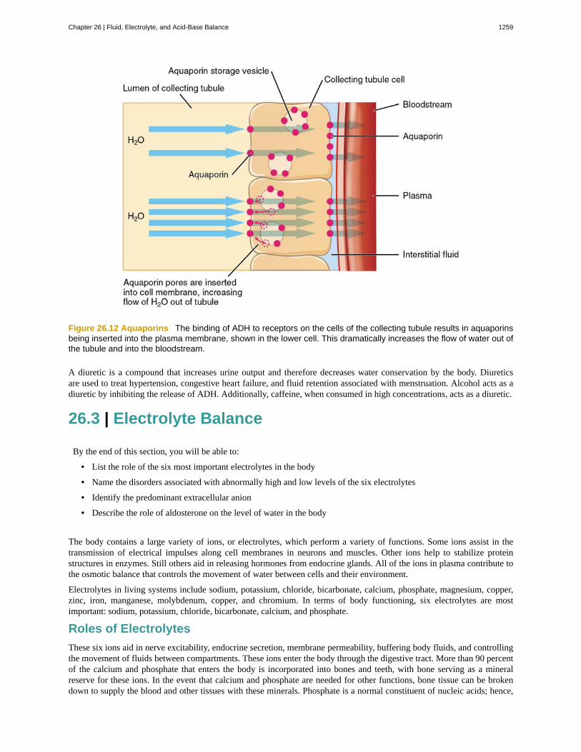
Figure 26.12 Aquaporins The binding of ADH to receptors on the cells of the collecting tubule results in aquaporinsbeing inserted into the plasma membrane, shown in the lower cell. This dramatically increases the flow of water out ofthe tubule and into the bloodstream.
A diuretic is a compound that increases urine output and therefore decreases water conservation by the body. Diureticsare used to treat hypertension, congestive heart failure, and fluid retention associated with menstruation. Alcohol acts as adiuretic by inhibiting the release of ADH. Additionally, caffeine, when consumed in high concentrations, acts as a diuretic.
26.3 | Electrolyte Balance
By the end of this section, you will be able to:
• List the role of the six most important electrolytes in the body
• Name the disorders associated with abnormally high and low levels of the six electrolytes
• Identify the predominant extracellular anion
• Describe the role of aldosterone on the level of water in the body
The body contains a large variety of ions, or electrolytes, which perform a variety of functions. Some ions assist in thetransmission of electrical impulses along cell membranes in neurons and muscles. Other ions help to stabilize proteinstructures in enzymes. Still others aid in releasing hormones from endocrine glands. All of the ions in plasma contribute tothe osmotic balance that controls the movement of water between cells and their environment.
Electrolytes in living systems include sodium, potassium, chloride, bicarbonate, calcium, phosphate, magnesium, copper,zinc, iron, manganese, molybdenum, copper, and chromium. In terms of body functioning, six electrolytes are mostimportant: sodium, potassium, chloride, bicarbonate, calcium, and phosphate.
Roles of Electrolytes
These six ions aid in nerve excitability, endocrine secretion, membrane permeability, buffering body fluids, and controllingthe movement of fluids between compartments. These ions enter the body through the digestive tract. More than 90 percentof the calcium and phosphate that enters the body is incorporated into bones and teeth, with bone serving as a mineralreserve for these ions. In the event that calcium and phosphate are needed for other functions, bone tissue can be brokendown to supply the blood and other tissues with these minerals. Phosphate is a normal constituent of nucleic acids; hence,
Chapter 26 | Fluid, Electrolyte, and Acid-Base Balance 1259

blood levels of phosphate will increase whenever nucleic acids are broken down.
Excretion of ions occurs mainly through the kidneys, with lesser amounts lost in sweat and in feces. Excessive sweatingmay cause a significant loss, especially of sodium and chloride. Severe vomiting or diarrhea will cause a loss of chlorideand bicarbonate ions. Adjustments in respiratory and renal functions allow the body to regulate the levels of these ions inthe ECF.
Table 26.1 lists the reference values for blood plasma, cerebrospinal fluid (CSF), and urine for the six ions addressed in thissection. In a clinical setting, sodium, potassium, and chloride are typically analyzed in a routine urine sample. In contrast,calcium and phosphate analysis requires a collection of urine across a 24-hour period, because the output of these ions canvary considerably over the course of a day. Urine values reflect the rates of excretion of these ions. Bicarbonate is the oneion that is not normally excreted in urine; instead, it is conserved by the kidneys for use in the body’s buffering systems.
Electrolyte and Ion Reference Values
Name Chemical symbol Plasma CSF Urine
Sodium Na+ 136.00–146.00 (mM) 138.00–150.00 (mM) 40.00–220.00 (mM)
Potassium K+ 3.50–5.00 (mM) 0.35–3.5 (mM) 25.00–125.00 (mM)
Chloride Cl- 98.00–107.00 (mM) 118.00–132.00 (mM) 110.00–250.00 (mM)
Bicarbonate HCO3- 22.00–29.00 (mM) ------ ------
Calcium Ca++ 2.15–2.55 (mmol/day) ------ Up to 7.49 (mmol/day)
Phosphate HPO42 −
0.81–1.45 (mmol/day) ------ 12.90–42.00 (mmol/day)
Table 26.1
Sodium
Sodium is the major cation of the extracellular fluid. It is responsible for one-half of the osmotic pressure gradient that existsbetween the interior of cells and their surrounding environment. People eating a typical Western diet, which is very highin NaCl, routinely take in 130 to 160 mmol/day of sodium, but humans require only 1 to 2 mmol/day. This excess sodiumappears to be a major factor in hypertension (high blood pressure) in some people. Excretion of sodium is accomplishedprimarily by the kidneys. Sodium is freely filtered through the glomerular capillaries of the kidneys, and although much ofthe filtered sodium is reabsorbed in the proximal convoluted tubule, some remains in the filtrate and urine, and is normallyexcreted.
Hyponatremia is a lower-than-normal concentration of sodium, usually associated with excess water accumulation in thebody, which dilutes the sodium. An absolute loss of sodium may be due to a decreased intake of the ion coupled with itscontinual excretion in the urine. An abnormal loss of sodium from the body can result from several conditions, includingexcessive sweating, vomiting, or diarrhea; the use of diuretics; excessive production of urine, which can occur in diabetes;and acidosis, either metabolic acidosis or diabetic ketoacidosis.
A relative decrease in blood sodium can occur because of an imbalance of sodium in one of the body’s other fluidcompartments, like IF, or from a dilution of sodium due to water retention related to edema or congestive heart failure.At the cellular level, hyponatremia results in increased entry of water into cells by osmosis, because the concentration ofsolutes within the cell exceeds the concentration of solutes in the now-diluted ECF. The excess water causes swelling of thecells; the swelling of red blood cells—decreasing their oxygen-carrying efficiency and making them potentially too large tofit through capillaries—along with the swelling of neurons in the brain can result in brain damage or even death.
Hypernatremia is an abnormal increase of blood sodium. It can result from water loss from the blood, resulting in thehemoconcentration of all blood constituents. Hormonal imbalances involving ADH and aldosterone may also result inhigher-than-normal sodium values.
Potassium
Potassium is the major intracellular cation. It helps establish the resting membrane potential in neurons and muscle fibersafter membrane depolarization and action potentials. In contrast to sodium, potassium has very little effect on osmoticpressure. The low levels of potassium in blood and CSF are due to the sodium-potassium pumps in cell membranes,
1260 Chapter 26 | Fluid, Electrolyte, and Acid-Base Balance
This OpenStax book is available for free at http://cnx.org/content/col11496/1.8
which maintain the normal potassium concentration gradients between the ICF and ECF. The recommendation for dailyintake/consumption of potassium is 4700 mg. Potassium is excreted, both actively and passively, through the renal tubules,especially the distal convoluted tubule and collecting ducts. Potassium participates in the exchange with sodium in the renaltubules under the influence of aldosterone, which also relies on basolateral sodium-potassium pumps.
Hypokalemia is an abnormally low potassium blood level. Similar to the situation with hyponatremia, hypokalemia canoccur because of either an absolute reduction of potassium in the body or a relative reduction of potassium in the blooddue to the redistribution of potassium. An absolute loss of potassium can arise from decreased intake, frequently related tostarvation. It can also come about from vomiting, diarrhea, or alkalosis.
Some insulin-dependent diabetic patients experience a relative reduction of potassium in the blood from the redistributionof potassium. When insulin is administered and glucose is taken up by cells, potassium passes through the cell membranealong with glucose, decreasing the amount of potassium in the blood and IF, which can cause hyperpolarization of the cellmembranes of neurons, reducing their responses to stimuli.
Hyperkalemia, an elevated potassium blood level, also can impair the function of skeletal muscles, the nervous system,and the heart. Hyperkalemia can result from increased dietary intake of potassium. In such a situation, potassium from theblood ends up in the ECF in abnormally high concentrations. This can result in a partial depolarization (excitation) of theplasma membrane of skeletal muscle fibers, neurons, and cardiac cells of the heart, and can also lead to an inability of cellsto repolarize. For the heart, this means that it won’t relax after a contraction, and will effectively “seize” and stop pumpingblood, which is fatal within minutes. Because of such effects on the nervous system, a person with hyperkalemia may alsoexhibit mental confusion, numbness, and weakened respiratory muscles.
Chloride
Chloride is the predominant extracellular anion. Chloride is a major contributor to the osmotic pressure gradient betweenthe ICF and ECF, and plays an important role in maintaining proper hydration. Chloride functions to balance cations in theECF, maintaining the electrical neutrality of this fluid. The paths of secretion and reabsorption of chloride ions in the renalsystem follow the paths of sodium ions.
Hypochloremia, or lower-than-normal blood chloride levels, can occur because of defective renal tubular absorption.Vomiting, diarrhea, and metabolic acidosis can also lead to hypochloremia. Hyperchloremia, or higher-than-normal bloodchloride levels, can occur due to dehydration, excessive intake of dietary salt (NaCl) or swallowing of sea water, aspirinintoxication, congestive heart failure, and the hereditary, chronic lung disease, cystic fibrosis. In people who have cysticfibrosis, chloride levels in sweat are two to five times those of normal levels, and analysis of sweat is often used in thediagnosis of the disease.
Read this article (http://openstaxcollege.org/l/saltwater) for an explanation of the effect of seawater on humans.What effect does drinking seawater have on the body?
Bicarbonate
Bicarbonate is the second most abundant anion in the blood. Its principal function is to maintain your body’s acid-basebalance by being part of buffer systems. This role will be discussed in a different section.
Bicarbonate ions result from a chemical reaction that starts with carbon dioxide (CO2) and water, two molecules that areproduced at the end of aerobic metabolism. Only a small amount of CO2 can be dissolved in body fluids. Thus, over 90
percent of the CO2 is converted into bicarbonate ions, HCO3–, through the following reactions:
CO2 + H2 O ↔ H2 CO3 ↔ HCO3 - + H+
Chapter 26 | Fluid, Electrolyte, and Acid-Base Balance 1261

The bidirectional arrows indicate that the reactions can go in either direction, depending on the concentrations of thereactants and products. Carbon dioxide is produced in large amounts in tissues that have a high metabolic rate. Carbondioxide is converted into bicarbonate in the cytoplasm of red blood cells through the action of an enzyme called carbonicanhydrase. Bicarbonate is transported in the blood. Once in the lungs, the reactions reverse direction, and CO2 is regeneratedfrom bicarbonate to be exhaled as metabolic waste.
Calcium
About two pounds of calcium in your body are bound up in bone, which provides hardness to the bone and serves as amineral reserve for calcium and its salts for the rest of the tissues. Teeth also have a high concentration of calcium withinthem. A little more than one-half of blood calcium is bound to proteins, leaving the rest in its ionized form. Calcium ions,
Ca2+, are necessary for muscle contraction, enzyme activity, and blood coagulation. In addition, calcium helps to stabilizecell membranes and is essential for the release of neurotransmitters from neurons and of hormones from endocrine glands.
Calcium is absorbed through the intestines under the influence of activated vitamin D. A deficiency of vitamin D leads to adecrease in absorbed calcium and, eventually, a depletion of calcium stores from the skeletal system, potentially leading torickets in children and osteomalacia in adults, contributing to osteoporosis.
Hypocalcemia, or abnormally low calcium blood levels, is seen in hypoparathyroidism, which may follow the removal ofthe thyroid gland, because the four nodules of the parathyroid gland are embedded in it. Hypercalcemia, or abnormallyhigh calcium blood levels, is seen in primary hyperparathyroidism. Some malignancies may also result in hypercalcemia.
Phosphate
Phosphate is present in the body in three ionic forms: H2 PO4 − , HPO42 − , and PO4
3 − . The most common form is
HPO42 − . Bone and teeth bind up 85 percent of the body’s phosphate as part of calcium-phosphate salts. Phosphate is found
in phospholipids, such as those that make up the cell membrane, and in ATP, nucleotides, and buffers.
Hypophosphatemia, or abnormally low phosphate blood levels, occurs with heavy use of antacids, during alcoholwithdrawal, and during malnourishment. In the face of phosphate depletion, the kidneys usually conserve phosphate, butduring starvation, this conservation is impaired greatly. Hyperphosphatemia, or abnormally increased levels of phosphatesin the blood, occurs if there is decreased renal function or in cases of acute lymphocytic leukemia. Additionally, becausephosphate is a major constituent of the ICF, any significant destruction of cells can result in dumping of phosphate into theECF.
Regulation of Sodium and Potassium
Sodium is reabsorbed from the renal filtrate, and potassium is excreted into the filtrate in the renal collecting tubule. Thecontrol of this exchange is governed principally by two hormones—aldosterone and angiotensin II.
Aldosterone
Recall that aldosterone increases the excretion of potassium and the reabsorption of sodium in the distal tubule. Aldosteroneis released if blood levels of potassium increase, if blood levels of sodium severely decrease, or if blood pressure decreases.Its net effect is to conserve and increase water levels in the plasma by reducing the excretion of sodium, and thus water, fromthe kidneys. In a negative feedback loop, increased osmolality of the ECF (which follows aldosterone-stimulated sodiumabsorption) inhibits the release of the hormone (Figure 26.13).
1262 Chapter 26 | Fluid, Electrolyte, and Acid-Base Balance
This OpenStax book is available for free at http://cnx.org/content/col11496/1.8

Figure 26.13 The Aldosterone Feedback Loop Aldosterone, which is released by the adrenal gland, facilitates
reabsorption of Na+ and thus the reabsorption of water.
Angiotensin II
Angiotensin II causes vasoconstriction and an increase in systemic blood pressure. This action increases the glomerularfiltration rate, resulting in more material filtered out of the glomerular capillaries and into Bowman’s capsule. AngiotensinII also signals an increase in the release of aldosterone from the adrenal cortex.
In the distal convoluted tubules and collecting ducts of the kidneys, aldosterone stimulates the synthesis and activation ofthe sodium-potassium pump (Figure 26.14). Sodium passes from the filtrate, into and through the cells of the tubules andducts, into the ECF and then into capillaries. Water follows the sodium due to osmosis. Thus, aldosterone causes an increasein blood sodium levels and blood volume. Aldosterone’s effect on potassium is the reverse of that of sodium; under itsinfluence, excess potassium is pumped into the renal filtrate for excretion from the body.
Chapter 26 | Fluid, Electrolyte, and Acid-Base Balance 1263
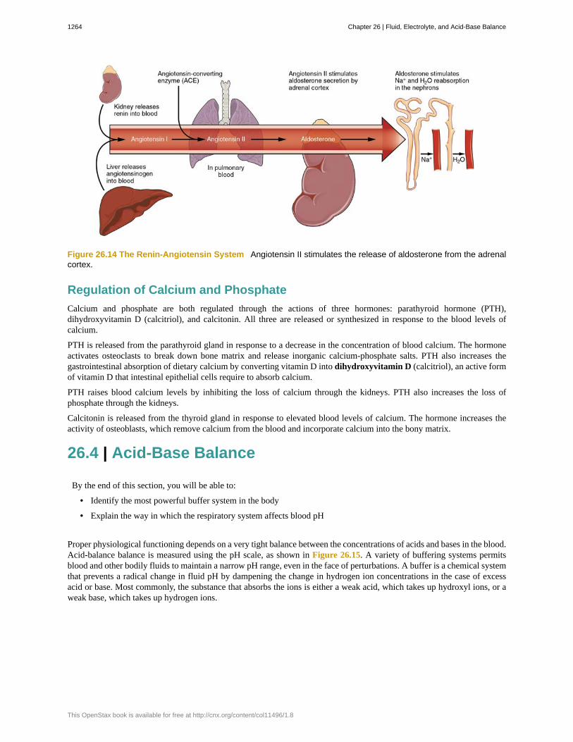
Figure 26.14 The Renin-Angiotensin System Angiotensin II stimulates the release of aldosterone from the adrenalcortex.
Regulation of Calcium and Phosphate
Calcium and phosphate are both regulated through the actions of three hormones: parathyroid hormone (PTH),dihydroxyvitamin D (calcitriol), and calcitonin. All three are released or synthesized in response to the blood levels ofcalcium.
PTH is released from the parathyroid gland in response to a decrease in the concentration of blood calcium. The hormoneactivates osteoclasts to break down bone matrix and release inorganic calcium-phosphate salts. PTH also increases thegastrointestinal absorption of dietary calcium by converting vitamin D into dihydroxyvitamin D (calcitriol), an active formof vitamin D that intestinal epithelial cells require to absorb calcium.
PTH raises blood calcium levels by inhibiting the loss of calcium through the kidneys. PTH also increases the loss ofphosphate through the kidneys.
Calcitonin is released from the thyroid gland in response to elevated blood levels of calcium. The hormone increases theactivity of osteoblasts, which remove calcium from the blood and incorporate calcium into the bony matrix.
26.4 | Acid-Base Balance
By the end of this section, you will be able to:
• Identify the most powerful buffer system in the body
• Explain the way in which the respiratory system affects blood pH
Proper physiological functioning depends on a very tight balance between the concentrations of acids and bases in the blood.Acid-balance balance is measured using the pH scale, as shown in Figure 26.15. A variety of buffering systems permitsblood and other bodily fluids to maintain a narrow pH range, even in the face of perturbations. A buffer is a chemical systemthat prevents a radical change in fluid pH by dampening the change in hydrogen ion concentrations in the case of excessacid or base. Most commonly, the substance that absorbs the ions is either a weak acid, which takes up hydroxyl ions, or aweak base, which takes up hydrogen ions.
1264 Chapter 26 | Fluid, Electrolyte, and Acid-Base Balance
This OpenStax book is available for free at http://cnx.org/content/col11496/1.8

Figure 26.15 The pH Scale This chart shows where many common substances fall on the pH scale.
Buffer Systems in the Body
The buffer systems in the human body are extremely efficient, and different systems work at different rates. It takes onlyseconds for the chemical buffers in the blood to make adjustments to pH. The respiratory tract can adjust the blood pHupward in minutes by exhaling CO2 from the body. The renal system can also adjust blood pH through the excretion of
hydrogen ions (H+) and the conservation of bicarbonate, but this process takes hours to days to have an effect.
The buffer systems functioning in blood plasma include plasma proteins, phosphate, and bicarbonate and carbonic acidbuffers. The kidneys help control acid-base balance by excreting hydrogen ions and generating bicarbonate that helpsmaintain blood plasma pH within a normal range. Protein buffer systems work predominantly inside cells.
Protein Buffers in Blood Plasma and Cells
Nearly all proteins can function as buffers. Proteins are made up of amino acids, which contain positively charged aminogroups and negatively charged carboxyl groups. The charged regions of these molecules can bind hydrogen and hydroxylions, and thus function as buffers. Buffering by proteins accounts for two-thirds of the buffering power of the blood andmost of the buffering within cells.
Hemoglobin as a Buffer
Hemoglobin is the principal protein inside of red blood cells and accounts for one-third of the mass of the cell. During the
Chapter 26 | Fluid, Electrolyte, and Acid-Base Balance 1265

conversion of CO2 into bicarbonate, hydrogen ions liberated in the reaction are buffered by hemoglobin, which is reducedby the dissociation of oxygen. This buffering helps maintain normal pH. The process is reversed in the pulmonary capillariesto re-form CO2, which then can diffuse into the air sacs to be exhaled into the atmosphere. This process is discussed indetail in the chapter on the respiratory system.
Phosphate Buffer
Phosphates are found in the blood in two forms: sodium dihydrogen phosphate ( Na2 H2 PO4− ), which is a weak acid,
and sodium monohydrogen phosphate ( Na2 HPO42- ), which is a weak base. When Na2 HPO4
2- comes into contact with
a strong acid, such as HCl, the base picks up a second hydrogen ion to form the weak acid Na2 H2 PO4− and sodium
chloride, NaCl. When Na2 HPO42 − (the weak acid) comes into contact with a strong base, such as sodium hydroxide
(NaOH), the weak acid reverts back to the weak base and produces water. Acids and bases are still present, but they holdonto the ions.
HCl + Na2 HPO4 → NaH2 PO4 + NaCl(strong acid) + (weak base) → (weak acid) + (salt)
NaOH + NaH2 PO4 → Na2 HPO4 + H2 O(strong base) + (weak acid) → (weak base) + (water)
Bicarbonate-Carbonic Acid Buffer
The bicarbonate-carbonic acid buffer works in a fashion similar to phosphate buffers. The bicarbonate is regulated in theblood by sodium, as are the phosphate ions. When sodium bicarbonate (NaHCO3), comes into contact with a strong acid,such as HCl, carbonic acid (H2CO3), which is a weak acid, and NaCl are formed. When carbonic acid comes into contactwith a strong base, such as NaOH, bicarbonate and water are formed.
NaHCO3 + HCl → H2 CO3 +NaCl(sodium bicarbonate) + (strong acid) → (weak acid) + (salt)
H2 CO3 + NaOH → HCO3- + H2 O(weak acid) + (strong base) → (bicarbonate) + (water)
As with the phosphate buffer, a weak acid or weak base captures the free ions, and a significant change in pH is prevented.Bicarbonate ions and carbonic acid are present in the blood in a 20:1 ratio if the blood pH is within the normal range.With 20 times more bicarbonate than carbonic acid, this capture system is most efficient at buffering changes that wouldmake the blood more acidic. This is useful because most of the body’s metabolic wastes, such as lactic acid and ketones,are acids. Carbonic acid levels in the blood are controlled by the expiration of CO2 through the lungs. In red blood cells,carbonic anhydrase forces the dissociation of the acid, rendering the blood less acidic. Because of this acid dissociation,CO2 is exhaled (see equations above). The level of bicarbonate in the blood is controlled through the renal system, wherebicarbonate ions in the renal filtrate are conserved and passed back into the blood. However, the bicarbonate buffer is theprimary buffering system of the IF surrounding the cells in tissues throughout the body.
Respiratory Regulation of Acid-Base Balance
The respiratory system contributes to the balance of acids and bases in the body by regulating the blood levels of carbonicacid (Figure 26.16). CO2 in the blood readily reacts with water to form carbonic acid, and the levels of CO2 and carbonicacid in the blood are in equilibrium. When the CO2 level in the blood rises (as it does when you hold your breath), the excessCO2 reacts with water to form additional carbonic acid, lowering blood pH. Increasing the rate and/or depth of respiration(which you might feel the “urge” to do after holding your breath) allows you to exhale more CO2. The loss of CO2 from thebody reduces blood levels of carbonic acid and thereby adjusts the pH upward, toward normal levels. As you might havesurmised, this process also works in the opposite direction. Excessive deep and rapid breathing (as in hyperventilation) ridsthe blood of CO2 and reduces the level of carbonic acid, making the blood too alkaline. This brief alkalosis can be remediedby rebreathing air that has been exhaled into a paper bag. Rebreathing exhaled air will rapidly bring blood pH down towardnormal.
1266 Chapter 26 | Fluid, Electrolyte, and Acid-Base Balance
This OpenStax book is available for free at http://cnx.org/content/col11496/1.8

Figure 26.16 Respiratory Regulation of Blood pH The respiratory system can reduce blood pH by removing CO2from the blood.
The chemical reactions that regulate the levels of CO2 and carbonic acid occur in the lungs when blood travels throughthe lung’s pulmonary capillaries. Minor adjustments in breathing are usually sufficient to adjust the pH of the blood bychanging how much CO2 is exhaled. In fact, doubling the respiratory rate for less than 1 minute, removing “extra” CO2,would increase the blood pH by 0.2. This situation is common if you are exercising strenuously over a period of time. Tokeep up the necessary energy production, you would produce excess CO2 (and lactic acid if exercising beyond your aerobicthreshold). In order to balance the increased acid production, the respiration rate goes up to remove the CO2. This helps tokeep you from developing acidosis.
The body regulates the respiratory rate by the use of chemoreceptors, which primarily use CO2 as a signal. Peripheralblood sensors are found in the walls of the aorta and carotid arteries. These sensors signal the brain to provide immediateadjustments to the respiratory rate if CO2 levels rise or fall. Yet other sensors are found in the brain itself. Changes in thepH of CSF affect the respiratory center in the medulla oblongata, which can directly modulate breathing rate to bring thepH back into the normal range.
Hypercapnia, or abnormally elevated blood levels of CO2, occurs in any situation that impairs respiratory functions,including pneumonia and congestive heart failure. Reduced breathing (hypoventilation) due to drugs such as morphine,barbiturates, or ethanol (or even just holding one’s breath) can also result in hypercapnia. Hypocapnia, or abnormally lowblood levels of CO2, occurs with any cause of hyperventilation that drives off the CO2, such as salicylate toxicity, elevatedroom temperatures, fever, or hysteria.
Chapter 26 | Fluid, Electrolyte, and Acid-Base Balance 1267

Renal Regulation of Acid-Base Balance
The renal regulation of the body’s acid-base balance addresses the metabolic component of the buffering system. Whereasthe respiratory system (together with breathing centers in the brain) controls the blood levels of carbonic acid by controllingthe exhalation of CO2, the renal system controls the blood levels of bicarbonate. A decrease of blood bicarbonate can resultfrom the inhibition of carbonic anhydrase by certain diuretics or from excessive bicarbonate loss due to diarrhea. Bloodbicarbonate levels are also typically lower in people who have Addison’s disease (chronic adrenal insufficiency), in whichaldosterone levels are reduced, and in people who have renal damage, such as chronic nephritis. Finally, low bicarbonateblood levels can result from elevated levels of ketones (common in unmanaged diabetes mellitus), which bind bicarbonatein the filtrate and prevent its conservation.
Bicarbonate ions, HCO3-, found in the filtrate, are essential to the bicarbonate buffer system, yet the cells of the tubule are
not permeable to bicarbonate ions. The steps involved in supplying bicarbonate ions to the system are seen in Figure 26.17and are summarized below:
• Step 1: Sodium ions are reabsorbed from the filtrate in exchange for H+ by an antiport mechanism in the apicalmembranes of cells lining the renal tubule.
• Step 2: The cells produce bicarbonate ions that can be shunted to peritubular capillaries.
• Step 3: When CO2 is available, the reaction is driven to the formation of carbonic acid, which dissociates to form abicarbonate ion and a hydrogen ion.
• Step 4: The bicarbonate ion passes into the peritubular capillaries and returns to the blood. The hydrogen ion is secretedinto the filtrate, where it can become part of new water molecules and be reabsorbed as such, or removed in the urine.
Figure 26.17 Conservation of Bicarbonate in the Kidney Tubular cells are not permeable to bicarbonate; thus,bicarbonate is conserved rather than reabsorbed. Steps 1 and 2 of bicarbonate conservation are indicated.
It is also possible that salts in the filtrate, such as sulfates, phosphates, or ammonia, will capture hydrogen ions. Ifthis occurs, the hydrogen ions will not be available to combine with bicarbonate ions and produce CO2. In such cases,bicarbonate ions are not conserved from the filtrate to the blood, which will also contribute to a pH imbalance and acidosis.
The hydrogen ions also compete with potassium to exchange with sodium in the renal tubules. If more potassium is presentthan normal, potassium, rather than the hydrogen ions, will be exchanged, and increased potassium enters the filtrate. Whenthis occurs, fewer hydrogen ions in the filtrate participate in the conversion of bicarbonate into CO2 and less bicarbonateis conserved. If there is less potassium, more hydrogen ions enter the filtrate to be exchanged with sodium and morebicarbonate is conserved.
Chloride ions are important in neutralizing positive ion charges in the body. If chloride is lost, the body uses bicarbonate
1268 Chapter 26 | Fluid, Electrolyte, and Acid-Base Balance
This OpenStax book is available for free at http://cnx.org/content/col11496/1.8

ions in place of the lost chloride ions. Thus, lost chloride results in an increased reabsorption of bicarbonate by the renalsystem.
Acid-Base Balance: KetoacidosisDiabetic acidosis, or ketoacidosis, occurs most frequently in people with poorly controlled diabetes mellitus. Whencertain tissues in the body cannot get adequate amounts of glucose, they depend on the breakdown of fatty acids forenergy. When acetyl groups break off the fatty acid chains, the acetyl groups then non-enzymatically combine to formketone bodies, acetoacetic acid, beta-hydroxybutyric acid, and acetone, all of which increase the acidity of the blood.In this condition, the brain isn’t supplied with enough of its fuel—glucose—to produce all of the ATP it requires tofunction.
Ketoacidosis can be severe and, if not detected and treated properly, can lead to diabetic coma, which can be fatal. Acommon early symptom of ketoacidosis is deep, rapid breathing as the body attempts to drive off CO2 and compensatefor the acidosis. Another common symptom is fruity-smelling breath, due to the exhalation of acetone. Other symptomsinclude dry skin and mouth, a flushed face, nausea, vomiting, and stomach pain. Treatment for diabetic coma isingestion or injection of sugar; its prevention is the proper daily administration of insulin.
A person who is diabetic and uses insulin can initiate ketoacidosis if a dose of insulin is missed. Among people withtype 2 diabetes, those of Hispanic and African-American descent are more likely to go into ketoacidosis than those ofother ethnic backgrounds, although the reason for this is unknown.
26.5 | Disorders of Acid-Base Balance
By the end of this section, you will be able to:
• Identify the three blood variables considered when making a diagnosis of acidosis or alkalosis
• Identify the source of compensation for blood pH problems of a respiratory origin
• Identify the source of compensation for blood pH problems of a metabolic/renal origin
Normal arterial blood pH is restricted to a very narrow range of 7.35 to 7.45. A person who has a blood pH below 7.35 isconsidered to be in acidosis (actually, “physiological acidosis,” because blood is not truly acidic until its pH drops below7), and a continuous blood pH below 7.0 can be fatal. Acidosis has several symptoms, including headache and confusion,and the individual can become lethargic and easily fatigued (Figure 26.18). A person who has a blood pH above 7.45 isconsidered to be in alkalosis, and a pH above 7.8 is fatal. Some symptoms of alkalosis include cognitive impairment (whichcan progress to unconsciousness), tingling or numbness in the extremities, muscle twitching and spasm, and nausea andvomiting. Both acidosis and alkalosis can be caused by either metabolic or respiratory disorders.
As discussed earlier in this chapter, the concentration of carbonic acid in the blood is dependent on the level of CO2 inthe body and the amount of CO2 gas exhaled through the lungs. Thus, the respiratory contribution to acid-base balance isusually discussed in terms of CO2 (rather than of carbonic acid). Remember that a molecule of carbonic acid is lost forevery molecule of CO2 exhaled, and a molecule of carbonic acid is formed for every molecule of CO2 retained.
Chapter 26 | Fluid, Electrolyte, and Acid-Base Balance 1269

Figure 26.18 Symptoms of Acidosis and Alkalosis Symptoms of acidosis affect several organ systems. Bothacidosis and alkalosis can be diagnosed using a blood test.
Metabolic Acidosis: Primary Bicarbonate Deficiency
Metabolic acidosis occurs when the blood is too acidic (pH below 7.35) due to too little bicarbonate, a condition calledprimary bicarbonate deficiency. At the normal pH of 7.40, the ratio of bicarbonate to carbonic acid buffer is 20:1. If aperson’s blood pH drops below 7.35, then he or she is in metabolic acidosis. The most common cause of metabolic acidosisis the presence of organic acids or excessive ketones in the blood. Table 26.2 lists some other causes of metabolic acidosis.
Common Causes of Metabolic Acidosis and Blood Metabolites
Cause Metabolite
Diarrhea Bicarbonate
Uremia Phosphoric, sulfuric, and lactic acids
Diabetic ketoacidosis Increased ketones
Strenuous exercise Lactic acid
Methanol Formic acid*
Paraldehyde β-Hydroxybutyric acid*
Isopropanol Propionic acid*
Ethylene glycol Glycolic acid, and some oxalic and formic acids*
Salicylate/aspirin Sulfasalicylic acid (SSA)*
Table 26.2 *Acid metabolites from ingested chemical.
The first three of the eight causes of metabolic acidosis listed are medical (or unusual physiological) conditions. Strenuousexercise can cause temporary metabolic acidosis due to the production of lactic acid. The last five causes result from theingestion of specific substances. The active form of aspirin is its metabolite, sulfasalicylic acid. An overdose of aspirincauses acidosis due to the acidity of this metabolite. Metabolic acidosis can also result from uremia, which is the retentionof urea and uric acid. Metabolic acidosis can also arise from diabetic ketoacidosis, wherein an excess of ketones is presentin the blood. Other causes of metabolic acidosis are a decrease in the excretion of hydrogen ions, which inhibits the
1270 Chapter 26 | Fluid, Electrolyte, and Acid-Base Balance
This OpenStax book is available for free at http://cnx.org/content/col11496/1.8

conservation of bicarbonate ions, and excessive loss of bicarbonate ions through the gastrointestinal tract due to diarrhea.
Metabolic Alkalosis: Primary Bicarbonate Excess
Metabolic alkalosis is the opposite of metabolic acidosis. It occurs when the blood is too alkaline (pH above 7.45) due totoo much bicarbonate (called primary bicarbonate excess).
A transient excess of bicarbonate in the blood can follow ingestion of excessive amounts of bicarbonate, citrate, or antacidsfor conditions such as stomach acid reflux—known as heartburn. Cushing’s disease, which is the chronic hypersecretionof adrenocorticotrophic hormone (ACTH) by the anterior pituitary gland, can cause chronic metabolic alkalosis. Theoversecretion of ACTH results in elevated aldosterone levels and an increased loss of potassium by urinary excretion. Othercauses of metabolic alkalosis include the loss of hydrochloric acid from the stomach through vomiting, potassium depletiondue to the use of diuretics for hypertension, and the excessive use of laxatives.
Respiratory Acidosis: Primary Carbonic Acid/CO2 Excess
Respiratory acidosis occurs when the blood is overly acidic due to an excess of carbonic acid, resulting from too much CO2
in the blood. Respiratory acidosis can result from anything that interferes with respiration, such as pneumonia, emphysema,or congestive heart failure.
Respiratory Alkalosis: Primary Carbonic Acid/CO2 Deficiency
Respiratory alkalosis occurs when the blood is overly alkaline due to a deficiency in carbonic acid and CO2 levels in theblood. This condition usually occurs when too much CO2 is exhaled from the lungs, as occurs in hyperventilation, whichis breathing that is deeper or more frequent than normal. An elevated respiratory rate leading to hyperventilation can bedue to extreme emotional upset or fear, fever, infections, hypoxia, or abnormally high levels of catecholamines, such asepinephrine and norepinephrine. Surprisingly, aspirin overdose—salicylate toxicity—can result in respiratory alkalosis asthe body tries to compensate for initial acidosis.
Watch this video (http://openstaxcollege.org/l/altitude) to see a demonstration of the effect altitude has on blood pH.What effect does high altitude have on blood pH, and why?
Compensation Mechanisms
Various compensatory mechanisms exist to maintain blood pH within a narrow range, including buffers, respiration, andrenal mechanisms. Although compensatory mechanisms usually work very well, when one of these mechanisms is notworking properly (like kidney failure or respiratory disease), they have their limits. If the pH and bicarbonate to carbonicacid ratio are changed too drastically, the body may not be able to compensate. Moreover, extreme changes in pH candenature proteins. Extensive damage to proteins in this way can result in disruption of normal metabolic processes, serioustissue damage, and ultimately death.
Respiratory Compensation
Respiratory compensation for metabolic acidosis increases the respiratory rate to drive off CO2 and readjust the bicarbonateto carbonic acid ratio to the 20:1 level. This adjustment can occur within minutes. Respiratory compensation for metabolicalkalosis is not as adept as its compensation for acidosis. The normal response of the respiratory system to elevated pHis to increase the amount of CO2 in the blood by decreasing the respiratory rate to conserve CO2. There is a limit to thedecrease in respiration, however, that the body can tolerate. Hence, the respiratory route is less efficient at compensating for
Chapter 26 | Fluid, Electrolyte, and Acid-Base Balance 1271

metabolic alkalosis than for acidosis.
Metabolic Compensation
Metabolic and renal compensation for respiratory diseases that can create acidosis revolves around the conservation ofbicarbonate ions. In cases of respiratory acidosis, the kidney increases the conservation of bicarbonate and secretion of
H+ through the exchange mechanism discussed earlier. These processes increase the concentration of bicarbonate in theblood, reestablishing the proper relative concentrations of bicarbonate and carbonic acid. In cases of respiratory alkalosis,
the kidneys decrease the production of bicarbonate and reabsorb H+ from the tubular fluid. These processes can be limited
by the exchange of potassium by the renal cells, which use a K+-H+ exchange mechanism (antiporter).
Diagnosing Acidosis and Alkalosis
Lab tests for pH, CO2 partial pressure (pCO2), and HCO3– can identify acidosis and alkalosis, indicating whether the
imbalance is respiratory or metabolic, and the extent to which compensatory mechanisms are working. The blood pHvalue, as shown in Table 26.3, indicates whether the blood is in acidosis, the normal range, or alkalosis. The pCO2 and
total HCO3– values aid in determining whether the condition is metabolic or respiratory, and whether the patient has been
able to compensate for the problem. Table 26.3 lists the conditions and laboratory results that can be used to classifythese conditions. Metabolic acid-base imbalances typically result from kidney disease, and the respiratory system usuallyresponds to compensate.
Types of Acidosis and Alkalosis
pH pCO2 Total HCO3–
Metabolic acidosis ↓ N, then ↓ ↓
Respiratory acidosis ↓ ↑ N, then ↑
Metabolic alkalosis ↑ N, then↑ ↑
Respiratory alkalosis ↑ ↓ N, then ↓
Table 26.3 Reference values (arterial): pH: 7.35–7.45;pCO2: male: 35–48 mm Hg, female: 32–45 mm Hg; totalvenous bicarbonate: 22–29 mM. N denotes normal; ↑denotes a rising or increased value; and ↓ denotes afalling or decreased value.
Metabolic acidosis is problematic, as lower-than-normal amounts of bicarbonate are present in the blood. The pCO2 wouldbe normal at first, but if compensation has occurred, it would decrease as the body reestablishes the proper ratio ofbicarbonate and carbonic acid/CO2.
Respiratory acidosis is problematic, as excess CO2 is present in the blood. Bicarbonate levels would be normal at first, butif compensation has occurred, they would increase in an attempt to reestablish the proper ratio of bicarbonate and carbonicacid/CO2.
Alkalosis is characterized by a higher-than-normal pH. Metabolic alkalosis is problematic, as elevated pH and excessbicarbonate are present. The pCO2 would again be normal at first, but if compensation has occurred, it would increase asthe body attempts to reestablish the proper ratios of bicarbonate and carbonic acid/CO2.
Respiratory alkalosis is problematic, as CO2 deficiency is present in the bloodstream. The bicarbonate concentration wouldbe normal at first. When renal compensation occurs, however, the bicarbonate concentration in blood decreases as thekidneys attempt to reestablish the proper ratios of bicarbonate and carbonic acid/CO2 by eliminating more bicarbonate tobring the pH into the physiological range.
1272 Chapter 26 | Fluid, Electrolyte, and Acid-Base Balance
This OpenStax book is available for free at http://cnx.org/content/col11496/1.8

antidiuretic hormone (ADH)
dehydration
dihydroxyvitamin D
diuresis
extracellular fluid (ECF)
fluid compartment
hydrostatic pressure
hypercalcemia
hypercapnia
hyperchloremia
hyperkalemia
hypernatremia
hyperphosphatemia
hypocalcemia
hypocapnia
hypochloremia
hypokalemia
hyponatremia
hypophosphatemia
interstitial fluid (IF)
intracellular fluid (ICF)
metabolic acidosis
metabolic alkalosis
plasma osmolality
respiratory acidosis
respiratory alkalosis
KEY TERMSalso known as vasopressin, a hormone that increases the volume of water reabsorbed
from the collecting tubules of the kidney
state of containing insufficient water in blood and other tissues
active form of vitamin D required by the intestinal epithelial cells for the absorption of calcium
excess production of urine
fluid exterior to cells; includes the interstitial fluid, blood plasma, and fluids found in otherreservoirs in the body
fluid inside all cells of the body constitutes a compartment system that is largely segregated fromother systems
pressure exerted by a fluid against a wall, caused by its own weight or pumping force
abnormally increased blood levels of calcium
abnormally elevated blood levels of CO2
higher-than-normal blood chloride levels
higher-than-normal blood potassium levels
abnormal increase in blood sodium levels
abnormally increased blood phosphate levels
abnormally low blood levels of calcium
abnormally low blood levels of CO2
lower-than-normal blood chloride levels
abnormally decreased blood levels of potassium
lower-than-normal levels of sodium in the blood
abnormally low blood phosphate levels
fluid in the small spaces between cells not contained within blood vessels
fluid in the cytosol of cells
condition wherein a deficiency of bicarbonate causes the blood to be overly acidic
condition wherein an excess of bicarbonate causes the blood to be overly alkaline
ratio of solutes to a volume of solvent in the plasma; plasma osmolality reflects a person’s state ofhydration
condition wherein an excess of carbonic acid or CO2 causes the blood to be overly acidic
condition wherein a deficiency of carbonic acid/CO2 levels causes the blood to be overlyalkaline
Chapter 26 | Fluid, Electrolyte, and Acid-Base Balance 1273
CHAPTER REVIEW
26.1 Body Fluids and Fluid Compartments
Your body is mostly water. Body fluids are aqueous solutions with differing concentrations of materials, called solutes.An appropriate balance of water and solute concentrations must be maintained to ensure cellular functions. If the cytosolbecomes too concentrated due to water loss, cell functions deteriorate. If the cytosol becomes too dilute due to waterintake by cells, cell membranes can be damaged, and the cell can burst. Hydrostatic pressure is the force exerted by a fluidagainst a wall and causes movement of fluid between compartments. Fluid can also move between compartments alongan osmotic gradient. Active transport processes require ATP to move some solutes against their concentration gradientsbetween compartments. Passive transport of a molecule or ion depends on its ability to pass easily through the membrane,as well as the existence of a high to low concentration gradient.
26.2 Water Balance
Homeostasis requires that water intake and output be balanced. Most water intake comes through the digestive tract vialiquids and food, but roughly 10 percent of water available to the body is generated at the end of aerobic respiration duringcellular metabolism. Urine produced by the kidneys accounts for the largest amount of water leaving the body. The kidneyscan adjust the concentration of the urine to reflect the body’s water needs, conserving water if the body is dehydrated ormaking urine more dilute to expel excess water when necessary. ADH is a hormone that helps the body to retain water byincreasing water reabsorption by the kidneys.
26.3 Electrolyte Balance
Electrolytes serve various purposes, such as helping to conduct electrical impulses along cell membranes in neurons andmuscles, stabilizing enzyme structures, and releasing hormones from endocrine glands. The ions in plasma also contribute tothe osmotic balance that controls the movement of water between cells and their environment. Imbalances of these ions canresult in various problems in the body, and their concentrations are tightly regulated. Aldosterone and angiotensin II controlthe exchange of sodium and potassium between the renal filtrate and the renal collecting tubule. Calcium and phosphate areregulated by PTH, calcitriol, and calcitonin.
26.4 Acid-Base Balance
A variety of buffering systems exist in the body that helps maintain the pH of the blood and other fluids within a narrowrange—between pH 7.35 and 7.45. A buffer is a substance that prevents a radical change in fluid pH by absorbing excesshydrogen or hydroxyl ions. Most commonly, the substance that absorbs the ion is either a weak acid, which takes up a
hydroxyl ion (OH-), or a weak base, which takes up a hydrogen ion (H+). Several substances serve as buffers in the body,including cell and plasma proteins, hemoglobin, phosphates, bicarbonate ions, and carbonic acid. The bicarbonate bufferis the primary buffering system of the IF surrounding the cells in tissues throughout the body. The respiratory and renalsystems also play major roles in acid-base homeostasis by removing CO2 and hydrogen ions, respectively, from the body.
26.5 Disorders of Acid-Base Balance
Acidosis and alkalosis describe conditions in which a person's blood is, respectively, too acidic (pH below 7.35) and tooalkaline (pH above 7.45). Each of these conditions can be caused either by metabolic problems related to bicarbonate levelsor by respiratory problems related to carbonic acid and CO2 levels. Several compensatory mechanisms allow the body tomaintain a normal pH.
INTERACTIVE LINK QUESTIONS
1. Watch this video (http://openstaxcollege.org/l/bodyfluids) to learn more about body fluids, fluidcompartments, and electrolytes. When blood volumedecreases due to sweating, from what source is water takenin by the blood?
2. Watch this video (http://openstaxcollege.org/l/dynamicfluid) to see an explanation of the dynamics offluid in the body’s compartments. What happens in tissueswhen capillary blood pressure is less than osmoticpressure?
3. Read this article (http://openstaxcollege.org/l/saltwater) for an explanation of the effect of seawater onhumans. What effect does drinking seawater have on thebody?
1274 Chapter 26 | Fluid, Electrolyte, and Acid-Base Balance
This OpenStax book is available for free at http://cnx.org/content/col11496/1.8
4. Watch this video (http://openstaxcollege.org/l/altitude) to see a demonstration of the effect altitude has onblood pH. What effect does high altitude have on blood pH,and why?
REVIEW QUESTIONS
5. Solute contributes to the movement of water betweencells and the surrounding medium by ________.
a. osmotic pressureb. hydrostatic pressurec. Brownian movementd. random motion
6. A cation has a(n) ________ charge.a. neutralb. positivec. alternatingd. negative
7. Interstitial fluid (IF) is ________.a. the fluid in the cytosol of the cellsb. the fluid component of bloodc. the fluid that bathes all of the body’s cells except
for blood cellsd. the intracellular fluids found between membranes
8. The largest amount of water comes into the body via________.
a. metabolismb. foodsc. liquidsd. humidified air
9. The largest amount of water leaves the body via________.
a. the GI tractb. the skin as sweatc. expirationd. urine
10. Insensible water loss is water lost via ________.
a. skin evaporation and in air from the lungsb. urinec. excessive sweatingd. vomiting or diarrhea
11. How soon after drinking a large glass of water will aperson start increasing their urine output?
a. 5 minutesb. 30 minutesc. 1 hourd. 3 hours
12. Bone serves as a mineral reserve for which two ions?
a. sodium and potassiumb. calcium and phosphatec. chloride and bicarbonated. calcium and bicarbonate
13. Electrolytes are lost mostly through ________.
a. renal functionb. sweatingc. fecesd. respiration
14. The major cation in extracellular fluid is ________.
a. sodiumb. potassiumc. chlorided. bicarbonate
15. The major cation in intracellular fluid is ________.
a. sodiumb. potassiumc. chlorided. bicarbonate
16. The major anion in extracellular fluid is ________.
a. sodiumb. potassiumc. chlorided. bicarbonate
17. Most of the body’s calcium is found in ________.
a. teethb. bonec. plasmad. extracellular fluids
18. Abnormally increased blood levels of sodium aretermed ________.
a. hyperkalemiab. hyperchloremiac. hypernatremiad. hypercalcemia
19. The ion with the lowest blood level is ________.
a. sodiumb. potassiumc. chlorided. bicarbonate
20. Which two ions are most affected by aldosterone?
a. sodium and potassiumb. chloride and bicarbonatec. calcium and phosphated. sodium and phosphate
Chapter 26 | Fluid, Electrolyte, and Acid-Base Balance 1275

21. Which of the following is the most important bufferinside red blood cells?
a. plasma proteinsb. hemoglobinc. phosphate buffersd. bicarbonate: carbonic acid buffer
22. Which explanation best describes why plasma proteinscan function as buffers?
a. Plasma proteins combine with bicarbonate tomake a stronger buffer.
b. Plasma proteins are immune to damage fromacids.
c. Proteins have both positive and negative chargeson their surface.
d. Proteins are alkaline.
23. The buffer that is adjusted to control acid-base balanceis ________.
a. plasma proteinb. hemoglobinc. phosphate bufferd. bicarbonate: carbonic acid buffer
24. Carbonic acid levels are controlled through the________.
a. respiratory systemb. renal systemc. digestive systemd. metabolic rate of cells
25. Bicarbonate ion concentrations in the blood arecontrolled through the ________.
a. respiratory systemb. renal systemc. digestive systemd. metabolic rate of cells
26. Which reaction is catalyzed by carbonic anhydrase?
a. HPO42- +H+ ↔ H2 PO4-
b. CO2 + H2 O ↔ H2 CO3
c. H2 PO4 − +OH− ↔ HPO42 − +H2 O
d. H2 CO3 ↔ HCO3 − + H+
27. Which of the following is a cause of metabolicacidosis?
a. excessive HCl lossb. increased aldosteronec. diarrhead. prolonged use of diuretics
28. Which of the following is a cause of respiratoryacidosis?
a. emphysema
b. low blood K+
c. increased aldosteroned. increased blood ketones
29. At a pH of 7.40, the carbonic acid ratio is ________.
a. 35:1b. 4:1c. 20:1d. 3:1
30. Which of the following is characterized as metabolicalkalosis?
a. increased pH, decreased pCO2, decreased HCO3–
b. increased pH, increased pCO2, increased HCO3–
c. decreased pH, decreased pCO2, decreased
HCO3–
d. decreased pH, increased pCO2, increased HCO3–
1276 Chapter 26 | Fluid, Electrolyte, and Acid-Base Balance
This OpenStax book is available for free at http://cnx.org/content/col11496/1.8
CRITICAL THINKING QUESTIONS
31. Plasma contains more sodium than chloride. How canthis be if individual ions of sodium and chloride exactlybalance each other out, and plasma is electrically neutral?
32. How is fluid moved from compartment tocompartment?
33. Describe the effect of ADH on renal collecting tubules.
34. Why is it important for the amount of water intake toequal the amount of water output?
35. Explain how the CO2 generated by cells and exhaled inthe lungs is carried as bicarbonate in the blood.
36. How can one have an imbalance in a substance, but notactually have elevated or deficient levels of that substancein the body?
37. Describe the conservation of bicarbonate ions in therenal system.
38. Describe the control of blood carbonic acid levelsthrough the respiratory system.
39. Case Study: Bob is a 64-year-old male admitted tothe emergency room for asthma. His laboratory results areas follows: pH 7.31, pCO2 higher than normal, and total
HCO3– also higher than normal. Classify his acid-base
balance as acidosis or alkalosis, and as metabolic orrespiratory. Is there evidence of compensation? Propose themechanism by which asthma contributed to the lab resultsseen.
40. Case Study: Kim is a 38-year-old women admittedto the hospital for bulimia. Her laboratory results are asfollows: pH 7.48, pCO2 in the normal range, and total
HCO3– higher than normal. Classify her acid-base balance
as acidosis or alkalosis, and as metabolic or respiratory. Isthere evidence of compensation? Propose the mechanismby which bulimia contributed to the lab results seen.
Chapter 26 | Fluid, Electrolyte, and Acid-Base Balance 1277

1278 Chapter 26 | Fluid, Electrolyte, and Acid-Base Balance
This OpenStax book is available for free at http://cnx.org/content/col11496/1.8


![27 [chapter 27 fluid, electrolyte and acid base homeostasis]](https://static.fdocuments.us/doc/165x107/5a6496117f8b9a2c568b5ff5/27-chapter-27-fluid-electrolyte-and-acid-base-homeostasis.jpg)


