26 midline congenital anomalies on ultrasonography
11
26 Midline Congenital Anomalies on Ultrasonography
-
Upload
muhammad-bin-zulfiqar -
Category
Education
-
view
70 -
download
0
Transcript of 26 midline congenital anomalies on ultrasonography

26 Midline Congenital Anomalies on Ultrasonography

CLINICAL IMAGAGINGAN ATLAS OF DIFFERENTIAL DAIGNOSIS
EISENBERG
DR. Muhammad Bin Zulfiqar PGR-FCPS III SIMS/SHL

• Fig SK 26-1 Vein of Galen malformation. (A) Coronal sonogram shows the large vein of Galen (VG) lying between the dilated lateral ventricles (LV). Note the parenchymal atrophy, seen as hypoechoic areas above the vein of Galen. (B) On the sagittal image, the dilated vein of Galen can be followed posteriorly into the straight sinus (S) and the torcular Herophili (TH).43

• Fig SK 26-2 Chiari II malformation. Sagittal sonogram shows the cerebellum (C) and fourth ventricle situated low in the posterior fossa and obliteration of the cisterna magna. An enlarged massa intermedia (M) partially fills the third ventricle (3).43

• Fig SK 26-3 Dandy-Walker syndrome. (A) Sagittal sonogram demonstrates such characteristic features as a posterior fossa cyst (CY) representing the ballooned fourth ventricle and partial or complete absence of the vermis. The often huge cyst displaces the cerebellum (C) and tentorium (arrows) superiorly. (B) Posterior coronal image shows the large cyst (CY) filling the posterior fossa.4.3

• Fig SK 26-4 Dandy-Walker variant. Sonogram through the posterior fontanelle demonstrates a large cyst (CY) and fourth ventricle (4), connected by a narrow vallecula (arrow).43

• Fig SK 26-5 Agenesis of corpus callosum. Coronal sonogram shows separation of the frontal horns (FH), which have concave medial borders.43

• Fig SK 26-6 Intracerebral lipoma with absent corpus callosum. Coronal sonogram shows an echogenic area (arrows) superior and to the left of the elevated third ventricle (3).43
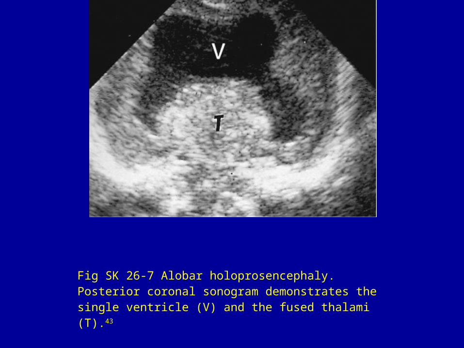
Fig SK 26-7 Alobar holoprosencephaly. Posterior coronal sonogram demonstrates the single ventricle (V) and the fused thalami (T).43