Diabetes Mellitus in pregnancy " Gestational diabetes mellitus''
Upload
jewel-billahCategory
view
111download
4
797
21 Clinical examination of the patient with
diabetes 798
Functional anatomy and physiology 800Normal glucose and fat metabolism 800Aetiology and pathogenesis of diabetes 802
Investigations 807
Presenting problems in diabetes mellitus 808Newly discovered hyperglycaemia 808Long-term supervision of diabetes 811Diabetic ketoacidosis 811Hyperglycaemic hyperosmolar state 814Hypoglycaemia 814Diabetes in pregnancy 817Children, adolescents and young adults with
diabetes 818Hyperglycaemia in acute myocardial
infarction 818Surgery and diabetes 818Complications of diabetes 820
Diabetes mellitus
E.R. PearsonR.J. McCrimmon
Management of diabetes 820Diet and lifestyle 820Drugs to reduce hyperglycaemia 821Insulin therapy 824Transplantation 826
Complications of diabetes 826Diabetic retinopathy 828Diabetic nephropathy 830Diabetic neuropathy 831The diabetic foot 833

Diabetes mellitus
798
Inset (Acanthosis nigricans) From Shotliff. In: Lim 2007 – see p. 836.
Observation
AbdomenHepatomegaly(fatty infiltration of liver)
9
• Weight loss in insulin deficiency• Obesity in type 2 diabetes• Mucosal candidiasis• Dehydration– dry mouth, ↓tissue turgor• Air hunger– Kussmaul breathing in ketoacidosis
LegsMuscle-wastingSensory abnormalityHair lossTendon reflexes
10
Hands(see opposite)
1
Exudativemaculopathy
7
Blood pressure 3
2SkinBullosis
PigmentationGranuloma annulare
Vitiligo
Axillae 4
6
Acanthosis nigricansin insulin resistance
Necrobiosis lipoidica
‘Prayer sign’
Neuropathic foot ulcer
Charcot neuroarthropathy
8
11 Feet (see opposite)InspectionPeripheral pulsesSensation
5NeckCarotid pulses
BruitsThyroid enlargement
HeadXanthelasma
Cranial nerve palsy/eye movements/ptosis
Eyes (see opposite)Visual acuity
Cataract/lens opacityFundoscopy
Insulin injection sites(see opposite)
CLINICAL EXAMINATION OF THE PATIENT WITH DIABETES

Clinical examination of the patient with diabetes
799
8 Insulin injection sitesMain areas used
• Anteriorabdominalwall• Upperthighs/buttocks• Upperouterarms
Inspection
• Bruising• Subcutaneousfatdeposition
(lipohypertrophy)• Subcutaneousfatloss(lipoatrophy;
associatedwithinjectionofunpurifiedanimalinsulins–nowrare)
• Erythema,infection(rare)
7 Examination of the eyesVisual acuity
• DistancevisionusingSnellenchartat6metres
• Nearvisionusingstandardreadingchart
Visualacuitycanalterreversiblywithacutehyperglycaemiaduetoosmoticchangesaffectingthelens.Mostpatientswithretinopathydonothavealteredvisualacuity,exceptafteravitreoushaemorrhageorinsomecasesofmaculopathy.
Lens opacification
• Lookfortheredreflexusingtheophthalmoscopeheld30cmfromtheeye
Fundal examination
• Eitheruseathree-fieldretinalcameraordilatepupilswithamydriatic(e.g.tropicamide)andexaminewithophthalmoscopeinadarkenedroom
• Notefeaturesofdiabeticretinopathy(p.828),includingphotocoagulationscarsfrompreviouslasertreatment
Diabetes can affect every system in the body. In routine clinical practice, examination of the patient with dia-betes is focused on 1 hands, 3 blood pressure, 4 and 5 axillae and neck, 7 eyes, 8 insulin injec-tion sites and 11 feet.
Background retinopathy.
Proliferative retinopathy.
1 Examination of the handsSeveralabnormalitiesaremorecommonindiabetes:• Limitedjointmobility
(‘cheiroarthropathy’)causespainlessstiffness.Theinabilitytoextend(to180°)themetacarpophalangealorinterphalangealjointsofatleastonefingerbilaterallycanbedemonstratedinthe‘prayersign’
• Dupuytren’scontracture(p.1134)causesnodulesorthickeningoftheskinandknucklepads
• Carpaltunnelsyndrome(p.1224)presentswithwristpainradiatingintothehand
• Triggerfinger(flexortenosynovitis)• Muscle-wasting/sensorychanges
maybepresentinperipheralsensorimotorneuropathy,althoughthisismorecommoninthelowerlimbs
Lipohypertrophy of the upper arm.
11 Examination of the feetInspection
• Lookforevidenceofcallusformationonweight-bearingareas,clawingofthetoes(inneuropathy),lossoftheplantararch,discolorationoftheskin(ischaemia),localisedinfectionandulcers
• Deformitymaybepresent,especiallyinCharcotneuroarthropathy
• Fungalinfectionmayaffectskinbetweentoes,andnails
Circulation
• Peripheralpulses,skintemperatureandcapillaryrefillmaybeabnormal
Sensation
• Abnormalinstockingdistributionintypicalperipheralsensorimotorneuropathy
• Testinglighttouchwithmonofilamentsissufficientforriskassessment;testothersensationmodalities(vibration,pain,proprioception)onlywhenneuropathyisbeingevaluated
Reflexes
• Lossofanklereflexesintypicalsensorimotorneuropathy
• Testplantarandanklereflexes
Monofilaments. The monofilament is applied gently until slightly deformed at 5 points on each foot. Callus should be avoided as sensation is reduced. If the patient feels fewer than 8 out of 10 touches, the risk of foot ulceration is increased 5–10-fold.
Diabetes mellitus
21
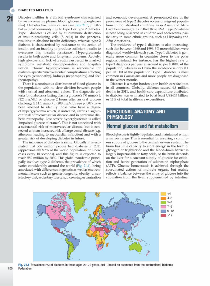
800Fig. 21.1 Prevalence (%) of diabetes in those aged 20–79 years, 2011, based on estimates from the International Diabetes Federation.
4–5<4
5–77–99–12>12
and economic development. A pronounced rise in the prevalence of type 2 diabetes occurs in migrant popula-tions to industrialised countries, as in Asian and Afro-Caribbean immigrants to the UK or USA. Type 2 diabetes is now being observed in children and adolescents, par-ticularly in some ethnic groups, such as Hispanics and Afro-Americans.
The incidence of type 1 diabetes is also increasing, such that between 1960 and 1996, 3% more children were diagnosed worldwide each year. Type 1 diabetes is gen-erally more common in countries closer to the polar regions. Finland, for instance, has the highest rate of type 1 diagnosis per year at around 40 per 100 000 of the population, whereas in China the incidence is only 0.1 per 100 000 of the population. Type 1 diabetes is most common in Caucasians and more people are diagnosed in the winter months.
Diabetes is a major burden upon health-care facilities in all countries. Globally, diabetes caused 4.6 million deaths in 2011, and health-care expenditure attributed to diabetes was estimated to be at least US$465 billion, or 11% of total health-care expenditure.
FUNCTIONAL ANATOMY AND PHYSIOLOGY
Normal glucose and fat metabolism
Blood glucose is tightly regulated and maintained within a narrow range. This is essential for ensuring a continu-ous supply of glucose to the central nervous system. The brain has little capacity to store energy in the form of glycogen or triglyceride and the blood–brain barrier is largely impermeable to fatty acids, so the brain depends on the liver for a constant supply of glucose for oxida-tion and hence generation of adenosine triphosphate (ATP). Glucose homeostasis is achieved through the coordinated actions of multiple organs, but mainly reflects a balance between the entry of glucose into the circulation from the liver, supplemented by intestinal
Diabetes mellitus is a clinical syndrome characterised by an increase in plasma blood glucose (hyperglycae-mia). Diabetes has many causes (see Box 21.5, p. 807) but is most commonly due to type 1 or type 2 diabetes. Type 1 diabetes is caused by autoimmune destruction of insulin-producing cells (β cells) in the pancreas, resulting in absolute insulin deficiency, whereas type 2 diabetes is characterised by resistance to the action of insulin and an inability to produce sufficient insulin to overcome this ‘insulin resistance’. Hyperglycaemia results in both acute and long-term problems. Acutely, high glucose and lack of insulin can result in marked symptoms, metabolic decompensation and hospital-isation. Chronic hyperglycaemia is responsible for diabetes-specific ‘microvascular’ complications affecting the eyes (retinopathy), kidneys (nephropathy) and feet (neuropathy).
There is a continuous distribution of blood glucose in the population, with no clear division between people with normal and abnormal values. The diagnostic cri-teria for diabetes (a fasting plasma glucose ≥ 7.0 mmol/L (126 mg/dL) or glucose 2 hours after an oral glucose challenge ≥ 11.1 mmol/L (200 mg/dL); see p. 807) have been selected to identify those who have a degree of hyperglycaemia which, if untreated, carries a signifi-cant risk of microvascular disease, and in particular dia-betic retinopathy. Less severe hyperglycaemia is called ‘impaired glucose tolerance’. This is not associated with a substantial risk of microvascular disease, but is con-nected with an increased risk of large vessel disease (e.g. atheroma leading to myocardial infarction) and with a greater risk of developing diabetes in future.
The incidence of diabetes is rising. Globally, it is esti-mated that 366 million people had diabetes in 2011 (approximately 8.3% of the world population, or 3 new cases every 10 seconds), and this figure is expected to reach 552 million by 2030. This global pandemic princi-pally involves type 2 diabetes, the prevalence of which varies considerably around the world (Fig. 21.1), being associated with differences in genetic as well as environ-mental factors such as greater longevity, obesity, unsat-isfactory diet, sedentary lifestyle, increasing urbanisation

Functional anatomy and physiology
21
801
Fig. 21.2 Major metabolic pathways of fuel metabolism and the actions of insulin. ⊕ indicates stimulation and ⊖ indicates suppression by insulin. In response to a rise in blood glucose, e.g. after a meal, insulin is released, suppressing gluconeogenesis and promoting glycogen synthesis and storage. Insulin promotes the peripheral uptake of glucose, particularly in skeletal muscle, and encourages storage (as muscle glycogen). It also promotes protein synthesis and lipogenesis, and suppresses lipolysis. The release of intermediate metabolites, including amino acids (glutamine, alanine), 3-carbon intermediates in oxidation (lactate, pyruvate) and free fatty acids (FFAs), is controlled by insulin. In the absence of insulin, e.g. during fasting, these processes are reversed and favour gluconeogenesis in liver from glycogen, glycerol, amino acids and other 3-carbon precursors.
Liver
Adipose tissue
Muscle
Gut
Gluconeogenesis
FFAs
Amino acids
LactatePyruvate
Proteolysis
Glucose
Ketonebodies
Oxidation
Glucose
Glycogen
Oxidation
FFAs
Triglycerides
Food
Glucose
GlycogenKetogenesis
Oxidation
Glycerol
FFAs
Glucose
Glycerol+
absorption of glucose after meals, and the uptake of glucose by peripheral tissues, particularly skeletal muscle and brain.
After ingestion of a meal containing carbohydrate, normal blood glucose levels are maintained by:• suppression of hepatic glucose production• stimulation of hepatic glucose uptake• stimulation of glucose uptake by peripheral tissues
(Fig. 21.2).Insulin, the primary regulator of glucose metabolism and storage (Box 21.1), is secreted from pancreatic β cells into the portal circulation in response to a rise in blood glucose (Fig. 21.3). A number of other factors released from the gut following food intake can augment insulin release, including amino acids and hormones such as glucagon-like peptide 1 (GLP-1) and gastrointestinal peptide (GIP). As a result, insulin release is greater when glucose is administered by mouth than when the same rise in plasma glucose is achieved by intravenous glucose infusion, a phenomenon termed the ‘incretin’ effect (see Fig. 21.3). The post-prandial rise in portal vein insulin and glucose, together with a fall in portal
Increase Decrease
Carbohydrate metabolismGlucosetransport(muscle,adiposetissue)GlucosephosphorylationGlycogensynthesisGlycolysisPyruvatedehydrogenaseactivityPentosephosphateshunt
GluconeogenesisGlycogenolysis
Lipid metabolismTriglyceridesynthesisFattyacidsynthesis(liver)Lipoproteinlipaseactivity(adiposetissue)
LipolysisLipoproteinlipase(muscle)KetogenesisFattyacidoxidation(liver)
Protein metabolismAminoacidtransportProteinsynthesis
Proteindegradation
21.1 Metabolic actions of insulin

Diabetes mellitus
21
802
Fig. 21.3 Pancreatic structure and endocrine function. A The normal adult pancreas contains about 1 million islets, which are scattered throughout the exocrine parenchyma. Histology is shown in Figure 21.4. B The core of each islet consists of β cells that produce insulin, and is surrounded by a cortex of endocrine cells that produce other hormones, including glucagon (α cells), somatostatin (δ cells) and pancreatic polypeptide (PP cells). C Pro-insulin in the pancreatic β cell is cleaved to release insulin and equimolar amounts of inert C-peptide (connecting peptide). Measurement of C-peptide can be used to assess endogenous insulin secretory capacity. D An acute first phase of insulin secretion occurs in response to an elevated blood glucose, followed by a sustained second phase. E The incretin effect describes the observation that insulin secretion is greater when glucose is given by mouth than when glucose is administered intravenously to achieve the same rise in blood glucose concentrations. The additional stimulus to insulin secretion is mediated by release of peptides from the gut and these actions are exploited in incretin-based therapies (p. 823).
Accessory ampulla
Ampullaof Vater
Duodenum
Islet core (β cells)
Arteriole
Other islet cells
Venules
Pro-insulin
Pancreaticβ cell
1st phase
2nd phase
Basalsecretion
Insulin secretion
The incretin effect
Glucose stimulus
0–5mins
Time
Time
After oralglucose
Afterintravenousglucose
Insu
lin
C-peptide
Insulin
C
B
D
E
A
glucagon concentrations, suppresses hepatic glucose production and results in net hepatic glucose uptake. Depending on the size of the carbohydrate load, around one-quarter to one-third of ingested glucose is taken up in the liver. In addition, insulin stimulates glucose uptake in skeletal muscle and fat, mediated by the glucose transporter, GLUT 4.
When intestinal glucose absorption declines between meals, portal vein insulin and glucose concentrations fall while glucagon levels rise. This leads to increased hepatic glucose output via gluconeogenesis and glyco-gen breakdown. The liver now resumes net glucose pro-duction and glucose homeostasis is maintained. The main substrates for gluconeogenesis are glycerol and amino acids, as shown in Figure 21.2.
Adipocytes (and the liver) synthesise triglyceride from non-esterified (‘free’) fatty acids (FFAs) and gly-cerol. Insulin is the major regulator not only of glucose metabolism but also of fatty acid metabolism. High insulin levels after meals promote triglyceride accumu-lation. In contrast, in the fasting state, low insulin levels permit lipolysis and the release into the circulation of FFAs (and glycerol), which can be oxidised by many tissues. Their partial oxidation in the liver provides energy to drive gluconeogenesis and also produces ketone bodies (acetoacetate, which can be reduced to 3-hydroxybutyrate or decarboxylated to acetone), which are generated in hepatocyte mitochondria. Ketone bodies are organic acids which, when formed in small amounts, are oxidised and utilised as metabolic fuel. However, the rate of utilisation of ketone bodies by peripheral tissues is limited, and when the rate of pro-duction by the liver exceeds their removal, hyper-ketonaemia results. This occurs physiologically during starvation, when low insulin levels and high catecho-lamine levels increase lipolysis and delivery of FFAs to the liver.
Aetiology and pathogenesis of diabetes
In both of the common types of diabetes, environmental factors interact with genetic susceptibility to determine which people develop the clinical syndrome, and the

Functional anatomy and physiology
21
803
including thyroid disease (p. 738), coeliac disease (p. 880), Addison’s disease (p. 777), pernicious anaemia (p. 1025) and vitiligo (p. 1295).
Genetic predispositionGenetic factors account for about one-third of the sus-ceptibility to type 1 diabetes, the inheritance of which is polygenic (Box 21.2). Over 20 different regions of the human genome show some linkage with type 1 diabetes but most interest has focused on the human leucocyte antigen (HLA) region within the major histocompatibil-ity complex on the short arm of chromosome 6; this locus is designated IDDM 1. The HLA haplotypes DR3 and/or DR4 are associated with increased susceptibility to type 1 diabetes in Caucasians and are in ‘linkage disequilibrium’, i.e. they tend to be transmitted together, with the neighbouring alleles of the HLA-DQA1 and DQB1 genes. The latter may be the main determinants of genetic susceptibility, since these HLA class II genes code for proteins on the surface of cells which present foreign and self antigens to T lymphocytes (p. 87). Candidate gene and genome-wide association studies have also implicated other genes in type 1 diabetes, e.g. CD25, PTPN22, IL2RA and IL-10, which are involved in immune recognition of pancreatic islet antigens, T-cell development and immune regulation. The genes associ-ated with type 1 diabetes overlap with those for other
timing of its onset. However, the underlying genes, precipitating environmental factors and pathophysiol-ogy differ substantially between type 1 and type 2 diabetes. Type 1 diabetes was previously termed ‘insulin-dependent diabetes mellitus’ (IDDM) and is invariably associated with profound insulin deficiency requiring replacement therapy. Type 2 diabetes was previously termed ‘non-insulin-dependent diabetes mellitus’ (NIDDM) because patients retain the capacity to secrete some insulin but exhibit impaired sensitivity to insulin (insulin resistance) and initially can usually be treated without insulin replacement therapy. However, 20% or more of patients with type 2 diabetes will ultimately develop profound insulin deficiency requiring re placement therapy, so that IDDM and NIDDM were misnomers.
Type 1 diabetesPathologyType 1 diabetes is a T cell-mediated autoimmune disease (p. 86) involving destruction of the insulin-secreting β cells in the pancreatic islets. Progressive loss of β cell function takes place over a prolonged period (months to years), but marked hyperglycaemia, accompanied by the classical symptoms of diabetes, occurs only when 80–90% of the functional capacity of β cells has been lost.
The pathology in the pre-diabetic pancreas is charac-terised by ‘insulitis’ (Fig. 21.4), with infiltration of the islets by mononuclear cells con taining activated macro-phages, helper cytotoxic and suppressor T lymphocytes, natural killer cells and B lymphocytes. Initially, these lesions are patchy and, until a very late stage, lobules containing heavily infiltrated islets are seen adjacent to unaffected lobules. The destructive process is β cell-specific, the glucagon and other hormone-secreting cells in the islet remaining intact.
Islet cell antibodies are present before the clinical presentation of type 1 diabetes, and their detection can be useful in confirming a diagnosis of type 1 diabetes, but they are poorly predictive of disease progression and disappear over time (see Fig. 21.4). Type 1 diabetes is associated with other autoimmune disorders (Ch. 4),
Fig. 21.4 Pathogenesis of type 1 diabetes. Proposed sequence of events in the development of type 1 diabetes. Environmental triggers are described in the text.
Normal islet
β-ce
ll m
ass
Environmental triggers
Geneticsusceptibility toimmune dysfunction
Inflammatory cell infiltration of isletAntibody-mediated β-cell destructionAutoantibodies present in blood
Overtdiabetes
Loss of first phaseinsulin secretionImpaired glucosetolerance
Insulitis β-cell destruction
Time
Relative with type 1 diabetes % overall risk
Identicaltwin 35
Non-identicaltwin 20
HLA-identicalsibling 16
Non-HLA-identicalsibling 3
Father 9
Mother 3
Bothparents Upto30
21.2 Risk of type 1 diabetes among first-degree relatives of patients with type 1 diabetes

Diabetes mellitus
21
804
protein expressed by β cells. It has also been proposed that reduced exposure to microorganisms in early child-hood limits maturation of the immune system and increases susceptibility to autoimmune disease (the ‘hygiene hypothesis’).
Metabolic disturbances in type 1 diabetesPatients with type 1 diabetes present when progressive β-cell destruction has crossed a threshold at which ade-quate insulin secretion and normal blood glucose levels can no longer be sustained. Above a certain level, high glucose levels may be toxic to the remaining β cells, so that profound insulin deficiency rapidly ensues, causing the metabolic sequ elae shown in Figure 21.5. Hyper-glycaemia leads to glycosuria and dehydration, causing fatigue, polyuria, nocturia, thirst and polydipsia, sus-ceptibility to urinary and genital tract infections, and later tachycardia and hypotension. Unrestrained lipo-lysis and proteolysis result in weight loss. Ketoacidosis occurs when generation of ketone bodies exceeds the capacity for their metabolism. Elevated blood H+ ions drive K+ out of the intracellular compartment, while sec-ondary hyperaldosteronism encourages urinary loss of K+. Thus patients usually present with a short history (typically a few weeks) of hyperglycaemic symptoms (thirst, polyuria, nocturia and fatigue), infections and weight loss, and may have developed ketoacidosis (p. 811).
autoimmune disorders, such as coeliac disease and thyroid disease, consistent with clustering of these con-ditions in individuals or families.
Environmental predispositionAlthough genetic susceptibility appears to be a pre-requisite for type 1 diabetes, the concordance rate between monozygotic twins is less than 40% (see Box 21.2), and wide geographic and seasonal variations in incidence suggest that environmental factors have an important role in precipitating disease.
Although hypotheses abound, the nature of these environmental factors is uncertain. They may trigger type 1 diabetes through direct toxicity to β cells or by stimulating an autoimmune reaction directed against β cells. Potential candidates fall into three main categories: viruses, specific drugs or chemicals, and dietary con-stituents. Viruses implicated in the aetiology of type 1 diabetes include mumps, Coxsackie B4, retroviruses, rubella (in utero), cytomegalovirus and Epstein–Barr virus. Various dietary nitrosamines (found in smoked and cured meats) and coffee have been proposed as potentially diabetogenic toxins. Bovine serum albumin (BSA), a major constituent of cow’s milk, has been impli-cated, since children who are given cow’s milk early in infancy are more likely to develop type 1 diabetes than those who are breastfed. BSA may cross the neonatal gut and raise antibodies which cross-react with a heat-shock
Fig. 21.5 Acute metabolic complications of insulin deficiency. (FFA = free fatty acids.)
↓Glucose uptakeand utilisation ↑Glycogenolysis
Hyperglycaemia
Glycosuria
Osmotic diuresis
Dehydration
Hyperosmolarity
Secondaryhyperaldosteronism
K+ deficiency
↑Gluconeogenesis
↑Lipolysis↑Proteolysis
↑FFA and glycerolto liver
↓Renal function ↑Lactate
↑Ketogenesis
Metabolic acidosis
Insufficient insulin
Functional anatomy and physiology
21
805
The primary cause of insulin resistance remains unclear; it is likely that there are multiple defects in insulin signalling, affecting several tissues. One theory is centred around the adipocyte; this is particularly appealing, as obesity is a major cause of increased insulin resistance. Intra-abdominal ‘central’ adipose tissue is metabolically active, and releases large quanti-ties of FFAs, which may induce insulin resistance because they compete with glucose as a fuel supply for oxidation in peripheral tissues such as muscle. In addi-tion, adipose tissue releases a number of hormones (including a variety of peptides, called ‘adipokines’ because they are structurally similar to immunological ‘cytokines’) which act on specific receptors to influence sensitivity to insulin in other tissues. Because the venous drainage of visceral adipose tissue is into the portal vein, central obesity may have a particularly potent influence on insulin sensitivity in the liver, and thereby adversely affect gluconeogenesis and hepatic lipid metabolism.
Physical activity is another important determinant of insulin sensitivity. Inactivity is associated with down-regulation of insulin-sensitive kinases and may promote accumulation of FFAs within skeletal muscle. Sedentary people are therefore more insulin-resistant than active people with the same degree of obesity. Moreover, physical activity allows non-insulin-dependent glucose uptake into muscle, reducing the ‘demand’ on the pan-creatic β cells to produce insulin.
Deposition of fat in the liver is a common association with central obesity and is exacerbated by insulin resist-ance and/or deficiency. Many patients with type 2 dia-betes have evidence of fatty infiltration of the liver (non-alcoholic fatty liver disease (NAFLD)). This condi-tion may improve with effective treatment of the diabe-tes and dyslipidaemia, but despite this, a few patients progress to non-alcoholic steatohepatitis (NASH, p. 959) and cirrhosis.
Type 2 diabetesPathologyType 2 diabetes is a diagnosis of exclusion, i.e. it is made when type 1 diabetes and other types of diabetes (see Box 21.5, p. 807) are ruled out, and is highly heterogene-ous. The natural history of typical type 2 diabetes is shown in Figure 21.6. Initially, insulin resistance leads to elevated insulin secretion in order to maintain normal blood glucose levels. However, in susceptible individu-als, the pancreatic β cells are unable to sustain the increased demand for insulin and a slowly progressive insulin deficiency develops. Some patients develop dia-betes at a young age, usually driven by insulin resistance due to obesity and ethnicity; others, particularly the elderly, develop dia betes despite being non-obese and may have more pronounced β-cell failure. The key feature is a ‘relative’ insulin deficiency, such that there is insufficient insulin production to overcome the resist-ance to insulin action. This contrasts with type 1 dia-betes, in which there is rapid loss of insulin production and an absolute deficiency, resulting in ketoacidosis and death if the insulin is not replaced.
Insulin resistanceType 2 diabetes, or its antecedent, impaired glucose tol-erance, is one of a cluster of conditions thought to be caused by resistance to insulin action. Thus, patients with type 2 diabetes often have associated disorders including hypertension, dyslipidaemia (characterised by elevated levels of small dense low-density lipopro-tein (LDL) cholesterol and triglycerides, and a low level of high-density lipoprotein (HDL) cholesterol), non-alcoholic fatty liver (p. 959) and, in women, polycystic ovarian syndrome. This cluster has been termed the ‘insulin resistance syndrome’ or ‘metabolic syndrome’, and is much more common in patients who are obese.
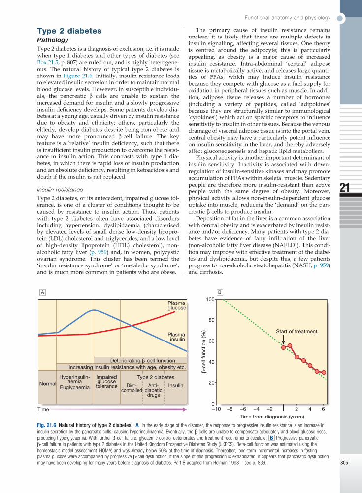
Fig. 21.6 Natural history of type 2 diabetes. A In the early stage of the disorder, the response to progressive insulin resistance is an increase in insulin secretion by the pancreatic cells, causing hyperinsulinaemia. Eventually, the β cells are unable to compensate adequately and blood glucose rises, producing hyperglycaemia. With further β-cell failure, glycaemic control deteriorates and treatment requirements escalate. B Progressive pancreatic β-cell failure in patients with type 2 diabetes in the United Kingdom Prospective Diabetes Study (UKPDS). Beta-cell function was estimated using the homeostasis model assessment (HOMA) and was already below 50% at the time of diagnosis. Thereafter, long-term incremental increases in fasting plasma glucose were accompanied by progressive β-cell dysfunction. If the slope of this progression is extrapolated, it appears that pancreatic dysfunction may have been developing for many years before diagnosis of diabetes. Part B adapted from Holman 1998 – see p. 836.
Impairedglucose
tolerance
Type 2 diabetesHyperinsulin-aemiaNormal
Time
Plasmaglucose
100
β-ce
ll fu
nctio
n (%
)
80
60
40
20
−10 −8 −6 −4 −2
Time from diagnosis (years)
Start of treatment
2 4 60
Plasmainsulin
Deteriorating β-cell functionIncreasing insulin resistance with age, obesity etc.
Euglycaemia Diet-controlled
Anti-diabeticdrugs
Insulin
A B
Diabetes mellitus
21
806
AgeType 2 diabetes is more common in the middle-aged and elderly (Box 21.4). In the UK, it affects 10% of the popula-tion over 65, and over 70% of all cases of diabetes occur after the age of 50 years.
Metabolic disturbances in type 2 diabetesPatients with type 2 diabetes have a slow onset of ‘rela-tive’ insulin deficiency. Relatively small amounts of insulin are required to suppress lipolysis, and some glucose uptake is maintained in muscle, so that, in con-trast with type 1 diabetes, lipolysis and proteolysis are not unrestrained and weight loss and ketoacidosis seldom occur. In type 2 diabetes, hyperglycaemia tends to develop slowly over months or years; because of this insidious onset many cases of type 2 diabetes are discov-ered coincidentally and a large number are undetected. At diagnosis, patients are often asymptomatic or give a long history (typically many months) of fatigue, with or without ‘osmotic symptoms’ (thirst and polyuria). In some patients with type 2 diabetes, presentation is late and pancreatic β-cell failure has reached an advanced stage of insulin deficiency (see type 1 diabetes, p. 803). These patients may present with weight loss but ketoaci-dosis is uncommon. However, in some ethnic groups, such as African Americans, half of those whose first presentation is with diabetic ketoacidosis have type 2 diabetes.
Intercurrent illness, e.g. with infections, increases the production of stress hormones which oppose insulin action, such as cortisol, growth hormone and catecho-lamines. This can precipitate an acute exacerbation of insulin resistance and insulin deficiency, and result in more severe hyperglycaemia and dehydration (see hyperglycaemic hyperosmolar state, p. 814).
Other forms of diabetesOther causes of diabetes are shown in Box 21.5. In most cases, there is an obvious cause of destruction of pancre-atic β cells. Some acquired disorders, notably other endocrine diseases such as acromegaly (p. 792) or Cushing’s syndrome (p. 773), can precipitate type 2 diabetes in susceptible individuals.
A number of unusual genetic diseases are associated with diabetes. In rare families, diabetes is caused by single gene defects with autosomal dominant inherit-ance. These subtypes constitute less than 5% of all cases of diabetes and typically present as ‘maturity-onset dia-betes of the young’ (MODY), i.e. non-insulin-requiring
Pancreatic β-cell failureIn the early stages of type 2 diabetes, reduction in the total mass of pancreatic islet tissue is modest. At the time of diagnosis, around 50% of β-cell function has been lost and this declines progressively (see Fig. 21.6B). Some pathological changes are typical of type 2 dia-betes, the most consistent of which is deposition of amyloid in the islets. In addition, elevated plasma glucose and FFAs exert toxic effects on pancreatic β cells to impair insulin secretion. However, while β-cell numbers are reduced, β-cell mass is unchanged and glucagon secretion is increased, which may contribute to hyperglycaemia.
Genetic predispositionGenetic factors are important in type 2 diabetes, as shown by marked differences in susceptibility in dif-ferent ethnic groups and by studies in monozygotic twins where concordance rates for type 2 diabetes approach 100%. However, many genes are involved and the chance of developing diabetes is also influenced very powerfully by environmental factors (Box 21.3). Genome-wide association studies have identified over 65 genes or gene regions that are associated with type 2 diabetes, each exerting a small effect. The largest effect is seen with variation in TCF7L2; the 10% of the population with two copies of the risk variant for this gene have a nearly twofold increase in risk of develop-ing type 2 diabetes. Most of the genes known to con-tribute to risk of type 2 diabetes are involved in β-cell function or in regulation of cell cycling and turnover, suggesting that altered regulation of β-cell mass is a key factor.
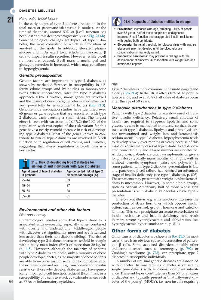
• Prevalence:increaseswithage,affecting~10%ofpeopleover65years.Halfofthesepeopleareundiagnosed.Impairedβ-cellfunctionandexaggeratedinsulinresistancewithageingbothcontribute.
• Glycosuria:therenalthresholdforglucoseriseswithage,soglycosuriamaynotdevelopuntilthebloodglucoseconcentrationismarkedlyraised.
• Pancreatic carcinoma:maypresentinoldagewiththedevelopmentofdiabetes,inassociationwithweightlossanddiminishedappetite.
21.4 Diagnosis of diabetes mellitus in old age
Age at onset of type 2 diabetes in proband
Age-corrected risk of type 2 diabetes for siblings (%)
25–44 53
45–54 37
55–64 38
65–80 31
21.3 Risk of developing type 2 diabetes for siblings of and individuals with type 2 diabetes
Environmental and other risk factorsDiet and obesityEpidemiological studies show that type 2 diabetes is associated with overeating, especially when combined with obesity and underactivity. Middle-aged people with diabetes eat significantly more and are fatter and less active than their non-diabetic siblings. The risk of developing type 2 diabetes increases tenfold in people with a body mass index (BMI) of more than 30 kg/m2 (p. 115). However, although the majority of patients with type 2 diabetes are obese, only a minority of obese people develop diabetes, as the majority of obese patients are able to increase insulin secretion to compensate for the increased demand resulting from obesity and insulin resistance. Those who develop diabetes may have genet-ically impaired β-cell function, reduced β-cell mass, or a susceptibility of β cells to attack by toxic substances such as FFAs or inflammatory cytokines.

Investigations
21
807
INVESTIGATIONS
Urine testingGlucoseTesting the urine for glucose with dipsticks is a common screening procedure for detecting diabetes. If possible, testing should be performed on urine passed 1–2 hours after a meal to maximise sensitivity. Glycosuria always warrants further assessment by blood testing (see below). The greatest disadvantage of urinary glucose measurement is the individual variation in renal thresh-old for glucose. The most frequent cause of glycosuria is a low renal threshold, which is common during preg-nancy and in young people; the resulting ‘renal glycos-uria’ is a benign condition unrelated to diabetes. Another disadvantage is that some drugs (such as β-lactam anti-biotics, levodopa and salicylates) may interfere with urine glucose tests.
KetonesKetone bodies can be identified by the nitroprusside reaction, which measures acetoacetate, using either tablets or dipsticks. Ketonuria may be found in normal people who have been fasting or exercising strenuously for long periods, who have been vomiting repeatedly, or who have been eating a diet high in fat and low in carbohydrate. Ketonuria is therefore not pathognomonic of diabetes but, if associated with glycosuria, the diag-nosis of diabetes is highly likely. In diabetic ketoacidosis (p. 811), ketones can also be detected in plasma using test sticks (see below).
ProteinStandard dipstick testing for albumin detects urinary albumin at concentrations above 300 mg/L, but smaller amounts (microalbuminuria, see Box 17.13, p. 476) can only be measured using specific albumin dipsticks or by quantitative biochemical laboratory measurement. Microalbuminuria or proteinuria, in the absence of urinary tract infection, is an important indicator of dia-betic nephropathy and/or increased risk of macrovascu-lar disease (p. 830).
Blood testingGlucoseLaboratory glucose testing in blood relies upon an enzym-atic reaction (glucose oxidase) and is cheap, usually automated and highly reliable. However, blood glucose levels depend on whether the patient has eaten recently, so it is important to consider the circumstances in which the blood sample was taken.
Blood glucose can also be measured with colorimet-ric or other testing sticks, which are often read with a portable electronic meter. These are used for capillary (fingerprick) testing to monitor diabetes treatment (p. 810). There is some debate as to whether self-monitoring in people with type 2 diabetes improves glycaemic control. Many countries now only offer self-monitoring to people with type 2 diabetes taking insulin therapy. To make the diagnosis of diabetes, the blood glucose concentration should be estimated using an accurate laboratory method rather than a portable technique.
Functional defect Main type* Gene mutated*
β-cell glucose sensing MODY2 GCK
Thesetpointforbasalinsulinreleaseisaltered,causingahighfastingglucose,butsufficientinsulinisreleasedaftermeals.Asaresult,theHbA1cisoftennormalandmicrovascularcomplicationsarerare.Treatmentisrarelyrequired
β-cell transcriptional regulation
MODY3 HNF-1αMODY5 HNF-1βMODY1 HNF-4α
Diabetesdevelopsduringadolescence/earlyadulthoodandcanbemanagedwithdietandtabletsformanyyears,butultimately,insulintreatmentisrequired.TheHNF-1αand4αformsrespondparticularlywelltosulphonylureadrugs.Alltypesareassociatedwithmicrovascularcomplications.HNF-1βmutationsalsocauserenalcystsandrenalfailure
*Other gene mutations have been found in rare cases. For further information, see http://diabetesgenes.org.
21.6 Monogenic diabetes mellitus: maturity-onset diabetes of the young (MODY)
Type 1 diabetes• Immune-mediated• Idiopathic
Type 2 diabetesOther specific types• Geneticdefectsofβ-cellfunction(seeBox21.6)• Geneticdefectsofinsulinaction(e.g.leprechaunism,
lipodystrophies)• Pancreaticdisease(e.g.pancreatitis,pancreatectomy,
neoplasticdisease,cysticfibrosis,haemochromatosis,fibrocalculouspancreatopathy)
• Excessendogenousproductionofhormonalantagoniststoinsulin,e.g.Growthhormone–acromegalyGlucocorticoids–Cushing’ssyndromeGlucagon–glucagonomaCatecholamines–phaeochromocytomaThyroidhormones–thyrotoxicosis
• Drug-induced(e.g.corticosteroids,thiazidediuretics,phenytoin)
• Uncommonformsofimmune-mediateddiabetes(e.g.IPEX(immunodysregulationpolyendocrinopathyX)syndrome)
• Associatedwithgeneticsyndromes(e.g.Down’ssyndrome;Klinefelter’ssyndrome;Turner’ssyndrome;DIDMOAD(Wolfram’ssyndrome)–diabetesinsipidus,diabetesmellitus,opticatrophy,nervedeafness;Friedreich’sataxia;myotonicdystrophy)
Gestational diabetes
21.5 Aetiological classification of diabetes mellitus
diabetes presenting before the age of 25 years (Box 21.6). Very rarely, diabetes can develop at or soon after birth. This neonatal diabetes is usually genetic in origin, with 50% due to mutations in the KATP channel of the pancre-atic β cell causing insulin deficiency and diabetic keto-acidosis. However, sulphonylurea drugs overcome the defect in potassium channel signalling, so that insulin therapy is not necessary in these cases.

Diabetes mellitus
21
808
To allow worldwide comparisons of HbA1c values, the International Federation of Clinical Chemistry and Laboratory Medicine (IFCC) has developed a standard method; IFCC-standardised HbA1c values are reported in mmol/mol. In 2011, many countries adopted the IFCC reference method (Box 21.8).
HbA1c estimates may be erroneously diminished in anaemia or during pregnancy, and may be difficult to interpret with some assay methods in patients who have uraemia or a haemoglobinopathy.
PRESENTING PROBLEMS IN DIABETES MELLITUS
Newly discovered hyperglycaemia
Hyperglycaemia is a very common biochemical abnor-mality. It is frequently detected on routine biochemical analysis of asymptomatic patients, following routine dipstick testing of urine showing glycosuria, or during severe illness (‘stress hyperglycaemia’). Alternatively, hyperglycaemia may present with the symptoms described in Box 21.9. Occasionally, patients present as an emergency with acute metabolic decompensation (see below). The key goals are to establish whether the patient has diabetes, and if so, what type of diabetes it is and how it should be treated.
Establishing the diagnosis of diabetesGlycaemia can be classified into three categories: normal, impaired (pre-diabetes) and diabetes (Boxes 21.10 and 21.11). The glucose cut-off that defines diabetes is based upon the level above which there is a significant risk of microvascular complications (retinopathy, nephropathy, neuropathy). People categorised as having pre-diabetes have blood glucose levels that carry a negligible risk of microvascular complications but are at increased risk of developing diabetes. Also, because there is a continuous risk of macrovascular disease (atheroma of large conduit blood vessels) with increasing glycae mia in the popula-tion, people with pre-diabetes have increased risk of cardiovascular disease (myocardial infarction, stroke and peripheral vascular disease).
When a person has symptoms of diabetes, the diag-nosis can be confirmed with either a fasting glucose
Glucose concentrations are lower in venous than arte-rial or capillary (fingerprick) blood. Whole blood glucose concentrations are lower than plasma concentrations because red blood cells contain relatively little glucose. Venous plasma values are usually the most reliable for diagnostic purposes (Boxes 21.10 and 21.11).
KetonesBlood ketone monitoring is increasingly available. Urinary ketone measurements described above are semi-quantitative, difficult to perform and retrospective (i.e. the urine has accumulated over several hours), and do not measure the major ketone in blood during diabetic ketoacidosis (DKA), beta-hydroxybutyrate (β-OHB). Whole blood ketone monitoring detects β-OHB and is useful in assisting with insulin adjustment during intercurrent illness or sustained hyperglycaemia to prevent or detect DKA. Blood ketone monitoring is also useful in monitoring resolution of DKA in hospitalised patients (Box 21.7).
DCCT units (%) IFCC units (mmol/mol)
4 20
5 31
6 42
7 53
8 64
9 75
10 86
IFCCHbA1c(mmol/mol)=[DCCTHbA1c(%)−2.15]×10.929
21.8 Conversion between DCCT and IFCC units for HbA1c
(DCCT = Diabetes Control and Complications Trial; IFCC = International Federation of Clinical Chemistry and Laboratory Medicine
Measurement* Interpretation
< 0.6 mmol/L Normal;noactionrequired
0.6–1.5 mmol/L Suggestsmetaboliccontrolmaybedeteriorating;continuetomonitorandseekmedicaladviceifsustained/progressive
1.5–3.0 mmol/L Withhighbloodglucose(>10mmol/L),thereisahighriskofdiabeticketoacidosis;seekmedicaladvice
> 3.0 mmol/L Severeketosis;inthepresenceofhighglucose(>10mmol/L)suggestspresenceofdiabeticketoacidosis;seekurgentmedicalhelp
21.7 Interpretation of capillary blood ketone measurements
*To convert to mg/dL, divide values by 0.098.
Glycated haemoglobinGlycated haemoglobin provides an accurate and objec-tive measure of glycaemic control integrated over a period of weeks to months.
In diabetes, the slow non-enzymatic covalent attach-ment of glucose to haemoglobin (glycation) increases the amount in the HbA1 (HbA1c) fraction relative to non-glycated adult haemoglobin (HbA0). These fractions can be separated by chromatography; laboratories may report glycated haemoglobin as total glycated haemo-globin (GHb), HbA1 or HbA1c. In most countries, HbA1c is the preferred measurement. The rate of formation of HbA1c is directly proportional to the ambient blood glucose concentration; a rise of 1% in HbA1c corresponds to an approximate average increase of 2 mmol/L (36 mg/dL) in blood glucose. Although HbA1c concen-tration reflects the integrated blood glucose control over the lifespan of erythrocytes (120 days), HbA1c is most sensitive to changes in glycaemic control occurring in the month before measurement.
Various assay methods are used to measure HbA1c, but most laboratories have been reporting HbA1c values (as %) aligned with the reference range that was used in the Diabetes Control and Complications Trial (DCCT).
Presenting problems in diabetes mellitus
21
809
When a diagnosis of diabetes is confirmed, other investigations should include plasma urea, creatinine and electrolytes, lipids, liver and thyroid function tests, and urine testing for ketones, protein or microalbuminuria.
Clinical assessment and classificationHyperglycaemia causes a wide variety of symptoms (see Box 21.9). The classical clinical features of the two main types of diabetes are compared in Box 21.12. Symptoms of thirst, polyuria, nocturia and rapid weight loss are prominent in type 1 diabetes, but are often absent in patients with type 2 diabetes, many of whom are asymp-tomatic or have non-specific complaints such as chronic fatigue and malaise. Uncontrolled diabetes is associated
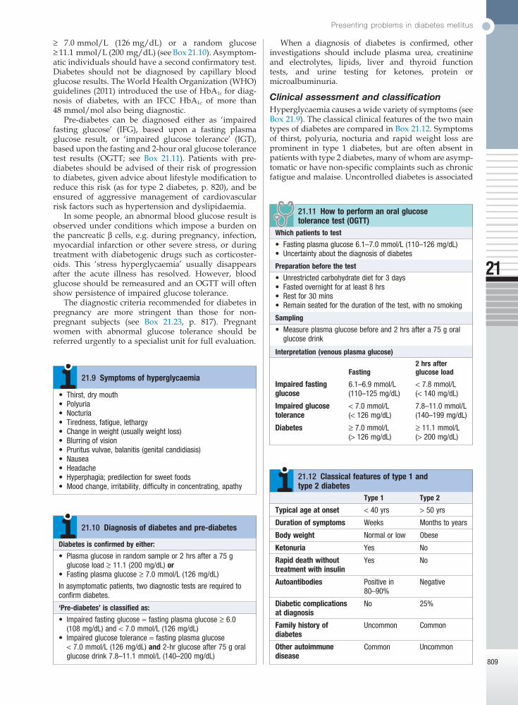
• Thirst,drymouth• Polyuria• Nocturia• Tiredness,fatigue,lethargy• Changeinweight(usuallyweightloss)• Blurringofvision• Pruritusvulvae,balanitis(genitalcandidiasis)• Nausea• Headache• Hyperphagia;predilectionforsweetfoods• Moodchange,irritability,difficultyinconcentrating,apathy
21.9 Symptoms of hyperglycaemia
Diabetes is confirmed by either:
• Plasmaglucoseinrandomsampleor2hrsaftera75gglucoseload≥11.1(200mg/dL)or
• Fastingplasmaglucose≥7.0mmol/L(126mg/dL)
Inasymptomaticpatients,twodiagnostictestsarerequiredtoconfirmdiabetes.
‘Pre-diabetes’ is classified as:
• Impairedfastingglucose=fastingplasmaglucose≥6.0(108mg/dL)and<7.0mmol/L(126mg/dL)
• Impairedglucosetolerance=fastingplasmaglucose<7.0mmol/L(126mg/dL)and2-hrglucoseafter75goralglucosedrink7.8–11.1mmol/L(140–200mg/dL)
21.10 Diagnosis of diabetes and pre-diabetes
Fasting2 hrs after glucose load
Impaired fasting glucose
6.1–6.9mmol/L <7.8mmol/L(110–125mg/dL) (<140mg/dL)
Impaired glucose tolerance
<7.0mmol/L 7.8–11.0mmol/L(<126mg/dL) (140–199mg/dL)
Diabetes ≥7.0mmol/L ≥11.1mmol/L(>126mg/dL) (>200mg/dL)
Which patients to test
• Fastingplasmaglucose6.1–7.0mmol/L(110–126mg/dL)• Uncertaintyaboutthediagnosisofdiabetes
Preparation before the test
• Unrestrictedcarbohydratedietfor3days• Fastedovernightforatleast8hrs• Restfor30mins• Remainseatedforthedurationofthetest,withnosmoking
Sampling
• Measureplasmaglucosebeforeand2hrsaftera75goralglucosedrink
Interpretation (venous plasma glucose)
21.11 How to perform an oral glucose tolerance test (OGTT)
Type 1 Type 2
Typical age at onset <40yrs >50yrs
Duration of symptoms Weeks Monthstoyears
Body weight Normalorlow Obese
Ketonuria Yes No
Rapid death without treatment with insulin
Yes No
Autoantibodies Positivein80–90%
Negative
Diabetic complications at diagnosis
No 25%
Family history of diabetes
Uncommon Common
Other autoimmune disease
Common Uncommon
21.12 Classical features of type 1 and type 2 diabetes
≥ 7.0 mmol/L (126 mg/dL) or a random glucose ≥ 11.1 mmol/L (200 mg/dL) (see Box 21.10). Asymptom-atic individuals should have a second confirmatory test. Diabetes should not be diagnosed by capillary blood glucose results. The World Health Organization (WHO) guidelines (2011) introduced the use of HbA1c for diag-nosis of diabetes, with an IFCC HbA1c of more than 48 mmol/mol also being diagnostic.
Pre-diabetes can be diagnosed either as ‘impaired fasting glucose’ (IFG), based upon a fasting plasma glucose result, or ‘impaired glucose tolerance’ (IGT), based upon the fasting and 2-hour oral glucose tolerance test results (OGTT; see Box 21.11). Patients with pre-diabetes should be advised of their risk of progression to diabetes, given advice about lifestyle modification to reduce this risk (as for type 2 diabetes, p. 820), and be ensured of aggressive management of cardiovascular risk factors such as hypertension and dyslipidaemia.
In some people, an abnormal blood glucose result is observed under conditions which impose a burden on the pancreatic β cells, e.g. during pregnancy, infection, myocardial infarction or other severe stress, or during treatment with diabetogenic drugs such as corticoster-oids. This ‘stress hyperglycaemia’ usually disappears after the acute illness has resolved. However, blood glucose should be remeasured and an OGTT will often show persistence of impaired glucose tolerance.
The diagnostic criteria recommended for diabetes in pregnancy are more stringent than those for non-pregnant subjects (see Box 21.23, p. 817). Pregnant women with abnormal glucose tolerance should be referred urgently to a specialist unit for full evaluation.

Diabetes mellitus
21
810
• Licensingregulationsvaryconsiderablybetweencountries.IntheUK,diabetesrequiringinsulintherapyoranycomplicationthatcouldaffectdrivingshouldbedeclaredtotheDriverandVehicleLicensingAgency;ordinarydrivinglicencesare‘period-restricted’forinsulin-treateddrivers;andvocationallicences(largegoodsvehiclesandpublicservicevehicles)maybegrantedbutrequireverystrictcriteriatobemet
• Themainrisktodrivingperformanceishypoglycaemia.Visualimpairmentandothercomplicationsmayoccasionallycauseproblems
• Insulin-treateddiabeticdriversshould:Checkbloodglucosebeforedrivingand2-hourlyduringlongjourneysKeepanaccessiblesupplyoffast-actingcarbohydrateinthevehicleTakeregularsnacksormealsduringlongjourneysStopdrivingifhypoglycaemiadevelopsRefrainfromdrivinguntilatleast45minsaftertreatmentofhypoglycaemia(delayedrecoveryofcognitivefunction)Carryidentificationincaseofinjury
21.13 Diabetes and driving
Educating patientsIt is essential that people with diabetes understand their disorder and learn to handle all aspects of their manage-ment as comprehensively and quickly as possible. Ideally, this can be achieved by a multidisciplinary team (doctor, dietitian, specialist nurse and podiatrist) in the outpatient setting. For those with newly diagnosed type 2 diabetes, structured education can be given in groups by trained educators. Those requiring insulin need to learn how to measure doses of insulin accurately with an insulin syringe or pen device, how to inject, and how to adjust the dose on the basis of blood glucose values and in relation to factors such as exercise, illness and episodic hypoglycaemia. They must therefore acquire a working knowledge of diabetes, be familiar with the symptoms of hypoglycaemia (see Box 21.19, p. 815), and have ready access to medical advice when the need arises. Information should be provided about driving (national statutory regulations and practical safety measures, Box 21.13). Providing this education is time-consuming but essential if patients are to undertake normal activities safely while maintaining good control.
Self-assessment of glycaemic controlIn people with type 2 diabetes there is not usually a need for regular self-assessment of blood glucose, unless the patient is treated with insulin, or at risk of hypoglycae-mia while taking sulphonylureas. Blood glucose testing can be used for self-education (i.e. demonstrating how different food and exercise regimes affect blood glucose), and may be useful in acute illness. Blood glucose targets vary according to individual circumstances, but, in general, pre-meal values between 4 and 7 mmol/L (72 and 126 mg/dL) and 2-hour post-meal values between 4 and 8 mmol/L represent optimal control.
Insulin-treated patients should be taught how to monitor their own blood glucose using capillary blood glucose meters. Immediate knowledge of blood glucose levels can be used by patients to guide their insulin dosing and to manage exercise and illness. This can be
with an increased susceptibility to infection and patients may present with skin sepsis (boils) or genital candidia-sis, and complain of pruritus vulvae or balanitis.
While the distinction between type 1 and type 2 dia-betes is usually obvious, overlap occurs, particularly in age at onset, duration of symptoms and family history. There are many patients in whom the type of diabetes is not immediately apparent. For example, patients with type 2 diabetes may present with marked and rapid weight loss and even diabetic ketoacidosis, and type 2 diabetes is increasingly diagnosed in children and young adults. Type 1 diabetes can occur at any age, not just in younger people, and may develop more insidiously; the presence of pancreatic autoantibodies confirms the diagnosis of slow-onset type 1 diabetes, termed latent autoimmune diabetes of adults (LADA). Pancreatic autoantibodies are detectable at high titre in 80–90% of patients with type 1 diabetes, so a negative result should prompt consideration of other aetiologies. Other causes of diabetes (see Box 21.5, p. 807), such as MODY, should not be forgotten, particularly in those presenting in childhood or as young adults. A history of pancreatic disease, particularly in patients with a history of alcohol excess, makes insulin deficiency more likely. Sometimes the definitive classification of the type of diabetes is only made later, once the natural history or responsiveness to different therapies becomes apparent.
Physical signs in patients with type 2 diabetes at diagnosis depend on the mode of presentation. In Western populations, more than 80% are overweight, and the obesity is often central (truncal or abdominal). Obesity is much less evident in Asians. Hypertension is present in at least 50% of patients with type 2 diabetes. Although dyslipidaemia is also common, skin lesions such as xanthelasma and eruptive xanthomas are rare. An increasing number of patients now present with NAFLD, usually identified by their elevated blood transaminase values, but they may also have non-tender hepatomegaly.
ManagementThe aims of management are to improve symptoms of hyperglycaemia and to minimise the risks of long-term microvascular and macrovascular complications. Treat-ment methods for diabetes include dietary/lifestyle modification, oral anti-diabetic drugs and injected thera-pies. These are described in detail on page 821. In patients with suspected type 1 diabetes, urgent treat-ment with insulin is required and prompt referral to a specialist is usually needed. In patients with suspected type 2 diabetes, first-line therapy involves advice about dietary and lifestyle modification. Oral anti-diabetic drugs are usually added in those who do not achieve glycaemic targets as a result, or who have severe symp-tomatic hyperglycaemia at diagnosis and a high HbA1c. However, the guidelines in some countries are to intro-duce medication immediately upon diagnosis of dia-betes without waiting to assess the impact of diet and lifestyle changes.
In parallel with treatment of hyperglycaemia, other risk factors for complications of diabetes need to be addressed, including treatment of hypertension (p. 609) and dyslipidaemia (p. 456), and advice on smoking cessation (p. 100).
Presenting problems in diabetes mellitus
21
811
supplemented with blood testing for ketones when blood glucose is high and/or during intercurrent illness.
Urine testing for glucose is not recommended because variability in renal threshold means that some patients with inadequate glycaemic control will not find glucose in their urine.
Long-term supervision of diabetes
Diabetes is a complex disorder which progresses in severity with time, so people with diabetes should be seen at regular intervals for the remainder of their lives, either at a specialist diabetic clinic or in primary care where facilities are available and staff are trained in diabetes care. A checklist for follow-up visits is given in Box 21.14. The frequency of visits is variable, ranging from weekly during pregnancy to annually in the case of patients with well-controlled type 2 diabetes.
Therapeutic goalsThe target HbA1c depends on the individual patient. Early on in diabetes (i.e. patients managed by diet or one or two oral agents), a target of 48 mmol/mol (6.5%) or less may be appropriate. However, a higher target of 58 mmol/mol (7.5%) may be more appropriate in older
Lifestyle issues• Generalhealth• Workorschool• Smoking• Alcoholintake
• Stressordepression• Sexualhealth• Exercise
Body weight and BMIBlood pressure• Individualisedtargetof130–140/70–80mmHg,depending
onriskfactorsandpresenceofnephropathyUrinalysis• Analysefastingspecimenforglucose,ketones,albumin(both
macro-andmicro-albuminuria)Biochemistry• Renal,liverandthyroidfunction• Lipidprofileandestimated10-yrcardiovascularrisktoguide
needforlipid-loweringtherapy(p.581)Glycaemic control• Glycatedhaemoglobin(HbA1c);individualisedtargetbetween
48and58mmol/mol(6.5and7.5%)• Inspectionofhomebloodglucosemonitoringrecord(if
carriedoutbypatient)Hypoglycaemic episodes• Numberandcauseofsevere(requiringassistancefor
treatment)eventsandfrequencyofmild(self-treated)episodesandbiochemicalhypoglycaemia
• Awarenessofhypoglycaemia• DrivingadviceAssessment of injection sites if insulin-treatedEye examination• Visualacuities(nearanddistance)• Ophthalmoscopy(withpupilsdilated)ordigitalphotographyExamination of lower limbs and feet• Assessmentoffootrisk(p.799)
21.14 How to review a patient in the diabetes clinic
patients with pre-existing cardiovascular disease, or those treated with insulin and therefore at risk of hypoglycaemia. In general, the benefits of lower target HbA1c (primarily a lower risk of microvascular disease) need to be weighed against any increased risks (prima-rily hypoglycaemia in insulin-treated patients). Type 2 diabetes is usually a progressive condition (Fig. 21.7), unless there are major diet and lifestyle changes, so that there is usually a need to increase diabetes medication over time to achieve the individualised target HbA1c.
In people with type 2 diabetes, treatment of coexist-ing hypertension and dyslipidaemia is usually required. This can be decided by assessing absolute risk of a car-diovascular disease event (p. 581) and adjusting targets to individual circumstances. The target for blood pres-sure is usually below 140/80 mmHg, although some guidelines suggest 130/80 mmHg. For lipid-lowering, there is a reduction in cardiovascular risk even with normal cholesterol levels, but statin therapy is usually recommended when the 10-year cardiovascular event risk is at least 20%. As a general rule, this means that anyone with type 2 diabetes who is over the age of 40 years should receive a statin, irrespective of baseline cholesterol levels. Some guidelines do not suggest a target level once the patient is started on a statin but others suggest a total cholesterol of less than 4.0 mmol/L (~150 mg/dL) and an LDL cholesterol of less than 2.0 mmol/L (~75 mg/dL). Similar targets are appropri-ate in type 1 diabetes, although there is a shortage of data from clinical trials in this group.
Diabetic ketoacidosis
Diabetic ketoacidosis (DKA) is a medical emergency and remains a serious cause of morbidity, principally in people with type 1 diabetes. Mortality is low in the UK (approximately 2%) but remains high in developing countries and among non-hospitalised patients. Mortal-ity in DKA is most commonly caused in children and adolescents by cerebral oedema and in adults by hypokalaemia, acute respiratory distress syndrome and
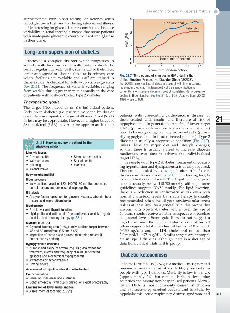
Fig. 21.7 Time course of changes in HbA1c during the United Kingdom Prospective Diabetes Study (UKPDS). In the UKPDS there was loss of glycaemic control with time in patients receiving monotherapy, independently of their randomisation to conventional or intensive glycaemic control, consistent with progressive decline in β-cell function (see Fig. 21.6, p. 805). Adapted from UKPDS 1998 – see p. 836.
6
Conventional
Upper limit of normal
Years from randomisation
Intensive
00 3 6 9 12 15
7
8
Hb
A1c
(%)
9

Diabetes mellitus
21
812
Symptoms
• Polyuria,thirst• Weightloss• Weakness• Nausea,vomiting
• Legcramps• Blurredvision• Abdominalpain
Signs
• Dehydration• Hypotension(posturalor
supine)• Coldextremities/peripheral
cyanosis• Tachycardia
• Airhunger(Kussmaulbreathing)
• Smellofacetone• Hypothermia• Confusion,drowsiness,
coma(10%)
21.16 Clinical features of diabetic ketoacidosis
comorbid conditions such as acute myocardial infarc-tion, sepsis or pneumonia.
DKA is characteristic of type 1 diabetes (see Box 21.12) and is often the presenting problem in newly diagnosed patients. However, an increasing number of patients presenting with DKA have underlying type 2 diabetes. This appears to be particularly prevalent in African-American and Hispanic populations. In estab-lished type 1 diabetes, DKA may be precipitated by an intercurrent illness because of failure to increase insulin dose appropriately to compensate for the stress response. Sometimes, there is no evidence of a precipitating infec-tion and DKA develops because of errors in self-management. In young patients with recurrent episodes of DKA, up to 20% may have psychological problems complicated by eating disorders.
PathogenesisA clear understanding of the biochemical basis and pathophysiology of DKA is essential for its efficient treatment (see Fig. 21.5, p. 804). The cardinal biochemi-cal features are:• hyperketonaemia (≥ 3 mmol/L) and ketonuria
(more than 2+ on standard urine sticks)• hyperglycaemia (blood glucose ≥ 11 mmol/L
(~200 mg/dL))• metabolic acidosis (venous bicarbonate
< 15 mmol/L and/or venous pH < 7.3).The hyperglycaemia causes a profound osmotic diu-
resis leading to dehydration and electrolyte loss, par-ticularly of sodium and potassium. Potassium loss is exacerbated by secondary hyperaldosteronism as a result of reduced renal perfusion. Ketosis results from insulin deficiency, exacerbated by elevated catecho-lamines and other stress hormones, leading to unre-strained lipolysis and supply of FFAs for hepatic ketogenesis. When this exceeds the capacity to metabo-lise acidic ketones, these accumulate in blood. The resulting metabolic acidosis forces hydrogen ions into cells, displacing potassium ions.
The average loss of fluid and electrolytes in moder-ately severe DKA in an adult is shown in Box 21.15. About half the deficit of total body water is derived from the intracellular compartment and occurs comparatively early in the development of acidosis with relatively few clinical features; the remainder represents loss of extra-cellular fluid sustained largely in the later stages, when marked contraction of extracellular fluid volume occurs, with haemoconcentration, a decreased blood volume, and finally a fall in blood pressure with associated renal ischaemia and oliguria.
Every patient in DKA is potassium-depleted, but the plasma concentration of potassium gives very little indi-cation of the total body deficit. Plasma potassium may even be raised initially due to disproportionate loss of
21.15 Average loss of fluid and electrolytes in adult diabetic ketoacidosis of moderate severity
water, catabolism of protein and glycogen, and displace-ment of potassium from the intracellular compartment by H+ ions. However, soon after treatment is started, there is likely to be a precipitous fall in the plasma potas-sium due to dilution of extracellular potassium by administration of intravenous fluids, the movement of potassium into cells induced by insulin, and the continu-ing renal loss of potassium.
The magnitude of the hyperglycaemia does not cor-relate with the severity of the metabolic acidosis; moder-ate elevation of blood glucose may be associated with life-threatening ketoacidosis. In some cases, hyper-glycaemia predominates and acidosis is minimal, with patients presenting in a hyperosmolar state (p. 814).
Clinical assessmentThe clinical features of ketoacidosis are listed in Box 21.16. In the fulminating case, the striking features are those of salt and water depletion, with loss of skin turgor, furred tongue and cracked lips, tachycardia, hypotension and reduced intra-ocular pressure. Breath-ing may be deep and sighing, the breath is usually fetid, and the sickly-sweet smell of acetone may be apparent. Mental apathy, confusion or a reduced conscious level may be present, although coma is uncommon. Indeed, a patient with dangerous ketoacidosis requiring urgent treatment may walk into the consulting room. For this reason, the term ‘diabetic ketoacidosis’ is to be preferred to ‘diabetic coma’, which implies that there is no urgency until unconsciousness supervenes. In fact, it is impera-tive that energetic treatment is started at the earliest possible stage.
Abdominal pain is sometimes a feature of DKA, par-ticularly in children, and vomiting is common. Serum amylase may be elevated but rarely indicates coexisting pancreatitis. In infected patients, pyrexia may not be present initially because of vasodilatation secondary to acidosis.
InvestigationsThe following are important but should not delay the institution of intravenous fluid and insulin replacement:• Venous blood: for urea and electrolytes, glucose and
bicarbonate (severe acidosis is indicated by a venous plasma bicarbonate < 12 mmol/L).
• Urine or blood analysis for ketones (p. 807).
Presenting problems in diabetes mellitus
21
813
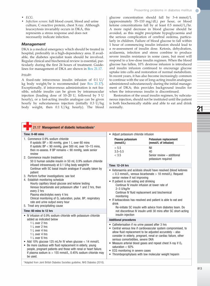
glucose concentration should fall by 3–6 mmol/L (approximately 55–110 mg/dL) per hour, or blood ketone concentrations fall by at least 0.5 mmol/L/hr. A more rapid decrease in blood glucose should be avoided, as this might precipitate hypoglycaemia and the serious complication of cerebral oedema, particu-larly in children. Failure of blood glucose to fall within 1 hour of commencing insulin infusion should lead to a re-assessment of insulin dose. Ketosis, dehydration, acidaemia, infection and stress combine to produce severe insulin resistance in some cases, but most will respond to a low-dose insulin regimen. When the blood glucose has fallen, 10% dextrose infusion is introduced and insulin infusion continued to encourage glucose uptake into cells and restoration of normal metabolism. In recent years, it has also become increasingly common to continue with the use of long-acting insulin analogues administered subcutaneously during the initial manage-ment of DKA; this provides background insulin for when the intravenous insulin is discontinued.
Restoration of the usual insulin regimen, by subcuta-neous injection, should not be instituted until the patient is both biochemically stable and able to eat and drink normally.
• ECG.• Infection screen: full blood count, blood and urine
culture, C-reactive protein, chest X-ray. Although leucocytosis invariably occurs in DKA, this represents a stress response and does not necessarily indicate infection.
ManagementDKA is a medical emergency which should be treated in hospital, preferably in a high-dependency area. If avail-able, the diabetes specialist team should be involved. Regular clinical and biochemical review is essential, par-ticularly during the first 24 hours of treatment. Guide-lines for management of DKA are shown in Box 21.17.
InsulinA fixed-rate intravenous insulin infusion of 0.1 U/kg body weight/hr is recommended (see Box 21.17). Exceptionally, if intravenous administration is not fea-sible, soluble insulin can be given by intramuscular injection (loading dose of 10–20 U, followed by 5 U hourly), or a fast-acting insulin analogue can be given hourly by subcutaneous injection (initially 0.3 U/kg body weight, then 0.1 U/kg hourly). The blood
Time: 0–60 mins
1. Commence0.9%sodiumchlorideIfsystolicBP>90mmHg,give1Lover60minsIfsystolicBP<90mmHg,give500mLover10–15mins,thenre-assess.IfBPremains<90mmHg,seekseniorreview
2. Commenceinsulintreatment50Uhumansolubleinsulinin50mL0.9%sodiumchlorideinfusedintravenouslyat0.1U/kgbodyweight/hrContinuewithSCbasalinsulinanalogueifusuallytakenbypatient
3. Performfurtherinvestigations:seetext4. Establishmonitoringschedule
HourlycapillarybloodglucoseandketonetestingVenousbicarbonateandpotassiumafter1and2hrs,thenevery2hrsPlasmaelectrolytesevery4hrsClinicalmonitoringofO2saturation,pulse,BP,respiratoryrateandurineoutputeveryhour
5. Treatanyprecipitatingcause
Time: 60 mins to 12 hrs
• IVinfusionof0.9%sodiumchloridewithpotassiumchlorideaddedasindicatedbelow
1Lover2hrs1Lover2hrs1Lover4hrs1Lover4hrs1Lover6hrs
• Add10%glucose125mL/hrIVwhenglucose<14mmol/L• Bemorecautiouswithfluidreplacementinelderly,young
people,pregnantpatientsandthosewithrenalorheartfailure.Ifplasmasodiumis>155mmol/L,0.45%sodiumchloridemaybeused.
21.17 Management of diabetic ketoacidosis*
*Adapted from Joint British Diabetes Societies guideline, NHS Diabetes (2010).
• Adjustpotassiumchlorideinfusion
Plasma potassium (mmol/L)
Potassium replacement (mmol/L of infusion)
>5.5 Nil3.5–5.5 40<3.5 Seniorreview–additional
potassiumrequired
Time: 12–24 hrs
• Ketonaemiaandacidosisshouldhaveresolved(bloodketones<0.3mmol/L,venousbicarbonate>18mmol/L).Requestseniorreviewifnotimproving
• IfpatientisnoteatinganddrinkingContinueIVinsulininfusionatlowerrateof2–3U/kg/hrContinueIVfluidreplacementandbiochemicalmonitoring
• Ifketoacidosishasresolvedandpatientisabletoeatanddrink
Re-initiateSCinsulinwithadvicefromdiabetesteam.DonotdiscontinueIVinsulinuntil30minsafterSCshort-actinginsulininjection
Additional procedures
• Catheterisationifnourinepassedafter3hrs• Centralvenouslineifcardiovascularsystemcompromised,to
allowfluidreplacementtobeadjustedaccurately–alsoconsiderinelderly,pregnant,renalorcardiacfailure,otherseriouscomorbidities,severeDKA
• MeasurearterialbloodgasesandrepeatchestX-rayifO2saturation<92%
• ECGmonitoringinseverecases• Thromboprophylaxiswithlowmolecularweightheparin
Diabetes mellitus
21
814
Fluid replacementIn adults, rapid fluid replacement in the first few hours is usually recommended (as in Box 21.17). Caution is recommended in children and young adults because of the risk of cerebral oedema. Most current guidelines favour correction of the extracellular fluid deficit with isotonic saline (0.9% sodium chloride). If the plasma sodium is greater than 155 mmol/L, 0.45% saline may be used initially.
PotassiumCareful monitoring of potassium is essential to the man-agement of diabetic ketoacidosis because both hypo- and hyperkalaemia can occur and are potentially life-threatening. Potassium replacement is not usually recommended with the initial litre of fluid because pre-renal failure may be present secondary to dehydration. Treatment with 0.9% sodium chloride with potassium chloride 40 mmol/L is recommended if the serum potas-sium is below 5.5 mmol/L and the patient is passing urine (see Box 21.17). If the potassium falls below 3.5 mmol/L, the potassium replacement regimen needs to be reviewed. Cardiac rhythm should be monitored in severe DKA because of the risk of electrolyte-induced cardiac arrhythmia.
BicarbonateAdequate fluid and insulin replacement should resolve the acidosis. The use of intravenous bicarbonate therapy is currently not recommended. Acidosis may reflect an adaptive response, improving oxygen delivery to the tissues, and so excessive bicarbonate may induce a para-doxical increase in cerebrospinal fluid acidosis and has been implicated in the pathogenesis of cerebral oedema in children and young adults.
Hyperglycaemic hyperosmolar state
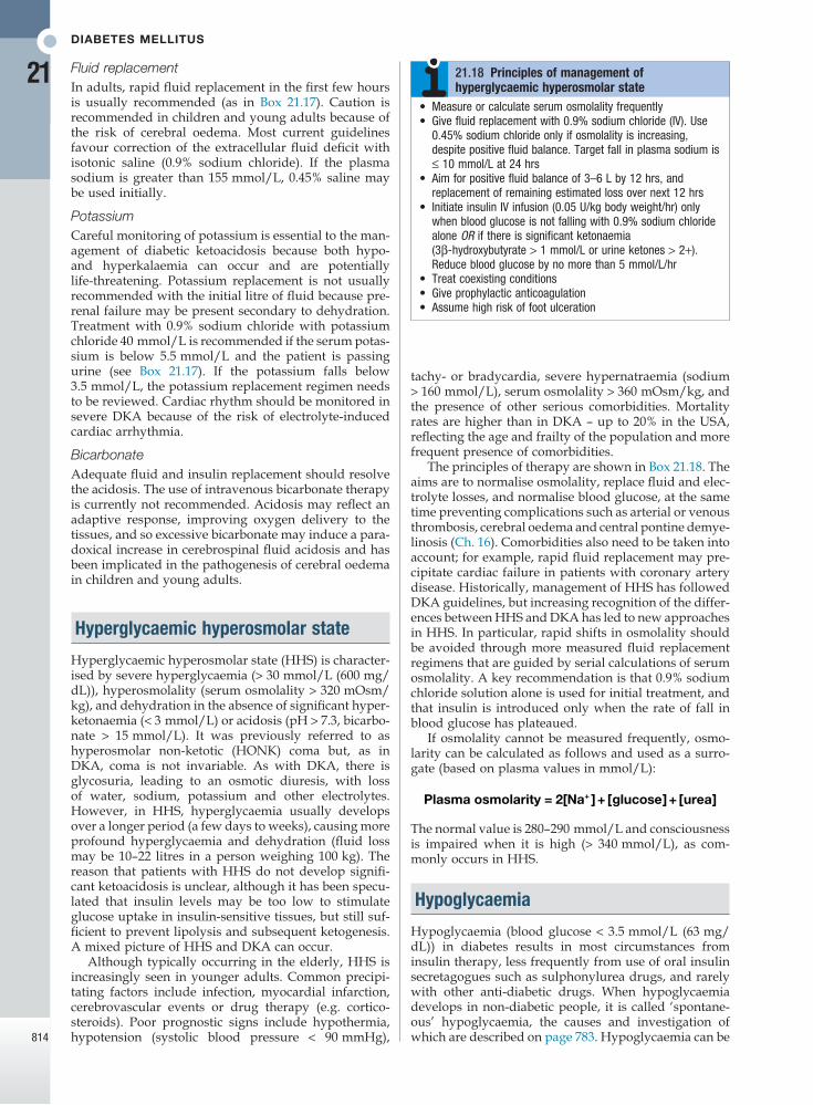
Hyperglycaemic hyperosmolar state (HHS) is character-ised by severe hyperglycaemia (> 30 mmol/L (600 mg/dL)), hyperosmolality (serum osmolality > 320 mOsm/kg), and dehydration in the absence of significant hyper-ketonaemia (< 3 mmol/L) or acidosis (pH > 7.3, bicarbo-nate > 15 mmol/L). It was previously referred to as hyperosmolar non-ketotic (HONK) coma but, as in DKA, coma is not invariable. As with DKA, there is glycosuria, leading to an osmotic diuresis, with loss of water, sodium, potassium and other electrolytes. However, in HHS, hyperglycaemia usually develops over a longer period (a few days to weeks), causing more profound hyperglycaemia and dehydration (fluid loss may be 10–22 litres in a person weighing 100 kg). The reason that patients with HHS do not develop signifi-cant ketoacidosis is unclear, although it has been specu-lated that insulin levels may be too low to stimulate glucose uptake in insulin-sensitive tissues, but still suf-ficient to prevent lipolysis and subsequent ketogenesis. A mixed picture of HHS and DKA can occur.
Although typically occurring in the elderly, HHS is increasingly seen in younger adults. Common precipi-tating factors include infection, myocardial infarction, cerebrovascular events or drug therapy (e.g. cortico-steroids). Poor prognostic signs include hypothermia, hypotension (systolic blood pressure < 90 mmHg),
tachy- or bradycardia, severe hypernatraemia (sodium > 160 mmol/L), serum osmolality > 360 mOsm/kg, and the presence of other serious comorbidities. Mortality rates are higher than in DKA – up to 20% in the USA, reflecting the age and frailty of the population and more frequent presence of comorbidities.
The principles of therapy are shown in Box 21.18. The aims are to normalise osmolality, replace fluid and elec-trolyte losses, and normalise blood glucose, at the same time preventing complications such as arterial or venous thrombosis, cerebral oedema and central pontine demye-linosis (Ch. 16). Comorbidities also need to be taken into account; for example, rapid fluid replacement may pre-cipitate cardiac failure in patients with coronary artery disease. Historically, management of HHS has followed DKA guidelines, but increasing recognition of the differ-ences between HHS and DKA has led to new approaches in HHS. In particular, rapid shifts in osmolality should be avoided through more measured fluid replacement regimens that are guided by serial calculations of serum osmolality. A key recommendation is that 0.9% sodium chloride solution alone is used for initial treatment, and that insulin is introduced only when the rate of fall in blood glucose has plateaued.
If osmolality cannot be measured frequently, osmo-larity can be calculated as follows and used as a surro-gate (based on plasma values in mmol/L):
Plasma osmolarity 2 Na glucose urea= + ++[ ] [ ] [ ]
The normal value is 280–290 mmol/L and consciousness is impaired when it is high (> 340 mmol/L), as com-monly occurs in HHS.
Hypoglycaemia
Hypoglycaemia (blood glucose < 3.5 mmol/L (63 mg/dL)) in diabetes results in most circumstances from insulin therapy, less frequently from use of oral insulin secretagogues such as sulphonylurea drugs, and rarely with other anti-diabetic drugs. When hypoglycaemia develops in non-diabetic people, it is called ‘spontane-ous’ hypoglycaemia, the causes and investigation of which are described on page 783. Hypoglycaemia can be
• Measureorcalculateserumosmolalityfrequently• Givefluidreplacementwith0.9%sodiumchloride(IV).Use
0.45%sodiumchlorideonlyifosmolalityisincreasing,despitepositivefluidbalance.Targetfallinplasmasodiumis≤10mmol/Lat24hrs
• Aimforpositivefluidbalanceof3–6Lby12hrs,andreplacementofremainingestimatedlossovernext12hrs
• InitiateinsulinIVinfusion(0.05U/kgbodyweight/hr)onlywhenbloodglucoseisnotfallingwith0.9%sodiumchloridealoneORifthereissignificantketonaemia(3β-hydroxybutyrate>1mmol/Lorurineketones>2+).Reducebloodglucosebynomorethan5mmol/L/hr
• Treatcoexistingconditions• Giveprophylacticanticoagulation• Assumehighriskoffootulceration
21.18 Principles of management of hyperglycaemic hyperosmolar state

Presenting problems in diabetes mellitus
21
815
of hyperinsulinaemia. Suppression of endogenous insulin secretion to allow increased hepatic glucose production to meet the increased metabolic demand is key to the normal physiological response to exercise. In insulin-treated diabetes, insulin levels may actually increase with exercise because of improved blood flow at the site of injection and this increases the risk of hypoglycaemia.
Awareness of hypoglycaemiaFor most individuals, the glucose level (threshold) at which they first become aware of hypoglycaemia is not
a frequent occurrence in the lives of people with type 1 diabetes and has a major impact on their willingness and ability to achieve target glucose levels.
In health, a number of mechanisms are in place to ensure that glucose homeostasis is maintained. If blood glucose falls, three primary physiological defence mech-anisms operate: endogenous insulin release from pan-creatic β cells is suppressed; release of glucagon from pancreatic α cells is increased; and the autonomic nervous system is activated, with release of catechol-amines both systemically and within the tissues. In addition, stress hormones, such as cortisol and growth hormone, are increased in the blood. These actions reduce whole-body glucose uptake and increase hepatic glucose production, maintaining a glucose supply to the brain. People with type 1 diabetes cannot regulate insulin once it is injected subcutaneously, and so it con-tinues to act, despite developing hypoglycaemia. In addition, within 5 years of diagnosis, most patients will have lost their ability to release glucagon specifically during hypoglycaemia. This is thought to result mainly from loss of α-cell regulation by β cells. These two primary defects mean that hypoglycaemia occurs much more frequently in people with type 1 and longer-duration type 2 diabetes.
Clinical assessmentSymptoms of hypoglycaemia (Box 21.19) comprise two main groups: those related to acute activation of the autonomic nervous system and those secondary to glucose deprivation of the brain (neuroglycopenia). Symptoms of hypoglycaemia are idiosyncratic and differ with age and duration of diabetes. Hypoglycae-mia also affects mood, inducing a state of increased tension and low energy. Learning to recognise the early onset of hypoglycaemia is an important aspect of the education of diabetic patients treated with insulin. The severity of hypoglycaemia is defined by the ability to self-treat; ‘mild’ episodes are self-treated, while ‘severe’ episodes require assistance for recovery.
Circumstances of hypoglycaemiaRisk factors and causes of hypoglycaemia in patients taking insulin or sulphonylurea drugs are listed in Box 21.20. Severe hypoglycaemia can have serious morbidity (e.g. convulsions, coma, focal neurological lesions) and has a mortality of up to 4% in insulin-treated patients. Rarely, sudden death during sleep occurs in otherwise healthy young patients with type 1 diabetes (‘dead-in-bed syndrome’) and may result from hypoglycaemia-induced cardiac arrhythmia. Severe hypoglycaemia is very disruptive and impinges on many aspects of the patient’s life, including employment, driving (see Box 21.13, p. 810), travel, sport and personal relationships.
Nocturnal hypoglycaemia in patients with type 1 diabetes is common but often undetected, as hypo-glycaemia does not usually waken a person from sleep. Patients may describe poor quality of sleep, morning headaches and vivid dreams or nightmares, or a partner may observe profuse sweating, restlessness, twitching or even seizures. The only reliable way to identify this problem is to measure blood glucose during the night.
Exercise-induced hypoglycaemia occurs in people with well-controlled, insulin-treated diabetes because
Autonomic
• Sweating• Trembling• Poundingheart
• Hunger• Anxiety
Neuroglycopenic
• Confusion• Drowsiness• Speechdifficulty
• Inabilitytoconcentrate• Incoordination• Irritability,anger
Non-specific
• Nausea• Tiredness
• Headache
21.19 Most common symptoms of hypoglycaemia
N.B. Symptoms differ with age; children exhibit behavioural changes (such as naughtiness or irritability), while elderly people experience more prominent neurological symptoms (such as visual disturbance and ataxia).
Causes of hypoglycaemia
• Missed,delayedorinadequatemeal• Unexpectedorunusualexercise• Alcohol• Errorsinoralanti-diabeticagent(s)orinsulindose/schedule/
administration• Poorlydesignedinsulinregimen,particularlyifpredisposing
tonocturnalhyperinsulinaemia• Lipohypertrophyatinjectionsitescausingvariableinsulin
absorption• Gastroparesisduetoautonomicneuropathycausingvariable
carbohydrateabsorption• Malabsorption,e.g.coeliacdisease• Unrecognisedotherendocrinedisorder,e.g.Addison’s
disease• Factitious(deliberatelyinduced)• Breastfeeding
Risk factors for severe hypoglycaemia
• Strictglycaemiccontrol• Impairedawarenessofhypoglycaemia• Age(veryyoungandelderly)• Longdurationofdiabetes• Sleep• C-peptidenegativity(indicatingcompleteinsulindeficiency)• Historyofpreviousseverehypoglycaemia• Renalimpairment• Genetic,e.g.angiotensin-convertingenzyme(ACE)genotype
21.20 Hypoglycaemia in diabetes: common causes and risk factors
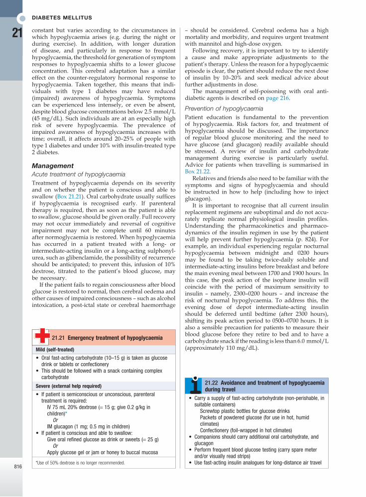
Diabetes mellitus
21
816
– should be considered. Cerebral oedema has a high mortality and morbidity, and requires urgent treatment with mannitol and high-dose oxygen.
Following recovery, it is important to try to identify a cause and make appropriate adjustments to the patient’s therapy. Unless the reason for a hypoglycaemic episode is clear, the patient should reduce the next dose of insulin by 10–20% and seek medical advice about further adjustments in dose.
The management of self-poisoning with oral anti-diabetic agents is described on page 216.
Prevention of hypoglycaemiaPatient education is fundamental to the prevention of hypoglycaemia. Risk factors for, and treatment of hypoglycaemia should be discussed. The importance of regular blood glucose monitoring and the need to have glucose (and glucagon) readily available should be stressed. A review of insulin and carbo hydrate management during exercise is particularly useful. Advice for patients when travelling is summarised in Box 21.22.
Relatives and friends also need to be familiar with the symptoms and signs of hypoglycaemia and should be instructed in how to help (including how to inject glucagon).
It is important to recognise that all current insulin replacement regimens are suboptimal and do not accu-rately replicate normal physiological insulin profiles. Understanding the pharmacokinetics and pharmaco-dynamics of the insulin regimen in use by the patient will help prevent further hypoglycaemia (p. 824). For example, an individual experiencing regular nocturnal hypoglycaemia between midnight and 0200 hours may be found to be taking twice-daily soluble and intermediate-acting insulins before breakfast and before the main evening meal between 1700 and 1900 hours. In this case, the peak action of the isophane insulin will coincide with the period of maximum sensitivity to insulin – namely, 2300–0200 hours – and increase the risk of nocturnal hypoglycaemia. To address this, the evening dose of depot intermediate-acting insulin should be deferred until bedtime (after 2300 hours), shifting its peak action period to 0500–0700 hours. It is also a sensible precaution for patients to measure their blood glucose before they retire to bed and to have a carbohydrate snack if the reading is less than 6.0 mmol/L (approximately 110 mg/dL).
constant but varies according to the circumstances in which hypoglycaemia arises (e.g. during the night or during exercise). In addition, with longer duration of disease, and particularly in response to frequent hypoglycaemia, the threshold for generation of symptom responses to hypoglycaemia shifts to a lower glucose concentration. This cerebral adaptation has a similar effect on the counter-regulatory hormonal response to hypoglycaemia. Taken together, this means that indi-viduals with type 1 diabetes may have reduced (impaired) awareness of hypoglycaemia. Symptoms can be experienced less intensely, or even be absent, despite blood glucose concentrations below 2.5 mmol/L (45 mg/dL). Such individuals are at an especially high risk of severe hypoglycaemia. The prevalence of impaired awareness of hypoglycaemia increases with time; overall, it affects around 20–25% of people with type 1 diabetes and under 10% with insulin-treated type 2 diabetes.
ManagementAcute treatment of hypoglycaemiaTreatment of hypoglycaemia depends on its severity and on whether the patient is conscious and able to swallow (Box 21.21). Oral carbohydrate usually suffices if hypoglycaemia is recognised early. If parenteral therapy is required, then as soon as the patient is able to swallow, glucose should be given orally. Full recovery may not occur immediately and reversal of cognitive impairment may not be complete until 60 minutes after normoglycaemia is restored. When hypoglycaemia has occurred in a patient treated with a long- or intermediate-acting insulin or a long-acting sulphonyl-urea, such as glibenclamide, the possibility of recurrence should be anticipated; to prevent this, infusion of 10% dextrose, titrated to the patient’s blood glucose, may be necessary.
If the patient fails to regain consciousness after blood glucose is restored to normal, then cerebral oedema and other causes of impaired consciousness – such as alcohol intoxication, a post-ictal state or cerebral haemorrhage
Mild (self-treated)
• Oralfast-actingcarbohydrate(10–15g)istakenasglucosedrinkortabletsorconfectionery
• Thisshouldbefollowedwithasnackcontainingcomplexcarbohydrate
Severe (external help required)
• Ifpatientissemiconsciousorunconscious,parenteraltreatmentisrequired:
IV75mL20%dextrose(=15g;give0.2g/kginchildren)*
OrIMglucagon(1mg;0.5mginchildren)
• Ifpatientisconsciousandabletoswallow:Giveoralrefinedglucoseasdrinkorsweets(=25g)
OrApplyglucosegelorjamorhoneytobuccalmucosa
21.21 Emergency treatment of hypoglycaemia
*Use of 50% dextrose is no longer recommended.
• Carryasupplyoffast-actingcarbohydrate(non-perishable,insuitablecontainers)
ScrewtopplasticbottlesforglucosedrinksPacketsofpowderedglucose(foruseinhot,humidclimates)Confectionery(foil-wrappedinhotclimates)
• Companionsshouldcarryadditionaloralcarbohydrate,andglucagon
• Performfrequentbloodglucosetesting(carrysparemeterand/orvisuallyreadstrips)
• Usefast-actinginsulinanaloguesforlong-distanceairtravel
21.22 Avoidance and treatment of hypoglycaemia during travel

Presenting problems in diabetes mellitus
21
817
carbohydrate. Women with gestational diabetes should undertake regular pre- and post-prandial self-monitoring of blood glucose, aiming for pre-meal blood glucose levels of less than 5.5 mmol/L (100 mg/dL) or post-meal blood glucose levels of less than 7.0 mmol/L (125 mg/dL). If treatment is necessary, metformin or glibenclamide is considered safe to use in pregnancy. Glibenclamide should be used rather than other sulpho-nylureas because it does not cross the placenta. Other oral therapies or injectable incretin-based therapies should not be given in pregnancy. Insulin may be required, especially in the later stages of pregnancy. If the maternal blood glucose is not well controlled prior to, and during, delivery, the resulting fetal hyperinsulin-aemia leads to neonatal hyperinsulinaemia, which in turn can cause neonatal hypoglycaemia.
After delivery, maternal glucose usually rapidly returns to pre-pregnancy levels. Woman should be tested at least 6 weeks post-partum with an oral glucose tolerance test. In women with persistent mild fasting hyperglycaemia (glucose over 5.5 mmol/L (100 mg/dL)) who have a small increment (less than 3.5 mmol/L) in plasma glucose 2 hours after oral glucose, monogenic diabetes due to a mutation in GCK (encoding gluco-kinase) should be considered (see Box 21.6, p. 807). Those who have returned to normal glucose tolerance remain at considerable risk for developing type 2 diabe-tes, with a 5-year risk between 15 and 50%, depending on the population. Therefore, all women who have had gestational diabetes should be given diet and lifestyle advice to reduce their risk of developing type 2 diabetes (p. 820).
Pregnancy in women with established diabetesMaternal hyperglycaemia early in pregnancy (during the first 6 weeks post-conception) can adversely affect fetal development. Consequences include cardiac, renal and skeletal malformations, of which the caudal regression syndrome is the most characteristic. The risk of fetal anomaly is about 2% for non-diabetic women,
Diabetes in pregnancy
During pregnancy, maternal glucose metabolism changes to optimise glucose and other nutrient delivery to the fetus. This is particularly apparent in the second half of pregnancy, when there is an increase in maternal tissue insulin resistance, such that glucose is preferen-tially supplied to the fetus rather than maternal tissue. This is largely driven by the maternal hormonal environ-ment, with increased oestrogens and progestogens, and, in particular, human placental lactogen (hPL). The deliv-ery of the placenta results in rapid decline in hPL with a rapid reversal of insulin resistance soon after birth. During pregnancy, fasting plasma glucose decreases slightly, while post-prandial blood glucose may be increased. The renal threshold for glycosuria (p. 807) is reduced in pregnancy.
In the fetus, insulin secretion is driven by fetal blood glucose levels, which are determined by the maternal glucose concentrations. Thus maternal hyperglycaemia drives fetal hyperinsulinaemia. Since insulin is a major fetal growth factor, hyperinsulinaemia in turn drives increased fetal growth, resulting in increased birth weight (‘macrosomia’).
Gestational diabetesGestational diabetes is defined as diabetes with first onset or recognition during pregnancy. This definition will include a few patients who develop type 1 diabetes during pregnancy, where prompt action and early insulin treatment will be required, and some patients who develop type 2 diabetes, or had unknown pre-existing type 2 diabetes, in whom the diabetes does not remit after pregnancy. However, the majority of gestational diabetes develops due to an inability to increase insulin secretion adequately to compensate for pregnancy-induced insulin resistance, and most women can expect to return to normal glucose tolerance imme-diately after pregnancy.
In contrast to non-gestational diabetes, for which the diagnostic thresholds for diabetes are based upon risk of microvascular complications, the diagnosis of gesta-tional diabetes is based upon maternal blood glucose measures that are associated with increased fetal growth. An international consensus recommended that glucose values diagnostic of gestational diabetes should be lower than those for non-gestational diabetes (Box 21.23). Controversy remains about who should be screened, and in part the screening strategy depends on the population risk. It is widely accepted that women at high risk for gestational diabetes should have an oral glucose tolerance test at 24–28 weeks, with some guide-lines recommending that all are screened by measuring HbA1c, fasting blood glucose or random blood glucose at the first booking visit. With the increasing use of HbA1c to diagnose diabetes, it should be noted that HbA1c is unreliable after early pregnancy, when it falls due to increased red cell turnover.
Management of gestational diabetesThe aim is to normalise the maternal blood glucose and thereby reduce excessive fetal growth. The first element of management is dietary modification, in par-ticular by reducing consumption of quick-acting refined
Women at high risk of gestational diabetes
• BMI>30kg/m2
• Apreviousmacrosomicbabyweighing≥4.5kgatbirth• Previousgestationaldiabetes• Afirst-degreerelativewithdiabetes• Ahigh-riskethnicity–SouthAsian,blackCaribbeanor
MiddleEastern
Diagnosis
• High-riskwomenshouldhavea75goralglucosetolerancetestbefore28weeks’gestation
• Gestationaldiabetesisdiagnosedwhen:Fastingplasmaglucose≥5.1mmol/L(92mg/dL)or1-hrplasmaglucose(afterglucoseload)≥10mmol/L(180mg/dL)or2-hrplasmaglucose(afterglucoseload)≥8mmol/L(144mg/dL)
• Considertestinghigh-riskwomenatfirstbookingvisitwithanHbA1corfastingbloodglucose
21.23 Identifying patients with gestational diabetes

Diabetes mellitus
21
818
• Type of diabetes:type1diabetesispredominantinchildrenandadolescents,buttype2diabetesisnowpresentinginunprecedentednumbersofobese,inactiveteenagers.Monogenicdiabetes(MODY)shouldalsobeconsidered(seeBox21.6,p.807).
• Physiological changes:hormonal,physicalandlifestylechangesinpubertyaffectdietaryintake,exercisepatternsandsensitivitytoinsulin,necessitatingalterationsininsulinregimen.
• Emotional changes:adolescenceisaphaseoftransitionintoindependence(principallyfromparentalcare).Periodsofrebellionagainstparentalcontrol,experimentation(e.g.withalcohol)andamorechaoticlifestylearecommon,andoftenimpactadverselyoncontrolofdiabetes.
• Glycaemic control:atemporarydeteriorationincontroliscommon,althoughnotuniversal.Itissometimesmoreimportanttomaintaincontactandengagementwithayoungpersonthantoinsistontightglycaemiccontrol.
• Diabetic ketoacidosis:afewadolescentsandyoungadultspresentwithfrequentepisodesofDKA,oftenbecauseofnon-adherencetoinsulintherapy.Thisismorecommoninfemales.Motivatingfactorsmayincludeweightloss,rebellion,andmanipulationoffamilyorschoolingcircumstances.
• Adolescent diabetes clinics:thesechallengesarebesttackledwithsupportfromaspecialisedmultidisciplinaryteam,includingpaediatricians,physicians,nursesandpsychologists.Supportisrequiredforthepatientandparents.
21.24 Diabetes in adolescence
Surgery and diabetes
Patients with diabetes are reported to have up to 50% higher peri-operative mortality than patients without diabetes. Surgery causes catabolic stress and secretion of counter-regulatory hormones (including catecholamines and cortisol) in both normal and diabetic subjects. This results in increased glycogenolysis, gluconeogenesis, lipolysis, proteolysis and insulin resistance. Starvation exacerbates this process by increasing lipolysis. In the non-diabetic person, these metabolic effects lead to a secondary increase in the secretion of insulin, which exerts a controlling influence. In diabetic patients, either there is absolute deficiency of insulin (type 1 diabetes) or insulin secretion is delayed and impaired (type 2 diabetes), so that in untreated or poorly controlled diabetes, the uptake of metabolic substrate into tissues is significantly reduced, catabolism is increased and, ultimately, metabolic decompensation in the form of diabetic ketoacidosis may develop in both types of diabetes. In addition, hyperglycaemia impairs wound healing and innate immunity, leading to increased risk of infection. Patients with diabetes are also more likely to have underlying pre-operative morbidity, especially cardiovascular disease. Finally, management errors with diabetes may cause dangerous hyperglycae-mia or hypoglycaemia. Careful pre-operative assess-ment and peri-operative management are therefore essential, ideally with support from the diabetes special-ist team.
about 4% for women with well-controlled diabetes (HbA1c below 53 mmol/mol (7%)), but more than 20% for those with poor glycaemic control (HbA1c greater than 97 mmol/mol (11%)). Therefore, a woman with diabetes should, if possible, be helped to achieve excel-lent glycaemic control before becoming pregnant. In addition, high-dose folic acid (5 mg, rather than the usual 400 µg, daily) should be initiated before concep-tion to reduce the risk of neural tube defects.
As for gestational diabetes, mothers should attempt to maintain near-normal blood glucose levels whilst avoiding hypoglycaemia throughout their pregnancy, as this minimises excessive fetal growth and neonatal hypoglycaemia. However, this is often difficult to achieve. Pregnancy is also associated with an increased potential for ketosis, particularly, but not exclusively, in women with type 1 diabetes. Ketoacidosis during preg-nancy is dangerous for the mother and is associated with a high rate (10–35%) of fetal mortality.
Pregnancy is associated with a worsening of diabetic complications, most notably retinopathy and nephro-pathy, so careful monitoring of eyes and kidneys is required throughout pregnancy. If heavy proteinuria and/or renal dysfunction exist prior to pregnancy, there is a marked increase in the risk of pre-eclampsia, and renal function can deteriorate irreversibly during preg-nancy. These risks need to be carefully discussed before a woman with diabetes is considering pregnancy. While the outlook for mother and child has been vastly improved over recent years, pregnancy outcomes are still not equivalent to those of non-diabetic mothers. Perinatal mortality rates remain 3–4 times those of the non-diabetic population (at around 30–40 per 1000 preg-nancies) and the rate of congenital malformation is increased 5–6-fold overall.
Children, adolescents and young adults with diabetes
The management of diabetes in children and adoles-cents presents particular challenges, which should be addressed in specialised clinics (Box 21.24).
Hyperglycaemia in acute myocardial infarction
Hyperglycaemia is often found in patients who have sustained an acute myocardial infarction. In some, this represents stress hyperglycaemia (p. 808), some have previously undiagnosed diabetes, and many have estab-lished diabetes. Many patients with stress hyper-glycaemia will have impaired glucose tolerance on a subsequent glucose tolerance test. Over and above the standard management of myocardial infarction (p. 593), hyperglycaemia should be treated with insulin rather than oral anti-diabetic agents in the peri-infarct period, aiming for near-normalisation of blood glucose. Studies have suggested that good glycaemic control using insulin therapy in hyperglycaemic patients with acute myocardial infarction may reduce their long-term mor-tality from coronary heart disease.

Presenting problems in diabetes mellitus
21
819
Peri-operative managementFigure 21.8 outlines a general approach to peri-operative management of diabetes, although this may need to be adapted according to the patient, the surgical procedure and local guidelines. Patients with diabetes who are con-sidered low-risk can attend as day cases or be admitted on the day of surgery. However, patients are often admitted the night before to ensure optimal manage-ment, and to begin intravenous insulin to optimise blood glucose, if required.
Post-operative managementPatients who need to continue fasting after surgery should be maintained on intravenous insulin and fluids until they are able to eat and drink (see Fig. 21.8). During this time, care must be taken with fluid balance and electrolyte levels. Insulin infusion necessitates dextrose infusion to maintain a supply of glucose, but this com-bination drives down plasma potassium (p. 440) and can result in hyponatraemia. Intravenous fluids during pro-longed insulin infusion should therefore include saline and potassium supplementation. UK guidelines recom-mend use of dextrose/saline (0.45% saline with 5% dex-trose and 0.15% potassium chloride).
Once a patient’s usual treatment has been reinstated, care must be taken to continue to control the blood glucose, ideally between 4 and 10 mmol/L (70–180 mg/dL), in order to optimise wound healing and recovery.
Pre-operative assessmentUnless a surgical intervention is an emergency, patients with diabetes should be assessed well in advance of surgery so that poor glycaemic control and other risk factors can be addressed (Box 21.25). There is good evi-dence that higher HbA1c is associated with adverse peri-operative outcome. In general, an upper limit for an acceptable HbA1c should be between 64 and 75 mmol/mol (8 and 9%). However, since optimisation of care may take weeks or months to achieve, the benefits need to be weighed against the need for early surgical intervention.
• AssessglycaemiccontrolConsiderdelayingsurgeryandreferraltothediabetesteamifHbA1c>75mmol/mol(9%).Thisshouldbeweighedagainsttheneedforsurgery
• AssesscardiovascularstatusOptimisebloodpressureECGforevidenceof(possiblysilent)ischaemicheartdiseaseandtoassessQTc(p.532)
• Assessfootrisk(p.833)Patientswithhigh-riskfeetshouldhavesuitablepressurereliefprovidedduringpost-operativenursing
• Forminor/moderateoperationswhereonlyonemealwillbeomitted,planforthepatienttobefirstonthelist
21.25 How to carry out pre-operative assessment of patients with diabetes
Fig. 21.8 Management of diabetic patients undergoing surgery and general anaesthesia. (Glucose > 14 mmol/L ≅ 250 mg/dL) (eGFR = estimated glomerular filtration rate; GLP-1 = glucagon-like peptide 1)
Minor operation/only one meal omitted
HbA1C ≤ 64 mmol/mol (8%)?
Yes
No
Aim for ‘first on list’
Omit any oral anti-diabetic drugs andGLP-1 analogues on morning of operation
Omit quick- or medium-acting insulins priorto missed meal. Continue long-actinginsulin (e.g. glargine or detemir) but
consider reducing dose if surgical listis in afternoon
No need for IV insulin unless unable to eatpost-operatively, blood glucose > 14 mmol/L,
or ketones present in urine or blood
Resume usual medication with first meal;if it is lunch for patient using mixed
insulin, give half of usual morning dose
Major operation/prolonged fast
Omit any oral anti-diabetic drugs andGLP-1 analogues on morning of operation
Omit quick- or medium-acting insulins priorto missed meal. Continue long-acting
insulin (e.g. glargine or detemir)
Start IV insulin infusion and IV fluidsearly on morning of operation
and maintain on IV insulin until eatingand drinking
Once patient is eating, prescribe usual oralor injectable treatment with a meal anddiscontinue insulin infusion 1 hr later
Withhold metformin if eGFR < 50 mmol/min
Check U&Es at least daily whilst on IVinsulin and fluids; ensure adequatepotassium replacement and avoid
hyponatraemia from dextrose infusion

Diabetes mellitus
21
820
Aims of dietary management
• Achievegoodglycaemiccontrol• Reducehyperglycaemiaandavoidhypoglycaemia• Assistwithweightmanagement:
Weightmaintenancefortype1diabetesandnon-obesetype2diabetesWeightlossforoverweightandobesetype2diabetes
• Reducetheriskofmicro-andmacrovascularcomplications• Ensureadequatenutritionalintake• Avoid‘atherogenic’dietsorthosethataggravate
complications,e.g.highproteinintakeinnephropathy
Dietary constituents and recommended % of energy intake
• Carbohydrate:45–60%Sucrose:upto10%
• Fat(total):<35%n-6Polyunsaturated:<10%n-3Polyunsaturated:eat1portion(140g)oilyfishonceortwiceweeklyMonounsaturated:10–20%Saturated:<10%
• Protein:10–15%(donotexceed1g/kgbodyweight/day)• Fruit/vegetables:5portionsdaily
21.26 Dietary management of diabetes
underestimated in improving glycaemic control, but many people, particularly the middle-aged and elderly, find them difficult to sustain. Patients should also be encouraged to stop smoking.
Healthy eatingAll people with diabetes need to pay special attention to their diet (Box 21.26; see also p. 110). They should have access to a dietitian at diagnosis, at review and at times of treatment change. Nutritional advice should be tai-lored to individuals and take account of their age and lifestyle. Many people with type 2 diabetes require dietary advice for achieving weight loss, to include caloric restriction and, in particular, reduced fat intake. Structured education programmes are available for both common types of diabetes.
CarbohydrateBoth the amount and source of carbohydrate determine post-prandial glucose (p. 112). The glycaemic index (GI) of a carbohydrate-containing food is a measure of the change in blood glucose following its ingestion relative to the rise in blood glucose observed following a liquid oral glucose tolerance test. Different foods can be ranked by their effect on post-prandial glycaemia. Consumption of foods with a low GI is encouraged because they produce a slow, gradual rise in blood glucose. Examples include starchy foods such as basmati rice, spaghetti, porridge, noodles, granary bread, and beans and lentils. However, different methods of food processing and preparation can influence the GI of foods, as can the ripeness of some foods and differences in strains of rice. In addition, while consideration of GI is useful in choos-ing between types of carbohydrate, it does not address the total amount consumed. Recent studies suggest short-term (6 months), very low carbohydrate diets (con-taining as little as 50 g carbohydrate per day – 13% of daily energy) are safe and effective means of reducing
Patients normally controlled on tablets may require temporary subcutaneous insulin treatment until the increased ‘stress’ of surgery, wound healing or infection has resolved.
Complications of diabetes
Patients with long-standing diabetes are at risk of devel-oping a variety of complications (see Box 21.31, p. 826). Moreover, as many as 25% of people with type 2 diabe-tes have evidence of diabetic complications at the time of initial diagnosis. Thus, diabetes may be first suspected when a patient visits an optometrist or podiatrist, or presents with hypertension or a vascular event such as an acute myocardial infarction or stroke. Blood glucose should therefore be checked in all patients presenting with such pathology. The detailed investigation and management of diabetic complications are described on pages 826–836.
MANAGEMENT OF DIABETES
In new cases of diabetes, adequate glycaemic control can be obtained by diet and lifestyle advice alone in approxi-mately 50%, 20–30% will need oral anti-diabetic medica-tion, and 20–30% will require insulin. Regardless of aetiology, the choice of treatment is determined by the adequacy of residual β-cell function. However, this cannot be determined easily by measurement of plasma insulin concentration because a level which is adequate in one patient may be inadequate in another, depending upon sensitivity to insulin. Consideration of the features in Box 21.12 (p. 809), and in particular the age and weight of the patient at diagnosis, usually indicate the type of treatment required. However, in each individual, the regimen adopted is effectively a therapeutic trial and should be reviewed regularly.
The ideal management for diabetes would allow the person to lead a completely normal life, to remain not only symptom-free but in good health, to achieve a normal metabolic state and to escape the long-term com-plications of diabetes. This is achievable to a variable degree. Setting individual goals for each patient is dis-cussed on page 811.
Patients whose glycaemic control deteriorates after a period of satisfactory control need their therapy to be adjusted. However, this is not a homogeneous group; it includes some patients with late-onset type 1 diabetes who develop an absolute deficiency of insulin, some with type 2 diabetes whose β-cell failure is advanced, and others who are not adhering to the recommended lifestyle changes or medication. Weight loss suggests worsening β-cell function. During continuing follow-up, the majority of patients will require combinations of anti-diabetic drugs, often with additional insulin replace-ment, to obtain satisfactory glycaemic control.
Diet and lifestyle
The importance of lifestyle changes such as undertaking regular physical activity, observing a healthy diet and reducing alcohol consumption should not be

Management of diabetes
21
821
of vigorous-intensity exercise, or a combination thereof. The aerobic (moderate-intensity) activity should be per-formed for at least 10 minutes each time and spread throughout the week, with at least 30 minutes on at least 5 days of the week. Older adults should also follow these guidelines as far as their abilities allow. Recently, it has also been suggested that a combination of both aerobic and resistance exercise may lead to greater improvements in glycaemic control.
In type 1 diabetes, exercise can increase the risk of hypoglycaemia, so patients should seek specialist advice on taking extra carbohydrate, reducing insulin doses and choosing an injection site.
AlcoholAlcohol is recognised as having both beneficial and harmful effects on cardiovascular disease and this also appears to apply in patients with diabetes. Alcohol can therefore be taken in moderation in diabetes with the aim of keeping within national guidelines relating to recommendations for people without diabetes (e.g. in the UK, the weekly recommended maximum is 14 U for women and 21 U for men). However, alcohol can reduce hypoglycaemia awareness and, by suppressing gluco-neogenesis, increase hypoglycaemia risk. In addition, all patients with diabetes should be made aware of the high calorie content of alcohol and the implications for body weight management, which are often overlooked.
Drugs to reduce hyperglycaemia
For many years, there were only a few choices of drugs available for type 2 diabetes – the biguanide metformin, the sulphonylureas and insulin. Insulin is the only treat-ment for type 1 diabetes. Acarbose was also available but little used in most countries. Since the late 1990s, however, several new classes of agents have been approved for use in type 2 diabetes, with more in development. Newer drugs include thiazolidinediones, dipeptidyl peptidase 4 (DPP-4) inhibitors, glucagon-like peptide 1 (GLP-1) receptor agonists, and sodium and glucose transporter 2 (SGLT2) inhibitors. The effects of these drugs are compared in Box 21.27. This makes for an exciting time in diabetes pharmacotherapy, but exactly how, when and in what order these agents should be used remains uncertain. The older drugs are cheaper and have established benefits for reducing microvascular disease; they are therefore usually recom-mended as first-line therapy. Use of the newer drugs is not supported by evidence for reduction in microvascu-lar disease (because the trials have not yet been done) and they are much more expensive, so are often reserved for later therapy after failure of metformin and sulpho-nylureas. One guideline which follows these principles is shown in Figure 21.9. However, this does not neces-sarily reflect the optimum positioning of these newer drugs. As large trials, which aim to establish their car-diovascular benefit, report in the next few years, these recommendations may change dramatically.
BiguanidesMetformin is the only biguanide now available. Its long-term benefits were shown in the UK Prospective Dia-betes Study (UKPDS) (p. 827), and it is now widely used
weight and improving glycaemic control. However, high dropout rates and poor adherence suggest that this type of diet is not widely applicable.
In type 1 diabetes, the development of modern insulin regimens, particularly using insulin analogues or continuous subcutaneous insulin infusion (CSII; p. 824), has allowed increased flexibility in the timing and choice of carbohydrate intake. It is now possible to match the amount of carbohydrate in a meal with a dose of short-acting insulin using methods such as Dose Adjustment for Normal Eating (DAFNE), although this is demanding and requires extensive patient education. This approach enables motivated individuals with type 1 diabetes to achieve and maintain good glycaemic control, while avoiding post-prandial hyper- and hypoglycaemia.
FatThe intake of total fat should be restricted to less than 35% of energy intake, with less than 10% as saturated fat, and 10–20% from monounsaturated fat through con-sumption of oils and spreads made from olive, rapeseed or groundnut oils (see Box 21.26). The influence of dietary fats on plasma lipid profile and cardiovascular disease is discussed on page 113.
SaltPeople with diabetes should follow the advice given to the general population: namely, to limit sodium intake to no more than 6 g daily.
Weight managementIn patients with diabetes, weight management is impor-tant, as a high percentage of people with type 2 diabetes are overweight or obese, and many anti-diabetic drugs, including insulin, encourage weight gain. Obesity, particularly central obesity with increased waist cir-cumference, also predicts insulin resistance and cardio-vascular risk.
Management of obesity is described on page 117. Weight loss can be achieved through a reduction in energy intake and an increase in energy expenditure through physical activity. Lifestyle interventions or pharmacotherapy for obesity when associated with weight reduction have beneficial effects on HbA1c, but long-term benefits in terms of glycaemic control and microvascular disease have not been adequately assessed. More recently, bariatric surgery has been shown to induce marked weight loss in obese individu-als with type 2 diabetes and this is often associated with significant improvements in HbA1c and withdrawal of or reduction in diabetes medications.
ExerciseAll patients with diabetes should be advised to achieve a significant level of physical activity and to maintain this in the long term. This can include activities such as walking, gardening, swimming or cycling. Super-vised and structured exercise programmes may be of particular benefit to people with type 2 diabetes. Various guidelines exist for physical activity in the general popu-lation. Those from the US Department of Health and Human services (2008) suggest that adults (18–64 years) should build up to achieve a weekly minimum of 2.5 hours of moderate-intensity exercise or 75 minutes
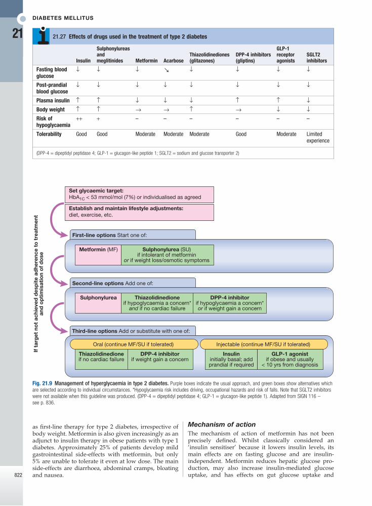
Diabetes mellitus
21
822
Fig. 21.9 Management of hyperglycaemia in type 2 diabetes. Purple boxes indicate the usual approach, and green boxes show alternatives which are selected according to individual circumstances. *Hypoglycaemia risk includes driving, occupational hazards and risk of falls. Note that SGLT2 inhibitors were not available when this guideline was produced. (DPP-4 = dipeptidyl peptidase 4; GLP-1 = glucagon-like peptide 1). Adapted from SIGN 116 – see p. 836.
Thiazolidinedioneif no cardiac failure
Oral (continue MF/SU if tolerated)
DPP-4 inhibitorif weight gain a concern
DPP-4 inhibitorif hypoglycaemia a concern*or if weight gain a concern
Insulininitially basal; addprandial if required
Injectable (continue MF/SU if tolerated)
GLP-1 agonistif obese and usually
< 10 yrs from diagnosis
Third-line options Add or substitute with one of:
Sulphonylurea
If t
arg
et n
ot
achi
eved
des
pit
e ad
here
nce
to t
reat
men
tan
d o
pti
mis
atio
n o
f d
ose
Thiazolidinedioneif hypoglycaemia a concern*
and if no cardiac failure
Second-line options Add one of:
Metformin (MF)
Set glycaemic target:HbA1C < 53 mmol/mol (7%) or individualised as agreed
Establish and maintain lifestyle adjustments:diet, exercise, etc.
Sulphonylurea (SU)if intolerant of metformin
or if weight loss/osmotic symptoms
First-line options Start one of:
Insulin
Sulphonylureas and meglitinides Metformin Acarbose
Thiazolidinediones (glitazones)
DPP-4 inhibitors (gliptins)
GLP-1 receptor agonists
SGLT2 inhibitors
Fasting blood glucose
↓ ↓ ↓ ↘ ↓ ↓ ↓ ↓
Post-prandial blood glucose
↓ ↓ ↓ ↓ ↓ ↓ ↓ ↓
Plasma insulin ↑ ↑ ↓ ↓ ↓ ↑ ↑ ↓Body weight ↑ ↑ → → ↑ → ↓ ↓Risk of hypoglycaemia
++ + – – – – – –
Tolerability Good Good Moderate Moderate Moderate Good Moderate Limitedexperience
(DPP-4 = dipeptidyl peptidase 4; GLP-1 = glucagon-like peptide 1; SGLT2 = sodium and glucose transporter 2)
21.27 Effects of drugs used in the treatment of type 2 diabetes
Mechanism of actionThe mechanism of action of metformin has not been precisely defined. Whilst classically considered an ‘insulin sensitiser’ because it lowers insulin levels, its main effects are on fasting glucose and are insulin-independent. Metformin reduces hepatic glucose pro-duction, may also increase insulin-mediated glucose uptake, and has effects on gut glucose uptake and
as first-line therapy for type 2 diabetes, irrespective of body weight. Metformin is also given increasingly as an adjunct to insulin therapy in obese patients with type 1 diabetes. Approximately 25% of patients develop mild gastrointestinal side-effects with metformin, but only 5% are unable to tolerate it even at low dose. The main side-effects are diarrhoea, abdominal cramps, bloating and nausea.

Management of diabetes
21
823
utilisation. At the molecular level, metformin acts as a weak inhibitor of mitochondrial respiration, which increases intracellular adenosine monophosphate (AMP) and reduces adenosine triphosphate (ATP). This has direct effects on the flux through gluconeogenesis, and activates the intracellular energy sensor AMP-activated protein kinase (AMPK), leading to multiple beneficial metabolic effects.
Clinical useMetformin is a potent blood glucose-lowering treatment that is weight-neutral, does not cause hypoglycaemia and has established benefits in microvascular disease. It is employed as first-line therapy in all patients who tol-erate it, and its use is maintained when additional agents are added as glycaemia deteriorates (see Fig. 21.9). Met-formin is usually introduced at low dose (500 mg twice daily) to minimise the risk of gastrointestinal side-effects. The usual maintenance dose is 1 g twice daily. There is a modified-release formulation of metformin which may be better tolerated by patients with gastro-intestinal side-effects.
Metformin can increase susceptibility to lactic acido-sis, although this is much less common than was previ-ously thought. As metformin is cleared by the kidneys, it can accumulate in renal impairment, so the dose should be halved when estimated glomerular filtration rate (eGFR) is 30–45 mL/min, and it should not be used below an eGFR of 30 mL/min. Its use is also contraindi-cated in patients with impaired hepatic function and in those who drink alcohol in excess, in whom the risk of lactic acidosis is significantly increased. It should be dis-continued, at least temporarily, if any other serious medical condition develops, especially one causing severe shock or hypoxia. In such circumstances, treat-ment with insulin should be substituted if required.
SulphonylureasSulphonylureas are ‘insulin secretagogues’, i.e. they promote pancreatic β-cell insulin secretion. Similar to metformin, the long-term benefits of sulphonylureas in lowering microvascular complications of diabetes were established in the UK Prospective Diabetes Study (p. 827).
Mechanism of actionSulphonylureas act by closing the pancreatic β-cell ATP-sensitive potassium (KATP) channel, decreasing K+ efflux, which ultimately triggers insulin secretion. Meg-litinides (e.g. repaglinide and nateglinide) also work in this way and, although short-acting, are essentially sulphonyl urea-like drugs.
Clinical useSulphonylureas are an effective therapy for lowering blood glucose and are often used as an add-on to met-formin, if glycaemia is inadequately controlled on met-formin alone (see Fig. 21.9). The main adverse effects of sulphonylureas are weight gain and hypoglycaemia. The weight gain is not ideal in patients with diabetes who are already overweight or obese, although sulphonyl-ureas are effective treatments in this group. Hypo-glycaemia occurs because the closure of KATP channels brings about unregulated insulin secretion, even with normal or low blood glucose levels.
There are a number of sulphonylureas. In the UK, gliclazide is the most commonly used; in contrast, in the USA, glibenclamide (also known as glyburide) is widely used. Glibenclamide, however, is long-acting and prone to induce hypoglycaemia, so should be avoided in the elderly. Other sulphonylureas include glimepiride and glipizide. The dose–response of all sulphonylureas is steepest at low doses; little additional benefit is obtained when the dose is increased to maximal levels.
Alpha-glucosidase inhibitorsThe α-glucosidase inhibitors delay carbohydrate absorp-tion in the gut by inhibiting disaccharidases. Acarbose and miglitol are available and are taken with each meal. Both lower post-prandial blood glucose and modestly improve overall glycaemic control. They can be com-bined with a sulphonylurea. The main side-effects are flatulence, abdominal bloating and diarrhoea. They are used widely in the Far East but infrequently in the UK.
ThiazolidinedionesMechanism of actionThese drugs (also called TZDs, ‘glitazones’ or PPARγ agonists) bind and activate peroxisome proliferator-activated receptor-γ, a nuclear receptor present mainly in adipose tissue that regulates the expression of several genes involved in metabolism. TZDs enhance the actions of endogenous insulin, in part directly (in the adipose cells) and in part indirectly (by altering release of ‘adi-pokines’, such as adiponectin, which alter insulin sensi-tivity in the liver). Plasma insulin concentrations are not increased and hypoglycaemia does not occur. TZDs increase pre-adipocyte differentiation, resulting in an increase in fat mass and in body weight.
Clinical useTZDs have been prescribed widely since the late 1990s, but recently a number of adverse effects have become apparent and their use has declined. One popular TZD, rosiglitazone, was reported to increase the risk of myo-cardial infarction and was withdrawn in 2010. The other TZD in common use, pioglitazone, does not appear to increase the risk of myocardial infarction but it does exacerbate cardiac failure by causing fluid retention, and recent data show that it increases the risk of bone frac-ture, and possibly bladder cancer. These observations have reduced the use of pioglitazone dramatically.
Pioglitazone can be very effective at lowering blood glucose in some patients and appears more effective in insulin-resistant patients. In addition, it has a beneficial effect in reducing fatty liver and NASH (p. 805). Piogli-tazone is usually added to metformin with or without sulphonylurea therapy (see Fig. 21.9). It may be given with insulin therapy, when it can be very effective, but the combination of insulin and TZDs markedly increases fluid retention and risk of cardiac failure, so should be used with caution.
Incretin-based therapies: DPP-4 inhibitors and GLP-1 analoguesThe incretin effect is the augmentation of insulin secre-tion seen when a glucose stimulus is given orally rather than intravenously, and reflects the release of incretin peptides from the gut (see Fig. 21.3, p. 802). The incretin

Diabetes mellitus
21
824
solution, can be extended by the addition of protamine and zinc at neutral pH (isophane or NPH insulin) or excess zinc ions (lente insulins). These modified ‘depot’ insulins are cloudy preparations. Pre-mixed formula-tions containing short-acting and isophane insulins in various proportions are available. The pharmacokinetics of these various insulins are shown in Box 21.28.
In most countries, the insulin concentration in avail-able formulations has been standardised at 100 U/mL.
Subcutaneous multiple dose insulin therapyIn most patients, insulin is injected subcutaneously several times a day into the anterior abdominal wall, upper arms, outer thighs and buttocks (Box 21.29). Acci-dental intramuscular injection often occurs in children and thin adults. The rate of absorption of insulin may be influenced by many factors other than the insulin for-mulation, including the site, depth and volume of injec-tion, skin temperature (warming), local massage and exercise. Absorption is delayed from areas of lipohyper-trophy at injection sites (p. 799), which results from the local trophic action of insulin, so repeated injection at the same site should be avoided. Other routes of admin-istration (intravenous and intraperitoneal) are reserved for specific circumstances.
Once absorbed into the blood, insulin has a half-life of just a few minutes. It is removed mainly by the liver and also the kidneys, so plasma insulin concentrations are elevated in patients with liver disease or renal failure. Rarely, the rate of clearance can be affected by binding to insulin antibodies (induced by use of animal insulins).
Insulin can be administered using a disposable plastic syringe with a fine needle (which can be re-used several times), but this has largely been replaced by pen
hormones are primarily glucagon-like peptide 1 (GLP-1) and gastric inhibitory polypeptide (GIP). These are rapidly broken down by the peptidase DPP-4 (dipepti-dyl peptidase 4). The incretin effect is diminished in type 2 diabetes, and this has stimulated the development of two incretin-based therapeutic approaches.
The ‘gliptins’, or DPP-4 inhibitors, prevent break-down and therefore enhance concentrations of endo-genous GLP-1 and GIP. The first DPP-4 inhibitor to market was sitagliptin; others now available include vildagliptin, saxagliptin and linagliptin. These drugs are very well tolerated and are weight-neutral (see Box 21.27).
The GLP-1 receptor agonists have a similar structure to GLP-1 but have been modified to resist breakdown by DPP-4. These agents are not orally active and have to be given by subcutaneous injection. However, they have a key advantage over the DPP-4 inhibitors: because the GLP-1 activity achieved is supra-physiological, it delays gastric emptying and, at the level of the hypothalamus, decreases appetite. Thus, injectable GLP-1 analogues lower blood glucose and result in weight loss – an appealing therapy, as the majority of patients with type 2 diabetes are obese. Currently available GLP-1 receptor agonists include exenatide (twice daily), exenatide MR (once weekly) and liraglutide (once daily).
Unlike sulphonylureas, both incretin-based therapies only promote insulin secretion when there is a glucose ‘trigger’ for insulin secretion. Thus, when the blood glucose is normal, the insulin secretion is not augmented and so these agents do not cause hypoglycaemia.
SGLT2 inhibitorsThe sodium and glucose transporter 2 (SGLT2) inhibitor, dapagliflozin, was licensed for use in 2012. Glucose is filtered freely in the renal glomeruli and reabsorbed in the proximal tubules. SGLT2 is involved in reabsorption of glucose. Inhibition results in approximately 25% of the filtered glucose not being reabsorbed, with conse-quent glycosuria. Although this helps to lower blood glucose and results in calorie loss and subsequent weight loss, the glycosuria does result in increased urinary tract and genital fungal infections. With limited experience and evidence to date, the most appropriate position for SGLT2 inhibitors in the therapy of type 2 diabetes has yet to be established.
Insulin therapy
Manufacture and formulationInsulin was discovered in 1921 and transformed the management of type 1 diabetes, until then a fatal disor-der. Until the 1980s, insulin was obtained by extraction and purification from pancreata of cows and pigs (bovine and porcine insulins), and some patients still prefer to use animal insulins. Recombinant DNA tech-nology enabled large-scale production of human insulin. More recently, the amino acid sequence of insulin has been altered to produce analogues of insulin, which differ in their rate of absorption from the site of injection.
The duration of action of short-acting, unmodified insulin (‘soluble’ or ‘regular’ insulin), which is a clear
Insulin Onset Peak Duration
Rapid-acting(insulinanalogues:lispro,aspart,glulisine)
<0.5 0.5–2.5 3–4.5
Short-acting(soluble(regular))
0.5–1 1–4 4–8
Intermediate-acting(isophane(NPH),lente)
1–3 3–8 7–14
Long-acting(bovineultralente)
2–4 6–12 12–30
Long-acting(insulinanalogues:glargine,detemir)
1–2 None 18–24
21.28 Duration of action (in hours) of insulin preparations
• Needlesitedatrightangletotheskin• Subcutaneous(notintramuscular)injection• Deliverydevices:glasssyringe(requiresre-sterilisation),
plasticsyringe(disposable),pendevice(reusable,somedisposable),infusionpump
21.29 How to inject insulin subcutaneously
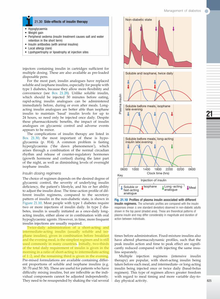
Management of diabetes
21
825
times before administration. Fixed-mixture insulins also have altered pharmacodynamic profiles, such that the peak insulin action and time to peak effect are signifi-cantly reduced compared with injecting the same insu-lins separately.
Multiple injection regimens (intensive insulin therapy) are popular, with short-acting insulin being taken before each meal, and intermediate- or long-acting insulin being injected once or twice daily (basal-bolus regimen). This type of regimen allows greater freedom with regard to meal timing and more variable day-to-day physical activity.
injectors containing insulin in cartridges sufficient for multiple dosing. These are also available as pre-loaded disposable pens.
For the most part, insulin analogues have replaced soluble and isophane insulins, especially for people with type 1 diabetes, because they allow more flexibility and convenience (see Box 21.28). Unlike soluble insulin, which should be injected 30 minutes before eating, rapid-acting insulin analogues can be administered immediately before, during or even after meals. Long-acting insulin analogues are better able than isophane insulin to maintain ‘basal’ insulin levels for up to 24 hours, so need only be injected once daily. Despite these pharmacokinetic benefits, the impact of insulin analogues on glycaemic control and adverse events appears to be minor.
The complications of insulin therapy are listed in Box 21.30; the most important of these is hypo-glycaemia (p. 814). A common problem is fasting hyperglycaemia (‘the dawn phenomenon’), which arises through a combination of the normal circadian rhythm and release of counter-regulatory hormones (growth hormone and cortisol) during the later part of the night, as well as diminishing levels of overnight isophane insulin.
Insulin dosing regimensThe choice of regimen depends on the desired degree of glycaemic control, the severity of underlying insulin deficiency, the patient’s lifestyle, and his or her ability to adjust the insulin dose. The time–action profile of dif-ferent insulin regimens, compared to the secretory pattern of insulin in the non-diabetic state, is shown in Figure 21.10. Most people with type 1 diabetes require two or more injections of insulin daily. In type 2 dia-betes, insulin is usually initiated as a once-daily long-acting insulin, either alone or in combination with oral hypoglycaemic agents. However, in time, more frequent insulin injections are usually required.
Twice-daily administration of a short-acting and intermediate-acting insulin (usually soluble and iso-phane insulins), given in combination before breakfast and the evening meal, is the simplest regimen and is still used commonly in many countries. Initially, two-thirds of the total daily requirement of insulin is given in the morning in a ratio of short-acting to intermediate-acting of 1 : 2, and the remaining third is given in the evening. Pre-mixed formulations are available containing differ-ent proportions of soluble and isophane insulins (e.g. 30 : 70 and 50 : 50). These are useful for patients who have difficulty mixing insulins, but are inflexible as the indi-vidual components cannot be adjusted independently. They need to be resuspended by shaking the vial several
• Hypoglycaemia• Weightgain• Peripheraloedema(insulintreatmentcausessaltandwater
retentionintheshortterm)• Insulinantibodies(withanimalinsulins)• Localallergy(rare)• Lipohypertrophyorlipoatrophyatinjectionsites
21.30 Side-effects of insulin therapy
Fig. 21.10 Profiles of plasma insulin associated with different insulin regimens. The schematic profiles are compared with the insulin responses (mean ± one standard deviation) observed in non-diabetic adults shown in the top panel (shaded area). These are theoretical patterns of plasma insulin and may differ considerably in magnitude and duration of action between individuals.
Pla
sma
insu
lin
Soluble before meals, long-actinginsulin late evening
0600020022001800140010000600Clock time (hrs)
Soluble before meals, isophane late evening
Soluble and isophane, twice daily
Non-diabetic state
Injection of insulin
Soluble orfast-actinganalogue
Isophane Long -actinganalogue
Meal
Key

Diabetes mellitus
21
826
Alternative insulin therapies‘Open-loop’ systems are battery-powered portable pumps providing continuous subcutaneous (CSII), intraperitoneal or intravenous infusion of insulin without reference to the blood glucose concentration. The rate of insulin infusion is variable; it can be pro-grammed to match the patient’s diurnal variation in requirements and then manually boosted at mealtimes. In practice, the ‘loop’ is closed by the patient performing blood glucose estimations, and the use of these devices requires a high degree of patient motivation. These increasingly sophisticated systems can achieve excellent glycaemic control but widespread therapeutic use is limited by cost. The ‘Artificial Pancreas’ project aims to close the loop by using miniaturised glucose sensors to communicate wirelessly with the insulin pump, which would automatically adjust its rate, but this has not yet reached widespread clinical practice.
Alternative routes of insulin delivery have been investigated. Clinical trials with intrapulmonary (inha-lation), transdermal and oral insulins are ongoing but as yet none has proven commercially viable.
Transplantation
Whole pancreas transplantation is carried out in a small number of patients with diabetes each year, but it presents problems relating to exocrine pancreatic secre-tions and long-term immunosuppression is necessary. While results are steadily improving, they remain less favourable than for renal transplantation. At present, the procedure is usually undertaken only in patients with end-stage renal failure who require a combined pancreas/kidney transplantation and in whom diabetes control is particularly difficult, e.g. because of recurrent hypoglycaemia.
Transplantation of isolated pancreatic islets (usually into the liver via the portal vein) has been achieved safely in an increasing number of centres around the world. Progress is being made towards meeting the needs of supply, purification and storage of islets, but the problems of transplant rejection, and of destruction by the patient’s autoantibodies against β cells, remain. Nevertheless, the development of methods of inducing tolerance to transplanted islets and the potential use of stem cells (p. 69) means that this may still prove the most promising approach in the long term.
COMPLICATIONS OF DIABETES
Despite all the treatments now available, the outcome for patients with diabetes remains disappointing. Long-term complications of diabetes still cause significant morbidity and mortality (Boxes 21.31 and 21.32).
Excess mortality in diabetes is caused mainly by large blood vessel disease, particularly myocardial infarction and stroke. Macrovascular disease also causes sub-stantial morbidity from myocardial infarction, stroke, angina, cardiac failure and intermittent claudication. The pathological changes of atherosclerosis in diabetic patients are similar to those in the non-diabetic popula-tion but occur earlier in life and are more extensive and severe. Diabetes amplifies the effects of the other major
Microvascular/neuropathic
Retinopathy, cataract• Impairedvision
Nephropathy• Renalfailure
Peripheral neuropathy• Sensoryloss• Pain• Motorweakness
Autonomic neuropathy• Gastrointestinalproblems(gastroparesis;alteredbowelhabit)• Posturalhypotension
Foot disease• Ulceration• Arthropathy
Macrovascular
Coronary circulation• Myocardialischaemia/infarction
Cerebral circulation• Transientischaemicattack• Stroke
Peripheral circulation• Claudication• Ischaemia
21.31 Complications of diabetes
cardiovascular risk factors: smoking, hypertension and dyslipidaemia (Fig. 21.11). Moreover, patients with type 2 diabetes are more likely to have additional cardiovas-cular risk factors, which co-segregate with insulin resist-ance in the metabolic syndrome (p. 805).
Disease of small blood vessels is a specific complica-tion of diabetes and is termed diabetic microangiopathy. It contributes to mortality through renal failure caused
21.32 Mortality in diabetes

Complications of diabetes
21
827
There was a 60% overall reduction in the risk of develop-ing diabetic complications in patients with type 1 dia-betes on intensive therapy with strict glycaemic control, compared with those on conventional therapy (Box 21.33). No single factor other than glycaemic control had a significant effect on outcome. However, the group who were intensively treated to lower blood glucose had three times the rate of severe hypoglycaemia. The UKPDS showed that, in type 2 diabetes, the frequency of diabetic complications is lower and progression is slower with good glycaemic control and effective treat-ment of hypertension, irrespective of the type of therapy used (Box 21.34). Extrapolation from the UKPDS sug-gests that, for every 11 mmol/mol (1%) reduction in HbA1c, there is a 21% reduction in death related to dia-betes, a 14% reduction in myocardial infarction and 30–40% reduction in risk of microvascular complications (see Fig. 21.11).
These landmark trials demonstrated that diabetic complications are preventable and that the aim of treat-ment should be ‘near-normal’ glycaemia. However, more recent studies, such as the Action to Control Cardiovascular Risk in Diabetes (ACCORD), showed increased mortality in a subgroup of patients who were aggressively treated to lower HbA1c to a target of less than 48 mmol/mol (6.5%). The patients in this study had poor glycaemic control at baseline, a long duration of diabetes and a high prevalence of cardiovascular disease. It appears that, whilst a low target HbA1c is appropriate in younger patients with earlier diabetes who do not
by diabetic nephropathy, and is responsible for substan-tial morbidity and disability: for example, blindness from diabetic retinopathy, difficulty in walking, chronic ulceration of the feet from peripheral neuropathy, and bowel and bladder dysfunction from autonomic neuro-pathy. The risk of microvascular disease is positively correlated with the duration and degree of sustained hyperglycaemia, however caused and at whatever age it develops.
PathophysiologyThe histopathological hallmark of diabetic micro-angiopathy is thickening of the capillary basement membrane, with associated increased vascular perme-ability, which occurs throughout the body. The devel-opment of the characteristic clinical syndromes of diabetic retinopathy, nephropathy, neuropathy and accelerated atherosclerosis is thought to result from the local response to generalised vascular injury. For example, in the wall of large vessels, increased perme-ability of arterial endothelium, particularly when com-bined with hyperinsulinaemia and hypertension, may increase the deposition of atherogenic lipoproteins. The mechanisms linking hyperglycaemia to these pathologi-cal changes are, however, poorly characterised.
Preventing diabetes complicationsGlycaemic controlThe evidence that improved glycaemic control decreases the risk of developing microvascular complications of diabetes was established by the Diabetes Control and Complications Trial (DCCT) in type 1 diabetes, and the UK Prospective Diabetes Study (UKPDS) in type 2 dia-betes. The DCCT was a large study that lasted 9 years.
Fig. 21.11 Association between HbA1c and risk of microvascular and macrovascular diabetes complications. These data were obtained amongst participants in the UK Prospective Diabetes Study and were adjusted for effects of age, sex and ethnicity; the incidences show what could be expected amongst white men aged 50–54 years at diagnosis of type 2 diabetes, followed up for 10 years. Microvascular disease included retinopathy requiring photocoagulation, vitreous haemorrhage and renal failure. Macrovascular disease included fatal and non-fatal myocardial infarction and sudden death. A 1% change in HbA1c is equivalent to a reduction of 11 mmol/mol.
< 6 6–7 7–8 8–9 9–10 > 10HbA1C (%)
0
20
40
60In
cid
ence
per
100
0 p
atie
nt y
ears
Microvascular disease
Macrovascular disease(myocardial infarction) ‘Inpatientswithtype1diabetes,strictglycaemiccontrol(mean
HbA1c53mmol/mol(7%))reducedthedevelopmentofretinopathyandothermicrovascularcomplicationsby76%comparedwithconventionaltherapy(meanHbA1c75mmol/mol(9%)).Onlonger-termfollow-up,strictglycaemiccontrolalsoreducedcardiovascularevents,includingmyocardialinfarction,strokeanddeathfromcardiovasculardisease.’
• DiabetesControlandComplicationsTrialResearchGroup.NEnglJMed1993;329:977–986.
• DiabetesControlandComplicationsTrial/EpidemiologyofDiabetesInterventionsandComplications(DCCT/EDIC)StudyResearchGroup.NEnglJMed2005;353:2643–2653.
Forfurtherinformation: www.diabetes.niddk.nih.gov/dm/pubs/control/
21.33 Glycaemic control in type 1 diabetes
‘Inpatientswithtype2diabetes,intensiveglycaemiccontrol(meanHbA1c53mmol/mol(7%))withoralanti-diabeticdrugsorinsulinreducedthedevelopmentofmicrovascularcomplications,particularlyretinopathy,by25%comparedwithconventionaltreatment(meanHbA1c64mmol/mol(8%)).Onlonger-termfollow-up,therewasasignificantreductioninmyocardialinfarctionandall-causemortalityinintensivelycontrolledpatients.’
• UKPDSGroup.Lancet1998;352:837–853,854–865.• HolmanRR,etal.NEnglJMed2008;359:1577–1589.
Forfurtherinformation: www.dtu.ox.ac.uk
21.34 Glycaemic control in type 2 diabetes

Diabetes mellitus
21
828
• Glycaemic control:theoptimaltargetforglycaemiccontrolinolderpeoplehasyettobedetermined.Strictglycaemiccontrolshouldbeavoidedinfrailpatientswithcomorbiditiesandinolderpatientswithlongdurationofdiabetes.
• Cognitive function and affect:maybenefitfromimprovingglycaemiccontrol.
• Hypoglycaemia:olderpeoplehavereducedsymptomaticawarenessofhypoglycaemiaandlimitedknowledgeofsymptoms,andareatgreaterriskof,andfrom,hypoglycaemia.
• Mortality:themortalityrateofolderpeoplewithdiabetesismorethandoublethatofage-matchednon-diabeticpeople,largelybecauseofincreaseddeathsfromcardiovasculardisease.
21.35 Diabetes management in old age
Clinical featuresThe major risk factors for DR are shown in Box 21.36. DR is a progressive condition, often classified into two stages: non-proliferative (‘background’) and pro-liferative. The earliest signs of non-proliferative DR are microaneurysms and retinal haemorrhages, some-times inaccurately called ‘dot’ and ‘blot’ haemorrhages, respectively (Fig. 21.12A and B). As DR progresses and there is continuing capillary hypoperfusion, cotton wool spots, venous beading and intra-retinal microvascular abnormalities can be seen (Fig. 21.12C–E); this stage is referred to as pre-proliferative DR. The disease may then progress to proliferative DR, which is characterised by growth of new blood vessels on the retina or optic disc (Fig. 21.12F and G). The new vessels are abnormal and often bleed, leading to vitreous haemorrhage, sub-sequent fibrosis and scarring, and finally tractional retinal detachment.
In addition to proliferative and non-proliferative DR, patients may also develop clinically significant macular oedema (CSMO; see Fig. 21.12C). This can occur at any stage of DR and is characterised by increased vascular permeability and deposition of hard exudates in the central retina. CSMO is the most common cause of loss of vision in people with diabetes.
Proliferative retinopathy and severe ocular ischaemia may stimulate new vessels to grow on the anterior surface of the iris: ‘rubeosis iridis’. These vessels may obstruct the drainage angle of the eye and the outflow of aqueous fluid, causing secondary glaucoma.
Loss of visual acuityMicroaneurysms, abnormalities of the veins, and small haemorrhages and exudates situated in the periphery will not interfere with vision. However, if these changes are observed near the macula, and in particular if they are accompanied by loss of visual acuity, CSMO should be suspected. Macular oedema can cause impair-ment of visual acuity even if this is associated with only mild peripheral non-proliferative retinopathy and no other obvious pathology. Macular oedema can only be confirmed or excluded on slit lamp retinal biomicroscopy.
Sudden visual loss occurs with vitreous haemorrhage or retinal detachment. In pre-proliferative and prolifera-tive retinopathy, whether or not visual acuity is impaired, prompt laser treatment is important to reduce the risk of haemorrhage, fibrosis/gliosis and severe irreversible visual impairment.
PreventionGlycaemic, blood pressure and lipid profile controlMany epidemiological studies have shown a clear rela-tionship between glycaemic control and the incidence of DR. Large randomised controlled trials have shown con-vincingly that improved glycaemic control, particularly
have underlying cardiovascular disease, aggressive glucose-lowering is not beneficial in older patients with long duration of diabetes and multiple comorbidities (Box 21.35).
Control of other risk factorsRandomised controlled trials have shown that aggres-sive management of blood pressure minimises the microvascular and macrovascular complications of dia-betes. Angiotensin-converting enzyme (ACE) inhibitors are valuable in improving outcome in heart disease and in treating diabetic nephropathy (p. 830). The manage-ment of dyslipidaemia with a statin limits macrovascu-lar disease in people with diabetes (p. 456). This often results in the necessary use of multiple medications, which exacerbates the problem of adherence to therapy by patients; it is not unusual for a patient to be taking two or more diabetes therapies, two or more blood pres-sure drugs and a statin.
Diabetic retinopathy
Diabetic retinopathy (DR) is one of the most common causes of blindness in adults between 30 and 65 years of age in developed countries. The prevalence of DR increases with duration of diabetes, and almost all indi-viduals with type 1 diabetes and the majority of those with type 2 diabetes will have some degree of DR after 20 years.
PathogenesisHyperglycaemia increases retinal blood flow and dis-rupts intracellular metabolism in retinal endothelial cells and pericytes (pericytes wrap around the outside of the capillary wall and influence blood flow and capillary permeability). This leads to impaired vascular autoregu-lation, increased production of vasoactive substances and endothelial cell proliferation. The resulting capillary hypoperfusion and closure cause chronic retinal ischae-mia, stimulating the production of growth factors, including vascular endothelial growth factor (VEGF), which further stimulates deleterious endothelial cell growth (causing new vessel formation) and increased vascular permeability (causing retinal leakage and exudation).
• Longdurationofdiabetes• Poorglycaemiccontrol• Hypertension• Hyperlipidaemia
• Pregnancy• Nephropathy/renaldisease• Others:obesity,smoking
21.36 Risk factors for diabetic retinopathy

Complications of diabetes
21
829
organised and audited programme. The preferred method is a digital photographic system for retinal imaging, with prompt referral of patients with sight-threatening retinopathy to an ophthalmologist for examination with slit lamp biomicroscopy. If direct ophthalmoscopy is used, the pupils should be dilated for adequate examination. Unfortunately, many people with diabetes receive no regular supervision and do not attend for eye screening.
ManagementGood glycaemic (HbA1c around 53 mmol/mol (7%)) control and an appropriate blood pressure (< 130/80 mmHg) should be maintained to prevent onset and delay progression of diabetic eye disease. Novel agents are emerging, including ranibizumab, a monoclonal antibody fragment that binds to VEGF-A and is anti-angiogenic; it is used for diabetic macular oedema.
Retinal photocoagulation (laser treatment) is indi-cated in: severe proliferative or very severe non-proliferative retinopathy; new vessels elsewhere with vitreous haemorrhage; new vessels without vitreous haemorrhage in type 2 diabetes; or CSMO. Photocoagu-lation is used:• to treat leaking microaneurysms and areas of retinal
thickening in the macular area, and to reduce macular oedema (focal laser)
in the early stages of disease, reduces both the incidence and progression of DR in type 1 (see Box 21.33) and type 2 (see Box 21.34) diabetes. When blood glucose is lowered, there can be a transient deterioration of retin-opathy. This relates to loss of hyperglycaemia-induced hyperperfusion in the retinal circulation and a conse-quent increase in ischaemia. However, this effect wears off within 18 months. Improvement in glycaemic control should therefore be effected gradually in patients with retinopathy, particularly when glycaemic control is ini-tially poor.
Evidence from randomised controlled trials suggests that blood pressure control is also warranted to reduce the incidence and progression of DR. Early trials sug-gested specific benefit from angiotensin II receptor antagonists, but later studies indicate that other anti-hypertensive agents are similarly effective. Most guide-lines recommend achieving a blood pressure of less than 130/80 mmHg. Observational studies suggest that hyperlipidaemia is a risk factor for DR, but intervention trials have not been conclusive.
ScreeningAnnual screening for retinopathy is essential in all diabetic patients, as the disease is asymptomatic in the early stages, when treatment is most effective. Screening is particularly important in those with risk factors. It should be undertaken by trained personnel in an
Fig. 21.12 Diabetic retinopathy. A Microaneurysms. Usually the earliest clinical abnormality, these tiny aneurysms arise mainly from the venous end of capillaries and appear as discrete, circular, dark red dots near to, but apparently separate from, the retinal vessels and no wider than a vessel at the optic disc margin (arrows). B Haemorrhages. Larger than a microaneurysm, with indistinct margins and at least as wide as a vessel at the optic disc margin, these occur in deeper layers of the retina (arrows). They result either from microaneurysms that have burst or from leaky capillaries. Superficial flame-shaped haemorrhages in the nerve fibre layer may also occur, particularly if the patient is hypertensive. C Hard exudates. These irregularly shaped lesions are formed from leaking of cholesterol, often through microaneurysms (black arrows). They can be associated with retinal oedema; if this affects the centre of the macula, it can cause clinically significant macular oedema (CSMO, white arrows), which is sight-threatening. D Cotton wool spots. These white, feathery, fluffy lesions indicate capillary infarcts within the nerve fibre layer (arrows). They are most often seen in rapidly advancing retinopathy or in association with uncontrolled hypertension. E Venous beading. In extensive retinal ischaemia, walls of veins develop saccular bulges, looking like a string of sausages (black arrow). Intra-retinal microvascular anomalies (IRMA) are spidery vessels, often with sharp corners that indicate dilatations of pre-existing capillaries (white arrow). F and G Neovascularisation. New vessel formation in response to widespread retinal ischaemia may arise from the venous circulation either on the optic disc (NVD, arrows in F) or elsewhere in the retina (NVE, arrows in G). Initially, fine tufts of delicate vessels form arcades on the surface of the retina; later, they may extend forwards on to the posterior surface of the vitreous. Leaking of serous products from new vessels stimulates a connective tissue reaction, with gliosis and fibrosis, that first appears as a white, cloudy haze among the network of new vessels and later extends to obliterate the area with a dense white sheet. H Vitreous haemorrhage. New vessels are fragile and liable to rupture during vitreous movement, causing a pre-retinal (‘subhyaloid’) or a vitreous haemorrhage (arrows), which may lead to sudden visual loss.
A B C D
E F G H

Diabetes mellitus
21
830
• to destroy areas of retinal ischaemia and hence lower intraocular levels of VEGF, which play a major role in the development of neovascularisation
• to reduce the risk of recurrent haemorrhage by inducing gliosis and fibrosis of new vessels (pan-retinal photocoagulation (PRP)).Argon laser photocoagulation is the usual method.
This simple procedure can be carried out under topical anaesthesia. Patients should be reviewed regularly to look for further development of new vessels and/or maculopathy. Extensive bilateral photocoagulation can cause significant visual field loss, which may interfere with driving ability and reduce night vision.
Vitrectomy is used in selected cases of advanced dia-betic eye disease due to type 1 diabetes where visual loss has been caused by recurrent vitreous haemorrhage that has failed to clear or by tractional retinal detachment threatening the macula. The value of vitrectomy in type 2 diabetes is less certain. Rubeosis iridis is a severe com-plication requiring early and extensive PRP.
Other causes of visual loss in people with diabetesAround 50% of visual loss in people with type 2 diabetes results from causes other than diabetic retinopathy. These include cataract, age-related macular degenera-tion, retinal vein occlusion, retinal arterial occlusion, non-arteritic ischaemic optic neuropathy and glaucoma. Some of these conditions are to be expected in this group, as they relate to cardiovascular risk factors (e.g. hypertension, hyperlipidaemia and smoking), all of which are prevalent in people with type 2 diabetes.
CataractCataract is a permanent lens opacity and is a common cause of visual deterioration in the elderly. The lens thickens and opacifies with age; with diabetes, the increased metabolic insult to the lens causes these changes to accelerate and occur prematurely. A rare type of ‘snow-flake’ cataract occurs in young patients with poorly controlled diabetes. This does not usually affect vision but tends to make fundal examination difficult.
The indications for cataract extraction are similar to those for the non-diabetic population and depend on the degree of visual impairment. An additional indication in diabetes is when adequate assessment of the fundus, or laser treatment to the retina, is prevented.
Diabetic nephropathy
Diabetic nephropathy is an important cause of morbid-ity and mortality, and is now among the most common causes of end-stage renal failure in developed countries. About 30% of patients with type 1 diabetes have devel-oped diabetic nephropathy 20 years after diagnosis, but the risk after this time falls to less than 1% per year, and from the outset the risk is not equal in all patients (Box 21.37). Indeed, some patients do not develop nephro-pathy, despite having long-standing, poorly controlled diabetes, suggesting that they are genetically protected from it. Whilst variants in a few genes have been impli-cated in diabetic nephropathy, the major differences in individual risk remain unexplained. Epidemiological data have indicated that the overall incidence is
• Poorglycaemiccontrol• Longdurationofdiabetes• Presenceofothermicrovascularcomplications• Ethnicity(e.g.Asians,PimaIndians)• Pre-existinghypertension• Familyhistoryofdiabeticnephropathy• Familyhistoryofhypertension
21.37 Risk factors for diabetic nephropathy
declining as standards of glycaemic and blood pressure control have improved.
The pattern of progression of renal abnormalities in diabetes is shown schematically in Figure 21.13. Patho-logically, the first changes coincide with the onset of microalbuminuria and include thickening of the glomer-ular basement membrane and accumulation of matrix material in the mesangium. Subsequently, nodular deposits (Fig. 21.14) are characteristic, and glomerulo-sclerosis worsens as heavy proteinuria develops, until
Fig. 21.13 Natural history of diabetic nephropathy. In the first few years of type 1 diabetes mellitus, there is hyperfiltration, which declines fairly steadily to return to a normal value at approximately 10 years (blue line). In susceptible patients (about 30%), after about 10 years, there is sustained proteinuria, and by approximately 14 years it has reached the nephrotic range (red line). Renal function continues to decline, with the end stage being reached at approximately 16 years.
MicroalbuminuriaSustained proteinuria
Nephrotic range proteinuria
10
5
120
0 10 20
GFR(mL / min)
Proteinuria(g / 24 hrs)
Years
HyperfiltrationHypertension
Renalfailure
Fig. 21.14 Nodular diabetic glomerulosclerosis. There is thickening of basement membranes and mesangial expansion, and a Kimmelstiel–Wilson nodule (arrow), which is pathognomonic of diabetic kidney disease.

Complications of diabetes
21
831
Halving the amount of albuminuria with an ACE or ARB results in a nearly 50% reduction in long-term risk of progression to end-stage renal disease. However, some patients do progress, with worsening renal func-tion. Renal replacement therapy (p. 488) may benefit diabetic patients at an earlier stage than other patients with end-stage renal failure.
Renal transplantation dramatically improves the life of many, and any recurrence of diabetic nephropathy in the allograft is usually too slow to be a serious problem, but associated macrovascular and microvas-cular disease elsewhere may still progress. Pancreatic transplantation (generally carried out at the same time as renal transplantation) can produce insulin independ-ence and delay or reverse microvascular disease, but the supply of organs is limited and this option is avail-able to few. For further information on management, see Chapter 17.
Diabetic neuropathy
Although there is some evidence that the central nervous system is affected in long-term diabetes, the clinical impact of diabetes is mainly manifest in the peripheral nervous system. Diabetic neuropathy causes substantial morbidity and increases mortality. It is diagnosed on the basis of symptoms and signs, and after the exclusion of other causes of neuropathy (p. 1223). Depending on the criteria used for diagnosis, it affects between 50 and 90% of patients with diabetes, and of these, 15–30% will have painful diabetic neuropathy (PDN). Like retinopathy, neuropathy occurs secondary to metabolic disturbance, and prevalence is related to the duration of diabetes and the degree of metabolic control.
Pathological features can occur in any peripheral nerves. They include axonal degeneration of both myeli-nated and unmyelinated fibres, with thickening of the Schwann cell basal lamina, patchy segmental demyeli-nation, and abnormal intraneural capillaries (with base-ment membrane thickening and microthrombi).
Various classifications of diabetic neuropathy have been proposed. One is shown in Box 21.40, but motor, sensory and autonomic nerves may be involved in varying combinations so that clinically mixed syn-dromes usually occur.
Clinical featuresSymmetrical sensory polyneuropathyThis is frequently asymptomatic. The most common clinical signs are diminished perception of vibration
glomeruli are progressively lost and renal function deteriorates.
Diagnosis and screeningMicroalbuminuria (Box 21.38) is the presence in the urine of small amounts of albumin, at a concentration below that detectable using a standard urine dipstick. Overt nephropathy is defined as the presence of macro-albuminuria (albumin to creatinine ratio (ACR) > 300 mg/mmol; detectable on urine dipstick). Micro-albuminuria is a good predictor of progression to nephro pathy in type 1 diabetes. It is a less reliable pre-dictor of nephropathy in older patients with type 2 dia-betes, in whom it may be accounted for by other diseases (p. 476), although it is a potentially useful marker of an increased risk of macrovascular disease.
ManagementThe presence of established microalbuminuria or overt nephropathy should prompt vigorous efforts to reduce the risk of progression of nephropathy and of cardiovas-cular disease by:• aggressive reduction of blood pressure• aggressive cardiovascular risk factor reduction
(Box 21.39).In type 1 diabetes, ACE inhibitors have been shown
to provide greater protection than equal blood pressure reduction achieved with other drugs (p. 611), and sub-sequent studies have shown similar benefits from angio-tensin II receptor blockers (ARBs) in patients with type 2 diabetes. This benefit from blockade of the renin–angiotensin system arises from a reduction in the angio-tensin II-mediated vasoconstriction of efferent arterioles in glomeruli (see Fig 17.1D, p. 465). The resulting dilata-tion of these vessels decreases glomeruli filtration pres-sure and therefore the hyperfiltration and protein leak. Both ACE inhibitors and ARBs increase risk of hyper-kalaemia (p. 442) and, in the presence of renal artery stenosis (p. 494), may induce marked deterioration in renal function. Therefore, electrolytes and renal function should be checked after initiation or each dose increase. Non-dihydropyridine calcium antagonists (diltiazem, verapamil) may be suitable alternatives.
• Identifiesincipientnephropathyintype1andtype2diabetes;isanindependentpredictorofmacrovasculardiseaseintype2diabetes
• Riskfactorsincludehighbloodpressure,poorglycaemiccontrol,smoking
• Whotoscreen:Patientswithtype1diabetesannuallyfrom5yrsafterdiagnosisPatientswithtype2diabetesannuallyfromtimeofdiagnosis
• Earlymorningurinemeasuredforalbumin:creatinineratio(ACR).Microalbuminuriapresentif:
MalesACR2.5–30mg/mmolcreatinineFemalesACR3.5–30mg/mmolcreatinine
• AnelevatedACRshouldbefollowedbyarepeattest:Establishedmicroalbuminuriaif2outof3testspositiveAnACR>30mg/mmolcreatinineisconsistentwithovertnephropathy
21.38 Screening for microalbuminuria
‘Intype2patientswithmicroalbuminuria,intensivetreatment(includingcontrolofglycaemiaandhypertension,withuseofACEinhibitors,statinsandaspirin)reducedtheriskofcardiovasculardiseaseby53%,ofnephropathyby61%,andofretinopathyby58%comparedwithconventionaltreatment.Onlonger-termfollow-up,therewasareductionintotalmortalityanddeathfromcardiovascularcauses.’
• GaedeP,etal.NEnglJMed2003;348:383–393.• GaedeP,etal.NEnglJMed2008;358:580–591.
21.39 Multiple risk factor intervention in type 2 diabetes

Diabetes mellitus
21
832
Visceral (autonomic)
• Cardiovascular• Gastrointestinal• Genitourinary
• Sudomotor• Vasomotor• Pupillary
Somatic
• PolyneuropathySymmetrical,mainlysensoryanddistalAsymmetrical,mainlymotorandproximal(includingamyotrophy)
• Mononeuropathy(includingmononeuritismultiplex)
21.40 Classification of diabetic neuropathyneuro pathies, mononeuropathies are severe and of rapid onset, but they eventually recover. The nerves most commonly affected are the 3rd and 6th cranial nerves (resulting in diplopia), and the femoral and sciatic nerves. Rarely, involvement of other single nerves results in paresis and paraesthesiae in the thorax and trunk (truncal radiculopathies).
Nerve compression palsies are more common in dia-betes, frequently affecting the median nerve, giving the clinical picture of carpal tunnel syndrome, and less com-monly the ulnar nerve. Lateral popliteal nerve compres-sion occasionally causes foot drop. Compression palsies may be more common because of glycosylation and thickening of connective tissue and/or because of increased susceptibility of nerves affected by diabetic microangiopathy.
Autonomic neuropathyThis is not necessarily associated with peripheral somatic neuropathy. Parasympathetic or sympathetic nerves may be predominantly affected in one or more visceral systems. The resulting symptoms and signs are listed in Box 21.41 and tests of autonomic function in Box 21.42. The development of autonomic neuropathy is less clearly related to poor metabolic control than somatic neuropa-thy, and improved control rarely results in improved symptoms. Within 10 years of developing overt symp-toms of autonomic neuropathy, 30–50% of patients are dead, many from sudden cardiorespiratory arrest. Patients with postural hypotension (a drop in systolic pressure of 30 mmHg or more on standing from the supine position) have the highest subsequent mortality.
sensation distally, ‘glove and stocking’ impairment of all other modalities of sensation (see Fig. 21.15), and loss of tendon reflexes in the lower limbs. In symptomatic patients, sensory abnormalities are predominant. Symp-toms include paraesthesiae in the feet (and, rarely, in the hands), pain in the lower limbs (dull, aching and/or lancinating, worse at night, and mainly felt on the ante-rior aspect of the legs), burning sensations in the soles of the feet, cutaneous hyperaesthesia and an abnormal gait (commonly wide-based), often associated with a sense of numbness in the feet. Weakness and atrophy, in particular of the interosseous muscles, may develop, leading to structural changes in the foot with loss of lateral and transverse arches, clawing of the toes and exposure of the metatarsal heads. This results in increased pressure on the plantar aspects of the metatar-sal heads, with the development of callus skin at these and other pressure points. Electrophysiological tests (p. 1151) demonstrate slowing of both motor and sensory conduction, and tests of vibration sensitivity and thermal thresholds are abnormal.
A diffuse small-fibre neuropathy causes altered perception of pain and temperature, and is associated with symptomatic autonomic neuropathy; characteris-tic features include foot ulcers and Charcot neuro-arthropathy.
Asymmetrical motor diabetic neuropathySometimes called diabetic amyotrophy, this presents as severe and progressive weakness and wasting of the proximal muscles of the lower (and occasionally the upper) limbs. It is commonly accompanied by severe pain, mainly felt on the anterior aspect of the leg, and hyperaesthesia and paraesthesiae. Sometimes there may also be marked loss of weight (‘neuropathic cachexia’). The patient may look extremely ill and be unable to get out of bed. Tendon reflexes may be absent on the affected side(s). Sometimes there are extensor plantar responses and the cerebrospinal fluid protein is often raised. This condition is thought to involve acute infarction of the lower motor neurons of the lumbosacral plexus. Other lesions involving this plexus, such as neoplasms and lumbar disc disease, must be excluded. Although recov-ery usually occurs within 12 months, some deficits are permanent. Management is mainly supportive.
MononeuropathyEither motor or sensory function can be affected within a single peripheral or cranial nerve. Unlike the gradual progression of distal symmetrical and autonomic
Cardiovascular
• Posturalhypotension• Restingtachycardia
• Fixedheartrate
Gastrointestinal
• Dysphagia,duetooesophagealatony• Abdominalfullness,nauseaandvomiting,unstable
glycaemia,duetodelayedgastricemptying(‘gastroparesis’)• Nocturnaldiarrhoea±faecalincontinence• Constipation,duetocolonicatony
Genitourinary
• Difficultyinmicturition,urinaryincontinence,recurrentinfection,duetoatonicbladder
• Erectiledysfunctionandretrogradeejaculation
Sudomotor
• Nocturnalsweatswithouthypoglycaemia
• Gustatorysweating
• Anhidrosis;fissuresinthefeet
Vasomotor
• Feetfeelcold,duetolossofskinvasomotorresponses• Dependentoedema,duetolossofvasomotortoneand
increasedvascularpermeability• Bullousformation
Pupillary
• Decreasedpupilsize• Resistancetomydriatics
• Delayedorabsentreflexestolight
21.41 Clinical features of autonomic neuropathy
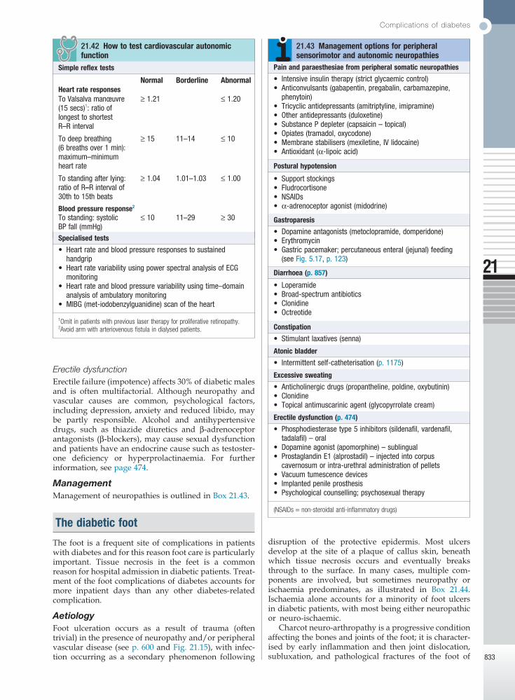
Complications of diabetes
21
833
Erectile dysfunctionErectile failure (impotence) affects 30% of diabetic males and is often multifactorial. Although neuropathy and vascular causes are common, psychological factors, including depression, anxiety and reduced libido, may be partly responsible. Alcohol and antihypertensive drugs, such as thiazide diuretics and β-adrenoceptor antagonists (β-blockers), may cause sexual dysfunction and patients have an endocrine cause such as testoster-one deficiency or hyperprolactinaemia. For further information, see page 474.
ManagementManagement of neuropathies is outlined in Box 21.43.
The diabetic foot
The foot is a frequent site of complications in patients with diabetes and for this reason foot care is particularly important. Tissue necrosis in the feet is a common reason for hospital admission in diabetic patients. Treat-ment of the foot complications of diabetes accounts for more inpatient days than any other diabetes-related complication.
AetiologyFoot ulceration occurs as a result of trauma (often trivial) in the presence of neuropathy and/or peripheral vascular disease (see p. 600 and Fig. 21.15), with infec-tion occurring as a secondary phenomenon following
disruption of the protective epidermis. Most ulcers develop at the site of a plaque of callus skin, beneath which tissue necrosis occurs and eventually breaks through to the surface. In many cases, multiple com-ponents are involved, but sometimes neuropathy or ischaemia predominates, as illustrated in Box 21.44. Ischaemia alone accounts for a minority of foot ulcers in diabetic patients, with most being either neuropathic or neuro-ischaemic.
Charcot neuro-arthropathy is a progressive condition affecting the bones and joints of the foot; it is character-ised by early inflammation and then joint dislocation, subluxation, and pathological fractures of the foot of
Pain and paraesthesiae from peripheral somatic neuropathies
• Intensiveinsulintherapy(strictglycaemiccontrol)• Anticonvulsants(gabapentin,pregabalin,carbamazepine,
phenytoin)• Tricyclicantidepressants(amitriptyline,imipramine)• Otherantidepressants(duloxetine)• SubstancePdepleter(capsaicin–topical)• Opiates(tramadol,oxycodone)• Membranestabilisers(mexiletine,IVlidocaine)• Antioxidant(α-lipoicacid)
21.43 Management options for peripheral sensorimotor and autonomic neuropathies
Postural hypotension
• Supportstockings• Fludrocortisone• NSAIDs• α-adrenoceptoragonist(midodrine)
Gastroparesis
• Dopamineantagonists(metoclopramide,domperidone)• Erythromycin• Gastricpacemaker;percutaneousenteral(jejunal)feeding
(seeFig.5.17,p.123)
Diarrhoea (p. 857)
• Loperamide• Broad-spectrumantibiotics• Clonidine• Octreotide
Constipation
• Stimulantlaxatives(senna)
Atonic bladder
• Intermittentself-catheterisation(p.1175)
Excessive sweating
• Anticholinergicdrugs(propantheline,poldine,oxybutinin)• Clonidine• Topicalantimuscarinicagent(glycopyrrolatecream)
Erectile dysfunction (p. 474)
• Phosphodiesterasetype5inhibitors(sildenafil,vardenafil,tadalafil)–oral
• Dopamineagonist(apomorphine)–sublingual• ProstaglandinE1(alprostadil)–injectedintocorpus
cavernosumorintra-urethraladministrationofpellets• Vacuumtumescencedevices• Implantedpenileprosthesis• Psychologicalcounselling;psychosexualtherapy
(NSAIDs = non-steroidal anti-inflammatory drugs)
Simple reflex tests
Normal Borderline AbnormalHeart rate responsesToValsalvamanœuvre(15secs)1:ratiooflongesttoshortestR–Rinterval
≥1.21 ≤1.20
Todeepbreathing(6breathsover1min):maximum–minimumheartrate
≥15 11–14 ≤10
Tostandingafterlying:ratioofR–Rintervalof30thto15thbeats
≥1.04 1.01–1.03 ≤1.00
Blood pressure response2
Tostanding:systolicBPfall(mmHg)
≤10 11–29 ≥30
21.42 How to test cardiovascular autonomic function
Specialised tests
• Heartrateandbloodpressureresponsestosustainedhandgrip
• HeartratevariabilityusingpowerspectralanalysisofECGmonitoring
• Heartrateandbloodpressurevariabilityusingtime–domainanalysisofambulatorymonitoring
• MIBG(met-iodobenzylguanidine)scanoftheheart
2Avoid arm with arteriovenous fistula in dialysed patients.
1Omit in patients with previous laser therapy for proliferative retinopathy.
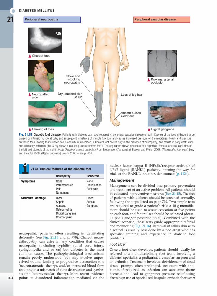
Diabetes mellitus
21
834
neuropathic patients, often resulting in debilitating deformity (see Fig. 21.15 and p. 798). Charcot neuro-arthropathy can arise in any condition that causes neuropathy (including syphilis, spinal cord injury, syringomyelia and so on) but diabetes is the most common cause. The pathophysiological mechanisms remain poorly understood, but may involve unper-ceived trauma leading to progressive destruction (the ‘neurotraumatic’ theory), and/or increased blood flow resulting in a mismatch of bone destruction and synthe-sis (the ‘neurovascular’ theory). More recent evidence points to disordered inflammation mediated via the
nuclear factor kappa B (NFκB)/receptor activator of NFκB ligand (RANKL) pathway, opening the way for trials of the RANKL inhibitor, denosumab (p. 1124).
ManagementManagement can be divided into primary prevention and treatment of an active problem. All patients should be educated in preventive measures (Box 21.45). The feet of patients with diabetes should be screened annually, following the steps listed on page 799. Two simple tests are required to grade a patient’s risk: a 10 g monofila-ment should be used to assess sensation at five points on each foot, and foot pulses should be palpated (dorsa-lis pedis and/or posterior tibial). Combined with the clinical scenario, these tests guide appropriate referral and monitoring (Fig. 21.16). Removal of callus skin with a scalpel is usually best done by a podiatrist who has specialist training and experience in diabetic foot problems.
Foot ulcerOnce a foot ulcer develops, patients should ideally be referred to a multidisciplinary foot team, involving a diabetes specialist, a podiatrist, a vascular surgeon and an orthotist. Treatment involves: débridement of dead tissue; prompt, often prolonged, treatment with anti-biotics if required, as infection can accelerate tissue necrosis and lead to gangrene; pressure relief using dressings; use of specialised bespoke orthotic footwear;
Neuropathy Ischaemia
Symptoms NoneParaesthesiaePainNumbness
NoneClaudicationRestpain
Structural damage UlcerSepsisAbscessOsteomyelitisDigitalgangreneCharcotjoint
UlcerSepsisGangrene
21.44 Clinical features of the diabetic foot
Fig. 21.15 Diabetic foot disease. Patients with diabetes can have neuropathy, peripheral vascular disease or both. Clawing of the toes is thought to be caused by intrinsic muscle atrophy and subsequent imbalance of muscle function, and causes increased pressure on the metatarsal heads and pressure on flexed toes, leading to increased callus and risk of ulceration. A Charcot foot occurs only in the presence of neuropathy, and results in bony destruction and ultimately deformity (this X-ray shows a resulting ‘rocker bottom foot’). The angiogram shows disease of the superficial femoral arteries (occlusion of the left and stenosis of the right). Insets (Proximal arterial occlusion) From Medscape; (Toe clawing) Bowker and Pfeifer 2008; (Neuropathic foot ulcer) Levy and Valabhji 2008; (Digital gangrene) Swartz 2006 – see p. 836.
Glove andstocking
neuropathy
Clawing of toes Digital gangrene
Neuropathiculcer
Dry, cracked skinCallus
Loss of leg hair
Peripheral vascular diseasePeripheral neuropathy
Charcot foot
Proximal arterialocclusion
Absent pulsesCold feet

Further information and acknowledgements
21
835
As about 40% of patients with a Charcot joint also have a foot ulcer, it can be difficult to differentiate from osteomyelitis. Magnetic resonance imaging (MRI) of the foot is often helpful. The mainstay of treatment for an active Charcot foot is immobilisation and, ideally, avoid-ance of weight-bearing on the affected foot. The ration-ale is that if no pressure is applied through the foot, the destructive process involving the bones will not result in significant deformity when the acute inflam-matory process subsides. Immobilisation is often achieved by a total contact plaster cast or ‘aircast’ boot. The acute phase often lasts 3–6 months and sometimes longer. In the post-acute phase, there is consolidation and remodelling of fracture fragments, eventually resulting in a stable foot.
Further information and acknowledgements
Websiteswww.cdc.gov/diabetes/ Diabetes Public Health Resource.
Useful American site with resources for patients and health-care professionals.
www.diabetes.org American Diabetes Association. Includes information on research and advocacy issues.
www.diabetes.org.uk Diabetes UK. Includes information for patients and leaflets.
www.idf.org International Diabetes Federation. Useful information on international aspects of care and education.
www.joslin.org Joslin Diabetes Center. Well-written resource for patients and health-care professionals, and information on diabetes research.
www.mydiabetesmyway.scot.nhs.uk/ An interactive diabetes website for patients with diabetes and their carers.
www.ndei.org National Diabetes Education Initiative. Web-based education for health-care professionals, including case studies and slides.
• Preventiveadvicetoalldiabeticpatientsincludes:InspectfeeteverydayWashfeeteverydayMoisturiseskinifdryCutorfiletoenailsregularlyChangesocksorstockingseverydayAvoidwalkingbarefootCheckfootwearforforeignbodiesWearsuitable,well-fittingshoesCoverminorcutswithsteriledressingsDonotburstblistersAvoidover-the-countercorn/callusremedies
• Advicetomoderate-andhigh-riskpatientsisasaboveplus:DonotattemptcornremovalAvoidhighandlowtemperatures
• Apodiatristisanintegralpartofthediabetesteamtoensureregularandeffectivepodiatryandtoeducatepatientsincareofthefeet
• SpeciallymanufacturedandfittedorthoticfootwearisrequiredtopreventrecurrenceofulcerationandtoprotectthefeetofpatientswithCharcotneuroarthropathy
21.45 Care of the feet in patients with diabetes
Fig. 21.16 Risk assessment and management of foot problems in diabetes. Adapted from SIGN 116 – see p. 836.
Moderate
Low
High
Active Urgent referral tospecialist team
Current foot ulcer, infection,critical ischaemia, gangrene or
unexplained hot, red swollen foot
Previous foot ulcer or amputation
Sensation impairedand
foot pulses absent
Skin callus orfoot deformity?
Yes
No
Inability to self-care for feet
Sensation impairedand
foot pulses absent
Sensationunimpaired,
foot pulses present
Annual assessmentby podiatrist
Annual assessmentby podiatrist
Annual screening byhealth-care professional
ManagementScreening
and sometimes total contact plaster cast or irremovable aircast boot. If an ulcer is neuro-ischaemic, a vascular assessment is often carried out, by ultrasound or angio-graphy, as revascularisation by angioplasty or surgery may be required to allow the ulcer to heal. In cases of severe secondary infection or gangrene, an amputation may be required. This can be limited to the affected toe or involve more extensive limb amputation.
Charcot footAcute Charcot arthropathy almost always presents with signs of inflammation – a hot, red, swollen foot. Initial X-ray may show bony destruction but is often normal.

Diabetes mellitus
21
836
Figure acknowledgements
Page 798 inset (Acanthosis nigricans) Shotliff K. Diabetes and other metabolic diseases. In: Lim E (ed). Medicine and surgery: an integrated textbook. Elsevier; 2007, Fig. 7.11. Copyright © 2007 Edinburgh: Elsevier Ltd. All rights reserved.
Fig. 21.6B Adapted from Holman RR. Diabetes Res Clin Pract 1998; 40 (Suppl.):S21–S25.
Fig. 21.7 Adapted from UK Prospective Diabetes Study Group. UKPDS 33. Lancet 1998; 352: 837–853.
Figs 21.9 and 21.16 Adapted from Scottish Intercollegiate Guidelines Network (SIGN) guideline number 116.
Fig. 21.15 insets (Proximal arterial occlusion) http://emedicine.medscape.com/article/460178-overview#a0104; (Toe clawing) Bowker JH, Pfeifer MA. Levin and O’Neal’s The diabetic foot, 7th edn. Philadelphia: Mosby; 2008; Fig. 26-9. Copyright © 2008 by Mosby, Inc., an affiliate of Elsevier; (Neuropathic foot ulcer) Levy MJ, Valabhji J. Vascular II: The diabetic foot. Surgery 2008; 26:25–28, Fig. 2; copyright © 2007 Elsevier Ltd; (Digital gangrene) Swartz MH. Textbook of physical diagnosis, 5th edn. Philadelphia: WB Saunders, 2006.