2018 Endodontic Board Review and Scientific Update ... · 2018 Endodontic Board Review and...
21
1 2018 Endodontic Board Review and Scientific Update University of Texas Health, San Antonio Endodontic microbiology Christine Sedgley MDS, MDSc(Endo), FRACDS, MRACDS(ENDO), PhD February 2018 Overview Part 1 Basic microbiology update Part 2 Methods used for evaluating endodontic microflora Part 3 Microorganisms in primary endodontic infections Part 4 Microorganisms and unsuccessful endodontic treatment Part 5 Microorganisms in periapical lesions Part 6 Microorganisms and regenerative endodontics Part 7 Antimicrobial efficacy of endodontic treatment Overview 1 Part 1 (Very) basic microbiology update Bugs Biofilms Virulence Basic microbiology Prokaryotes bacteria No nucleus Eukaryotes fungi, animals Chromosomes in nucleus Gram-positive Gram-negative Classification - cell wall Gram positive and Gram negative What are biofilms? ØAggregates of microbial cells enclosed in a self-produced matrix adherent to a surface Biofilm Composition Bacterial cells ± 20% by volume EPS/glycocalyx, biofilm matrix ± 80% by volume
Transcript of 2018 Endodontic Board Review and Scientific Update ... · 2018 Endodontic Board Review and...

1
2018 Endodontic Board Review and Scientific Update �University of Texas Health, San Antonio�
Endodontic microbiology
Christine Sedgley �MDS, MDSc(Endo), FRACDS, MRACDS(ENDO), PhD �
February 2018"
Overview
Part 1 Basic microbiology update
Part 2 Methods used for evaluating endodontic microflora
Part 3 Microorganisms in primary endodontic infections
Part 4 Microorganisms and unsuccessful endodontic treatmentPart 5 Microorganisms in periapical lesionsPart 6 Microorganisms and regenerative endodontics Part 7 Antimicrobial efficacy of endodontic treatment
Overview 1
Part 1 (Very) basic microbiology update
Bugs Biofilms Virulence
Basic microbiology
ProkaryotesbacteriaNo nucleus
Eukaryotes fungi, animalsChromosomes
in nucleus
Gram-positive
Gram-negative
Classification - cell wall �Gram positive and Gram negative
What are biofilms?
Ø Aggregates of microbial cells enclosed in a self-produced matrix adherent to a surface
Biofilm CompositionBacterial cells ± 20% by volume
EPS/glycocalyx, biofilm matrix
± 80% by volume

2
EPS - Extracellular Polymeric Substance� What’s happening in the EPS?
Flemming 2016
• Means of regulating gene expression and diverse physiological activities within community
How is activity in the EPS regulated?
Quorum sensing = how bacteria talk
• Regulation is dynamic:§ Stressors
§ Population density
• Spatial wisdom of crowds:§ Texts, tweets etc
Ø Microbes in biofilms can easily interact with each other and exchange genes encoding:Ø Virulence factorsØ Antibiotic resistance
Ø Biofilm microorganisms are more resistant to antimicrobial agents than planktonic bacteria
Biofilms and antimicrobial resistance Intracanal biofilms
Ø First observed by Nair using microscopy in 1987
Ø Described as clusters of “self-aggregating” colonies of one distinct type or “coaggregating” communities of several types
Ø Described as “biofilms” by Svensater & Bergenholtz (2005)
LM, TEM: Nair 1990
Nair et al. 2005
Root canal biofilms are complex, polymicrobial and heterogenous
Biofilm morphology can vary between and within root canal systems
“Entombed biofilms” in the root canal system
Ø Proposed that “apical periodontitis” be included in the set of “biofilm-induced diseases”
Ricucci and Siqueira 2010

3
What is virulence?
Ø The degree of pathogenicity or disease-producing ability of a microorganism
Ø The pathogenicity of an organism is determined by its virulence factors
Potential virulence factors associated with a bacterial cell
Sedgley 2016
Microbial virulence �Specific virulence factors and endodontics
Endotoxin (LPS)
Others?Hemolysin (Sedgley et al. 2005)Lipoteichoic acid (Baik et al. 2008)Peptidoglycan (Hahn and Liewehr 2007)Gelatinase (Sedgley 2007, Sato et al. 2009)Short chain fatty acids (Ho and Chang 2007, Provenzano et al. 2015)Fimbriae (Figdor and Davies 1997, Rôças and Siqueira 2010)Proteases (Ogawa et al. 2006, Nandakumar et al. 2009)
Microbial virulence �Gram negative cell wall - Endotoxin
Endotoxin is an established pathogenic factor in endodontic infections
Microbial virulence �Endotoxin and endodontics
Endotoxin is positively associated with:
ü Pulpal pain and periapical inflammationSchein and Schilder 1975, Horiba et al. 1991,
Yamasaki et al. 1992, Khabbaz et al. 2001, and others
ü Bone destruction Dahlén et al. 1981, Dwyer and Torabinejad 1981,
Pitts et al. 1982, Mattison et al. 1987and others
Ø Periapical pathology is multifactorial
Ø The host determines the response to microorganisms ………….
Microbial virulence �What about host factors?

4
Periapical pathology is multifactorial
Pulpal infection
Immune response
Systemic factors
Bone resorption
Cytokines
Adapted from Stashenko
Overview 2
Part 1 Basic microbiology updatePart 2 Methods used for evaluating endodontic microflora
Methods used for evaluating endodontic microflora
1. Histology
2. Culturing and biochemical tests
3. Microbial bioinformatics
Evaluating endodontic infections �1. Histology
Limited to observing morphology, and more recently, viability
• Light microscopy (LM) (Ricucci and Bergenholtz 2003, Ricucci et al. 2015)• Transmission electron microscopy (TEM) (Nair 2005)• Scanning electron microscopy (SEM) (Leonardo et al. 2002)• Environmental SEM (Bergmans et al. 2005)• Confocal laser SEM (Parmar et al. 2011)• Combination LM, TEM, SEM (Richardson et al. 2009)
LIVEDEAD
à Standardized sampling methodà Use of clinical sterility controls
à Pre-reduced transport mediaà Anaerobic culturing
à Pure culture and identification
sampletransfer
analyses
Evaluating endodontic infections �2. Culturing
anaerobic culturingidentification
Evaluating endodontic infections – now and the future �3. Microbial bioinformatics
Adapted from Libault et al. 2010
Rela%onalDatabasesCompare,Cluster&Integrate
BiologicalKnowledge,Gene%c&MetabolicEngineering
Correla%onofGenes&GeneProductsIden%fica%onofGeneFunc%ons
GeneAnnota%onIden%fica%onofPathways
DNASequenceDatabases
Genome
GeneExpression
Transcriptome Proteome Metabolome

5
Siqueira and Rôças 2005 Malmberg et al. 2016, Figdor et al. 2016
Sampling controls should be subjected to same investigations as root canals sample
“Critical evaluation and standardization of the disinfection methods and aseptic procedures in endodontics are needed”
“The high binding affinity between DNA and hydroxyapatitepresents a special challenge for decontamination”
Evaluating endodontic infections �Genomics – DNA based identification
ü Polymerase chain reaction (PCR)
ü Nested PCR
ü Multiplex-PCR
ü Real-time PCR
ü DNA-DNA hybridization
ü Pyrosequencing
Li et al. 2010
ü Enables more comprehensive analysis than traditional (Sanger) sequencing
Genomics – DNA based identification�Pyrosequencing
600-fold difference
Bottom line �Advantages of genomic approaches
é More sensitive than culturing
é Many microbes that are not easily cultured, or are unculturable, have been identified
é Multiple studies have clarified and confirmed that flora is diverse and not limited to a single species
Disadvantages of using genomic methods to study endodontic infections
! Strong binding affinity between DNA and dentin may compromise root canal sampling (Brundin et al. 2014, Figdor et al. 2016)
! Methods are very technique sensitive
! Preparation methods destroy microorganisms so cannot do further phenotypic analyses
! Most methods used don’t address cell viability
! Many nucleic acid sequences are unidentifiable

6
Evaluating endodontic infections �3. Microbial bioinformatics
Adapted from Libault et al. 2010
Rela%onalDatabasesCompare,Cluster&Integrate
BiologicalKnowledge,Gene%c&MetabolicEngineering
Correla%onofGenes&GeneProductsIden%fica%onofGeneFunc%ons
GeneAnnota%onIden%fica%onofPathways
DNASequenceDatabases
Genome Transcriptome Proteome Metabolome
GeneExpression
þ Microbial proteins in 7 samples:§ adhesins, autolysins, proteases, virulence factors,
and antibiotic-resistance proteins
þ Microbial and human proteins in 24 cases:§ Microbial proteins: metabolism and housekeeping,
adhesion, biofilm formation, antibiotic resistance, stress proteins, exotoxins, invasins, proteases
§ Human proteins: cellular processes and metabolism, immune defense
Evaluating endodontic infections �Proteomics
Provenzano et al. 2013, 2016
Nandakumar et al. 2009
Tip of the iceberg
3. Microbial bioinformatics�At what stage are we in the bioinformatics era?
Overview 3
Part 1 Basic microbiology update
Part 2 Methods used for evaluating endodontic microflora
Part 3 Microorganisms in primary endodontic infections
Are microorganisms the cause of apical periodontitis?
YESþ Animal studies have proven that bacteria in
the pulp are essential to the development of periapical disease
Kakehashi et al. 1965, Möller et al. 1981, Fabricius et al. 1982a, 1982b

7
Used germ-free rats to prove that bacteria in the pulp are essential to the development
of periapical disease
Kakehashi, Stanley and Fitzgerald 1965
Microbial etiologyClassic papers - Rats
1. Relative number of obligate anaerobes increases with time
2. Proportionally more anaerobes apically with time
3. Mixed infections show greatest capacity for inducing apical periodontitis
Möller, Fabricius, Dahlén et al. 1981, 1982
Microbial etiologyClassic papers - Monkeys
Primary endodontic infectionsClassic CULTURE studies - bottom line
Mixed flora dominated by Gram negative anaerobes
“Polymicrobial”
Möller 1966
Bergenholtz 1974Sundqvist 1976Cvek et al. 1976
Kantz and Henry 1974and others
Specific microorganisms and symptoms
Are specific symptoms associated with specific microorganisms?§ Yes
• Griffee et al. 1980, Haapasalo 1986, Yoshida et al. 1987, Hashioka et al. 1992, Hahn et al. 1993, Chavez 2002 and many others
§ No • Baumgartner et al. 1999, Jung et al. 2000, Rôças et al.
2001, Fouad et al. 2002, Chu et al. 2005 and many others
Geographic location
• Microflora of infected root canals may vary according to geographic location
Siqueira et al. 2005, Rôças et al. 2006
ü Biofilms
ü Polymicrobial
ü Predominantly anaerobic Gram negative rods
Summary - Primary endodontic infectionsHistology and culture-based studies
_____________________________________________________________ Facultative anaerobes Anaerobes Gram positive cocci
Streptococcus Micromonas Enterococcus Peptostreptococcus
Peptococcus Gram negative cocci
Neisseria Veillonella Gram positive rods
Lactobacillus Actinomyces Eubacterium Propionibacterium
Gram negative rods Enterobacter Porphyromonas Pseudomonas Prevotella Eikonella Fusobacterium Capnocytophaga Bacteroides family Dialister
Filifactor Spirochetes Treponema
_______________________________________________________________________ Fungi Candida Viruses HIV, Epstein-Barr virus, human cytomegalovirus, Herpes simplex virus-1

8
Summary - Primary endodontic infectionsMolecular-based studies
ü Multiple species (highly “diverse”)
ü Significantly more species detected
than in culture-based studies
ü Many species as yet unidentified and
unculturable
“The Rest”
Pyrosequencing studies:Li et al. 2010, Siqueira et al. 2011, Saber et al. 2012, Hong et al. 2013, Tzanetakis et al. 2015
Li et al. 2010
Overview 4
Part 1 Basic microbiology update
Part 2 Methods used for evaluating endodontic microfloraPart 3 Microorganisms in primary endodontic infections
Part 4 Microorganisms and unsuccessful endodontic treatment
Nair et al. 2005
How well can root canal systems be cleaned in a single visit ….. or ever?
Microorganisms remain after debridement in isthmuses
Microflora in primary and secondary endodontic infections differs?
Primary endodontic infections• Biofilms• Polymicrobial• Predominantly Gram negative anaerobic rods• Many species as yet unidentified and unculturable
Previously root filled canals• Biofilms• Polymicrobial, reduced microbial load• Frequently recovered in culture-based studies -
Gram positive facultatively anaerobic cocci: Enterococcus faecalis
• Many species as yet unidentified and unculturable
Secondary endodontic infections �Enterococci
ü Enterococci predominate in culture-positive root-filled teeth with chronic apical periodontitis
Molander et al. 1998, Sundqvist et al. 1998, Hancock et al. 2001, Peciuliene et al. 2001, Pinheiro et al. 2003 and many others
ü Enterococci are associated with persistence of infection during endodontic treatment
Siren et al. 1997
Reasons E. faecalis could resist root canal treatment procedures and survive
L Ability to invade dentinal tubules and adhere to collagen in the presence of human serum
Love et al. 2001
L A proton pump which allows E. faecalis to survive CaOH2 treatment
Evans et al. 2002
L Expression of virulence factors Sedgley et al. 2005, Mathew et al. 2010, Wang et al. 2011
L An efflux pump that may render E. faecalis biofilms more susceptible to antimicrobials
Upadya et al. 2011

9
E. faecalis can survive for extended periods in obturated root canals ex vivo
§ Viable E. faecalis were recovered from teeth for 2 years after obturation
48 hours 12 months 2 years
Sedgley et al. 2005, 2014
E. faecalis in secondary endodontic infections�
Bottom line Ø They survive
• obturation for at least 2 years• and have the potential to be virulent
Ø Is there proof that residual E. faecalis cause endodontic treatment failure?
NoØ Are they harmless hitchhikers?Ø Are they “persisters” in biofilms?
Tzanetakis et al. 2015Firmicutes phylum e.g. Enterococcus, Lactobacillus, Eubacterium
ü Fewer species and reduced microbial load compared with primary infection
ü Polymicrobialü E. faecalis frequently recovered, but not
exclusively
Summary – Secondary endodontic infectionsCulture-based studies
________________________________________________________________ Facultative anaerobes Anaerobes ____________________________________________________________ Gram positive cocci
Enterococcus Micromonas (formerly Peptostreptococcus) Streptococcus Staphylococcus
Gram negative cocci
Veillonella Anaeroglobus
Gram positive rods Lactobacillus Actinomyces Bacillus Propionibacterium (formerly Arachnia) Pseudoramibacter Eubacterium
Gram negative rods Escherichia Porphyromonas Pseudomonas Prevotella Proteus Fusobacterium Klebsiella Bacteroides family Enterobacter Wolinella Tannerella
________________________________________________________________ Fungi Candida ________________________________________________________________
Summary - Secondary endodontic infectionsMolecular-based studies
Siqueira et al. 2016
ü Multiple species (highly “diverse”)
ü Less predominance of E. faecalis
than in culture studies
ü Many species as yet unidentified
and unculturableChugal et al. 2011
Anderson et al. 2012Tzanetakis et al. 2015Siqueira et al. 2016
and others…
Overview 5
Part 1 Basic microbiology update
Part 2 Methods used for evaluating endodontic microfloraPart 3 Microorganisms in primary endodontic infections Part 4 Microorganisms and unsuccessful endodontic treatment
Part 5 Microorganisms in periapical lesions

10
Ricucci et al. 2015
Periapical lesions�Histology study - humans
Ø Not detected in granulomas
Ø Bacteria only detected in abscesses or cysts
Ricucci et al. 2006
Microorganisms in asymptomatic periapical lesions�Tronstad et al. 1987
“Anaerobic bacteria are able to survive and maintain an infectious disease
process in periapical tissues”
8 PA lesionsCulture study
Microorganisms in periapical lesions�Sampling
Microbiological sampling of periapical tissue presents difficulties
J Submarginal incision better than marginal incision to avoid contamination of surgery site
Sunde et al. 2000, Gatti et al. 2000
However…..L Disinfection procedures do not “destroy” the
DNA
Ø Evaluated 39 PA lesions (root-filled teeth with asymptomatic apical periodontitis)
Ø Used CLSM for 3D observations, and FISH to detect specific microorganisms
ü Observed microcolonies throughout
ü Detected P. gingivalis, P. intermedia, T. forsythia, Streptococcus spp. and unidentified morphotypes
Sunde et al. 2003 Subramanian and Mickel 2009
Ø Examined 34 apicoectomy samples for bacterial DNA in root end and soft tissue (including one “control” sample which was neg for DNA)Ø Persistent PA lesions – 1/3 symptomaticØ All except one root end and six periradicular tissue samples showed bacterial DNA
11
ü Sampling challenges
ü Polymicrobial
ü Predominantly Gram positive and Gram negative anaerobes
Summary – Periapical lesion infectionsCulture-based studies
_______________________________________________________ Facultative anaerobes Anaerobes Gram positive cocci
Streptococcus Micromonas (formerly Peptostreptococcus) Enterococcus Gemella Staphylococcus
Gram negative cocci Veillonella
Gram positive rods
Lactobacillus Actinomyces Bacillus Eubacterium Propionibacterium (formerly Arachnia)
Gram negative rods
Enterobacter Porphyromonas Pseudomonas Prevotella Vibrio Fusobacterium Capnocytophaga Bacteroides family Tannerella
Fungi Aspergillus Candida?
Sunde et al. 2003
ü Sampling challenges
ü Evidence of bacterial DNA
ü Multiple species (“polymicrobial”)
ü Many species as yet unidentified
and unculturable
ü Viruses
Summary – Periapical lesion infectionsMolecular-based studies
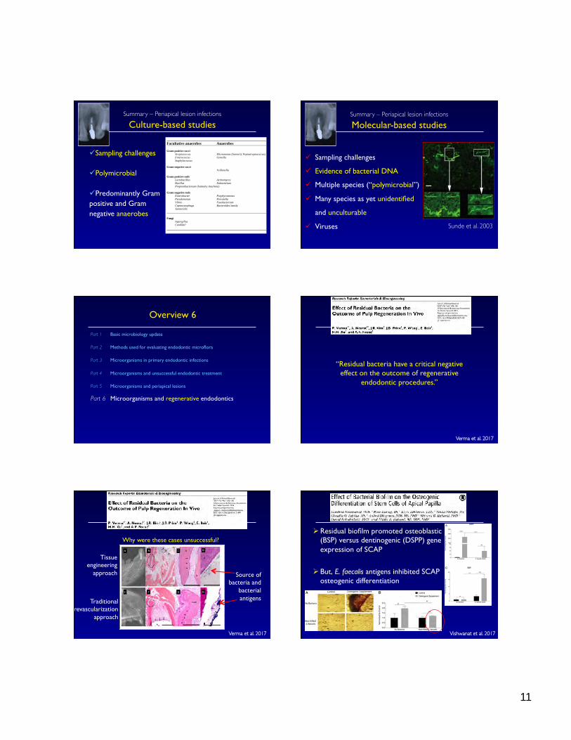
Overview 6
Part 1 Basic microbiology update
Part 2 Methods used for evaluating endodontic microfloraPart 3 Microorganisms in primary endodontic infections Part 4 Microorganisms and unsuccessful endodontic treatmentPart 5 Microorganisms and periapical lesions
Part 6 Microorganisms and regenerative endodontics
Verma et al. 2017
“Residual bacteria have a critical negative effect on the outcome of regenerative
endodontic procedures.”
Verma et al. 2017
Tissue engineering
approach
Traditional revascularization
approach
Source of bacteria and
bacterial antigens
Why were these cases unsuccessful?Ø Residual biofilm promoted osteoblastic
(BSP) versus dentinogenic (DSPP) gene expression of SCAP
Ø But, E. faecalis antigens inhibited SCAP osteogenic differentiation
Vishwanat et al. 2017

12
Diogenes and Hargreaves 2017
Microbes take control!
Overview 7
Part 1 Basic microbiology update
Part 2 Methods used for evaluating endodontic microfloraPart 3 Microorganisms in primary endodontic infections Part 4 Microorganisms and unsuccessful endodontic treatmentPart 5 Microorganisms and periapical lesionsPart 6 Microorganisms and regenerative endodontics
Part 7 Antimicrobial efficacy of endodontic treatment
?
Clinical management of infected root canals
Aiming to remove:Ø Microorganisms
Ø in biofilm and planktonic state Ø dead and alive
Ø whole cells and parts of cells
Ø Debris from instrumentation, smear layer Ø Pulpal remnants - cellular, fibrousØ Metabolic by-productsØ Previous root filling material Siqueira and Rôças 2011
Pathways of the Pulp
Biofilms are involved in all stages of root canal infection
Biofilms grow on root canals walls and in dentinal tubules
Siqueira and Rôças 2011Pathways of the Pulp
Microbe tactics and strategies �Why it’s hard to get rid of biofilms
Howard Hughes Medical Institute

13
Microbe tactics and strategies
WinnersVirulence factors
Biofilm interactionsResistance
LosersAccessible?
DisruptibleAntimicrobials
Antimicrobials
Mechanical
removal
Mechanicaldisruption
Clinical management of infected root canals
Antimicrobials
Mechanical
removal
Mechanicaldisruption
Clinical management of infected root canals
Ø Conventional irrigation
Ø Activation of irrigant
Ø Different file systems
Clinician tactics and strategies �Mechanical disruption
Clinician tactics and strategies �Conventional irrigation of root canals
Do irrigants reach apical part of the canal?
L There is little fluid exchange and displacement of particles beyond the tip of the needle �
Chow 1983
Needle tip positioned 3mm short of WL
Boutsioukis et al. 2010
�Apical pressure varies according to needle type�
�

14
�What influences irrigant flow?�
Ø Increased canal taper and apical size improves
irrigant replacement and wall shear stress and reduces irrigant extrusion
Albrecht et al. 2004, Falk and Sedgley 2005, Boutsioukis et al. 2010Ø Needle depth placement influences irrigation
efficacySedgley et al. 2005, Boutsioukis et al. 2010
Ø Irrigation significantly less effective in curved compared to straight canals
Nguy and Sedgley 2005
Ø Conventional irrigation
Ø Activation of irrigant
Ø Different file systems
Clinician tactics and strategies �Mechanical disruption
Mechanical disruption
Sonic activation
Ø Lower frequency than ultrasonic EndoActivatorTM Sonicare CanalBrushTM VibringeTM GentleWaveTM☺ All remove debris/smear layer in vitro ☹ Need independent data on antimicrobial activity
and biofilm removal in clinical situations Salman et al. 2010, Kanter et al. 2011, Rodig et al. 2011, Molina et al. 2015
Photon-Initiated Photoacoustic Streaming (PIPSTM)
Uses pulsed erbium:YAG laser to create photon-initiated photoacoustic streamingIn vitro study – culture of samples infected with oral flora
Ø Extracted premolars prepared to apical #20.07Ø PIPSTM generated more negative bacterial
samples than conventional and ultrasonic activation
Peters et al. 2011
Photodynamic therapy
Rationale
§ Solution binds to microbial cell§ Laser light applied (via plastic
flexible fiber) activates dye§ Free radicals produced destroy
cell
Soukos et al. 2006
Ø Uses photosensitizer solutions (e.g. methylene blue, tolonium chloride) and low-power laser light
Ø Grew multispecies biofilms (4 species) in root canalsØ Photodynamic therapy : up to 80% reduction of CFU
Fimple et al. 2008
B. Viable biofilms (arrows)
C. After PDT : destruction of biofilms, with some foci of live bacteria (arrow)
Photodynamic therapy (PDT) reduced biofilms in root canals

15
Ø 3 clinical studies reviewedØ All showed PDT had a “positive effect” in
private office clinical settingØ Review authors conclusions:
Ø Limited clinical information available on the use of PDT in root canal disinfection
Ø If supported by future clinical research, PDT may have efficacy for additional root canal disinfection, especially in the presence of multi–drug-resistant bacteria
Chrepa et al. 2014
Ø Conventional irrigation
Ø Activation of irrigant
Ø Different file systems
Clinician tactics and strategies �Mechanical disruption
In vitro studies comparing different file systems to reduce microbial load
No difference between:u SAF, Twisted File and Reciproc
Siqueira et al. 2013
u WaveOne and One ShapeNabeshima et al. 2014
u Hand (K-file) and ProTaperNakamura et al. 2013
u Single Reciproc file and BioRaCe seriesAlves et al. 2012
u WaveOne and ProTaperPinheiro et al. 2016
Clinical studies comparing different file systems to reduce microbial load
No difference between:u Hand NiTi using alternated rotation motion
or BioRaceRôças and Siqueira 2013
u Self-Adjusting File and Twisted File Adaptive for retreatment cases
Rodrigues et al. 2015
u Single Reciproc file and BioRaCe seriesNeves et al. 2016
Studies used real-time qPCR
Clinician challenges�Uninstrumented niches for biofilms
Peters 2004
Antimicrobials
Mechanical
removal
Mechanicaldisruption
Clinical management of infected root canals

16
Clinician tactics and strategies �Antimicrobials
Ø Sodium hypochlorite
Ø Chlorhexidine
Ø EDTA
Ø Calcium hydroxide
Ø Antibiotics
Sodium hypochlorite (NaOCl)
Is sodium hypochlorite an effective antimicrobial agent?
YES
NaOCl + H2O çè NaOH + HOCl çè Na+ + OH- + H+ + OCl-
J NaOCl inactivates the lipid moiety of lipotechoic acid through deacylation of the lipid moiety
Hong et al. 2016
Sodium hypochlorite �Classic clinical studies using culture-based techniques
J 0.5% NaOCl better than saline Byström and Sundqvist 1983
K No sig difference between 0.5% & 5% NaOCl Cvek et al. 1976, Byström and Sundqvist 1985
J 5% NaOCl effects enhanced by EDTA Byström and Sundqvist 1985
Sodium hypochlorite
• Toxic - dramatic effects if injected into tissues• Minimize risk with use of safety-ended irrigation
needles
Chlorhexidine
Is CHX an effective antimicrobial agent?
Ø Yes, but not as effective as NaOCl
Ø Does not have tissue solvent capacity
Chlorhexidine�Antimicrobial spectrum
J Bacteriostatic or bacteriocidal (depending on
concentration used) for a wide range of Gram
positive and Gram negative bacteria. Disrupts
cell membrane
L CHX has limited activity against non-enveloped
viruses and some bacterial and fungal spores

17
Ø Bacteria in mature biofilms and nutrient-limited biofilms are more resistant to CHX killing than bacteria in young biofilms
Shen et al. 2011
2 days 2 weeks 12 weeks
Chlorhexidine �Effect on biofilms
NaOCl versus CHX �Effect on biofilms
In vitro – multispecies
Ø 6% NaOCl rendered bacteria nonviable and physically removed polymicrobial biofilms from root segments (EDTA, MTAD, CHX did not)
Clegg et al. 2006
Ø 2% NaOCl was more effective against multispecies biofilms in dentin than 2% CHX
Yang et al. 2016
Clinical studies using molecular methods
Ø Canals with necrotic pulp - 2.5% NaOCl killed more
MOs than 2% CHX gel and removed more cells Vianna et al. 2006
Ø Teeth with AsAP - no difference between 0.12% CHX and 2.5% NaOCl
Rôças and Siqueira 2011
Ø CONSORT trial - Retreatment cases: no difference between 2% CHX and 1% NaOCl
Zandi et al. 2016
1. Standard2. OR153. OR174. OR375. OR316. OR477. OR628. OR649. OR6810. OR9211. ER312. ER513. JG214. OG115. JH2-2
1 2 3 4 5 6 7 8 9 10 11 12 13 14 15
NaOCl versus CHX �Antimicrobial activity
Chlorhexidine + NaOCl precipitates
In vitro studies L CHX and NaOCl precipitates when combined
Basrani et al. 2007
L CHX and NaOCl precipitate blocks dentinal tubules Bui et al. 2008
Neg control 6% NaOCl + 2% CHX
Other chlorhexidine interactions
L CHX and EDTA precipitates when combined
Rasimick et al. 2008
L CHX and Ca(OH)2 results in immediate degradation of CHX
Barbin et al. 2008
Clinician tactics and strategies �Antimicrobials
Ø Sodium hypochlorite
Ø Chlorhexidine
Ø EDTA
Ø Calcium hydroxide
Ø Antibiotics

18
Ø Introduced to endo as a chelating agent
Nygaard-Ostby 1957
Ø In vitro: EDTA can act as an antibiofilm acid for limiting S. aureus biofilm attachment by decreasing iron availability
Al-Azemi et al. 2011
Ø Destabilizes biofilms by sequestering calcium, magnesium, zinc, and iron
Finnegan and Percival 2015
EDTAEPS - Extracellular Polymeric Substance�
What’s happening in the EPS?
Flemming 2016
Alternate irrigation with EDTA and NaOCl
ü Biofilm removal enhanced by alternate irrigation with NaOCl and EDTA rather than using EDTA all at once as a final rinse after NaOCl
Soares et al. 2010
Clinician tactics and strategies �Antimicrobials
Ø Sodium hypochlorite
Ø Chlorhexidine
Ø EDTA
Ø Calcium hydroxide
Ø Antibiotics
Calcium hydroxide
Can calcium hydroxide kill endodontic microflora?
YES ….. but not always
Calcium hydroxide�Effect on biofilms in dentinal tubules
Ø Canals infected with E. faecalisØ Tooth slices (1mm) obtained
Parmar et al. 2011
Top. Treated with CaOH2: Survival 29%-50%
Bottom. Untreated controls: Survival 83%-96%
LIVE
LIVE
DEAD
DEAD

19
Clinical study - cultureJ 7 day intracanal application effective in
eliminating bacteriaL 10 minute application ineffective K Intracanal Ca(OH)2 for at least 1 wk rendered
92.5% of canals bacteria-freeSjögren et al. 1991, Shuping et al. 2000
K Intracanal medication for 4 weeks with Ca(OH)2
limited but did not prevent regrowth of endodontic bacteria
Peters et al. 2002
Calcium hydroxide�Antimicrobial effectiveness
Prospective randomized clinical trialØ 69 single rooted adult teethØ Used real-time qPCR and viable counts to
compare antimicrobial effectiveness of intracanal 2% CHX gel and Ca(OH)2 paste
Ø 14 day dressing with Ca(OH)2 paste was significantly more effective, particularly in cases with apical periodontitis
Teles et al. 2014
Calcium hydroxide versus CHX medicament�Antimicrobial effectiveness
Clinical study (case series, retrospective)�K 2-4 yr follow-up on previous study of 22 teeth with
apical periodontitis medicated with CHX
K No sig difference in healing outcome between 2% CHX liquid (94%) and Ca(OH)2 (90%)(historical control)
Calcium hydroxide versus CHX medicament�Healing outcome
Tervit et al. 2009
Epigallocatechin: Kwon et al. 2017
High-purity nisin: Kajwadkar et al. 2017
Chitosan nanoparticles abd propolis: Carpio-Perochena et al. 2017
Synthetic human beta-defensin-3-C15: Yoo et al. 2017
Clindamycin-modified TAP nanofibers: Karczewski et al. 2017
Mixed alkaline EDTA/NaOCl: Solana et al. 2017
Peptide LL-37: Milhan et al. 2017
2-Hydroxyisocaproic acid: Sakko et al. 2017
D-Enantiomeric peptide: Zhang et al. 2016
Other recent antimicrobial approaches….
In vitro studies
Clinician tactics and strategies �Antimicrobials
Ø Sodium hypochlorite
Ø Chlorhexidine
Ø EDTA
Ø Calcium hydroxide
Ø Antibiotics
Antibiotic resistance in endodontic microflora
A clinical problem?

20
Biofilms and resistance�Gene transfer occurs in biofilms
JOE 2008
Resistance phenotype (Culture, MICs) to:Ø Metronidazole, beta-lactams
Khemaleelakul et al. 2002, Baumgartner and Xia 2003
Genes (PCR) associated with resistance to:Ø Tetracycline, erythromycin, beta-lactams
Jungermann et al. 2011, Rôças and Siqueira 2013
Proteins (enzymes) related to resistance:Ø TetR, beta-lactamase, MarR regulator, efflux pump
Provenzano et al. 2013, 2016
Antibiotic resistance in endodontic microflora: �A clinical problem?
Reasons for prescribing antibiotics that are not necessary
Germack et al. 2017
Sample question….�How do you control microbes in endo Rx?
ü Aseptic technique
ü Effective debridement
ü Local antimicrobials
ü Systemic antibiotics only if indicated
ü Apical and coronal sealRoss Mitchell
Sample question….�What are the main developments in endo micro?1960s-1970s
• Causative role of microorganisms in endo infections • Aseptic sampling, controls, anaerobes
1970s-1980s• Anaerobic species, symptom correlations
1990s• Microflora of “failed” endodontic treatment
2000s• Non-culturable species using molecular methods • Biofilms - observation and clinical management
Current and Future???• Bioinformatics • Targeting inaccessible root canal biofilms
Rochas et al. 2008
Bottom line….the present �