ACC/AHA/HRS 2008 Guidelines for Device-Based Therapy of Cardiac Rhythm Abnormalities

2017 ACC/AHA/HRS Guideline for the Evaluation and
Management of Patients With Syncope
A Report of the American College of Cardiology/ American Heart Association
Task Force on Clinical Practice Guidelines, and the Heart Rhythm Society
Cardiconcept.com

Epidemiology and Demographics
19% reported an episode of syncope in their lifetime
Female (22%) > Male (15%)
Trimodal distribution
20 Yo 60 Yo 80 Yo

Causes of Syncope
%
Unknown 37%
Reflex syncope 21%
Cardiac syncope 9%
Orthostatic syncope 9% Others 24%

Initial evaluation
T-LOC
Hx, PE, ECG (class I)
Cause Certain Cause Uncertain Risk ?
Suspected Syncope
Treatment Further evaluation
Evaluation as clinically indicated
N
Y

History and Physical Examination
A detailed history and physical examination should be performed in patients with syncope I

Cardiac Versus Non-cardiac
Cardiac
Non Cardiac
Older Male sex Brief prodome (or no prodome) Syncope on exertion Syncope in supine position Low in number (1 or 2) Abnormal cardiac examination FHx of SCD
Younger No known cardiac disease
Standing position Presence of prodome (N/V)
Triggers (dehydration, pain, stress) Postional change (supine sitting)
Situation trigger (micturation, defecation, cough, largh)

Electrocardiography
I In the initial evaluation of patients with syncope, a resting 12-lead electrocardiogram (ECG) is useful
Brady & Tachyarrhythmia WPWs Long QT Short QT Brugada pattern ARVC HCM
Find the CLUES !!

Risk Assessment:
Evaluation of the cause and assessment for the short- and long-term morbidity and mortality risk of syncope are recommended
I
Use of risk stratification scores IIb

Risk factors Short term risk factors (< 30 days)
Male sex Older age > 60 yo No prodome Palpitation precede LOC Exertional syncope Structural heart disease Heart failure Cerebrovascular disease FHx of SCD Trauma
Long term risk factors (> 30 days)
Male sex Older age Absence of N/V precede LOC Ventricular arrhythmia Cancer Structural heart disease Heart failure Cerebrovascular disease Diabetes mellitus High CHADS2
Abnormal ECG Low GFR
Evidence of bleeding Persistent abnormal vital signs Abnormal ECG Positive troponin
LAB
Hx

Exam
ple
of
syn
cop
e ri
sk s
core

Patient Disposition After Initial Evaluation for Syncope
Syncope initial evaluation
Serious medical condition ?
Inpatient evaluation (I)
Y
Mx as reflex-mediated
syncope in OPD (IIa)
Observe at ED in intermediate risk
(IIa)
Mx selected pts. With suspected cardiac
syncope in OPD (IIb)
N
No serious medical condition No admission

Serious medical condition ?
Cardiac arrhythmic condition
Cardiac or vascular Nonarrhythmic condition
Noncardiac condition
1. Sustain or symptomatic VT 2. Symptomatic conduction
system or mobitz II or 3rd degree heart block
3. Symptomatic bradycardia or sinus pause
4. Symptomatic SVT 5. Pacemaker or ICD
malfunction 6. Inheritable predisposing to
arrhythmia
1. Cardiac ischemia 2. Severe aortic stenosis 3. Cardiac tamponade 4. HCM 5. Severe prosthetic valve
dysfunction 6. Pulmonary embolism 7. Aortic dissection 8. Acute HF 9. Moderate to severe LV
dysfunction
1. Severe anemia/GI bleeding
2. Major traumatic injury due to syncope
3. Persistent vital sign abnormalities

Recommendations for Disposition After Initial Evaluation
I Hospital evaluation and treatment are recommended for patients
presenting with syncope who have a serious medical condition potentially relevant to the cause of syncope identified during initial evaluation
IIa It is reasonable to manage patients with presumptive reflex-mediated syncope in the outpatient setting in the absence of serious medical conditions
In intermediate-risk patients with an unclear cause of syncope, use of a structured ED observation protocol can be effective in reducing hospital admission
IIa It may be reasonable to manage selected patients with suspected cardiac syncope in the outpatient setting in the absence of serious medical conditions
IIb

Additional Evaluation and Diagnosis
Syncope additional evaluation and diagnosis
Hx, PE, ECG (class I)
Initial evaluation Clear
No additional investigation needed
Initial evaluation Unclear
See below….

Initial evaluation Unclear
Targeted blood testing(IIa)
Suggested Neurogenic orthostatic
hypotension
Suggested Reflex
syncope
Suggested Cardiac syncope
Autonomic evaluation
(IIa)
Tilt table test (IIa)
Cardiac monitoring
(I)
optional
Stress test (IIa)
TTE (IIa)
EPS (IIa)
MRI/CT (IIb)
CBC and electrolyte panel (IIa)
BNP and troponin (IIb)
Routine broad panel laboratory testing (III)

Cardiovascular testing
IIa in selected patients presenting with syncope if structural heart disease is suspected
in selected patients presenting with syncope of suspected cardiac etiology IIb
Transthoracic echocardiography
Cardiac CT / MRI
Routine cardiac imaging
III Routine cardiac imaging is not useful in the evaluation of patients with syncope unless cardiac etiology is suspected– particularly transthoracic echocardiography

Stress Testing
can be useful to establish the cause of syncope in selected patients who experience syncope or presyncope during exertion
IIa

Cardiac monitoring
I The choice of a specific cardiac monitor should be determined on the basis of the frequency and nature of syncope events.
the following external cardiac monitoring approaches can be useful Holter monitoring 24-72 hr Event monitor 2-6 weeks External loop recorder 2-6 weeks External patch recorder 2-14 days Mobile cardiac outpatient telemetry realtime monitoring
IIa
IIb Implantable cardiac monitoring

In-Hospital Telemetry
I Continuous ECG monitoring is useful for hospitalized patients admitted for syncope evaluation with suspected cardiac etiology

Electrophysiological Study
IIa EPS can be useful for evaluation of selected patients with syncope of suspected arrhythmic etiology
EPS is not recommended for syncope evaluation in patients with a normal ECG and normal cardiac structure and function, unless an arrhythmic etiology is suspected III

Tilt-Table Testing
IIa useful for patients with suspected VVS useful for patients with syncope and suspected delayed OH when initial evaluation is not diagnostic reasonable to distinguish convulsive syncope from epilepsy in selected patients establish a diagnosis of pseudosyncope
Tilt-table testing is not recommended to predict a response to medical treatments for VVS III

Recommendation for Autonomic Evaluation
IIa Referral for autonomic evaluation can be useful to improve diagnostic and prognostic accuracy in selected patients with syncope and known or suspected neurodegenerative disease
CNS multiple system atrophy , Parkinson’s disease , and Lewy Body dementia . Peripheral autonomic dysfunctions pure autonomic failure , or may accompany autonomic peripheral neuropathies diabetes amyloidosis, immunemediated neuropathies hereditary sensory and autonomic neuropathies, inflammatory neuropathies. Peripheral neuropathies vitamin B12 deficiency, neurotoxic exposure, HIV and other infections, and porphyria
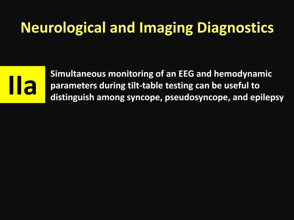
Neurological and Imaging Diagnostics
Simultaneous monitoring of an EEG and hemodynamic parameters during tilt-table testing can be useful to distinguish among syncope, pseudosyncope, and epilepsy
IIa

Neurological and Imaging Diagnostics
MRI and CT of the head are not recommended in the routine evaluation of patients with syncope in the absence of focal neurological findings or head injury that support further evaluation
III
Carotid artery imaging is not recommended in the routine evaluation of patients with syncope in the absence of focal neurological findings that support further evaluation
Routine recording of an EEG is not recommended in the evaluation of patients with syncope in the absence of specific neurological features suggestive of a seizure

Management of Cardiovascular Conditions

Rx the disease Bradycardia, SVT, AF, VA
I In patients with syncope associated with bradycardia, GDMT is recommended. In patients with syncope and SVT, GDMT is recommended In patients with AF, GDMT is recommended. In patients with syncope and VA, GDMT is recommended

Rx the disease Cardiomyopathy, VHD, HCM
I In patients with syncope associated with ischemic and nonischemic cardiomyopathy, GDMT is recommended In patients with syncope associated with valvular heart disease, GDMT is recommended In patients with syncope associated with HCM, GDMT is recommended

Arrhythmogenic Right Ventricular Cardiomyopathy (ARVC)
I ICD implantation is recommended in patients with ARVC who present with syncope and have a documented sustained VA
IIa ICD implantation is reasonable in patients with ARVC who present with syncope of suspected arrhythmic etiology
Syncope With Documented sustained VA
Syncope Without Documented sustained VA

Brugada Syndrome
ICD implantation is reasonable in patients with Brugada ECG pattern and syncope of suspected arrhythmic etiology IIa
IIb Invasive EPS may be considered in patients with Brugada ECG pattern and syncope of suspected arrhythmic etiology .
III ICD implantation is not recommended in patients with Brugada ECG pattern and reflex-mediated syncope in the absence of other risk factors

Short-QT Syndrome
ICD implantation may be considered in patients with short-QT pattern and syncope of suspected arrhythmic etiology.
IIb

Long-QT Syndrome
Beta-blocker therapy, in the absence of contraindications, is indicated as a first-line therapy in patients with LQTS and suspected arrhythmic syncope.
I
IIa ICD implantation is reasonable in patients with LQTS and suspected arrhythmic syncope who are on beta-blocker therapy or are intolerant to beta-blocker therapy
Left cardiac sympathetic denervation (LCSD) is reasonable in patients with LQTS and recurrent syncope of suspected arrhythmic mechanism who are intolerant to beta-blocker therapy or for whom beta-blocker therapy has failed

Catecholaminergic Polymorphic Ventricular Tachycardia (CPVT)
I Exercise restriction is recommended in patients with CPVT presenting with syncope of suspected arrhythmic etiology
Beta blockers lacking intrinsic sympathomimetic activity are recommended in patients with CPVT and stress-induced syncope.
Exercise restriction, Betablocker

Catecholaminergic Polymorphic Ventricular Tachycardia (CPVT)
Flecainide is reasonable in patients with CPVT who continue to have syncope of suspected VA despite beta-blocker therapy
IIa
ICD therapy is reasonable in patients with CPVT and a history of exercise or stress-induced syncope despite use of optimal medical therapy or LCSD
Flecainide, ICD despite OMT

Catecholaminergic Polymorphic Ventricular Tachycardia (CPVT)
In patients with CPVT who continue to experience syncope or VA, verapamil with or without beta-blocker therapy may be considered IIb
LCSD may be reasonable in patients with CPVT, syncope, and symptomatic VA despite optimal medical therapy
Verapamil, Left cardiac sympathetic denervation

Early Repolarization Pattern
IIb ICD implantation may be considered in patients with early repolarization pattern and suspected arrhythmic syncope in the presence of a family history of early repolarization pattern with cardiac arrest.
EPS should not be performed in patients with early repolarization pattern and history of syncope in the absence of other indications.
III

Management of VVS

Vasovagal Syncope
Patient education on the diagnosis and prognosis of VVS is recommended. I
IIa Physical counter-pressure maneuvers can be useful in patients with VVS who have a sufficiently long prodromal period
Midodrine is reasonable in patients with recurrent VVS with no history of hypertension, HF, or urinary retention

Vasovagal Syncope orthostatic training Fludrocortisone Betablocker in patients with >/= 42 year old Encorage increase salt and water intake reduce or withdra medications that cause hypotension SSRI Dual-chamber pacing might be reasonable in a select population ofpatients 40 years of age or older with recurrent VVS and prolonged spontaneous pauses
IIb

VVS
Education on diagnosis and prognosis (I)
Counter pressure maneuver (IIa)
Increase salt and water intake (IIb)
VVS recurs
Midodrine (IIa) Fludrocortisone(IIb) Betablocker (in > 42
yo )(IIb) Orthostatic training IIb)
SSRI (IIb) Dual chamber
pacemaker (IIb)
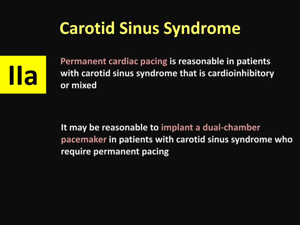
Carotid Sinus Syndrome
Permanent cardiac pacing is reasonable in patients with carotid sinus syndrome that is cardioinhibitory or mixed IIa
It may be reasonable to implant a dual-chamber pacemaker in patients with carotid sinus syndrome who require permanent pacing

Management of Neurogenic OH

Recommendations for Neurogenic OH
I Acute water ingestion is recommended in patients with syncope caused by neurogenic OH for occasional, temporary relief
Physical counter-pressure maneuvers Compression garments Midodrine Droxidopa Fludrocortisone Encorage salt and water intake
IIa
Pyridostigmine Octreotide IIb
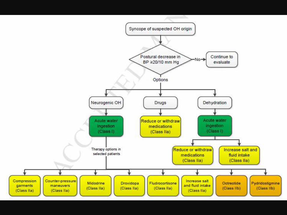

Dehydration and Drugs: Recommendations
I Fluid resuscitation via oral or intravenous bolus is recommended in patients with syncope due to acute dehydration
IIa Reducing or withdrawing medications that may cause hypotension can be beneficial in selected patients with syncope In selected patients with syncope due to dehydration, it is reasonable to encourage increased salt and fluid intake

Uncommon Conditions Associated With Syncope

Uncommon Conditions Associated With Syncope

Uncommon Conditions Associated With Syncope


Uncommon Conditions Associated With Syncope

Special population

Recommendations for Adult congenital heart disease
For evaluation of patients with ACHD and syncope, referral to a specialist with expertise in ACHD can be beneficial
IIa
EPS is reasonable in patients with moderate or severe ACHD and unexplained syncope

Recommendations for Geriatric Patients
For the assessment and management of older adults with syncope, a comprehensive approach in collaboration with an expert in geriatric care can be beneficial.
It is reasonable to consider syncope as a cause of nonaccidental falls in older adults
IIa
IIb

Driving syncope
It can be beneficial for healthcare providers managing patients with syncope to know the driving laws and restrictions in their regions and discuss implications with the patient.
IIa

Avo
idan
ce o
f Private
Drivin
g Afte
r an
Episo
de
of Syn
cop
e