· PDF file · 2017-06-07SUBMITTED TO H. N. B. GARHWAL UNIVERSITY . SRINAGAR...
109
SUBMITTED TO H. N. B. GARHWAL UNIVERSITY SRINAGAR GARHWAL (UTTARAKHAND) (A CENTRAL UNIVERSITY) FOR THE AWARD OF DEGREE OF DOCTOR OF PHILOSOPHY IN BIOTECHNOLOGY 2011 Under the Supervision of Prof. Anoop K. Dobriyal Head,Department of Zoology and Biotechnology H.N.B.Garhwal University Campus, Pauri Garhwal Submitted by Maryada Goyal Registration No. HNBGU/Res/ Studies on Isolation, Purification, Kinetic Characterisation and Immobilisation of α-amylase from some legumes of Garhwal Himalayan Region. DEPARTMENT OF ZOOLOGY AND BIOTECHNOLOGY, H.N.B. Garhwal (Central) University Campus Pauri, Garhwal-246001
Transcript of · PDF file · 2017-06-07SUBMITTED TO H. N. B. GARHWAL UNIVERSITY . SRINAGAR...

SUBMITTED TO H. N. B. GARHWAL UNIVERSITY SRINAGAR GARHWAL (UTTARAKHAND)
(A CENTRAL UNIVERSITY)
FOR THE AWARD OF DEGREE OF
DOCTOR OF PHILOSOPHY IN
BIOTECHNOLOGY 2011
Under the Supervision of
Prof. Anoop K. Dobriyal
Head,Department of Zoology and Biotechnology H.N.B.Garhwal University Campus, Pauri Garhwal
Submitted by
Maryada Goyal
Registration No. HNBGU/Res/
Studies on Isolation, Purification, Kinetic Characterisation and Immobilisation of α-amylase from some legumes of Garhwal Himalayan Region.
DEPARTMENT OF ZOOLOGY AND BIOTECHNOLOGY, H.N.B. Garhwal (Central) University Campus
Pauri, Garhwal-246001
HEMWATI NANDAN BAHUGUNA GARHWAL UNIVERSITY
SRINAGAR-GARHWAL, 246174 UTTARAKHAND, INDIA
Prof. Anoop K. Dobriyal Head, FZSI,FSB,FIFSI,FIAES,FAEB, Department of Zoology and Biotechnology FMAFS,FANSF, Paul Harris Fellow, HNB Garhwal Central University, Campus Pauri, Young Scientist Award-AFS, IB(1987) Pauri Garhwal-246001 (Uttarakhand) Group Study Exchange Award USA (1992-93) e-mail: [email protected] Scientist of Year Award-NESA (2002) Phone: 01368-222925mob. 9412960687 ASEA FISHERIES Award-India (2006) RASHTRIYA SHIKSHA SAMMAN AWARD-CEGR (2009) Ref………………… Date…………………
Certificate
This is to certify that the thesis entitled “Studies on Isolation, Purification, Kinetic
Characterisation, and Immobilisation of α-amylase from some legumes of
Garhwal Himalayan Region” submitted by Maryada Goyal Reg. No. HNBGU/Res/
to the school of Life sciences HNB Garhwal Central University ,Srinagar (Garhwal),
Uttarakhand for the award of degree of Doctor of Philosophy in Biotechnology,
carries out a record of original research by her and has not been submitted in full or
in part for any diploma, degree or associate ship in this or any other University. It is
also certified that she has put in more than 200 days work in biotechnology
laboratory, Department of Zoology and Biotechnology, HNB Garhwal Central
University, Campus Pauri, Pauri Garhwal (Uttarakhand), India. I wish her every
success in life.
(Prof. A.K. Dobriyal)
Acknowledgements
I shall first of all pay reverence towards Almighty God without whose grace
and benevolence it would have not been possible for me to accomplish this task.
Thou not only motivated me to start this work, but also enlightened my mind to
achieve my goals.
My sincere regards to my esteemed guide, Prof. Anoop K. Dobriyal (Head,
Department of Zoology and Biotechnology, HNB Garhwal Central University, Pauri
Campus, Pauri) for his solemn efforts, guidance, constructive criticism, constant
support, and guidance throughout my thesis work.
I cannot forget to quote the reference of Dr. R. M. Saxena, Ex- Head,
Department of Zoology, DAV (PG) College, Dehradun, whose initial ideas and help
paved my way for start-up of this present work.
It is not enough to convey my thankfulness to Dr. M.S. Bisht, Associate
Professor, Dr. C.B. Kotnala, Dr. V. P. Balodi, Dr. A. Thapliyal, and Dr. H.K. Joshi,
Assistant Professors, at HNB Garhwal Central University, Pauri Campus, Pauri for
providing necessary help during the course of progress of thesis work.
I cannot forget the pains taken by my parents from start till end of this work.
Their constant guidance and moral support cannot be expressed in words. The
meticulous care of my sister Divya Goyal is notwithstanding in this research work. I
would also heartily express my sincere regards to my husband and family members
in supporting me for accomplishing this work.
Last but not the least, I am rich with friends whose perspiration, criticism,
affection and care is necessary for each and every individual. There indebtedness
cannot be mentioned in words.
(Maryada Goyal Garg)
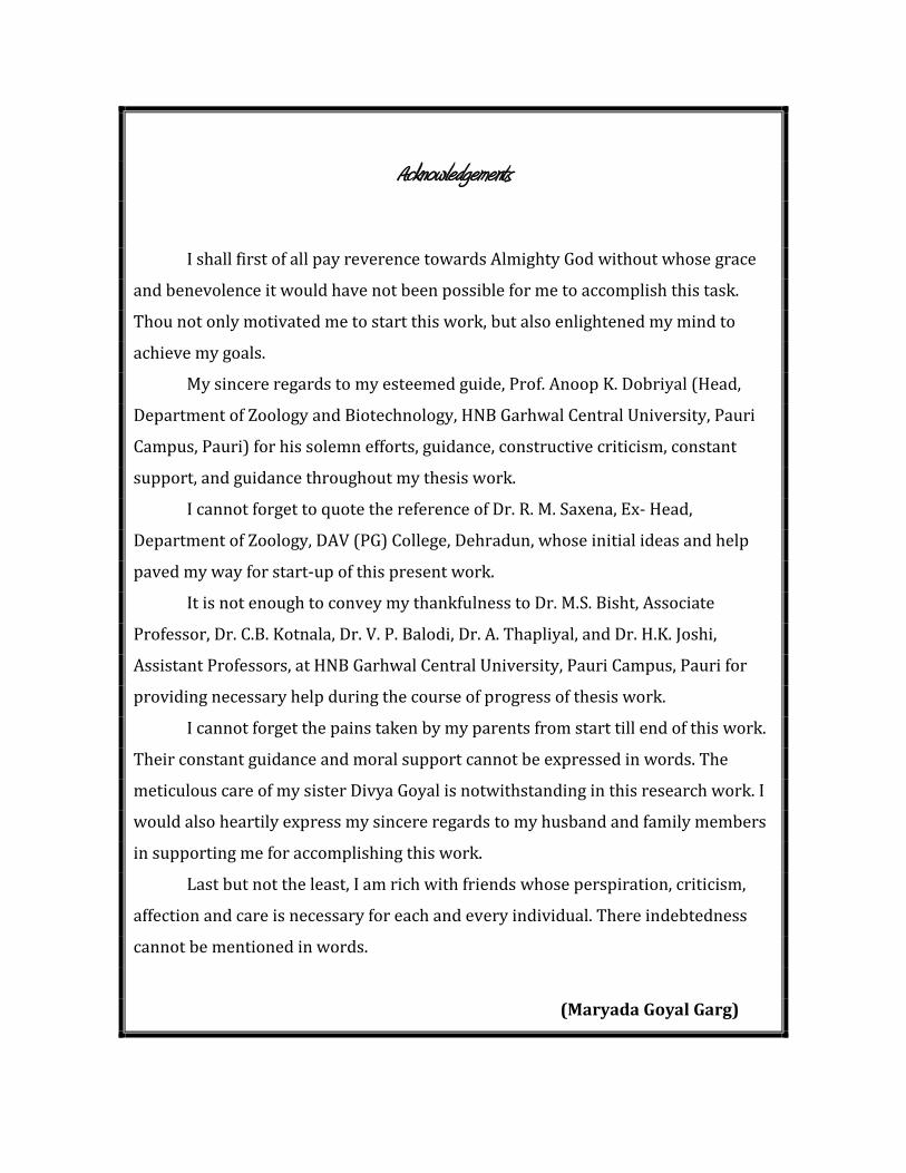
ABBREVIATIONS
• α : alpha
• A° : Angstrom
• CaCl2 :
• CCM : Cellulose coated magnetite
Calcium chloride
• CNBr : Cyanogen bromide
• CTMAB : Cetyl- tri-methyl ammonium bromide
• DEAE : Di-ethyl-amino-ethyl
• DNS : di-nitro salicylic acid
• HFD : High fat diet
• Hrs. : Hours
• I.U. : International Unit
• Km
• KDa : Kilodaltons.
: Michaelis-Menton’s constant
• mM : Mili molar
• Min : Minute
• Nm : nano meter.
• O.D. : Optical Density
• Ppm : parts per million
• Rpm : Revolutions per minute.
• Pp : pages
• Sec : Second
• Spp : species
• So :
• UV-Vis : Ultra violet-visible
Initial starch substrate concentration
• Vo
• V
: Initial velocity
max
: Maximum velocity

Contents
Page No. Chapter-1 INTRODUCTION……………………………………………………………….1-12 Origin of the current problem……………………………………………………1 Brief introduction about the enzyme………………………………………..........2 Sources……………………………………………………………………….......3
Dolichos biflorus……………………..……………………………………….5 Phaseolus vulgaris HUR 15…………………………………………………8 Brief Introduction about the methodology used…………..…………………....11 Variation of this work from earlier work done……………………………….…12 Importance to science and society………………………………………..……..12 Chapter-2 REVIEW OF LITERATURE…………………………………………..………13-25 Chapter-3 MATERIALS AND METHODS……………………………………………….26-38 Sampling materials and sampling sites……………………………...………….26 Selection of material…………………………………………..………………..26 Initial treatment of legumes…………………………………………………….27 Sprouting of seedlings…………………………………………..………………27 Initial studies of sprouted seedlings…………………………...………………..28 Germination percentage………………………………...……………….28 Seedling length……………………………………….………………….28 Enzyme extraction/isolation…………………………………..…………………29 Enzyme assay……………………………………………………………………29 Preparation of standard graph………………………………...…………………30 Enzyme assay………………………………………………...………………….31 For crude Dolichos biflorus extract……………………..……………….31 For crude Phaseolus vulgaris HUR 15 extract…………………………..31 Purification………………………………………………………………………32 Ultrafiltration…………………………………………………………….32 Column chromatography…………………….…………………………..32
Kinetic Characterisation……………………………….…………………………33 Optimum pH…………………………………..…………………………33 Optimum Temperature…………………………..……………………….34 Thermal Stability……………………………..………………………….35 Substrate Concentration………………………………………………….35 Time…………………………………………….………………………..35 Vmax and Km
Immobilisation…………………………………………………………………...36 ...............................................................................................36
Preparation of Sodium alginate……………………………………….….37
Calcium Chloride solution……………………………………...………..37 Preparation of beads…………………………………………...…………37 Kinetic Characterisation of Immobilised Enzyme………………….……38
Comparison ………………………………………………………..…….38 Chapter-4 RESULTS AND DISCUSSION……………………………………………...39-73
Initial studies……………………………………..………………………39 Germination percentage…………………...……………………..39 Seedling length……………………………..…………………….42 Preparation of standard graph………….……………….………………..42 Isolation………………………………………………….……………….43 Purification of crude extracts…………………………………………….45 Ultrafiltration………………………………………...…………..45 Dolichos biflorus…………………………………….………..45 Phaseolus vulgaris HUR 15……………………...………45 Ion-exchange chromatography………………………………..….46 Kinetic characterization………………………………………………….48
Optimum pH……………………………………………………..48 Optimum temperature and thermal stability…………………..…52 Km and Vmax…………………………………………………....56 Time…………………………………………………...…………59
Yield………………………………………………………………...……61 Immobilisation………………………………………………….………..62
Kinetic characteristics of immobilized enzyme………………….62 Comparison……………………………………………………..………..68 Conclusion……………………………………………………………….72 Future perspectives of the study…………………………………...…….73
Chapter-5 SUMMARY………………………………………………………………….74-83 Introduction and methodology………………………………………..….74 Results and Discussion…………………………………………………..77 Conclusion…………………………………………………………….....83 Chapter-6
REFERENCES……………………………………………………………….84-99 Annexures:- 1. 2.
List of tables

Table No. Page No.
1. Standard graph via DNS method………………………………………………...42
2. Ion exchange chromatography results for Dolichos biflorus enzyme…………...46
3. Ion exchange chromatography results for Phaseolus vulgaris HUR
15 enzyme………………………………………………………………………..47
4. Table showing optimum pH of crude kulath extract…………………………….48
5. Table showing optimum pH of purified kulath extract…………………………..49
6. Table showing pH optima of white rajma extract………………………………..50
7. Table showing optimum pH of purified white rajma extract…………………….51
8. Table showing optimum temperature of purified enzyme in kulath………..……52
9. Table showing thermal stability of purified kulath enzyme………………….….54
10. Table showing optimum temperature of purified enzyme of white rajma……….54
11. Table showing thermal stability of white rajma enzyme………………………...56
12. Table showing So against Vo (kulath)…………………………………………...57
13. Table showing So against Vo (white rajma)……………………………………..58
14. Table showing optimum time of α-amylase of kulath…………………………...59
15. Table showing optimum time of α-amylase of white rajma……………………..60
16. Table showing comparison between enzyme activities of crude and immobilized
kulath enzyme at various pH concentrations…………………………………….62
17. Table showing comparison between enzyme activities of crude and immobilized
kulath enzyme at various temperatures…………………………………………..65
18. Table showing comparison between enzyme activities of crude and immobilized
white rajma enzyme at various pH concentrations……………………………....66
19. Table showing comparison between enzyme activities of crude and immobilized
white rajma enzyme at various temperatures…………………………………….67
20. Table showing comparison between temperature and pH for plant α-amylase….69
21. Table showing comparison between temperature and pH for bacterial α-
amylase…………………………………………………………………………..70
22. Molecular weight of α-amylase from bacterial sources………………………….70
23. Molecular weight of α-amylase from plant sources……………………………...71
24. Comparison of Km
values for α-amylase from plant and bacterial sources……...71
List of FIGURES.
Figure No. Page No.
1. Plant of Dolichos biflorus showing pods………………………………………….7
2. Seeds of Dolichos biflorus………………………………………………………...7
3. Seeds of Phaseolus vulgaris HUR 15……………………………………………10

4. Plant of Phaseolus vulgaris HUR 15 showing flowers and pods………………..10
5. 5 day old sprouts of white rajma with secondary roots (Phaseolus vulgaris HUR
15)………………………………………………………………………………..40
6. 1-5 day seedling length of white rajma…………………………………………..40
7. 5 day seedling length of kulath…………………………………………………..41
8. Crude extract of kulath…………………………………………………………...41
9. 2 day old sprouts of kulath……………………………………………………….41
10. Graph showing O.D. values against varying glucose concentrations……………43
11. Standard Graph also showing enzyme activity of crude and white rajma……….44
12. Graph showing optimum pH of crude kulath extract…………………………….49
13. Graph showing optimum pH of purified kulath extract………………………….50
14. Graph showing pH optima of white rajma extract……………………………….51
15. Graph showing optimum pH of purified white rajma extract……………………52
16. Graph showing optimum temperature of purified enzyme in kulath…………….53
17. Graph showing optimum temperature of purified enzyme of white rajma………55
18. Graph showing Km and Vmax
19. Graph showing K
of α-amylase of
kulath…………………………….57
m and Vmax
20. Graph showing optimum time of α-amylase of kulath…………………………..59
of white rajma…………………………………….58
21. Graph showing optimum time of α-amylase of white rajma…………………….60
22. Shiny yellow brown beads of crude extract of Dolichos biflorus..........................63
23. Shiny beads of ultrafiltered extract of Dolichos biflorus………………………...63
24. White beads of Phaseolus vulgaris HUR 15 (white rajma)……………………...63
25. Graph showing comparison between enzyme activities of crude and immobilized
kulath enzyme at various pH concentrations…………………………………….62
26. Graph showing comparison between enzyme activities of crude and immobilized
kulath enzyme at various temperatures…………………………………………..63
27. Graph showing comparison between enzyme activities of crude and immobilized
white rajma enzyme at various pH concentrations………………………………64
28. Graph showing comparison between enzyme activities of crude and immobilized
white rajma enzyme at various temperatures…………………………………….65


Chapter I
Introduction
The Garhwal Himalayan Region of Uttarakhand has been largely known for its
genetic and ecological diversity since times immemorial. Ever since, the state of
Uttarakhand came into being on 9
Origin of the current problem:
th November 2000 as the 27th
The Dicots dominate with 3493 species under 1163 genera and 182 families.
Among the dicots, family Asteraceae has the largest number of genera, i.e., 125, followed
by Fabaceae which has 76 genera. It is interesting to note that the family Fabaceae has
the second largest number of species i.e., 327. The flowering plants provide wide range
of useful products ranging from timber, medicine, food, vegetable oils, gums, resins and
spices etc. Among the edible plants, pulses, also known as grain legumes, are second only
to cereals as a source of human food. These account for nearly 20 species all belonging to
the family Fabaceae. Major pulse crops are Cajanus cajan, Cicer arietinum, Dolichos
state of Indian Republic,
the rich angiospermic flora of this state became a topic of investigation for large number
of biotechnologists. This state has 4700 species under 1503 genera and 213 families, thus
accounting roughly for 27% of total Indian angiospermic flora. Interestingly the state
hosts approximately 50% of genera of Indian flowering plants.
spp., Phaseolus spp., Vigna spp., Glycine max, Lens culinaris, Pisum sativum and Vicia
faba (Uniyal et.al, 2007).
Biotechnological practices in this area are not new to people. Biotechnologists of
this area are working largely on subjects related to biotechnology like molecular biology,
enzymology and tissue culture, etc. It is interesting to note that despite the large number
of products; timber, medicine, food, vegetable oils, gums, resins and spices; exploited
from these multi-faceted pulse crops, the enzymatic stamina of these crops has not been
explored yet. Despite many biotechnological advances in this area, the scientists were
least interested regarding the idea of enzyme extraction from pulse crops. Therefore the
present research on “Studies on isolation, purification, kinetic characterization and
immobilization of α-amylase from some legumes of Garhwal Himalayan Region” has
been carried out.
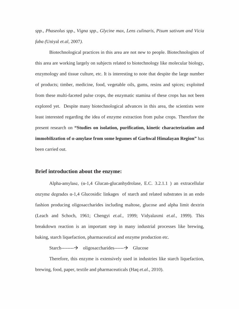
Brief introduction about the enzyme:
Alpha-amylase, (α-1,4 Glucan-glucanhydrolase, E.C. 3.2.1.1 ) an extracellular
enzyme degrades α-1,4 Glucosidic linkages of starch and related substrates in an endo
fashion producing oligosaccharides including maltose, glucose and alpha limit dextrin
(Leach and Schoch, 1961; Chengyi et.al., 1999; Vidyalaxmi et.al., 1999). This
breakdown reaction is an important step in many industrial processes like brewing,
baking, starch liquefaction, pharmaceutical and enzyme production etc.
Starch-------- oligosaccharides------ Glucose
Therefore, this enzyme is extensively used in industries like starch liquefaction,
brewing, food, paper, textile and pharmaceuticals (Haq et.al., 2010).

According to a survey, the European food enzyme market was worth $ 200
millions in 2004. Although it was contributed by starch processing, sugar processing,
bakery and dairies, but the highest growth rate was witnessed in the nutrition and dietary
supplements market. Here, amylases are said to contribute 25% of the enzymes. These
uses have placed greater stress on increasing indigenous α-amylase production and search
for more efficient sources of the enzyme.
Among various extracellular enzymes, it ranks first in terms of commercial
exploitation. Spectrum of application of α-amylase has widened in many sectors such as
clinical, medicinal and analytical chemistry. Besides their use in starch saccharification, it
was also found applicable in baking, brewing, detergent, textile, paper and distilling
industry.
Sources:
Large numbers of sources have been recognized by scientists and researches all
around the world for extraction of α-amylase. These sources include micro-organisms,
plants and animals. The enzyme can be isolated from micro-organisms (Morgan et.al.,
1981; Farez-Vidal et.al., 1992; Abe et.al., 1994; Aguloglu et.al., 2000; Primarini and
Ohta, 2000 and Lin et.al., 2002), human saliva (Shainkin and Birk, 1966; Mayo and
Carlson, 1974 and Goyal, 2005); and porcine pancreas (Strumeyer et.al., 1988) etc.
A close investigation of various enzyme firms shows that most of the α-amylase
production is done from genetically modified micro-organisms. Most of the enzyme firms
are dependent on genetically modified sources of bacteria like Bacillus
amyloliquefaciens, Bacillus subtilis and Bacillus caldolyticus. Among plants, work has
been done on sorghum, broad beans, coconut oil cake, cereals and mung beans.
From literature survey it was revealed that leguminous plants also contain α-
amylase (Beers and Duke, 1988; Greenwood et.al., 1965a and b; Tripathi et.al., 2007;
Kumari et.al., 2010). The Garhwal Himalayan region is a rich source of various
economic important crops. This region also exclusively produces some legumes such as
kulath, tohar, rajma, mung etc. which can be a great source of α-amylase in future. No
work has been reported regarding α-amylase of legumes of this region. Therefore, it was
found useful to investigate the legumes found in Garhwal Himalayan region for α-
amylase.
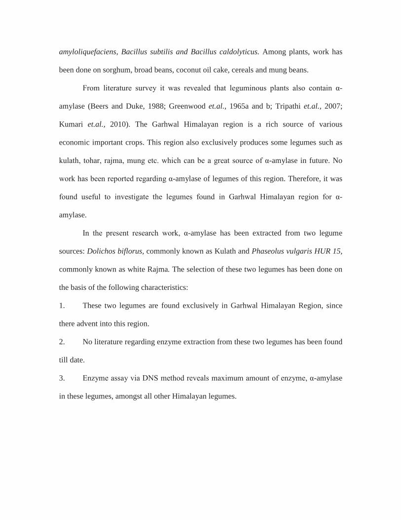
In the present research work, α-amylase has been extracted from two legume
sources: Dolichos biflorus, commonly known as Kulath and Phaseolus vulgaris HUR 15,
commonly known as white Rajma. The selection of these two legumes has been done on
the basis of the following characteristics:
1. These two legumes are found exclusively in Garhwal Himalayan Region, since
there advent into this region.
2. No literature regarding enzyme extraction from these two legumes has been found
till date.
3. Enzyme assay via DNS method reveals maximum amount of enzyme, α-amylase
in these legumes, amongst all other Himalayan legumes.

Dolichos biflorus (kulath):
Synonyms: Horse gram, Kulthee, Gahat
Taxonomic lineage:
Kingdom: Plantae
Phylum: Magnoliophyta
Class: Magnoliopsida
Order: Fabales
Family: Fabaceae
Genus: Dolichos
Species: biflorus
It is a branched or trailing annual with small trifoliate leaves, and very wide
climbing slender stem. On maturity, it gives narrow, flat, curved pods. The pods contain
5-7 flattened ellipsoid seeds, 1/8” to ¼” long.
Archaeological investigations have revealed the use of Kulath as food around
2000 BC. Commonly known as Horse Gram, this twining herb of old world tropics is
cultivated for its seed in tropical and sub-tropical regions of the world. In India, it is
found up to 1500 MSL (mean sea level). Interestingly, the entire Garhwal himalayan
region is well suited for the production of this legume crop.
In experimental studies, the seeds of Dolichos biflorus have shown to be effective
in preventing the deposition of stone material in urinary bladder of rats (Kumar et.al.,
1981). The extracts of the seeds of Dolichos biflorus have shown inhibiting effect on the
formation of phosphate precipitate and has shown better potential in preventing the
formation of calcium precipitate (Garimella et.al., 2001). It has also been reported to

lower lipids contents in rats and high fat diet (HFD)-induced oxidative stress in rabbits.
Singh and Kumar (1973) have opined that the seeds of Dolichos biflorus are effective in
dissolving kidney stones in human patients. Alpha-amylase extraction from this pulse
crop revealed 27.7IU/ml of the enzyme (Garg and Dobriyal, 2011).
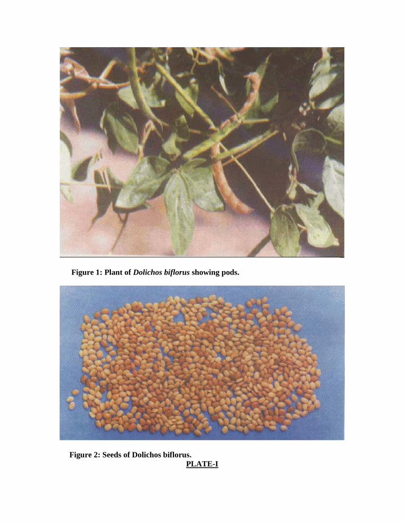
Figure 1: Plant of Dolichos biflorus showing pods.
Figure 2: Seeds of Dolichos biflorus.
PLATE-I
Phaseolus vulgaris var HUR15 (white rajma):
Taxonomic lineage:
Synonyms: Kidney bean, French bean
Kingdom: Plantae
Phylum: Magnoliophyta
Class: Magnoliopsida
Order: Fabales
Family: Papilionaceae
Genus: Phaseolus
Species: vulgaris
First developed at BHU, Varanasi, this Rabi crop was first identified in 1984. It
has been cultivated for its seeds in UP, Uttarakhand, Bihar, MP and Maharashtra. Four
varieties of this crop are identified: out of which white Rajma i.e., the HUR15 variety is
mainly grown in tropical hilly regions of Uttarakhand as a Rabi crop. Rajma fits well in
existing cropping system. It can be successfully grown after all kharif crops maturing by
middle of October and spring crops may follow thereafter.
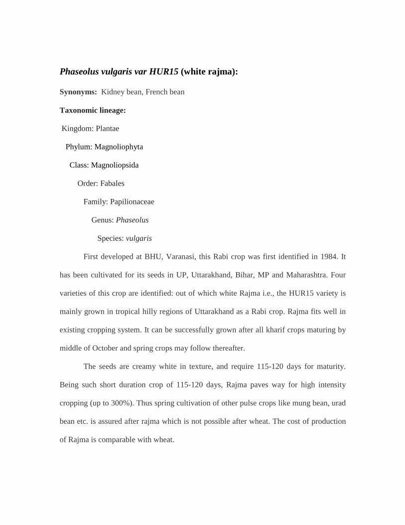
The seeds are creamy white in texture, and require 115-120 days for maturity.
Being such short duration crop of 115-120 days, Rajma paves way for high intensity
cropping (up to 300%). Thus spring cultivation of other pulse crops like mung bean, urad
bean etc. is assured after rajma which is not possible after wheat. The cost of production
of Rajma is comparable with wheat.

Unlike other pulse crops, Rajma is inefficient in symbiotic nitrogen fixation and
therefore need heavy dose of nitrogen fertilizer. Seeds should be treated with fungicides
to control seed borne diseases. The recommended fungicides are bavistin, thiram, and
captan. Rajma needs fine seed bed and adequate moisture for proper germination of
seeds. It has been estimated that under good management conditions the Rajma
production is 25 to 30 quintals /ha (Ali and Lal, 1991).
In the present study α-amylase extraction from the grains has been done and the
results obtained were remarkable.
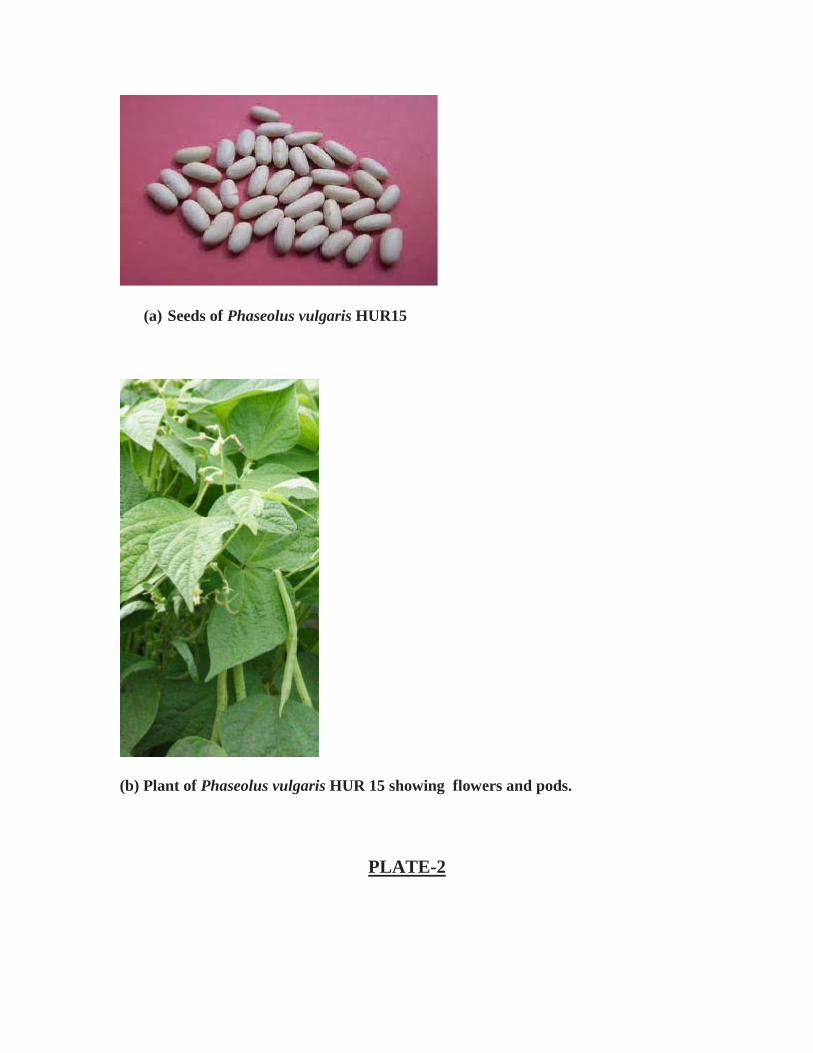
(a) Seeds of Phaseolus vulgaris HUR15
(b) Plant of Phaseolus vulgaris HUR 15 showing flowers and pods.
PLATE-2

Brief introduction about the methodology used:
Isolation of enzyme from legume source is accompanied by initial crushing of the
presoaked, imbibed seed using mortar-pestle. The crushed seed is then passed through
four layers of cheese cloth. Then filtration is done in refrigerated centrifuge at 13000 rpm
for 15 min. The supernatant is collected and pellet is discarded. This supernatant is
considered as crude enzyme extract and can be stored at 4˚C.
Purification of the enzyme is then done from the stored supernatant. Two-fold
purification is done: Ultrafiltration and ion-exchange chromatography. Although
ultrafiltration is generally employed for concentration of very dilute enzymes, but in the
present study it has been utilized for purification of low molecular weight amylases (Garg
and Dobriyal, 2011).
The crude extract of the enzyme is passed through ultrafiltration membrane of
10KDa and 30KDa sizes. Then enzyme activity is measured by Miller’s DNS method
(Miller, 1959) in both the ultrafiltrate and the concentrated enzyme fraction. Further
purification of the ultrafiltrate is performed by using ion exchange chromatography. And
DEAE cellulose was used for column packing.
Kinetic characterization on the basis of following parameters; optimum pH,
optimum temperature, substrate concentration, thermal stability, time, Michaelis
Menton’s constant (Km) and Maximum velocity (Vmax) is then performed. These kinetic
characteristics are calculated via enzyme assays using DNS method in the ion exchange
fractions of both the ultrafiltrate; for low molecular weight amylases; and dialysed
sample; for large molecular weight amylases.
After kinetic characterization, the immobilization of this purified enzyme is also
done and the enhanced characteristics are studied. A close comparison is made between
the characteristics of immobilized and free enzyme.
It was interesting to note that the enzyme extracted from these pulse crops was as
better as the traditional enzyme extracted from bacterial sources. Results prove that plant
enzymes showed nearly same characteristics just like bacterial enzymes. Thus, besides
finding out new, low cost sources of α-amylase, we could also deduce that these new
sources can be made usable on industrial scale.
Variation of this work from earlier work done:
Earlier researchers have extracted α-amylase from all sources including plants,
bacteria, fungi, and animals. But only genetically modified bacteria have been applied to
commercial usage. The present research focuses on extraction of enzyme from
leguminous source, which is a totally new concept.
Importance to science and society:
The increasing demand of α-amylase in food, pharmaceutical and starch industry
have led researchers to think about some cheap as well as newer sources for extraction of
the enzyme. In this context, this research would prove to be stepping stone towards this
approach.

CHAPTER-2
REVIEW OF LITERATURE
Alpha- amylase is ubiquitous in bacteria, fungi, plants and animals. Literature
survey reveals that work on α-amylase started in the mid of 20th
Further studies on human amylases included human pancreas. In this context,
Ferey-Roux et.al., (1998) studied human pancreatic α-amylase isoforms. Hokari et.al.,
century. In this context,
work of Shainkin and Birk (1966) is worth mentioning, who isolated pure α-amylase
from human saliva. After that Mayo and Carlson (1974) isolated four α-amylase
isozymes from human sub-mandibular saliva by gel-filtration and iso-electric focusing
and studied there different properties. This indicates that human beings and their
digestive process was the main source of study of this enzyme. Now it is a well known
fact that the enzyme α-amylase is present in human saliva and is responsible for
degradation of starch into glucose. This glucose is further utilized in the human body for
the life controlling cycle, glycolysis (discovered by Embden, Meyerhoff and Parnas in
1940) which further ends up into Kreb’s cycle. These two cycles are responsible for
formation of ATP (Adenosine tri-phosphate) in body which is utilized for providing
energy to all other cells and cycles of the human body.
(2002) performed a restriction endonuclease assay for expression of human α-amylase
isozymes.
As it is known that all animal cells are same, and glycolysis and Kreb’s cycle
occurs in all animal cells, therefore it became necessary to investigate the presence of α-
amylase enzyme in other animal cells also. In the meanwhile, various researchers isolated
and characterized the enzyme from other animal sources as well. In this context,
Strumeyer et.al., (1988) studied the population distribution of isozymes of α-amylase in
porcine pancreas. Perera and Hoover (1998) studied the reactivity of porcine pancreatic
α-amylase towards native, defatted and heat-moisture treated potato starches before and
after hydroxypropylation. It was found that among animals, α-amylase has been studied
mostly in case of porcine, almost till the beginning of 21st
Further, among animals Oosthuizen et.al., (1992) performed the isolation and
partial characterization of α-amylase from pancreas of Ostrich (Struthio camelus).
Oosthuizen et.al., (1994) also performed purification and characterization of pancreatic
isozymes of Ostrich α-amylase. They discovered four isozymes. Nakatani and Kobayashi
(1996) studied the enzymatic properties of α-amylase from digestive tube of sea urchin,
Strongylocentrotus nudas. Benkel et.al., (1997) perfomed cloning and expression of an α-
amylase gene in chicken. Strobl et.al., (1998) studied crystal structure of yellow meal
worm α-amylase at 1.64 A resolution. Moreau et.al., (2001) studied isolation, structural
studies and inhibition kinetics of α-amylases from two tilapias Oreochromis niloticus and
Sarotherodon melanotheron. They used ammonium sulfate precipitation, affinity
chromatography and chromato-focusing procedures for purification of the enzyme from
the intestinal cavity of these two fishes. Mohamed (2004) studied purification and
century.
characterization of α-amylase from the infective juveniles of the nematode
Heterorhabditis bacteriophora. Lombrana et.al., (2005) discovered two forms of α-
amylase in mantle tissue of Mytilus galloprovincialis. Th is was th e first case when α-
amylase activity was discovered in a non-digestive tissue. Wu et.al., (2006) discovered
amylase gene from Ophiostoma floccosum. Similarly α-amylase has also been discovered
in other mammals and insects by Silva et.al., (2009). Silva et.al., (2009) studied
inhibitory action of Cerrado plants against α-amylase of mammals and insects. The insect
species studied were Zabrotes subfasciatus and Acanthoscelides obtectus while the
mammalian sample tested was human saliva. Thus it can be said that till date there are
more than fifty animals in which the enzyme has been discovered, purified and studied
kinetically as well as biochemically, including human beings. Further, it is interesting to
note that there has been no animal species till date whose α-amylase has been
immobilized.
For industrial purposes most of the workers have proposed to isolate the enzyme α-
amylase from micro-organisms like bacteria, fungi and also the genetically modified
micro-organisms. Amongst micro-organisms, most of the work regarding isolation,
purification, kinetic characterization and immobilization of the enzyme α-amylase has
been done in bacteria. Various properties like size, thermal stability, optimum pH, Km,
Vmax etc. have been studied by different workers. Research on bacterial α-amylase started
very early in the mid of 20th century, and by the mid of 20th century it was realized that
on an industrial scale, strains of Bacillus are best suited for the production of enzyme.
This is so because the enzyme α-amylase exists in both membrane associated and
extracellular forms in Bacillus sp. and in other bacteria (Farez-Vidal et.al., 1992). The α-
amylase synthesized by Bacillus subtilis (pAMY430) which is a wild type gene is almost
completely secreted from the cell by the aid of various amino acids in media. Cysteine in
media shows highest secretion of α-amylase (Aguloglu et.al., 2000).
A close investigation of bacterial amylases indicated that more than 20 species of
the genus Bacillus have been investigated for the enzyme α-amylase on an industrial
scale. These include wild type as well as genetically modified bacteria. Among these the
work of Welker and Campbell (1967) is quite interesting. They performed experiments
on comparison of α-amylase of Bacillus subtilis and Bacillus amyloliquefaciens. Both
these strains have been considered to be commercial α-amylase concentrates. Yutani
(1973) studied molecular weight of a thermostable α-amylase from Bacillus
stearothermophilus. Heinen and Lauwers (1975) worked on molecular size of
extracellular amylase produced from intact cells and protoplasts of Bacillus caldolyticus.
This enzyme had a typical property of disintegrating into smaller units of less than 10,000
when subjected to ultrafiltration. Ivanova and Dobriva (1994) studied catalytic properties
of immobilized purified thermostable α-amylase from Bacillus licheniformis (44MB82-
A). Raabe and Knorr (1996) studied the kinetics of starch hydrolysis in Bacillus
amyloliquefaciens α-amylase under high hydrostatic pressure.
With the beginning of 21st century, research work on bacterial strains was further
strengthened with increase in demand of enzyme. These strains mostly included
genetically modified strains of the genus Bacillus. In 2001, Hagihara et.al., produced a
novel α-amylase that is highly resistant to chelating reagents and chemical oxidants from
the alkaliphilic isolate, Bacillus KSM-K38. Further, Jin et.al., (2001) studied and
modeled extracellular thermostable α-amylase from aerobic Bacillus sp. JF strain. Lin

et.al., (2002) performed the isolation of a recombinant Bacillus sp. TS-23 α-amylase by
adsorption elution on raw starch. Das et.al., 2004 performed purification and biochemical
characterization of a thermostable, alkaliphilic, extracellular α-amylase from Bacillus
subtilis DM-03, a strain isolated from traditional fermented food of India. This traditional
fermented food was a starter culture used for the production of alcohol by local Assam
tribes. The enzyme was purified by ion-exchange, gel filtration and reverse phase HPLC.
Bernhardsdotter et.al., (2005) studied the enzymatic properties of an alkaline chelator
resistant α-amylase from an alkaliphilic Bacillus sp. isolate L1711. This Bacillus isolate
was selected from 13 soda lakes isolates. Anto et.al., (2006) performed α-amylase
production by Bacillus cereus MTCC 1305 using solid state fermentation, while
Murakami et.al., (2008) performed purification and characterization of five alkaline
thermotolerant, and maltotetraose producing α-amylases from Bacillus halodurans MS-2-
5. Gangadharan et.al., (2009) used submerged fermentation for isolation of the enzyme
from Bacillus amyloliquefaciens. They further utilized ion-exchange chromatography for
purification of raw-starch digesting α-amylase. Riaz et.al., (2009) performed
immobilization of a thermostable α-amylase on Calcium alginate beads from Bacillus
subtilis KIBGE-HAR. Haq et.al., (2010) performed production of α-amylase from a
randomly induced mutant strain of Bacillus amyloliquefaciens. Femi-Ola and Olowe
(2011) performed characterization of α-amylase from Bacillus subtilis BS5 isolated from
hindgut of wood eating termite Amitermes evuncifer Silvestri.
Today it is the Bacillus spp. whose strain is being used world wide by industry for
production of this industrially important enzyme. It is interesting to note that various
researchers have used various methods for purification of the enzyme from time to time.
Brena et.al., (1996) studied a range of chromatographic methods for separation of
amylases from complex extracts, including separation of isozymes. Till date, almost all
types of chromatographic methods have been utilized for purification of extracellular/
membrane bound α-amylase from various bacterial isolates.
Besides Bacillus, work on other bacterial genus has also been done from time to
time. In this regard, Freer (1993) performed purification and characterization of
extracellular α-amylase from Streptococcus bovis JB1. Abe et.al., (1994) performed
purification and characterization of periplasmic α-amylase from Xanthomonas campestris
K-11151. This α-amylase isolated by Abe et.al., was of new type as it showed the
activities of cyclodextrinase and neopullanase also along with α-amylase. In 1977,
Glymph and Stutzenberger performed production, purification and characterization of α-
amylase from Thermomonospora curvata. Further, Primarini and Ohta (2000) performed
purification and characterization of some raw starch digesting amylases of Streptomyces
praecox NA- 273 and Streptomyces aureofaciens. Talamond et.al., (2002) studied α-
amylases of Lactobacillus species. They performed isolation, characterization and
inhibition by acarbose of α-amylase from Lb. fermentum. They also compared the α-
amylase of Lb. fermentum with Lb. manihotivorans and Lb. plantarum. Ezeji and Bahl
(2006) studied purification, characterization and synergistic action of phytate-resistant α-
amylase and α-glucosidase from Geobacillus thermodenitrificans HRO10. Shafiei et.al.,
(2010) performed purification and biochemical characterization of a novel SDS and
surfactant stable, raw starch digesting, and halophilic α-amylase from a moderately
halophilic bacterium, Nesterenkonia sp. strain F. Mollania et.al., (2010) performed

purification and characterization of a thermostable phytate- resistant α-amylase from
Geobacillus sp. LH8.
Work on fungal strains regarding isolation of α-amylase enzyme started a bit later
than bacteria. A significant contribution in this field is made by Ramachandran et.al.,
(2004) who discovered that coconut oil cake is a potent raw material for production of α-
amylase. They used solid state fermentation for production of α amylase using fungal
culture of Aspergillus oryzae.
Some workers have isolated and characterized α-amylase from plant sources as
well. Among plants also, isolation, purification, kinetic characterization and
immobilization has been done from a variety of species. The earliest known research
among plant amylases is from the cereal family, done by Dube and Nordin (1961). They
purified α-amylase in good yield from Sorghum (also known as jowar) malt by
procedures based on ultracentrifugation, salt fractionation and adsorption on starch
granules and studied their properties as well. Greenwood et.al., (1965a) studied the
properties of purified enzyme from broad beans Vicia faba belonging to the family
Fabaceae/leguminoceae. As a result of this study, characteristic α-amylolytic degradation
patterns for the action of the enzyme on various substrates were obtained. In the same
year, Greenwood et.al., (1965b) performed studies on purification and properties of
starch-degrading enzymes of one more leguminoseae member, soyabean (Glycine max).
Beers and Duke (1988) observed that the enzyme α-amylase is present in apoplast of pea
(Pisum sativum L) stems. They found that virtually all α-amylase activity of pea is
located in stems. Kumari et.al., (2010) worked on α-amylase from germinating soybean
(Glycine max) seeds. Along with purification and characterization, they also studied the
sequence similarity of conserved and catalytic amino acid residues in soybean. Thus we
can say that the earliest research in plant amylases was from the grass family, Poaceae
and legume family, Leguminoseae also known as Fabaceae.
With the classification and evolution of α-amylase genes of plants (Huang et.al.,
1992), the studies on isolation and characterization of α-amylase from plant sources
speeded up. Warner and Knutson (1991) performed isolation of α-amylase and other
starch-degrading enzymes from endosperm of germinating maize (family Poaceae). Their
scientific work was significant improvement over use of protein precipitation and affinity
chromatography for purification of α-amylase. Later, Rashad et.al., (1995) experimented
on localization of Radish (family, Brassicaceae) β-amylases in root. They carried out
extraction, purification and characterization of these radish root β-amylases, unlike stem
α-amylases of pea. Berbezy et.al., (1996) performed the purification and characterization
of α-amylase from vine shoot internodes (family, Vitaceae). Azad et.al., (2009) studied
isolation and characterization of a novel thermostable α-amylase from Korean pine seeds
(family Pinaceae). These amylases also showed good prospects for industrial application.
At the start of 21st century, work on the family Poaceae (grass family) once again
got momentum. Mohamed et.al., (2009) studied partial purification and characterization
of five α-amylase from a wheat local variety (Balady) during germination. They found
that activity of α-amylase increased from day 0 to day 6 of germination, followed by
decrease of activity till day 16. The highest level of activity was discovered on day 6 with
2300 units/ g seeds. Biazus et.al., (2009) performed production and characterization of
amylases from Zea mays malt. Nour and Yagoub, (2010) performed partial purification

and characterization of α and β-amylases isolated from Sorghum bicolor cv. (Feterita)
Malt.
It is worth mentioning that certain α-amylase inhibitors have been discovered in
all sorts of plants. In this context, Gibbs and Alli (1998) studied the characterization of
purified α-amylase inhibitor from white kidney beans (Phaseolus vulgaris).
With increase in industrial applicability of the enzyme, immobilization of enzyme
on various matrices started. Most immobilization processes are based on covalent
immobilization. Bayramoglu et.al., (1992) performed immobilization of α-amylase into
photographic gelatin by chemical cross-linking. Chemical cross-linking was done with
chromium (III) acetate and chromium (III) sulphate. In this context, Somers et.al., (1995)
performed isolation of α-amylase on cross-linked starch. This has been proved to be one
of the most economically attractive purification processes because the adsorbent is cheap
and easy to prepare. Tanyolac et.al., (1998) performed immobilization of a thermostable
α-amylase Termamyl® onto nitrocellulose membrane by Cibacron Blue F3GA dye
binding. He et.al., (2000) proposed a new method for immobilization of α-amylase by
UV-curing coating on piezoelectric crystal. The activity of immobilized α-amylase is
monitored by a technique based on bulk-acoustic wave (BAW) sensor. Experimental
results show that immobilized α-amylase entrapped by this method can be reused more
than 50 times under experimental conditions. Tumturk et.al., (2000) performed covalent
immobilization of α-amylase onto p-(HEMA), (poly(2-hydroxyethyl methacrylate)), and
p(St-HEMA), (poly(styrene -2-hydroxyethyl methacrylate)), microspheres. Bryjak (2003)
performed glucoamylase, α-amylase and beta amylase immobilization on acrylic carriers.
Bayramoglu et.al., (2004) performed immobilization of a thermostable α-amylase on
reactive membranes. They also studied kinetics, characterization and application to
contimuous starch hydrolysis. Liao and Syu (2005) performed affinity binding of α-
amylase on beta cyclodextrin matrix. Beta cyclodextrin was cross linked with
epichlorohydrin to improve its rigidity. El-Batal et.al., (2005) described the entrapment
of α-amylase into butyl acrylate-acrylic acid copolymer using γ-radiation. The effect of
an anionic surfactant (AOT), the reuse efficiency, and kinetic behaviour of immobilized
α-amylase were studied. Kara et.al., (2005) performed immobilization of α-amylase on
Cu+2
Among fungi, immobilization of α-amylase of Aspergillus niger has been
performed. Pascoal et.al., (2010) immobilized this fungal α-amylase onto polyaniline.
chelated poly (ethylene glycol dimethacrylate –n-vinyl imidazole) matrix via
adsorption. Kahraman et.al., (2007) performed immobilization of α-amylase on
functional glass-beads by covalent attachment. This covalently bound α-amylase lost all
its activity within 25 days unlike the free enzyme which was stable upto 15 days.
Konieczna-Molenda et.al., (2009) performed immmobilisation of α-amylase on six
poly(vinylamines) and three poly(vinylformamides) hydrogels. Here also the enzyme was
covalently bound to the supports using glutaraldehyde as a spacer. Turunc et.al., (2009)
performed immobilization of α-amylase onto cyclic carbonate bearing hybrid material.
Hybrid matrix was prepared using sol-gel method and α-amylase was covalently bounded
onto matrix via cyclic carbonate functionality. Sedaghat et.al., (2009) studied
immobilization of α-amylase onto sodium bentonite and modified bentonite. Bentonite
was modified with cetyl trimethyl ammonium bromide (CTMAB).
Most studies on α-amylase indicate that the enzyme is calcium-dependent. In this context
Sajedi et.al., (2005) discovered a calcium independent α-amylase from Bacillus species
KR-8104. It is interesting to note that this α-amylase was active and stable at low pH.
In India, some scanty references are available on α-amylase sources. Shah et.al.,
(1989) isolated a high yielding stable mutant of Bacillus subtilis, which yielded 5-fold
more α-amylase activity by subjecting the strain to irradiation. Lonsane and Ramesh
(1990) performed production of bacterial thermostable α-amylase by solid state
fermentation; this process is highly economic in enzyme production and starch hydrolysis
Dubey et.al., (2000) discovered α-amylase in Aspergillus niger, and proved that this α-
amylase is a proteolytically processed product of a precursor enzyme. Gupta et.al., (2003)
studied microbial α-amylases and their application with a biotechnological perspective.
Gopal et.al., (2008) studied porcine pancreatic α-amylase and its isoforms- effect of
deglycosylation by peptide-N-glycosidase F. Prakash et.al., (2009) studied production,
purification and characterization of two extremely halotolerant, thermostable and alkali
stable α-amylases from Chromahalobacter sp. TVSP 101. These two α-amylases were
named as amylase I and amylase II. These enzymes efficiently hydrolysed carbohydrates
to yield maltotetraose, maltotriose, maltose and glucose as end products.
In this context of plant α-amylases the work performed by Nirmala and
Muralikrishna since the year 2000 is worth mentioning. They mostly studied the
properties of α-amylases of cereals. First of all, Nirmala et.al., (2000) studied
carbohydrate and their degrading enzymes from malted finger millet (Ragi, Eleusine
coracana, Indaf-15). Indaf -15 is a hybrid ragi crop developed at CFTRI, Mysore. They
observed that Indaf-15 is a potential variety for malting purposes and it develops high
levels of amylases during germination. They also found that its malt form is a rich source
of reducing sugar. Nirmala and Muralikrishna (2003a) studied purification and partial
characterization of three α-amylases from this same malted finger millet (Ragi, Eleusine
coracana Indaf-15). These three α-amylases were designated as α-1, α-2 and α-3
respectively. In the same year, they performed in-vitro digestibility studies of cereal
flours and starches using these three purified finger millet amylases. In these studies α3
was found to be the most efficient followed by α1 and α2 in their hydrolyzing capacity
(Nirmala and Muralikrishna, 2003b). Later on Nirmala and Muralikrishna (2003c)
together studied the properties of these three purified α-amylases from this same malted
finger millet once again. Muralikrishna and Nirmala (2005), studied various facets of
cereal α-amylases regarding definition, history, types, sources, classification, assay
methods, molecular basis of α-amylase during malting, isolation, fractionation,
purification etc. Emphasis was also given to recently characterized finger millet α-
amylases.
Immobilization from α-amylases has also been done in India to a certain extent.
Sahukhan et.al., (1993) performed immobilization of α-amylase from Myceliophthora
thermophila D-14 (ATCC 48104). In this research, three immobilization methods were
involved; covalently bound to CNBr-activated Sepharose, and entrapped within
crosslinked poly-acrylamide gels and Calcium alginate beads. Of the three methods,
Calcium alginate beads proved to be the best carrier for immobilization. Reshmi et.al.,
(2007) performed immobilization of α-amylase from zirconia. Zirconia is a heterogenous
biocatalyst for starch hydrolysis. Tripathi et.al., (2007) performed immobilization of α-
amylase from mung beans (Vigna radiata). They performed the immobilization on

Amberlite MB-150 and Chitosan beads. Gangadharan et.al., (2009) immobilized bacterial
α-amylase for effective hydrolysis of raw and soluble starch. The α-amylase produced by
Bacillus amyloliquefaciens ATCC 23842 was immobilized in calcium –alginate beads
and used for effective hydrolysis of soluble and raw potato starch which was comparable
to the free enzyme. Namdeo and Bajpai (2009) performed immobilization of α-amylase
onto cellulose-coated magnetite (CCM) nanoparticles. CCM nanoparticles were obtained
by coagulation of aqueous solution of cellulose containing magnetite nanoparticles.
Kumari and Kayastha (2011) performed immobilization of soyabean α-amylase onto
Chitosan and Amberlite MB-150 beads. Optimisation and characterization studies were
also performed on the immobilized enzyme. Singh and Kumar (2011) used a very
different approach for immobilization of α-amylase. They used carboxymethyl tamarind
gum-silica nanohybrids for effective immobilization of the enzyme.
All the above work studied shows that a lot of work on α-amylase enzyme has
been done all around the world. Till date, around 25,000 research papers based on the
enzyme α-amylase have been published in various journals, both India and abroad.
Among these, around 500 research papers, reviews, short communications etc. have been
published regarding isolation, purification, kinetic characterization, and immobilization
of α-amylase. Still, the above literature clearly reveals complete lacunae of literature on
legume α-amylase in India. Therefore, the present study entitled “Studies on Isolation,
purification, kinetic characterization and Immobilisation of α-amylase from some
legumes of Garhwal Himalayan Region” has been undertaken.
CHAPTER-3
MATERIALS AND METHODS
Sampling material and sampling sites:
The legumes were collected from local markets of Garhwal Himalayan Region
and were considered as source of α-amylase enzyme. Various legumes including
Dolichos biflorus (kulath), Phaseolus vulgaris (Rajma), Vigna ungiculata (lobia),
Cajanus cajan (Tohar), Pisum sativum (pea), Glycine max (soyabean) and mung etc.,
were collected initially. These legumes were studied based on their different varieties
obtained exclusivity in Garhwal Himalayan Region. We found four varieties of Rajma,
one of kulath, two of lobia, and one variety each of tohar, pea, soyabean and mung.
Selection of material:
In the present research work, it was decided to extract α-amylase from two
legume sources: Dolichos biflorus, commonly known as Kulath and Phaseolus vulgaris
HUR 15, commonly known as white Rajma. The selection of these two legumes has been
done due to following reasons:

1. These two legumes are found exclusively and extensively in Garhwal Himalaya,
since their advent into this region.
2. No literature regarding enzyme extraction from these two legumes has been found
till date.
3. Enzyme assay via DNS method reveals maximum amount of enzyme, α-amylase
in these legumes amongst all other himalayan legumes.
Initial treatment of legumes:
All these legumes were first of all washed with tap water followed by a wash with
distilled water. Then chemical treatments were given to the legumes:
First, these were thoroughly washed with mild non-ionic detergent Tween-20
(Molecular formula= (C58H114O27
)20) for 20 minutes. Tween-20, also known as
polysorbate-20 is a polysorbate surfactant whose stability and relative non- toxicity
allows it to be used as detergent and emulsifier in a number of domestic, scientific and
pharmacological applications. Secondly, these were treated with an ionic surfactant,
0.01% mercuric chloride. At last, both the legume samples were thoroughly washed with
distilled water before proceeding to the next step.
Sprouting of seedlings:
As well known, the α-amylase enzyme shows maximum activity in germinating
seedlings, therefore all the seedlings were imbibed overnight (Warner and Knutson,
1991). Then these were kept in incubator at 37C for 24 hrs. to initiate germination. The

seedlings were kept in a petri-plate on the top of wet cotton to retain moisture during
germination.
Adequately long sprouts were obtained for Dolichos biflorus within 24 hrs. For
Phaseolus vulgaris HUR15 adequate sprouting was attained in 48 hours. These results
defied earlier results of Warner and Knutson (1991) who stated adequate time for
sprouting of seedlings to be 6 days in case of cereals. It was further witnessed that
sprouting time can be shortened by use of incubator for germination. The seeds were
further induced to higher sprouting for a maximum of 6 days. Higher the sprouting, larger
will be the production of enzyme. After 6 days, further sprouting could not be witnessed
due to appearance of visible moulds. It was further observed that within 5-6 days visible
secondary roots could be seen in the sprouts
Initial studies of sprouted seedlings:
Germination percentage and seedling length were calculated for both the legumes
after five days.
• Germination percentage: 100 seeds each of Dolichos biflorus and Phaseolus
vulgaris were kept in three Petri plates. These were made to sprout for 5 days. Then total
germinated seeds were calculated.
• Seedling length: Seeds of each of the legumes were kept by side of ruler and
measured for the length of their axis. Maximum length was calculated for up to three
seedlings.
Enzyme Extraction/Isolation:
All germinated seeds were first of all homogenized in phosphate buffer (pH=7)
using mortar-pestle. Homogenate was filtered through four layers of cheese cloth. Filtrate
was centrifuged at 13,000 rpm for 15 minutes in cooling centrifuges (manufactured by
Remi) at 4C. The supernatant was taken as crude extract and stored at 4˚C in
Refrigerator (Figure 8). This crude extract was utilized for further enzyme assays, kinetic
characterization and immobilization studies.
Enzyme assay:
The α-amylase activity was assayed in crude extract by DNS method given by
Miller (1959). The α-amylase catalyses the hydrolysis of α-1, 4 linkages in starch with
the production of reducing sugars like glucose and maltose. As the hydrolysis reaction
proceeds, there is an increase in the number of reducing end groups. To determine the
increase in number of these reducing end groups, DNS solution, i.e. 3, 5 di-nitrosalicylic
acid is added to the reaction mixture. DNS is an oxidizing agent, which on reaction with
reducing end groups converts to 3-amino, 5-nitro salicylic acid. DNS is a yellow coloured
reagent. It serves two purposes in the reaction-
• First, it stops the reaction between starch and α-amylase immediately. Thus, the
amount of reducing sugars obtained within the given period of time under given
conditions can be determined.
• Second, it imparts colour to the reaction mixture which further enables us to
detect the reagent in visible spectrum range.
Since, the reagent is solely responsible for the entire reaction to take place,
therefore the name of method is DNS method. Substrate utilized for DNS method was D-
glucose (Dextrose Fisher Scientific, Product No. 15405, Molecular weight = 180.16) and
standard graph was plotted (Figure 10).
Preparation of Standard graph:
Concentrations of glucose starting from 50 ppm, 100 ppm (mg.l-1
), 200 ppm, 300
ppm, 400 ppm, 500 ppm, 600 ppm, 700 ppm, 800 ppm, 900 ppm, and 1000 ppm were
prepared first of all. For the same, first of all a stock of 10,000 ppm glucose was
prepared. 1 ml of each glucose concentration sample was added in separate test tube.
One test tube was kept aside as blank. In this test tube 1ml water was added. Then, in all
the 12 test tubes, 3ml DNS solution (prepared 1% in 1% NaOH) was added. Then all
these test tubes were kept in water bath (manufactured by Remi Equipments Ltd.), heated
up to a temperature of 45C. After 10 minutes, 1 -1 ml of Sodium potassium tartarate
(40%) was added to each of the test tubes. Then, 15 ml of distilled water was added to all
the tubes. All these tubes were kept in boiling water bath at 80˚C for 15 minutes until
colour (orange-yellow) change was observed. O.D was recorded in UV-Visible
spectrophotometer at 620 nm. A graph was plotted taking glucose concentration (in ppm)
on X-axis and O.D. on Y-axis.

Enzyme assay
(a) For crude extract of Dolichos biflorus:
Step 1: A set of test tubes were taken. One tube was marked as main “M” and the other
tube was marked as reference “R”.
Step 2: To each of the test tubes 1ml of 1% starch solution was added.
Step 3: Then, 2.5 ml buffer solution, of pH 7 was added to both the tubes. The tubes were
kept in hot water bath at 45C. The enzyme was also kept in hot water bath at the same
temperature for equilibration.
Step 4: After 10minutes, 0.1 ml of crude enzyme, was added to the test tube marked as
“M” and equal amount of distilled water was added to the test tube marked as “R”. The
reaction was allowed for 10min
Step 5: Then 3 ml DNS solution was added to both the test tubes. These tubes were put
to boiling for 15min.
Step 6: Then, 1ml of sodium potassium tartarate and 15ml distilled water was added to
both the tubes.
Step 7: The absorbance was recorded at 620nm in UV-Visible spectrophotometer.
One unit of enzyme was calculated as micromoles of glucose formed per ml of
enzyme per unit time (in min) (Palmer, 2004), from standard graph. Enzyme activity is
based on conversion of DNS to 3-amino 5-nitro salicylic acid.
(b) For crude extract of Phaseolus vulgaris HUR 15:
Similarly enzyme assay was performed with the crude extract of Phaseolus
vulgaris HUR 15.
Purification:
Basically two methods were employed for purification of enzyme from the crude
extract:-ultrafiltration and column chromatography.
Ultrafiltration:
The supernatant was made to pass through 30kDa and 10kDa ultra filtration
membrane (Millipore) respectively at 55psi pressure and 4˚C temperature. Enzyme
assays were performed with the ultra filtered extracts, for calculating the percent recovery
of α-amylase in these extracts. This is accompanied with DNS method for quantitative
estimation of enzymes.
Column chromatography:
Ion-exchange chromatography was performed for further purification of the
ultrafiltered extract.
* Column packing: DEAE cellulose was used for column packing (Welker and
Campbell, 1967). The DEAE cellulose was washed with buffer of pH 8 and packed to a
height of 5cm in chromatographic column, 2cm in diameter.
* Equilibration of column: Then the column was washed exhaustively with
equilibration buffer, pH 8, twice.
* Sample addition: Then α-amylase solution was applied to the column. Care
should be taken that no air-bubbles enter the column during packing of column, addition
of equilibration buffer, and addition of sample. The column is covered on the top each
time to avoid any evaporation losses and contamination of sample with micro-organisms
from the air.
* Elution: The α-amylase was eluted by stepwise elution with increasing molarities
of sodium-phosphate buffer (0.05, 0.1, 0.15, 0.2, 0.25, 0.3, 0.35, 0.4, 0.5M) pH 7.2. The
fractionation was carried out at 4 C and the eluate was collected in 2 -2ml fractions. Each
fraction was assayed for enzyme activity. The fraction showing maximum amylase
activity (via DNS method) was further subjected to kinetic characterization.
Kinetic Characterisation:
Following biochemical properties were determined to characterize the purified
enzyme in both the samples. All assays were done via DNS method.
Optimum pH:
There are a number of distinct effects that a change in pH can have on an enzyme
catalyzed reaction. Outside a certain pH range,
* inactivation of enzyme can occur.
* Also there can be a change in ionization state of a substrate
* There could be a change in equilibrium position and changes in the equilibrium
position might displace the equilibrium of the reaction in favor of the product.
* The most important effect of pH is that it results in changes in the ionization state
of the amino acid side chains that are essential for catalysis of the enzyme.
For determining optimum pH, phosphate buffer (pH 6.2-8.2), tris buffer (pH>8),
and acetate buffer (pH 3-8) were prepared. From these prepared buffers, 8 pH solutions
were taken. A separate set of test tubes were taken, for each of the 8 pH solution taken
Test tube of each set was marked as Reference “R” and Main “M” respectively along
with the pH . Then, enzyme assay was done at a range of pH concentrations from pH 3-9,
from step 1-7 as mentioned in section Enzyme assays (discussed ahead). The only
difference was in Step 3, where the buffer of requisite pH value was added to each set.
That pH was noted down at which maximum O.D was obtained. From the standard
graph, enzyme activity corresponding to these O.D values was determined.
A graph is plotted with pH on X-axis and O.D (620nm) on the Y-axis. From this
graph, that pH was determined at which maximum enzyme activity was obtained. This
pH value is known as optimum pH for the given enzyme.
Optimum Temperature:
Every enzyme has a particular temperature at which its enzyme activity is
maximal. This optimum temperature is calculated by checking the enzyme activity of α-
amylase at different temperatures. The temperature at which maximum enzyme activity is
obtained is said to be optimum temperature of the enzyme. While performing experiment
with a particular enzyme knowledge of this temperature is necessary. Heating above the
optimum temperature disrupts the folded structure of the enzyme by increasing the
vibrational and rotational motions of atoms.
Enzyme assay is done as mentioned earlier in section ‘Enzyme assays’. Optimum
temperature was investigated by incubating the enzyme extracts at 0˚C, 10˚C, 20˚C,
30˚C, 40˚C, 50˚C, 60˚C, 70˚C, and 80˚C respectively in Step-3.
Thermal Stability:
Checking the thermal stability means finding that maximum temperature up till
which an enzyme is stable. It is also calculated that for how much time, an enzyme
remains stable at maximum temperature. For this first of all, the optimum temperature is
found. Then O.D. of enzyme is calculated 10 °C above optimum temperature for 10, 15,
20, 25 and 30 minutes respectively. That temperature above the optimum temperature,
was obtained at which the enzyme remains stable. The difference was in Step-4 where the
reaction was carried out with five sets of test tubes for 10, 15, 20 and 25 min respectively
for each temperature above the optimum temperature. O.D. was calculated up till that
time till which it became almost nullified.
Substrate Concentration:
The difference was in Step-1 where enzyme reaction was performed at various
substrate concentrations of starch ranging from 200ppm, 300ppm----- 5000ppm.
Optimum substrate concentration was that concentration at which maximum O.D. was
obtained.
Time:
Optimum time is calculated as that time duration in which best reaction was
obtained. For this, enzyme assay was done as usual using DNS method as mentioned
earlier. The only difference was that the reaction time was varied in Step-4. The enzyme
reaction was performed at various time ranges:- 2, 4, 6, 8, 10, 12, and 14 minutes.

Vmax and Km
After a particular substrate concentration, the enzyme activity stops increasing.
The enzyme activity obtained at this maximum substrate concentration is known as V
:
max.
For this, the enzyme activity of the enzyme was checked at various substrate
concentrations of starch. The substrate concentration, at which maximum enzyme activity
(Vmax) became half, was calculated as Michaelis- Menton’s constant (Km). A graph was
plotted between substrate concentration (So) and enzyme activity (Vo) and Km and Vmax
marked accordingly.
Immobilisation:
The major concern in an enzymatic process is the instability of enzyme under
repetitive or prolonged use and inhibition by high substrate and product concentration.
Immobilization is a very effective alternative in overcoming problems of instability and
repetitive use of enzymes. Entrapment method of immobilization is advantageous over
other methods as they do not involve chemical modification of the enzyme (Gangadharan
et.al., 2009).
Both the purified enzyme extracts were subjected to entrapment method for
immobilization. In this approach, enzyme molecules are held or entrapped within
Calcium alginate gels. This is one of the most widely used application.
Preparation of Sodium Alginate:
100ml water was kept on stirrer. In stirring conditions, 3% sodium alginate was
added to the water. Stirring was allowed for 10-15 minutes. Then the solution was left for
½ hour so that any bubbles present in the solution disappear. Then at room temperature,
2ml enzyme extract was added to the sodium alginate solution.
Calcium Chloride solution:
0.2M CaCl2
solution was prepared. This solution was kept in freezer for ½ hour
to maintain chilled conditions.
Preparation of beads:
The sodium alginate- enzyme solution was filled in a 10ml syringe. This solution
was added drop wise to chilled CaCl2
solution. Small rounded beads about 2cm in
diameter were obtained. These beads are the immobilized α-amylase enzyme beads.
Kinetic characterization of the immobilized enzymes was performed using these beads. In
the subsequent experiments, these beads will be added to DNS extract instead of free
enzyme. These beads can be recovered after each experiment. And the subsequent
experiment can be performed using the same beads.

Kinetic Characterization of the immobilized enzyme:
The kinetic characteristics were calculated using the immobilized enzyme. To
find out these kinetic characteristics, DNS method was applied as mentioned earlier in
text. The only difference was that in place of free enzyme, the immobilized enzyme beads
were added to the mixture. Enhanced characteristics were determined.
Comparison:
A close comparison between the kinetic characteristics of immobilized as well as
free enzyme was done. Also the kinetic characteristics of the free enzyme were compared
with the kinetic characteristics of α-amylase of other plant and bacterial species
discovered so far.
Chapter 4
Results and Discussion
(All the experiments were performed in triplicates, and the mean values were taken for
results.)
Initially the legume samples of Kulath and white Rajma procured from local
markets of Garhwal Himalayan Region were soaked and made to germinate for a
maximum of 6 days. These 6 day old legumes were used for further studies.
Initial studies
Germination percentage: • Germination percentage of seedlings for white Rajma :
Total seeds taken = 41
Ungerminated seeds= 6
Germinated seeds= 36
Germination percentage= (36 / 41) x 100
= 87.8%
• Germination percentage of seedlings for Kulath:
Total seeds taken = 279
Ungerminated seeds= 80
Germinated seeds= 199
Germination percentage= (199 / 279) x 100
=71.3%
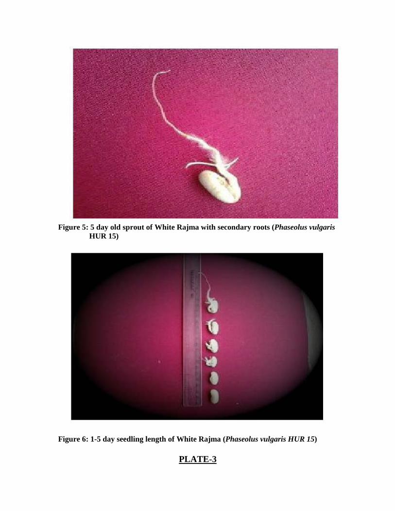
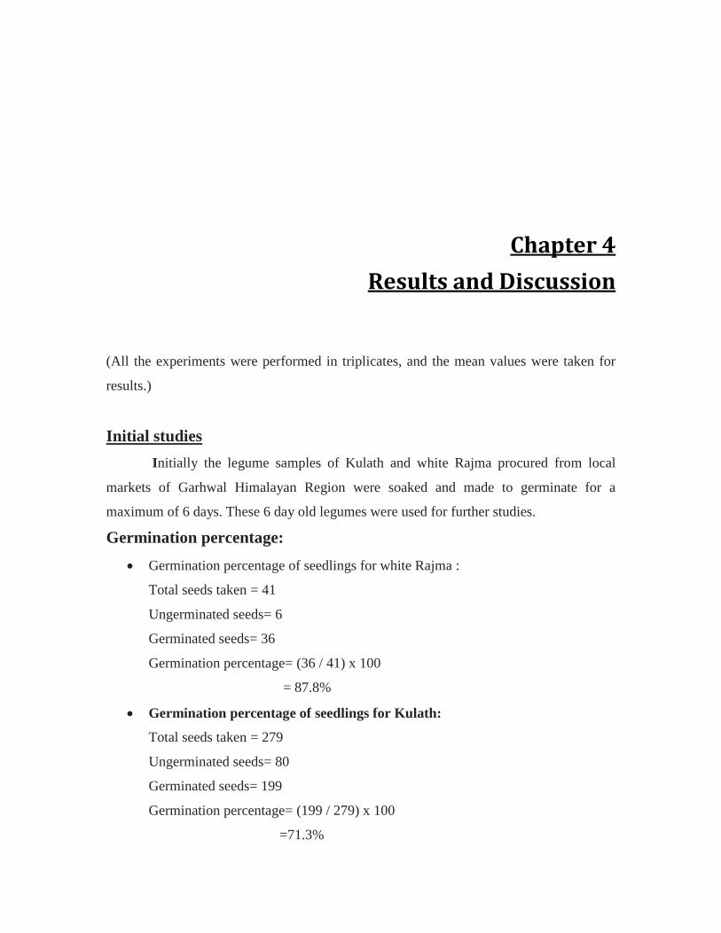
Figure 5: 5 day old sprout of White Rajma with secondary roots (Phaseolus vulgaris HUR 15)
Figure 6: 1-5 day seedling length of White Rajma (Phaseolus vulgaris HUR 15)
PLATE-3
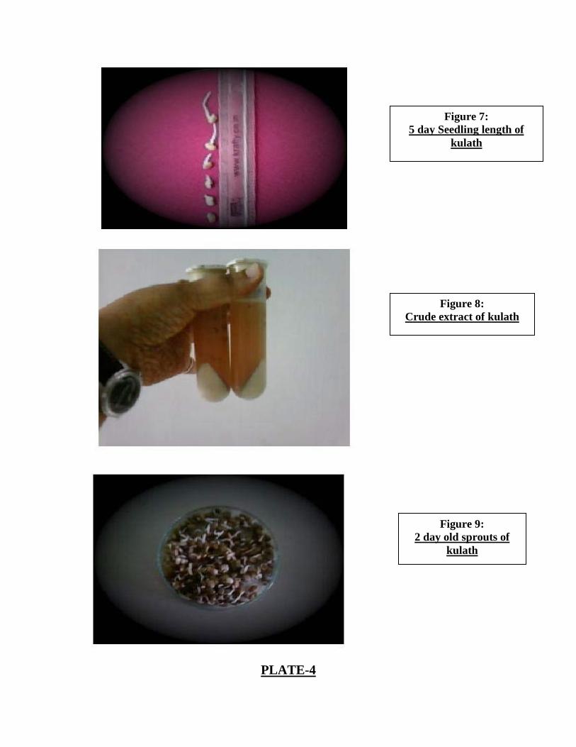
PLATE-4
Figure 7: 5 day Seedling length of
kulath
Figure 8: Crude extract of kulath
Figure 9: 2 day old sprouts of
kulath
Seedling length: From these germinated seedlings, seedling length was calculated. For Rajma,
highest seedling length was 6.5cm, 6.1cm and 5.9cm (Figure 6). In case of kulath, highest
seedling length was 3.6cm, 1.7cm and 1.6cm respectively (Figure 7).
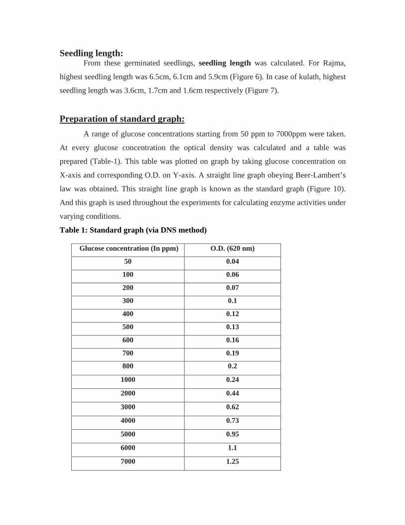
A range of glucose concentrations starting from 50 ppm to 7000ppm were taken.
At every glucose concentration the optical density was calculated and a table was
prepared (Table-1). This table was plotted on graph by taking glucose concentration on
X-axis and corresponding O.D. on Y-axis. A straight line graph obeying Beer-Lambert’s
law was obtained. This straight line graph is known as the standard graph (Figure 10).
And this graph is used throughout the experiments for calculating enzyme activities under
varying conditions.
Preparation of standard graph:
Table 1: Standard graph (via DNS method)
Glucose concentration (In ppm) O.D. (620 nm)
50 0.04
100 0.06
200 0.07
300 0.1
400 0.12
500 0.13
600 0.16
700 0.19
800 0.2
1000 0.24
2000 0.44
3000 0.62
4000 0.73
5000 0.95
6000 1.1
7000 1.25

Figure 10: Graph showing O.D. values against varying glucose concentrations.
The isolated enzyme was found in the supernatant. O.D. of crude Dolichos
biflorus extract was found to be 0.17 while that of crude Phaseolus vulgaris HUR15
extract was found to be 0.09.
Isolation:
From the standard graph (Figure 11), the corresponding glucose concentration
against these O.D values came out to be 680ppm for Dolichos biflorus and 290ppm for
Phaseolus vulgaris HUR 15.
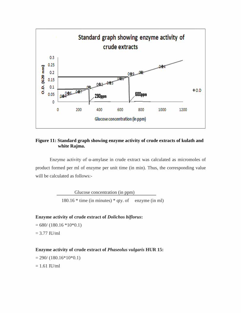
Figure 11: Standard graph showing enzyme activity of crude extracts of kulath and white Rajma.
Enzyme activity of α-amylase in crude extract was calculated as micromoles of
product formed per ml of enzyme per unit time (in min). Thus, the corresponding value
will be calculated as follows:-
Glucose concentration (in ppm)
180.16 * time (in minutes) * qty. of enzyme (in ml)
Enzyme activity of crude extract of Dolichos biflorus:
= 680/ (180.16 *10*0.1)
= 3.77 IU/ml
Enzyme activity of crude extract of Phaseolus vulgaris HUR 15:
= 290/ (180.16*10*0.1)
= 1.61 IU/ml

Further, these isolated crude enzyme extracts were subjected to purification.
Two fold purification was carried out first by ultra filtration and then by ion
exchange chromatography.
Purification of Crude Extracts:
a) Ultra filtration:
The ultra filtered extract from 10kDa extract showed an O.D of 0.01, of which
glucose concentration could not be determined from standard graph. Thus, 10kDa extract
does not have any α-amylase.
Dolichos biflorus:
The ultra filtered extract from 30kDa extract showed an O.D of 0.13. From standard
graph, the corresponding glucose concentration came out to be 500 ppm. Thus, amount of
reducing sugars were estimated to be (as per formula)
= 500/ (180.16*10*0.1)
= 2.77 IU/ml
As the crude extract contained 3.78 IU/ml enzyme, therefore it was deduced that
nearly 2/3 of alpha amylase of this plant is below 30kDa size. This 2/3 enzyme was
further purified by ion exchange chromatography. Further 1/3 of the enzyme which is
above 30kDa was not investigated.
In case of Phaseolus vulgaris HUR15 (white Rajma),10 kDa extract showed an
O.D. of 0.01 while 30kDa extract showed an O.D. of 0.08. From standard graph, the
corresponding glucose concentration came out to be 275 ppm. Thus, amount of reducing
sugars were estimated to be (as per formula):
b) Phaseolus vulgaris HUR15
= 275/ (180.16*10*0.1)
= 1.52 IU/ml.

Thus, 94% of enzyme has been found to be below 30KDa size. Therefore, this 94%
enzyme was used for kinetic characterization.
It was finally concluded that there are two types of α-amylases, viz., Small and
large. Small α-amylase is that whose size ranges upto<= 30kDa while large alpha
amylase is that whose size is above > 30kDa.
In the present research, α-amylase of Kulath as well as white Rajma comes under
small alpha amylases. α-amylases of bacteria generally come under large amylases
(discussed later).
It is also important to note that the recent research focuses basically on the kinetic
characteristics of purified alpha amylases of these two plants, therefore the exact size of
alpha amylases was not required, thus not calculated.
Ion exchange chromatography of 30 kDa extract was done.
Ion exchange chromatography
In case of Dolichos biflorus, highly purified α-amylase was obtained at 250 mM
concentration (Table 2).
Table 2: Ion exchange chromatography results for Dolichos biflorus extract.
Molar Concentration (In mM) O.D. (620 nm)
50 0.06
100 0.07
150 0.07
200 0.10
250 0.14
300 0.13
350 0.13
400 0.08
500 0.07
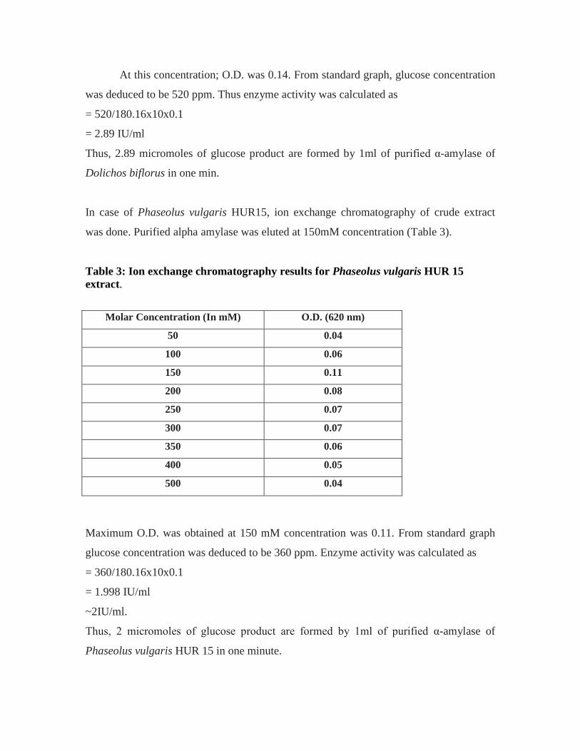
At this concentration; O.D. was 0.14. From standard graph, glucose concentration
was deduced to be 520 ppm. Thus enzyme activity was calculated as
= 520/180.16x10x0.1
= 2.89 IU/ml
Thus, 2.89 micromoles of glucose product are formed by 1ml of purified α-amylase of
Dolichos biflorus in one min.
In case of Phaseolus vulgaris HUR15, ion exchange chromatography of crude extract
was done. Purified alpha amylase was eluted at 150mM concentration (Table 3).
Table 3: Ion exchange chromatography results for Phaseolus vulgaris HUR 15 extract.
Maximum O.D. was obtained at 150 mM concentration was 0.11. From standard graph
glucose concentration was deduced to be 360 ppm. Enzyme activity was calculated as
= 360/180.16x10x0.1
= 1.998 IU/ml
~2IU/ml.
Thus, 2 micromoles of glucose product are formed by 1ml of purified α-amylase of
Phaseolus vulgaris HUR 15 in one minute.
Molar Concentration (In mM) O.D. (620 nm)
50 0.04
100 0.06
150 0.11
200 0.08
250 0.07
300 0.07
350 0.06
400 0.05
500 0.04

To increase this enzyme activity of the purified enzyme, further kinetic
characterization is done. As it is a well known fact the under optimum reaction conditions
best enzyme activity will be reached.
Study of kinetic characteristics of enzymes is important because only after
studying the kinetic characteristics, a particular enzyme can be used on industrial scale.
More the stability of enzyme in a wide range of conditions more is the usefulness of
enzyme on industrial scale.
Kinetic Characterisation
We calculated the kinetic characteristics of pure enzyme as well as the crude
extracts of the enzyme. And we found out that the characteristics of both the extracts
were almost the same. This is so because reference on standard graph is glucose.
• pH optima of kulath extract was 6.1. From table 4, O.D. values against each pH
concentration starting from pH 3.6 to 7 could be tabulated. The table simply shows
maximum O.D. value of 0.94 at pH 6.1. Therefore, pH 6.1 was considered as optimum
pH.
Optimum pH
Table 4: Table showing optimum pH of crude kulath extract.
pH O.D. (620 nm) 3.6 0.24 4 0.27 5 0.67
6.1 0.94 6.3 0.91 6.7 0.67 7 0.17
On plotting a graph, a bell shaped graph was obtained (Figure 12). Figure 12 shows
that the enzyme showed maximum activity between pH 5.5 to 7. But the activity reduced
in alkaline range i.e.>7. The drop in activity was more in phosphate buffer than in Tris-
HCl buffer, indicating the stability of enzyme in Tris-HCl buffer. After pH 7.5 the

enzyme activity almost nullified. These results correspond with previous results of
Nirmala and Muralikrishna (2002).
Figure 12: Optimum pH of crude kulath extract.
pH optima of purified kulath enzyme obtained via ion-exchange chromatography
was also calculated. Maximum O.D. was obtained to be 1 at pH 6.1. Thus, pH 6.1 was
considered to be optimum pH of the purified enzyme (Table 5).
Table 5: Table showing optimum pH of purified kulath extract.
pH O.D. (620 nm) 3.6 0.33 4 0.46 5 0.53
6.1 1 6.3 0.94 6.7 0.93 7 0.14
A graph was also plotted taking pH on X-axis and corresponding O.D. values on
the Y-axis. Again a bell shaped graph was obtained showing pH optima of purified
enzyme to be 6.1 pH. Purified enzyme also showed best stability in acidic pH range, i.e.
5.5 to 6 (Figure 13).

Figure 13: Optimum pH of purified kulath extract.
pH optima of white rajma extract was calculated similarly. Maximum O.D. was
obtained to be 0.14 at pH 6.5 (Table 6).
Table 6: Table showing pH optima of white Rajma extract (crude).
pH O.D. (620 nm) 4 0.11 5 0.12
6.1 0.13 6.3 0.13 6.5 0.14 6.7 0.11 7 0.09
7.5 0.07 8 0.06
8.6 0.01
On plotting a graph of pH (X-axis) against O.D values (Y-axis), a bell shaped
graph was obtained. The enzyme showed best stability in 4 to 6.5 pH range. Enzyme
activity totally nullified after pH range 8 (Figure 14).
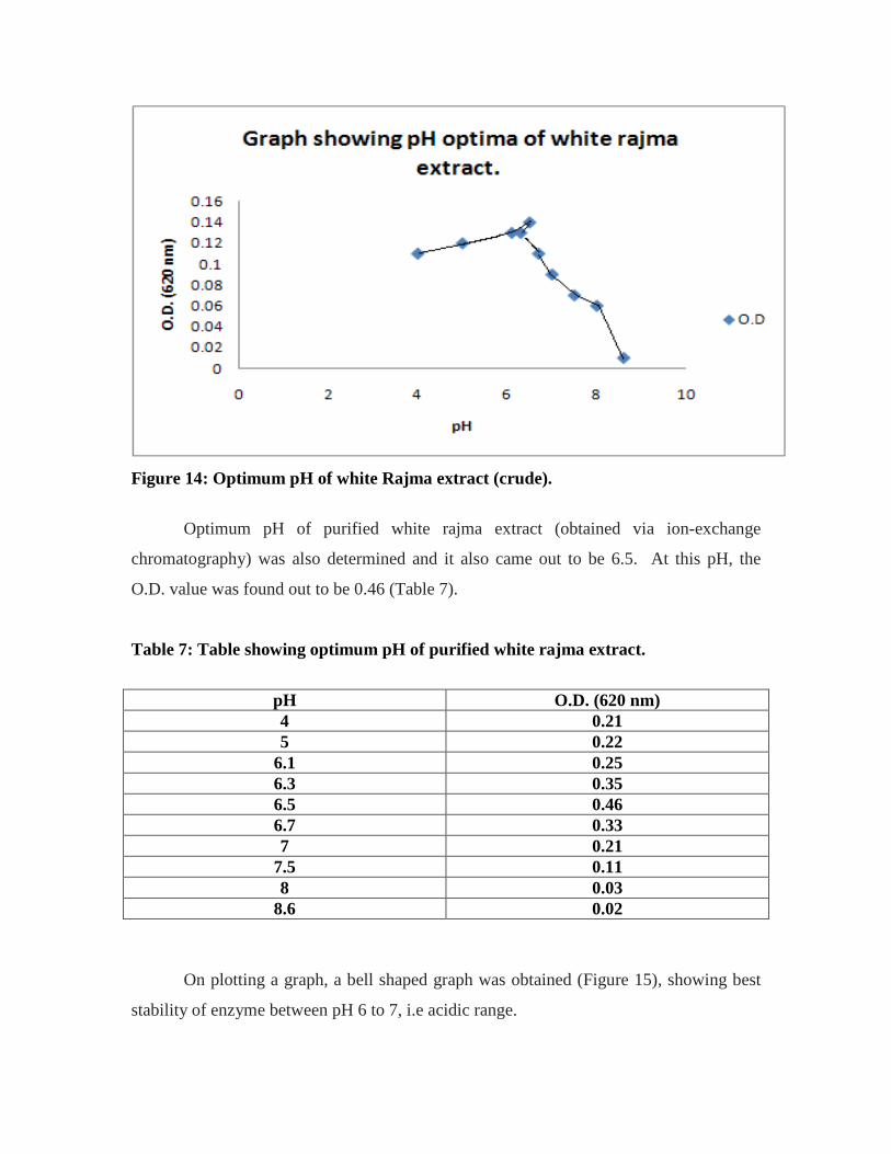
Figure 14: Optimum pH of white Rajma extract (crude).
Optimum pH of purified white rajma extract (obtained via ion-exchange
chromatography) was also determined and it also came out to be 6.5. At this pH, the
O.D. value was found out to be 0.46 (Table 7).
Table 7: Table showing optimum pH of purified white rajma extract.
pH O.D. (620 nm) 4 0.21 5 0.22
6.1 0.25 6.3 0.35 6.5 0.46 6.7 0.33 7 0.21
7.5 0.11 8 0.03
8.6 0.02
On plotting a graph, a bell shaped graph was obtained (Figure 15), showing best
stability of enzyme between pH 6 to 7, i.e acidic range.

Figure 15: Optimum pH of purified white rajma extract.
We can finally deduce that α- amylase of these two legumes remains stable in
acidic pH range and is an acidic amylase.
(Further kinetic characteristics were determined only with the purified enzyme.)
The optimum temperature of kulath α-amylase was found out to be 45°C. At this
temperature, a maximum O.D. of 0.88 was obtained (Table 8).
Optimum temperature and thermal stability
Table- 8: Optimum temperature of purified enzyme in kulath.
Temperature (°C) O.D. (620 nm) 0 0.02 4 0.05 10 0.22 20 0.44 30 0.57 40 0.78 45 0.88 50 0.76 60 0.77 70 0.21 80 0.06

A graph was plotted taking temperature in (°C) on X-axis and temperature on Y-
axis. A slightly distorted bell shaped graph was obtained. The enzyme showed best
stability in temperature range 42°C to 48°C (Figure 16).
Figure 16: Optimum temperature of purified kulath enzyme.
Since optimum temperature is found to be 45°C, therefore determination of
thermal stability was started at 55°C (i.e. 10°C above the optimum temperature). Enzyme
assay was done at 55°C, 65°C and 75°C for 10, 15, 20, 25 and 30 minutes respectively. It
was observed that O.D keeps on decreasing with increasing temperature. A very slight
O.D. change was observed at 55°C; the O.D change increased extensively at 65°C; and at
75°C, after 20minutes, the enzyme was almost totally unstable (Table-9). This enzyme
was considered to be highly thermostable as it shows stability upto 75°C. Table 9 shows
exact values of changing O.D. with increasing temperature and time.
The optimum temperature of white rajma α-amylase was found out to be 50°C.
(Table-10). At this temperature, the maximum O.D. value obtained was 0.2

Table- 9: Table showing thermal stability of purified kulath enzyme.
At 55°C temperature and Time (in minutes)
O.D.
10 0.77 15 0.76 20 0.65 25 0.62 30 0.61
At 65°C temperature and Time (in minutes)
O.D.
10 0.48 15 0.31 20 0.22 25 0.21 30 0.20
At 75°C temperature and Time (in minutes)
O.D.
10 0.08 15 0.08 20 0.07 25 0.06 30 0.05
Table -10: Table showing optimum temperature of white rajma purified enzyme.
Temperature (°C) O.D. (620 nm) 30 0.09 40 0.13 42 0.13 44 0.15 46 0.14 48 0.19 50 0.2 60 0.12 70 0.05 80 0.02
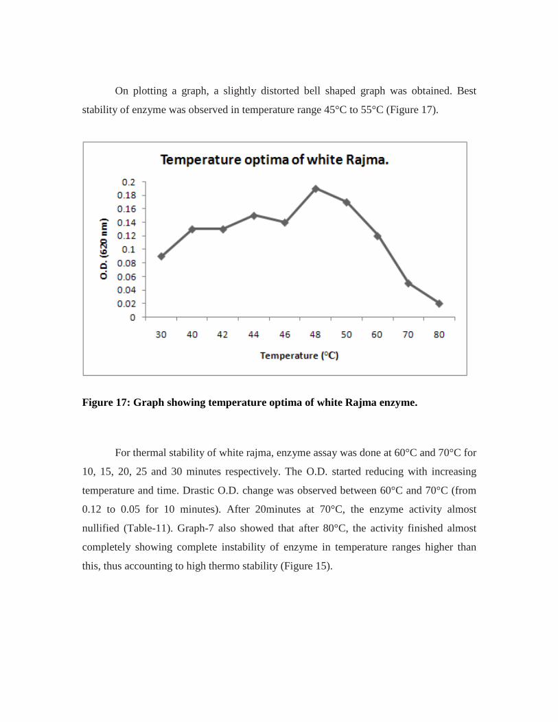
On plotting a graph, a slightly distorted bell shaped graph was obtained. Best
stability of enzyme was observed in temperature range 45°C to 55°C (Figure 17).
Figure 17: Graph showing temperature optima of white Rajma enzyme.
For thermal stability of white rajma, enzyme assay was done at 60°C and 70°C for
10, 15, 20, 25 and 30 minutes respectively. The O.D. started reducing with increasing
temperature and time. Drastic O.D. change was observed between 60°C and 70°C (from
0.12 to 0.05 for 10 minutes). After 20minutes at 70°C, the enzyme activity almost
nullified (Table-11). Graph-7 also showed that after 80°C, the activity finished almost
completely showing complete instability of enzyme in temperature ranges higher than
this, thus accounting to high thermo stability (Figure 15).

Table -11: Table showing thermal stability of white Rajma enzyme.
At 60°C temperature and Time (in minutes)
O.D.
10 0.12 15 0.11 20 0.09 25 0.08 30 0.08
At 70°C temperature and Time (in minutes)
O.D.
10 0.05 15 0.05 20 0.04 25 0.02 30 0.02
Km and Vmax
For calculating Km and Vmax
values, O.D. of the enzyme at various substrate
concentrations was calculated first of all. Then that substrate concentration was
determined at which maximum O.D. was obtained. In case of kulath, optimum
substrate concentration was found to be 2.4mg/ml. At this substrate concentration,
O.D. was found to be 0.90. This value is known as the optimum substrate
concentration (Table 12).
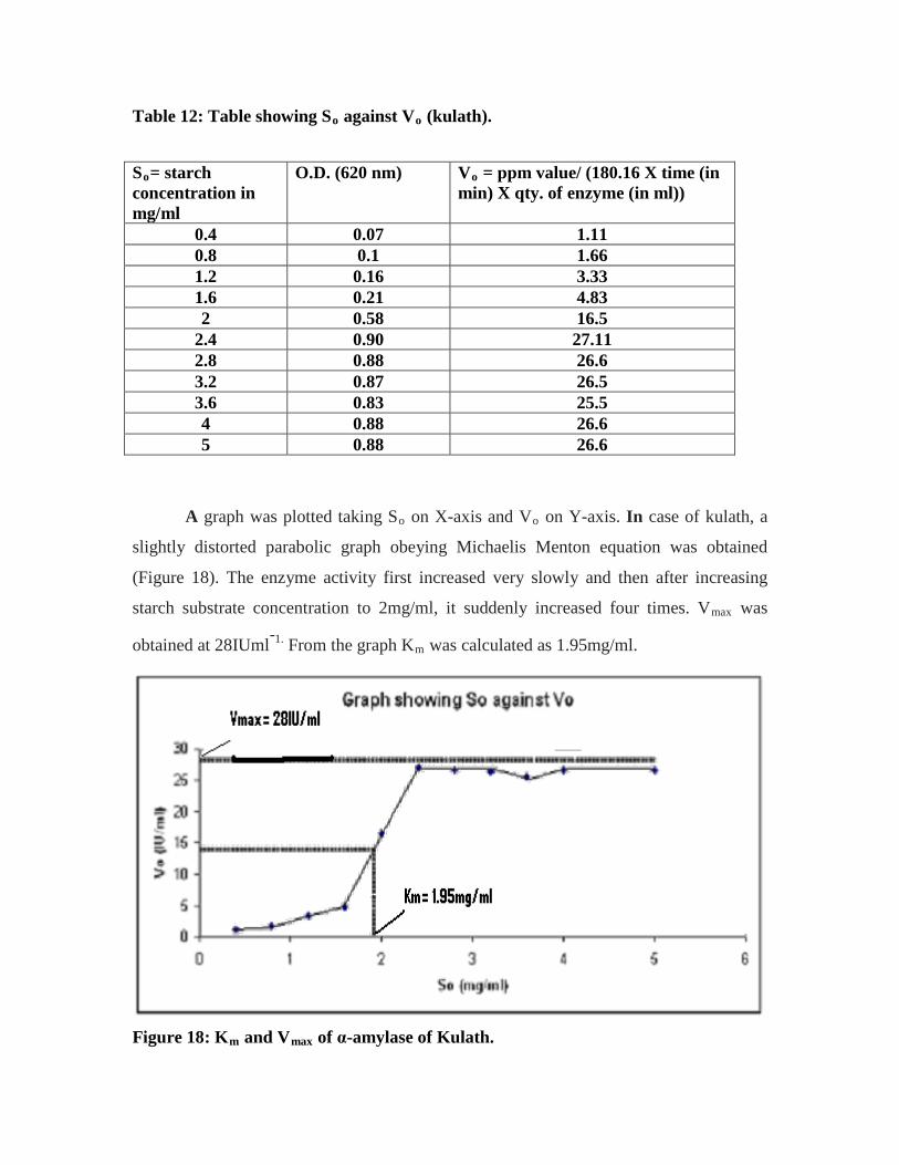
Table 12: Table showing So against Vo
(kulath).
So O.D. (620 nm) = starch concentration in mg/ml
Vo
= ppm value/ (180.16 X time (in min) X qty. of enzyme (in ml))
0.4 0.07 1.11 0.8 0.1 1.66 1.2 0.16 3.33 1.6 0.21 4.83 2 0.58 16.5
2.4 0.90 27.11 2.8 0.88 26.6 3.2 0.87 26.5 3.6 0.83 25.5 4 0.88 26.6 5 0.88 26.6
A graph was plotted taking So on X-axis and Vo on Y-axis. In case of kulath, a
slightly distorted parabolic graph obeying Michaelis Menton equation was obtained
(Figure 18). The enzyme activity first increased very slowly and then after increasing
starch substrate concentration to 2mg/ml, it suddenly increased four times. Vmax was
obtained at 28IUml-1. From the graph Km was calculated as 1.95mg/ml.
Figure 18: Km and Vmax of α-amylase of Kulath.
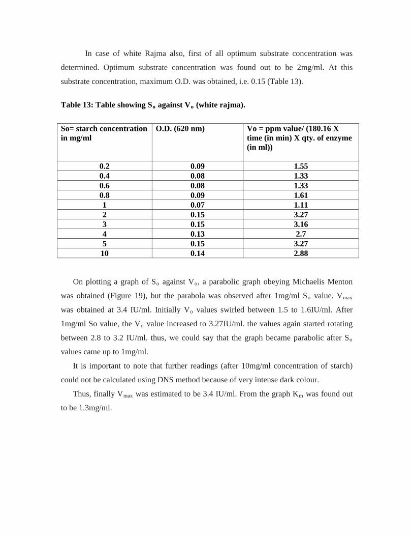
In case of white Rajma also, first of all optimum substrate concentration was
determined. Optimum substrate concentration was found out to be 2mg/ml. At this
substrate concentration, maximum O.D. was obtained, i.e. 0.15 (Table 13).
Table 13: Table showing So against Vo
(white rajma).
So= starch concentration in mg/ml
O.D. (620 nm) Vo = ppm value/ (180.16 X time (in min) X qty. of enzyme (in ml))
0.2 0.09 1.55 0.4 0.08 1.33 0.6 0.08 1.33 0.8 0.09 1.61 1 0.07 1.11 2 0.15 3.27 3 0.15 3.16 4 0.13 2.7 5 0.15 3.27 10 0.14 2.88
On plotting a graph of So against Vo, a parabolic graph obeying Michaelis Menton
was obtained (Figure 19), but the parabola was observed after 1mg/ml So value. Vmax
was obtained at 3.4 IU/ml. Initially Vo values swirled between 1.5 to 1.6IU/ml. After
1mg/ml So value, the Vo value increased to 3.27IU/ml. the values again started rotating
between 2.8 to 3.2 IU/ml. thus, we could say that the graph became parabolic after So
It is important to note that further readings (after 10mg/ml concentration of starch)
could not be calculated using DNS method because of very intense dark colour.
values came up to 1mg/ml.
Thus, finally Vmax was estimated to be 3.4 IU/ml. From the graph Km
was found out
to be 1.3mg/ml.

Figure 19: Km and Vmax
of alpha- amylase of white Rajma.
In case of kulath, maximum enzyme activity was observed in 10 minutes in this
time maximum O.D. = 0.18 was obtained (Table 14).
Time
Table 14: Table showing optimum time of α-amylase of kulath.
Time (in minutes) O.D. (620 nm) 2 0.04 4 0.06 6 0.09 8 0.14 10 0.18 12 0.11 14 0.09
A graph was plotted with time on X-axis and corresponding O.D. on Y-axis.
Again a bell shaped graph was obtained (Figure 20)
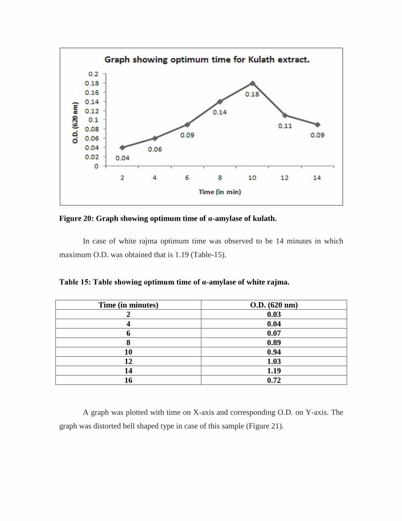
Figure 20: Graph showing optimum time of α-amylase of kulath.
In case of white rajma optimum time was observed to be 14 minutes in which
maximum O.D. was obtained that is 1.19 (Table-15).
Table 15: Table showing optimum time of α-amylase of white rajma.
Time (in minutes) O.D. (620 nm) 2 0.03 4 0.04 6 0.07 8 0.89 10 0.94 12 1.03 14 1.19 16 0.72
A graph was plotted with time on X-axis and corresponding O.D. on Y-axis. The
graph was distorted bell shaped type in case of this sample (Figure 21).
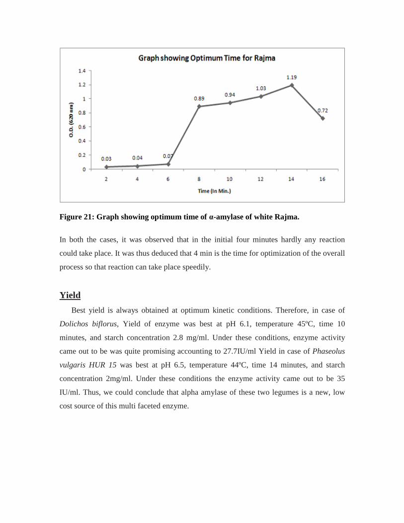
Figure 21: Graph showing optimum time of α-amylase of white Rajma.
In both the cases, it was observed that in the initial four minutes hardly any reaction
could take place. It was thus deduced that 4 min is the time for optimization of the overall
process so that reaction can take place speedily.
Best yield is always obtained at optimum kinetic conditions. Therefore, in case of
Dolichos biflorus, Yield of enzyme was best at pH 6.1, temperature 45ºC, time 10
minutes, and starch concentration 2.8 mg/ml. Under these conditions, enzyme activity
came out to be was quite promising accounting to 27.7IU/ml Yield in case of Phaseolus
vulgaris HUR 15 was best at pH 6.5, temperature 44ºC, time 14 minutes, and starch
concentration 2mg/ml. Under these conditions the enzyme activity came out to be 35
IU/ml. Thus, we could conclude that alpha amylase of these two legumes is a new, low
cost source of this multi faceted enzyme.
Yield

Immobilisation
Immobilization of both enzymes was done on calcium alginate matrix. Shiny
transparent beads of two centimeter diameter with slightly yellowish brown coloration
were obtained in case of kulath. Beads of ultra filtered kulath extract (~30kDa) were also
prepared which were shiny yellow in colouration (Plate 5).
In case of rajma opaque white color beads of two centimeter diameter were
obtained.
Kinetic characteristics of immobilized enzyme:
Kinetic characteristics of these immobilised enzymes beads were calculated and it
was found out that the characteristics were highly enhanced. Although much change in
optimum pH was not observed, still we observed a remarkable increase in enzyme
activity. Optimum pH of free enzyme came out to be 6.1 and that of immobilized enzyme
came out to be 6.3 (Table 16).
Table -16: Comparison between enzyme activities of crude and immobilized kulath enzyme at different pH concentrations.
pH Enzyme activity (IU/ml) of crude enzyme.
Enzyme activity (IU/ml) of immobilized enzyme.
3.6 5.55 4.44 4 6.11 6.67 5 18.87 18.05
6.1 27.22 22.78 6.3 26.11 37.22 6.7 18.87 44.44 7 3.39 38.89 8 <0.20 16.96
8.6 <0.20 17.19 9 <0.20 2.08
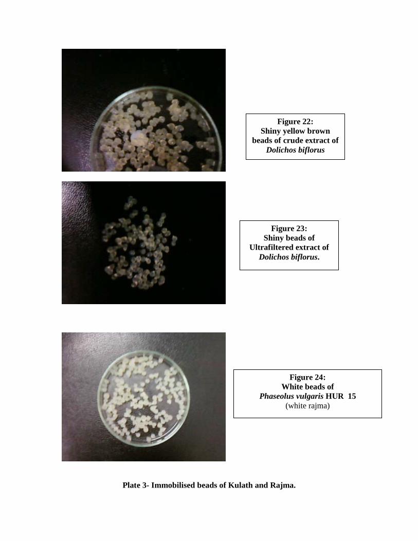
Plate 3- Immobilised beads of Kulath and Rajma.
Figure 24: White beads of
Phaseolus vulgaris HUR 15 (white rajma)
Figure 22: Shiny yellow brown
beads of crude extract of Dolichos biflorus
Figure 23: Shiny beads of
Ultrafiltered extract of Dolichos biflorus.

Figure 25 shows a close comparison between optimum pH of free and
immobilized enzyme. Most enzymes are stable in pH range 6 to 8 (Welker and Campbell,
1967). This table further puts light on this statement. Uptil pH 6.1, we could not observe
much increase in enzyme activity. Drastic increase in enzyme activity was observed after
pH 6.3. We observed that at pH 7 enzyme activity increased more than 11 times (~11.5
times). Although we could not observe much increase in optimum pH; Free enzyme
showed an optimum pH of 6.1 (Table 5) while the immobilized enzyme showed an
optimum pH of 6.7 (Table 16). We also observed that the immobilized enzyme was stable
at much higher pH values than the free enzyme. While the free enzyme showed no
activity at pH 8, the immobilized enzyme showed an activity of 16.96 IU/ml at pH 8.
An important point of consideration was that at pH 3.6, the enzyme activity of
immobilized enzyme was slight lower than that of free enzyme. Free enzyme showed
enzyme activity of 5.55 IU/ml while immobilized enzyme showed enzyme activity of
4.44 IU/ml. This might be so because immobilized enzyme takes higher time for
optimization than the free enzyme.
Figure 25: Graph showing comparison between enzyme activities of free and immobilized kulath at various pH concentrations.

Certain similar results were seen in case of optimum temperature. Optimum
temperature of free enzyme was 45°C (Table 8) while optimum temperature of
immobilized enzyme was 50°C (Table 17). This simply proves the increase in thermal
stability. Around 9 times increase in enzyme activity was observed at 70° C (Figure 26).
While the free enzyme showed very less activity at 80°C, the immobilized enzyme
showed a high activity of 10.74IU/ml at 80°C.
Table -17: Comparison between enzyme activities of crude and immobilized kulath enzyme at various temperatures.
Temperature (°C) Enzyme activity (IU/ml) of crude enzyme.
Enzyme activity (IU/ml) of immobilized enzyme.
0 <0.20 0.28 4 0.42 0.55 10 5 5.55 20 11.11 14.44 30 15.55 17.22 40 22.22 22.78 45 25.55 28.94 50 21.67 >44.4 60 22.19 >44.4 70 4.44 38.89 80 0.55 24.8 85 -- 10.42 90 -- 1.11 95 -- 0.55
Kinetic characterization of immobilized α-amylase of white rajma also revealed
similar results. Optimum pH of immobilized white rajma extract came out to be 6.5 . This
was same as that of free enzyme (Table 7). Enzyme activity of immobilized enzyme
increased 20 times at pH 8. While the free enzyme showed no activity at pH 8.6, the
immobilized enzyme showed an activity of 8.89 IU/ml at pH 8.6. Thus, it was finally
proved that characteristics are enhanced in immobilized enzyme. While the pH stability
range for free enzyme is 4 to 7, the pH stability range for immobilized enzyme is
observed to be 4 to 9 (Table 18; Figure 27).

Figure 26: Graph showing comparison between enzyme activities of free and immobilized kulath enzyme at various temperatures. Table 18: Comparison between enzyme activities of crude and immobilized white rajma enzyme at various pH concentrations.
pH Enzyme activity (IU/ml) of crude enzyme.
Enzyme activity (IU/ml) of immobilized enzyme.
4 2 1.67 5 2.22 2.22
6.1 2.78 6.78 6.3 2.78 10.5 6.5 2.89 35.55 6.7 2 30.55 7 1.55 29.72
7.5 1.11 21.39 8 0.55 10.89
8.6 <0.20 8.89
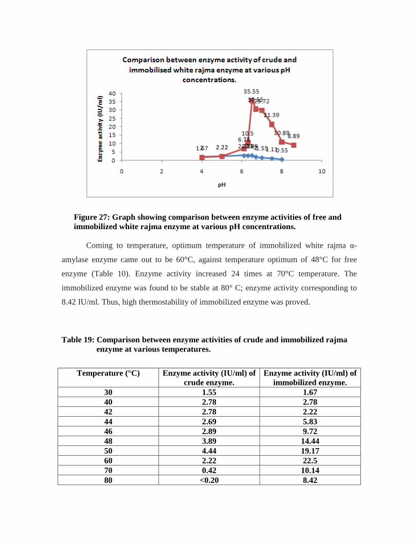
Figure 27: Graph showing comparison between enzyme activities of free and immobilized white rajma enzyme at various pH concentrations.
Coming to temperature, optimum temperature of immobilized white rajma α-
amylase enzyme came out to be 60°C, against temperature optimum of 48°C for free
enzyme (Table 10). Enzyme activity increased 24 times at 70°C temperature. The
immobilized enzyme was found to be stable at 80° C; enzyme activity corresponding to
8.42 IU/ml. Thus, high thermostability of immobilized enzyme was proved.
Table 19: Comparison between enzyme activities of crude and immobilized rajma enzyme at various temperatures.
Temperature (°C) Enzyme activity (IU/ml) of crude enzyme.
Enzyme activity (IU/ml) of immobilized enzyme.
30 1.55 1.67 40 2.78 2.78 42 2.78 2.22 44 2.69 5.83 46 2.89 9.72 48 3.89 14.44 50 4.44 19.17 60 2.22 22.5 70 0.42 10.14 80 <0.20 8.42

Figure 25: Graph showing comparison between enzyme activities of free and immobilized white rajma enzyme at various temperatures.
Re-utilisation of immobilized beads was not possible more than twice as during
enzyme assay, when the beads came to boiling (Refer Step 5 of enzyme assay, Chapter-
3); they almost dissolved, thus leaving only the contaminated solution for taking out the
O.D values. But since, immobilized beads could be reused 2 times, therefore usage of
immobilized enzyme in comparison to free enzyme is easy and user friendly.
Further preparation of immobilized beads which could be used more than twice
remains a topic of further research.

Comparison: We also performed a small comparison between 14 plant and bacterial species for
α-amylase enzyme present in them (Garg and Dobriyal, 2010). Comparison was made on
the basis of optimum pH, optimum temperature, Km, Vmax
α-amylase of Dolichos biflorus and Phaseolus vulgaris (family Fabaceae) showed
complete instability in highly alkaline pH range. Optimum pH of white Rajma came out
to be 6.5 while that of Kulath came out to be 6.1. These were completely unstable above
pH 7.5. However, these were compared with α-amylase of the family Poaceae, which
were stable in acidic pH range only, and become completely unstable above pH 7.
Eleusine coracana Indaf-15, Sorghum bicolor, Zea mays and Triticum aestivum were
studied. All these have pH stability range and optimum pH below 7 (Table 20).
, pH stability range and thermal
stability of enzyme. We found out that the plant enzyme is equally good than the bacterial
enzyme in terms of characteristics.
Table -20: Comparison between temperature and pH for plant α-amylase.
S.No. α-amylase source
Common name
pH stability range
Optimum pH
Thermal stability
(°C)
Optimum temperature
(°C) 1 Dolichos
biflorus Kulath 5.5-7 6.1 40-75 45
2 Phaseolus vulgaris HUR 15
White rajma
4-7.5 6.5 30-80 50
3 Glycine max
Soyabean 4-6 5.5 25-85 50
4 Eleusine coracana Indaf-15
Ragi or finger millet
5-5.5 5.3 45-50 47
5 Sorghum bicolor
Jowar 4.5-6 5.5 30-70 70
6 Trticum aestivum
(5 isozymes)
Wheat 5.5-7 5.5-7 (for all five
isozymes)
40-50 50
7 Zea mays Corn 4-6.5 5.5 50-90 55
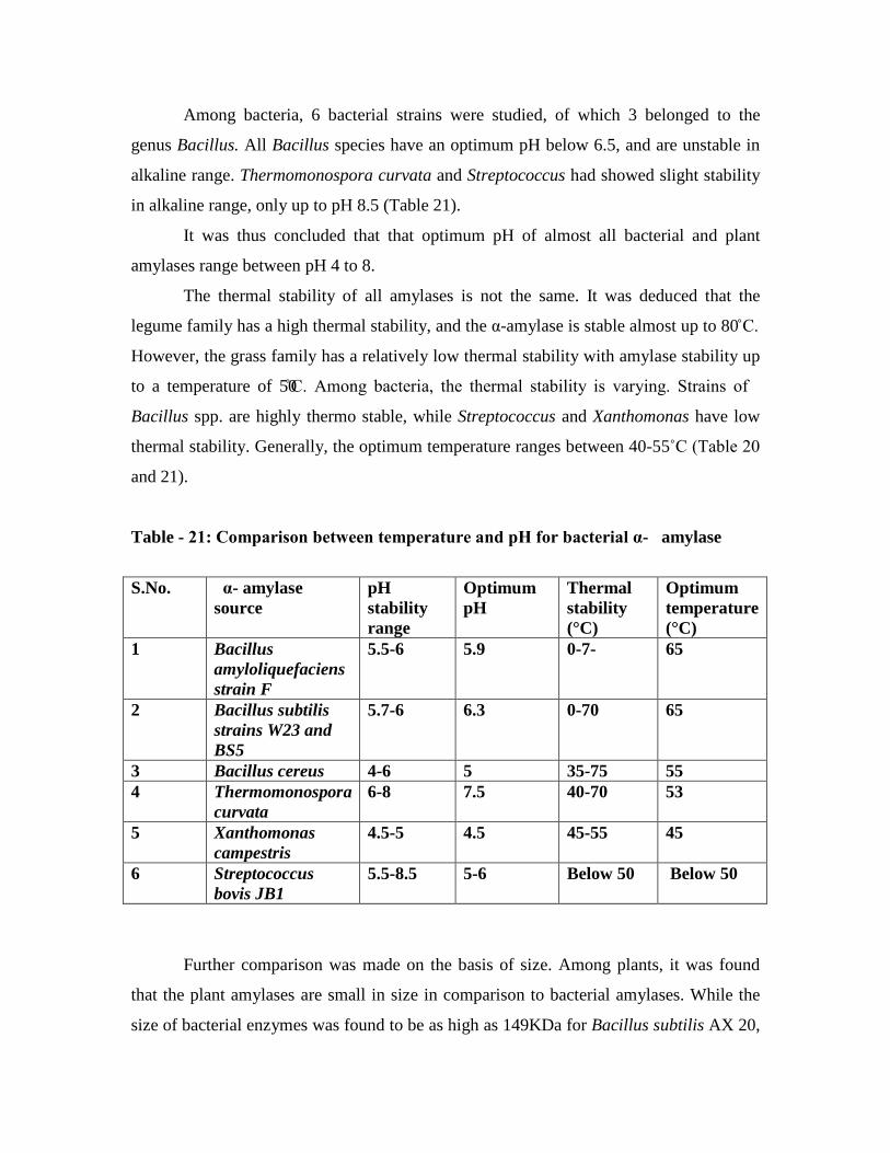
Among bacteria, 6 bacterial strains were studied, of which 3 belonged to the
genus Bacillus. All Bacillus species have an optimum pH below 6.5, and are unstable in
alkaline range. Thermomonospora curvata and Streptococcus had showed slight stability
in alkaline range, only up to pH 8.5 (Table 21).
It was thus concluded that that optimum pH of almost all bacterial and plant
amylases range between pH 4 to 8.
The thermal stability of all amylases is not the same. It was deduced that the
legume family has a high thermal stability, and the α-amylase is stable almost up to 80 C.
However, the grass family has a relatively low thermal stability with amylase stability up
to a temperature of 50C. Among bacteria, the thermal stability is varying. Strains of
Bacillus spp. are highly thermo stable, while Streptococcus and Xanthomonas have low
thermal stability. Generally, the optimum temperature ranges between 40-55˚C (Table 20
and 21).
Table - 21: Comparison between temperature and pH for bacterial α- amylase
S.No. α- amylase source
pH stability range
Optimum pH
Thermal stability (°C)
Optimum temperature (°C)
1 Bacillus amyloliquefaciens strain F
5.5-6 5.9 0-7- 65
2 Bacillus subtilis strains W23 and BS5
5.7-6 6.3 0-70 65
3 Bacillus cereus 4-6 5 35-75 55 4 Thermomonospora
curvata 6-8 7.5 40-70 53
5 Xanthomonas campestris
4.5-5 4.5 45-55 45
6 Streptococcus bovis JB1
5.5-8.5 5-6 Below 50 Below 50
Further comparison was made on the basis of size. Among plants, it was found
that the plant amylases are small in size in comparison to bacterial amylases. While the
size of bacterial enzymes was found to be as high as 149KDa for Bacillus subtilis AX 20,
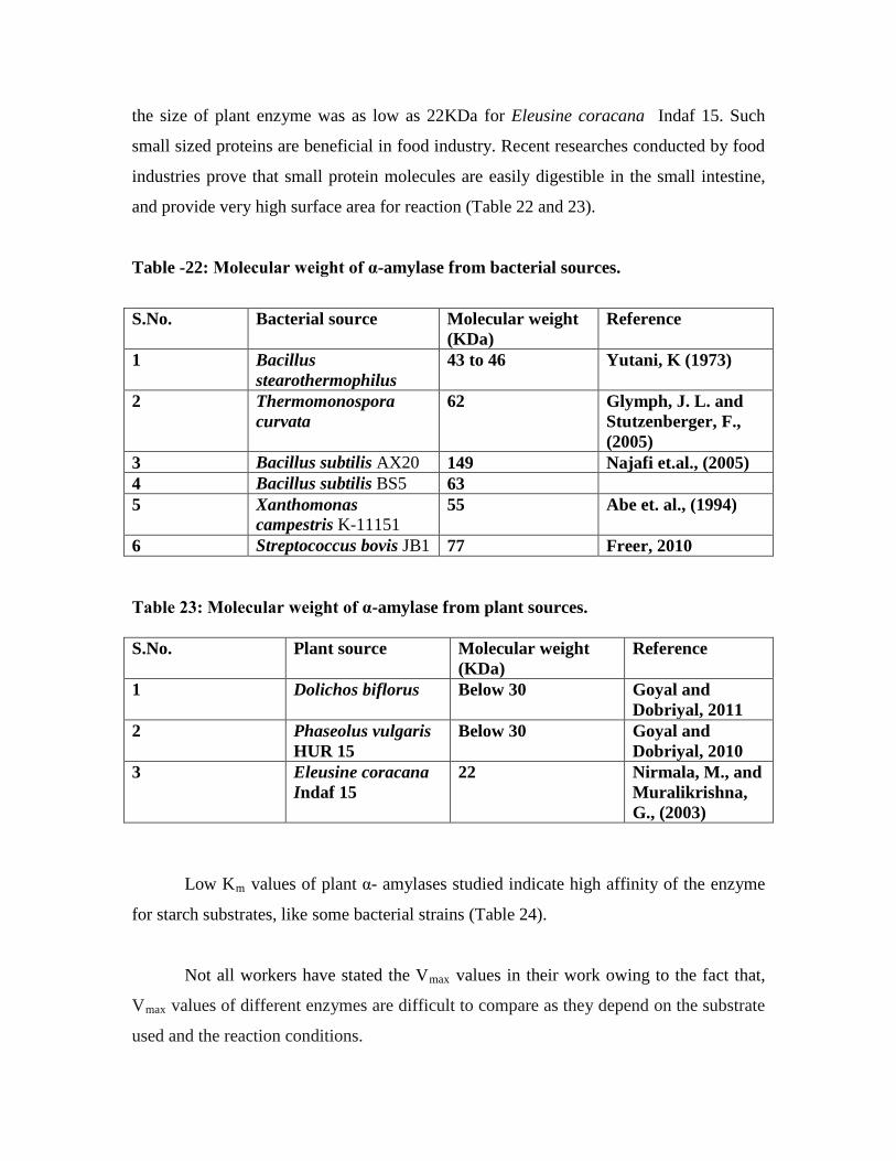
the size of plant enzyme was as low as 22KDa for Eleusine coracana Indaf 15. Such
small sized proteins are beneficial in food industry. Recent researches conducted by food
industries prove that small protein molecules are easily digestible in the small intestine,
and provide very high surface area for reaction (Table 22 and 23).
Table -22: Molecular weight of α-amylase from bacterial sources.
S.No. Bacterial source Molecular weight (KDa)
Reference
1 Bacillus stearothermophilus
43 to 46 Yutani, K (1973)
2 Thermomonospora curvata
62 Glymph, J. L. and Stutzenberger, F., (2005)
3 Bacillus subtilis AX20 149 Najafi et.al., (2005) 4 Bacillus subtilis BS5 63 5 Xanthomonas
campestris K-11151 55 Abe et. al., (1994)
6 Streptococcus bovis JB1 77 Freer, 2010 Table 23: Molecular weight of α-amylase from plant sources. S.No. Plant source Molecular weight
(KDa) Reference
1 Dolichos biflorus Below 30 Goyal and Dobriyal, 2011
2 Phaseolus vulgaris HUR 15
Below 30 Goyal and Dobriyal, 2010
3 Eleusine coracana Indaf 15
22 Nirmala, M., and Muralikrishna, G., (2003)
Low Km
values of plant α- amylases studied indicate high affinity of the enzyme
for starch substrates, like some bacterial strains (Table 24).
Not all workers have stated the Vmax values in their work owing to the fact that,
Vmax values of different enzymes are difficult to compare as they depend on the substrate
used and the reaction conditions.
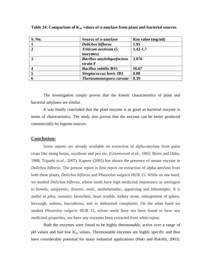
Table 24: Comparison of Km
values of α-amylase from plant and bacterial sources.
S. No. Source of α-amylase Km value (mg/ml) 1 Dolichos biflorus 1.95 2 Triticum aestivum (5
isozymes) 1.42-1.7
3 Bacillus amyloliquefaciens strain F
3.076
4 Bacillus subtilis BS5 16.67 5 Streptococcus bovis JB1 0.88 6 Thermomonospora curvata 0.39
The investigation simply proves that the kinetic characteristics of plant and
bacterial amylases are similar.
It was finally concluded that the plant enzyme is as good as bacterial enzyme in
terms of characteristics. The study also proves that the enzyme can be better produced
commercially by legume sources.
Conclusion: Some reports are already available on extraction of alpha-amylase from pulse
crops like mung beans, soyabean and pea etc. (Greenwood et.al., 1965; Beers and Duke,
1988; Tripathi et.al., 2007). Kapoor (2005) has shown the presence of urease enzyme in
Dolichos biflorus. The present report is first report on extraction of alpha amylase from
both these plants, Dolichos biflorus and Phaseolus vulgaris HUR 15. While on one hand,
we studied Dolichos biflorus, whose seeds have high medicinal importance as astringent
to bowels, antipyretic, diuretic, tonic, antihelminthic, appetizing and lithontriptic. It is
useful in piles, tumours, bronchitis, heart trouble, kidney stone, enlargement of spleen,
hiccough, asthma, leucoderma, and in abdominal complaints. On the other hand we
studied Phaseolus vulgaris HUR 15, whose seeds have not been found to have any
medicinal properties, nor have any enzymes been extracted from white rajma.
Both the enzymes were found to be highly thermostable, active over a range of
pH values and had low Km values. Thermostable enzymes are highly specific and thus
have considerable potential for many industrial applications (Haki and Rakshit, 2003).
Activeness in a range of pH values also increases the industrial applicability of the
enzyme. Although low Km values, did not prove to be much effective because substrate
was not limiting, still we could say that this characteristic is important when the enzyme
will be produced on laboratory scale; and further industrial scale.
Till date, very less literature could be cited regarding immobilization of plant α-
amylase. No work regarding immobilization of plant amylase on Calcium alginate could
be found. This research proved to be a very new decent approach on immobilization of
plant α-amylase on Calcium alginate. We used Calcium alginate because Calcium
alginate has been found to be the best matrix material for immobilization of bacterial α-
amylases (Refer Chapter-2).
Previously researches avoided working on plant amylases because of the
tediousness witnessed during extraction of the enzyme and further purification. A large
amount of spent material is to be removed during enzyme extraction. In the present work,
we also proved that the kinetic characteristics of crude as well as purified enzyme are
almost the same. Thus, even the crude enzyme can be used for calculating the kinetic
characteristics. This approach can reduce not only the tediousness of the work, but also
made the work cost- effective.
The very basic thought behind starting this research work was to find out whether
the α-amylase enzyme present in these two legumes is of industrial importance or not.
We could finally deduce that even this α-amylase can be used in industries like starch
industry, brewing and baking industry and detergent industry.
Future perspectives of the study:
• We have proved the high enzyme potential of Kulath and White Rajma in the
present study. This high enzyme potential can be further enhanced by application
of biotechnology and genetic engineering in this field. Studies can be performed
on the same.
• Further studies on other legume sources, (of Garhwal and outside Garhwal) can
also be performed.

• We have immobilized the enzyme only on Calcium alginate matrix. Studies can
be perfomed on other matrices also. Also we used beads only of 2cm diameter.
Further studies can be performed with different bead sizes and different matrices.
• Research can also be performed to find out the suitable matrix whose beads could
be used more than twice.
1
Chapter 5
Summary
The thesis entitled, “Studies on isolation, purification, kinetic characterisation and
immobilisation of α-amylase from some legumes of Garhwal Himalayan Region” can be
summarised as follows:
1. There is a bulk of genetic and ecological diversity in the Garhwal Himalayan
region of Uttarakhand. This diversity includes 4700 species under 1503 genera
and 213 families. Among all these, the dicots dominate with 3493 species under
1163 genera and 182 families. Among dicots, the family Asteraceae has the
largest number of genera, i.e., 125 followed by Fabaceae which has 76 genera.
The flowering plants provide wide range of useful products ranging from timber,
medicine, food, vegetable oils, gums, resins and spices etc. Among the edible
plants, pulses, also known as grain legumes, are second only to cereals as source
of human food. These account for nearly 20 species all belonging to the family
Fabaceae. Major pulse crops are Cajanus cajan, Cicer arientum, Dolichos spp,
Phaseolus spp, Vigna spp, Glycine max, Lens culinaris, Pisum sativum and Vicia
faba.
2. Biotechnologists of this area are known to work on subjects related to
biotechnology like molecular biology, enzymology and tissue culture, etc. It is
interesting to note that despite the large number of products; timber, medicine,
food, vegetable oils, gums, resins and spices; exploited from these multi-faceted
pulse crops, the enzymatic stamina of these pulse crops has not been explored yet.
2
Despite many biotechnological advances in this area, the scientists were found to
be least interested regarding the idea of enzyme extraction from pulse crops.
Therefore the present research on “Studies on isolation, purification, kinetic
characterisation and immobilisation of α-amylase from some legumes of Garhwal
Himalayan Region” has been carried out.
3. α- amylase (α-1,4 glucan-glucanhydrolase; E.C. 3.2.1.1) is an extracellular
enzyme which degrades α-1,4 glucosidic linkages of starch and related substrates
in an endo fashion producing oligosaccharides including maltose, glucose and
alpha limit dextrins. This breakdown reaction is an important step in many
industrial processes like brewing, baking, starch liquefaction, pharmaceutical and
enzyme production etc. Therefore, this enzyme is extensively used in industries
like starch liquefaction, brewing, food, paper, textile and pharmaceuticals.
4. A large number of sources have been recognised by scientists and researchers all
around the world for extraction of α-amylase. These sources include micro-
organisms, plants and animals. A close investigation of various enzyme firms
shows that most of the α-amylase production is done from genetically modified
micro-organisms of Bacillus spp. During the research it was thoroughly
investigated that even though the enzyme is present in all sorts of sources
discussed above, still no plant or animal source has been utilised on large scale for
production of the enzyme. Although the enzyme has been located in plant sources
like sorghum, broad beans, coconut oil cake, cereals and mung beans, no
researcher has tried to locate the enzyme in the flora of Garhwal Himalayan
Region. In the present research, the enzyme has been extracted from two
leguminous sources, Dolichos biflorus, commonly known as Kulathha and
Phaseolus vulgaris HUR15, commonly known as white rajma. Enzyme assays via
DNS method (given by Miller, 1959) revealed maximum amount of enzyme α-
amylase in these two legumes, amongst all other Himalayan legumes.
5. Dolichos biflorus, commonly known as kulthee, gahat or horse gram is a branched
or trailing annual with small trifoliate leaves, and very wide climbing slender
stem. On maturity, it gives narrow, flat, curved pods containing ellipsoid seeds
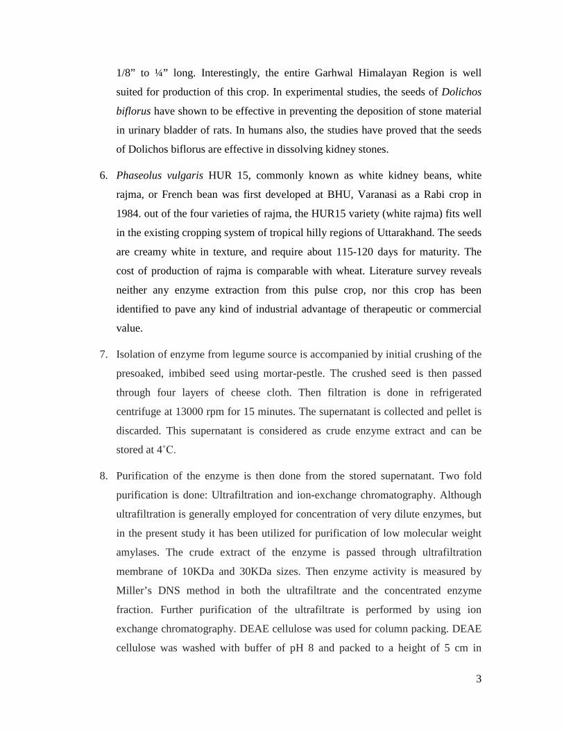
3
1/8” to ¼” long. Interestingly, the entire Garhwal Himalayan Region is well
suited for production of this crop. In experimental studies, the seeds of Dolichos
biflorus have shown to be effective in preventing the deposition of stone material
in urinary bladder of rats. In humans also, the studies have proved that the seeds
of Dolichos biflorus are effective in dissolving kidney stones.
6. Phaseolus vulgaris HUR 15, commonly known as white kidney beans, white
rajma, or French bean was first developed at BHU, Varanasi as a Rabi crop in
1984. out of the four varieties of rajma, the HUR15 variety (white rajma) fits well
in the existing cropping system of tropical hilly regions of Uttarakhand. The seeds
are creamy white in texture, and require about 115-120 days for maturity. The
cost of production of rajma is comparable with wheat. Literature survey reveals
neither any enzyme extraction from this pulse crop, nor this crop has been
identified to pave any kind of industrial advantage of therapeutic or commercial
value.
7. Isolation of enzyme from legume source is accompanied by initial crushing of the
presoaked, imbibed seed using mortar-pestle. The crushed seed is then passed
through four layers of cheese cloth. Then filtration is done in refrigerated
centrifuge at 13000 rpm for 15 minutes. The supernatant is collected and pellet is
discarded. This supernatant is considered as crude enzyme extract and can be
stored at 4˚C.
8. Purification of the enzyme is then done from the stored supernatant. Two fold
purification is done: Ultrafiltration and ion-exchange chromatography. Although
ultrafiltration is generally employed for concentration of very dilute enzymes, but
in the present study it has been utilized for purification of low molecular weight
amylases. The crude extract of the enzyme is passed through ultrafiltration
membrane of 10KDa and 30KDa sizes. Then enzyme activity is measured by
Miller’s DNS method in both the ultrafiltrate and the concentrated enzyme
fraction. Further purification of the ultrafiltrate is performed by using ion
exchange chromatography. DEAE cellulose was used for column packing. DEAE
cellulose was washed with buffer of pH 8 and packed to a height of 5 cm in

4
chromatographic column, 2 cm in diameter. Then the column was washed
exhaustively with equilibration buffer, pH 8, twice. Then sample of α-amylase
was added to the column with utmost care avoiding any air bubbles. Elution was
done using stepwise elution method with increasing molarities of sodium-
phosphate buffer (0.05, 0.1, 0.15, 0.2, 0.25, 0.3, 0.35, 0.4, 0.5M), pH 7.2. The
fractionation of was carried out at 4° C and the eluate was collected in 2-2ml
fractions. Each fraction was assayed for enzyme activity. The fraction showing
maximum α-amylase activity was further subjected to kinetic characterization.
9. Kinetic characterization on the basis of following parameters; optimum pH,
optimum temperature, substrate concentration, thermal stability, time, Michaelis
Menton’s constant (Km) and Maximum velocity (Vmax
10. After kinetic characterization, the immobilization of this purified enzyme is also
done and the enhanced characteristics are studied. A close comparison is made
between the characteristics of immobilized and free enzyme.It was interesting to
note that the enzyme extracted from these pulse crops was as better as the
traditional enzyme extracted from bacterial sources. Results prove that plant
enzymes showed nearly same characteristics just like bacterial enzymes. Thus,
besides finding out new, low cost sources of α-amylase, we could also deduce that
these new sources can be made usable on industrial scale.
) is then performed. These
kinetic characteristics are calculated via enzyme assays using DNS method in the
ion exchange fractions
11. Initial studies were performed on the legume samples of kulath and white rajma
procured from local markets of Garhwal Himalayan Region. Germination
percentage of seedling for white rajma came out to be 87.8% while the
germination percentage of kulath came out to be 71.3%. Seedling length for white
rajma was calculated as 6.5cm, 6.1cm and 5.9cm. in case of kulath, the seedling
length came out to be 3.6cm, 1.7cm and 1.6cm. Then isolation of the enzyme was
done. The supernatant obtained from centrifuged filtered extract of seeds was
considered as isolated crude extract of the enzyme. Optical density of crude
Dolichos biflorus extract was found to be 0.17 while that of Phaseolus vulgaris

5
HUR 15 extract was found to be 0.09. From standard graph, corresponding
glucose concentration against these O.D. values came out to be 680 ppm for
Dolichos biflorus and 290 ppm for Phaseolus vulgaris HUR15. Enzyme activity
of α-amylase was calculated as micromoles of product formed per ml of enzyme
per unit time (in minutes).
12. Enzyme activity of crude extract of Dolichos biflorus came out to be 3.77 IU/ml
while that of crude extract of Phaseolus vulgaris HUR 15 came out to be 1.61
IU/ml.Further these isolated crude enzymes were subjected to two-fold
purification, first by ultrafiltration and then by ion-exchange chromatography.
The ultrafiltered extract from 10kDa extract of Dolichos biflorus showed and
O.D. of 0.01, of which glucose concentration concentration could not be
determined from standard graph. Thus, 10kDa extract does not have any α-
amylase. The ultra filtered extract from 30kDa extract showed an O.D of 0.13.
From standard graph, the corresponding glucose concentration came out to be 500
ppm. Thus, amount of reducing sugars were estimated to be 2.77 IU/ml. As the
crude extract contained 3.78 IU/ml enzyme, therefore it was deduced that nearly
2/3 of α-amylase of this plant is below 30kDa size. This 2/3 enzyme was further
purified by ion exchange chromatography. Further 1/3 of the enzyme which is
above 30kDa was not investigated.
13. In case of Phaseolus vulgaris HUR15 (white Rajma),10 kDa ultrafiltered extract
showed an O.D. of 0.01 while 30kDa extract showed an O.D. of 0.08. From
standard graph, the corresponding glucose concentration came out to be 275 ppm.
Thus, amount of reducing sugars were estimated to be 1.52 IU/ml. Thus, 94% of
enzyme has been found to be below 30KDa size. Therefore, this 94% enzyme was
used for kinetic characterization.
14. It was finally concluded that there are two types of α-amylases:- Small and large.
Small α-amylase is that whose size ranges upto<= 30kDa while large α-amylase is
that whose size is greater than 30kDa. In the present research, alpha amylase of
Kulath as well as white Rajma comes under small α-amylases.

6
15. Ion exchange chromatography of 30 kDa extract was done. In case of Dolichos
biflorus, highly purified alpha amylase was obtained at 250 mM. At this
concentration; O.D. was 0.14. From standard graph, glucose concentration was
deduced to be 520 ppm. Thus enzyme activity was calculated as 2.89 IU/ml (as
per formula). Thus, 2.89 micromoles of glucose product are formed by 1ml of
purified α-amylase of Dolichos biflorus in one minute.Purified α-amylase of
white rajma was obtained at 150mM concentration. Maximum O.D. was obtained
at 150 mM concentration was 0.11. From standard graph, glucose concentration
was deduced to be 360 ppm. Enzyme activity was calculated as 1.998 IU/ml i.e.
~2IU/ml. Thus, 2 micromoles of glucose product are formed by 1ml of purified α-
amylase of Phaseolus vulgaris HUR 15 in one minute.To increase this enzyme
activity of the purified enzyme, further kinetic characterization is done. As it is a
well known fact that under optimum reaction conditions best enzyme activity will
be reached.
16. pH optima of kulath extract was 6.1. A bell shaped graph was obtained. The
enzyme showed maximum activity between pH 5.5 to 7. But the activity reduced
in alkaline range i.e.>7. The drop in activity was more in phosphate buffer than in
Tris-HCl buffer, indicating the stability of enzyme in Tris-HCl buffer. After pH
7.5 the enzyme activity almost nullified. These results correspond with previous
results of Nirmala and Muralikrishna (2002). pH optima of purified kulath
enzyme also came out to be 6.1. pH optima of white rajma extract was 6.5. In this
case also, a bell shaped graph was obtained. The enzyme showed best stability in
4 to 6.5 pH range. Enzyme activity totally nullified after pH range 8. We can
deduce that alpha amylase of these two legumes remains stable in acidic pH range
and is an acidic amylase. Optimum pH of purified white rajma extract (obtained
via ion-exchange chromatography) was also determined and it also came out to be
6.5.
17. The optimum temperature of kulath α-amylase was found out to be 45°C. A
slightly distorted bell shaped graph was obtained. The enzyme showed best
stability in temperature range 42°C to 48°C. Thermal stability of the enzyme was
checked by calculating the enzyme activity at temperatures above 45°C. This
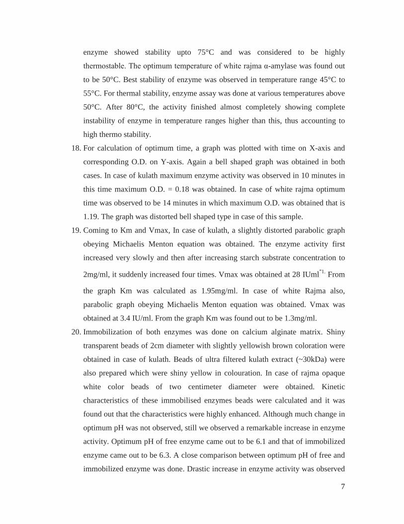
7
enzyme showed stability upto 75°C and was considered to be highly
thermostable. The optimum temperature of white rajma α-amylase was found out
to be 50°C. Best stability of enzyme was observed in temperature range 45°C to
55°C. For thermal stability, enzyme assay was done at various temperatures above
50°C. After 80°C, the activity finished almost completely showing complete
instability of enzyme in temperature ranges higher than this, thus accounting to
high thermo stability.
18. For calculation of optimum time, a graph was plotted with time on X-axis and
corresponding O.D. on Y-axis. Again a bell shaped graph was obtained in both
cases. In case of kulath maximum enzyme activity was observed in 10 minutes in
this time maximum O.D. = 0.18 was obtained. In case of white rajma optimum
time was observed to be 14 minutes in which maximum O.D. was obtained that is
1.19. The graph was distorted bell shaped type in case of this sample.
19. Coming to Km and Vmax, In case of kulath, a slightly distorted parabolic graph
obeying Michaelis Menton equation was obtained. The enzyme activity first
increased very slowly and then after increasing starch substrate concentration to
2mg/ml, it suddenly increased four times. Vmax was obtained at 28 IUml-1.
20. Immobilization of both enzymes was done on calcium alginate matrix. Shiny
transparent beads of 2cm diameter with slightly yellowish brown coloration were
obtained in case of kulath. Beads of ultra filtered kulath extract (~30kDa) were
also prepared which were shiny yellow in colouration. In case of rajma opaque
white color beads of two centimeter diameter were obtained. Kinetic
characteristics of these immobilised enzymes beads were calculated and it was
found out that the characteristics were highly enhanced. Although much change in
optimum pH was not observed, still we observed a remarkable increase in enzyme
activity. Optimum pH of free enzyme came out to be 6.1 and that of immobilized
enzyme came out to be 6.3. A close comparison between optimum pH of free and
immobilized enzyme was done. Drastic increase in enzyme activity was observed
From
the graph Km was calculated as 1.95mg/ml. In case of white Rajma also,
parabolic graph obeying Michaelis Menton equation was obtained. Vmax was
obtained at 3.4 IU/ml. From the graph Km was found out to be 1.3mg/ml.
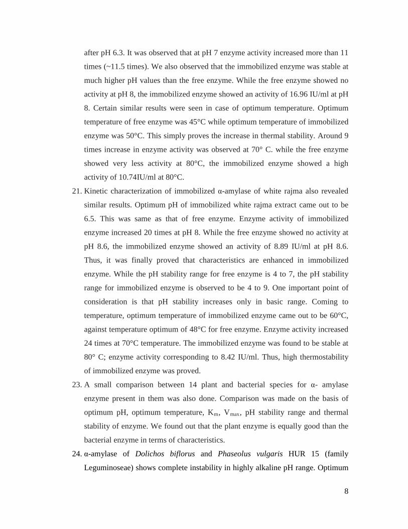
8
after pH 6.3. It was observed that at pH 7 enzyme activity increased more than 11
times (~11.5 times). We also observed that the immobilized enzyme was stable at
much higher pH values than the free enzyme. While the free enzyme showed no
activity at pH 8, the immobilized enzyme showed an activity of 16.96 IU/ml at pH
8. Certain similar results were seen in case of optimum temperature. Optimum
temperature of free enzyme was 45°C while optimum temperature of immobilized
enzyme was 50°C. This simply proves the increase in thermal stability. Around 9
times increase in enzyme activity was observed at 70° C. while the free enzyme
showed very less activity at 80°C, the immobilized enzyme showed a high
activity of 10.74IU/ml at 80°C.
21. Kinetic characterization of immobilized α-amylase of white rajma also revealed
similar results. Optimum pH of immobilized white rajma extract came out to be
6.5. This was same as that of free enzyme. Enzyme activity of immobilized
enzyme increased 20 times at pH 8. While the free enzyme showed no activity at
pH 8.6, the immobilized enzyme showed an activity of 8.89 IU/ml at pH 8.6.
Thus, it was finally proved that characteristics are enhanced in immobilized
enzyme. While the pH stability range for free enzyme is 4 to 7, the pH stability
range for immobilized enzyme is observed to be 4 to 9. One important point of
consideration is that pH stability increases only in basic range. Coming to
temperature, optimum temperature of immobilized enzyme came out to be 60°C,
against temperature optimum of 48°C for free enzyme. Enzyme activity increased
24 times at 70°C temperature. The immobilized enzyme was found to be stable at
80° C; enzyme activity corresponding to 8.42 IU/ml. Thus, high thermostability
of immobilized enzyme was proved.
23. A small comparison between 14 plant and bacterial species for α- amylase
enzyme present in them was also done. Comparison was made on the basis of
optimum pH, optimum temperature, Km, Vmax
24. α-amylase of Dolichos biflorus and Phaseolus vulgaris HUR 15 (family
Leguminoseae) shows complete instability in highly alkaline pH range. Optimum
, pH stability range and thermal
stability of enzyme. We found out that the plant enzyme is equally good than the
bacterial enzyme in terms of characteristics.
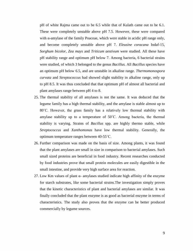
9
pH of white Rajma came out to be 6.5 while that of Kulath came out to be 6.1.
These were completely unstable above pH 7.5. However, these were compared
with α-amylase of the family Poaceae, which were stable in acidic pH range only,
and become completely unstable above pH 7. Eleusine coracana Indaf-15,
Sorghum bicolor, Zea mays and Triticum aestivum were studied. All these have
pH stability range and optimum pH below 7. Among bacteria, 6 bacterial strains
were studied, of which 3 belonged to the genus Bacillus. All Bacillus species have
an optimum pH below 6.5, and are unstable in alkaline range. Thermomonospora
curvata and Streptococcus had showed slight stability in alkaline range, only up
to pH 8.5. It was thus concluded that that optimum pH of almost all bacterial and
plant amylases range between pH 4 to 8.
25. The thermal stability of all amylases is not the same. It was deduced that the
legume family has a high thermal stability, and the amylase is stable almost up to
80˚C. However, the grass family has a relatively low thermal stability with
amylase stability up to a temperature of 50˚C. Among bacteria, the thermal
stability is varying. Strains of Bacillus spp. are highly thermo stable, while
Streptococcus and Xanthomonas have low thermal stability. Generally, the
optimum temperature ranges between 40-55˚C.
26. Further comparison was made on the basis of size. Among plants, it was found
that the plant amylases are small in size in comparison to bacterial amylases. Such
small sized proteins are beneficial in food industry. Recent researches conducted
by food industries prove that small protein molecules are easily digestible in the
small intestine, and provide very high surface area for reaction.
27. Low Km values of plant α- amylases studied indicate high affinity of the enzyme
for starch substrates, like some bacterial strains.The investigation simply proves
that the kinetic characteristics of plant and bacterial amylases are similar. It was
finally concluded that the plant enzyme is as good as bacterial enzyme in terms of
characteristics. The study also proves that the enzyme can be better produced
commercially by legume sources.
10
The present report is first report on extraction of alpha amylase from both
these plants, Dolichos biflorus and Phaseolus vulgaris HUR 15. While on one
hand, we studied Dolichos biflorus, whose seeds have high medicinal importance
as astringent to bowels, antipyretic, diuretic, tonic, antihelminthic, appetizing and
lithontriptic. It is useful in piles, tumours, bronchitis, heart trouble, kidney stone,
enlargement of spleen, hiccough, asthma, leucoderma, and in abdominal
complaints. On the other hand we studied Phaseolus vulgaris HUR 15, whose
seeds have not been found to have any medicinal properties, nor have any
enzymes been extracted from white rajma.
Conclusion:
Both the enzymes were found to be highly thermostable, active over a
range of pH values and had low Km values. Thermostable enzymes are highly
specific and thus have considerable potential for many industrial applications.
Activeness in a range of pH values also increases the industrial applicability of
the enzyme. Although low Km values, did not prove to be much effective because
substrate was not limiting, still we could say that this characteristic is important
when the enzyme will be produced on laboratory scale; and further industrial
scale.
Till date, very less literature could be cited regarding immobilization of
plant α-amylase. No work regarding immobilization of plant amylase on Calcium
alginate could be found. This research proved to be a very new decent approach
on immobilization of plant α-amylase on Calcium alginate. We used Calcium
alginate because Calcium alginate has been found to be the best matrix material
for immobilization of bacterial α-amylases.
Previously researches avoided working on plant amylases because of the
tediousness witnessed during extraction of the enzyme and further purification. A
large amount of spent material is to be removed during enzyme extraction. In the
present work, we also proved that the kinetic characteristics of crude as well as
purified enzyme are almost the same. Thus, even the crude enzyme can be used
for calculating the kinetic characteristics. This approach can reduce not only the
tediousness of the work, but also made the work cost- effective.

11
The very basic thought behind starting this research work was to find out
whether the α-amylase enzyme present in these two legumes is of industrial
importance or not. We could finally deduce that even this α-amylase can be used
in industries like starch industry, brewing and baking industry and detergent
industry.
12
Chapter-6
References
Abe, J., Onitsuka, N., Nakano, T., Shibata, Y., Hizukuri, S., and Entaniz, E., (1994).
“Purification and characterization of periplasmic α-amylase from Xanthomonas
campestris K-1151”, Journal of Bacteriology, 176(12): pp 3584-3588.
Aguloglu, S., Ensari, N.Y., Uyar, F., and Otludil, B., (2000). “The effects of amino acids on
production and transport of α-amylase through bacterial membranes”, Starch, 52: pp 290-
295.
Ali, M., and Lal, S., (1991). “Rabi Rajmash-Potentialities, limitations and Technology of
Production” Technical Bulletin No. 3 at Directorate of Pulses Research, Kanpur.
Anto, H., Trivedi, U., and Patel, K., (2006). “α- amylase production by Bacillus cereus MTCC
1305 Using Solid- State Fermentation”, Food Technology Biotechnology, 44(2): pp 241-
245.
Azad, M.A. K., Bae, J., Kim, J., Lim, J., Song, K., Shin, B., and Kim, H., (2009). “Isolation and
characterization of a novel thermostable α-amylase from Korean pine seeds”, New
Biotechnology 26(3-4): pp143-149.
Bayramoglu, G., Yilmaz, M., and Arica, M.Y., (2004). “Immobilisation of a thermostable α-
amylase onto reactive membranes: kinetics characterization and application to continuous
starch hydrolysis” Food Chemistr, 84(4): pp 591-599.

13
Bayramoglu, Z., Akbulut, U., and Sungur, S., (1992). “Immobilisation of α-amylase into
photographic gelatin by chemical cross linking” Biomaterials, 13(10): pp 704-708.
Beers, E.P. and Duke, S.H., (1988). “Localisation of α-amylase in apoplast of pea (Pisum
sativum L.) Stems” Plant physiology, 87(4): pp 799-802.
Benkel, B.F., Nguyen, T., Ahluwalia, N., Benkel, K. I. and Hickey, D. A., (1997). “Cloning and
expression of a chicken α-amylase gene” Gene, 192(2): pp 261-270.
Berbezy, P., Legendre, L., and Maujean, A., (1996). “Purification and characterization of α-
amylase from vine shoot internodes” Plant Physiology and Biochemistry, 34: pp 353-361.
Bernhardsdotter, E.C.M.J., Ng, J.D., Garriott, O. K., and Pusey, M.L., (2005). “Enzymic
properties of an alkaline chelator-resistant α-amylase from an alkaliphilic Bacillus sp.
Isolate L1711” Process Biochemistry, 40(7): pp 2401-2408.
Biazus, J.P.M., Souza, R.R., Marquez, J.E., Franco, T.T., Santana, J.C.C., and Tanbourgi, E.B.,
(2009). “Production and characterization of amylases from Zea mays malt” Brazilian
Archives of Biology and Technology, 52(4): 991-1000
Brena, B.M., Pazos, C., Franco- Fraguas, L., and Batista-Viera, F., (1996). “Chromatographic
methods for amylases” Journal of Chromatography B: Biomedical Sciences and
Applications, 684(1-2): pp 217-237.
Bryjak, J., (2003). “Glucoamylase, α-amylase and β-amylase immobilization on acrylic carriers”
Biochemical Engineering Journal, 16(3): pp 347-355.
Chengyi, W.H., Ming, M., and Jiang, R., (1999). “Studies on the properties of α-amylase
produced by Bacillus pumilus 289 (PBX96)” Acta Microbial Sin, 32: pp 400-404.
14
Das, K., Doley, R., and Mukherjee, A.K., (2004). “Purification and biochemical characterization
of a thermostable, alkaliphilic, extracellular α- amylase from Bacillus subtilis DM-03, a
strain isolated from traditional fermented food of India” Biotechnology and Applied
Biochemistry, 40: pp 291-298.
Dube, S.K., and Nordin, P., (1961). “Isolation and properties of sorghum α-amylase” Archives of
Biochemistry and Biophysics, 94(1): pp 121-127.
Dubey, A. K., Suresh, C., Kavitha, R., Karanth, N.G., and Umesh-Kumar, S., (2000). “Evidence
that the glucoamylases and α-amylase secreted by Aspergillus niger are proteolytically
processed products of precursor enzyme” FEBS Letters, 471(2-3): pp 251-255.
El-Batal, A.I., Atia, K.S. and Eid, M., (2005). “Stabilisation of α-amylase by using anionic
surfactant during the immobilization process” Radiation physics and Chemistry, 74(2):
pp 96-101.
Ezeji, T. C., and Bahl, H., (2006). “Purification, characterization, and synergistic action of
phytate-resistant α-amylase and α-glucosidase from Geobacillus thermodenitrificans
HRO10” Journal of Biotechnology, 125(1): pp 27-38.
Farez-Vidal, M.E., Fernandez-Vivaz, A., and Arias, J.M., (1992). “Production of α-amylase by
Myxococcus coralloides D.” Journal of Applied Bacteriology, 73: pp 148-156.
Femi-Ola, T, O., and Olowe, B.M., (2011). “Characterisation of α- amylase from Bacillus
subtilis BS5 isolated from Amitermes evuncifer Silvestri” Research Journal of
Microbiology, 6(2): pp 140-146.
Ferey-Roux G., Perrier, J., Forest, E., Marchis-Moren, G., Puigserver, A., and Santimone, M.,
(1998). “The human pancreatic α-amylase isoforms: isolation, structural studies and
kinetics of inhibition by acarbose” Biochimica et Biophysica Acta (BBA)-Protein
Structure and Molecular Enzymology, 1388(1): pp10-20.
15
Freer, S.N., (1993). “Purification and characterization of the extracellular α- amylase from
Streptococcus bovis JB1” Applied Environment Microbiology, 59(5): pp 1398-1402.
Gangadharan, D., Nampoothiri, K.M., Sivaramakrishnan, S., and Pandey, A., (2009).
“Immobilised bacterial α-amylase for effective hydrolysis of raw and soluble starch”
Food Research International, 42(4): pp 436-442.
Gangadharan, D., Nampoothiri, K.M., Sivaramakrishnan, S., and Pandey, A., (2009).
“Biochemical Characterisation of raw-starch digesting α-amylase purified from Bacillus
amyloliquefaciens” Applied Biochemistry and Biotechnology, 158: pp 653-662.
Garg, M., and Dobriyal, A.K., (2011). “Partial purification and characterization of some low
molecular weight α-amylases from Dolichos biflorus” Journal of Applied and Natural
Sciences, 3(1): pp 75-77.
Garg, M and Dobriyal, A K. (2010). Comparison of α -amylase from bacterial and plant sources
Environmental Conservation Journal. 11: 149-152.
Garimella, T. S., Jolly, C.L., and Narayanan, S., (2001). “In-vitro studies on antilithiatic activity
of seeds of Dolichos biflorus Linn.and rhizomes of Bergenia ligulata Wall.” Phytother.
Res., 15: pp 351-355.
Gibbs, B.F., and Alli, I., (1998). “Characterisation of purified α-amylase inhibitor from white
kidney beans (Phaseolus vulgaris)” Food Research International, 31(3): pp 217-225.
Greenwood, C.T., MacGregor, A.W., and Milne, E.A., (1965a). “The α-amylase from broad
beans: Purification and properties” Archives of Biochemistry and Biophysics, 112(3): pp
459-465.
16
Greenwood, C.T., MacGregor, A.W., and Milne, E.A., (1965b). “Studies on starch-degrading
enzymes Part II the Z-enzyme from soyabeans; purification and properties”
Carbohydrate Research, 1(3): pp 229-241.
Glymph, J. L., and Stutzenberger, F. J.,(1977). “Production, purification and characterization of
α- amylase from Thermomonospora curvata” Applied and Environmental Microbiology,
34(4): pp 391-397.
Gopal, B.A., Singh, S,.A., and Muralikrishna, G., (2008). “Porcine pancreatic α- amylase and its
isoforms-Effect of deglycosylation by peptide-N-glycosidase F” International Journal of
Biological Macromolecules, 43(2): pp 100-105.
Goyal, M., (2005). “A project report on isolation, purification, kinetic characterization and
immobilization of α-amylase” PG project submitted to DIBNS, Dehradun, pp 1-24.
Gupta, R., Gigras, P., Mohapatra, H., Goswami, V.K., and Bhavna., (2003). “Microbial α-
amylases: a biotechnological perspective” Process Biochemistry, 38(11): pp 1599-1616.
Hagihara, H., Igarashi, K., Hayashi, Y., Endo, K., Ikawa-Kitayama, K., Ozaki, K., Kawai, S.,
and Ito, S., (2001). “Novel α- amylase that is highly resistant to chelating reagents and
chemical oxidants from the Alkaliphilic Bacillus isolate KSM-K38” Applied and
Environment Microbiology, 67(4): pp 1744-1750.
Haki, G. D., and Rakshit, S. K. (2003). “Developments in industrially important thermostable
enzymes: a review” Bioresource Technology, 89 (1):17-34.
Haq, I., Ali, S., Javed, M.M., Hameed, U., Saleem, A., Adnan, F., and Qadeer, M.A., (2010).
“Production of α- amylase from a randomly induced mutant strain of Bacillus
amyloliquefaciens and its application as a desizer in textile industry” Pakistan Journal of
Botany, 42(1): pp 473-484.

17
He, D., Cai, Y., Wei, W., Nie, L., and Yao, S., (2000). “α- amylase immobilized on bulk
acoustic-wave sensor by UV-curing coating” Biochemical Engineering Journal, 6(1): pp
7-11.
Heinen, W., and Lauwers, A.M., (1975). “Variability of the Molecular Size of extracellular
amylase produced by intact cells and protoplasts of Bacillus caldolyticus” Archives of
Microbiology, 106: pp 201-207.
Hokari, S., Miura, K., Koyana, I., Kobayashi, M., Komine, S., and Komoda, T., (2002). “A
restriction endonuclease assay for expression of human CE B1 amylase isozymes”
Clinica Chimica Acta, 322(1-2): pp 113-116.
Huang, N., and Stebbins, G. L., and Rodriguez, R.L., (1992). “Classification and evolution of α-
amylase genes of plants” Proc Natl Acad Sci, 89: pp 7526-7530.
Ivanova, V., and Dobreva, E., (1994). “Catalytic properties of immobilized purified
thermostable α- amylase from Bacillus licheniformis 44 MB82-A” Process
Biochemistry, 29(7): pp 607-612.
Jin, F., Li, Y., Zhang, C., and Yu, H., (2001). “Thermostable α-amylase and α- galactosidase
from the thermophilic and aerobic Bacillus sp. JF strain” Process Biochemistry, 36(6): pp
559-564.
Kahraman, M.V., Bayramoglu, G., Kayaman-Apohan, N., and Gungor, A., (2007). “α-amylase
immobilization on functionalized glass beads by covalent attachment” Food chemistry,
104(4): pp 1385-1392.
Kara, A., Osman, B., Yavuz, H., Besirli, N., and Denizli, A., (2005). “Immobilisation of α-
amylase on Cu+2
chelated poly (ethylene glycol dimethacrylate-n-vinyl imidazole) matrix
via adsorption” Reactive and functional polymers, 62(1): pp 61-68.
18
Konieczna-Molenda, A., Kochanowski, A., Walaszek, A., Bortel, E., and Tomasik, P., (2009).
“Immobilisation of α-amylase on poly(vinylamine) and poly (vinylformamide) supports
and its performance” Chemical Engineering Journal, 146(3): pp 515-519.
Kumar, P., Deshpande, P.S., and Singh, C.M., (1981). “Studies on urolithiatic actions of
indigenous drugs” Bull. Med. Ethnobot. Res., 2: pp 277-284.
Kumari, A., Singh, V.K., Fitter, J., Polen, T., and Kayastha, A.M., (2010). “α – amylase from
germinating soybean (Glycine max) seeds- Purification, characterization and sequential
similarity of conserved and catalytic amino acid residues” Phytochemistry, 71(14-15): pp
1657-1666.
Kumari, A., and Kayastha, A.M., (2011). “Immobilisation of soybean (Glycine max) α-amylase
onto Chitosan and Amberlite MB-150 beads: Optimisation and characterisation” Journal
of Molecular Catalysis B: Enzymatic, 69(1-2): pp 8-14.
Leach, H. W., and Schoch, T.J., (1961). “Structure of the starch granule. II. Action of various
amylases on granular starches.” Cereal Chemistry, 38: pp 34-46.
Liao, Y., and Syu, M., (2005). “Novel immobilized metal ion affinity adsorbent based on cross-
linked β-cyclodextrin matrix for repeated adsorption of α-amylase” Biochemical
Engineering Journal, 23(1): pp 17-24.
Lin, L., Lo, H., Chen, J., Ku, K., and Hsu, W., (2002). “Isolation of Recombinant Bacillus sp.
TS-23 α-amylase by adsorption elution on raw starch” Starch, 54: pp 338-342.
Lombrana, M., Suarez, P., and San Juan, F., (2005). “Two forms of α- amylase in mantle tissue
of Mytilus galloprovincialis: Purification and molecular properties of form II”
Comparative Biochemistry and Physiology Part B: Biochemistry and Molecular Biology,
142(1): pp 56-66.
19
Lonsane, B. K., and Ramesh, M.V., (1990). “Production of Bacterial Thermostable α-amylase by
Solid-State Fermentation: A Potential Tool for Achieving Economy in Enzyme
Production and Starch Hydrolysis” Advances in Applied Microbiology, 35: pp 1-56.
Mayo, J.W. and Carlson, D.M. (1974). “Isolation and properties of four alpha-amylase isozymes
from human submandibular saliva” Archives of Biochemistry and Biophysics, 163 (2): pp
498-506.
Miller, G.L., (1959). “Use of dinitrosalicylic acid reagent for determination of reducing sugar”
Analytical Chemistry, 31(3-4): pp 426-428.
Mohamed, M.A., (2004). “Purification and characterization of α- amylase from the infective
juveniles of the nematode Heterorhabditis bacteriophora” Comparative Biochemistry
and Physiology Part B: Comparative Biochemistry, 139(1): pp 1-9.
Mohamed, S.A., Al-Malki, A., and Kumosani, T. A., (2009). “Partial purification and
characterization of five α-amylases from a wheat local variety (Balady) during
germination” Australian Journal of Basic and Applied Sciences, 3(3): pp 1749-1748.
Mollania, N., Khajeh, K., Hosseinkhani, S., and Dabirmanesh, B., (2010). “Purification and
characterization of a thermostable phytate resistant α- amylase from Geobacillus sp.
LH8” International Journal of Biological Macromolecules, 46(1): pp 27-36.
Moreau, Y., Desseaux, V., Koukiekolo, R., Marchis- Mouren, G., and Santimone, M., (2001).
“Starch digestion in tropical fishes: isolation, structural studies and inhibition kinetics of
α-amylases from two tilapias Oreochromis niloticus and Sarotherodon melanotheron”
Comparative Biochemistry and Physiology Part B: Biochemistry and Molecular Biology,
128(3): pp 543-552.
Morgan, F.J., and Priest, F. G. (1981). “Characterisation of a thermostable α-amylase from
Bacillus licheniformis NCIB 6346” Journal of Applied Microbiology, 50(1): pp 107-114.
20
Murakami, S., Nagasaki, K., Nishimoto, H., Shigematu, R., Umesaki, J., Takenaka, S.,
Kaulpiboon, J., Prousoontorn, M., Limpaseni, T., Pongsawasdi, P., and Aoki, K., (2008).
“Purification and characterization of five alkaline, thermotolerant, and maltotetraose-
producing α-amylases from Bacillus halodurans MS-2-5, and production of recombinant
enzymes in Escherichia coli” Enzyme and Microbial Technology, 43(4-5): pp 321-328.
Najafi, M. F., Deobaqkar, D., and Deobaqkar, D., (2005). “Purification and characterization of
an extracellular α- amylase from Bacillus subtilis AX20” Protein Expression and
Purification, 41(2): pp 349-54.
Nakatani, H., and Kobayashi, I., (1996). “Enzymatic properties of α-amylase from sea-urchin
Strongylocentrotus nudas” Comparative Biochemistry and Physiology Part B:
Biochemistry and Molecular Biology, 113(2): pp 383-386.
Namdeo, M., and Bajpai, S. K., (2009). “Immobilisation of α-amylase onto cellulose-coated
magnetite (CCM) nanoparticles and preliminary starch degradation study” Journal of
Molecular Catalysis B: Enzymatic 59(1-3): pp 134-139.
Nirmala, M., Subba Rao, M.V.S.S.T. and Muralikrishna, G., (2000). “Carbohydrates and their
degrading enzymes from native and malted finger millet (Ragi, Eleusine coracana, Indaf-
15)” Food Chemistry, 69(2): pp 175-180.
Nirmala, M., and Muralikrishna, G., (2003a). “Three α- amylases from malted finger millet
(Ragi, Eleusine coracana, Indaf-15)- purification and partial characterization”
Phytochemistry, 62(1): pp 21-30.
Nirmala, M., and Muralikrishna, G., (2003b). “In vitro digestibility studies of cereal flours and
starches using purified finger millet (Eleusine coracana, ragi, Indaf-15)” Carbohydrate
Polymers, 52(3): pp 275-283.
21
Nirmala, M., and Muralikrishna, G., (2003c). “Properties of three purified α- amylases from
malted finger millet (Ragi, Eleusine coracana, Indaf-15)” Carbohydrate Polymers, 54(1):
pp 43-50.
Nirmala, M., and Muralikrishna, G., (2005). “Cereal α-amylases- an overview” Carbohydrate
Polymers, 60(2): pp 163-173.
Nour, M.E.M.E.L., and Yagoub, S.O., (2010). “Partial Purification and characterization of α and
β- amylases isolated from Sorghum bicolor cv. (Feterita) Malt” Journal of Applied
Sciences, 10(13): pp 1314-1319.
Oosthuizen, V., Naude, R.J., and Oelofsen, W., (1992). “The isolation and partial
characterization of α-amylase from the pancreas of the ostrich (Struthio camelus)”
Comparative Biochemistry and Physiology Part B: Comparative Biochemistry, 101(1-2):
pp 277-282.
Oosthuizen, V., Naude, R.J., and Oelofsen, W., (1994). “Ostrich α-amylase: Purification and
characterization of the pancreatic isoenzymes” International Journal of Biochemistry,
26(6): pp 833-841.
Palmer, T., () “Enzymes: Biochemistry, Biotechnology, Clinical Chemistry” 2nd
Edition, pp 470.
Pascoal, A.M., Mitidieri, S., Fernandes, K.F., (2011). “Immobilisation of α-amylase from
Aspergillus niger onto polyaniline” Food and Bioproducts processing, 84(4): pp 300-306.
Perera, C., and Hoover, H., (1998). “The reactivity of porcine pancreatic α-amylase towards
native, defatted and heat-moisture treated potato starches before and after
hydroxypropylation” Starch, 50(5): pp 206-213.

22
Prakash, B., Vidyasagar, M., Madhukumar, M.S., Muralikrishna, G., and Sreeramulu, K., (2009).
“Production, purification, and characterization of two extremely halotolerant,
thermostable, and alkali-stable α-amylases from Chromohalobacter sp. TVSP 101”
Process Biochemistry, 44(2): pp 210-215.
Primarini, D., and Ohta, Y., (2000). “Some enzyme properties of raw starch digesting amylases
from Streptomyces sp. No. 4” Starch, 52(1): pp 28-32.
Raabe, E., and Knorr, D., (1996). “Kinetics of starch hydrolysis with Bacillus amyloliquefaciens
α-amylase under high hydrostatic pressure.” Starch, 48(11-12): pp 409-414.
Ramachandran, S., Patel, A.K., Nampoothiri, K.M., Francis, F., Nagy, V., Szakacs, G., and
Pandey, A., (2004). “Coconut oil cake-a potential raw material for the production of α-
amylase” Bioresource Technology, 93: pp 169-174.
Rashad, M.M., Jwanny, E.W., El-Sayed, S. T., Mahmoud, A.E., and Abdallah, N.M., (1995).
“Glycosidases in tissues of some Brassicaceae. Radish-root β-amylase: Extraction,
purification and characterization” Bioresource Technology, 51(2-3): pp 183-186.
Reddy, N.S., Nimmagadda, A., and Rao, K.R. S. S. (2003). “An overview of the microbial α-
amylase family” African Journal of Biotechnology, 2(12): pp 645-648.
Reshmi, R., Sanjay, G., and Sugunan, S., (2007). “Immobilisation of α-amylase on zirconia: A
heterogeneous biocatalyst for starch hydrolysis” Catalysis Communications, 8(3): pp
393-399.
Riaz, A., Qader, S. A.U., Anwar, A., and Iqbal, S., (2009). “Immobilisation of a thermostable α-
amylase on Calcium Alginate Beads from Bacillus subtilis KIBGE-HAR” Australian
Journal of Basic and Applied Sciences, 3(3): pp 2883-2887.
23
Sahukhan, R., Roy, S. K. and Chakrabarty, S.L. (1993). “Immobilisation of α-amylase from
Myceliophthora thermophila D-14 (ATCC 48104)” Enzyme and Microbial Technology,
15(9): pp 801-804.
Sajedi, R.H., Naderi-Manesh, H., Khajeh, K., Ahmadvand, R., Ranjbar, B., Asoodeh, A., and
Moradian, F., (2005). “A Ca-independent α-amylase that is active and stable at low pH
from the Bacillus sp. KR-8104” Enzyme and Microbial Technology, 36(5-6): pp 666-671.
Sedaghat, M.E., Ghiaci, M., Aghaei, H., and Soleimanian-Zad, S., (2009). “Enzyme
immobilization. Part 3: Immobilisation of α-amylase on Na-bentonite and modified
bentonite” Applied Clay Science, 46(2): pp 125-130.
Shafeie, M., Ziaee, A., and Amoozegar., (2010). “Purification and biochemical characterization
of a novel SDS and surfactant stable, raw starch digesting, and halophilic α-amylase from
a moderately halophilic bacterium, Nesterenkonia sp. Strain F” Process Biochemistry,
45(5): pp 694-699.
Shah, N.K., Nehete, P.N., Shah, V.D., and Kothari, R.M., (1989). “Isolation of a stable and high
yielding α-amylase mutant of Bacillus subtilis” Journal of Biotechnology, 11(1): pp 67-
74.
Shainkin, R., Birk, Y. (1966). “Isolation of pure α- amylase from human saliva” Biochimica et
Biophysica Acta (BBA)-Enzymology and Biological Oxidation, 122(6): pp 153-156.
Silva, E.M., Valencia, A., Grossi-de-Sa, M.F., Rocha, T.L., Freire, E., de Paula J.E., and
Espindola, L.S., (2009). “Inhibitory action of Cerrado plants against mammalian and
insect α-amylases” Pesticide Biochemistry and Physiology, 95(3): pp 141-146.
Singh, L.M., and Kumar, P., (1973). “Management of urolithiasis by an indigenous drug
(Kulatha)” Indian Journal of Medical Research, 8(3): pp 1-6.

24
Singh, V., and Kumar, P., (2011). “Carboxymethyl tamarind gum-silica nanohybrids for
effective immobilisation of amylase” Journal of Molecular Catalysis B: Enzymatic, 70(1-
2): pp 67-73.
Somers, W.A.C., Koenen, P.H.M., Rozie, H.J., Visser, J., Rombouts, F.M., and Riet, K.V.,
(1995). “Isolation of α-amylase on crosslinked starch” Enzyme and Microbial
Technology, 17(1): pp 56-62.
Strobl, S., Maskos, K., Betz, M., Wiegand, G., Huber, R., Gomis-Ruth, F.X., and Glockshuber,
R., (1998). “Crystal structure of yellow meal worm α- amylase at 1.64 Ǻ resolution”
Journal of Molecular Biology, 278 (3): pp 617-62.
Strumeyer, D.H., Kao, W., Roberts, D.F., (1988). “Isozymes of α-amylase in the porcine
pancreas: Population Distribution” Comparative Biochemistry and Physiology Part B:
Biochemistry and Molecular Biology, 91(2): pp 351-357.
Talamond, P., Desseaux, V., Moreau, Y., Santimone, M., and Marchis-Mouren G., (2002).
“Isolation, characterization, and inhibition by acarbose of the α-amylase from
Lactobacillus fermentum: comparison with Lb. manihotivorans and Lb. plantarum
amylases” Comparative Biochemistry and Physiology Part B: Biochemistry and
Molecular Biology 133(3): pp 351-360.
Tanyolac, D., Yuruksoy, B.I., and Ozdural, A.R., (1998). “Immobilisation of a thermostable α-
amylase Termamyl®
, onto nitrocellulose membrane by Cibacron Blue F3GA dye
binding” Biochemical Engineering Journal, 2(3): pp 179-186.
Tripathi, P., Kumari, A., Rath, P., and Kayastha, A.M., (2007). “Immobilisation of α-amylase
from mung beans (Vigna radiata) on Amberlite MB 150 and chitosan beads: A
comparative study” Journal of Molecular Catalysis B: Enzymatic, 49(1-4): pp 69-74.
25
Tumturk, H., Aksoy, S., and Hasirci, N., (2000). “Covalent immobilization of α-amylase onto
poly (2-hydroxyethyl methacrylate) and poly (styrene- 2-hydroxyethyl methacrylate)
microspheres and the effect of Ca+2
ions on the enzyme activity” Food Chemistry, 68(3):
pp 259-266.
Turunc, O., Kahraman, M.V., Akdemir, Z.S., Kayaman-Apohan, N., Gungor, A., (2009).
“Immobilisation of α-amylase onto cyclic carbonate bearing hybrid material” Food
Chemistry, 112(4): pp 992-997.
Uniyal, B.P., Sharma, J.R., Choudhery, U., and Singh, D.K., (2007). “Flowering Plants of
Uttarakhand” Ed. Bishen Singh Mahendra Pal Singh pp 1-41.
Vidyalakshmi, R., Paranthaman, R., and Indhumathi, J., (2009). “Amylase Production on
Submerged Fermentation by Bacillus sp.” World Journal of Chemistry, 4(1): pp 89-91.
Warner, D.A., and Knutson, C.A., (1991). “Isolation of α-amylase and other starch degrading
enzymes from endosperm of germinating maize” Plant Science, 78(2): pp 143-150.
Welker, N. E., and Campbell, L. L., (1967). “Comparison of α- amylase of Bacillus subtilis and
Bacillus amyloliquefaciens” Journal of Bacteriology, 94(4): pp 1131-1135.
Wu, C., Te’o, V.S.J., Farrell, R. L., Bergquist, P.L., and Nevalainen, K. M. H., (2006).
“Improvement of the secretion of extracellular proteins and isolation and characterization
of the amylase I (amy1) gene from Ophiostoma floccosum” Gene, 384: pp 96-103.
Yutani, K., (1973). “Molecular weight of thermostable α-amylase from Bacillus
stearothermophilus” The Journal of Biochemistry, 74(3): pp 581-586.