2013 AAIM Pathology Workshopaaimedicine.org/annualmeetingpresentations/documents/Pathology... ·...
29
9/25/2013 1 2013 AAIM Pathology Workshop John Schmieg, M.D., Ph.D. Disclosures • None
-
Upload
truongthuy -
Category
Documents
-
view
218 -
download
0
Transcript of 2013 AAIM Pathology Workshopaaimedicine.org/annualmeetingpresentations/documents/Pathology... ·...
9/25/2013
1
2013 AAIM Pathology Workshop
John Schmieg, M.D., Ph.D.
Disclosures
• None

9/25/2013
2
Pathology Workshop Objectives
• Define the general philosophy of reviewing pathology reports– Review the various components of
• Bone marrow aspirate and biopsy
• Flow Cytometry
• Cytogenetic Studies
• Molecular studies
– Outline a general approach to interpreting pathology reports• Define the difference between a non-diagnostic bone marrow biopsy and a negative
bone marrow biopsy
• Discuss case studies with relevant pathology reports / bone marrow biopsies in relation to staging and prognosis / mortality risk. If new treatments improve prognosis, briefly outline the details.– Review a case of Anemia of Unknown Etiology
– Review a case of Early Myelodysplastic Syndrome
– Review a case of MGUS / Multiple Myeloma
– Review a case of Chronic Lymphocytic Leukemia/Small Lymphocytic Lymphoma (CLL/SLL)

9/25/2013
3
Bone Marrow Biopsy and Aspirate
Report
• Biopsy and aspirate findings usually reported together
• Biopsy: architecture, cellularity, some cytology
• Aspirate: cytology, quantitative data (cell %s)
• Biopsy and aspirate findings oftentimes suffice for a definitive diagnosis
• Flow cytometry, cytogenetic, and molecular findings are sometimes needed for a definitive diagnosis, and are usually incorporated in the biopsy/aspirate report, oftentimes as addenda
Bone Marrow Biopsy
Bone Marrow Aspirate
9/25/2013
4
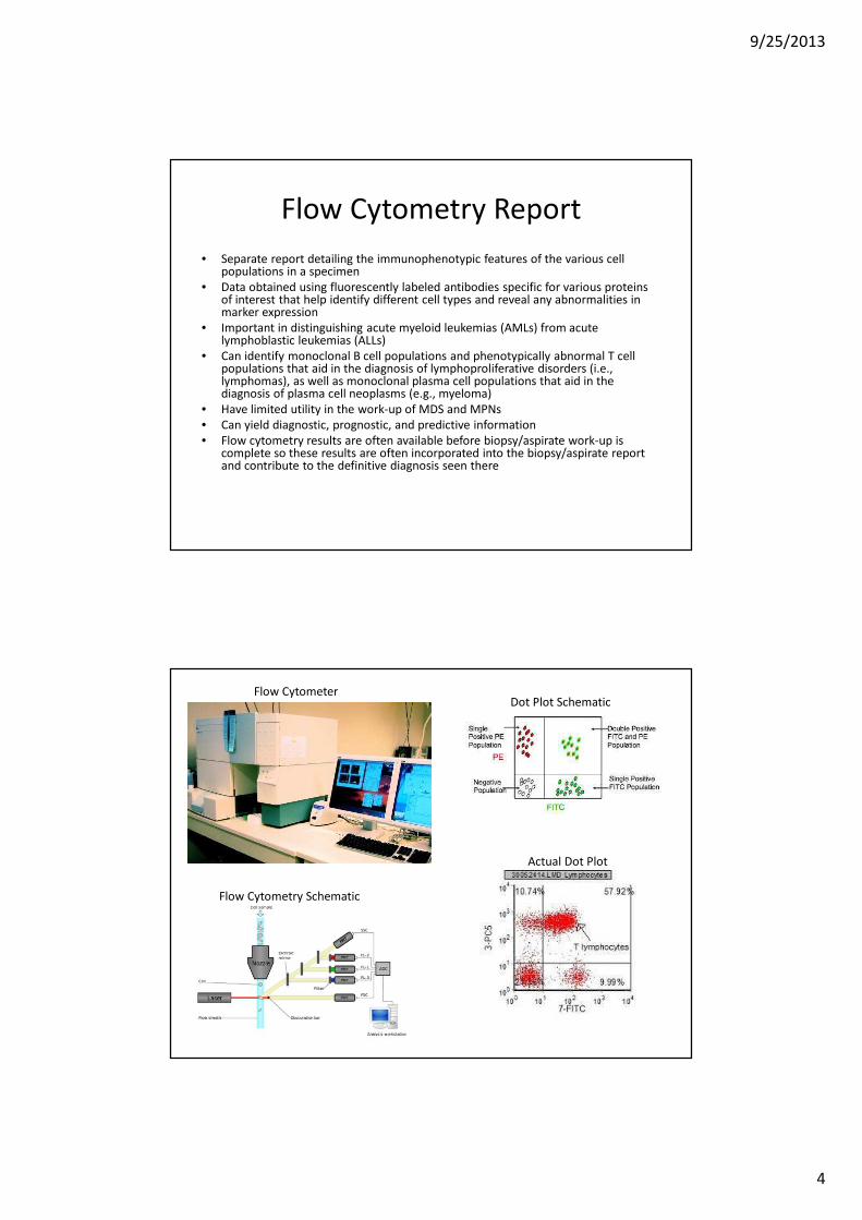
Flow Cytometry Report
• Separate report detailing the immunophenotypic features of the various cell populations in a specimen
• Data obtained using fluorescently labeled antibodies specific for various proteins of interest that help identify different cell types and reveal any abnormalities in marker expression
• Important in distinguishing acute myeloid leukemias (AMLs) from acute lymphoblastic leukemias (ALLs)
• Can identify monoclonal B cell populations and phenotypically abnormal T cell populations that aid in the diagnosis of lymphoproliferative disorders (i.e., lymphomas), as well as monoclonal plasma cell populations that aid in the diagnosis of plasma cell neoplasms (e.g., myeloma)
• Have limited utility in the work-up of MDS and MPNs
• Can yield diagnostic, prognostic, and predictive information
• Flow cytometry results are often available before biopsy/aspirate work-up is complete so these results are often incorporated into the biopsy/aspirate report and contribute to the definitive diagnosis seen there
Flow Cytometer
Flow Cytometry Schematic
Dot Plot Schematic
Actual Dot Plot

9/25/2013
5
Cytogenetics Report
• Separate report detailing any chromosomal abnormalities detected in the cells of a specimen.
• Two main methods: classic karyotyping and Fluorescence In Situ Hybridization (FISH)
• Very important in the work-up of MDS, AMLs, ALLs, and myelomas
• Also important in the evaluation of some lymphoproliferative disorders/lymphomas
• Occasionally relevant in the work-up of MPNs
• Can yield diagnostic, prognostic, and predictive information
Male Karyotype (normal)
FISH

9/25/2013
6
Molecular Report
• Separate report detailing any specific gene mutations or monoclonal T or B cell populations present in a specimen
• Numerous different methodologies and genes can be looked at depending on the situation
• Important in the evaluation of AMLs, MPNs, and lymphomas
• Can yield diagnostic, prognostic, and predictive information
Pathology Workshop Objectives
• Define the general philosophy of reviewing pathology reports– Review the various components of
• Bone marrow aspirate and biopsy
• Flow Cytometry
• Cytogenetic Studies
• Molecular studies
– Outline a general approach to interpreting pathology reports• Define the difference between a non-diagnostic bone marrow biopsy and a negative
bone marrow biopsy
• Discuss case studies with relevant pathology reports / bone marrow biopsies in relation to staging and prognosis / mortality risk. If new treatments improve prognosis, briefly outline the details.– Review a case of Anemia of Unknown Etiology
– Review a case of Early Myelodysplastic Syndrome
– Review a case of MGUS / Multiple Myeloma
– Review a case of Chronic Lymphocytic Leukemia/Small Lymphocytic Lymphoma (CLL/SLL)

9/25/2013
7
Approach to Interpreting a Hemepath
Report
• Biopsy/aspirate report will generally incorporate
flow cytometric, cytogenetic, and molecular
information (as they become available), and yield
the most comprehensive diagnosis
• If biopsy/aspirate report does not mention flow
cytometry, cytogenetics, or molecular, see if
there are separate reports for these studies
because information in these reports can make
an otherwise non-diagnostic biopsy diagnostic
“Non-diagnostic” vs. “Negative” Bone
Marrow Biopsies
• “Non-diagnostic” Biopsy: Findings (morphologic, immunophenotypic, cytogenetic, and molecular) not normal but not sufficient in and of themselves to render a definitive diagnosis based on defined criteria, but do not exclude the possibility of certain disorders
• “Negative” Biopsy: Findings (morphologic, immunophenotypic, cytogenetic, and molecular) are normal, and do not support the diagnosis of a hematologic (or any other) disorder

9/25/2013
8
Pathology Workshop Objectives
• Define the general philosophy of reviewing pathology reports– Review the various components of
• Bone marrow aspirate and biopsy
• Flow Cytometry
• Cytogenetic Studies
• Molecular studies
– Outline a general approach to interpreting pathology reports• Define the difference between a non-diagnostic bone marrow biopsy and a negative
bone marrow biopsy
• Discuss case studies with relevant pathology reports / bone marrow biopsies in relation to staging and prognosis / mortality risk. If new treatments improve prognosis, briefly outline the details.– Review a case of Anemia of Unknown Etiology
– Review a case of Early Myelodysplastic Syndrome
– Review a case of MGUS / Multiple Myeloma
– Review a case of Chronic Lymphocytic Leukemia/Small Lymphocytic Lymphoma (CLL/SLL)

9/25/2013
9
Anemia of Unknown Etiology
• 55-year old Hispanic female with no significant past medical history presented to her PCP with a primary complaint of fatigue, and was found to have a normochromic normocytic anemia (Hgb 8, Hct 24) with mild anisopoikilocytosis; WBC, ANC, and platelet counts are normal; spleen not enlarged
• Serum iron, ferritin, folate, and vitamin B12 levels are all normal
• LDH, total bilirubin, and haptoglobin all wnl
• Reticulocyte count is decreased; EPO levels are elevated
• Bone marrow biopsy is performed
Anemia of Unknown Etiology (cont.)Bone Marrow Biopsy

9/25/2013
10
Anemia of Unknown Etiology (cont.)
Bone Marrow Aspirate
• Immunohistochemistry for Parvovirus B19
performed on the biopsy is negative
• Flow cytometry: No phenotypically abnormal
cell population detected; no increase in blasts
• Cytogenetic studies: Karyotype: 46,XX [20];
normal MDS FISH panel
• Negative for BCR-ABL1 gene rearrangement
and JAK2 V617F mutation
Anemia of Unknown Etiology (cont.)

9/25/2013
11
• Final bone marrow report: Normocellular marrow (40-50%) with marked erythroid hypoplasia (M:E ratio >10:1) with an associated left-shift. Blasts are not increased. No myelodysplasia is identified. No evidence of a lymphoproliferative disorder or plasma cell dyscrasia
• DDx: Acquired pure red cell aplasia secondary to viral infection (not parvovirus B19), drug, autoimmunity, thymoma, or idiopathic
• Example of “non-diagnostic” bone marrow biopsy
Anemia of Unknown Etiology (cont.)
Approach to Anemia of Unknown
Etiology
• Blood loss
– GI
– Menstrual
– Other
• Increased destruction
– Hemolysis
• Impaired production
– Bone marrow dysfunction

9/25/2013
12
Underwriting Concerns
• Would you insure this woman?
– If so, how would you assess her mortality?
– If not, what would need to change to consider
insuring her?
• Are there any factors present that you would
consider positive in terms of mortality?
• Are there any factors present that you would
consider negative in terms of mortality?
Questions? Comments?
9/25/2013
13
Pathology Workshop Objectives
• Define the general philosophy of reviewing pathology reports– Review the various components of
• Bone marrow aspirate and biopsy
• Flow Cytometry
• Cytogenetic Studies
• Molecular studies
– Outline a general approach to interpreting pathology reports• Define the difference between a non-diagnostic bone marrow biopsy and a negative
bone marrow biopsy
• Discuss case studies with relevant pathology reports / bone marrow biopsies in relation to staging and prognosis / mortality risk. If new treatments improve prognosis, briefly outline the details.– Review a case of Anemia of Unknown Etiology
– Review a case of Early Myelodysplastic Syndrome
– Review a case of MGUS / Multiple Myeloma
– Review a case of Chronic Lymphocytic Leukemia/Small Lymphocytic Lymphoma (CLL/SLL)
Early Myelodysplastic Syndrome (MDS)
• 67-year old Caucasian male with no significant past medical history presents to his PCP for an annual check-up and a routine CBC showed a normochromic normocytic anemia (Hgb 9, Hct 27) with mild anisopoikilocytosis; WBC, ANC, and platelet counts are normal; spleen is not enlarged
• Serum iron, ferritin, folate, and vitamin B12 levels are all normal
• LDH, total bilirubin, and haptoglobin all wnl
• Although serum iron, ferritin, folate, and vitamin B12 levels are all normal, the patient is placed on a multivitamin and iron supplement , and asked to return in 6 months
• A follow-up CBC performed 6 months later shows a persistent normochromic normocytic anemia (Hgb 9, Hct 27) with mild anisopoikilocytosis refractory to multivitamin and iron supplement treatment; WBC, ANC, and platelet counts are still normal
• Reticulocye count performed at the time is decreased and EPO levels are elevated
• Bone marrow biopsy is performed

9/25/2013
14
Early MDS (cont.)
Bone Marrow Biopsy
Early MDS (cont.)Bone Marrow Aspirate
Wright-Giemsa stain Iron stain

9/25/2013
15
• Flow cytometry: No phenotypically abnormal
cell population detected; no increase in blasts
• Cytogenetic studies: Karyotype: 45,XY,-
5[7]/46,XY[13]; MDS FISH panel: -5 detected
in 70 of 200 cells analyzed
• Negative for BCR-ABL1 gene rearrangement
and JAK2 V617F mutation
Early MDS (cont.)
Early MDS (cont.)
• Bone marrow report before cytogenetic studies: Mildly hypercellular marrow (~50%) with mild erythroid hyperplasia and mild dyserythropoiesis, pending cytogenetics. Blasts are not increased. DDX includes MDS, megaloblastic anemia, toxin exposure, and medication effect
• Bone marrow report after cytogenetic studies: MDS morphologically consistent with refractory anemia with unilineage dysplasia (good prognostic factor) with isolated chromosome 5 deletion (good prognostic factor) and no increase in blasts (good prognostic factor), IPPS score of 0 (low risk group)

9/25/2013
16
Myelodysplastic Syndrome
• Results from ineffective function or
production of myeloid blood cells
• Classification:
– Refractory cytopenia with unilineage dysplasia
– Refractory anemia with ringed sideroblasts
– Refractory cytopenia with multilineage dysplasia
– Refractory anemia with excess blasts
– MDS with isolated del(5q)
IPSS-R Cytogenetic Risk Groups
Cytogenetic Prognostic Subgroups Cytogenetic Abnormalities
Very good -Y, del(11q)
Good Normal, del(5q), del(12p), del(20q)
double including del(5q
Intermediate del(7q), +8, +19, i(17q), any other single
or double independent clones
Poor -7, inv(3)/t(3q)/del(3q), double including
-7/del(7q), Complex: 3 abnormalities
Very poor Complex: >3 abnormalities

9/25/2013
17
Revised International Prognostic
Scoring System (IPSS-R)Prognostic
Variable
Score
0 0.5 1.0 1.5 2.5 3 4
Cytogenetics Very
good
Good Int Poor Very
poor
Bone marrow
blast (%)
≤5 >2 - <5 5 - 10 11 -33 >10
Hemoglobin
(g/dl)
≥10 8 - <10 <8
Platelets ≥100 50 - 100 <50
Absolute
neutrophil
count
≥0.8 <0.8
IPPS-R: Prognostic Subgroup Clinical
Outcomes
Risk Group IPSS-R Score Median Overall
Survival (years)
Median Time to
25% AML Evolution
Very low ≤1.5 8.8 >14.5
Low >1.5 - 3 5.3 10.8
Intermediate >3.5 - 4.5 3.0 3.2
High >4.5 - 6 1.6 1.4
Very high >6 0.8 0.7

9/25/2013
18
Underwriting Concerns
• Would you insure this man?
– If so, how would you assess his mortality?
– If not, what would need to change to consider
insuring him?
• Are there any factors present that you would
consider positive in terms of mortality?
• Are there any factors present that you would
consider negative in terms of mortality?
Questions? Comments?

9/25/2013
19
Pathology Workshop Objectives
• Define the general philosophy of reviewing pathology reports– Review the various components of
• Bone marrow aspirate and biopsy
• Flow Cytometry
• Cytogenetic Studies
• Molecular studies
– Outline a general approach to interpreting pathology reports• Define the difference between a non-diagnostic bone marrow biopsy and a negative
bone marrow biopsy
• Discuss case studies with relevant pathology reports / bone marrow biopsies in relation to staging and prognosis / mortality risk. If new treatments improve prognosis, briefly outline the details.– Review a case of Anemia of Unknown Etiology
– Review a case of Early Myelodysplastic Syndrome
– Review a case of MGUS / Multiple Myeloma
– Review a case of Chronic Lymphocytic Leukemia/Small Lymphocytic Lymphoma (CLL/SLL)
MGUS/Multiple Myeloma
• 64-year old African American male with no significant past medical history presents to his PCP for an annual check-up
• A CBC with manual differential is performed, which shows normal counts; however the slide is flagged for pathologist review because of mild rouleaux formation
• A complete metabolic panel shows mildly increased total protein (8.5 g/dL) with normal albumin (4.0 g/dL) and normal creatinine (1.0 mg/dL)
• An SPEP is ordered, which shows a small M spike in the gamma region of 1.5 g/dL; serum IFE shows a monoclonal IgG kappa protein
• Bone marrow biopsy is performed

9/25/2013
20
MGUS/Multiple Myeloma (cont.)
Peripheral blood smear showing rouleaux formation
MGUS/Multiple Myeloma (cont.)

9/25/2013
21
MGUS/Multiple Myeloma (cont.)
Bone Marrow Aspirate
• Flow cytometry: Small (0.5%) kappa-restricted
monoclonal plasma cell population detected
• Cytogenetic studies: Karyotype: 46,XY[20];
Multiple Myeloma FISH panel: 17p13 (TP53)
deletion detected in 15 of 200 cells analyzed
• No molecular testing performed
MGUS/Multiple Myeloma (cont.)

9/25/2013
22
MGUS/Multiple Myeloma (cont.)
• Bone marrow report: Normocellular marrow (~40%) involved by plasma cell myeloma (monoclonal kappa-restricted plasma cells account for ~20% of the bone marrow cellularity)
• Follow-up bone scan shows no bone lesions
• Serum Beta2-microglobulin level: 1.5 mg/L
• Since no evidence of end organ damage or myeloma-related symptoms, clinical diagnosis of low stage smoldering multiple myeloma with 17q13 deletion (poor prognostic factor)
Monoclonal Gammopathy
• Monoclonal Gammopathy of Unknown
Significance (MGUS)
– Monoclonal paraprotein <3g/dl
– Bone marrow biopsy with <10% plasma cells
– Absence of any end-organ sequelae
– Risk of progression to myeloma – 1%/yr

9/25/2013
23
Monoclonal Gammopathy – cont.
• Asymptomatic (smoldering) Myeloma
– Serum IgA or IgG monoclonal protein ≥3.0 g/dl
and/or
– ≥10% more plasma cells in bone marrow
– No evidence of end-organ sequelae
– Risk of progression to myeloma 10%/yr for first
five years, 3%/yr for next five years, then 1%/yr
Underwriting Concerns
• Would you insure this man?
– If so, how would you assess his mortality?
– If not, what would need to change to consider
insuring him?
• Are there any factors present that you would
consider positive in terms of mortality?
• Are there any factors present that you would
consider negative in terms of mortality?

9/25/2013
24
Questions? Comments?
Pathology Workshop Objectives
• Define the general philosophy of reviewing pathology reports– Review the various components of
• Bone marrow aspirate and biopsy
• Flow Cytometry
• Cytogenetic Studies
• Molecular studies
– Outline a general approach to interpreting pathology reports• Define the difference between a non-diagnostic bone marrow biopsy and a negative
bone marrow biopsy
• Discuss case studies with relevant pathology reports / bone marrow biopsies in relation to staging and prognosis / mortality risk. If new treatments improve prognosis, briefly outline the details.– Review a case of Anemia of Unknown Etiology
– Review a case of Early Myelodysplastic Syndrome
– Review a case of MGUS / Multiple Myeloma
– Review a case of Chronic Lymphocytic Leukemia/Small Lymphocytic Lymphoma (CLL/SLL)

9/25/2013
25
CLL/SLL
• 59-year old Caucasian female is referred to a hematologist for work-up of a mild leukocytosis with relative and absolute lymphocytosis (WBC count 14,000, 70% lymphocytes), mild normochromic normocytic anemia (Hgb 10, Hct 30), and thrombocytopenia (platelets 100,000)
• Peripheral blood smear review shows a lymphocytosis consisting mostly of small mature lymphocytes with occasional smudge cells; the red blood cells show occasional microspherocytes and platelets are mildly reduced but morphologically unremarkable
• Physical exam shows no lymphadenopathy and no hepatosplenomegaly
• Flow cytometry, cytogenetics, and molecular studies of the peripheral blood are performed
• Also, because of the anemia and thrombocytopenia a bone marrow biopsy is performed
CLL/SLL (cont.)
• PB Flow cytometry results: 50% (of all WBCs) population of kappa-restricted B cells positive for CD19 (mod), CD20 (dim), CD22 (dim), CD5 (dim), CD23 (mod), CD38 (mod), and ZAP-70 (mod) c/w CLL/SLL
• PB Cytogenetic results: Karyotype: 46,XX,del(17p13)[10]/46,XX[10]; CLL FISH panel: 17p13 (TP53) deletion detected in 100 of 200 cells analyzed, negative for t(11;14)(q13;q32)
• PB Molecular results: Non-hypermutated
Peripheral blood smear

9/25/2013
26
CLL/SLL (cont.)
Bone Marrow Biopsy Bone Marrow Aspirate
CLL/SLL (cont.)
• BM flow cytometry results: No monoclonal B
cell or phenotypically abnormal T cell
population detected
• BM cytogenetics: Karyotype 46,XX[20]; CLL
FISH panel: WNL
• BM molecular studies: No monoclonal B cell
population detected (IgH PCR studies)

9/25/2013
27
CLL/SLL (cont.)
• Peripheral blood report: CLL/SLL, CD38 and ZAP-70+ (poor prognostic factors), with 17p13 (TP53) deletion (poor prognostic factor), non-hypermutated (poor prognostic factor)
• Bone marrow report: Normocellular marrow (~40%) with trilineage hematopoieis; no evidence of involvement by a B or T cell lymphoproliferative disorder; no increase in blasts
• Example of a “negative” bone marrow
CLL/SLL (cont.)
• Follow up lab studies showed elevated LDH, elevated total and indirect bilirubin with normal direct bilirubin, decreased haptoglobin, and a positive DAT
• Given these lab studies, the negative bone marrow biopsy, and the presence of occasional microspherocytes on the PB smear, a diagnosis of CLL/SLL-associated Evan’s syndrome was made to explain the patient’s anemia and thrombocytopenia

9/25/2013
28
RAI Clinical Staging System
Revised Staging
System
Original Staging
System
Clinical Features at
Diagnosis
Median Survival,
Years
Low risk 0 Blood & marrow
lymphocytes
12
I Lymphocytosis &
enlarged LN
11
Intermediate risk II Lymphocytosis &
enlarged spleen
&/or liver
8
High risk III Lymphocytosis &
anemia (Hgb <11)
5
IV Lymphocytosis &
thromocytopenia
(Plt <100,000)
7
Underwriting Concerns
• Would you insure this woman?
– If so, how would you assess her mortality?
– If not, what would need to change to consider
insuring her?
• Are there any factors present that you would
consider positive in terms of mortality?
• Are there any factors present that you would
consider negative in terms of mortality?

9/25/2013
29
Questions? Comments?