2013 519 allslides - ualberta.cacampbell//resources/2013_519_7_SPR.pdf · through a prism on top of...
7
84 Bioanalytical chemistry 7. Surface plasmon resonance
Transcript of 2013 519 allslides - ualberta.cacampbell//resources/2013_519_7_SPR.pdf · through a prism on top of...
84
Bioanalytical chemistry
7. Surface plasmon resonance

Total Internal Reflection Fluorescence (TIRF) Microscopy
85
Light is totally reflected when it strikes an interface at an
angle greater than the critical angle (defined as the angle above which total reflection occurs, recall Snell’s law)
http://snowflakemorayeel.wordpress.com/category/pets/http://en.wikipedia.org/wiki/File:TIR_in_PMMA.jpg
When light is internally reflected, a standing wave known as an evanescent wave is formed at the boundaryThe strength of this wave decays
exponentially with distance from the boundary

Total Internal Reflection Fluorescence (TIRF) Microscopy
86
The energy of the evanescent wave is equal to the energy of that reflected light. Accordingly, the
evanescent wave can be used to excite appropriate fluorophores to the excited state.
This allows the selective excitation of fluorophores that are in close proximity (i.e., within 200 nm) to the interface. For example, if a cell is attached to
the surface, only those fluorophores in the membrane, or very close to the membrane, will be excited and observed in the fluorescence image.
focal adhesionswidefield TIRF
tubulinwidefield TIRF Overlay
• TIRF microscopy is one of the most sensitive fluorescence microscopy techniques and is routinely used for single molecule imaging. The big advantage is that the fluorescence background is very low since fluorophores further from the surface are not being excited.
• Wilson, W.D., Analyzing Biomolecular Interactions, Science 295, 2002, 2103-2105.
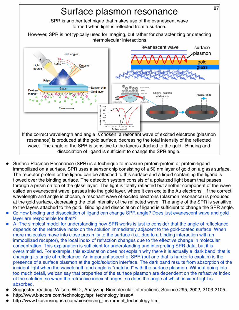
Surface plasmon resonance 87
However, SPR is not typically used for imaging, but rather for characterizing or detecting intermolecular interactions.
SPR is another technique that makes use of the evanescent wave formed when light is reflected from a surface.
If the correct wavelength and angle is chosen, a resonant wave of excited electrons (plasmon resonance) is produced at the gold surface, decreasing the total intensity of the reflected wave. The angle of the SPR is sensitive to the layers attached to the gold. Binding and
dissociation of ligand is sufficient to change the SPR angle.
goldglass
surface plasmon
evanescent wave
• Surface Plasmon Resonance (SPR) is a technique to measure protein-protein or protein-ligand immobilized on a surface. SPR uses a sensor chip consisting of a 50 nm layer of gold on a glass surface. The receptor protein or the ligand can be attached to this surface and a liquid containing the ligand is flowed over the binding surface. The detection system consists of a polarized light beam that passes through a prism on top of the glass layer. The light is totally reflected but another component of the wave called an evanescent wave, passes into the gold layer, where it can excite the Au electrons. If the correct wavelength and angle is chosen, a resonant wave of excited electrons (plasmon resonance) is produced at the gold surface, decreasing the total intensity of the reflected wave. The angle of the SPR is sensitive to the layers attached to the gold. Binding and dissociation of ligand is sufficient to change the SPR angle.
• Q: How binding and dissociation of ligand can change SPR angle? Does just evanescent wave and gold layer are responsible for that/?
• A: The simplest model for understanding how SPR works is just to consider that the angle of reflectance depends on the refractive index on the solution immediately adjacent to the gold-coated surface. When more molecules move into close proximity to the surface (i.e., due to a binding interaction with an immobilized receptor), the local index of refraction changes due to the effective change in molecular concentration. This explanation is sufficient for understanding and interpreting SPR data, but it is oversimplified. For example, this explanation does not explain why there it is actually a 'dark band' that is changing its angle of reflectance. An important aspect of SPR (but one that is harder to explain) is the presence of a surface plasmon at the gold/solution interface. The dark band results from absorption of the incident light when the wavelength and angle is "matched" with the surface plasmon. Without going into too much detail, we can say that properties of the surface plasmon are dependent on the refractive index of the solution, so when the refractive index changes, so does the angle at which incident light is absorbed.
• Suggested reading: Wilson, W.D., Analyzing Biomolecular Interactions, Science 295, 2002, 2103-2105.• http://www.biacore.com/technology/spr_technology.lasso#• http://www.biosensingusa.com/biosensing_instrument_technology.html

Surface plasmon resonance 88
In a typical sensorgram, a baseline signalwith no change in RU over time is followed
sample injection, which produces the as-sociation phase where RU increases withtime. If the reaction rates are fast enough, itis possible to reach a steady state region,
here the rates of association and dissocia-tion are equal. Resuming buffer flow causesthe complex to dissociate, and the kinetics of
). At adesired time, a regeneration solution can beinjected to remove remaining P bound to thesurface, and the original RU value is re-es-tablished (see schematic). Thus, both kinetics
25
20
15
10
5
0
0 100 200 300 400 500Time (s)
Res
pons
e (R
U)
Set of sensorgrams.
In principle, you could get a rough estimate of the Kd by plotting response (RU) as a function of sample concentration. However, this is not how it
is typically done.
Rather, the association and dissociation phases of the RU vs time trace are fit in order to extract
the kon and koff kinetic constants.
Kd = koff kon
recall that
• http://www.biosensingusa.com/biosensing_instrument_technology.html#• http://www.biacore.com/technology/spr_technology.lasso#

Antibodies shown to scale
Penetration depth (arbitrary definition)
Penetration depth of the evanescent wave on a SPR chip with a gold film
89
• Defined arbitrary penetration depth = 1/e (.37) * maximum intensity• This arbitrary cutoff occurs around 150-200 nm• This is equivalent to ~10 antibodies stacked side by side (shown to scale) • The dimensions of an antibody are ~13 x ~16 nm• The point is that the further you get from the surface, the weaker the SPR effect will be. However, it is
difficult to imagine a case where the signal could drop below ~50% due to distance from the chip.• The dashed line represents the strength of the evanescent wave in the absence of the gold film. The solid
line is the strength with the gold film.

A SPR test for biotin in food samples
How does this test work?Why not just use
immobilized avidin?
http://www.biacore.com/pdf/products/foodkits/Biotin_Kit_PIS.pdf
90
• Q. Testing for biotin in food - is the reason that we don't use avidin or streptavidin immobilized and capturing biotin because we have no way of attaching an unknown quantity of biotin post translationally with an antigen in order to use antibodies against that antigen? or is it also because of the cost of using avidin/streptavidin? or because of the difficulty of reusing the surface again in another sample due to the very low Kd of the avidins and biotin?
• A. One reason is that biotin is a small molecule and would produce only a small change in SPR signal when it binds to the avidin or streptavidin. The second reason is that it would be nearly impossible to reuse the chip, due to the high affinity of biotin for avidin.