2009 03 Heister - Triple functionalisation of SWNTswith ... author: Elena Heister...
27
Corresponding author: Elena Heister ([email protected]) 1 Triple functionalisation of single-walled carbon nanotubes with doxorubicin, a monoclonal antibody, and a fluorescent marker for targeted cancer therapy Elena Heister 1* , Vera Neves 1 , Carmen Tîlmaciu 2 , Kamil Lipert 3 , Vanesa Sanz Beltrán 1 , Helen M. Coley 1 , S. Ravi P. Silva 4 , and Johnjoe McFadden 1 1 Faculty of Health and Medical Sciences, University of Surrey, Guildford, GU27XH, UK 2 CIRIMAT-LCMIE, CNRS-UMR 5085, Université Paul Sabatier, 118 Route de Narbonne, 31077 Toulouse, Cedex 9, France 3 Leibniz Institute for Solid State and Materials Research (IFW), Helmholtzstraße 20, 01069 Dresden, Germany 4 Nanoelectronics Centre, Advanced Technology Institute, University of Surrey, Guildford, GU27XH, UK Abstract Single-walled carbon nanotubes (SWCNTs) have been identified as a transporter for anti- cancer drugs, as they are capable of penetrating mammalian cell membranes and allow for a high drug loading due to their nanoscale dimensions and high aspect ratio. In addition, they can assist the targeting of therapeutic agents to the desired site of action by conjugation to antibodies or ligands of cancer cell surface receptors, which increases the effectiveness of the treatment and reduces side effects. In this work, we present a method for the triple
Transcript of 2009 03 Heister - Triple functionalisation of SWNTswith ... author: Elena Heister...
Corresponding author: Elena Heister ([email protected]) 1
Triple functionalisation of single-walled carbon nanotubes with
doxorubicin, a monoclonal antibody, and a fluorescent marker
for targeted cancer therapy
Elena Heister1*, Vera Neves1, Carmen Tîlmaciu2, Kamil Lipert3, Vanesa Sanz Beltrán1,
Helen M. Coley1, S. Ravi P. Silva4, and Johnjoe McFadden1
1Faculty of Health and Medical Sciences, University of Surrey, Guildford, GU27XH, UK
2CIRIMAT-LCMIE, CNRS-UMR 5085, Université Paul Sabatier, 118 Route de Narbonne,
31077 Toulouse, Cedex 9, France
3Leibniz Institute for Solid State and Materials Research (IFW), Helmholtzstraße 20, 01069
Dresden, Germany
4Nanoelectronics Centre, Advanced Technology Institute, University of Surrey, Guildford,
GU27XH, UK
Abstract
Single-walled carbon nanotubes (SWCNTs) have been identified as a transporter for anti-
cancer drugs, as they are capable of penetrating mammalian cell membranes and allow for a
high drug loading due to their nanoscale dimensions and high aspect ratio. In addition, they
can assist the targeting of therapeutic agents to the desired site of action by conjugation to
antibodies or ligands of cancer cell surface receptors, which increases the effectiveness of the
treatment and reduces side effects. In this work, we present a method for the triple
2
functionalisation of oxidised SWCNTs with the anti-cancer drug doxorubicin, a monoclonal
antibody, and a fluorescent marker at non-competing binding sites. The proposed
methodology allows for the targeted delivery of the anticancer drug to cancer cells and the
visualisation of the cellular uptake of SWCNTs by confocal microscopy. We show that the
complex is efficiently taken up by cancer cells with subsequent intracellular release of
doxorubicin, which then translocates to the nucleus while the nanotubes remain in the
cytoplasm.
1. Introduction
The development of novel anti-cancer therapies is often limited by administration problems
of drugs, such as insolubility, inefficient distribution, lack of selectivity, and the inability of
drugs to cross cellular barriers. Currently, most of these problems are subject of intense
studies and in this context, many different types of drug delivery systems have been
investigated, including quantum dots, silica nanoparticles, dendrimers, micelles, molecular
conjugates, and liposomes [1-4]. Within the family of nanomaterials, carbon nanotubes have
emerged as a novel tool for the delivery of therapeutic molecules into cells [5]. Carbon
nanotubes are undoubtedly one of the most striking discoveries in the quest for new materials
in recent years, as these unique structures possess tremendous strength [6], an extreme aspect
ratio, and are excellent thermal and electrical conductors [7,8]. Apart from that, carbon
nanotubes can immobilise therapeutic molecules, such as proteins, antibodies, DNA or drugs
on their surface [9,10] or in the hollow cavity [11] and are capable of penetrating mammalian
cell membranes [12], which makes them ideal candidates for drug delivery systems.
3
In this article, we present a new approach to functionalise single-walled carbon nanotubes
(SWCNTs) with three different agents for multimodal drug delivery. This includes the anti-
cancer drug doxorubicin, a monoclonal antibody for molecular targeting, and the fluorescent
marker fluorescein, all of which are attached to oxidised SWCNTs at non-competing binding
sites. Doxorubicin was chosen as a therapeutic agent, as it is a widely used anti-cancer drug
for the treatment of many cancers and furthermore possesses a fluorescent hydroxy-
substituted anthraquinone chromophore with an emission spectrum ranging from 500 to 750
nm that enables intracellular tracking of the drug by confocal microscopy. The monoclonal
antibody recognizes carcinoembryonic antigen (CEA), which is a tumour marker for the
identification of metastatic disease following surgical resection and is relevant for a variety
of adenocarcinomas, such as colon cancer. Fluorescein is used to label SWCNTs in order to
colocalise them inside cells by confocal microscopy next to doxorubicin.
The attachment of the three agents was accomplished by two different chemical approaches;
non-covalent and covalent binding. Non-covalent chemistry has the advantage that it
preserves the structure of SWCNTs and thus their unique properties. However, a non-
covalent bond is susceptible to environmental factors, such as pH and salt concentration, and
is in general less stable than a covalent bond. In contrast to this, covalent attachment of
molecules to carbon nanotubes depends on the introduction of chemically reactive groups to
their relatively inert sp2 structure, which can only be achieved by harsh treatments, such as
oxidation with concentrated inorganic acids [13], fluorination by elemental fluorine [14] or a
1,3 dipolar cycloaddition reaction [15] and inevitably introduces defects to the nanotube
structure.
4
On the basis of these considerations, non-covalent functionalisation was chosen for
attachment of doxorubicin to allow for its release after cellular uptake, whereas covalent
functionalisation was employed to attach fluorescein and CEA antibodies to oxidised
SWCNTs. The latter was achieved by using the hydrophilic protein bovine serum albumin
(BSA) as a multifunctional linker. In total, BSA possesses 60 amino groups in lysine chains
and 99 carboxylic groups as part of glutamic and aspartic acid residues. Those residues
located on the protein surface are available for conjugation reactions and thus provide two
different types of binding sites for the covalent attachment of molecules. In our approach,
BSA is conjugated to carboxylic groups of oxidised SWCNTs by one of its amine groups, to
NHS-fluorescein via the remaining amine groups, and to monoclonal CEA antibodies via its
carboxylic groups. Since multiple copies of fluorescein and antibodies can be attached to one
BSA molecule, the degree of coupling is increased manifold. Figure 1 shows a schematic
illustration of the structure, which we propose for the nanotube-drug-BSA conjugates. The
distribution of functional groups after acid oxidation of carbon nanotubes has already been
investigated in previous studies, which showed that functional groups are found at the ends
of the tubes and along the sidewalls [16,17]. Results obtained by atomic force microscopy
(AFM) demonstrate a similar distribution for BSA molecules attached to functional groups of
oxidised, doxorubicin-loaded SWCNTs in our experiments (insert of Figure 1).

5
Figure 1 Schematic illustration of the doxorubicin-fluorescein-BSA-antibody-SWCNT
complexes (red=doxorubicin, green=fluorescein, light blue=BSA, dark blue=antibodies).
Insert: AFM image of doxorubicin-fluorescein-BSA-SWCNT complexes (without
antibodies).
The cellular uptake of drug-antibody-SWCNT complexes was studied by means of confocal
microscopy using CEA-expressing WiDr colon cancer cells. The design of the nanotube-drug
complexes is based on the hypothesis that functionalised SWCNTs are taken up by WiDr
cells by endocytosis. Following uptake, the lower pH inside endosomes is expected to trigger
the release of doxorubicin from the nanotube due to increased hydrophilicity of the drug,
which can then translocate to the nucleus and exert its cytotoxic action.
2. Experimental Details
6
2.1. Material
SWCNTs were purchased from NanoLab, Inc. (produced by CVD using iron (ferrocene) as
catalyst, purity > 60%, length 1-5 µm, Lot No. 90907). WiDr human colon cancer cells were
obtained from LGC Promochem (Teddington, UK; Catalog No. CCL-218). The monoclonal
CEA antibody with specificity for CD66e was purchased from AbD Serotec (MCA1744F).
MEM (31095-029) and Opti-MEM medium (31985-047) were obtained from Invitrogen
(Paisley, UK). Doxorubicin hydrochloride (44583), 5(6)-carboxyfluorescein N-
hydroxysuccinimide ester (NHS-fluorescein, 21878), bovine serum albumin (BSA, A3294),
1-ethyl-3-[3-dimethyl-aminopropyl] carbodiimide hydrochloride (EDC, E1769), N-
hydroxysulfo-succinimide (sulfo-NHS, 56485), 2-mercaptoethanol (63700), and
ethanolamine hydrochloride (E6133) were purchased from Sigma-Aldrich (Poole, UK).
2.2. Oxidation and Purification of SWCNTs
As-prepared SWCNTs are usually contaminated with metal catalyst particles, amorphous
carbon and graphitic nanoparticles. However, the application of carbon nanotubes for
biomedical purposes has created a demand for high purity material, especially if the sidewalls
of carbon nanotubes are to serve as platform for the non-covalent attachment of
biomolecules. Li et al. have compared different oxidation methods for CVD-synthesized
SWCNTs and found that nitric acid pre-sonication, followed by refluxing in a mixture of
concentrated acids yield the best results with respect to purity [18]. This two-step process
was slightly adapted for the work presented here. In the first step, 100 mg of SWCNTs were
incubated in 20 mL of concentrated nitric acid and sonicated with a tip sonicator for 6x 10s
7
to disentangle the tubes and break up bundles. Subsequently, the mixture was incubated in a
95 °C water bath for 2h. To remove dissolved metal catalyst particles, the oxidation treatment
was followed by a centrifugation step at 3000 g for 30 min. After discarding the supernatant,
the purified tubes were washed twice with bi-distilled water. In the second step, the purified
sample of SWCNTs (~100 mg) was dispersed in a 3:1 mixture of concentrated nitric and
sulfuric acid and sonicated for 6x 10s with a tip sonicator. Subsequently, the mixture was
refluxed at 110 °C for 2h, followed by three washing steps with water and centrifugation at
3000 g for 30 min to remove excess acid in the supernatant. Finally, the oxidised SWCNTs
were vacuum filtered using a 0.2 µm polycarbonate filter (Whatman) until the eluate was
clear and of neutral pH. The filter cake was dried overnight at room temperature, weighed,
and resuspended in water to a concentration of 5 mg/mL. This material will henceforth be
referred to as “oxSWCNTs”.
2.3. Characterisation of pristine and oxidised SWCNTs
The term “carbon nanotube” does not describe a simple, chemical structure, but a whole class
of materials, which vary in their numbers of walls, diameter distribution, length distribution,
chirality, purity, catalyst material, impurity species and defects. Thus, characterisation of the
applied material is crucial in order to ensure quality and reproducibility. The nanotubes in
this work were characterised by scanning electron microscopy (SEM), high resolution
transmission electron microscopy (HR-TEM), atomic force microscopy (AFM), Raman
spectroscopy, and thermogravimetric analyis (TGA).
2.3.1. SEM
8
All SEM images in this study were obtained using a FEI 200 Nova NanoSEM system.
Powder samples were prepared by placing a small amount of SWCNT powder onto a piece
of double-sided adhesive tape stuck to a metal stub. Liquid samples were prepared by placing
a 2 µL drop of a 10 µg/mL SWCNT suspension onto a small piece of silicon stuck to a metal
stub and evaporating residual water using a hot plate. The metal stubs holding the samples
were then placed into the vacuum chamber of the microscope and images acquired using a
voltage of 10 kV and a spot size of 3.0 to 5.0.
2.3.2. HR-TEM
The HR-TEM work presented in this report was carried out using a High Resolution
Transmission Electron Microscope JEOL-EM 2100. For sample preparation, oxSWCNTs
were diluted with ethanol and briefly sonicated in a water bath for dispersion. 3-5 drops of
this suspension were then placed onto a 300 mesh copper TEM grid and excess liquid was
removed by touching one edge of the grid with filter paper.
2.3.3. AFM
For AFM sample preparation, oxSWCNTs were diluted with water to obtain a concentration
of 10 µg/mL. A droplet of 2 µL was placed onto a freshly cleaved mica substrate (1 cm2) and
was dried with help of compressed air. AFM measurements were performed using a Veeco
Dimension 3100 Atomic Force Microscope in tapping mode. To obtain a length distribution,
several AFM images were taken at a frame size of 5 µm x 5 µm and the lengths of 500
nanotubes was measured using “Gwyddion 2.9”, a free SPM data visualization and imaging
tool released under the GNU General Public License.
9
2.3.4. Raman Spectroscopy
Samples for Raman spectroscopy were prepared by placing a small quantity of carbon
nanotube powder or a droplet of an oxSWCNT suspension between a glass slide and a cover
slip. Measurements were performed using a NT-MDT NTEGRA Spectra Probe
NanoLaboratory with excitation at 633 nm.
2.3.5. TGA
For a typical TGA experiment, ~ 1 mg of nanotube material was placed in the sample holder
in the furnace of a Rheometric Scientific TG 760 series and the material was heated up at a
rate of 10 °C/min, while the weight was measured and recorded continuously.
2.4. Triple functionalisation of SWCNTs with doxorubicin, fluorescein, and CEA
antibodies
Labeling of BSA with fluorescein
3 mg NHS-fluorescein (10 mg/mL in DMSO) was mixed with 150 mg BSA in sodium
phosphate buffer 20 mM pH 8.5, followed by incubation for 2h in darkness at room
temperature while stirring. To remove excess fluorescein, the mixture was filtered and
washed repeatedly using Amicon Ultra® 30 kDa centrifugal filtration devices (Millipore)
until the eluate was clear. The fluorescein-BSA conjugates were resuspended in sodium
phosphate buffer 20 mM (pH 7.4) at a BSA concentration of 5 mg/mL.
10
The degree of labeling (fluorescein/BSA molar ratio) was determined by separately
calculating the protein and fluorophore molar concentrations of the conjugate based on
absorbance measurements and expressing these concentrations as a ratio. Absorbance at 280
nm (A280) was used to determine the protein concentration in the sample. However, because
fluorescent dyes also absorb at 280 nm, a correction factor must be used to adjust for the
amount of A280 contributed by the dye. The correction factor (CF) equals the A280 of the dye
divided by the Amax of the dye and was determined to be 0.26 for NHS-fluorescein (data not
shown).
In the first step, the molar concentration of BSA ( BSAc ) was calculated according to equation
(1) which is derived from Beer-Lambert’s Law ( lcA ) with A being the absorbance of
the respective molecules, CF being the correction factor, ε being the molar extinction
coefficient, l being the path length of the cuvette and DF being the dilution factor:
DFl
CFAAc
BSA
nfluoresceiBSABSA
) ( 494,280,
(1)
In the second step, the degree of labeling (moles fluorescein per mole protein) was calculated
according to equation (2):
DFc
A
n
n
BSAnfluorescei
nfluorescei
BSA
nfluorescei
494, (2)
11
Non-covalent attachment of doxorubicin to oxidised SWCNTs
150 µL of a 5 mg/mL oxSWCNT suspension was dispersed in 15 mL sodium phosphate
buffer 20 mM pH 8.5 and 3 mL of a 5 mM doxorubicin hydrochloride solution was added.
The mixture was sonicated in a water bath for 15 min and incubated overnight while stirring.
Unbound doxorubicin was removed by filtering and washing using Amicon Ultra® 30 kDa
centrifugal filter devices (Millipore). Finally, the doxorubicin-loaded SWCNTs were
resuspended in 15 mL sodium phosphate buffer 20 mM pH 7.4 at a SWCNT concentration of
50 µg/mL and 5 mL of these complexes were kept as a control sample, whereas the other 10
mL were used for further conjugation steps. In order to determine how much doxorubicin
was bound to the nanotubes, the eluate of the centrifugation steps containing unbound
doxorubicin were collected and analysed by UV/vis absorption spectroscopy by measuring
the absorbance at the first excitation maximum of doxorubicin at 490 nm.
Connection of fluorescein-labeled BSA and doxorubicin-loaded SWCNTs
10 mL doxorubicin-loaded SWCNTs were dispersed in 10 mL sodium phosphate buffer 20
mM pH 8.5. If necessary, the pH was adjusted to 8.5 by means of a trisodium phosphate 20
mM solution. Next, 1 mL of a 20 mM EDC solution and 1 mL of a 50 mM sulfo-NHS
solution were added and the mixture was incubated for 5 min at room temperature. To
quench excess EDC, 70 µL 2-mercaptoethanol was added to a final concentration of 20 mM.
To start the coupling reaction, 10 mL of the fluorescein-labeled BSA solution (5 mg/mL
BSA) was added and the reaction allowed to proceed for 2h at room temperature. Finally, 1
mL of a 250 mM ethanolamine hydrochloride solution was added to quench the reaction. The
nanotube complexes were washed by vacuum filtration using 0.2 µM polycarbonate filters
12
(Whatman) and were resuspended in 10 mL sodium phosphate buffer 20 mM pH 7.4. 5 mL
of these complexes were kept for experiments and the remaining 5 mL were used for the last
conjugation step.
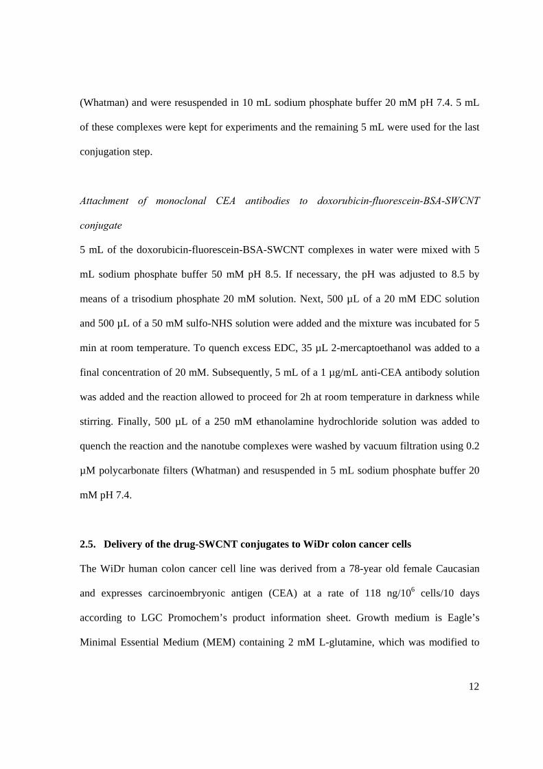
Attachment of monoclonal CEA antibodies to doxorubicin-fluorescein-BSA-SWCNT
conjugate
5 mL of the doxorubicin-fluorescein-BSA-SWCNT complexes in water were mixed with 5
mL sodium phosphate buffer 50 mM pH 8.5. If necessary, the pH was adjusted to 8.5 by
means of a trisodium phosphate 20 mM solution. Next, 500 µL of a 20 mM EDC solution
and 500 µL of a 50 mM sulfo-NHS solution were added and the mixture was incubated for 5
min at room temperature. To quench excess EDC, 35 µL 2-mercaptoethanol was added to a
final concentration of 20 mM. Subsequently, 5 mL of a 1 µg/mL anti-CEA antibody solution
was added and the reaction allowed to proceed for 2h at room temperature in darkness while
stirring. Finally, 500 µL of a 250 mM ethanolamine hydrochloride solution was added to
quench the reaction and the nanotube complexes were washed by vacuum filtration using 0.2
µM polycarbonate filters (Whatman) and resuspended in 5 mL sodium phosphate buffer 20
mM pH 7.4.
2.5. Delivery of the drug-SWCNT conjugates to WiDr colon cancer cells
The WiDr human colon cancer cell line was derived from a 78-year old female Caucasian
and expresses carcinoembryonic antigen (CEA) at a rate of 118 ng/106 cells/10 days
according to LGC Promochem’s product information sheet. Growth medium is Eagle’s
Minimal Essential Medium (MEM) containing 2 mM L-glutamine, which was modified to
13
contain 1.0 mM sodium pyruvate, 0.1 mM non-essential amino acids, 10% fetal bovine
serum, and penicillin/streptomycin (100 units/mL penicillin and 100 µg/mL streptomycin in
the final formulation). The medium is formulated for use with a 5% CO2 in air atmosphere.
For the cell studies, WiDr cells were grown on cover slips in a 6-well plate until 70-80%
confluent. The prepared doxorubicin-fluorescein-BSA-SWCNT conjugates and the controls
were mixed with Opti-MEM medium at a 1:1 ratio (500 µL complexes + 500 µL medium),
resulting in final SWCNT concentrations of 25 µg/mL. The mixtures were sonicated for 3x
10s using a tip sonicator and remaining agglomerates removed by centrifugation for 60 s at
3000 g. Next, WiDr cells were incubated with 1 mL of the sample or control solutions in
Opti-MEM medium for 4h at 37 °C. The cells were then washed three times with PBS and
fixed by incubation with freshly prepared 4% paraformaldehyde solution at room
temperature for 30 min. Finally, the cells were washed twice with PBS and mounted on a
glass slide.
Confocal microscopy was carried out using a Zeiss LSM 510 confocal microscope equipped
with an Argon/2 laser in multi-channel mode. The excitation wavelength was set to 488 nm
for fluorescein and doxorubicin. Emission of fluorescein was detected between 500 and 530
nm and emission of doxorubicin was detected from 650 to 710 nm. Images were taken at 63x
magnification by means of a C-Apochromat 63x/1.4 Oil objective.
3. Results and Discussion
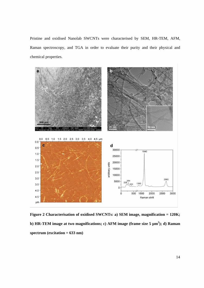
3.1. Characterisation of oxidised SWCNTs
14
Pristine and oxidised Nanolab SWCNTs were characterised by SEM, HR-TEM, AFM,
Raman spectroscopy, and TGA in order to evaluate their purity and their physical and
chemical properties.
Figure 2 Characterisation of oxidised SWCNTs: a) SEM image, magnification = 120K;
b) HR-TEM image at two magnifications; c) AFM image (frame size 5 µm2); d) Raman
spectrum (excitation = 633 nm)
15
The SEM image of oxidised SWCNTs (Fig. 2a) demonstrates that acid oxidation entangles
clumps of nanotubes that are present in the bulk sample. The oxidised SWCNTs seem to
form small bundles which are covered with a thin layer of a material that has been shown to
consist of decomposed products of carbonaceous impurities [19], such as partially oxidised
graphitic fragments, and amorphous carbon. Thermal oxidation can be applied to remove
these remaining impurities; however, they improve the solubilisation properties of carbon
nanotubes by acting as a surfactant [20]. The TEM picture in Fig. 2b reveals more detailed
information about the bundling state of the nanotubes. Apart from single nanotubes, bundles
of up to ~15 nanotubes can be observed, which are again covered with the same oxidation
debris. AFM analysis of carbon nanotubes was applied to extract a height profile and to
generate a length distribution of the oxidised SWCNTs (Fig. 2c, Fig. 3). The height profile
features seven SWCNT bundles with diameters in the range of 2.0 to 4.0 nm. Given that the
average diameter of a single SWCNT is about 1 nm, this corresponds to bundles consisting of
3 to 10 nanotubes. The length distribution was used to quantify the extent of shortening by
the oxidation process. The maximum of the size distribution after oxidation lies at 450 nm
and 83% of the tubes were found to be between 200 nm and 1000 nm long. Considering that
the lengths of SWCNTs in the pristine sample ranged from 1 to 5 µm, this corresponds to a
shortening of 80%. The Raman spectrum (Fig. 2d) shows all features that are characteristic
for SWCNTs: the radial breathing mode (RBM), the D-band and the G-band [21,22]. The
RBM results from low-energy radial vibrations of carbon atoms in the nanotube backbone
and hence its frequency is inversely proportional to the tube diameter according to
12.5 /5.223 d , where d is the tube diameter in nm and is Raman shift in wave
numbers [23]. The RBM in the Raman spectrum of Fig. 2d ranges from 170 cm-1 to 410 cm-1;

16
corresponding to a diameter distribution between 0.56 and 1.42 nm with an average diameter
of approximately 1 nm. Maximum intensities are observed at 228 cm-1, 284 cm-1, and 403
cm-1; relating to accumulated diameters at 1.06 nm, 0.82 nm, and 0.57 nm. The disorder-
induced D-band at 1380 cm-1 indicates the presence of defective, sp3 hybridised sites on
SWCNTs and the G-band at 1640 cm-1 is a tangential vibrational mode characteristic to all
graphitic materials. The G/D intensity ratio is a good indicator for the quality of bulk
samples. Here, the G/D ratio was found to be 13.95, demonstrating a low quantity of defects.
Figure 3 Height profile (a) of oxSWCNTs extracted from Fig. 2c and length distribution
(b) of oxSWCNTs (n=500)
Last, but not least, oxidised SWCNTs were analysed by TGA to determine the amount of
metal catalyst in the bulk sample. The metal residue was found to be 11.2% for pristine
SWCNTs and 11.6% for oxidised SWCNTs (data not shown). This indicates that the present
metallic catalyst particles are encapsulated in carbon shells (so-called nanoonions) [24],
which protect them from acid oxidation, but also prevent them from being released in
biological environments and causing toxic effects.
17
3.2. Triple functionalisation of SWCNTs with doxorubicin, fluorescein, and CEA
antibodies
We developed a method for the triple functionalisation of SWCNTs, which allows for the
attachment of a monoclonal antibody for targeting purposes, an anti-cancer drug, and a
fluorescent dye at non-competing binding sites to enable visualisation of cellular uptake. The
functionalisation procedure is a critical process comprising 4 steps and can easily result in
many kinds of unwanted crosslinking reactions. Hence, the reaction parameters have to be
selected and adjusted carefully. In the first step, the protein BSA is labeled with the amine-
reactive dye NHS-fluorescein. Here, it is crucial to find the optimal NHS-fluorescein
concentration in order to leave enough free amino groups on the BSA for the later attachment
of carbon nanotubes. However, leaving too many free binding sites would lead to
crosslinking of nanotubes via BSA and thus to precipitation. Optimisation of the
fluorescein/BSA ratio was performed by testing three different weight ratios (1:20, 1:50, and
1:100) in their binding properties to SWCNTs (see next paragraph). Furthermore, the degree
of fluorescein labeling was determined by UV/vis absorption spectroscopy (see equations 1
and 2). A 1:20 weight ratio of fluorescein/BSA resulted in a degree of labeling of 3.99,
whereas a 1:50 weight ratio correlated with a degree of 2.19 and a 1:100 weight ratio with a
degree of 1.19.
In the second step of the coupling reaction, the anti-cancer drug doxorubicin is non-
covalently attached to the sidewalls of oxidised carbon nanotubes via -stacking and
hydrophilic interactions with carboxylic groups. The strength of this non-covalent binding is
18
pH dependent: At a low pH, the amino group in the sugar moiety of doxorubicin is
protonated, which increases the molecule’s hydrophilicity and thus its solubility in water.
However, at a higher pH, the amino group becomes deprotonated, resulting in stronger
hydrophobic interactions with the nanotubes’ side walls and lower solubility in water. Liu et
al. have attached doxorubicin non-covalently to the side walls of SWCNTs by overnight
incubation at pH 9 [25]. However, Janssen et al. reported that doxorubicin hydrochloride has
its maximum stability at pH 4 [26] and processing the drug at pH 9 may cause its degradation
and thus the loss of its therapeutic efficiency. Thus, to ensure a high drug loading whilst
maintaining doxorubicin’s therapeutic properties, the reaction was carried out at a slightly
lower pH of 8.5. According to Beijnen et al., who generated a pH-dependent decomposition
profile for doxorubicin at 50 °C corrected for buffer and ionic strength influences [27], the
decomposition rate of doxorubicin at pH 8.5 is approximately 35% less than at pH 9.0.
The amount of doxorubicin bound to the nanotubes was calculated by determining the
quantity of unbound doxorubicin in the eluate of the filtration step by UV/vis absorption
spectroscopy. It was found that 87.5% of doxorubicin had attached to the nanotubes, whereas
12.5% had been washed off. In terms of weight, the ratio of doxorubicin to oxidised
SWCNTs is 20:1, which demonstrates the enormous binding capacity of the nanotubes. Liu
et al. have determined a weight ratio of 4:1 in a similar experiment [25] – however, the
surface of their (non-oxidised) SWCNTs was already covered with phospholipid-PEG-
molecules to a certain extent, which were previously applied to solubilise the nanotubes.
In the third step, the fluorescein-labeled BSA and doxorubicin-loaded SWCNTs are
conjugated. In order to avoid non-specific binding of BSA to the nanotubes’ sidewalls, it is
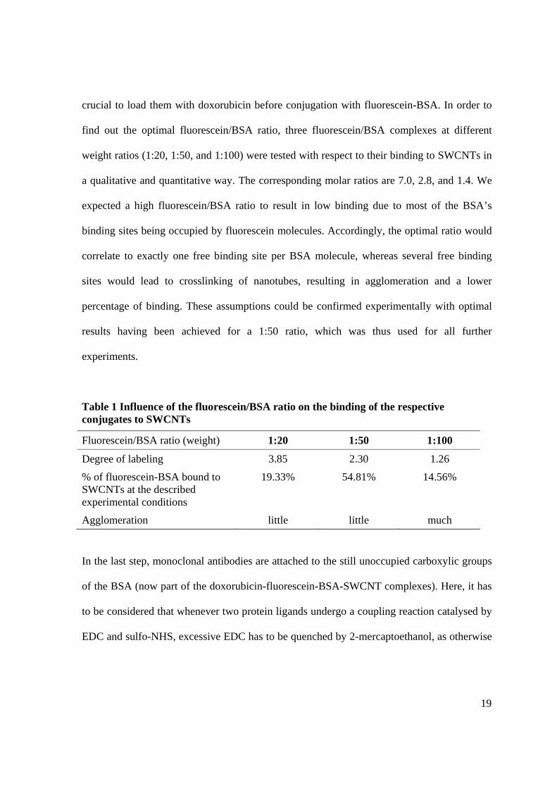
19
crucial to load them with doxorubicin before conjugation with fluorescein-BSA. In order to
find out the optimal fluorescein/BSA ratio, three fluorescein/BSA complexes at different
weight ratios (1:20, 1:50, and 1:100) were tested with respect to their binding to SWCNTs in
a qualitative and quantitative way. The corresponding molar ratios are 7.0, 2.8, and 1.4. We
expected a high fluorescein/BSA ratio to result in low binding due to most of the BSA’s
binding sites being occupied by fluorescein molecules. Accordingly, the optimal ratio would
correlate to exactly one free binding site per BSA molecule, whereas several free binding
sites would lead to crosslinking of nanotubes, resulting in agglomeration and a lower
percentage of binding. These assumptions could be confirmed experimentally with optimal
results having been achieved for a 1:50 ratio, which was thus used for all further
experiments.
Table 1 Influence of the fluorescein/BSA ratio on the binding of the respective conjugates to SWCNTs
Fluorescein/BSA ratio (weight) 1:20 1:50 1:100
Degree of labeling 3.85 2.30 1.26
% of fluorescein-BSA bound to SWCNTs at the described experimental conditions
19.33% 54.81% 14.56%
Agglomeration little little much
In the last step, monoclonal antibodies are attached to the still unoccupied carboxylic groups
of the BSA (now part of the doxorubicin-fluorescein-BSA-SWCNT complexes). Here, it has
to be considered that whenever two protein ligands undergo a coupling reaction catalysed by
EDC and sulfo-NHS, excessive EDC has to be quenched by 2-mercaptoethanol, as otherwise
20
carboxylic groups of both coupling partners (BSA and CEA antibodies) would be activated
and cause extensive crosslinking.
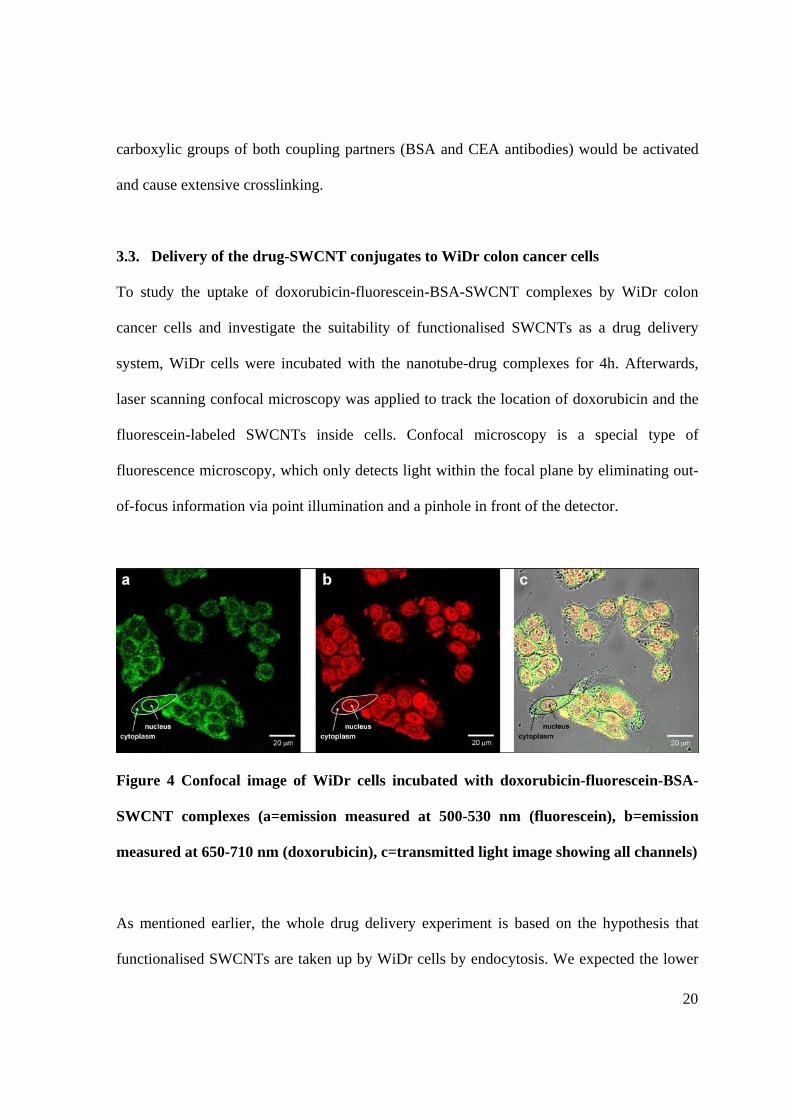
3.3. Delivery of the drug-SWCNT conjugates to WiDr colon cancer cells
To study the uptake of doxorubicin-fluorescein-BSA-SWCNT complexes by WiDr colon
cancer cells and investigate the suitability of functionalised SWCNTs as a drug delivery
system, WiDr cells were incubated with the nanotube-drug complexes for 4h. Afterwards,
laser scanning confocal microscopy was applied to track the location of doxorubicin and the
fluorescein-labeled SWCNTs inside cells. Confocal microscopy is a special type of
fluorescence microscopy, which only detects light within the focal plane by eliminating out-
of-focus information via point illumination and a pinhole in front of the detector.
Figure 4 Confocal image of WiDr cells incubated with doxorubicin-fluorescein-BSA-
SWCNT complexes (a=emission measured at 500-530 nm (fluorescein), b=emission
measured at 650-710 nm (doxorubicin), c=transmitted light image showing all channels)
As mentioned earlier, the whole drug delivery experiment is based on the hypothesis that
functionalised SWCNTs are taken up by WiDr cells by endocytosis. We expected the lower
21
pH inside endosomes to trigger the release of doxorubicin from the nanotubes due to
increased hydrophilicity. Indeed, Fig. 4 clearly demonstrates, that doxorubicin (red) does not
colocalise any longer with the fluorescein-labeled SWCNTs (green) after internalization by
WiDr cells, but was found to accumulate in the nuclei of cells (seen as round structures
positioned in the in the centre of the cell). The fluorescently-labeled nanotubes, however, are
mainly observed outside the nuclei within the cytoplasm. Areas in the cell where doxorubicin
and fluorescein-labeled nanotubes colocalise can be observed along the border between the
cytoplasm and the nucleus, the so-called “nuclear envelope”, and on the cell membrane. The
delivery efficiency in this experiment was 100%, meaning that all cells have taken up the
SWCNT complexes.
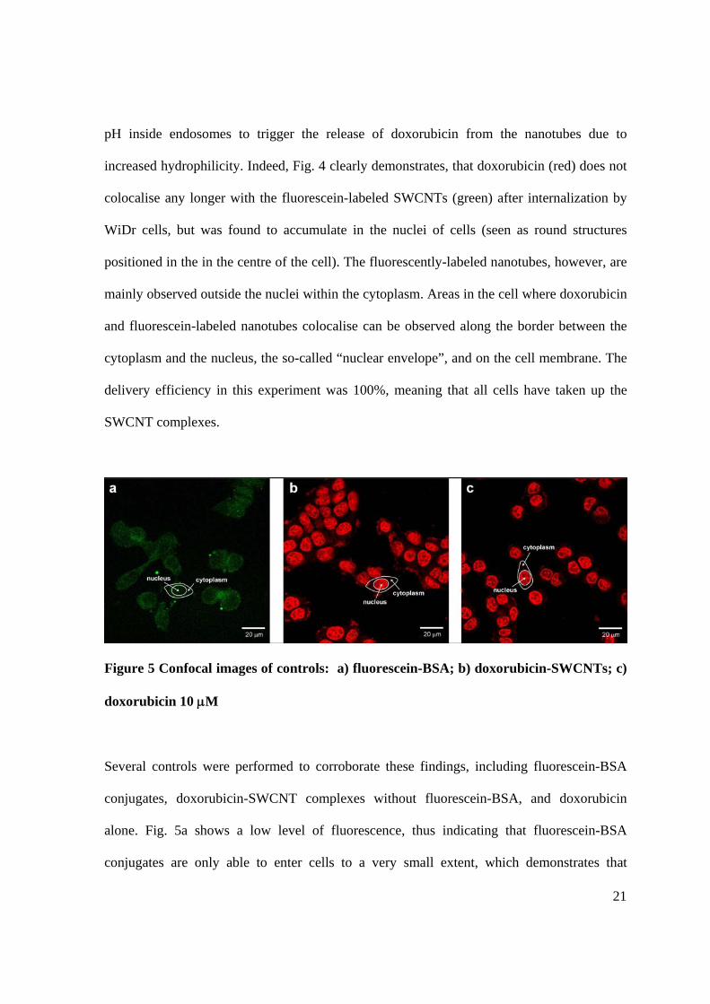
Figure 5 Confocal images of controls: a) fluorescein-BSA; b) doxorubicin-SWCNTs; c)
doxorubicin 10 M
Several controls were performed to corroborate these findings, including fluorescein-BSA
conjugates, doxorubicin-SWCNT complexes without fluorescein-BSA, and doxorubicin
alone. Fig. 5a shows a low level of fluorescence, thus indicating that fluorescein-BSA
conjugates are only able to enter cells to a very small extent, which demonstrates that
22
SWCNTs play an important role as a delivery system. However, when comparing Fig. 5b and
5c it becomes obvious that this is not the case for doxorubicin: due to its molecular structure,
the drug is able to cross cell membranes without the help of a carrier system and accumulates
in the cell nuclei, as shown before. Nevertheless, the nanotubes still play an important role,
as they enable molecular targeting via the attachment of monoclonal antibodies and are
crucial for the delivery of drugs that are not taken up by cells under normal conditions.
Apart from this, more work needs to be done with regard to the question whether carbon
nanotubes are able to enter nuclei of cells. This study clearly showed that the nanotubes
remained in the cytoplasm under the applied conditions. The same behavior was
demonstrated by Kam et al., who studied the uptake of Cy3-DNA-wrapped SWCNTs by
Hela cells and detected fluorescence in the cytoplasm only [28]. However, Pantarotto et al.
have conducted a study where two different carbon nanotube conjugates were introduced into
human and murine fibroblasts and found that water-soluble SWCNTs conjugated to
fluorescein isothiocyanate (FITC) accumulated in the cytoplasm, whereas SWCNTs
conjugated to a FITC-labeled peptide were observed to distribute inside the nucleus [12].
Furthermore, Cheng et al. recently showed that FITC-PEG-SWCNTs accumulated in the
nuclei of several mammalian cell lines [29]. This indicates that the ability of CNTs to cross
cellular barriers, such as the nuclear envelope, is dependent on the type of functionalisation.
4. Conclusion
This work presents a novel functionalisation approach to equip oxidised SWCNTs with three
different agents for multimodal drug delivery. It is demonstrated that SWCNTs can
23
successfully transport a potent anti-cancer drug to human cancer cells with subsequent
translocation of the drug to the nucleus, while the SWCNTs remain in the cytoplasm. This
finding suggests that SWCNTs may be used to enhance cellular pharmacokinetics and have
considerable implications for improving anti-cancer drug delivery. However, targeting and
toxicity of SWCNTs remain important issues that need to be examined in greater depth and
will be addressed in future studies. The triple functionalisation methodology presented in this
article already incorporates the attachment of monoclonal antibodies for molecular targeting
and current work is in progress in order to investigate, whether the uptake of CEA antibody-
tagged SWCNT conjugates by WiDr colon cancer cells is increased in comparison to
untagged conjugates.
Acknowledgements
This work has been performed in the framework of the FP6 Marie Curie Research Training
Network CARBIO (Multifunctional carbon nanotubes for biomedical applications) funded by
the European Union.
24
References
[1] Panyam J, Labhasetwar V. Biodegradable nanoparticles for drug and gene delivery to
cells and tissue. Advanced Drug Delivery Reviews. 2003 Feb 24;55(3):329-47.
[2] Okuda T, Kawakami S, Akimoto N, Niidome T, Yamashita F, Hashida M. PEGylated
lysine dendrimers for tumor-selective targeting after intravenous injection in tumor-
bearing mice. Journal of Controlled Release. 2006 Dec 1;116(3):330-6.
[3] Merdan T, Kopecek J, Kissel T. Prospects for cationic polymers in gene and
oligonucleotide therapy against cancer. Advanced Drug Delivery Reviews. 2002 Sep
13;54(5):715-58.
[4] Park JW. Liposome-based drug delivery in breast cancer treatment. Breast Cancer
Research. 2002;4(3):93-7.
[5] Klingeler R, Hampel S, Büchner B. Carbon nanotube based biomedical agents for
heating, temperature sensoring and drug delivery. International Journal of
Hyperthermia. 2008;24(6):496-505.
[6] Yakobson BI, Avouris P. Mechanical properties of carbon nanotubes. In: Dresselhaus
MS, Dresselhaus G, Avouris P, eds. Carbon nanotubes: Synthesis, structure,
properties, and applications. Berlin/Heidelberg: Springer 2001:287-327.
[7] Odom TW, Huang JL, Kim P, Lieber CM. Structure and electronic properties of
carbon nanotubes. Journal of Physical Chemistry B. 2000 Apr 6;104(13):2794-809.
[8] Hone J, Llaguno MC, Biercuk MJ, Johnson AT, Batlogg B, Benes Z, et al. Thermal
properties of carbon nanotubes and nanotube-based materials. Applied Physics A:
Materials Science & Processing. 2002 Mar;74(3):339-43.
[9] Lin Y, Allard LF, Sun YP. Protein-affinity of single-walled carbon nanotubes in
water. Journal of Physical Chemistry B. 2004 Mar 25;108(12):3760-4.
[10] Shim M, Kam NWS, Chen RJ, Li YM, Dai HJ. Functionalization of carbon nanotubes
for biocompatibility and biomolecular recognition. Nano Letters. 2002 Apr;2(4):285-
8.
25
[11] Hampel S, Kunze D, Haase D, Kramer K, Rauschenbach M, Ritschel M, et al. Carbon
nanotubes filled with a chemotherapeutic agent: a nanocarrier mediates inhibition of
tumor cell growth. Nanomedicine. 2008 Apr;3(2):175-82.
[12] Pantarotto D, Briand JP, Prato M, Bianco A. Translocation of bioactive peptides
across cell membranes by carbon nanotubes. Chemical Communications. 2004 Jan
7(1):16-7.
[13] Liu J, Rinzler AG, Dai HJ, Hafner JH, Bradley RK, Boul PJ, et al. Fullerene pipes.
Science. 1998 May 22;280(5367):1253-6.
[14] Mickelson ET, Huffman CB, Rinzler AG, Smalley RE, Hauge RH, Margrave JL.
Fluorination of single-wall carbon nanotubes. Chemical Physics Letters. 1998 Oct
30;296(1-2):188-94.
[15] Georgakilas V, Tagmatarchis N, Pantarotto D, Bianco A, Briand JP, Prato M. Amino
acid functionalisation of water soluble carbon nanotubes. Chemical Communications.
2002(24):3050-1.
[16] Azamian BR, Coleman KS, Davis JJ, Hanson N, Green MLH. Directly observed
covalent coupling of quantum dots to single-wall carbon nanotubes. Chemical
Communications. 2002(4):366-7.
[17] Cech J, Curran SA, Zhang DH, Dewald JL, Avadhanula A, Kandadai M, et al.
Functionalization of multi-walled carbon nanotubes: Direct proof of sidewall
thiolation. Physica Status Solidi B: Basic Solid State Physics. 2006
Nov;243(13):3221-5.
[18] Li Y, Zhang XB, Luo JH, Huang WZ, Cheng JP, Luo ZQ, et al. Purification of CVD
synthesized single-wall carbon nanotubes by different acid oxidation treatments.
Nanotechnology. 2004 Nov;15(11):1645-9.
[19] Nagasawa S, Yudasaka M, Hirahara K, Ichihashi T, Iijima S. Effect of oxidation on
single-wall carbon nanotubes. Chemical Physics Letters. 2000 Oct 6;328(4-6):374-80.
[20] Rosca ID, Watari F, Uo M, Akaska T. Oxidation of multiwalled carbon nanotubes by
nitric acid. Carbon. 2005 Dec;43(15):3124-31.
26
[21] Saito R, Kataura H. Optical properties and Raman spectroscopy of carbon nanotubes.
In: Dresselhaus MS, Dresselhaus G, Avouris P, eds. Carbon nanotubes: Synthesis,
structure, properties, and applications. Berlin/Heidelberg: Springer 2001:213-46.
[22] Dresselhaus MS, Dresselhaus G, Saito R, Jorio A. Raman spectroscopy of carbon
nanotubes. Physics Reports: Review Section of Physics Letters. 2005 Mar;409(2):47-
99.
[23] Bachilo SM, Strano MS, Kittrell C, Hauge RH, Smalley RE, Weisman RB. Structure-
assigned optical spectra of single-walled carbon nanotubes. Science. 2002 Dec
20;298(5602):2361-6.
[24] Pumera M. Carbon nanotubes contain residual metal catalyst nanoparticles even after
washing with nitric acid at elevated temperature because these metal nanoparticles are
sheathed by several graphene sheets. Langmuir. 2007 May 22;23(11):6453-8.
[25] Liu Z, Sun XM, Nakayama-Ratchford N, Dai HJ. Supramolecular chemistry on
water-soluble carbon nanotubes for drug loading and delivery. ACS Nano. 2007
Aug;1(1):50-6.
[26] Janssen MJH, Crommelin DJA, Storm G, Hulshoff A. Doxorubicin decomposition on
storage - Effect of pH, type of buffer and liposome encapsulation. International
Journal of Pharmaceutics. 1985;23(1):1-11.
[27] Beijnen JH, Vanderhouwen OAGJ, Underberg WJM. Aspects of the degradation
kinetics of doxorubicin in aqueous solution. International Journal of Pharmaceutics.
1986 Oct;32(2-3):123-31.
[28] Kam NWS, O'Connell M, Wisdom JA, Dai HJ. Carbon nanotubes as multifunctional
biological transporters and near-infrared agents for selective cancer cell destruction.
Proceedings of the National Academy of Sciences of the United States of America.
2005 Aug 16;102(33):11600-5.
[29] Cheng J, Fernando KAS, Veca LM, Sun Y-P, Lamond AI, Lam YW, et al. Reversible
accumulation of PEGylated single-walled carbon nanotubes in the mammalian
nucleus. ACS Nano. 2008;2(10):2085-94.
27
List of Figures
Figure 1 Schematic illustration of the doxorubicin-fluorescein-BSA-antibody-SWCNT
complexes (red=doxorubicin, green=fluorescein, light blue=BSA, dark
blue=antibodies). Insert: AFM image of doxorubicin-fluorescein-BSA-SWCNT
complexes (without antibodies)................................................................................ 5
Figure 2 Characterisation of oxidised SWCNTs: a) SEM image, magnification = 120K; b)
HR-TEM image at two magnifications; c) AFM image (frame size 5 µm2); d)
Raman spectrum (excitation = 633 nm) ................................................................. 14
Figure 3 Height profile (a) of oxSWCNTs extracted from Fig. 2c and length distribution (b)
of oxSWCNTs (n=500) .......................................................................................... 16
Figure 4 Confocal image of WiDr cells incubated with doxorubicin-fluorescein-BSA-
SWCNT complexes (a=emission measured at 500-530 nm (fluorescein),
b=emission measured at 650-710 nm (doxorubicin), c=transmitted light image
showing all channels) ............................................................................................. 20
Figure 5 Confocal images of controls: a) fluorescein-BSA; b) doxorubicin-SWCNTs; c)
doxorubicin 10 M................................................................................................. 21
List of Tables
Table 1 Influence of the fluorescein/BSA ratio on the binding of the respective conjugates
to SWCNTs ............................................................................................................ 19