2007 - Membrane Protein Structure
19
Membrane Protein Structure: Prediction versus Reality Arne Elofsson and Gunnar von Heijne Center for Biomembrane Research, Stockholm Bioinformatics Center, Department of Biochemistry and Biophysics, Stockholm University, SE-106 91 Stockholm, Sweden; email: [email protected], [email protected] Annu. Rev. Biochem. 2007. 76:125–40 First published online as a Review in Advance on January 11, 2007 The Annual Review of Biochemistry is online at biochem.annualreviews.org This article’ s doi: 10.1146/annurev.biochem.76.052705.163539 Copyright c 2007 by Annual Reviews. All rights reserved 0066-4154/07/0707-0125$20.00 Key Words bioinformatics, membrane protein structure prediction, topology Abstract Since high-resolution structural data are still scarce, different kinds of theoretical structure prediction algorithms are of major impor- tance in membrane protein biochemistry. But how well do the cur- rent prediction methods perform? Which structural features can be predicted and which cannot? An d what can we expect in the next few years? 125 A n n u . R e v . B i o c h e m . 2 0 0 7 . 7 6 : 1 2 5 1 4 0 . D o w n l o a d e d f r o m w w w . a n n u a l r e v i e w s . o r g A c c e s s p r o v i d e d b y U n i v e r s i d a d N a c i o n a l A u t o n o m a d e M e x i c o o n 0 9 / 0 1 / 1 5 . F o r p e r s o n a l u s e o n l y .
-
Upload
jason-parsons -
Category
Documents
-
view
6 -
download
0
description
un pequeño articulo cientifico donde se escalrecen las conformaciones estructurales de las proteinas de membrana
Transcript of 2007 - Membrane Protein Structure

7/18/2019 2007 - Membrane Protein Structure
http://slidepdf.com/reader/full/2007-membrane-protein-structure 1/19
Membrane ProteinStructure: Prediction versus Reality
Arne Elofsson and Gunnar von Heijne
Center for Biomembrane Research, Stockholm Bioinformatics Center, Department oBiochemistry and Biophysics, Stockholm University, SE-106 91 Stockholm, Sweden;email: [email protected], [email protected]
Annu. Rev. Biochem. 2007. 76:125–40
First published online as a Review in Advance on January 11, 2007
The Annual Review of Biochemistry is online at
biochem.annualreviews.org This article’s doi:10.1146/annurev.biochem.76.052705.163539
Copyright c 2007 by Annual Reviews. All rights reserved
0066-4154/07/0707-0125$20.00
Key Words
bioinformatics, membrane protein structure prediction, topology
Abstract
Since high-resolution structural data are still scarce, different kindsof theoretical structure prediction algorithms are of major importance in membrane protein biochemistry. But how well do the cur-
rent prediction methods perform? Which structural features can bepredicted and which cannot? And what can we expect in the next few
years?
125

7/18/2019 2007 - Membrane Protein Structure
http://slidepdf.com/reader/full/2007-membrane-protein-structure 2/19
Contents
INTRODUCTION... . . . . . . . . . . . . . . 126 MEMBRANE PROTEIN
STRUCTURES: THE BASICFACTS . . . . . . . . . . . . . . . . . . . . . . . . . . 126
MEMBRANE PROTEIN
BIOSYNTHESIS, FOLDING, AND OLIGOMERIZATION . . . 127 Membrane Targeting and
I n s e r t i o n . . . . . . . . . . . . . . . . . . . . . . 1 2 7Folding and Stability . . . . . . . . . . . . . 128
MEMBRANE PROTEIN
BIOINFORMATICS: WHAT THE SEQUENCES TELL . . . . . . 129
MEMBRANE PROTEINBIOINFORMATICS: WHAT
THE STRUCTURES TELL . . . . 129
MEMBRANE PROTEINSTRUCTURE PREDICTION:FROM 2D TO 2.5D AND 3D. . . . 130
2D Predictions . . . . . . . . . . . . . . . . . . . 130B en ch m ar kin g . . . . . . . . . . . . . . . . . . . . 1 3 1
Genome Annotation.... . . . . . . . . . . 1322.5D Predictions . . . . . . . . . . . . . . . . . 132
3D Predictions . . . . . . . . . . . . . . . . . . . 134
MEMBRANE PROTEINCLASSIFICATION SCHEMES
AND DATABASES . . . . . . . . . . . . . . 135
PROTEIN-PROTEININTERACTIONS.. . . . . . . . . . . . . . 135
CONCLUSIONS AND
O U T L O O K . . . . . . . . . . . . . . . . . . . . . 1 3 6
INTRODUCTION
Membrane proteins are crucial players in thecell and take center stage in processes rang-
ing from basic small-molecule transport tosophisticated signaling pathways. Many are
also prime contemporary or future drug tar-gets, and it has been estimated that more than
half of all drugs currently on the market are
directed against membrane proteins (1). By contrast, it is still frustratingly hard to ob-
tain high-resolution three-dimensional (3D)
structures of membrane proteins, and they
represent less than 1% of the structures in theProtein Data Bank (2). Even if the number
of experimentally known membrane proteinstructures is on the rise (3, 4), methods to pre-
dict their topology (i.e., the transmembranesegments and their in-out orientation across
the membrane) and fold type from the aminoacid sequence will be needed for many yearsto come.
In this review, we discuss current topol-ogy and structure prediction methods against
a background of knowledge that has beengleaned from membrane protein structures
andfrom studiesof protein insertion andfold-ingin cellular membranes. We attemptto pro-
vide a realistic picture of what one may andmay not expect from the various prediction
schemes and to identify major issues yet to beresolved.
MEMBRANE PROTEIN STRUCTURES: THE BASICFACTS
Integral membrane proteins come in two ba-
sic architectures: the α -helix bundle and the
β-barrel. Helix-bundle proteins are found in
all cellular membranes and represent an es-timated 20% to 25% of all open reading
frames (ORFs) in fully sequenced genomes
(5). The number of β-barrel membrane pro-teins is more uncertain because they are more
difficult to identify by sequence gazing; forbacteria, a rough estimate, based on the fact
that all known β-barrel proteins are in theouter membrane and hence are made with
an (easily predicted) N-terminal signal pep-tide, suggests that they account for no more
than a few percent of all ORFs. The EcoCycdatabase (6)currently lists 58 outer membrane
and 511 inner membrane proteins out of atotal of 4332 proteins; considering that the
number of inner membrane proteins in Es-
cherichia coli has been estimated to be closeto 1000 (5), one may guess at somewhere be-
tween 100 and 150 outer membrane proteins
126 Elofsson· von Heijne
7/18/2019 2007 - Membrane Protein Structure
http://slidepdf.com/reader/full/2007-membrane-protein-structure 3/19
in total. This number of ∼100 E. coli outer
membrane proteins is consistent with theresults from recent attempts to identify
bacterial outer membrane proteins computa-tionally (7–9).
Whether a helix bundle orβ-barrel, all in-tegral membrane proteins share common sur-
face characteristics with a belt of hydrophobic(mainly aliphatic) amino acids flanked by two“aromatic girdles” composed of Trp and Tyr
residues (10–12). This mirrors the structureof the surrounding lipid bilayer, with the lipid
headgroup regions corresponding to the aro-matic girdles and the hydrocarbon tail region
to the hydrophobic belt, and ensures a seam-less fit of the proteins to the membrane.
Even though their surface structures aresimilar, the two classes of proteins have com-
pletely different secondary structures andfolds. As their names imply, helix-bundle pro-
teins are built from long transmembrane α -
helices that pack together into more or lesscomplicated bundles, whereas β-barrel pro-
teins are large antiparallel β-sheets rolled upinto a barrel closed by the first and last strands
in the sheet. In both cases, all backbone hy-drogen bonds in the membrane-buried parts
of the protein are internally satisfied withinthe helices or between the β-strands. Another
fundamental difference between the helix-bundle and β-barrel proteins pertains to their
biosynthesis and mechanism of membrane in-sertion; this is discussed in the next section.
Because all current membrane protein
topology and structure prediction schemesfirst seek to identify the transmembrane seg-
ments, they are obviously quite different, andtheir variations depend on the class of pro-
tein for which they are designed. Generally speaking, long hydrophobic transmembrane
helices are easier to recognize in an aminoacid sequence than the much shorter and less
hydrophobic transmembrane β-strands, andpartly for this reason, much more bioinfor-
matics work has been devoted to the helix-bundle proteins—another instance of the
well-known dictum “always go for the easy
problems.”
Translocon: aprotein complex thatassures thetranslocation of proteins across acellular membrane
Endoplasmicreticulum (ER):organelle into whichsecretory andmembrane proteinsare delivered uponsynthesis on theribosome
MEMBRANE PROTEIN BIOSYNTHESIS, FOLDING,
AND OLIGOMERIZATION
Membrane Targeting and Insertion
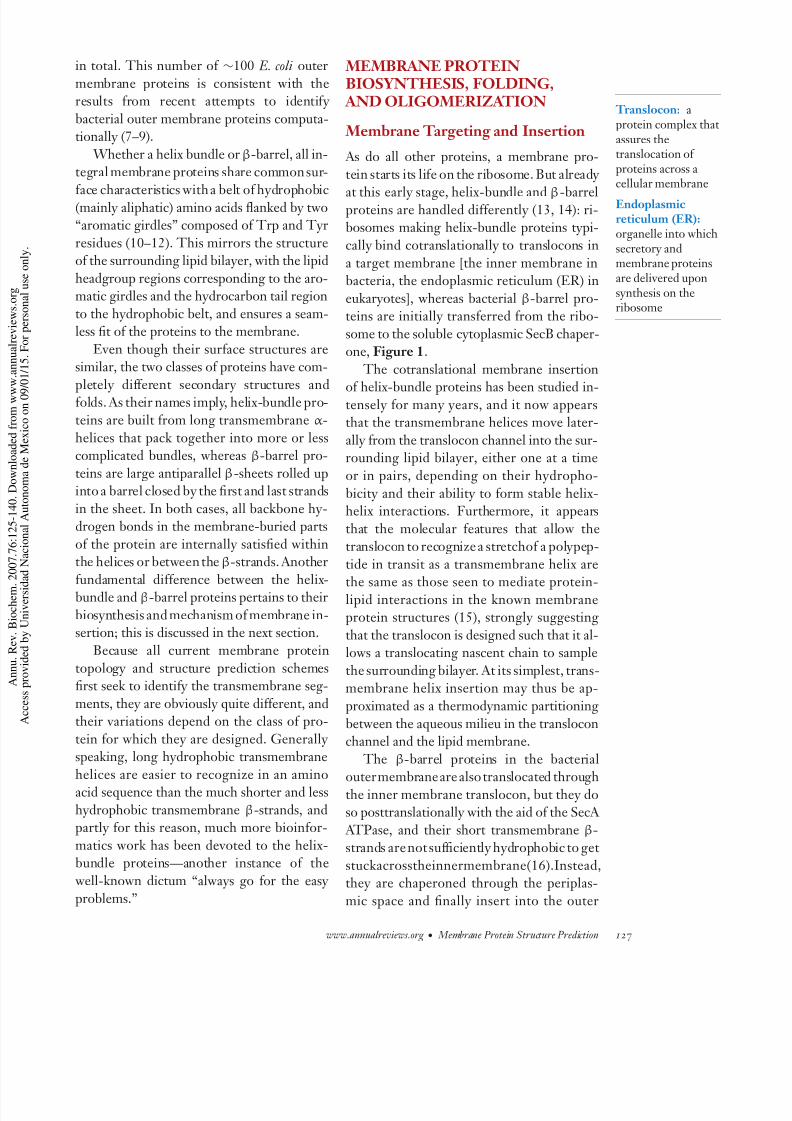
As do all other proteins, a membrane pro-
tein starts its life on the ribosome. But already
at this early stage, helix-bundle and β-barrelproteins are handled differently (13, 14): ri-bosomes making helix-bundle proteins typi-
cally bind cotranslationally to translocons in
a target membrane [the inner membrane inbacteria, the endoplasmic reticulum (ER) in
eukaryotes], whereas bacterial β-barrel pro-teins are initially transferred from the ribo-
some to the soluble cytoplasmic SecB chaper-one, Figure 1.
The cotranslational membrane insertion
of helix-bundle proteins has been studied in-tensely for many years, and it now appearsthat the transmembrane helices move later-
ally from the translocon channel into the sur-rounding lipid bilayer, either one at a time
or in pairs, depending on their hydropho-
bicity and their ability to form stable helix-helix interactions. Furthermore, it appears
that the molecular features that allow thetranslocon to recognize a stretchof a polypep-
tide in transit as a transmembrane helix are
the same as those seen to mediate protein-lipid interactions in the known membraneprotein structures (15), strongly suggesting
that the translocon is designed such that it al-lows a translocating nascent chain to sample
the surrounding bilayer. At its simplest, trans-membrane helix insertion may thus be ap-
proximated as a thermodynamic partitioning
between the aqueous milieu in the transloconchannel and the lipid membrane.
The β-barrel proteins in the bacterial
outermembraneare also translocated throughthe inner membrane translocon, but they doso posttranslationally with the aid of the SecA
ATPase, and their short transmembrane β-strands are not sufficiently hydrophobic to get
stuckacrosstheinnermembrane(16).Instead,they are chaperoned through the periplas-
mic space and finally insert into the outer
www.annualreviews.org • Membrane Protein Structure Prediction 127

7/18/2019 2007 - Membrane Protein Structure
http://slidepdf.com/reader/full/2007-membrane-protein-structure 4/19
Figure 1
Biogenesis of α -helix bundle (left ) and β-barrel (right ) membrane proteins in Escherichia coli .
membrane with the aid of the resident YaeT
hetero-oligomeric outer membrane integra-
tion complex (14).
Folding and Stability
Once inserted into the membrane, transmem-
brane helices pack into the typical helix-
bundle folds, and many then go on to
form homo- or hetero-oligomeric complexes.
Membrane proteins form closely packed
structures, and it is believed that an im-
portant driving force for folding is better
shape complementarity between the trans-
membrane helices than between the helices
and the lipid (17). Other factors that come
into play are hydrogen bonding between po-
lar side chains (18) and possibly even the for-
mation of Cα −H−O hydrogen bonds (19, 20
but also see Reference 21).
Many membrane proteins, both helix bun-
dle and β-barrel, form stable structures with
little flexibility, whereas others undergo sub-
stantial rearrangements of their transmem-
brane domains as part of a reaction cycle. Pro-
teins involved in proton and electron transfer
typically coordinate a range of cofactors that
need to be positioned relative to each other
with ˚ A-levelprecision and hence must be quite
rigidly packed (22), whereas small-molecule
transporters must flip between dramatically
different conformations open either toward
128 Elofsson· von Heijne

7/18/2019 2007 - Membrane Protein Structure
http://slidepdf.com/reader/full/2007-membrane-protein-structure 5/19
the external or the internal side of the cell
(23, 24).
MEMBRANE PROTEIN BIOINFORMATICS: WHAT THESEQUENCES TELL
For the helix-bundle membrane proteins,amino acid sequences told their story long be-fore the first high-resolution structures were
determined: the typical transmembrane seg-ment is formed by a stretch of predominantly
hydrophobic residues long enough to spanthe lipid bilayer as an α -helix (25–29). The
early topology prediction methods were con-sequently little more than plots of the seg-
mental hydrophobicity (averaged over 10–20 residues) along the sequence (30–32). With
more sequences came the realizations that aromatic Trp and Tyr residues tend to clus-ter near the ends of the transmembrane seg-
ments (10, 33) and that the loops connectingthe helices differ in amino acid composition,
depending on whether they face the inside oroutside of the cell (the “positive-inside” rule)
(34–36). More recent analyses have focusedon the higher-than-random appearance of se-
quence motifs, such as the GxxxG-motif intransmembrane segments (37, 38) as well as
other periodic patterns within the membranehelices (39), with the aim of providing infor-
mation that may help in predicting helix-helixpacking and 3D structure.
MEMBRANE PROTEIN BIOINFORMATICS: WHAT THESTRUCTURES TELL
For a long time, the general view has beenthat membrane proteins form simple he-
lix bundles, with their transmembrane he-lices crisscrossing the membrane in more or
less perpendicular orientations. Indeed, many membrane proteins abide by this principle.
However, some more recently solved mem-
brane protein structures show that reality isnot always this simple. This is illustrated by
the structure of the glutamate transporter ho-
Figure 2
(a) The glutamate transporter homolog (1XFH) contains both disruptedtransmembrane helices and reentrant loops. Disrupted helices are shown(cyan and green), and reentrant loops are also shown. The mesh indicates theapproximate extent of the lipid tail region ( ±15 ˚ A). (b) Topology (upper
part ) and z-coordinate plot. The z-coordinate plot shows the distance fromthe center of the membrane for each residue. The coloring is the same as inpanel a. Modified with permission of Oxford University Press (79).
Reentrant loop: astructural motif in which thepolypeptide dipsonly partway acrossthe membrane
mologfrom Pyrococcus horikoshii (40), shown in
Figure 2. This protein has six typical trans-membrane helices and two irregular helices
with breaks inside the lipid bilayer. The struc-
ture also contains two reentrant loops that go only halfway through the membrane and
then turn back to the side from which they
www.annualreviews.org • Membrane Protein Structure Prediction 129

7/18/2019 2007 - Membrane Protein Structure
http://slidepdf.com/reader/full/2007-membrane-protein-structure 6/19
originate. The two reentrant loops meet in
the middle of the membrane, a feature alsoseen in the aquaporin structures (41).
Other structural elements, largely ignoreduntil recently in statistical studies of mem-
brane protein structure, are found in thoseparts of the protein that are located in the
membrane-water interface region. Here onefinds irregular structure andinterfacial helicesrunning roughly parallel to the membrane
surface, while β-strands are extremely rare(42–44). The average amino acid composition
is different between the interfacial helices, theparts of the transmembrane helices located in
the interface region, and the irregular struc-tures. Hydrophobic and aromatic residues in
this region tend to point toward the centerof the membrane, whereas charged and polar
residues tend to point away from the mem-brane. The interface region thus imposes dif-
ferent constraints on protein structure than
do the central hydrocarbon core of the mem-brane and the surrounding aqueous phase.
For β-barrel membrane proteins, a num-ber of structural rules have been deduced from
the known structures (45): The number of β-strands is even; the N and C termini are at
the periplasmic barrel end; the β-strand tilt is
∼45◦; all β-strands are antiparallel and con-
nected locally to their next neighbors alongthe chain; and the β-barrel surface, contact-
ing the nonpolar membrane interior, consists
of a belt of aliphatic side chains lined by twogirdles of aromatic side chains. These gener-
alizations provide a framework for the devel-opment of topology prediction algorithms.
MEMBRANE PROTEIN STRUCTURE PREDICTION:FROM 2D TO 2.5D AND 3D
Over the years, many topology and structure
prediction schemes have been developed forhelix-bundle membrane proteins. In general,
there has been a logical progression from sim-
ple to more complicated models. This hasbeenenabledbytheincreaseinavailabletrain-
ing data, a better understanding of membrane
protein structure, and advances in machine-
learning methods.
2D Predictions
The earliest topology prediction methods re-
lied only on the fact that transmembrane he-
lices are on average more hydrophobic thanloop regions. Although these simple methods worked surprisingly well, they left a lot to be
desired. Inclusion of the positive-inside ruleled to a significant improvement in thepredic-
tions (46), and a further step was taken whenhidden Markov models (HMMs), Figure 3
and other machine-learning techniques wereemployed to extract the relevant sequence fea-
tures (5, 47–50). In addition, HMM-basedmethods that also include evolutionary and/or
limited experimental information to improvetopology predictions have been developed
(51–53).
One particular problem faced by all topol-ogy predictors is to discriminate between sig-
nal peptides and transmembrane helices—twokinds of topogenic elements that look quite
similar. This problem has recently been ad-dressed with thedevelopment of Phobius (54)
an HMM-based method that predicts bothsignal peptides and transmembrane segments
simultaneously and thereby significantly de-creases the confusion between them.
Much less work has been devoted to the de- velopment of topology prediction schemes for
β-barrel membrane proteins, in part because
the membrane-spanning β-strands are bothconsiderably shorter and much less conspicu-
ous in terms of amino acid sequence than thelong, hydrophobic transmembrane helices in
the helix-bundle proteins. Thesimplest meth-ods attempt to identify bacterial outer mem-
brane β-barrel proteins using only two cri-teria: the presence of an N-terminal signal
peptide [predicted using a program such asSignalP (55, 56)] and the overall amino acid
composition of the protein (9, 57). The moreadvanced methods also predict the individual
β-strands and the topology of the protein, in
most cases using HMMs (58–60).
130 Elofsson· von Heijne

7/18/2019 2007 - Membrane Protein Structure
http://slidepdf.com/reader/full/2007-membrane-protein-structure 7/19
Figure 3
Typical hidden Markov models(HMMs) for
topology predictionsof (a) helix-bundlemembrane proteins(5) and (b) β-barrelmembrane proteins. Modified withpermission of OxfordUniversity Press(58).
Benchmarking
The topology of more than 400 helix-bundle
membrane proteins has been determined ex-perimentally by a variety of genetic, biochem-ical, and structural techniques, and such data
has been used both to train and to bench-mark the various topology prediction meth-
ods. However, much of the experimental dataare of quite low resolution and not always cor-
rect. In addition, when benchmarking differ-
ent methods, many diverse quality measureshave been used. In principle, these can be di-
vided into residue-based and topology-basedinstruments. In our experience, benchmark-
ing with residue-based measures is not opti-mal as (a) all methods perform more or less
equally well by such measures, and (b) most experimental topology information is not of
high enough quality to exactly define the bor-ders between membrane and nonmembrane
parts of the proteins.
The different data sets used in bench-
marking studies can also impact the results. An especially important point is if single-
spanning membrane proteins are included ornot. On average, transmembrane helices in
single-spanning proteins are more hydropho-
bic than in multispanning (polytopic) mem-brane proteins (A. Bernsel & G. von Heijne,
unpublished data). Including single-spanningproteins in training data may compromise
the performance on multispanning proteinsand vice versa. Ideally, single-spanning pro-
teins should, therefore, be treated differently than multispanning proteins. However, to our
knowledge no method has successfully in-cluded this distinction in a prediction scheme,
and it is rarely taken into account in bench-marking studies.
Given these caveats, it is nevertheless clear
from several recent benchmarking studies(61–63) that HMM-based methods that also
www.annualreviews.org • Membrane Protein Structure Prediction 131

7/18/2019 2007 - Membrane Protein Structure
http://slidepdf.com/reader/full/2007-membrane-protein-structure 8/19
include evolutionary information, e.g., poly-
Phobius (64), HMMTOP (50), and prodiv- TMHMM (52), perform best. These methods
predict the correct topology (i.e., the correct number of transmembrane helices and the
correct overall orientation of the protein inthe membrane) for close to 70% of all mem-
brane proteins, and this number can be im-proved even further when additional experi-mental information is available to constrain
the predictions (51, 53, 65, 66). A recent benchmarking of β-barrel mem-
brane protein topology predictors concludedthat HMM-based methods also perform bet-
ter than other methods for these proteins (67). The best HMM methods predicted the cor-
rect topology for 14 out of 20 test proteins(70%), although the real accuracy is likely
lower than this because many of the proteinsin the test set were used also to develop the
different methods.
It should be kept in mind that a correct topology prediction does not mean that the
predicted starts and ends of the transmem-brane α -helices or β-strands can be trusted;
only the number of transmembrane helicesandtheir approximate positions arecorrect. In
fact, this part of the structure predictionprob-lem has not yet been satisfactorily resolved.
With the availability of more exact structuralinformation, it should be possible to evalu-
ate how well different methods can predict the exact helix locations. Our experience has
shown that it is possible to predict the points
of entrance and exit from the membrane en- vironment with acceptable accuracy, whereas
the prediction of helix start and terminationpoints is very hard.
The rapid increase in high-resolutionstructural data for membrane proteins means
that in the future both benchmarking anddevelopment of novel prediction methods
should be based on structural data only.Luckily, the general conclusions from using
structure-based benchmarks are similarto those of earlier studies (52, 68). One
remaining problem with the structural data
is that the precise location of the protein in
the lipid membrane is often not immediately
available because crystals are grown fromdetergent-solubilized proteins. Automatic
methods that optimize the fit between a 3Dprotein structure and a model membrane are
available (69, 70), although it is difficult toassess their accuracy.
Genome Annotation
An important application of topology predic-tion algorithms is to annotate genome se-
quencing data. It has been reported that al-
gorithms such as TMHMM can discriminatehelix-bundle proteins from other proteins
with better than 95% sensitivity and speci-ficity (5), meaning that the helix-bundle mem-
brane proteome of an organism can be quitereliably predicted from its genome sequence.
It was initially observed that the distri-bution of helix-bundle membrane protein
topologies in a genome seemed to follow apower law with respect to the number of
transmembrane helices, i.e., that proteins withfew transmembrane helices are more frequent
than proteins with many transmembrane he-
lices (71–73). As the topology predictors im-proved, several exceptions to this general
trend were noted (5, 65, 66, 74). In particularbacterial genomes encode large numbers of
small-molecule transporters with 6 or (moreoften) 12 transmembrane helices, whereas
mammalian genomes are strongly enrichedfor G protein–coupled receptors (GPCRs)
with 7 transmembrane helices, as well asfor small-molecule transporters. With the
availability of more accurate predictors andgenome-wide experimental topology data, it
was also noted that there is a strong overrep-resentation of proteins with an even number
of transmembrane helices and with their N
and C termini located on the cytoplasmic sideof membrane, both in bacteria and eukaryotes
(5, 65, 66).
2.5D Predictions
As noted above, the general view, until re-
cently, has been that the basic structural
132 Elofsson· von Heijne

7/18/2019 2007 - Membrane Protein Structure
http://slidepdf.com/reader/full/2007-membrane-protein-structure 9/19
feature of helix-bundle membrane proteins
is the perpendicularly penetrating transmem-brane helices. But as many 3D structures now
show, membrane protein structures are of-ten too complex to fit completely into such
a simple topology model. To advance further,a more fine-grained definition of topology,
“a two-and-a-half dimensional (2.5D) struc-ture,” is needed where structural elements,such as interfacial helices and reentrant loops,
are taken into account. In addition, severalother limitations of the current generation of
predictors exist, e.g., it has been noted that such a trivial characteristic as the exact length
of transmembrane helices is very difficult topredict using current methodologies.
Reentrant loops are a common feature inmany membrane proteins. They were first
seen in the aquaporin-1 water channel (75)and in the KcsA potassium channel (76). De-
tailed analysis suggests that reentrant loops
can be divided into three distinct categoriesbased on secondary structure content: long
loops with a helix-coil-helixstructure, loops of medium length with a helix-coil or coil-helix
structure,andloopsofshorttomediumlengthconsisting entirely of an irregular secondary
structure (77). Residues in reentrant loops aresignificantly smaller on average compared to
other parts of the protein, and they can be de-tected in regions between the transmembrane
helices with ∼70% accuracy based on theiramino acid composition. Reentrant loops of-
ten contain particular functional motifs that
enable them to be detected (78). On the ba-sis of a novel predictor for reentrant loops
(TOP-MOD), it appears that more than 10%of all multispanning membrane proteins con-
tain such loops (77). Reentrant loops seem tobe most commonly found in ion and water
channel proteins and least commonly in cellsurface receptors.
Although the division of a membrane pro-tein into different substructures is clearly use-
ful, distinguishing different types of struc-tural elements is not always straighforward.
Reentrant loops can vary quite dramatically in
their secondary structure and depth of pen-
etration into the membrane, and the length
of transmembrane helices varies significantly. An alternative approach to membrane protein
2.5D structure predictionis to directly predict the distance from the center of the membrane
(i.e., the z-coordinate) for each residue in aprotein, rather than the type of structural el-
ement of which it is a part. One recent al-gorithm of this kind correctly classified 88%
of all residues in the test set proteins to be
inside or outside the membrane, with an aver-age error of 2.5 ˚ A in the predicted residue dis-
tances from the center of the membrane (79). A similar z-coordinate predictor has also been
developed for β-barrel membrane proteins(80).
An important characteristic of residues intransmembrane helices is their degree of lipid
exposure in the folded structure. In contrast to globular proteins, membrane proteins do
not show a large difference in hydrophobicity between thelipid-exposed andburiedresidues
in the membrane-embedded region, and the
prediction of surface accessibility becomesmuch harder. The major features distinguish-
ing the lipid-exposed and buried residues arethepolarity of the side chain (more hydropho-
bicresiduestendtobemorelipidexposed)andthe degree of sequence conservation (less con-
servedresiduestendtobemorelipidexposed). Many attempts to predict lipid exposure have
been published; one of the most recent studiesreports the prediction of lipid-exposedsurface
patches in transmembrane helices that inter-face with lipid molecules with a per residue
accuracy of 88% (81).
A final 2.5D characteristic that can be pre-dicted with reasonable accuracy is the pres-
ence of proline-induced kinks in the trans-membrane helices. Interestingly, such kinks
can be preserved even when the Pro residue ismutated (82, 83), and a kink can confidently
be predicted if proline is conserved in a par-ticular position in a transmembrane helix in
more than 10% of the sequences in a multiplealignment (83).
The prediction of 2.5D features of mem-brane proteins should not only be useful as a
www.annualreviews.org • Membrane Protein Structure Prediction 133

7/18/2019 2007 - Membrane Protein Structure
http://slidepdf.com/reader/full/2007-membrane-protein-structure 10/19
step toward 3D predictions. Such predictors
will also help in the classification of mem-brane protein families because the different
substructures provide unique sequence signa-tures separating different membrane protein
families with the same topology. In addition,the identification of suitable peptide antibody
epitopes may be facilitated if z-coordinatescan be accurately predicted.
3D Predictions
Interestingly, 3D structure predictions of membrane proteins were attempted even be-
fore the first high-resolution structure of any membrane proteins was solved. Using infor-
mation from low-resolution experiments, inparticular electron microscopy, quite accurate
models of bacteriorhodopsin (84) as well asGPCRs (85, 86) were made.
Despite these early successes, the field of
nonhomology-based 3D structure predictionfor membrane proteins has followed a simi-
lar trend as that seen in the globular proteinstructure predictionfield, whereinthe general
experience is that most methods when testedin blind predictions show a much lower ac-
curacy than first reported. However, throughrounds of iterative refinement, the best meth-
ods can now predict the structure of smallglobular proteins quite accurately (87). As it
turns out, one of the most importantimprove-mentstothemethodologyhasbeentobasethe
3D prediction on short sequence fragments
extracted from known protein structures (88–90).
It is, however, not straightforward toapply similar schemes to membrane pro-
teins because the different environment introduced by the membrane has to be mod-
eled in some way, and because most mem-brane proteins are significantly larger than
the globular proteins successfully predicted sofar. Therefore the success to date has been
quite limited even using the most advancedmethods adapted from the globular protein
field (91, 92). If experimentally derived dis-
tance constraints from techniques, such as
Fourier transform infrared spectroscopy, elec-tron paramagnetic resonance spectroscopy
and chemical cross-linking, or low-resolutionmodels based on electron microscopy, are
available, more reliable models can be built(93).
An interesting attempt to model allGPCRs of the human proteome was recently
made by Skolnick and coworkers (94) using
the TASSER algorithm. Although the accu-racy of the predicted rhodopsin structure was
quite good, the correctness of the GPCRstructures can notbe verified until more struc-
tures are available. Many membrane proteins, in particular
channels and transporters, undergo substan-tial structural changes during a reaction cy-
cle. Often the structure of only one of thestates is available, and methodology to reli-
ably model structural changes will be neededfor a long time to come. In a recent study, the
ROSETTA membrane folding algorithm was
used to model the closed and open states ofa voltage-dependent potassium channel (95)
generating a number of testable hypothesesthat may guide further experimental work
For a more thorough review of ab initiostructure prediction methods, see Reference
96. While ab initio structure modeling can at
best predict the overall fold of a protein, struc-ture modeling based on a preexisting struc-
ture of a close homologue promises atomic-level structural detail. Homology modeling
of membrane proteins is still in its infancy
however, because so fewstructures are known A recent benchmarking study suggests that
when a template is available, homology mod-els of membrane proteins are comparable in
quality to those that can be made for globularproteins; i.e., when the sequence identity be-
tween the template and the target is >30%one can expect the root mean-square devia-
tion between the modeled and correct struc-ture to be less than 2 ˚ A in the transmembrane
regions (97).
134 Elofsson· von Heijne

7/18/2019 2007 - Membrane Protein Structure
http://slidepdf.com/reader/full/2007-membrane-protein-structure 11/19
MEMBRANE PROTEIN CLASSIFICATION SCHEMES
AND DATABASES
Hierarchically organized databases of pro-tein structure have found many uses, both
in studies of protein evolution, as a meansto put together nonredundant sequence and
structure collections for statistical studies,and as test beds for benchmarking fold
recognition and structure prediction algo-rithms. For globular proteins, several well-
established hierarchical, structure-based do-main classification schemes, such as SCOP
(98) and CATH (99), exist. However, the
number of membrane protein structuresis still too low for such classifications to
cover an important part of the membraneprotein universe (4). All currently known
high-resolution membrane protein structuresare listed at http://blanco.biomol.uci.edu/
MemPro resources.html and have alsobeen organized into a database at http://
pdbtm.enzim.hu/ (100).Sequence-based, nonhierarchical classi-
fications of membrane protein domains are
available in Pfam (101) and other similardatabases, and specialized databases col-
lecting families of membrane transporters(http://www.tcdb.org/ ), GPCRs (http://
www.gpcr.org/7tm/ ), andpotassium channelproteins (http://www.receptors.org/KCN )
have been set up. Because these databases arenot based on 3D structure information, they
do not contain complete information about very distant evolutionary relationships (e.g.,
the SCOP class, fold, and superfamily levels). The detection of distantly related glob-
ular proteins has seen great progress dur-
ing the past decade through the use of fold-recognition methods, improved use of
evolutionary relationships, and careful bench-marking. Because of the low incidence of po-
lar and charged residues in transmembranehelices, the use of algorithms optimized for
globular proteins for the detection of distantly related membrane proteins is problematic. To
compound these difficulties, a major obstacle
for the development of improved methods to
identify distantly related membrane proteinsis the lack of a structure-based“gold standard”
such as SCOP.Nevertheless, large, divergent membrane
protein families, such as the GPCRs, canbe used to benchmark fold-recognition algo-
rithms for membrane proteins. As for glob-ular proteins, it appears that HMM-basedsequence family models, profile-profile simi-
larity searches, and the inclusion of secondary structure information in the form of predicted
topology models all help in the detection of distant homologues (102, 103). It has also
been shown that the best alignment of re-lated membrane proteins is obtained using
profile-profile methods in combination withpredicted secondary structures (97).
PROTEIN-PROTEIN INTERACTIONS
The final step on the structure prediction
ladder is the prediction of quaternary struc-ture, i.e., protein-protein interactions. This
is especially pertinent for membrane proteinsbecause membrane-integral protein domains
in most cases seem to be encoded by sep-arate polypeptides rather than as multido-
main polypeptides as often found in globu-lar proteins (104). Large-scale experimentalprotein-protein interaction studies tend to ig-
nore membrane proteins, although some dataare now starting to appear in the literature
(105–107). The current state of predicting interac-
tions between membrane proteins may besummarized in a few words: much remains to
be done. For example, a recent attempt to pre-dict interacting proteins in the Saccharomyces
cerevisiae membrane proteome by integratingdata, such as amino acid sequence, annotated
function, subcellular localization, mRNA and
protein abundance, transcriptional coregula-tion, and gene knock-out phenotype, resulted
in a predictor that could identify ∼40% of 304 experimentally well-documented gold
www.annualreviews.org • Membrane Protein Structure Prediction 135
7/18/2019 2007 - Membrane Protein Structure
http://slidepdf.com/reader/full/2007-membrane-protein-structure 12/19
standard interactions while minimizing
the number of false-positive predictions(108).
CONCLUSIONS AND OUTLOOK
If bioinformatics methods are evaluated by
how they are received by the scientific com-munity at large, it is clear that membrane pro-tein structure prediction algorithmsholdtheir
ground; to give but one example, TMHMM(5,47)hasbeencitedwellover1200times.But
precisely what sort of information can one ex-pect to get from the various prediction meth-
ods? And what sort of advances can we see onthe horizon?
First and foremost, do not expect the com-puter to tell you the truth! Topology predic-
tions are just predictions. True, high-scoringpredictions are nearly always right (51, 63,109), but this only means that the really clear-
cut cases (i.e., those that can equally wellbe done by hand) are easy to predict. Still,
if taken with a grain of salt, topology mod-els, predicted lipid-exposed residues, poten-
tial reentrant loops, and lists of possibly in-teracting partner proteins can be invaluable
guides for planning experiments and inter-preting results. Andlarge-scalecomputational
studies of entire genomes can provide tanta-
lizing clues to everything from the basic pat-terns of membrane protein evolution (110
111) to differences in lifestyle between differ-ent organisms—show me your transporters
and I will tell you where you live.Still, much remains to be done, both in
perfecting the current arsenal of predictionmethods and devising entirely newalgorithmsto do new things. Our current representation
of membrane protein topology as a simplestring of membrane-spanning α -helices or β-
strands does not fully capture the structuraldiversity seen in membrane proteins; defin-
ing a fuzzy area between the 2D and 3Dstructure is in need of more exploration. The
rapid growth in known membrane protein 3Dstructures improves the prospects for effec-
tive fold-recognition and homology model-ing approaches, although the day when most
of membrane protein fold space has been
mapped experimentally seems desperately faroff (4). Computational means to map out the
membrane interactome will become an im-portant complement to high-throughput (but
error-prone) experimental studies, and hereas in so many other areas, tight integration
between the “wet” and “dry” approaches iscertainly the best way forward.
SUMMARY POINTS
1. Integral membrane proteins come in two basic architectures: α -helix bundles and
β-barrels.
2. The lipid-facing surface of integral membrane proteins is composed of a central
“hydrophobic belt” flanked by two “aromatic girdles.”
3. In the helix-bundle proteins, nontranslocated loops are enriched in Lys and Arg com-
pared to translocated loops (the positive-inside rule).
4. Helix-bundle membrane proteins are built from transmembrane α -helices, interfacial
helices lying flat on the membrane, reentrant loops, and extramembraneous globulardomains.
5. For the β-barrel protein, the number of β-strands is even, the N and C termini are at the periplasmic barrel end, the β-strand tilt is ∼45◦, and all β-strands are antiparallel
and connected locally to their next neighbors along the chain.
136 Elofsson· von Heijne

7/18/2019 2007 - Membrane Protein Structure
http://slidepdf.com/reader/full/2007-membrane-protein-structure 13/19
6. The best topology prediction algorithms forecast the correct topology for ≤70%of all proteins but cannot accurately predict the start and end of a transmembrane
segment.
7. Only a few recent prediction algorithms attempt to identify surface helices and reen-
trant loops.
8. Ab initio high-resolution 3D structure prediction is still not feasible for membraneproteins. Homology-based structure modeling of membrane proteins performs on apar with homology modeling of globular proteins.
ACKNOWLEDGMENTS
The authors’ laboratories are supported by grants from the Swedish Foundation for Strategic
Research, the Marianne and Marcus Wallenberg Foundation, the Swedish Cancer Founda-tion, the Swedish Research Council, and the European Commission (BioSapiens, Genefun,
EMBRACE).
LITERATURE CITED
1. Klabunde T, Hessler G. 2002. ChemBioChem 3:928–44
2. Berman HM, Westbrook J, Feng Z, Gilliland G, Bhat TN, et al. 2000. Nucleic Acids Res.
28:235–42
3. White SH. 2004. Protein Sci. 13:1948–49
4. Oberai A, Ihm Y, Kim S, Bowie JU. 2006. Protein Sci. 15:1723–34
5. Krogh A, Larsson B, von Heijne G, Sonnhammer EL. 2001. J. Mol. Biol. 305:567–80
6. Keseler IM, Collado-Vides J, Gama-Castro S, Ingraham J, Paley S, et al. 2005. Nucleic
Acids Res. 33:D334–37
7. Wimley WC. 2002. Protein Sci. 11:301–12
8. Casadio R, Fariselli P, Finocchiaro G, Martelli PL. 2003. Protein Sci. 12:1158–68
9. Garrow AG, Agnew A, Westhead DR. 2005. BMC Bioinformatics 6:56
10. Wallin E, Tsukihara T, Yoshikawa S, vonHeijne G, Elofsson A. 1997. Protein Sci. 6:808–15
11. Seshadri K, Garemyr R, Wallin E, von Heijne G, Elofsson A. 1998. Protein Sci. 7:2026–32
12. Ulmschneider MB, Sansom MS, Di Nola A. 2005. Proteins 59:252–65
13. Luirink J, von Heijne G, Houben E, de Gier JW. 2005. Annu. Rev. Microbiol. 59:329–55
14. Ruiz N, Kahne D, Silhavy TJ. 2006. Nat. Rev. Microbiol. 4:57–66
15. White SH, von Heijne G. 2004. Curr. Opin. Struct. Biol. 14:397–404
16. MacIntyre S, Freudl R, Eschbach ML, Henning U. 1988. J. Biol. Chem. 263:19053–59
17. Popot J-L, Engelman DM. 2000. Annu. Rev. Biochem. 69:881–922
18. Curran AR, Engelman DM. 2003. Curr. Opin. Struct. Biol. 13:412–17
19. Senes A, Ubarretxena-Belandia I, Engelman DM. 2001. Proc. Natl. Acad. Sci. USA
98:9056–61
20. Arbely E, Arkin IT. 2004. J. Am. Chem. Soc. 126:5362–63
21. Yohannan S, Faham S, Yang D, Grosfeld D, Chamberlain AK, Bowie JU. 2004. J. Am.
Chem. Soc. 126:2284–85
22. Brzezinski P, Adelroth P. 2006. Curr. Opin. Struct. Biol. 16:465–72
23. Guan L, Kaback HR. 2006. Annu. Rev. Biophys. Biomol. Struct. 35:67–91
www.annualreviews.org • Membrane Protein Structure Prediction 137

7/18/2019 2007 - Membrane Protein Structure
http://slidepdf.com/reader/full/2007-membrane-protein-structure 14/19
24. Toyoshima C, Nomura H, Tsuda T. 2004. Nature 432:361–68
25. Henderson R, Unwin PNT. 1975. Nature 257:28–3226. Tomita M, Marchesi VT. 1975. Proc. Natl. Acad. Sci. USA 72:2964–68
27. Ovchinnikov YA, Abdulaev NG, Feigina MY, Kiselev AV, Lobanov NA. 1977. FEBS Lett
84:1–4
28. Khorana HG, Gerber GE, Herlihy WC, Gray CP, Anderegg RJ, et al. 1979. Proc. Natl
Acad. Sci. USA 76:5046–50
29. Ovchinnikov YA, Abdulaev NG, Feigina MY, Kiselev AV, Lobanov NA. 1979. FEBS Lett100:219–2430. von Heijne G, Blomberg C. 1979. Eur. J. Biochem. 97:175–81
31. Engelman DM, Steitz TA. 1981. Cell 23:411–2232. Kyte J, Doolittle RF. 1982. J. Mol. Biol. 157:105–32
33. Weiss MS, Kreusch A, Schiltz E, Nestel U, Welte W, et al. 1991. FEBS Lett. 280:379–8234. von Heijne G. 1986. J. Mol. Biol. 189:239–42
35. von Heijne G. 1986. EMBO J. 5:3021–2736. von Heijne G. 1989. Nature 341:456–58
37. Senes A, Gerstein M, Engelman DM. 2000. J. Mol. Biol. 296:921–3638. Kim S, Jeon TJ, Oberai A, Yang D, Schmidt JJ, Bowie JU. 2005. Proc. Natl. Acad. Sci
USA 102:14278–8339. Samatey FA, Xu C, Popot J-L. 1995. Proc. Natl. Acad. Sci. USA 92:4577–81
40. Yernool D, Boudker O, Jin Y, Gouaux E. 2004. Nature 431:811–18
41. Hedfalk K, Tornroth-Horsefield S, Nyblom M, Johanson U, Kjellbom P, Neutze R2006. Curr. Opin. Struct. Biol. 16:447–56
42. Orgel JPRO. 2004. J. Struct. Biol. 148:51–6543. Granseth E, von Heijne G, Elofsson A. 2005. J. Mol. Biol. 346:377–85
44. Liang J, Adamian L, Jackups RJ. 2005. Trends Biochem. Sci. 30:355–5745. Schulz GE. 2000. Curr. Opin. Struct. Biol. 10:443–47
46. von Heijne G. 1992. J. Mol. Biol. 225:487–9447. Sonnhammer ELL, von HeijneG, Krogh A. 1998. A hidden Markov model for predicting
transmembrane helices in protein sequences. Proc. Int. Conf. Intell. Syst. Mol. Biol. 6:175–8248. Jones DT, Taylor WR, Thornton JM. 1994. Biochemistry 33:3038–49
49. Tusnady GE, Simon I. 1998. J. Mol. Biol. 283:489–50650. Tusnady GE, Simon I. 2001. Bioinformatics 17:849–50
51. Mel´ en K, Krogh A, von Heijne G. 2003. J. Mol. Biol. 327:735–44
52. Viklund H, Elofsson A. 2004. Protein Sci. 13:1908–1753. Bernsel A, von Heijne G. 2005. Protein Sci. 14:1723–28
54. K ¨ all L, Krogh A, Sonnhammer ELL. 2004. J. Mol. Biol. 338:1027–3655. Nielsen H, Engelbrecht J, Brunak S, von Heijne G. 1997. Protein Eng. 10:1–6
56. Dyrløv-Bendtsen J, Nielsen H, von Heijne G, Brunak S. 2004. J. Mol. Biol. 340:783–9557. Nakai K, Horton P. 1999. Trends Biochem. Sci. 24:34–35
58. Martelli PL, Fariselli P, Krogh A, Casadio R. 2002. Bioinformatics 18(Suppl. 1):S46–5359. Bagos PG, Liakopoulos TD, Spyropoulos IC, Hamodrakas SJ. 2004. Nucleic Acids Res
32:W400–460. Bigelow HR, Petrey DS, Liu J, Przybylski D, Rost B. 2004. Nucleic Acids Res. 32:2566–77
61. Chen CP, Kernytsky A, Rost B. 2002. Protein Sci. 11:2774–91
62. Chen CP, Rost B. 2002. Protein Sci. 11:2766–7363. K ¨ all L, Sonnhammer ELL. 2002. FEBS Lett. 532:415–18
64. K ¨ all L, Krogh A, Sonnhammer ELL. 2005. Bioinformatics 21(Suppl. 1):i251–57
138 Elofsson· von Heijne

7/18/2019 2007 - Membrane Protein Structure
http://slidepdf.com/reader/full/2007-membrane-protein-structure 15/19
65. Daley DO, Rapp M, Granseth E, Mel´ en K, Drew D, von Heijne G. 2005. Science
308:1321–2366. KimH, ¨ OsterbergM,Mel´ enK,vonHeijneG.2006. Proc. Natl. Acad. Sci. USA 103:11142–
4767. Bagos PG, Liakopoulos TD, Hamodrakas SJ. 2005. BMC Bioinformatics 6:7
68. Cuthbertson JM, Doyle DA, Sansom MS. 2005. Protein Eng. Des. Sel. 18:295–30869. Tusnady GE, Dosztanyi Z, Simon I. 2005. Bioinformatics 21:1276–77
70. Lomize AL, Pogozheva ID, Mosberg HI. 2004. Protein Sci. 13:2600–1271. Wallin E, von Heijne G. 1998. Protein Sci. 7:1029–3872. Liu J, Rost B. 2001. Protein Sci. 10:1970–79
73. Gerstein M. 1998. Proteins: Struct. Funct. Genet. 33:518–3474. Lehnert U, Xia Y, Royce TE, Goh CS, Liu Y, et al. 2004. Q. Rev. Biophys. 37:121–46
75. Walz T, Hirai T, Murata K, Heymann J, Mitsuoka K, et al. 1997. Nature 387:624–2776. Doyle D, Cabral J, Pfuetzner R, Kuo A, Gulbis J, et al. 1998. Science 280:69–77
77. Viklund H, Granseth E, Elofsson A. 2006. J. Mol. Biol. 361:591–60378. Lasso G, Antoniw JF, Mullins JGL. 2006. Bioinformatics 22:e290–97
79. Granseth E, Viklund H, Elofsson A. 2006. Bioinformatics 22:e191–9680. Diederichs K, Freigang J, Umhau S, Zeth K, Breed J. 1998. Protein Sci. 7:2413–20
81. Adamian L, Liang J. 2006. BMC Struct. Biol. 6:1382. von Heijne G. 1991. J. Mol. Biol. 218:499–503
83. Yohannan S, Faham S, Yang D, Whitelegge JP, Bowie JU. 2004. Proc. Natl. Acad. Sci.
USA 101:959–6384. Baldwin JM. 1993. EMBO J. 12:1693–703
85. Baldwin JM, Schertler GF, Unger VM. 1997. J. Mol. Biol. 272:144–6486. Unger VM, Hargrave PA, Baldwin JM, Schertler GF. 1997. Nature 389:203–8
87. Bradley P, Misura KM, Baker D. 2005. Science 309:1868–7188. Jones TA, Thirup S. 1986. EMBO J. 5:819–22
89. Simons KT, Bonneau R, Ruczinski I, Baker D. 1999. Proteins 3(Suppl.):171–7690. Bowie JU, Eisenberg D. 1994. Proc. Natl. Acad. Sci. USA 91:4436–40
91. Pellegrini-Calace M, Carotti A, Jones DT. 2003. Proteins 50:537–4592. Yarov-Yarovoy V, Schonbrun J, Baker D. 2006. Proteins 62:1010–25
93. Fleishman SJ, Unger VM, Ben-Tal N. 2006. Trends Biochem. Sci. 31:106–1394. Zhang Y, Devries ME, Skolnick J. 2006. PLoS Comput. Biol. 2:e13
95. Yarov-Yarovoy V, Baker D, Catterall WA. 2006. Proc. Natl. Acad. Sci. USA 103:7292–97
96. Fleishman SJ, Ben-Tal N. 2006. Curr. Opin. Struct. Biol. 16:496–50497. Forrest LR, Tang CL, Honig B. 2006. Biophys. J. 91:508–17
98. Murzin AG, Brenner SE, Hubbard T, Chothia C. 1995. J. Mol. Biol. 247:536–4099. OrengoCA, Michie AD, Jones S, Jones DT, Swindells MB, Thornton JM. 1997. Structure
5:1093–108100. Tusnady GE, Dosztanyi Z, Simon I. 2005. Nucleic Acids Res. 33:D275–78
101. Bateman A, CoinL, DurbinR, FinnRD,Hollich V,et al. 2004. Nucleic Acids Res. 32:D138–41
102. Hedman M, DeLoof H, von Heijne G, Elofsson A. 2002. Protein Sci. 11:652–58103. Wistrand M, K ¨ all L, Sonnhammer EL. 2006. Protein Sci. 15:509–21
104. Liu Y, Gerstein M, Engelman DM. 2004. Proc. Natl. Acad. Sci. USA 101:3495–97
105. Gavin AC, Bosche M, Krause R, Grandi P, Marzioch M, et al. 2002. Nature 415:141–47106. Stenberg F, Chovanec P, Maslen SL, Robinson CV, Ilag LL, et al. 2005. J. Biol. Chem.
280:34409–19
www.annualreviews.org • Membrane Protein Structure Prediction 139

7/18/2019 2007 - Membrane Protein Structure
http://slidepdf.com/reader/full/2007-membrane-protein-structure 16/19
107. Lasserre JP, Beyne E, Pyndiah S, Lapaillerie D, Claverol S, BonneuM. 2006. Electrophore-
sis 27:3306–21108. Xia Y, Lu LJ, Gerstein M. 2006. J. Mol. Biol. 357:339–49
109. Nilsson J, Persson B, von Heijne G. 2000. FEBS Lett. 486:267–69110. Shimizu T, Mitsuke H, Noto K, Arai M. 2004. J. Mol. Biol. 339:1–15
111. Rapp M, Sepp ¨ al ¨ a S, Granseth E, von Heijne G. 2006. Nat. Struct. Mol. Biol. 13:112–16
140 Elofsson· von Heijne

7/18/2019 2007 - Membrane Protein Structure
http://slidepdf.com/reader/full/2007-membrane-protein-structure 17/19
Annual Review
Biochemistry
Volume 76, 2007Contents
Mitochondrial Theme
The Magic Garden
Gottfried Schatz 673
DNA Replication and Transcription in Mammalian Mitochondria
Maria Falkenberg, Nils-Göran Larsson, and Claes M. Gustafsson 679
Mitochondrial-Nuclear Communications
Michael T. Ryan and Nicholas J. Hoogenraad 701
Translocation of Proteins into Mitochondria
Walter Neupert and Johannes M. Herrmann 723
The Machines that Divide and Fuse Mitochondria
Suzanne Hoppins, Laura Lackner, and Jodi Nunnari 751
Why Do We Still Have a Maternally Inherited Mitochondrial DNA?
Insights from Evolutionary Medicine
Douglas C. Wallace
781
Molecular Mechanisms of Antibody Somatic Hypermutation
Javier M. Di Noia and Michael S. Neuberger 1
Structure and Mechanism of Helicases and Nucleic Acid Translocases
Martin R. Singleton, Mark S. Dillingham, and Dale B. Wigley 23
The Nonsense-Mediated Decay RNA Surveillance Pathway
Yao-Fu Chang, J. Saadi Imam, Miles F. Wilkinson
51
Functions of Site-Specific Histone Acetylation and Deacetylation
Mona D. Shahbazian and Michael Grunstein 75
The tmRNA System for Translational Surveillance and Ribosome Rescue
Sean D. Moore and Robert T. Sauer 101
Membrane Protein Structure: Prediction versus Reality
Arne Elofsson and Gunnar von Heijne 125
v

7/18/2019 2007 - Membrane Protein Structure
http://slidepdf.com/reader/full/2007-membrane-protein-structure 18/19
Structure and Function of Toll Receptors and Their Ligands
Nicholas J. Gay and Monique Gangloff
The Role of Mass Spectrometry in Structure Elucidation of Dynamic
Protein Complexes
Michal Sharon and Carol V. Robinson
Structure and Mechanism of the 6-Deoxyerythronolide B SynthaseChaitan Khosla, Yinyan Tang, Alice Y. Chen, Nathan A. Schnarr,
and David E. Cane
The Biochemistry of Methane Oxidation
Amanda S. Hakemian and Amy C. Rosenzweig
Anthrax Toxin: Receptor Binding, Internalization, Pore Formation,
and Translocation
John A.T. Young and R. John Collier
Synapses: Sites of Cell Recognition, Adhesion, and FunctionalSpecification
Soichiro Yamada and W. James Nelson
Lipid A Modification Systems in Gram-negative Bacteria
Christian R.H. Raetz, C. Michael Reynolds, M. Stephen Trent,
and Russell E. Bishop
Chemical Evolution as a Tool for Molecular Discovery
S. Jarrett Wrenn and Pehr B. Harbury
Molecular Mechanisms of Magnetosome Formation
Arash Komeili
Modulation of the Ryanodine Receptor and Intracellular Calcium
Ran Zalk, Stephan E. Lehnart, and Andrew R. Marks
TRP Channels
Kartik Venkatachalam and Craig Montell
Studying Individual Events in Biology
Stefan Wennmalm and Sanford M. Simon
Signaling Pathways Downstream of Pattern-Recognition Receptors
and Their Cross Talk Myeong Sup Lee and Young-Joon Kim
Biochemistry and Physiology of Cyclic Nucleotide Phosphodiesterases:
Essential Components in Cyclic Nucleotide Signaling
Marco Conti and Joseph Beavo
The Eyes Absent Family of Phosphotyrosine Phosphatases: Properties
and Roles in Developmental Regulation of Transcription
Jennifer Jemc and Ilaria Rebay
v i C on te nt s

7/18/2019 2007 - Membrane Protein Structure
http://slidepdf.com/reader/full/2007-membrane-protein-structure 19/19
Assembly Dynamics of the Bacterial MinCDE System and Spatial
Regulation of the Z Ring
Joe Lutkenhaus 539
Structures and Functions of Yeast Kinetochore Complexes
Stefan Westermann, David G. Drubin, and Georjana Barnes 563
Mechanism and Function of Formins in the Control of Actin Assembly Bruce L. Goode and Michael J. Eck 593
Unsolved Mysteries in Membrane Traffic
Suzanne R. Pfeffer 629
Structural Biology of Nucleocytoplasmic Transport
Atlanta Cook, Fulvia Bono, Martin Jinek, and Elena Conti 647
The Postsynaptic Architecture of Excitatory Synapses: A More
Quantitative View
Morgan Sheng and Casper C. Hoogenraad
823
Indexes
Cumulative Index of Contributing Authors, Volumes 72–76 849
Cumulative Index of Chapter Titles, Volumes 72–76 853
Errata
An online log of corrections to Annual Review of Biochemistry chapters (if any, 1997to the present) may be found at http://biochem.annualreviews.org/errata.shtml