
(2007) Do all epistaxis -...
51
Transcript of (2007) Do all epistaxis -...

References
1 Small M., Murray J.A.M. & Maran A.G.D. (1982) A study of
patients with epistaxis requiring admission to hospital. Health
Bulletin (Edinb). 40, 20–29
2 Ho E.C. & Chan J.Y. (2008) Front-line epistaxis management:
let’s not forget the basics. J. Laryngol. Otol. 122, 696–699
3 Kotecha B., Cocks R.A. & Rothera N.P. (1990) The management
of epistaxis in accident and emergency departments: a survey of
current practice. Arch Emerg. Med. 7, 35–41
4 Duvvi S., Khattab A., Khalil H.S. et al. (2006) Short falls in epi-
staxis management. A nationwide survey in UK. Clin. Otolaryn-
gol. 31, 560–561
5 Daudia A., Jaiswal V. & Jones N.S. (2008) Guidelines for the
management of idiopathic epistaxis: how we do it. Clin. Otolary-
gol. 33, 618–620
6 Van Wyk F.C., Nassey S., Worley G. et al. (2007) Do all epistaxis
patients with a nasal pack need admission? A retroepective study
of 116 patients managed in accident and emergency according to
a peer reviewed protocol. J. Laryngol. Otol. 121, 222–227
7 Moshaver A., Harris J.R., Liu R. et al. (2004) Early operative
intervention versus conventional treatment in epistaxis: random-
ized prospective trial. J. Laryngol. Otol. 33, 185–188
8 Srinivasan V., Sherman I.W. & O’Sullivan G. (2000) Surgical
management of intractable epistaxis: audit of results. J. Laryngol.
Otol. 114, 697–700
9 Jonas N., Viani L. & Walsh M. (2010) Sphenopalatine artery liga-
tion under local anesthesia: a report of two cases and review of
the literature. Local & Regional Anesthesia. 3, 1–4
Assessing the role of chronic hyperventilation in patientswith nasal congestion: Our experience in 118 patients
Hanna, B.C.,* Woodman, P.� & Adair, R.�
*The Queen Elizabeth Hospital, Adelaide, South Australia, Australia, �Craigavon Area Hospital, Craigavon, and�The Ulster Hospital, Belfast, Northern Ireland
Accepted for publication 16 March 2012
Dear Editor,
When assessing patients complaining of nasal obstruc-
tion ⁄ congestion, the possibility of a dysfunctional breathing
pattern should be considered in addition to restricted nasal
airflow. This has been aptly demonstrated by Bartley1 who
discovered a high prevalence of chronic hyperventilation
syndrome among a group of patients in whom surgery had
failed to relieve the symptom of nasal congestion. A simpli-
fied physiological definition of hyperventilation is breathing
in excess of metabolic requirements, a pathophysiological
process which can be acute or chronic.2 An extrapolation of
Bartley’s finding is that the initial surgery may not have
been performed if the chronic hyperventilation syndrome
had been detected earlier. We therefore introduced the Ni-
jmegen Questionnaire (for chronic hyperventilation syn-
drome) to our initial assessment of patients whose main
presenting complaint was nasal obstruction or congestion.
Of course, chronic hyperventilation could coexist with
other inflammatory and structural nasal problems. Ogata
et al.3 found 25% of patients with allergic rhinitis to have a
positive Nijmegen score. In such cases, the clinician has to
make a subjective judgement about the extent to which the
nasal obstruction can be attributed to chronic hyperventila-
tion. Over a period of 1 year we audited not only how many
patients had a positive Nijmegen score, but also the number
of cases in which this lead to a significant change in diagno-
sis and ⁄ or treatment.
Method
A prospective audit was performed for 1 year after the
introduction of the Nijmegen Questionnaire for the rou-
tine assessment of new patient referrals with nasal blockage
at a general ENT clinic in Northern Ireland. The Nijmegen
Questionnaire assesses 16 complaints related to different
organ systems affected by hyperventilation and has been
previously validated.4 It has been found to be a quick, easy
to administer and low-impact assessment tool for chronic
hyperventilation5 and its use in a routine ENT clinic has
been previously published.3 The questionnaire was com-
pleted either before the consultation or when waiting for
nasal endoscopy. However, the questionnaire was not
scored until after the history and examination were com-
plete and the clinician had recorded a provisional diagno-
CO
RR
ES
PO
ND
EN
CE
:O
UR
EX
PE
RI
EN
CE
Correspondence: B.C. Hanna, ENT department level 3c, The Queen
Elizabeth Hospital, 28 Woodville road, Woodville South, Adelaide,
South Australia SA5011, Australia. Tel.: +61 82227158; Fax:
+61 82227419; e-mail: [email protected]
Correspondence 155
� 2012 Blackwell Publishing Ltd • Clinical Otolaryngology 37, 148–161
sis and treatment plan. The Nijmegen score, age, sex and
alteration to diagnosis and ⁄ or treatment were recorded.
The questionnaire was used for patients aged 16 and over.
Results
Over 12 months, 118 new patients with nasal obstruction
presented to a general ENT clinic. 64 were men and 54
women. The age range was 16 to 80 with a mean of 38. The
Nijmegen score was positive in 28 patients (25%), 11 male
and 20 female. The initial diagnosis was changed to exclu-
sively hyperventilation syndrome in 12 patients (10%).
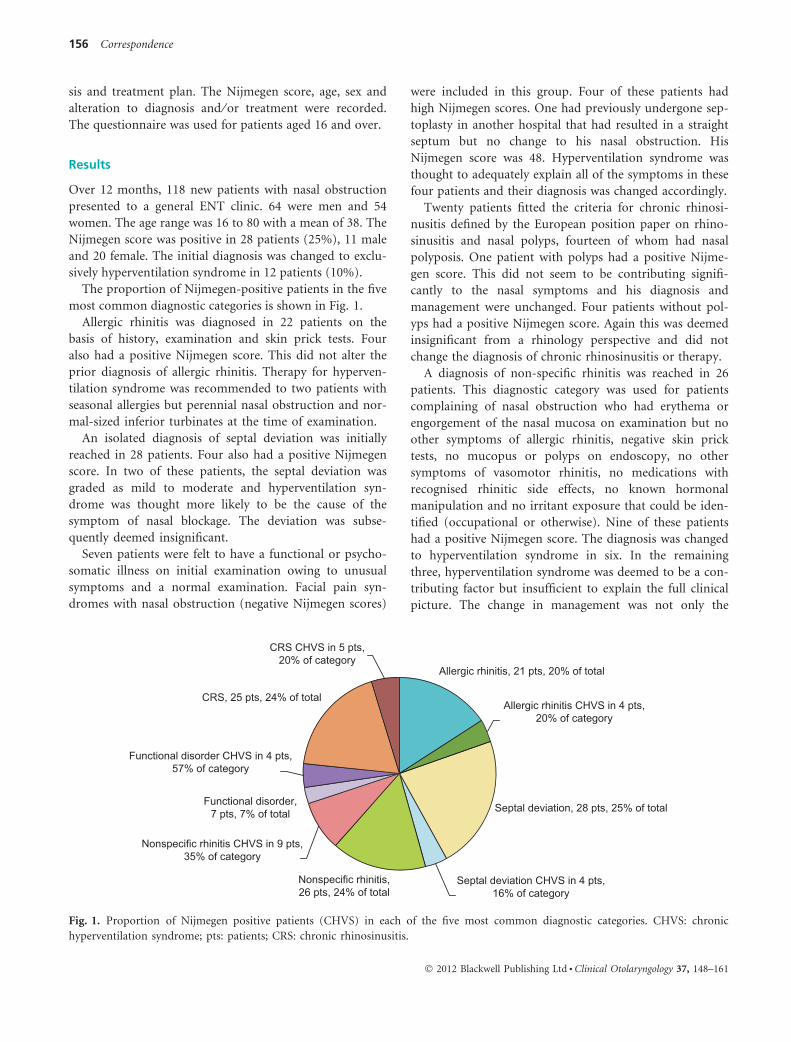
The proportion of Nijmegen-positive patients in the five
most common diagnostic categories is shown in Fig. 1.
Allergic rhinitis was diagnosed in 22 patients on the
basis of history, examination and skin prick tests. Four
also had a positive Nijmegen score. This did not alter the
prior diagnosis of allergic rhinitis. Therapy for hyperven-
tilation syndrome was recommended to two patients with
seasonal allergies but perennial nasal obstruction and nor-
mal-sized inferior turbinates at the time of examination.
An isolated diagnosis of septal deviation was initially
reached in 28 patients. Four also had a positive Nijmegen
score. In two of these patients, the septal deviation was
graded as mild to moderate and hyperventilation syn-
drome was thought more likely to be the cause of the
symptom of nasal blockage. The deviation was subse-
quently deemed insignificant.
Seven patients were felt to have a functional or psycho-
somatic illness on initial examination owing to unusual
symptoms and a normal examination. Facial pain syn-
dromes with nasal obstruction (negative Nijmegen scores)
were included in this group. Four of these patients had
high Nijmegen scores. One had previously undergone sep-
toplasty in another hospital that had resulted in a straight
septum but no change to his nasal obstruction. His
Nijmegen score was 48. Hyperventilation syndrome was
thought to adequately explain all of the symptoms in these
four patients and their diagnosis was changed accordingly.
Twenty patients fitted the criteria for chronic rhinosi-
nusitis defined by the European position paper on rhino-
sinusitis and nasal polyps, fourteen of whom had nasal
polyposis. One patient with polyps had a positive Nijme-
gen score. This did not seem to be contributing signifi-
cantly to the nasal symptoms and his diagnosis and
management were unchanged. Four patients without pol-
yps had a positive Nijmegen score. Again this was deemed
insignificant from a rhinology perspective and did not
change the diagnosis of chronic rhinosinusitis or therapy.
A diagnosis of non-specific rhinitis was reached in 26
patients. This diagnostic category was used for patients
complaining of nasal obstruction who had erythema or
engorgement of the nasal mucosa on examination but no
other symptoms of allergic rhinitis, negative skin prick
tests, no mucopus or polyps on endoscopy, no other
symptoms of vasomotor rhinitis, no medications with
recognised rhinitic side effects, no known hormonal
manipulation and no irritant exposure that could be iden-
tified (occupational or otherwise). Nine of these patients
had a positive Nijmegen score. The diagnosis was changed
to hyperventilation syndrome in six. In the remaining
three, hyperventilation syndrome was deemed to be a con-
tributing factor but insufficient to explain the full clinical
picture. The change in management was not only the
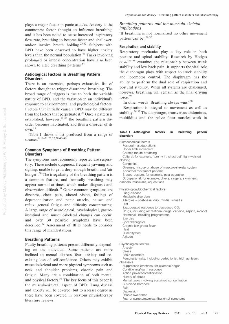
CRS, 25 pts, 24% of total
Allergic rhinitis, 21 pts, 20% of total
Septal deviation CHVS in 4 pts,16% of category
Septal deviation, 28 pts, 25% of total
Nonspecific rhinitis CHVS in 9 pts,35% of category
Nonspecific rhinitis,26 pts, 24% of total
Functional disorder,7 pts, 7% of total
Functional disorder CHVS in 4 pts,57% of category
CRS CHVS in 5 pts,20% of category
Allergic rhinitis CHVS in 4 pts,20% of category
Fig. 1. Proportion of Nijmegen positive patients (CHVS) in each of the five most common diagnostic categories. CHVS: chronic
hyperventilation syndrome; pts: patients; CRS: chronic rhinosinusitis.
156 Correspondence
� 2012 Blackwell Publishing Ltd • Clinical Otolaryngology 37, 148–161
recommendation of therapy for hyperventilation syn-
drome but the omission of a trial of topical steroid ther-
apy in the six patients with a change in diagnosis.
Three patients with vasomotor rhinitis had a positive
Nijmegen score and this was thought to be contributing
sufficiently to nasal symptoms in one case to alter manage-
ment. The diagnosis of vasomotor rhinitis was unchanged.
Three patients had rhinitis medicamentosa and one also
had a positive Nijmegen score. It was thought that nasal
obstruction because of hyperventilation syndrome may
have lead to the abuse of nasal decongestants, and again,
therapy for hyperventilation syndrome was recommended.
Of the remaining nine patients in the audit, two had
alar collapse. One of these patients had a positive Nijme-
gen score but this was not deemed relevant to the nasal
obstruction symptom and the diagnosis and management
were not changed. None of the seven patients left had a
positive Nijmegen score. Two were thought to have nasal
obstruction as a medication side effect, two had enlarged
inferior turbinates, two had acute infections at the time
of clinic attendance and one had a septal perforation.
Discussion
Hyperventilation
Acute hyperventilation causes lowering of the alveolar
pressure of carbon dioxide, lowering of the arterial pres-
sure of carbon dioxide and a respiratory alkalosis. The
resulting symptoms can usually be reproduced by a
hyperventilation provocation test. In contrast, although
chronic hyperventilation syndrome can be associated with
sustained arterial and alveolar hypocapnia, patients may
present with resting levels of carbon dioxide in the nor-
mal range. The respiratory rate may be elevated or the
tidal volume increased, often accompanied by deep sigh-
ing respirations. It is an idiopathic fluctuating disorder
which is identified by a combination of symptoms and is
therefore not able to be contained within a single diag-
nostic measurement. The wide range of symptoms
includes breathlessness, dyspnoea, light-headedness, par-
aesthesia, a variety of pains especially chest pains, palpita-
tions, sweating, anxiety, excessive sighing ⁄ yawning and
nasal congestion. The diagnosis was more common in
the past as hyperventilation syndrome lacks a single diag-
nostic measure for today’s era of testing technology.6 An
elaborate study by Howell comparing the hyperventila-
tion provocation test to a placebo test has demonstrated
that this test is invalid as a diagnostic test for chronic
hyperventilation syndrome.7 The Nijmegen Questionnaire
has been validated. It consists of 16 complaints relating
to different organ systems in which their frequency is
indicated on 5-point ordinal scale (0 = never, 4 = very
frequently). The maximum score is 64 and a score of
>23 is considered diagnostic for chronic hyperventilation
syndrome.4
Implications of chronic hyperventilation syndrome
This audit demonstrated that a significant proportion of
patients presenting with nasal obstruction or congestion
who would otherwise be diagnosed with a functional dis-
order or non-specific rhinitis can be reclassified as hyper-
ventilation syndrome according to the Nijmegen
Questionnaire. But what is the benefit? The therapy we
recommended was breathing awareness and retraining via
a self-help programme. In Bartley’s study, breathing
retraining was partially successful with two of five
patients enrolled having relief of their nasal congestion.
We did not audit the results of the self-help therapy. Per-
haps the greater benefit is the avoidance of trials of medi-
cal therapy. When the diagnosis is uncertain, the clinician
may prescribe a trial of topical steroid which can cause
epistaxis and, if it ultimately fails, may reduce the
patient’s confidence in the clinician. Perhaps more
importantly, when the role of a mild to moderately devi-
ated septum is uncertain, the significance of the patient’s
nasal congestion in the context of hyperventilation syn-
drome may reveal that a septoplasty is not indicated. The
patient is spared an unnecessary and unhelpful procedure
and the overall success of surgery is improved through
better patient selection. Only a small proportion of
patients with allergic rhinitis were recommended self-help
breathing exercises because of nasal congestion persisting
beyond the allergen exposure season and not accounted
for by hypertrophied inferior turbinates. In general
though, the Nijmegen Questionnaire was not a beneficial
exercise in patients with chronic rhinosinusitis or allergic
rhinitis.
Chronic hyperventilation syndrome and non-specific
rhinitis
The diagnostic category of non-specific rhinitis used in
this audit illustrates the challenge that the clinician faces
when forced to reach a diagnostic decision. Some of these
patients were subsequently reclassified as hyperventilation
syndrome. We do not know if the mild nasal erythema
and congestion found in these patients was a variation of
normal or a consequence of the hyperventilation syn-
drome. The Mayo Clinic described an inverse relationship
between nasal resistance and Pco2 levels and presumably
the elevated resistance was achieved by vascular engorge-
ment ⁄ congestion.8
Correspondence 157
� 2012 Blackwell Publishing Ltd • Clinical Otolaryngology 37, 148–161

Conclusion
Although the interpretation of the significance of the
results of the Nijmegen Questionnaire is subjective, we
propose that it is useful for those patients with mild to
moderately deviated septums and those who appear to
have a functional disorder or non-specific rhinitis.
Key points
• Hyperventilation syndrome has been demonstrated
to cause the symptom of nasal congestion ⁄ obstruc-
tion and to be a reason for failed nasal surgery.
• Routine use of the Nijmegen Questionnaire to
detect hyperventilation syndrome in all new
patients attending a general ENT clinic produced
positive results in a significant proportion of
patients (25%).
• Determining when the hyperventilation syndrome
was contributing significantly to a patient’s nasal
congestion was a subjective clinical decision
reached in 10% of the patients in this audit.
• The Nijmegen score was deemed useful for achiev-
ing a more accurate diagnosis in a significant num-
ber of patients who would otherwise have been
diagnosed as a functional disorder or non-specific
rhinitis.
• The Nijmegen Questionnaire was also deemed to
have improved patient selection for septoplasty.
Conflict of interest
None to declare.
References
1 Bartley J. (2005) Nasal congestion and hyperventilation syn-
drome. Am. J. Rhinol. 19, 607–611
2 Gardener W. (1990) Hyperventilation disorders. J R Soc Med. 83,
755–757
3 Ogata N., Bapat U. & Darby Y. (2006) Prevalence of hyperventi-
lation syndrome in an allergy clinic, compared with a routine
ENT clinic. J. Laryngol. Otol. 120, 924–926
4 Van Dixhoorn J. & Duivenvoorden H.J. (1985) Efficacy of Ni-
jmegen questionnaire in recognition of the hyperventilation syn-
drome. J. Psychosom. Res. 29, 199–206
5 Humphriss R.L., Baguley D.M., Andersson G. et al. (2004)
Hyperventilation in the vestibular clinic: use of the Nijmegen
Questionnaire. Clin. Otolaryngol. Allied Sci. 29, 232–237
6 Innocenti D.M. & Troup F.. (2008) Dysfunctional breathing. In
Physiotherapy for Respiratory and Cardiac Problems, 4th edition,
Ch 17, Pryor J.A. & Prasad S.A. (eds), pp. 529–548. Churchill
Livingstone, Elsevier, Philadelphia, USA.
7 Howell J.B.L. (1997) The hyperventilation syndrome: a syndrome
under threat? Thorax 52, s30–s34
8 Mertz J.S., McCaffrey T.V. & Kern E.B. (1984) Role of the nasal
airway in regulation of airway resistance during hypercapnia and
exercise. Otolaryngol. Head Neck Surg. 92, 302–307
The down-up bone bridge approach for cochlear and middleear implants: Our experience in 34 patients
Achena, F.,* Montaldo, C.� & Nucaro, A.L.�
*Otorhinolaryngology Division, CTO Hospital, Iglesias, �Department of Surgical Sciences, OBL, University, and�Genetics and Biomedical Research Institute- CNR, Cittadella Universitaria, Monserrato, Cagliari, Italy
Accepted for publication 16 March 2012
Dear Editor,
Over the last few years, different authors in various coun-
tries1–4 have proposed several minimal invasive approaches
for the fashioning of the bony recess and fixation of cochl-
ear and middle ear implants. The above-mentioned
approaches offer reduced surgical morbidity related to
wound complications and reduced hospital stay as com-
pared to wider access operations5. However, disagreement
still exists between authors about the best way to secure the
implants, either with a tie-down ligature1–3 or just by clo-
sure of the overlying periosteum4, particularly in teenage
patients.
CO
RR
ES
PO
ND
EN
CE
:O
UR
EX
PE
RI
EN
CE
Correspondence: A.L. Nucaro, Genetics and Biomedical Research Insti-
tute – IRGB- CNR, Cittadella Universitaria, ss 554 bivio Sestu, 09042
Monserrato, Cagliari, Italy. Tel.: +39 70 6754654; Fax: +39 70 6754652;
e-mail: [email protected]
158 Correspondence
� 2012 Blackwell Publishing Ltd • Clinical Otolaryngology 37, 148–161

34
Contribution
Breathing pattern disorders, motor control, and low back pain
L ChaitowSchool of Integrated Health, University of Westminster, London, United Kingdom.
AbstractMotor control is a key component in injury prevention. Loss of motor control involves failure tocontrol joints, commonly because of incoordination of the agonist-antagonist muscle co-activation.Three subsystems work together to maintain spinal stability: The central nervous subsystem(control), the osteoligamentous subsystem (passive), and the muscle subsystem (active).
There is evidence that the effects of breathing pattern disorders, such as hyperventilation, resultin a variety of negative psychological, biochemical, neurological and biomechanical influencesand interferences, capable of modifying each of these three subsystems. Breathing patterndisorders (the extreme form of which is hyperventilation), automatically increase levels of anxietyand apprehension, which may be sufficient to alter motor control and to markedly influence balancecontrol. Hyperventilation results in respiratory alkalosis, leading to reduced oxygenation of tissues(including the brain), smooth muscle constriction, heightened pain perception, speeding up ofspinal reflexes, increased excitability of the corticospinal system, hyperirritability of motor andsensory axons, changes in serum calcium and magnesium levels, and encouragement of thedevelopment of myofascial trigger points – all or any of which, in one way or another, are capableof modifying normal motor control of skeletal musculature.
Diaphragmatic and transversus abdominis tone are key features in provision of core stability,however it has been noted that reduction in the support offered to the spine, by the muscles ofthe torso, may occur if there is both a load challenge to the low back, combined with a breathingchallenge. It has been demonstrated that, after approximately 60 seconds of hypercapneoa, thepostural (tonic) and phasic functions of both the diaphragm and transversus abdominis are reducedor absent. Smooth muscle cells, now known to be widely embedded in connective tissues (includingspinal discs, and lumbar fascia) constrict during periods of respiratory alkalosis, with as yetundetermined effects on joint stability and fascial tone. Breathing rehabilitation offers the potentialfor reducing the negative influences resulting from breathing pattern disorders.
Keywords: breathing pattern disorder, hyperventilation, respiratory alkalosis, motor control,musculoskeletal pain
Leon Chaitow, ND, DO, School of Integrated Health, University of West-minster, 115 New Cavendish Street, London W1M 8JS, United [email protected] 04/02/04, Revised, 14/03/04, Accepted 15/03/04
INTRODUCTION
Motor control is a key component in injury prevention andloss of motor control involves failure to control joints,commonly because of incoordination of the agonist-antagonist muscle co-activation1. According to Panjabi2
three subsystems work together to maintain spinal stability:
• the central nervous subsystem (control)• the osteoligamentous subsystem (passive)• the muscle subsystem (active).
Anything that interferes with any aspect of these featuresof normal motor control, may contribute to dysfunction andpain.
An increased rate of ventilation, such as prevails withhyperventilation, during which the rate of carbon dioxide(CO2) exhalation exceeds the rate of its accumulation inthe tissues, produces respiratory alkalosis, characterised bythe decrease in CO2 and an increase in pH. This inducesvascular constriction, decreasing blood flow, as well asinhibiting transfer from haemoglobin, of oxygen, to tissuecells (due to the Bohr Effect).3
The Bohr effect states that an increase in alkalinity (decreasein CO2) increases the affinity of haemoglobin (Hb) foroxygen (O2). The Hb molecule is therefore less likely torelease its oxygen in tissues that have become increasinglyalkaline due to overbreathing.4 Increased O2-Hb affinity alsoleads to changes in serum calcium and red cell phosphatelevels.5 Additionally there is a loss of intra-cellular Mg2+ aspart of the renal compensation mechanism for correctingalkalosis.4,5 Muscles affected in this way inevitably become
Journal of Osteopathic Medicine, 2004; 7(1): 34-41 © 2004 Research Media

35
© 2004 Research Media Chaitow L. Breathing pattern disorders, motor control and low back pain
prone to fatigue, dysfunction (e.g. cramp), and trigger pointevolution.6
Acute episodes of hyperventilation represent onlyapproximately 1% of all cases, far outnumbering chronicpatterns.7 Chronic hyperventilation leads to hypocapnoea(reduced levels of carbon dioxide), and can present with amyriad of respiratory, cardiac, neurological orgastrointestinal symptoms, without any clinically apparentoverbreathing by the patient. In the United States as manyas 10% of patients in a general internal medicine practiceare reported to have HVS as their primary diagnosis.7,8
Studies show that, relative to men, women have a higherrate of respiration and a greater tendency to respiratoryalkalosis, which is exaggerated during the luteal(progesterone) phase of the menstrual cycle.9
Hyperventilation syndrome (HVS) and breathing patterndisorders (BPD) are therefore female dominated, with afemale:male ratio ranging from 2:1 to 7:1. Women may bemore at risk because of hormonal influences, sinceprogesterone stimulates respiration, and in the luteal (postovulation/pre-menstrual) phase, CO2 levels drop on average25%. Additional stress can subsequently, “increaseventilation at a time when carbon dioxide levels are alreadylow”.10
Lum11 points out that there are many people with BPD whohave been labelled as asthmatics. “Thirty percent of casesof asthma are known to be induced by emotion or exercise,and many symptoms are common to hyperventilation andto asthma: intermittent, labored breathing; relief frombronchodilators (transient in hyperventilation); exercise;cough; fear, anxiety and panic. It is thus a matter ofindividual preference whether the clinician calls such casesasthma or hyperventilation. The distinction is important.Treatment of hyperventilation cures the patient. Theasthmatic is condemned to a life of medication.”
While investigation as to the precipitating causes of episodesof hyperventilation may help with both the diagnosis andchoice of treatment, Nixon12 suggests that there are oftenattacks where there is no preceding stressful event. Inchronic hyperventilators the respiratory centre may havebeen reset to tolerate a lower than normal partial pressureof arterial carbon dioxide (PaCO2). In such patients a singlesigh, or one deep breath, may reduce the PaCO2 sufficientlyto trigger symptoms.
Lum7 has discussed the reasons for people becominghyperventilators: “Neurological considerations can leavelittle doubt that the habitually unstable breathing is the primecause of symptoms. Why they breathe in this way must bea matter for speculation, but manifestly the salientcharacteristics are pure habit.”
Respiratory alkalosis and its effects
Discussing hyperventilation syndrome and its links tovasospasm, Castro et al13 observe that both the acute andchronic forms of the syndrome are characterized byhypocapnoea and respiratory alkalosis. “The chronic formhas a blood pH closer to the normal range, and is usually
more symptomatic, in that only mild hyperventilation isnecessary to produce a substantial increase in the degree ofhypocapnoea … The underlying mechanisms of thesyndrome are cerebral vasoconstriction, due to hypocapnoeaand a decrease in the delivery of oxygen by haemoglobin.”
Respiratory alkalosis leads to an accumulation ofincompletely oxidised products of metabolism, due to theactivation of anaerobic energy pathways. The products ofthe anaerobic pathway are acids such as lactic acid. andpyruvic acid.14 This acidification is more extreme indeconditioned individuals. When ATP production issupplemented by anaerobic glycolysis, lactate accumulatesin muscle cells and the bloodstream – reducing pH. Relativeacidosis then encourages bicarbonate retention, resultingin increased CO2 production, stimulating a more rapidbreathing rate, leading to the respiratory threshold beingbreached. In a deconditioned individual this threshold islower, resulting in dyspnoea and fatigue early in aerobicactivity. The deconditioned individual relies more onanaerobic metabolism for energy supply.
Outcomes of deconditioning include:
1. Loss of muscle mass
2. Decreased ability to use energy substrates efficiently
3. Decreased neuromuscular transmission
4. Decreased efficiency in muscle fibre recruitment withindications of disruption of normal motor control beingapparent.15
Nixon and Andrews16 have summarised the emergingsymptoms resulting from hypocapnoea in a deconditionedindividual, as follows: “Muscular aching at low levels ofeffort; restlessness and heightened sympathetic activity;increased neuronal sensitivity; and, constriction of smooth-muscle tubes (e.g. the vascular, respiratory and gastric-intestinal) can accompany the basic symptom of inability tomake and sustain normal levels of effort.”
Lum7 notes, “Alkalosis alone cannot fully explain thesymptoms [of chronic hyperventilation]. Altitude adaptationallows residents of high altitudes to remain well, despitechronic respiratory alkalosis. In symptomatichyperventilation however, the PCO2 fluctuates, often wildly,causing constantly changing pH in nerve cells and tissuefluid to which no adaptation is possible…significant amountsof CO2 can be lost in a few minutes of overbreathing,immediately causing respiratory alkalosis. Compensation,by excretion of bicarbonate, is relatively slow and may takehours or days.”
Low back pain, balance and anxiety
Anxiety and apprehension are closely associated with alteredbreathing patterns, and breathing pattern disorders are inturn exaggerated by anxiety and apprehension.17,18
Maintaining body balance and equilibrium is a primary roleof functionally coordinated muscles, acting in task specificpatterns, and this is dependent on normal motor control.19

36
Balaban and Theyer 20 have examined the neurological basisof links between balance control and anxiety, based uponneural circuits that are shared by pathways that mediateautonomic control, vestibulo-autonomic interactions, andanxiety: “The core of this circuitry is a parabrachial nucleusnetwork, consisting of the parabrachial nucleus and itsreciprocal relationships with the extended centralamygdaloid nucleus, infralimbic cortex, and hypothalamus.Specifically, the parabrachial nucleus is a site of convergenceof vestibular information processing, and somatic andvisceral sensory information processing, in pathways thatappear to be involved in avoidance conditioning, anxiety,and conditioned fear.”
Klein17 reports that hyperventilation, and resultant alkalosis,is capable of triggering anxiety and/or panic (and associatedbalance control changes) when (as is commonly the case) itis interpreted by the individual as representing a danger ofsuffocation.
Abnormal breathing patterns such as hyperventilation leadto elevated reports of somatic symptoms, includingdisorientation. There is evidence that the central changesthat accompany hyperventilation may influence balancesystem functioning. Healthy individuals exhibit a substantialincrease in sway following voluntary hyperventilation, andthis postural instability may be linked to peripheral andcentral changes in somatosensory function.21
Low back pain often involves altered muscle lengthrelationships, postural changes, muscular imbalances,variations in location of the centres of mass and ofpressure.22,23 Unsurprisingly, in the presence of suchchanges, associated with chronic low back pain, the speedand intensity of muscular contractions are commonlyaltered24 with deep segmentally related muscles losing bothcontraction speed and intensity, while over activity and toniccontraction occurs in the larger multi-segmental muscles.25,26
All these changes lead to low back pain patients movingdifferently, compared to healthy individuals.27
Increased anxiety levels, caused or aggravated by disorderedbreathing patterns, such as hyperventilation, are capable ofamplifying many of these changes. Put simply, the responsesof the motor system alter under conditions of pain andanxiety, due to modified cerebral processing.28 Theamygdala appear to play a pivotal role in the transmissionand interpretation of fear and anxiety. The neuronalinteractions between the amygdala enable the individual toinitiate adaptive behaviours to threat, based upon the natureof the threat and prior experience. There is mediationbetween the efferent pathways involving the amygdala, locuscoeruleus, hypothalamus, and autonomic, neuroendocrine,and skeletal-motor responses associated with fear andanxiety.29
Anxious, apprehensive thoughts have been shown to havean effect on the functioning of muscles. Lotze et al30 usingfunctional MRI scans have demonstrated that the corticalactivity involved in thinking about a movement is similar tothe cortical activity associated with the movement itself. Itappears that simply talking about painful experiences
increases activity in associated muscles in chronic low backpain patients.31 Therefore, there is ample evidence thatanxiety regarding movement, pain and re-injury can allmodify motor behaviour.32,33
Anxiety and other emotions have also been shown toencourage recruitment of a small number of motor unitsthat display almost constant, or repeated, activity wheninfluenced psychogenically. In one study, low amplitudemyoelectric activity (measured using surfaceelectromyography) was evident even when muscles werenot being employed in situations of mental stress.34 “A smallpool of low-threshold motor units may be underconsiderable load for prolonged periods of time…motorunits with Type 1 [postural] fibres are predominant amongthese. If the subject repeatedly recruits the same motorunits, the overload may result in a metabolic crisis.” Thisaetiology parallels the proposed evolution of myofascialtrigger points, as suggested by Simons et al.6
Neuronal excitability
There appear to be both biochemically induced, as well aspsychological effects, deriving from breathing patterndisorders. Mogyoros35 states: “The thresholds of humansensory and motor axons are altered during hyperventilation.Hyperventilation does not alter conduction velocity,refractoriness or super-normality, implying that thehyperventilation-induced increase in excitability is not theresult of conventional depolarization, as seems to occurduring ischaemia. These results suggest thathyperventilation has a rather selective action on the thresholdchannels... The greater expression of threshold channels insensory [rather] than in motor fibres, can explain whyhyperventilation induces paraesthesiae before fasciculation,and why only paraesthesiae occur during ischaemia.” 35
Seyal et al36 note that hyperventilation increases theexcitability of both cutaneous and motor axons, and that inexperimental animals, HVS increases excitability ofhippocampal neurons. Their research, involving healthyhumans, demonstrates that hyperventilation increases theexcitability of the human corticospinal system.
Respiratory alkalosis, resulting from low PaCO2, which isalmost always the result of hyperventilation, automaticallylowers calcium ion levels in the plasma, precipitatinghyperirritability of motor and sensory axons 37 Lum 38
reports,: “During moderate hyperventilation, loss of CO2ions from neurons stimulates neuronal activity, causingincreased sensory and motor discharges, muscular tensionand spasm, speeding of spinal reflexes, heightenedperception (photophobia, hyperacusis) and other sensorydisturbances. More profound hypocapnoea, however,increasingly depresses activity. This parallels the clinicalstate: initial alertness with increased activity, progressingthrough decreased alertness, to stupor and coma.”
Combinations of inflammatory mediators, together withaltered tissue pH, effectively induce sensitisation moremarkedly than chemical mediators alone.39 Fluctuations inPaCO2, resulting from overbreathing, can have a
Journal of Osteopathic Medicine, 2004; 7(1): 34-41 © 2004 Research Media
37
destabilising effect on the autonomic nervous system,leading to sympathetic dominance, with patients often in astate of arousal.40 Mean urinary excretion of adrenaline inhyperventilators may up to three times greater thannormal.41
Influence of myofascial trigger points
Myofascial trigger points are commonly a source of painand dysfunction in the low back.42,43,6 There appear to be avariety of possible influences operating:
Simons and Travell6 have noted that ischemia is a precursorto the evolution of myofascial trigger points (MTrPs).Persistent ischemia, such as prevails with respiratoryalkalosis, seems to account for reduced O2 tension at MTrPsites44 They further report that, “a muscle that contains anactive trigger point shows electromyographic activity ‘atrest’ when it is stretched to, or beyond, the point of pain.”
Baldry45 observes that hyperventilation induced hypoxia,is a potent stimulator of bradykinin release, encouragingperpetuation of MTrP sensitisation, and persistence of pain.
Using a novel microdialysis technique Shah et al46 haveshown that at the nidus of an active trigger point, bradykininlevels are significantly higher (as were substance P,calcitonin gene-related peptide, norepinephrine, tumornecrosis factor-alpha, and IL-1) compared with latenttrigger points and normal tissue.
An altered pH in the local chemical environment ofperipheral nociceptors, such as occurs with respiratoryalkalosis, helps to induce mechanical sensitisation andischaemic pain.47,48
Bengtsson49 has suggested that a combination of circulatorystasis and hypoxia is probably responsible for the presenceof ‘ragged red’ fibres in the vicinity of MTrPs. Such tissues,found in both MTrP pain syndrome and fibromyalgia,apparently result from hypoxia induced alteration in ATPproduction50
Brucini et al51 have shown that trigger points, “increasemotor unit activity of muscles in both the pain and referencezone”
More recently Lucas52 has shown that the presence of latenttrigger points alters activation (firing) sequences in entirekinetic chains, for example involving latent trigger pointsin upper trapezius, on abduction at the shoulder joint.
A relevant question might be posed as to whether triggerpoints can at times be functional (to induce stabilisation ofhypermobile structures, for example) in local and/or targettissues53 since they represent an energy efficient means ofassisting sustained increased contracture, a chemical ratherthan action potential-mediated shortening of the musclefibers?54
The diaphragm
It seems likely that habitual, chronic, breathing patterndisorders interfere with normal function of key stabilizingmuscles such as transversus abdominis and the diaphragm.
Hypercapnoea (increased levels of CO2) can be induced byhaving the subject inhale through a long tube, increasing thedead space in the lungs, or by having the subject breath aircontaining higher than normal levels of CO2. Either methodappears to be preferable to voluntary hyperventilation whichcan have unpredictable outcomes. Hypercapnoea triggersan artificially rapid breathing rate, the effects of which canthen be studied.
Using a 10% CO2 gas mixture to elevate breathing, McGill55
noted that reduction in the support offered to the spine, bythe muscles of the torso, may occur if there is both a loadchallenge to the low back, combined with a breathingchallenge (shovelling snow is given as an easily understoodexample in real-life rather than under research conditions).“Modulation of muscle activity needed to facilitate breathingmay compromise the margin of safety of tissues that dependon constant muscle activity for support”.
Hodges56 demonstrated (using a long-tube breathing method)that after approximately 60 seconds of hypercapneoa thepostural (tonic) and phasic functions of both the diaphragmand transversus abdominis are reduced or absent. “Thepresent data suggest that increased central respiratory drivemay attenuate the postural commands reaching motoneurons.This attenuation can affect the key inspiratory and expiratorymuscles, and is likely to be co-ordinated at a pre-motoneuronal site.” Hodges further hypothesises: “Althoughinvestigation of spinal mechanics is required to confirm theextent to which spinal control is compromised by increasesin respiratory demand, it is hypothesised that such acompromise may lead to increased potential for injury tospinal structures and reduced postural control. Duringstrenuous exercise, when the physical stresses to the spineare greater, the physiological vulnerability of the spine toinjury is likely to be increased.”
Fascial considerations
Staubesand and Li57studied fascia in humans using electronphotomicroscopy and found smooth muscle cells (SMC)widely embedded within the collagen fibres. They describea rich intrafascial supply of capillaries, autonomic and sensorynerve endings, and concluded that these intrafascial smoothmuscle cells enable the autonomic nervous system to regulatea fascial pre-tension, independently of muscular tonus.
There is increasing interest on the possible effects that activeSMC contractility may have in the many fascial/connectivetissue sites in which their presence has now been identified,including ligaments, 58 menisci,59 spinal discs60 and, assuggested by the research of Yahia et al,61 on the lumbodorsalfascia, which has been shown by Barker and Briggs62 toextend from the pelvis to the cervical area: “Both superficialand deep laminae of the posterior layer are more extensivesuperiorly than previously thought.”
One result of respiratory alkalosis, with an as yet unspecifieddegree of impact on low back pain and function, as pH risesmarkedly, involves the potential for increased contractilityof SMC. The research of Yahia et al59 suggests the possibilityof (smooth) muscle cells in fascia offering a protective role,although at the time there was no histological proof of their
© 2004 Research Media Chaitow L. Breathing pattern disorders, motor control and low back pain

38
presence in these tissues. They have demonstrated aprogressive stiffening of lumbar fascia (human cadaverspecimens) when subjected to repetitive isometric strainforces. Yahia et al59 also cite research63,64,65 into the effectsof alterations in pH on modification of the viscosity ofconnective tissue (the ‘swelling rate’), another phenomenonwith a possibly protective, and certainly an influential, rolein low back stability.
SMC contractility directly impacts on circulation to muscleand brain tissues, by reducing blood vessel diameter andtherefore oxygenation, leading to increased likelihood offatigue.66
A further connective tissue consideration involveshypermobility which has been shown to be a major risk factorin the evolution of low back pain.67 Breathing patterndisorders have been found to be much more common inhypermobile individuals (where fascial stability is mostneeded) -often associated with chronic pain syndromes.68,69,70
A pertinent question arises: In a hypermobile individual whohyperventilates, is the altered breathing pattern functional– a means of increasing tone and stability in lax connectivetissue structures, via the effect of respiratory alkalosis oncontractile smooth muscle cells?
Breathing retraining
Reducing levels of apprehension, anxiety and fear may beseen to have the potential for allowing a variety of features,including motor control, to improve. Breathing retrainingis one way of achieving this objective. There is goodevidence that breathing rehabilitation is a useful methodfor achieving reduced anxiety/panic levels and for improvingpostural control and somatic complaints, such as low backpain.16,71,72,73
Nixon and Andrews16 suggest that recovery from BPDdepends upon: “Due attention to the restoration of propersleep, the modulation of arousal, the recovery of naturalbreathing, a salutary balance of rest and effort, and thesubject’s achievement of self-regulation and autonomy”.
Breathing retraining has been used to successfully correcthyperventilation. In one study7more than 1000 anxious andphobic patients were treated using a combination ofbreathing retraining, physical therapy and relaxation.Symptoms were usually abolished in one to six months withsome younger patients requiring only a few weeks. At 12months 75% were free of all symptoms, 20% had only mildsymptoms and about one patient in twenty had intractablesymptoms.
In another study72 breathing therapy was evaluated inpatients with HVS in which most of the patients met thecriteria for an anxiety disorder. The diagnosis was basedon the presence of several stress related complaints,reproduced by voluntary hyperventilation, patients withorganic diseases having been excluded. Therapy wasconducted in the following sequence:
1. Brief, voluntary hyperventilation to reproduce the
complaints in daily life
2. Reattribution of the cause of the symptoms tohyperventilation
3. Explaining the rationale of therapy—reduction ofhyperventilation by acquiring an abdominal breathingpattern, with slowing down of expiration
4. Breathing retraining for 2 to 3 months by aphysiotherapist
After breathing therapy, the sum scores of the NijmegenQuestionnaire74,75 were markedly reduced. A canonicalcorrelation analysis relating the changes of the variouscomplaints to the modifications of breathing variablesshowed that the improvement of the complaints wascorrelated mainly with the slowing down of breathingfrequency. The Nijmegen questionnaire provides a non-invasive test of high sensitivity (up to 91%) and specificity(up to 95%).75 This easily administered, internationallyvalidated74 diagnostic questionnaire is the simplest, kindestand to date most accurate indicator of acute and chronichyperventilation. The questions enquire as to the followingsymptoms, and their intensity:
• constriction in the chest,• shortness of breath,• accelerated or deepened breathing,• inability to breathe deeply,• feeling tense,• tightness around the mouth,• stiffness in the fingers or arms,• cold hands or feet,• tingling fingers,• bloated abdominal sensation,• dizzy spells,• blurred vision,• feeling of confusion or losing touch with environment.
Breath work can also be seen to offer prophylactic benefits.Aust and Fischer73 investigated whether psychophysicalbreath work influences postural control. The method usedinvolved optical patterns being projected onto a videoscreen, the test subjects having been instructed to shift theircentre of gravity according to the patterns projected. Thepatterns consisted of a line which had to be followed in theanterior-posterior and lateral plane, and a circle to befollowed clockwise and counter-clockwise. The resultsshowed that those participants with some experience ofbreath training had significantly better results in theposturographic test with visual feedback. Additionally, theposturographic results immediately following one hour ofbreath work demonstrated clear improvements in bodyequilibrium suggesting that breath work leads to a generalimprovement in maintaining equilibrium, which remainsstable over time.
There is also evidence of a degree of entrainment betweenactive movement and respiratory rate, suggesting that
Journal of Osteopathic Medicine, 2004; 7(1): 34-41 © 2004 Research Media

39
rhythmic slow movements (such as performed during Taichi exercise) can assist in reducing respiratory rate.76
Jasinskas77 reports that, “results strongly support theexistence of entrainment, and provide evidence forneurogenic input to ventilatory control during steady statework.”
The respiratory (and cardiovascular) effects of rosary prayer(‘Ave Maria’ in Latin) and recitation of a yoga mantra havebeen assessed.78 Results were similar for both methods,showing a slowing of respiration to approximately 6cpm,and synchronisation of all cardiovascular rhythms, Traube-Hering-Meyer oscillations, representing blood pressure,heart rate, cardiac contractility, pulmonary blood flow,cerebral blood flow and movement of cerebrospinal fluid).This positive influence on autonomic activity, may offer greatbenefits toward normalisation of sympathetic arousal andabnormal neural function resulting from BPD.
Biochemical influences on BPD, including allergy andpseudo-allergy
Lum79 reports that more than one third of patients sufferingfrom chronic hyperventilation have associated conditionsthat frustrate efforts to correct breathing. He reports that:
• “Allergies (e.g. hay fever) may keep patients sniffingand coughing for half the year, perpetuating irregularthoracic inspirations”
• Food intolerance, with bloating after meals, may ‘splint’diaphragmatic movement. Such cases need an expert indietary management.
• “Pseudo-allergy is common; many patients falselyattribute symptoms to an allergy to particular foods. Intwo-thirds of such cases of pseudo-allergy, the symptomshave been shown to be due to a conditioned reflex ofhyperventilation on exposure. A similar mechanism iscommon in allergy to perfumes, and industrial gases.”80
• Progesterone is a respiratory stimulant, making patientswith BPD most vulnerable during the post-ovulationphase of the menstrual cycle.10
• Blood sugar levels are, “clinically the most importantof these non-ventilatory factors. When blood glucose isbelow the middle of the normal range (i.e. below 4.4mmol/L) the effects of overbreathing are progressivelyenhanced at lower levels.” 81
SUMMARY POINTS
• Chronic BPD such as hyperventilation is widespread,more frequent in females, and leads to respiratoryalkalosis, constriction of smooth muscles, and a varietyof neurological, cardiac, gastrointestinal and emotionalsymptoms.
• Reduced CO2 levels (hypocapnoea), involvingrespiratory alkalosis, causes smooth muscle constriction,reduced blood, and therefore reduced oxygen, delivery
to tissues, and this is more pronounced in deconditionedindividuals.
• Breathing pattern disorders are associated with anxiety,and anxiety is associated with altered neuronal (includingmotor) function, muscular imbalances, disturbedpostural balance, and the enhanced evolution ofmyofascial trigger points.
• BPDs, such as hyperventilation, induce biochemicalchanges that increase neuronal excitability, enhancesensitisation processes, and destabilize the autonomicnervous system.
• BPDs encourage trigger point evolution, and triggerpoints can have a profound influence on motor functionand pain.
• Core stabilising muscles are compromised byhypercapnoea – an induced rise in breathing rate thatleads to respiratory alkalosis – compromising key coremuscles involved in spinal stability.
• SMC contractility, and its widespread presence inconnective tissues, appears to have a relevance tostability, however the precise relationship with conditionssuch as low back pain remains to be established, as doesthe connection between hyperventilation andhypermobility.
• Breathing retraining can have a positive effect innormalising BPD as well as associated neuraldysfunction.
• There appears to be an overlap between functional,habitual BPD and breathing pattern disorders associatedwith allergy.
CONCLUSION
It seems very likely that chronic BPD negatively influencesmotor control, neurological sensitisation, muscle behaviour,pain threshold and balance. There is evidence that breathingrehabilitation can reverse these tendencies and restore morenormal breathing patterns in many individuals. As with mostfeatures and functions not directly associated with thesymptoms, unless BPDs are looked for and evaluated, theyare unlikely to be recognized in a manual medicine setting.While seldom causative, BPD can be seen to potentially bea major factor in encouraging and maintainingmusculoskeletal dysfunction in general, and back pain inparticular.
REFERENCES1. McGill SM. Low back exercises: prescription for the healthy back
and when recovering from injury. In: Resources Manual for Guidelinesfor Exercise Testing and Prescription. 3rd ed. Indianapolis, Ind:American College of Sports Medicine. Baltimore: Williams andWilkins; 1998.
2. Panjabi M. The stabilizing system of the spine. Part 1. Function,dysfunction, adaptation, and enhancement. J Spinal Disorders. 1992;5:383-389.
© 2004 Research Media Chaitow L. Breathing pattern disorders, motor control and low back pain

40
3. Pryor J, Prasad S. Physiotherapy for respiratory and cardiacproblems. 3rd ed. Edinburgh: Churchill Livingstone; 2002.
4. Levitsky L. Pulmonary Physiology. 4th ed. McGraw Hill; 1995.
5. George S. Changes in serum calcium, serum phosphate and red cellphosphate during hyperventilation. New Engl J Med. 1964; 270:726-728.
6. Simons D, Travell J, Simons L. Myofascial pain and dysfunction:the trigger point manual, Vol 1, upper half of body. 2nd ed. Baltimore:Williams and Wilkins; 1999.
7. Lum L. Hyperventilation syndromes in medicine and psychiatry.Journal of the Royal Society of Medicine. 1987;229-231.
8. Newton E. Hyperventilation Syndrome. http://www.emedicine.com.Retrieved January 28th 2004.
9. Loeppky J Scotto P Charlton G et al. Ventilation is greater in womenthan men, but the increase during acute altitude hypoxia is the same.Respiration Physiology. 2001;125(3):225-237.
10. Damas-Mora J, Davies L, Taylor W, Jenner F A. Menstrual RespiratoryChanges and Symptoms. British Journal of Psychiatry. 1980;136:492-497.
11. Lum C. Hyperventilation and asthma: the grey area. BiologicalPsychology. 1996;43(3):262.
12. Nixon P. The grey area of effort syndrome and hyperventilation: fromThomas Lewis to today. Journal of the Royal College of Physicians.1993;27(4):377-383.
13. Castro P, Larrain G, Pérez O. Chronic hyperventilation syndromeassociated with syncope and coronary vasospasm The AmericanJournal of Medicine. 2000;109(1):78-80.
14. Fried R. Hyperventilation Syndrome. Baltimore: Johns HopkinsUniversity Press; 1987.
15. Wittink H, Michel T. Chronic Pain Management for PhysicalTherapists. 2nd ed. Boston: Butterworth Heinemann; 2002.
16. Nixon P, Andrews J. A study of anaerobic threshold in chronic fatiguesyndrome (CFS). Biological Psychology. 1996;43(3):264.
17. Klein DF. False suffocation alarms, spontaneous panics, and relatedconditions. Archives of General Psychiatry. 1993;50:306-317.
18. Zvolensky M, Eifert G. A review of psychological factors/ processesaffecting anxious responding during voluntary hyperventilation andinhalations of carbon dioxide-enriched air. Clinical PsychologyReview. 2001;21(3):375-400.
19. Winters J, Crago P. (eds.) Biomechanics and Neural Control ofPosture and Movement. New York: Springer; 2000.
20. Balaban C, Thayer J. Neurological bases for balance–anxiety links.Journal of Anxiety Disorders. 2001;15(1-2):53-79.
21. Yardley L, Redfern M. Psychological factors influencing recovery frombalance disorders. Journal of Anxiety Disorders. 2001;15(1-2):107-119.
22. Commerford M, Mottram S. Movement and stability dysfunction -contemporary developments. Manual Therapy. 2001;6:15-26.
23. Commerford M, Mottram S. Functional stability retraining. Principlesand strategies for managing mechanical dysfunction. Manual Therapy.2001;6:3-14.
24. Radebold A, Cholweicki J, Panjabi M, Patel TC. Muscle responsepattern to sudden trunk loading in healthy individuals and in patientswith chronic low back pain. Spine. 2000;24:947-954.
25. Hodges P, Richardson C. Altered trunk muscle recruitment in peoplewith low back pain with upper limb movement at different speeds.Archives of Physical Medicine Rehabilitation. 1999;80:1005-1012.
26. O’Sullivan P, Twomey L, Allison G et al. Altered patterns of abdominalmuscle activation in patients with chronic low back pain. AustralianPhysiotherapy. 1997;43:91-98.
27. Selles R. Wagenaar R. Smit T. et al. Disorders in trunk rotation during
walking in patients with low back pain: a dynamical systems approach.Clinical Biomechanics. 2001;16,175-181.
28. Butler D. The Sensitive Nervous System. Adelaide: NoigroupPublications; 2000:89.
29. Charney D, Deutch A. A functional neuroanatomy of anxiety and fear:implications for the pathophysiology and treatment of anxietydisorders. Critical Reviews In Neurobiology. 1996;10(3-4):419-446.
30. Lotze M, Montoya P, Erb M et al. Activation of cortical and cerebellarmotor areas during executed and imagined hand movements: an fMRIstudy. Journal of Cognitive Neuroscience. 1999;11:491-501.
31. Flor H, Birmbaumer N, Schugens M et al. Symptom specificpsychophysiological responses in chronic pain patients.Psychophysiology. 1992;29:452-460.
32. Crombez G Vlaeyen J Heurs P et al. Fear of pain is more disablingthan pain itself. Pain. 1999;80:329-340.
33. Vlaeyen J, Crombez G. Fear of movement.(re)injury, avoidance andpain disability in chronic low back pain patients. Manual Therapy.1999;4:187-195.
34. Waersted M, Eken T, Westgaard R. Psychogenic Motor Unit Activity- A possible muscle injury mechanism studied in a healthy subject.Journal of Musculoskeletal Pain. 1993;1(3 and 4):185.
35. Mogyoros I, Kiernan K, Burke D et al. Excitability changes in humansensory and motor axons during hyperventilation and ischaemia. Brain.1997;120(2):317-325.
36. Seyal M, Mull B, Gage B. Increased excitability of the humancorticospinal system with hyperventilation. Electroencephalographyand Clinical Neurophysiology/Electromyography and Motor Control.1998;109(3):263-267.
37. Macefield G, Burke D. Parasthesia and tetany induced by voluntaryhyperventilation. Brain. 1991:114:527-540.
38. Lum L. Hyperventilation Syndromes. In: Timmons B, Ley R. (eds)Behavioral and Psychological Approaches to Breathing Disorders.New York: Plenum Press; 1994.
39. Handwerker H, Reeh P. Pain and Inflammation. Proceedings V1thWorld Congress on Pain. Pain Research and Clinical Management.Amsterdam: Elsevier; 1991:59-70.
40. Freeman L, Nixon P. Chest pain and the hyperventilation syndrome.Postgraduate Medical Journal. 1985;61:957-961.
41. Folgering H. Beta-blockade in the hyperventilation syndrome.Respiration. 1983;44(1):19-25.
42. Gerwin R. Neurobiology of the Myofascial Trigger Point. Bailliere’sClinical Rheumatology. 1991; 8:747-762.
43. Njoo KH, Van der Does E. The Occurance and Inter-rater Reliabilityof Myofascial Trigger Points on Quadratus Lumborium and GluteusMedius – A Prospective Study in Non-specific Low Back Pain Patientsand Controls in General Practice. Pain. 1995;61:159.
44. Bruckle W et al. Gewebe-po2-messung in der verspanntenruckenmuskulatur. Zeitung Rheumatol. 1990;49:208-216.
45. Baldry P. Myofascial pain and fibromyalgia syndromes. Edinburgh:Churchill Livingstone; 2001.
46. Shah J, Phillips T et al. A novel microanalytical technique for assayingsoft tissue demonstrates significant quantitative biochemicaldifferences in 3 clinically distinct groups: normal, latent, and active[trigger points]. Archives of Physical Medicine and Rehabilitation.2003;84:9.
47. Steen K, Reeh P, Anton F. Protons selectively induce lasting excitationand sensitization to mechanical stimuli. Journal of Neuroscience.1992;12:86-9.
48. Dray A. Inflammatory mediators of Pain. British Journal ofAnasthesia. 1995;75:125-131.
49. Bengtsson A et al. Muscle Biopsy in primary FMS. ScandanavianJournal of Rheumatology. 1986;15:1-6.
Journal of Osteopathic Medicine, 2004; 7(1): 34-41 © 2004 Research Media

41
50. Henriksson K, Mense S.Pain and Nociception in FMS. Pain Reviews.1994;1:245-260.
51. Brucini M et al. Pain thresholds and EMG features of periarticularmuscles in patients with osteoarthritis of the knee. Pain. 1982;10:57-66.
52. Lucas K. Latent Myofascial Trigger Points: Their effects on MuscleActivation and Movement Efficiency. Journal of Bodywork andMovement Therapies. 2004;(in press).
53. Chaitow L, DeLany J. Neuromuscular Techniques in Orthopaedics.Techniques in Orthopaedics. 2003;18(1):74-86.
54. Hong C, Simons D. Pathophysiologic and ElectrophysiologicMechanisms of Myofascial Trigger Points. Archives of Physical andMedical Rehabilitation. 1998;79:863-872.
55. McGill S, Sharratt M, Seguin J. Loads on spinal tissues duringsimultaneous lifting and ventilatory challenge. Ergonomics.1995;38(9):1772-1792.
56. Hodges P, Heinjnen I, Gandevia S. Postural activity of the diaphragmis reduced in humans when respiratory demand increases. Journal ofPhysiology. 2001;537(3):999-1008.
57. Staubesand J, Li Y. Zum Feinbau der Fascia cruris mit besondererBerücksichtigung epi- und intrafaszialer Nerven. Manuelle Medizin.1996;34:196-200.
58. Meiss RA. Persistent mechanical effects of decreasing length duringisometric contraction of ovarian ligament smooth muscle. J MuscleRes Cell Motil. 1993;14(2):205-18.
59. Ahluwalia S. Distribution of smooth muscle actin-containing cells inthe human meniscus. Journal of Orthopaedic Research.2001;19(4):659-664.
60. Hastreite D et al. Regional variations in certain cellular characteristicsin human lumbar intervertebral discs, including the presence of -smooth muscle actin. Journal of Orthopaedic Research.2001;19(4):597-604.
61. Yahia L, Pigeon P DesRosiers E 1993 Viscoelastic properties of thehuman lumbodorsal fascia. Journal of Biomedical Engineering.1993;15:425-429.
62. Barker P, Briggs C. Attachments of the Posterior Layer of LumbarFascia. Spine. 1999:24(17):1757–1764.
63. Elden HR. Rate of swelling of collagen. Science. 1958;128:1624-1625.
64. Jackson DS et al. The swelling of bovine ligamentum nuchae as afunction of pH. Biochem J. 1965;96:813-817.
65. Price J et al. Biomechanics, Mechanical Properties of Living Tissues.New York: Springer-Verlag; 1981:371-379.
66. Nakao K, Ohgushi M, Yoshimura M et al. Hyperventilation as aSpecific Test for Diagnosis of Coronary Artery Spasm. The AmericanJournal of Cardiology. 1997;80(5):545-549.
67. Muller K Kreutzfeldt A Schwesig R et al. Hypermobility and chronicback pain. Manuelle Medizin. 2003;41:105-109.
68. Bulbena A et al. Anxiety disorders in the joint hypermobility syndromePsychiatry Research. 1993;46:59-68.
69. Martin-Santos R et al. Association between joint hypermobilitysyndrome and panic disorders. American Journal of Psychiatry.1998;155:1578-1583.
70. Chaitow L Bradley D Gilbert C. Multidisciplinary Approaches tobreathing pattern Disorders. Edinburgh: Churchill Livingstone; 2002.
71. Lum L. Editorial: Hyperventilation and anxiety state. Journal RoyalSociety of Medicine. 1984;Jan:1-4.
72. Han J Stegen K De Valck C et al. Influence of breathing therapy oncomplaints, anxiety and breathing pattern in patients withhyperventilation syndrome and anxiety disorders. Journal ofPsychosomatic Research. 1996;41(5):481-493.
73. Aust G, Fischer K. Changes in body equilibrium response caused bybreathing. A posturographic study with visual feedback.Laryngorhinootologie. 1997;76(10):577-82.
74. Van Dixhoorn J, Duivenvoorden HJ. Efficacy of Nijmegenquestionnaire in recognition of the hyperventilation syndrome. Journalof Psychosomatic Research. 1985;29:199-206.
75. Vansteenkiste J, Rochette F, Demedts M. Diagnostic tests ofhyperventilation syndrome. European Respiratory Journal.1991;4:393-399.
76. Bernasconi P, Kohl J. Analysis of co-ordination between breathingand exercise rhythms in man. The Journal of Physiology.1993;471:693-706.
77. Jasinskas C, Wilson B. Entrainment of breathing rate to movementfrequency during work at two intensities. Respiration Physiology.1980;42(3):199-209.
78. Bernardi L, Sleight P et al. Effect of Rosary Prayer and Yoga Mantrason Autonomic Cardiovascular Rhythms. British Medical Journal.2001;323:1446-144.
79. Lum L. Treatment difficulties and failures: causes and clinicalmanagement. Biological Psychology. 1996;43 (3):24.
80. Lum L, Lum C. Pseudo-allergy and hyperventilation. AbstractsBiological Psychology. 1995; II 1:83-IO2.
81. Brostoff J. Complete guide to food allergy. London: Bloomsbury;1992.
© 2004 Research Media Chaitow L. Breathing pattern disorders, motor control and low back pain

Pub
lishe
d by
Man
ey P
ublis
hing
(c)
W. S
. Man
ey &
Son
Lim
ited
Discussion Paper
Breathing pattern disorders andphysiotherapy: inspiration for our profession
Tania CliftonSmith, Janet Rowley
Breathing Works Physiotherapy Clinic, Auckland, New Zealand
Background: Breathing pattern disorders (BPDs), historically known as hyperventilation syndrome, arebeing increasingly recognized as an entity of their own. Breathing patterns reflect the functioning of therespiratory system and the biomechanical system as well as the cognitive state.Clinical relevance: It is essential, therefore, that physiotherapists from all areas of specialty consider theassessment and treatment of a patient’s breathing pattern. New literature is emerging which underpins therelevance of BPD in patients with lung disease, anxiety, and also in the comparatively new area of sportperformance. Physiotherapists are well placed to treat people with disordered breathing because of theirclinical skills and comprehensive knowledge base. Current treatment is briefly reviewed in this paper, andtrends for future treatment are also addressed.Conclusion: The potential for improving the patient’s state, by optimizing their breathing pattern in all theiractivities, is an important development in physiotherapy. It is a developing area of knowledge which ispertinent to physiotherapy practice as it develops in a biopsychosocial model.
Keywords: Breathing dysfunction, Breathing exercises, Breathing pattern disorders, Breathing retraining, Hyperventilation syndrome
IntroductionBreathing is a central aspect of our whole being and is
one of our most vital functions. A disordered breathing
pattern can be the first sign that all is not well, whether
it be a mechanical, physiological or psychological
dysfunction. It is essential, therefore, that breathing is
considered in all physiotherapy assessments.
Breathing practices historically span many centu-
ries, philosophies and cultures. Since the turn of the
century, Western medicine has been acknowledging
the role of the breath in wellbeing,1–3 and more
recently research has been critically evaluating the
role of the breath in both wellness and illness.4–6 The
concept of dysfunctional breathing, or breathing
pattern disorders (BPDs) has developed, to describe
the presentation of a poor breathing pattern that
produces symptoms.7 Defining BPD is an evolving
process, and various disciplines are providing unique
perspectives which give a multi-dimensional under-
standing of the multi-faceted function that is breath-
ing.4,8,9 Research is providing new knowledge which
underpins the comprehensive role physiotherapy can
provide in optimizing the breathing pattern, redu-
cing/eliminating symptoms and facilitating well-
being.10–12 To date the physiotherapy literature on
the topic of breathing pattern disorders and breathing
re-education is sparse. Breathing pattern disorders
are fast becoming recognized within the speciality
area of musculoskeletal and sports physiotherapy11
and private practice,13 whilst still having a significant
role in the more likely areas of lung disease5,6 and of
anxiety.9,14
A Developing Understanding of BreathingPattern DisordersThe symptoms of BPD first appeared in medical
literature in 1871 when DaCosta,1 noted a set of
symptoms predominately in American civil war soldiers
that were similar to those of heart disease: fatigue upon
exertion, palpitations, sweating, chest pain and a
disabling shortness of breath. DaCosta’s syndrome
became known as Soldier’s Heart (chest pain).15 As
early as 1876 the suggestion of a mechanical origin was
considered. Surgeon Arthur Davy attributed the
symptoms to military drill where ‘over-expanding’ the
chest caused dilatation of the heart, and so induced
irritability.16
Haldane and Poultons2 produced a paper linking
the symptoms to overbreathing. This gained further
support when Solely and Shock3 reported that
symptoms could be relieved by increasing partial
pressure of carbon dioxide (CO2), reinforcing an
underlying respiratory disorder as the cause. It was
the discovery of the role of hypocapnia in hyperven-
tilation syndrome (HVS), which placed it firmly in theCorrespondence to: T CliftonSmith, Breathing Works, 122 Remuera Road,Remuera 1541, Auckland, New Zealand. Email: [email protected]
� W. S. Maney & Son Ltd 2011DOI 10.1179/1743288X10Y.0000000025 Physical Therapy Reviews 2011 VOL. 16 NO. 1 75

Pub
lishe
d by
Man
ey P
ublis
hing
(c)
W. S
. Man
ey &
Son
Lim
ited
medical, biological framework, and subsequently,
research has focused on the phenomena of hyperven-
tilation, hypocapnia and symptoms.17–19
The term ‘hyperventilation’ was first used by Kerr
et al. 18 and has been frequently used since this time,
and more recently defined as, ‘breathing in excess of
metabolic demands, resulting in hypocapnia’.20,21
Although the syndrome was given various names,
the term inferred an anxiety state concurrent with
cardiovascular and emotional symptoms, hence
patients were considered neurotic and their condition
not appropriate for serious medical consideration.22
More recent psychology literature, however, focuses
on the symptoms relating to a broad range of
psychological influences on breathing, including
anticipation, suppressed emotion, association and
conditioned responses.23,24 Another recent develop-
ment is the significance of the musculo-skeletal aspect
of breathing patterns. Chaitow8 suggests that func-
tion and structure are so closely interconnected, that
change in one aspect will lead to change in the other.
He cites structural inadequacies, such as poor
posture, as key factors causing BPD.
All these aspects of BPD are succinctly summar-
ized by van Dixhoorn25 who described breathing as
having three functions, namely (1) gas exchange and
respiratory function – and with this the commu-
nicative properties of smell and speech, (2) muscu-
loskeletal movement – including moving body fluids,
enhancing organ function, and maintaining musculo-
skeletal mobility and trunk stability, and (3) con-
necting conscious awareness with the state of the
body.
Definition of Breathing Pattern DisordersBPD is a complex syndrome, and a concise definition
is evasive.26 Gardner27 questions whether HVS is an
appropriate term when it is the underlying cause of the
hyperventilation that needs diagnosis. He also suggests
that low arterial pressure of carbon dioxide (Pa CO2)
may not necessarily be pathological and therefore
indicative of HVS. Other authors have noted symp-
toms may occur without hypocapnia, suggesting there
are other mechanisms involved.24,28,29 Vickery11 refers
to breathing patterns disorders as long term abnormal
respiratory mechanics. Also, BPD is a distinct
syndrome, that is, BPDs are not an inevitable result
of pathologic changes due to illness/disease.19
Discussion at an international level as well as a local
level has failed to provide a succinct definition which
all parties support.30
A working definition by Rowley7 based on the
above perspectives, defines BPD as ‘Inappropriate
breathing which is persistent enough to cause symp-
toms, with no apparent organic cause’. Symptoms may
not interrupt daily life but may impact on specific
tasks, e.g. elite athletes and their performance, singers
and voice production, or the child playing Saturday
morning sport.
Mechanisms Underlying Breathing PatternDisordersThe mechanisms underlying disordered breathing
involve physiological, psychological and biomecha-
nical components, and these cannot be completely
separated.27 At a physiological level, hyperventilation
has been thought to be driven by central and
peripheral chemoreceptors, and cortical drive.19,31,32
Physiologically every cell in the body requires oxygen
to survive yet the body’s need to rid itself of carbon
dioxide is the most important stimulus for breathing
in a healthy person. CO2 is the most potent chemical
affecting respiration.33
Hyperventilation results in altered (CO2) levels,
and this is most commonly seen as lowered end tidal
CO2 (PET CO2), or fluctuating CO2 levels, and a
slower return to normal CO2 levels.34 The exact
mechanism by which CO2 influences BPD symptoms
remains under debate.29,35 Research into levels of
CO2 in the HVS/BPD population has produced
disparate results, therefore it may be that the effect
of hypocapnia appears highly dependent on the
individual.21
Common understanding is that the resulting
respiratory alkalosis creates a state of sympathetic
dominance, which invokes a ‘fright-flight’ response
throughout the body. This includes heightened
psychological and neuronal arousal, which leads to
increased muscle tone, parasthesia and altered rate
and depth of breathing.36,37 Respiratory alkalosis
also affects hemoglobin uptake of oxygen (O2),
coronary artery constriction and cerebral blood
flow.38 These changes in physiological, psychological,
and neuronal states affect the musculo-skeletal
system.
Musculo-skeletal imbalances may exist, as a result
or as a pre-existing contributing factor, and this can
be seen in areas such as loss of thoracic cage
compliance, constant overuse and tension in the
accessory respiratory muscles, and dysfunctional
postures. These may impede normal movement of
the chest wall, and exacerbate poor diaphragmatic
descent.8 The inefficient respiratory pattern and the
increased sympathetic drive contribute further to
muscle pain and fatigue, as well as psychological
traits such as anxiety.39
Psychological factors both influence and are
influenced by breathing patterns.40 Ley states breath-
ing should be examined as an independent variable
affecting the psychological process. For example
Ley41 calls dyspnoea a ‘harbinger of suffocation’
and believes that it is the fear of the dyspnoea that
CliftonSmith and Rowley Breathing pattern disorders and physiotherapy
76 Physical Therapy Reviews 2011 VOL. 16 NO. 1
Pub
lishe
d by
Man
ey P
ublis
hing
(c)
W. S
. Man
ey &
Son
Lim
ited
plays a major factor in panic attacks. Anxiety is the
commonest factor thought to influence breathing,
and it has been noted to cause increased inspiratory
flow rate, breathing to become faster and shallower,
and/or involve breath holding.23,42 Subjects with
BPD have been observed to have higher anxiety
levels than the normal population.43 Tasks involving
prolonged or intense concentration have also been
shown to alter breathing patterns.44
Aetiological Factors in Breathing PatternDisordersThere is an extensive, perhaps exhaustive list of
factors thought to trigger disordered breathing. The
broad range of triggers is due to both the variable
nature of BPD, and the variation in an individual’s
response to environmental and psychological factors.
Factors that initially cause a BPD may be different
from the factors that perpetuate it.38 Once a pattern is
established, however,21,45 the breathing pattern dis-
order becomes habituated, and thus a disorder of its
own.19
Table 1 shows a list produced from a range of
sources.8,18–21,23,32,38,46–49
Common Symptoms of Breathing PatternDisordersThe symptoms most commonly reported are respira-
tory. These include dyspnoea, frequent yawning and
sighing, unable to get a deep enough breath, and ‘air
hunger’.50 The irregularity of the breathing pattern is
a common feature, and ironically breathing may
appear normal at times, which makes diagnosis and
observation difficult.51 Other common symptoms are
dizziness, chest pain, altered vision, feelings of
depersonalization and panic attacks, nausea and
reflux, general fatigue and difficulty concentrating.
A large range of neurological, psychological, gastro-
intestinal and musculoskeletal changes can occur,
and over 30 possible symptoms have been
described.52 Assessment of BPD needs to consider
this range of manifestations.
Breathing PatternsFaulty breathing patterns present differently, depend-
ing on the individual. Some patients are more
inclined to mental distress, fear, anxiety and co-
existing loss of self-confidence. Others may exhibit
musculoskeletal and more physical symptoms such as
neck and shoulder problems, chronic pain and
fatigue. Many are a combination of both mental
and physical factors.53 The key focus of this paper is
the musculo-skeletal aspect of BPD. Lung disease
and anxiety will be covered, but to a lesser degree as
these have been covered in previous physiotherapy
literature reviews.
Breathing patterns and the musculo-skeletalimplications‘If breathing is not normalized no other movement
pattern can be’.54,55
Respiration and stability
Respiratory mechanics play a key role in both
posture and spinal stability. Research by Hodges
et al.56–58 examines the relationship between trunk
stability and low back pain. It supports the vital role
the diaphragm plays with respect to truck stability
and locomotor control. The diaphragm has the
ability to perform the dual role of respiration and
postural stability. When all systems are challenged,
however, breathing will remain as the final driving
force.59
In other words ‘Breathing always wins’.60
Respiration is integral to movement as well as
stability.56,57 The diaphragm, transversus abdominus,
multifidius and the pelvic floor muscles work in
Table 1 Aetiological factors in breathing patterndisorders
Biomechanical factorsPostural maladaptationsUpper limb movementChronic mouth breathingCultural, for example, ‘tummy in, chest out’, tight waisted
clothingCongenitalOveruse, misuse or abuse of musculo-skeletal systemAbnormal movement patternsBraced posture, for example, post-operativeOccupational, for example, divers, singers, swimmers,
dancers, musicians, equestrians
Physiological/biochemical factorsLung diseaseMetabolic disordersAllergies – post-nasal drip, rhinitis, sinusitisDietExaggerated response to decreased CO2
Drugs, including recreational drugs, caffeine, aspirin, alcoholHormonal, including progesteroneExerciseSpeech/laughterChronic low grade feverHeatHumidity/heatAltitude
Psychological factorsAnxietyStressPanic disordersPersonality traits, including perfectionist, high achiever,
obsessiveSuppressed emotions, for example angerConditioning/learnt responseAction projection/anticipationHistory of abuseMental tasks involving sustained concentrationSustained boredomPainDepressionPhobic avoidanceFear of symptoms/misattribution of symptoms
CliftonSmith and Rowley Breathing pattern disorders and physiotherapy
Physical Therapy Reviews 2011 VOL. 16 NO. 1 77
Pub
lishe
d by
Man
ey P
ublis
hing
(c)
W. S
. Man
ey &
Son
Lim
ited
unison to establish intra-abdominal pressure. All
structures add to stability and allow efficient respira-
tion, movement and continence control. Should there
be a deviation away from a normal recruitment
pattern, then pressure, ventilation volumes and
ultimately work of breathing is affected.59 Research
by O’Sullivan60 and Falla et al.61 further supports
Chaitow’s8 claims with respect to position/postures
and activation of muscle groups.
When considering total body pressure control, the
vocal folds and the surrounding musculature control
the top of the system, the diaphragm which sits in the
middle plays a key role in pressure generation, and
the pelvic muscle group support at the base.62 The
primary purpose of the human larynx is to function
as an exchange valve, controlling the flow of air in
and out of the lungs.63 This system adds to not only
to structural support but also contributes to motility
of fluid based systems within the body, i.e. gastro-
intestinal, lymphatic drainage, arterial and venous
circulation. It also creates phonation and voice
production.64 When a system is under load respira-
tion will dominate at the expense of voice and
locomotion and postural control.
It is important to consider how these diverse
functions are inter-related and can be co-ordinated
into physiotherapy treatment regimes, for example,
treatment regimes utilizing all systems, breath, move-
ment and voice.
Length–tension relationship
Pressure determines the length–tension relationship.
If a BPD is present respiratory accessory muscles
shorten, and the diaphragm is unable to return to its
optimal resting position, thus potentially contribut-
ing to dynamic hyperinflation, causing pressure
changes and further compounding the disorder. Not
only is accessory muscle load increased, but the
muscles are also working from a shortened disad-
vantaged position. Shortened muscles create less
force, hence the muscle length tension relationship
is altered.65 Patients with neck pain commonly have
faulty breathing patterns.66
It is advantageous to keep this in mind, musculo-
skeletal techniques will not address an altered length
tension ratio unless the driving BPD is addressed. It is
also important to note that sustained muscular
contraction may occlude local vasculature, momen-
tarily impeding blood flow to activated muscle; this
can lead to trigger point development in these
muscles.67
Dynamic hyperinflation
Dynamic hyperinflation can occur due to a phenom-
enon known as breath stacking. Traditionally thought
to occur in asthma, this also occurs significantly
during exercise, when incomplete exhalation can result
in residual air adding to the volume of the next
inhalation with eventual over-inflation of the
lungs. Airflow can become limited and the amount
of O2 reaching the alveoli decreases as dead
space volume increases. Inefficient ventilation
and dyspnoea are the end result.65 The suppor-
ting musculature also work in less than optimal
positions.
The concept of addressing dynamic hyperinflation
is not new in the physiotherapy literature: this has
been identified and clearly addressed regarding the
asthma patient. The idea of decreasing the dynamic
hyperinflation of the rib cage is based on the
assumption that this intervention will decrease the
elastic work of breathing and allow the inspiratory
muscles to work over a more advantageous part of
their length–tension relationship. There are several
treatment strategies that aim to reduce chest wall
hyperinflation.5 Similar strategies could be consid-
ered when treating dynamic inflation with no organic
lung disorder present.
Motor pattern changes
Dynamic hyperinflation can result due to habitual
motor patterns; e.g. increased resting tone of the
abdominal muscles in particular the oblique muscles
at rest. This can have a ‘corset’ effect preventing
diaphragm distension, resulting in the breathing
pattern changing to one of upper chest (apical); this
leads to over use of the respiratory accessory muscles,
pectoralis minor tightens lifting the chest apically,
their action opposed by the trapezii muscles which
work harder.68 Forward head posture occurs, and
temporomandibular joint compression may occur,
and potentially mouth breathing.69 The tension
relationship is altered, and consequently the dia-
phragm cannot return to optimal resting point, so
dynamic hyperinflation occurs. At rest the work
of breathing has exceeded the normal values.
Unbeknown to the fashion conscious or ‘fab ab’
seeker, there is a host of serious physiological and
mechanical, as well as psychological changes taking
place. This process challenges the deep motor
patterns that control trunk stability. The expiratory
reserve volume is increased where tidal volume may
remain the same but inspiratory reserve volume
decreases, suggesting a dynamic hyperinflated pat-
tern. If hypocapnia is present, this can further alter
the resting muscle tone and ultimately motor pattern
changes via the increased excitability in the nervous
system and muscular system.70–73
Sport/the AthleteVital capacity and oxygen deliveryLittle attention has been paid to the breathing pattern
of the athlete until recently. Historically this area of
research has been dominated by sports physiologists
CliftonSmith and Rowley Breathing pattern disorders and physiotherapy
78 Physical Therapy Reviews 2011 VOL. 16 NO. 1
Pub
lishe
d by
Man
ey P
ublis
hing
(c)
W. S
. Man
ey &
Son
Lim
ited
who have focused on ventilation and the delivery
of oxygen. Research is now beyond the capacity of
ventilation and starting to look at the muscles of
respiration and breathing patterns.11 The fundamen-
tal goal of our system is the protection of oxygen
delivery to the respiratory muscles, thus ensuring the
ability to maintain pulmonary ventilation, proper
regulation of arterial blood gases and pH and overall
homeostasis.
Harms et al.74 identified that the work of breathing
during maximal exercise resulted in marked changes
in locomotor muscle blood flow, cardiac output and
Figure 1 Breathing and the musculoskeletal connection (Tania Clifton-Smith122).
CliftonSmith and Rowley Breathing pattern disorders and physiotherapy
Physical Therapy Reviews 2011 VOL. 16 NO. 1 79
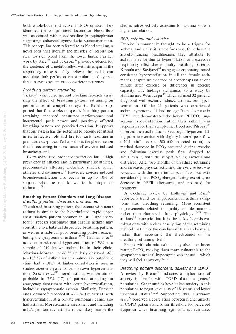
Pub
lishe
d by
Man
ey P
ublis
hing
(c)
W. S
. Man
ey &
Son
Lim
ited
both whole-body and active limb O2 uptake. They
identified the compromised locomotor blood flow
was associated with noradrenaline (norepinephrine)
suggesting enhanced sympathetic vasoconstriction.
This concept has been referred to as blood stealing, a
novel idea that literally the muscles of respiration
steal O2 rich blood from the lower limbs. Further
work by Sheel75 and St Croix76 provide evidence for
the existence of a metaboreflex, with its origin in the
respiratory muscles. They believe this reflex can
modulate limb perfusion via stimulation of sympa-
thetic nervous system vasoconstrictor neurones.
Breathing pattern retrainingVickery11 conducted ground breaking research asses-
sing the effect of breathing pattern retraining on
performance in competitive cyclists. Results sup-
ported that four weeks of specific breathing pattern
retraining enhanced endurance performance and
incremental peak power and positively affected
breathing pattern and perceived exertion. It appears
that our system has the potential to become sensitized
in its protective role and fire too early resulting in
premature dyspnoea. Perhaps this is the phenomenon
that is occurring in some cases of exercise induced
bronchospasm?
Exercise-induced bronchoconstriction has a high
prevalence in athletes and in particular elite athletes,
predominately affecting endurance athletes, winter
athletes and swimmers.77 However, exercise-induced
bronchoconstriction also occurs in up to 10% of
subjects who are not known to be atopic or
asthmatic.78
Breathing Pattern Disorders and Lung DiseaseBreathing pattern disorders and asthmaThe altered breathing pattern that occurs with acute
asthma is similar to the hyperinflated, rapid upper
chest, shallow pattern common in BPD, and there-
fore it appears reasonable that chronic asthma may
contribute to a habitual disordered breathing pattern,
as well as a habitual poor breathing pattern exacer-
bating the symptoms of asthma.52,79 Thomas et al.80
noted an incidence of hyperventilation of 29% in a
sample of 219 known asthmatics in their clinic.
Martinez-Moragon et al. 81 similarly observed 36%
(n517/157) of asthmatics at a pulmonary outpatient
clinic had a BPD. A higher correlation is seen in
studies assessing patients with known hyperventila-
tion. Saisch et al.82 noted asthma was certain or
probable in 78% (17) of patients attending an
emergency department with acute hyperventilation,
including asymptomatic asthma. Similarly, Demeter
and Cordasco83 recorded 80% (38/47) of patients with
hyperventilation, at a private pulmonary clinic, also
had asthma. More accurate assessment and including
mild/asymptomatic asthma is the likely reason the
studies retrospectively assessing for asthma show a
higher correlation.
BPD, asthma and exerciseExercise is commonly thought to be a trigger for
asthma, and whilst it is true for some, for others the
anxiety-inducing breathlessness they attribute to
asthma may be due to hyperinflation and excessive
respiratory effect due to faulty breathing patterns.
Kinnula and Sovijarvi84 using cycle ergometry, noted
consistent hyperventilation in all the female asth-
matics, despite no evidence of bronchospasm at one
minute after exercise or differences in exercise
capacity. The findings are similar to a study by
Hammo and Wienburger85 which assessed 32 patients
diagnosed with exercise-induced asthma, for hyper-
ventilation. Of the 21 patients who experienced
asthma symptoms, 11 had no significant decrease in
FEV1, but demonstrated the lowest PETCO2, sug-
gesting hyperventilation, rather than asthma, was
responsible for their symptoms. Hibbit and Pilsbury86
observed their asthmatic subject began hyperventilat-
ing prior to exercise, with slightly lowered peak flow
(470 L min21 versus 500–660 expected norm). A
marked decrease in PCO2 occurred during exercise
and following exercise peak flow dropped to
385 L min21, with the subject feeling anxious and
distressed. After two months of breathing retraining
and increased physical activities, the exercise test was
repeated, with the same initial peak flow, but with
considerably less PCO2 changes during exercise, no
decrease in PEFR afterwards, and no need for
treatment.
A Cochrane review by Holloway and Ram87
reported a trend for improvement in asthma symp-
toms after breathing retraining. More consistent
improvements related to quality of life markers
rather than changes in lung physiology.10,88 The
authors87 conclude that it is the lack of consistent,
robust data with a clear description of the retraining
method that limits the conclusions that can be made,
rather than necessarily the effectiveness of the
breathing retraining itself.
People with chronic asthma may also have lower
resting PeCO2 making them more vulnerable to the
sympathetic arousal hypocapnia can induce – which
they will feel as anxiety.82,89
Breathing pattern disorders, anxiety and COPDA review by Brenes90 indicates a higher rate of
anxiety in people with COPD than the general
population. Other studies have linked anxiety in this
population to negative quality of life status and lower
functional status.91,92 Supporting this, Livermore
et al.93 observed a correlation between higher anxiety
in COPD patients and lower threshold for perceived
dyspnoea when breathing against a set resistance
CliftonSmith and Rowley Breathing pattern disorders and physiotherapy
80 Physical Therapy Reviews 2011 VOL. 16 NO. 1

Pub
lishe
d by
Man
ey P
ublis
hing
(c)
W. S
. Man
ey &
Son
Lim
ited
increasing the exertion of breathing, compared to
perceived dsypnoea in matched subjects with COPD
and a normal control group.
For these populations, correcting the breathing
pattern to an efficient steady diaphragmatic pattern
can help reduce perceived dyspnoea by reducing the
inspiratory effort and anxiety, helping clarify symp-
toms attributable to actual lung disease rather than
functional factors.
Breathing pattern disorders and anxietyAnxiety may be driven by negative thoughts, but also
by physiology, for example autonomic disregulation,
and/or abnormal lung biomechanics causing a sensa-
tion of dyspnoea, not related to actual insufficiencies.
The factors surrounding anxiety are too complex and
interconnected to suggest there can be a simple causal
effect.94
Studies report greater changes in respiratory
patterns in subjects reporting high anxiety levels,
when completing a stressful task, with marked
increases in tidal volume and respiratory rate and
decreased expiratory time with significant drop in
FETCO2 in the high trait anxiety group.95,96 Similar
changes are seen with anticipatory anxiety.97 Con-
ditioned respiratory responses have also been shown
to occur prior to starting a computer task.98,99
In people with a confirmed diagnosis of an anxiety
disorder, such as panic disorder or post traumatic
stress disorder, there appears to be a loss of home-
ostasis, in particular regarding persistent hyperarou-
sal of the sympathetic control.100,101 Interestingly,
Blechert et al.102 noted the changes in firing of
vasoconstrictor fibers in panic disorder patients were
similar to those in subjects with increased muscle
sympathetic outflow induced by inhaled breath hold
and obstructive sleep apnoea, again reinforcing the
overlap between breathing pattern disorders and a
wide range of causes and symptoms.
Treatment of BPD in PhysiotherapyAssessmentPhysiotherapy treatment of BPD begins with assess-
ment. The lack of a definitive assessment tool for
BPD does make diagnosis difficult and sometimes it
is achieved only by a process of elimination.103
Assessment includes gaining an accurate clinical
history, observation of the person’s breathing and
musculo-skeletal status, and ‘hands on’ assessment of
breathing and muscle tension.4,104,105 Assessment
tools commonly used include the Nijmegen Ques-
tionnaire, breath hold test, peak expiratory flow rate,
and pulse oximetry.4 Spirometry and capnography
may be used, depending on the clinic resources.6
Treatment can then focus on areas of dysfunction
identified during assessment.
TreatmentThe role of breathing exercises in patients with
pulmonary disorders was documented as early as
1915.106,107 By 1919 it was recommended that many
medical and surgical patients be given breathing and
physical exercise as accessories to medical and
surgical treatment.108
The first literature referring to BPDs and breathing
re-education within the physiotherapy profession
was in the 1960s in cardiorespiratory physiotherapy.
At this time physiotherapists advocated breathing
retraining for BPD.105,109 The Papworth method
of breathing retraining evolved from the collabo-
ration of chest physician Claude Lum and phy-
siotherapists Diana Innocenti and Rosemary Cluff.
This focused on education, and a nose/abdominal
breathing pattern.45,105,109 Other key aspects in the
physiotherapy literature treatment are education,
reassurance, and breathing retraining.104,110–115
Most physiotherapy treatment protocols ap-
pear to have the following basic principles in
common.4,6,105,112,115
1. Education on the pathophysiology of the disorder2. Self-observation of one’s own breathing pattern3. Restoration to a basic physiological breathing pat-
tern: relaxed, rhythmical nose–abdominal breathing.4. Appropriate tidal volume5. Education of stress and tension in the body6. Posture7. Breathing with movement and activity8. Clothing Awareness9. Breathing and speech10. Breathing and nutrition11. Breathing and sleep12. Breathing through an acute episode
Education
Education is broader than breathing pattern alone.
Education includes the effects of abnormal versus
diaphragmatic breathing, and reassurance that HVS/
BPD symptoms have a physiological basis, and are
treatable. Education also involves identifying the
factors that initially caused the BPD, and/or may
trigger the poor breathing pattern in the future.4
Lifestyle issues are addressed, such as level of activity,
relaxation (both as a technique and as a recreational
activity), and sleep. Work issues, such as sustained
computer work, extended periods of intense con-
centration and speech are also addressed, as these
areas have been shown to impact on breathing
patterns.116–118
For the public domain, physiotherapists have
written and co-written books on hyperventilation/
BPD. ‘Asthma and Your Child’ by Thompson119
highlights many techniques to assist with breathing
pattern disorders and asthma treatment. In 1991
Bradley120 wrote the first patient handbook on
the subject of hyperventilation syndrome/breathing
pattern disorders. More recently, in collaboration with
CliftonSmith and Rowley Breathing pattern disorders and physiotherapy
Physical Therapy Reviews 2011 VOL. 16 NO. 1 81

Pub
lishe
d by
Man
ey P
ublis
hing
(c)
W. S
. Man
ey &
Son
Lim
ited
CliftonSmith, they have produced dynamic breathing
for asthma,121 and breathe stretch and move.122 All
these books place emphasis on self-management.
Breathing retraining
The terms breathing exercises, breathing retraining
and breathing pattern training are used interchange-
ably in the physiotherapy literature. There is varia-
tion, even within the physiotherapy discipline, of
what parameters of normal breathing are. Cluff105
states the rate should be 8–12 average sized breaths
per minute at rest, with gentle, silent, rhythmical
diaphragmatic (tummy) breathing, with little upper
chest movement. West123 reports breathing rate for
an adult at rest is 10–14 breaths per minute.
The treatment of BPD is under recognized.
Guidelines for the physiotherapy management of
the adult, medical, spontaneously breathing patient
have been recently published.124 These guidelines
represent an extensive amount of work collating and
analyzing research to support current physiothera-
peutic management in the area of cardiorespi-
ratory, neuromuscular diseases and musculoskeletal.
Breathing pattern disorders were not mentioned
during the review, except used in the context as a
historical reference when referring to the treatment
by physiotherapists in the management of disordered
breathing.125,126 Breathing retraining was only used
in reference to asthma and secondary disordered
breathing.
The BradCliff MethodH looks at breathing dys-
function as an indicator of physiological and mechan-
ical imbalances and psychological stress in the human
body. It is structured on current physiotherapy
research assessing and treating individuals from
children with asthma to elite athletes.127
We are now able to have a better informed
approach however, no longer assuming an adequate
breathing pattern at rest is necessarily an optimal
breathing pattern for all the activities our client is
involved with. Diaphragmatic breathing remains the
foundation of our treatment, but it is no longer the
only aspect of our treatment.
Musculo-skeletal componentMusculo-skeletal issues are addressed which are
impeding an effective breathing pattern. Alongside
the mechanical validation of respiratory muscle
contribution to motor control, research into the
training of respiratory muscle strength has gained
momentum,128,129 Much of the research was initially
surrounding dyspnoea and organic respiratory dis-
orders and it is well established that the respiratory
muscles could be strengthened.130 There is evidence
supporting the role of inspiratory muscle trainers to
strengthen the inspiratory muscles, to reduce dyspnoea
and improve function, whether it is for activities of
daily living or high performance sport.131–134
Massery has successfully incorporated breathing,
respiratory control and re-education into rehabilita-
tion covering many neurological conditions such as
cerebral palsy, complex paediatric cases, spinal cord
injuries, as well as respiratory and bio-mechanical
disorders. Massery utilizes a multi-system approach
with breathing/respiration as an integral part. Mas-
sery remains adamant breathing is the first step of
all rehabilitation.135 Massery incorporates breath
and movement into her treatment regimes at all
levels of functioning and views breathing retraining
and postural control strategies as simultaneous
interventions. ‘Motor impairments are never just
a musculoskeletal problem or just a neuromotor
problem. We are born with systems that interact
to give us the control we need for health and
participation.136
An extensive list of Mary Massery’s publica-
tions can be viewed: http://www.masserypt.com/html/
pub.html137
Research addressing treatment efficacy for BPDThe variability of treatment regimes and poor des-
cription of the regime details have made it difficult to
gain a cohesive understanding of what the research to
date has shown. Despite this variation, the authors
report improvements are achieved, suggesting key
elements are covered within the treatment pro-
gramme.138,139 The Papworth method has shown
favourable outcomes, significantly reducing respira-
tory symptoms and improving health-related quality
of life in a group of patients with asthma.140,141
Other papers from the UK also support breathing re-
education/training within physiotherapy practice.142,143
Singh144 reviewed the literature with respect to
physiotherapy treatment and hyperventilation. The
review concluded that the definition and diagnosis of
hyperventilation is difficult; however, once identified
physiotherapy intervention can provide an effective
intervention to significantly reduce the symptoms and
improve quality of life. The query over diagnosis was
the hyperventilation versus breathing pattern disor-
der debate. It has been shown clearly in studies that
breathing retraining has a positive effect on improv-
ing symptoms where the subject does not exhibit low
levels of CO2—highlighting that not only do we see
people with chronic hyperventilation (lowered CO2)
but perhaps a bigger group who present with
symptoms due to mechanisms directly related to
other pathways.145
A 2004 Cochrane review of breathing exercises
for asthma concluded that, due to the diversity of
breathing exercises and outcomes used, it was
impossible to draw conclusions from the available
CliftonSmith and Rowley Breathing pattern disorders and physiotherapy
82 Physical Therapy Reviews 2011 VOL. 16 NO. 1

Pub
lishe
d by
Man
ey P
ublis
hing
(c)
W. S
. Man
ey &
Son
Lim
ited
evidence.146 Thomas et al.147 randomized partici-
pants into a group to receive the Papworth breath-
ing re-education method or to see an experienced
respiratory nurse providing asthma education.
There were significant improvements in asthma-
related quality of life in both groups after 1 month,
but at 6 months a large difference between groups
was found, in favour of the breathing retraining
group, in asthma quality of life, anxiety and
depression, Nijmegen score and a trend for an
improvement in asthma control.147
Vickery11 investigated the effect of breathing
pattern retraining on 20-km time trial performance
and respiratory and metabolic measures in competi-
tive cyclists.
The results supported the performance enhancing
effect of four weeks of breathing pattern retraining in
cyclists. They suggested breathing pattern can be
retrained to exhibit a controlled pattern, without a
tachypnoeic shift (increased respiratory rate leading
potentially to breath stacking and an irregular
pattern that may impair alveolar ventilation) during
high intensity cycling. Results also showed that
respiratory and peripheral perceived effort was
diminished. This research could open avenues of
practice not yet proven before within the field of
sports physiotherapy, emphasizing the importance of
breathing patterns and ultimate performance.
Future trends for physiotherapy treatmentCurrently in western medicine, a fundamental push is
to encourage healthy life style skills. Education in one
of the most fundamental tools, and yet breathing has
not been emphasized enough as part of this healthy
lifestyle package.
Looking to the future the consensus of health in
the twenty-first century in the public domain, there
appears to be a move away from the twentieth
century biomedical model to a more global initiative,
promoting projects and programmes that reach all
human beings in a worldwide commitment to health
as a global public good.148 Keeping this in mind,
there is a push from within our professions to
run with this idea of ‘health for all’ and in parti-
cular involvement in the management, rehabilita-
tion education and prevention of the epidemic of
lifestyles diseases we are currently seeing, such
as obesity, ischemic heart disease, cancer, smoking
related conditions and pulmonary conditions.149,150
There is scope within this framework to explore
the concepts of breathing re-education within the
profession. Breathing re-education is drug free,
appealing to the new paradigm of health for all,
and a practice that requires little or no machinery so
a low running cost, and initial set-up is minimal for
the therapist.
ConclusionsFor the clinician the observation of breathing can
provide insight into many systems, including biome-
chanics, biochemistry/physiology, and psychology
reflecting the consideration of a multisystem approach.
Everyone is a complex integration of musculo/
neurological/respiratory systems, which combined
with individual personalities and lifestyles, reminds
us that these are never distinct groups, and everyone
we meet or treat works best when all systems are in
homeostasis as supported by an appropriate and
efficient breathing pattern.
There is a lack of robust evidence surrounding
breathing pattern disorders. Ongoing research is
needed that clearly describes treatment regimes and
assesses outcomes that are compatible with other
research and remains clinically relevant.
As a profession our diversity is an asset. The key
points of breathing pattern disorders are common to
whomever we treat. Our expertise is in our unique
assessment and treatment skills, which enable us to
develop specific programmes relevant to the indivi-
dual cases whether it is the child with asthma or the
elite athlete. The diversity of our profession enables
us to approach breathing pattern disorders from
different perspectives, yet allows us a cohesive
informed approach, as physiotherapy aims to treat
the whole person not just the system.
References1 DaCosta J. On irritable heart: a clinical study of a form of
functional cardiac disorder and its symptoms. Am J Med Sci1871;61:17–52.
2 Haldane J, Poulton E. The effects of want of oxygen onrespiration. Physiol 1908;37:390.
3 Solely M, Shock N. The etiology of effort syndrome. Am JMed Sci 1938;196,840.
4 Bradley D. Physiotherapy breathing rehabilitation strategies.In: Chaitow L, et al. editors. Multidisciplinary approaches tobreathing pattern disorders. Edinburgh: Churchill Livingstone;2002. p. 173–95.
5 Gosselink R. Breathing techniques in patients with chronicobstructive pulmonary disease (COPD). Chron Respir Dis2004;1:163–72.
6 Hough A. Physiotherapy in respiratory care: an evidencebased approach to respiratory and cardiac management. 3rded. Glos: Nelson Thornes Ltd; 2001.
7 Rowley J. The role of asthma, stress and posture asaetiological factors in breathing pattern disorders. 2002,unpublished.
8 Chaitow L. Biomechanical influences on breathing. In: ChaitowL, et al. Multidisciplinary approaches to breathing patterndisorders. Edinburgh: Churchill Livingstone; 2002. p. 83–110.
9 Gilbert, C. Clinical applications of breathing regulation.Behav Modif 2003;27:692–709.
10 Bruton A, Thomas M. The role of breathing training inasthma management. Current Opinion Allergy Clin Immunol2011;1(1):53–57.
11 Vickery R. The effect of breathing pattern retraining onperformance in competitive cyclists. 2007. Available from:http://repositoryaut.lconz.ac.nz/handle/10292/83.
12 Chaitow L. Breathing pattern disorders, motor control, andlow back pain. J Osteopath Med 2004;7:34–41.
13 Nicholls D, Walton JA, Price K. Making breathing yourbusiness: enterprising practices at the margins of orthodoxy.Sage Publications 2009;13:333–56.
CliftonSmith and Rowley Breathing pattern disorders and physiotherapy
Physical Therapy Reviews 2011 VOL. 16 NO. 1 83

Pub
lishe
d by
Man
ey P
ublis
hing
(c)
W. S
. Man
ey &
Son
Lim
ited
14 Magarian G. Hyperventilation syndromes: infrequently recog-nised common expression of anxiety and stress. Medicine1982;61:219–35.
15 Lewis T. The soldier’s heart and the effort syndrome. NewYork: Paul B. Hoeber; 1919.
16 Goetz CG, Turner C, Aminoff M, editors. Handbook ofclinical neurology. Amsterdam: Elsevier Science PublisherB. V.; 1993. p. 429–47.
17 Gardner W, Bass C. Hyperventilation in clinical practice. Br JHospital Med 1989;41:73–81.
18 Kerr W, Dalton J, Gliebe P. Some physical phenomenaassociated with the anxiety states and their relation tohyperventilation. Ann Int Med 1937;11:962–92.
19 Hornsveld H, Garssen B, Fiedeldij Dop M, van Spiegel P,de Haes J. Double-blind placebo-controlled study of the hyper-ventilation provocation test and the validity of the hyperven-tilation syndrome. Lancet 1996;348:154–8.
20 Gardner W. The pathophysiology of hyperventilation dis-orders. Chest 1996;109:516–35.
21 Newton E. Hyperventilation syndrome. 1997 [Retrieved 1999March 30]. Available from: http://www.emedicine.com/emerg/topic270.htm.
22 Lum C. Hyperventilation and anxiety state. J R Soc Med1981;74:1–4.
23 Gilbert C. Interaction of psychological and emotional effectswith breathing dysfunction. In: Chaitow L, et al, editors.Multidisciplinary approaches to breathing pattern disorders.Edinburgh: Churchill Livingstone; 2002. p. 11–130.
24 Ley R. Breathing and the psychology of emotion, cognition,and behavior. In: Timmons BH & Ley R (eds.), Behaviouraland psychological approaches to breathing disorders. NewYork: Plenum Press; 1994.
25 Dixhoorn J. Significance of breathing awareness and exercisetraining for recovery after myocardial infarction. In: CarlsonJG, et al, (eds.) Clinical applied psychophysiology. New York:Plenum Press; 1994. p. 113–32.
26 Gardner W. Orthostatic increase of respiratory gas exchangein hyperventilation syndrome. Thorax 2000;55:257–9.
27 Gardner W. Hyperventilation. Am J Respir Crit Care2004;170:105.
28 Garssen B, de Ruiter C, van Dyck R. Breathing retraining: arationale placebo? Clin Psychol Rev 1992;12:141–54.
29 Hornsveld H, Garssen B, Fiedeldu Dop M, van Spiegel P.Symptom reporting during voluntary hyperventilation andmental load: implications for diagnosing hyperventilationsyndrome. J Psychosom Res 1990;34:687–97.
30 Molema J, Folgering H. Introduction in abstracts of paperspresented at the 3rd International Society of the Advancementof Respiratory Psychophysiology (ISARP) Congress. Paperpresented at the 3rd International Society of the Advancementof Respiratory Psychophysiology (ISARP) Congress; 1996August 26–27; Nijmegen, The Netherlands.
31 Wilkins R, Krider S, Sheldon R. Clinical assessment inrespiratory care. 4th ed. St Louis, MO: Mosby; 2000.
32 Jack S, Rossiter H, Pearson M, Ward S. Ventilatory responsesto inhaled carbon dioxide, hypoxia, and exercise in idiopathichyperventilation. Am J Respir Crit Care Med 2004;170:118–25.
33 Marieb E. Human anatomy & physiology. 5th ed. SanFrancisco: Addison Wesley Longman; 2001.
34 Molema J, Folgering H. Introduction in abstracts of paperspresented at the 3rd International Society of the Advancementof Respiratory Psychophysiology (ISARP) Congress. Paperpresented at the 3rd International Society of the Advancementof Respiratory Psychophysiology (ISARP) Congress; 1996August 26–27; Nijmegen, The Netherlands.
35 Jack S, Rossiter H, Warburton C, Whipp B. Behaviouralinfluences and physiological indices of ventilatory control insubjects with idiopathic hyperventilation. Behav Modif2003;27:637–52.
36 Humphries B, Quaranta N, Wagstaff S, Baguley D. Tinnitusand cochlear implantation. Int J Audiol 2004;43:245–51.
37 Schleifer L, Ley R, Spalding TW. A hyperventilation theory ofjob stress and musculoskeletal disorders. Am J Ind Med2002;41:420–32.
38 Kazmaier S, Weyland A, Buhre W, Stephan H, Rieke H,Filoda K, et al. Effects of respiratory alkalosis and acidosis onmyocardial blood flow and metabolism in patients withcoronary artery disease. Anesthesiology 1998;89:831–7.
39 Clifton-Smith T. Breathe to succeed in all aspects of your life.Ringwood: Penguin Books; 1999.
40 Ley R. The modification of breathing behaviour: pavlovianand operant control in emotion and cognition. Behav Modif1999;23:441–79.
41 Ley R. Panic disorder and agoraphobia: fear of fear or fear ofthe symptoms produced by hyperventilation? J Behav TherExp Psychiatry 1987;18:305–16.
42 Umezawa A. Facilitation and inhibition of breathing duringchanges of emotion. In: Yutaka Haruki I, et al, editors.Respiration and emotion. Tokyo: Springer 2001. p. 139–47.
43 Boiten F. The effects of emotional behaviour on componentsof the respiratory cycle. Biol Psychol 1998;49:29–51.
44 Wientjes C, Grossman P, Gaillard A. Influence of drive andtiming mechanisms on breathing pattern and ventilationduring mental task performance. Biol Psychol 1998;49:53–70.
45 Lum L. Breathing exercises in the treatment of hyperventila-tion and chronic anxiety states. Chest, Heart Stroke J1977;2:6–11.
46 Gardner W. Diagnosis and organic causes of symptomatichyperventilation. In: Timmons BH, Ley R (eds.), Behavioraland psychological approaches to breathing disorders. 1994New York: Plenum Press; 1991. p.99–111.
47 Bugess J, Kovalchick D, Kyes K, Thompson J, Barnhart S.Hyperventilation following a large-scale hazardous-materialsincident. Int J Occup Environ Health 1999;5:194–7.
48 Hough A. Physiotherapy in respiratory care. London: StanleyThornes; 1996.
49 Pfeffer J. The aetiology of the hyperventilation syndrome: areview of the literature. Psychother Psychosom 1978;39:47–55.
50 Singh J. Management of hyperventilation: how to cope withheavy breathers. J Ass Chartered Physiother Resp Care2001;32:50–5.
51 Boiten F, Frijda N, Wientjes C. Emotions and respiratorypatterns: review and critical analysis. Int J Psychophysiol1994;17:103–28.
52 Hough A. Physiotherapy in respiratory care: a problem-solving approach to respiratory and cardiac management.Cheltenham: Stanley Thornes Ltd; 1997.
53 BradCliffH Manual. Bradley D, Clifton-Smith T, Writers Inc.Auckland, New Zeland, 2009.
54 Lewit K. Relation of faulty respiration to posture with clinicalimplication. J Am Osteopath Assoc 1980;79:525–9.
55 Lewit K. Manipulative therapy in rehabilitation of the motorsystem. 2nd ed. London: Butterworths; 1991. p. 1–4, , 79–82.
56 Hodges P, Gandevia S. Activation of the human diaphragmduring a repetitive postural task. J Physiol (Lond)2000;522:165–75.
57 Hodges P, Butler J, McKenzie D, Gandevia S. Contraction ofthe human diaphragm during postural adjustments. J Physiol(Lond) 1997;505:239–48.
58 Hodges P, McKenzie D, Heijnen I, Gandevia S. Reducedcontribution of the diaphragm to postural control in patientswith severe chronic airflow limitation. Proceedings of theThoracic Society of Australia and New Zealand; 2000 April 7–12; Melbourne, Australia.
59 Hodges P, Gandevia S. Changes in intra-abdominal pressureduring postural and respiratory activation of the humandiaphragm. J Appl Physiol 2000;89:967–976.
60 O’Sullivan P, Grahamslaw K, Kendell M, Lapenskie S, MollerN, Richards K. The effect of different standing and sittingpostures on trunk muscle activity in a pain-free population.Spine 2002;27:1238–44.
61 Falla D, Jull G, Russell T, Vicenzino B, Hodges P. Effect ofneck exercise on sitting posture in patients with chronic neckpain. Phys Ther 2007;87:408–17.
62 Massery M. The patient with multi-system impairmentsaffecting breathing mechanics and motor control. In:Frownfelter D, Dean E, editors. Cardiovascular and pulmon-ary physical therapy evidence and practice. 4th ed. St Louis,MO: Mosby & Elsevier Health Sciences; 2006. Chapter 39,p. 695–717.
63 Lennon J, Shealy N, Cady RK, Matta W, Cox R. Postural andrespiratory modulation of autonomic function, pain, andhealth. Am J Pain 1994; 4:36–39.
64 Hemborg B, Moritz U, Lowing H. Intra abdominal pressureand trunk muscle activity during lifting. The causal factors ofthe intra-abdominal pressure rise. Scand J Rehabil Med1985;17:25–38.
65 Johnson B, Saupe K, Dempsey J. Mechanical constraints onexercise hyperpnea in endurance athletes. J Appl Physiol1992;73:874–86.
CliftonSmith and Rowley Breathing pattern disorders and physiotherapy
84 Physical Therapy Reviews 2011 VOL. 16 NO. 1

Pub
lishe
d by
Man
ey P
ublis
hing
(c)
W. S
. Man
ey &
Son
Lim
ited
66 Perri M, Halford E. Pain and faulty breathing: a pilot study.J Bodyw Mov Ther 2008;4:297–306.
67 Simons DG, Travell JG, Simons IS. Myofascial pain anddysfunction: the trigger point manual. 2nd ed. Baltimore, MD:Williams and Wilkins; 1999. p. 19–20.
68 Hruska J. Influences of dysfunctional respiratory mechanicson orofacial pain. J Orofac Pain Relat Disord 1997;41:216–7.
69 Fabiano P, Cesar F, Amorim L, Henrique S, Oliveira F, DareG, et al. Physiology maximal clenching effort influence on theelectromyographic activity of the trapezius muscle in healthysubjects. J Bodyw Mov Ther 2010;14:346–51.
70 McLaughlin L. Breathing evaluation and retraining in manualtherapy. J Bodyw Mov Ther 2009;13:276–82.
71 Lee EK, Seyal M, Mull B, Gage B. Increased excitability of thehuman corticospinal system hyperventilation. Clin Neurophys1998;109:263–7.
72 Mogyoros I, Kiernan MC, Burke D. Strength-durationproperties of human peripheral nerve. Brain 1996;119:439–47.
73 Mogyoros I, Kiernan MC, Burke D, Bostock H. Strength-duration properties of sensory and motor axons in amyo-trophic lateral sclerosis. Brain 1998;121:851–9.
74 Harms C, Wetter T, McClaran S, Pegelow D, Nickele G,Nelson W, et al. Effects of respiratory muscle work on cardiacoutput and its distribution during maximal exercise. J ApplPhys 1998;85:609–18.
75 Sheel A, Derchak P, Morgan B, Pegelow D, Jaques A,Dempsey J. Fatiguing inspiratory work causes reflex reductionin resting leg blood flow in humans. J Physiol 2001;537:277–89.
76 St Croix C, Morgan B, Wetter T, Dempsey J. Fatiguinginspiratory muscle work causes reflex sympathetic activationin humans. J Physiol 2000;529:493–504.
77 Holzer K, Brukner P. Screening of athletes for exercise-induced bronchoconstriction. Clin J Sport Med 2004;14:134–8.
78 Gotshall RW. Exercise-induced bronchoconstriction. Drugs2002;62:1725–39.
79 Weiss J. Behavioural management of asthma. In Ley R,Timmons B., editors. Behavioural and psychologicalapproaches to breathing disorders. New York: Plenum Press;1994.
80 Thomas M, McKinley R, Freeman E, Foy C. Prevalence ofdysfunctional breathing in patients treated for asthma inprimary care: cross sectional survey. Br Med J 2001;322:1098–100.
81 Martinez-Moragon E, Perpifia M, Belloch A, de Diego A.Prevalence of hyperventilation syndrome in patientstreated for asthma in a pulmonary clinic. Archivos deBronconeumologi 2005;41:267–71.
82 Saisch S, Wessely S, Gardner W. Patients with acutehyperventilation presenting to an inner-city emergency depart-ment. Chest 1996;110:952–7.
83 Demeter S, Cordasco E. Hyperventilation and asthma. Am JMed 1986;81:989–94.
84 Kinnula V, Sovijarvi A. Hyperventilation during exercise:independence on exercise-induced bronchoconstriction in mildasthma. Respir Med 1996;90:145–51.
85 Hammo A, Weinberger M. Exercise-induced hyperventilation:a pseudoasthma syndrome. Ann Allergy Asthma Immunol1999;82:574–8.
86 Hibbit G, Pilsbury D. Demonstration and treatment ofhyperventilation causing asthma. Br J Psychiatry 1988;153:687–9.
87 Holloway E, Ram F. Breathing exercises for asthma. CochraneDatabase of Systematic Reviews 2004. Issue 1. Art. No.:CD001277. DOI: 10.1002/14651858.CD001277.pub2.
88 Thomas M, Taylor D, Bateman E, Boulet L, Boushey H,Busse W, et al. A new perspective on concepts of asthmaseverity and control. Eur Respir J 2008;32:545–54.
89 Osbourne C, O’Conner B, Lewis A, Kanabar V, Gardner W.Hyperventilation and asymptomatic chronic asthma. Thorax2000;55:1016–22.
90 Brenes G. Anxiety and chronic obstructive pulmonary disease:prevalence, impact and treatment. Psychosom Med2003;65:963–70.
91 Cully J, Graham D, Stanley M, Ferguson C, Sharafkhaneh A,Souchek J, et al. Quality of life in patients with chronicobstructive pulmonary disease and comorbidity anxiety ordepression. Psychosomatics 2006;47:312–9.
92 Kim HF, Kunik ME, Molinari VA, Hillman SL, Lalani S,Orengo CA, et al. Functional impairment in COPD patients:
the impact of anxiety and depression. Psychosomatics2000;41:465–71.
93 Livermore N, Butler J, Sharpe L, McBain R, Gandevia S,McKenzie D. Panic attacks and perception of inspiratoryresistive loads in chronic obstructive pulmonary disease. Am JRespir Crit Care Med 2008;178:7–12.
94 Chiatow L. Breathing, chronic pain, touch and the body-mind.J Holistic Care 2007;4:39–45.
95 Masaoka Y, Homma I. Anxiety and respiratory patterns: theirrelationship during mental stress and physical load. Int JPsychophys 1997;27:153–9.
96 Ley R, Yelich G. Fractional end-tidal CO2 as an index of theeffects of stress on math performance and verbal memory oftest-anxious adolescents. Biol Psychol 1998;49:83–94.
97 Masaoka Y, Homma I. Expiratory time determined byindividual anxiety levels in humans. J Appl Physiol1999;86(4):1329–1336.
98 Schleifer L, Ley R. End-tidal pCO2 as an index ofpsychophysiological activity during data-entry work andrelaxation. Ergonomics 1994;37:245–54.
99 Schleifer L, Ley R, Pan C. Breathing, psychological stress, andmusculoskeletal complaints in VDT data-entry work. Paperpresented at the Seventh International Conference on Human-Computer Interactions; 1997 August 24–29; San Francisco,CA, USA.
100 Lum LC. Breathing exercises in the treatment of hyperventila-tion and chronic anxiety states. Chest Heart Stroke J 1977;2:6–11.
101 Lambert E, Hotchkin E, Alvarenga M, Pier C, Richards J,Barton D, et al. Single-unit analysis of sympathetic nervousdischarges in patients with panic disorder. J Physiol2006;570:637–43.
102 Blechert J, Michael T, Grossman P, Lajtman M, Wilhelm F.Autonomic and respiratory characteristics of posttraumaticstress disorder and panic disorder. Psychosom Med2007;69:935–43.
103 Courtney R, Greenwood K, Cohen M. Relationships betweenmeasures of dysfunctional breathing in a population withconcerns about their breathing. J Body Work Movement Ther.2011;15:23–34.
104 Chaitow L, Bradley D. The structure and function ofbreathing. In: Chaitow L, et al, editors. Multidisciplinaryapproaches to breathing pattern disorders. Edinburgh:Churchill Livingstone; 2002.
105 Cluff R. Chronic hyperventilation and its treatment byphysiotherapy. J R Soc Med 1984;77:855–62.
106 MacMahon C. Breathing and physical exercise for use in casesof wounds in the pleura, lung and diaphragm. Lancet1915;2:796–70.
107 MacMahon C. Some cases of gunshot wounds and otheraffections of the chest treated by breathing and physicalexercises. Lancet 1919;1:697–9.
108 Kant S, Singh G. Breathing Exercises as adjunct in themanagement of COPD: an overview. Lung India 2006;23:165–9.
109 Innocenti D. Chronic hyperventilation syndrome. In: DowneyP A, editors. Cash’s textbook of chest, heart, and vasculardisorders for physiotherapists. 4th ed. LondonFaber & Faber;1987.
110 Rowbottom I. The physiotherapy management of chronichyperventilation syndrome. ACPRC J 1992;21:9–12.
111 Tweeddale PM, Rowbottom I, McHardy. Breathing retrain-ing: effect on anxiety and depression scores in behaviouralbreathlessness. J. Psychosom Res 1994;38:11–21.
112 Holloway, E. The role of the physiotherapist in the treatmentof hyperventilation. In: Timmons BH, Ley R., editors.Behavioural and psychological approaches to breathingdisorders. New York: Plenum Press; 1994.
113 Pitman A 1996. Physiotherapy for hyperventilation video.Physiotherapy for hyperventilation group, c/o Anne PitmanPhysiotherapy Dept. The London Clinic, 20 Devonshire Place,London, UK.
114 Bruton A. Breathing and relaxation training improvesrespiratory symptoms and quality of life in asthmatic adults.Aust J Physiother 2008;54:76.
115 Bartley J, Clifton-Smith T. Breathing matters. Auckland:Random House; 2006.
116 Peper E, Gibney R, Wilson V. Group training with healthycomputing practices to prevent repetitive strain injury (RSI): apreliminary study. Appl Psychophysiol Biofeedback 2004;29:279–87.
CliftonSmith and Rowley Breathing pattern disorders and physiotherapy
Physical Therapy Reviews 2011 VOL. 16 NO. 1 85
Pub
lishe
d by
Man
ey P
ublis
hing
(c)
W. S
. Man
ey &
Son
Lim
ited
117 Binazzi B, Lanini B, Bianchi R, Romagnoli I, Nerini M,Gigliotti F, Duranti R, Milic-Emili J, Scana G. Breathingpattern and kinematics in normal subjects during speech,singing and loud whispering. Acta Physiol 2006;186:233–246.
118 Schleifer L, Spalding T, Kerick S, Cram J, Ley R, Hatfield B.Mental stress and trapezius muscle activation under psycho-motor challenge: A focus on EMG gaps during computerwork. Psychophysiol 2008;45(3):356–65.
119 Thompson B. Asthma and your child. Christchurch: Pegasus;1967.
120 Bradley D. Hyperventilation syndrome/breathing patterndisorders. Auckland: Tandem Press; 1991.
121 Bradley D, Clifton-Smith T. Breathing works for asthma.Auckland: Tandem Press; 2002.
122 Bradley D, Clifton-Smith T. Breathe stretch & move.Auckland: Random House; 2002.
123 West, J. Respiratory physiology: the essentials. 7th ed.Baltimore: Lippincott Williams & Wilkins; 2005.
124 Bott J, Blumenthal S, Buxton M, Ellum S, Falconer C, GarrodR, et al. Guidelines for the physiotherapy management of theadult, medical, spontaneously breathing patient Joint BTS/ACPRC guideline. Thorax 2009;64:i1–52.
125 Innocenti D. Chest conditions. Physiotherapy 1969;55:181–9.126 Lum LC. Physiological consideration in the treatment of
hyperventilation syndromes. In: Vinken PJ, Bruyn GW,editors. Handbook of clinical neurology. Amsterdam/NewYork: North-Holland publishing company; 1979. Vol. 38,p. 309–60.
127 http://www.bradcliff.com/.128 Romer L, McConnell A, Jones D. Effects of inspiratory
muscle training upon recovery time during high intensity,repetitive sprint activity. Int J Sports Med 2002;23:353–60.
129 Romer L, McConnell A, Jones D. Effects of inspiratorymuscle training on time trial performance in trained cyclists. JSports Sci 2002;20:547–62.
130 Ries A, Bauldoff G, Carlin B, Casaburi R, Emery C, MahlerD, et al. Pulmonary rehabilitation joint ACCP/AACVPRevidence-based clinical practice guidelines. Chest 2007;131:4S–42S.
131 McConnell A, Romer L. Dyspnoea in health and obstructivepulmonary disease: the role of respiratory muscle function andtraining [Invited Review]. Sports Med 2004;34:117–32.
132 Volianitis S, McConnell A, Koutedakis Y, McNaughton L,Backx K, Jones D. Inspiratory muscle training improvesrowing performance. J Sports Sci 2000;18:551.
133 Volianitis S, McConnell A, Koutedakis Y, McNaughton L,Backx K, Jones D. The influence of inspiratory muscletraining upon rowing performance in competitive rowers.Med Sci Sports Exerc 2001;33:803–9.
134 Romer L, McConnell A, Jones D. Inspiratory muscle fatiguein highly trained cyclists: effects of inspiratory muscle training.Med Sci Sports Exerc 2002;34:785–92.
135 Massery M. Asthma: multi-system implications. In: CampbellS, et al., editors. Physical therapy for children. 3rd ed.Philadelphia, PA: Elsevier Health Science; 2006. Chapter 28,p. 851–79.
136 Massery M. The Linda Crane Memorial Lecture: the patientpuzzle – piecing it together. Cardiopulmonary Phys Ther J2009;20:19–27.
137 http://www.masserypt.com/html/pub.html.138 Bastow V. Hyperventilation syndrome and physiotherapy – a
retrospective audit (abstract). J Ass Chartered PhysiotherResp Care 2000;33:19.
139 Williams J. Audit of physiotherapy breathing re-education forpeople with hyperventilation syndrome (HVS). J AssChartered Physiother Resp Care 2000;1–43.
140 Holloway E, West R. Integrated breathing and relaxationtraining (the Papworth Method) for adults with asthma inprimary care: a randomised controlled trial. Thorax2007;62:1039–42.
141 Thomas M, McKinley RK, Freeman E, et al. Breathingretraining for dysfunctional breathing in asthma: a rando-mised controlled trial. Thorax 2003;58:110–5
142 Kraft A, Hoogduin C. The hyperventilation syndrome. A pilotstudy on the effectiveness of treatment. Br J Psychiatry1984;145:538–42.
143 Innocenti DM. Hyperventilation. In: Pryor J, Webber B,editors. Physiotherapy for respiratory and cardiac problems.Edinburgh: Churchill Livingston; 1998.
144 Singh J. Management of hyperventilation. ACPRC J 2001;34:50–5
145 Fried R, Grimaldi J. The psychology and physiology ofbreathing. In: Behavioral medicine, clinical psychology andpsychiatry. New YorkPlenum Press; 1993.
146 Holloway Cochrane reviews, 2004.147 Thomas M, McKinley RK, Mellor S, Watkin G, Holloway E,
Scullion J, et al. Breathing exercises for asthma: a randomisedcontrolled trial. Thorax 2009;64:55–61.
148 Kickbusch I. In search of the public health paradigm for the 21stcentury: the political dimensions of public health Portuguese.Journal of Public Health 25 anniversary Supplement Issue:current public health challenges. December 2010.
149 Dean E. Physical therapy in the 21st century (Part I): towardpractice informed by epidemiology and the crisis of lifestyleconditions. Physiother Theory Pract 2009;25:330–53.
150 Dean E, Physical therapy in the 21st century (Part II):evidence-based practice within the context of evidence-informed practice. Physiother Theory Pract 2009;25:354–68.
CliftonSmith and Rowley Breathing pattern disorders and physiotherapy
86 Physical Therapy Reviews 2011 VOL. 16 NO. 1

lable at ScienceDirect
International Journal of Osteopathic Medicine 12 (2009) 78–85
Contents lists avai
International Journal of Osteopathic Medicine
journal homepage: www.elsevier .com/i jos
Master class
The functions of breathing and its dysfunctions and their relationshipto breathing therapy
Rosalba Courtney*
RMIT University, School Health Science, 11 Binburra Ave, Avalon, N.S.W 2107, Australia
a r t i c l e i n f o
Article history:Received 8 April 2009Accepted 29 April 2009
Keywords:Dysfunctional breathingFunctions of breathingBreathing therapyHyperventilation
* Tel.: þ61 2 99183460; fax: þ61 2 99187489.E-mail address: [email protected]
1746-0689/$ – see front matter � 2009 Elsevier Ltd.doi:10.1016/j.ijosm.2009.04.002
a b s t r a c t
Breathing is unquestionably a key function of the human body; it sustains life by providing oxygenneeded for metabolism and removing the by-product of these reactions, carbon dioxide. Breathing,however, has other functions apart from the ventilation of air and the maintenance of oxygen and carbondioxide. Breathing affects motor control and postural stability and plays several roles in physiological andpsychological regulation. Breathing can influence homeostatic functions in other system including theautonomic nervous system, the circulatory system, chemical regulation and metabolism.
Breathing becomes dysfunctional when the person is unable to breathe efficiently or when breathingis inappropriate, unhelpful or inefficient in responding to environmental conditions and the changingneeds of the individual.
Impairment of the functions of breathing affects people’s lives, challenging homeostasis, creatingsymptoms and compromising health. The efficiency with which breathing fulfills its various functionscan be diminished because of musculo-skeletal dysfunction, disease, chronic psychological stress orother factors that affect respiratory drive and respiratory control. The neurological control of breathingshows high levels of neuroplasticity as shown by its ability to adapt to a wide range of internal andexternal conditions.
Breathing therapy generally aims to either correct dysfunctions of breathing or enhance its functions.Breathing, unlike most physiological functions, can be controlled voluntarily and it can serve as an entrypoint for physiological and psychological regulation.
� 2009 Elsevier Ltd. All rights reserved.
1. The functions and dysfunctions of breathing
There is a developing interest in impact of dysfunctionalbreathing in common conditions such as asthma, chronic back andneck pain, postural stability, cardiovascular disease, anxiety anddepression. Also breathing therapies are being increasingly used ascomponents of treatment strategies for these conditions. Osteo-paths have long recognized that breathing is a commonly disturbedfunction in the body, which if not addressed has far reaching effectson structure and function.1 Others have also argued that whiledysfunctional breathing (DB) is common, it is often overlooked andwhen untreated results in unnecessary suffering.2–7 The prevalencerate of DB in the general population has been suggested to be ashigh as 5–11% in the general population2,8,9 around 30% in asth-matics10 and up to 83% in anxiety sufferers.11
Understanding the true prevalence of dysfunctional breathing(DB) and its impact on health is difficult because the parameters
All rights reserved.
of DB are not clearly defined. Clinicians and breathing therapistsargue about the perimeters of DB and its definition and thecorrect approaches to use in its clinical assessment. A practicalapproach to DB that has heuristic value is to define it asbreathing which is unable to perform its various functions effi-ciently and is inappropriate for the needs of the individual atthat time.
To evaluate the significance of breathing dysfunctions in healthand to develop and refine the use of breathing therapy requirefurther understanding of the functions and dysfunctions ofbreathing. This article explores some of the key functions ofbreathing and discusses the multi faceted nature of breathingdysfunctions and some of their consequences on mental andphysical processes.
1.1. The respiratory pump and the movement of air
The 21,000 breaths per day taken by the average person comeabout as the breathing muscles attached to the chest wall act tochange its shape. As dimensions and form of the chest wall are

R. Courtney / International Journal of Osteopathic Medicine 12 (2009) 78–85 79
altered, resulting changes in pressure within the intrapleural andalveolar spaces drive the movement of air.12
In its normal relaxed state the 2 compartments of the chest wall,the rib cage or thorax and the abdomen, create an effective respi-ratory pump which moves in response to co-ordinated actionscreated by the diaphragm, scalenes, intercostals, abdominals andaccessory muscles of respiration.13,14 However under particularcircumstances this co-ordinated action is lost.
This pump becomes impaired and or distorted in variousdiseases such as kyphoscoliosis, neuromuscular diseases, obesity,emphysema and asthma.14 Its function can also change in responseto psychological stress.15–17 The changes in breathing pattern thatoccur in response to psychological states or various disease statesmay be appropriate responses to increased ventilatory or metabolicneeds or helpful compensations for pathology. Inappropriate habitsof muscle use may however be retained after the psychological,physiological or environmental conditions that initiated theirdevelopment have passed. In this case they are dysfunctional andcan complicate disease conditions and increase symptoms.
2. Dysfunctions of the biomechanics of breathing
2.1. The diaphragm
The diaphragm is the key to the function of other respiratorymuscles and the primary driver of respiration. If the diaphragm isdysfunctional then the other respiratory muscles will change theirfunction, often becoming overloaded. The normal phasic respira-tory action of this large domed shaped muscle is to descent andflatten during inhalation, lifting and widening the lower 6 ribs. Inmost cases this action is accompanied by slight anterior motion ofthe abdomen. When the diaphragm is functioning normally,forward displacement of the abdomen is accompanied by lateralexpansion and elevation of the lower 6 ribs. The extent of contri-bution from abdominal or lower rib cage motion can vary and stillremain functional. The abdomen can displace anteriorly duringinhalation or not move at all.18 Decreased anterior motion of theabdomen can be compensated by more expansion in the lateral ribcage and vice versa enabling the decreases in intra thoracic pres-sure necessary for inspiratory airflow through a combination ofabdominal and lower rib action.14
2.2. Asynchronous and paradoxical motion between rib cageand abdomen
Typically in the case of a dysfunctional diaphragm the abdom-inal muscles will alter their pattern of respiratory activity.14,18,19
Paradoxical or asynchronous motion of abdomen, where thedimensions of the abdomen decrease during inspiration can bea sign of diaphragm dysfunction, weakness or paralysis.18,20,21
However paradoxical inward motion of the abdomen duringinspiration is not always dysfunctional. In fact inward abdominalmotion during inspiration can be a normal and functional responseto increased lung volume, physical activity, rapid respiratorymaneuvers or standing posture that maintains abdominal pressureand helps the diaphragm to maintain a more ideal length andcurvature.21,22 During inhalation, paradoxical breathing is clearlydysfunctional when it is not adequately compensated by lateralmotion of the rib cage and it is observed that the lower rib cagenarrows instead of widening during inspiration.22
2.3. Upper body muscle dysfunctions
In the case of increased ventilatory demand or when breathing isinefficient the respiratory muscles of the upper rib cage, such as the
scalenes, sternomastoid, upper trapezius and other anterior neckmuscles such as hyoid and long colli increase their activity.19,23,24
This results in increased vertical motion of the rib cage and elevationof the shoulders during the inspiratory phase of breathing.
A tendency to carry the head forward of the body with anteriorrotation of the cranial base is a postural change commonly associatedwith breathing difficulty. Forward head posture is a well-knownresponse to obstructed breathing and is common in children withchronic nasal allergy and mouth breathing because this headposition opens the upper airways. Forward head posture can alsoindicate the presence of short flat diaphragm and weak abdominalsdue to the fact that positioning the head in front of the body increasesthe resting length of diaphragm.19,25 This head posture while facili-tating breathing has several adverse effects on the biomechanics ofthe head, neck and jaw and is associated with temporal mandibularjoint syndrome, neck pain and headache.19
Shoulder problems may also result because hypertonic trapeziusmuscles contract during the initial stages of shoulder movementrather than towards the end.25 This altered pattern of scapulohumeral motion is associated with shoulder pain and rotator cuffdysfunction.
3. Factors affecting efficiency of biomechanics of breathing
3.1. Hyperinflation and lung volumes
End expiratory lung volume is an important influence on thepower of the diaphragm affecting its ability to act efficiently on therib cage. Conditions like COPD, asthma and other conditions asso-ciated with increased inspiratory drive and inefficient expirationcan lead to trapping of air in the lungs or hyperinflation. When thisoccurs the diaphragm becomes shorter and loses its curvature, as itis forced to take a lower resting position in the thorax. This short-ening of the diaphragm fibers decreases the power and efficiency ofthe diaphragm due to the laws of length, tension relationshipswhich apply to all contracting muscles.26 It is also associated withloss of the curvature or doming of the diaphragm and a reduction ofthe zone of apposition. In this case the diaphragm fibers, which areattached to the lower 6 ribs, become orientated transversely ratherthan vertically. When the diaphragm contracts, it is ineffective inlifting and widen the lower rib cage. Instead there is a tendency forthe diaphragm to pull the rib the lower lateral rib cage inward,decreasing the transverse diameter of the lower rib cage duringinhalation.14,27,28
The more lung volume increases, the more the zone of apposi-tion is reduced. When the lungs are at 30% of inspiratory capacitythe costal diaphragm no longer expands the rib cage, and above 30%inspiratory capacity the costal diaphragm has a rib cage deflatingaction. At enlarged volumes there is also a tendency for the motionof the abdomen to reverse its timing so that inspiration is accom-panied by inward motion of the abdomen and expiration isaccompanied by outward motion of the abdomen.21
Reducing lung volumes can improve the function of the dia-phragm. This is clearly demonstrated in COPD patients who expe-rience reductions in lung volume after undertaking surgery toremove sections of their lungs. Lung reduction surgery has beenshown in several studies to improve neuromechanical coupling ofthe diaphragm and individuals who receive this surgery consis-tently report increased exercise tolerance and reduced symptomsof dyspnea.29–31
3.2. Abdominal weakness
Abdominal muscle weakness aggravates diaphragm dysfunc-tion.1,19 Both tonic and phasic contraction of abdominal muscles

R. Courtney / International Journal of Osteopathic Medicine 12 (2009) 78–8580
assist the function of the diaphragm during inspiration and expi-ration and can to some extent compensate for diaphragmdysfunction.22,26,32 Contraction of the abdomen during inspirationprevents the diaphragm from shortening excessively duringstanding posture and during rapid and large volume respiratorymaneuvers.22,33 During expiration contraction of the abdomenassists diaphragm doming, increasing its length and curvature inpreparation for effective contraction during inspiration. Peoplewith dysfunctional diaphragms, such as many individuals withCOPD, can become more breathless and develop more abnormalbreathing if tonic contraction of the abdomen is reduced.34 On theother hand, increasing abdominal tone artificially using abdominalbinding can assist these individuals.
3.3. Rib cage stiffness
During hyperinflation the ribs shift from their normal obliqueposition to a more horizontal position, impairing inspiratory actionof rib cage muscles and making the rib cage stiff and difficult toexpand.35 Restrictions of the rib cage then further inhibit dia-phragm function.36
3.4. Respiratory drive
When respiratory control centres in the brain receive messagesfrom the cortex, limbic system, chemoreceptors or mechanore-ceptors that ventilation is inadequate, the respiratory musclesadjust their functions to increase ventilation. If respiration isstimulated for prolonged periods, the diaphragm and accessorymuscles of breathing may become chronically hypertonic.13,21,37
Typical changes in breathing pattern that reflect increased respi-ratory drive include, upper chest breathing with decreased lateralexpansion of the lower rib cage and tendency to asynchronous andparadoxical breathing.
Disease processes can increase ventilatory needs, stimulaterespiratory drive and alter respiratory control often creating char-acteristic changes in breathing pattern. This is seen in respiratoryconditions such as asthma and COPD and in heart disease.38–40
Psychological and emotional states also alter respiratory controland respiratory rhythm generation.41–44 Subsequently changes inbreathing pattern which can be dysfunctional are very common inpeople with respiratory and cardiovascular disease and underpsychological stress.
4. Breathing in postural and motor control
The use of breathing muscles during respiration affects howthese muscles are used for non-breathing movement and forpostural support. Muscles such as the diaphragm, transverseabdominus and pelvic floor muscles are important for motorcontrol and postural support as well as for breathing. If theirfunction is compromised there is an increased susceptibility to backpain and injury.45–48 The respiratory functions of these musclesneed to be integrated with their many other functions, such asswallowing, speech, valsalva maneuvers, spinal stabilisation andmovement of the trunk and limbs. This need for integration of oftenunrelated functions places considerable demands on mechanism ofmotor control.49,50
In situations where respiratory drive is increased such as stress,disease or physical exercise, the ability of the respiratory muscles toperform their postural tasks is reduced. Interestingly the presenceof respiratory disease is a stronger predictor for lower back painthan other established risk factors.51 A study by McGill showed thatartificially stimulated respiration led to decreased support of thespine during a load challenge.52 Hodges in a subsequent study
showed that the postural functions of the diaphragm were signif-icantly reduced and in some cases abolished when respiration wasstimulated.53
Respiratory muscles that are responding to increased respira-tory drive due to stress or disease produce different breathingpatterns to those seen in normal subjects.33,54 The respiratorymuscles in these situations can become shortened or hypertonicand subsequently less powerful and less efficient.26 They also losetheir normal co-ordinated function and one would expect this toresult in increased demands on motor control mechanisms. Thesefactors compromise the ability of respiratory muscles to create thefine tuning adjustment required for postural support. The rela-tionship between dysfunctional breathing and postural stability,motor control and back pain has not been studied extensivelyhowever clinical observation supports the notion that patients withpoor breathing muscle co-ordination are more prone to chronicback pain and neck pain.56,57
5. Biomechanical influences on hemodynamicsand the lymphatic system
A functional respiratory pump creates rhythmic pressure fluc-tuations between the thorax and the abdomen that are importantfor the movement of body fluids such as blood and lymph. Normalpressure development during the respiratory cycle is characterizedby a decrease in intra thoracic pressure during inhalation and anincrease in intra abdominal pressure during expiration.58,59 Para-doxical motion of the abdomen and dysfunction of the diaphragmalters normal pressure relationships between the thoracic andabdominal compartment during inspiration and expiration. Inparadoxical motion of the abdomen during inspiration, abdominalpressures can decrease rather increase during inspiration.21
Osteopaths have long considered that restoration of lymphaticfunction and treatment of oedema and infection was related toproper function of the diaphragm, rib cage and abdomen.1,60,61
Miller, one of the early Osteopathic developers of manual pumptechniques for lymphatic drainage, noted that thoracic andabdominal pump techniques were exaggerations of respiratorymovements.62
Respiration is one of the important extrinsic influences onlymphatic flow, interacting with intrinsic motility of lymph vesselsand organs to either enhance or dampen their activity.63 Thepressure differentials created by respiration create fluctuations incentral venous pressure which directly affect lymph drainage fromthe lungs and abdomen64,65 and affect transmural pressure inlymphatic vessels which modulate the function of intrinisic pace-makers in the lymphatic system. Recent research also shows thatthe lymphatics in the diaphragm itself form a specialised system fordraining fluid from the peritoneal cavity and returning it to thevascular system.66 This suggests that breathing patterns whichalter normal respiratory pressure dynamics have detrimental effecton the function of the cardiovascular and lymphatic systems.
6. The role of breathing in physiological regulation
Breathing also affects physiological regulation because of itsability to entrain respiratory oscillations to oscillations in othersystems and its role in maintaining homestasis of oxygen, carbondioxide and pH.
7. Oxygen, carbon dioxide and pH
Breathing, by exchanging carbon dioxide (CO2) for oxygen (O2),controls the fundamental gaseous fuels of life’s energy and assistsin maintaining optimal conditions for the biochemical aspect of the

R. Courtney / International Journal of Osteopathic Medicine 12 (2009) 78–85 81
internal milieu. Oxygen concentrations are generally well main-tained by anyone who does not have severe pathology of the lungs,heart or central nervous system. Functional breathing disorders donot generally affect O2 however even relatively healthy people canover breathe sufficiently to become depleted in CO2.
Low CO2 levels can develop quickly because of the very highsolubility of CO2 (twenty times more soluble than oxygen). Itsease of excretion means that increased ventilation resulting fromnon-metabolic stimuli, for example stress, anxiety or increasesensations of dyspnea, can result in depletion of CO2. Breathing inexcess of metabolic demands is called hyperventilation. Theeffects of hyperventilation and carbon dioxide depletion are farreaching and include inhibiting the dissociation of oxygen fromhemoglobin in the blood, resulting in low oxygen concentration intissues. The effects on the brain and the nervous system ofhypocapnia are particularly pronounced and include reducedcerebral blood flow and increased neuronal excitability. Regula-tion of cerebro spinal fluid pressure, intracellular pH and cellularmetabolism are impaired. Hypocapnia produces bronchocon-striction in the lungs and vasoconstriction in the blood vessels.Blood pressure, myocardial contractility and cardiac blood flowcan also be adversely affected as can pH regulation and electrolytebalance.67,68
8. Hyperventilation and hypocapnia
Given the large number of physiological effects of hypocapnia itis not surprising that until the 1990s scientific literature primarilylinked breathing dysfunction with hyperventilation syndrome(HVS). The diagnosis of HVS was generally made on finding anassortment of symptoms believed to be caused by either acute orchronic hypocapnia or the consequences of respiratoryalkalosis.2,5,70
8.1. History of hyperventilation syndrome
Since the beginning of the nineteenth century it has beenknown that symptoms such as numbness, dizziness, musclehypertonicity and tingling sensations could be brought on byoverbreathing71 and that these symptoms could be attributed tohypocapnia and respiratory alkalosis.72 The idea of a hyperventila-tion syndrome characterized by a larger number of psychologicaland somatic symptoms that could be related to either acute orchronic hyperventilation began to develop after Kerr reported hisfindings on the effects of hyperventilation challenge on 35 patientswith unexplained symptom. His patients were able to reproducetheir symptoms by voluntary and prolonged hyperventilation.3 Thename Hyperventilation Syndrome began to be used at around thistime and was considered to exist mostly in neurotic patients and tobe a relatively rare condition.4 The range of symptoms attributed toHyperventilation Syndrome gradually increased until a largenumber of symptoms of central and peripheral neurovascular,muscular, respiratory, cardiac, gastrointestinal origin were attrib-uted to this syndrome.6 Dr. Claude Lum diagnosed 700 patientswith medically unexplained symptoms such as palpitationsincluding chest pain, dizziness, parasthesia, breathlessness,epigastric pain, muscle pain, tremor, tetany, dysphagia, tension andanxiety as suffering from hyperventilation syndrome. The diagnosisseemed to be confirmed when the majority of these patientsrecovered from most of their symptoms after 7 weeks of intensivebreathing training.2 Later studies such as those by Han, however,were to show that individuals with the symptoms generallyattributed to HVS who received benefit from breathing retrainingdid not necessarily improve because of changes in their carbondioxide levels.73
8.2. Doubts about the role of chronic hypocapniain hyperventilation syndrome
From the late 1980s scientists began to question the role ofchronic hypocapnia in HVS. Symptoms which were produced byvoluntary hyperventilation were assumed to be elicited by acutehypocapnia, but on closer investigation could not always beconsistently linked with chronic carbon dioxide deficit. Howellmeasured PCO2 levels in 31 patients with disproportionatebreathlessness and other symptoms of hyperventilation and foundthat they had mostly normal levels of carbon dioxide.74 Han incomparing 399 symptomatic hyperventilators with 347 normalsfound no difference in ETCO2.75 In a major review of hyperventi-lation Hardonk and Beumer found in their own and other studiesthat ETCO2 levels were not significantly different in symptomatic ornormal controls when measured in the laboratory. By the mid1990s Gardener in a review of HVS described the ‘‘ uncertainty andlack of consensus about the boundaries and even existence of thissyndrome’’. He reported that patients could have low carbondioxide but no symptoms, while other people could have relativelynormal carbon dioxide levels but still exhibit the symptoms ofHVS.76
Acute hypocapnia can be induced by the hyperventilationprovocation test (HVPT) and the onset of symptoms in HVS patientsafter performing the HVPT was believed to be due to the acutehypocapnia produced by this voluntary overbreathing.77 A debili-tating and almost fatal blow to the hyperventilation syndromecame from Hornsveld and Garssen whose research appeared toindicate that acute hypocapnia was not the mechanism of the goldstandard of HVS diagnosis, the HVPT. In their study 115 patientsbelieved to have HVS were given the Hyperventilation ProvocationTest (HVPT). 74% of their subjects were positive on the test andreported the onset of their symptoms after hyperventilation.However 65% of these responders were also positive on a placebotest, during which CO2 levels were kept stable through manualtitration. A second stage of this study, involving transcutaneousmonitoring of CO2 levels of patients in their daily lives, showed thatpatients suffering from attacks of the HVS symptoms suffered onlya very slight drop in CO2 levels at the onset of their symptoms andthis usually followed rather than preceded the onset of symp-toms.78 As their study, and others, found that neither chronic oracute deficiency of CO2 could be experimentally linked to HVS theseauthors recommended that the term hyperventilation syndrome bediscontinued.8 In recognition of the fact that causes of breathingrelated symptoms were unclear and often associated withpsychological disturbance, researchers proposed that the termhyperventilation syndrome be replaced with behavioral breath-lessness74 or unexplained breathing disorder (UBD) or chronicsymptomatic hyperventilation.79 Subsequently the term hyper-ventilation syndrome is used infrequently and the terms dysfunc-tional breathing10 breathing pattern disorder80 have become morecommon.
However the door on hyperventilation and hypocapnia mayhave been shut prematurely. Hypocapnia may not be the primesuspect in HVS symptoms but an accomplice whose contributionvaries according to individual susceptibility and exact symptoms.Recent studies indicate that carbon dioxide functions as onecontributing factor to symptoms and its influence is probablymoderated by neurological and other factors that influencesymptom perception.81 Not all symptoms of HVS appear to beequally related to carbon dioxide levels, a greater relationshipexists for neurovascular symptoms than for uncomfortable respi-ratory sensations.82
The physiological effects of hypocapnia have been too welldocumented to be irrelevant.67,68 Modern day researchers need to

R. Courtney / International Journal of Osteopathic Medicine 12 (2009) 78–8582
interpret the finding of Hornsveld et al. while still keeping in mindHaldanes experiments in 1908 which clearly shown that hyper-ventilation caused central and peripheral neurovascular symptomsof dizziness, sensations of numbness and tingling and tetany inhealthy people and the long history of studies on symptomaticindividuals which repeatedly showed that voluntary acute hyper-ventilation caused the recurrence of patients primary andsecondary complaints which could not be explained in otherways.70 Also worth remembering are the studies which demon-strated that administration of carbon dioxide either through re-breathing or by administration of CO2 enriched gas mixtures wasable to eliminate the symptoms of hyperventilation that eithercame on spontaneously or were brought on by hyperventilationprovocation tests.4,5
Chronic hypocapnia when found can and should be normalised,particularly in symptomatic individuals and individual responses toacute hypocapnia. Clinical experience also suggests that thehyperventilation provocation test may indeed be relevant to diag-nosis and treatment if used in conjunction with otherinvestigations.
9. Breathing, homeostasis and oscillations
Breathing, by creating fluctuations in the chemical compositionof the blood, circulation and vascular pressure and by its effects onautonomic reflexes acts as an extrinsic influence on other oscil-lating physiological systems. Oscillations, which are defined assystematic rhythms in physiological variables, are found in mostliving system including those of the human body. Oscillations insingle systems and synchronisation between oscillating systemshelp physiological control systems to maintain homeostasis andappropriate and rapid responsiveness to the continual changingneeds of the body. When oscillations of two or more systems aresynchronised it increases physiological efficiency by enabling thefunctions of these systems to be co-ordinated. This prevents energybeing wasted on non productive functions.85,86 Cyclic activity alsoallows systems to rest and renew themselves during cycles ofdecreased activity.
One aspect of breathing functionality is the ability of breathingoscillations to interact with oscillations in other physiologicalsystems in ways that optimise their functions. Breathing oscilla-tions interact with oscillations of heart rate and blood pressure,87,88
the lymphatic system63 the digestive system,89,90 brain waves91 andprobably the rhythmic fluctuations occurring in cellular metabo-lism.92 When respiratory oscillations entrain other oscillatingsystems it can enhance the physiological function of both systems.A well-known example is the phenomenon of respiratory sinusarrhythmia (RSA), where heart rate variability is entrained torespiratory frequencies. RSA improves the efficiency of gasexchange by coupling increased heat rate to the inspiratory phaseof respiration.93
9.1. Breathing pattern dysfunctions and resonant frequenciesof breathing
Breathing can be consciously manipulated to increase its abilityto entrain other oscillations and increase physiological regulation.When breathing frequency is slowed to between 4 and 6 breathsper minute (0.06–0.1 Hz) oscillations in blood pressure, heart rateand autonomic nervous system tend to synchronise at thisfrequency and be amplified due to resonance effects between thesesystems.94–96 These resonance effects between cardiorespiratoryoscillations and autonomic function are important for homeostasisand maintenance of health as evidence by the fact that training thebodies ability to increase them assists people with a range of
conditions including asthma, COPD, depression, hypertension andirritable bowel syndrome.97–100 Individuals with paradoxicalbreathing and thoracic dominant breathing have a decreased abilityto achieve resonance effects between oscillators (unpublisheddata), indicating that dysfunctions of breathing pattern impair thebody’s ability to regulate itself through co-ordinating homeostaticcardiorespiratory oscillations.
10. Breathing, stress, emotion and the autonomicnervous system
Chronic emotional stress and increased mental load can alterrespiratory regulation in several ways: 1) with regard to drive; 2)breathing pattern and timing; and 3) metabolic appropriateness ofthe respiratory response.42
Breathing is particularly sensitive to states of hyperarousal,during which signs of increased respiratory drive are evidence ofthe body’s readiness for action. Hyperarousal brought on by mentaland emotional processes contributes to allostatic load and affectsthe capacity of the body to maintain its stability and response tochange.101 Anticipation of coming physical and emotional eventshas the distinct effect of increasing respiratory rate, reducing timeof exhalation and changing respiratory pattern.44,102 Breathingirregularity is a common feature of patients with anxiety and panicdisorder, indicating dysregulation of normal breathing controlmechanisms in these individuals.75,103 Specific effects on the dia-phragm are also seen. Fluroscopic studies show that in situations ofemotional stress, the diaphragm shows signs of hypertonicitybecoming flattened and immobile.15,16
Breathing which is responding to feelings and thoughts,rather than metabolic cues from chemoreceptors, may not bealigned to the actual physical needs of the body or its metabolicrequirements.44,88 When mental and emotional factors such asfear, grief, anxiety or depression drive breathing regulation,homeostatic and biomechanical functions of breathing can bedisturbed.42,43 Emotions are drives to particular types of actionor expression. Repeatedly frustrated ability to act on strongemotional drives can lead to physiologically and biomechanicallyinappropriate breathing, tuned to the anticipation of action thatdoes not occur.
A fundamental survival function of increased respiratory driveis to prepare the body for fight and flight. However homeostasis isbest served through the functions of the parasympathetic nervoussystem, whose activity is associated with a relaxed, slow andabdominal breathing pattern.104 Rapid, shallow and thoracicdominant breathing with high levels of tonic contraction ofrespiratory muscles as found in situations requiring high levels ofventilation probably indicates a system that is having difficultyreturning to a state of rest. In this situation energy is wasted andhomeostatic functions needed for repair and renewal areimpaired.
Controlled respiration can help the system to return to a physi-ological rest state. It appears to act on the brain and the autonomicnervous system, synchronising neural elements in the brain andautonomic nervous system and creating a state of pycho-phsyio-logical coherence.105,106 The changes induced by controlledbreathing appear to order and regulate neurological function,improving psychological and emotional states.107,108 The regulatoryeffects of breathing on the autonomic nervous system have beeninvestigated from several perspectives. Several studies have shownthat in the short term controlled breathing can decrease sympa-thetic nervous system activity and increase parasympatheticnervous system and with continued, regular practice can createlong term improvements in autonomic balance and increased vagaltone.109–113

R. Courtney / International Journal of Osteopathic Medicine 12 (2009) 78–85 83
11. Breathing as a therapeutic tool
There are a large number of breathing therapies utilizing a widerange of techniques and several of these appear beneficial. Themain rationales for breathing therapies are that they: 1) correctsome aspect of dysfunctional breathing; 2) support one or severalof the functions of breathing and thus stimulate healing; or 3)provide a means for regulation of mental and emotional states. Theterritory covered by breathing therapies is large covering a broadarea ranging over psychology, physiology, spirituality and biome-chanics. The evidence for the ability of breathing therapies tocorrect breathing dysfunctions and support the functions ofbreathing is relatively sparse as research has tended to focus ondisease or psychological outcomes rather than investigating effectson breathing parameters.
There are several breathing therapies that aim to correcthyperventilation and restore normal carbon dioxide tension.Breathing biofeedback using a capnometer to monitor end-tidalcarbon dioxide levels during breathing training is one approach totreating hypocapnia. Biofeedback training that employs the use ofa capnometer aims at normalizing end-tidal CO2 at approximately5%. There are various systems for capnometry biofeedback availableto the practitioner and in recent years these have become increas-ingly available.114,115 Other breathing therapists use no instrumen-tation and rely on a combination of slow controlled breathing,breathing pattern correction and relaxation strategies.116,117 TheButeyko Breathing Technique (BBT) is yet another technique whoseprimary aim is the correction of acute and chronic hypocapnia. Ituses a unique set of breathing techniques in which breath holding iscombined with reduced volume breathing. BBT exercises aim toincrease carbon dioxde and reset chemoreceptor thresholdshowever they may also be useful in reducing hyperinflation.118
The practice of controlled breathing as a means of self-regula-tion and restoration of mental and emotional balance is a part ofIndian and Taoist Yoga and is also in the domain of modern respi-ratory psychophysiology. Modulation of the breath and mindfulattention to the breath are important parts of many meditationtechniques.119 Ancient systems such as Indian Yoga pranayama andthe breathing techniques of Qi Gong teach that specific types ofbreathing exercises can direct vital force or energy and through thisbenefit the health of the mind and body.120–123
Respiratory psycholphysiology uses a scientific approach tounderstanding the physiological processes that link mental andemotional states to breathing. Practitioners of respiratory psycho-physiology utilize evidence based breathing techniques includingresonant frequency breathing to promote emotional self regulationand to achieve efficient physiological states that promote healing.95
Another interesting breathing therapy used mostly to enhanceathletic performance in athletes is Intermittent Hypoxic Training(IHT).124 This therapy exposes individuals to carefully controlledlevels of hypoxia interspersed with rest periods during which theybreathe normal atmospheric air or oxygen enriched air. IHT has theeffect of enhancing adaptation to the specific stress of hypoxia butalso results in a general increase in stress tolerance. Other benefitsof IHT include enhanced antioxidant capacity, improved metabo-lism and increased aerobic capacity. IHT is believed to enhancemitochondrial function as it has been shown to increase the effi-ciency with which the body uses oxygen. In Russia IHT has beenstudied extensively for over 30 years and is used to treat a largenumber of diseases.125
There is a growing body of scientific evidence for the effective-ness of breathing therapies in a range of diseases including asthma,heart disease, anxiety and depression, and they are being increas-ingly included in therapeutic protocols. Research into ButeykoBreathing Techniques has focused on asthma and there have been at
least five published clinical trials on BBT for this condition.126–132
These clinical trials indicate that people learning the ButeykoMethod are able to substantially reduce medication with no dete-rioration in their lung function or asthma control, although nostudies have demonstrated objective changes in lung function. Thequality of evidence of the Buteyko Method according to an Austra-lian Department of Health report is stronger than any othercomplementary medicine treatment of asthma.133 Recent studiesindicate that several other types of breathing therapies also helpasthma including resonant frequency breathing biofeedback,97
capnometry biofeedback134 and breathing rehabilitation.116,135
Breathing therapy has also been found to be very helpful forcardiovascular disease. In a study comparing patients who receivedstandard cardiac rehabilitation with those receiving additionaltraining in breathing therapy after myocardial infarction (MI), itwas found that the breathing therapy group had about a 30%decreased in cardiac events at 5 year follow-up.136 Another studyshowed that exercise training in patients with MI was not alwayssuccessful in preventing future cardiac events, however the risk oftreatment failure was reduced by half when relaxation andbreathing training was added to exercise training.137 Otherbreathing therapy based on yoga breathing was also found toimprove hemodynamics and various cardiorespiratory risk factorsin cardiac patients.138,139
The effectiveness of breathing therapies in psychological condi-tions and chronic stress has also been shown in several studies. Inmajor depression, both resonant frequency biofeedback and yogabased breathing techniques appear to be effective.99,139,140 Peoplewith anxiety and panic disorder also show beneficial response tocapnometry and other breathing therapy protocols.73,141,142 Theeffects of breathing on the autonomic nervous system have beendemonstrated in several studies on yoga pranayama. Regular andprolonged practice of several brief breathing protocols combiningslow breathing, long breath retentions and nostril breathing havethe effect of increasing resetting autonomic balance and amplifyingparasympathetic nervous system function.109
This sampling of the literature on breathing dysfunction andbreathing therapies, while not exhaustive, strongly suggests thatbreathing therapies have the potential to be of benefit to patientswith many types of conditions. It appears that use of breathingtherapies is not proportional to the level of evidence supportingtheir efficacy. One reason for this may be the lack of coherentmodels for explaining the mechanisms of breathing therapies.Understanding of these mechanisms needs to be developed torefine and direct the use of breathing interventions. I propose thatthere are likely to be a range of mechanisms for breathing therapiesand these should be sought within the various functions ofbreathing and its dysfunctions.
Part two of this paper will elaborate on the role of breathingdysfunction in various diseases and present some diverseapproaches to evaluation of various aspects of breathingdysfunction.
Acknowledgements
The Australian Osteopathic Association for financial andadministrative support for research associated with this reviewarticle.
References
1. McConnell C. The diaphragm. J Am Osteopath Assoc 1928;31:87–91.2. Lum LC. Hyperventilation: the tip and the iceberg. J Psychom Res
1975;19:375–83.3. Kerr WJ, Dalton JA, Gliebe PA. Some physical phenomena associated with the
anxiety states and their relation to hyperventilation. Ann Int Med 1937;11:961.

R. Courtney / International Journal of Osteopathic Medicine 12 (2009) 78–8584
4. Soley MH, Shock NW. The aetiology of effort syndrome. Am J Med Sci1938;196:840.
5. Magarian G. Hyperventilation syndromes: infrequently recognized commonexpressions of anxiety and stress. Medicine 1982;61:219–36.
6. Lewis BI. The hyperventilation syndrome. Ann Int Med 1953;38:918–27.7. Nixon PGF. The grey area of effort syndrome and hyperventilation. J R Coll
Physicians of Lond 1993;27.8. Hornsveld HK, Garsson B. Hyperventilation syndrome: an elegant but scien-
tifically untenable concept. Nether J Med 1997;50:13–20.9. Fried R. The hyperventilation syndrome. Baltimore/London: John Hopkins
University Press; 1987.10. Thomas M, McKinley RK, Freeman E, Foy C. Prevalence of dysfunctional
breathing in patients treated for asthma in primary care: cross sectionalsurvey. BMJ 2001;322:1098–100.
11. Cowley DS. Hyperventilation and panic disorder. Am J Med 1987;83:923–9.12. West J. Respiratory physiology. 3rd ed. London: Williams and Wilkins; 1985.13. Konno K, Mead J. Measurement of separate volume changes of rib cage and
abdomen during breathing. J Appl Physiol 1966;22:407–22.14. De Troyer A, Estenne M. Functional anatomy of the respiratory muscles. Clin
Chest Med 1988;9:175–93.15. Faulkner WB. The effect of the emotions upon diaphragm function: observa-
tions in 5 patients. Psychosom Med 1941;3:187–9.16. Wolf S. Diaphragmatic spasm: a neglected cause of dyspnea and chest pain.
Integr Physiol Behav Sci 1994;29:74–6.17. Masaoka Y, Homma I. Anxiety and respiratory patterns: their relationship
during mental stress and physical load. Int J Psychophysiol 1997;27:153–9.18. Fitting JW, Grassino AE. Diagnosis of diaphragm dysfunction. Clin Chest Med
1987;8:91–103.19. Hruska R. Influences of dysfunctional respiratory mechanics on orofacial pain.
Dent Clin North Am 1997;41:211–27.20. Jubran A, Tobin M. The effect of hyperinflation on rib cage-abdominal motion.
Am Rev Respiratory Diseases 1992;146:1378–82.21. Wolfson D, Strohl K, Dimarco A, Altose M. Effects of an increase in end-expi-
ratory volume on the pattern of thoracoabdominal movement. Respir Physiol1983;53:273–83.
22. De Troyer A. Mechanical role of the abdominal muscles in relation to posture.Respir Physiol 1983;53:341–53.
23. Verschakelen JA, Demedts MG. Normal thoracoabdominal motions: influence ofsex, age, posture, and breath size. Am J Respir Crit Care Med 1995;151:399–405.
24. De Troyer A, Estenne M. Coordination between rib cage muscles and dia-phragm during quiet breathing in humans. J Appl Physiol 1984;57:899–906.
25. Pryor JA, Prasad SA. Physiotherapy for respiratory and cardiac problems. 3rd ed.Edinburg: Churchill Livingstone; 2002.
26. Finucane K, Panizza J, Singh B. Efficiency of the normal human diaphragmwith hyperinflation. J Appl Physiol 2005;99:1402–11.
27. Zocchi L, et al. Effect of hyperinflation and equalization of abdominal pressureon diaphragmatic action. J Appl Physiol 1987;62:1655–64.
28. Celli B. Clinical and physiological evaluation of respiratory muscle function.Clin Chest Med 1989;10.
29. Laghi F, et al. Effect of lung volume reduction surgery on neuromechanicalcoupling of the diaphragm. Am J Respir Crit Care Med 1998;157:475–83.
30. Lahrmann H, et al. Neural drive to the diaphragm after lung volume reductionsurgery*10.1378/chest.116.6.1593. Chest 1999;116:1593–600.
31. Cassart M, et al. Effects of lung volume reduction surgery for emphysema ondiaphragm dimensions and configuration. Am J Respir Crit Care Med2001;163:1171–5.
32. Cassart M, et al. Effect of chronic hyperinflation on diaphragm length andsurface area. Am J Respir Care Med 1997;156:504–8.
33. Sharp JT, et al. Relative contributions of rib cage and abdomen to breathing innormal subjects. J Appl Physiol 1975;39:608–18.
34. Cahalin L, et al. Efficacy of diaphragmatic breathing in persons with chronicobstructive pulmonary disease: a review of the literature. J Cardiopulm Rehabil2002;22:7–21.
35. Tobin M. Respiratory muscles in disease. Clin Chest Med 1988;9:263–86.36. Hussain S, et al. Effects of separate rib cage and abdominal restriction on
exercise performance in normal humans. J Appl Physiol 1985;58:2020–6.37. Muller J, Bryan A, Zamel N. Tonic inspiratory muscle activity as a cause of
hyperinflation in asthma. J Appl Physiol 1981;50:279–82.38. Loveridge B, et al. Breathing pattern in patients with chronic obstructive
pulmonary disease. Am Rev Respir Dis 1984;130:730–3.39. Allen J, et al. Thoracoabdominal asynchrony in infants with airflow obstruc-
tion. Am Rev Respir Dis 1990;141:337–42.40. Bernardi L, Gabutti A. Slow breathing reduces chemoreflex response to
hypoxia and hypercapnia and increases baroreflex sensitivity. J Hypertens2001;19:2221–8.
41. Manning H, et al. Reduced tidal volume increases ‘‘air hunger’’ at fixed PCO2 inventilated quadriplegics. Respir Physiol; 1992::9019–30.
42. Wientjes C. Respiration in psychophysiology: methods and applications. BiolPsychol 1992;34:179–203.
43. Boiten FA. The effects of emotional behavior on components of the respiratorycycle. Biol Psychol 1998;49:29–51.
44. Homma I, Masaoka Y. Breathing rhythms and emotions. Exp Physiol2008;93:1011–21.
45. Lewit K. Relationship of faulty respiration to posture with clinical implica-tions. J Am Osteopath Assoc 1980;79: 525/75–529/79.
46. Hodges P, Gandevia SC, Richardson CA. Contractions of specific abdominalmuscles in postural tasks are affected by respiratory maneuvers. J Appl Physiol1997;83:753–60.
47. Hodges P, Cresswell A, Thorstensson A. Preparatory trunk motion accompaniesrapid upper limb movement. Exp Brain Res 1999;124:69–79.
48. Hungerford B, Gilleard W, Hodges P. Evidence of altered lumbopelvicmuscle recruitment in the presence of sacroiliac joint pain. Spine2003;28:1593–600.
49. Gandevia SC, et al. Balancing acts: respiratory sensations, motor control andhuman posture. Clin Exp Pharmacol Physiol 2002;29:118–21.
50. Rassler B, Kohl J. Coordination-related changes in the rhythms of breathingand walking in humans. Eur J Appl Physiol 2000;82:280–8.
51. Smith MD, Russell A, Hodges PW. Disorders of breathing and continence havea stronger association with back pain than obesity and physical activity. Aust JPhysiother 2006;52:11–6.
52. McGill S, Sharratt M, Sequin J. Loads on spinal tissues during simultaneouslifting and ventilatory challenge. Ergononomics 1995;38:1772–92.
53. Hodges P, Heijnen I, Gandevia SC. Postural activity of the diaphragm isreduced in humans when respiratory demand increases. J Physiol2001;537:999–1008.
54. Tobin M, et al. Breathing patterns 2. Diseased subjects. Chest 1983;84:286–94.56. Perri MA, Halford E. Pain and faulty breathing: a pilot study. J Bodyw Mov Ther
2004;8:297–306.57. Chaitow L. Breathing pattern disorders, motor control and low back pain. J
Osteopath Med 2004;7:33–40.58. Dornhorst AC, Howard P, Leathart GL. Respiratory variations in blood pressure.
Circulation 1952;6:553–8.59. DeBoer RW, Karemaker JM, Strakee J. Hemodynamic fluctuations and baror-
eflex sensitivity in humans: a beat to beat model. Am J Physiol Heart Circ Physio1987;253:680–9.
60. Degenhardt B, Kuchera M. Update on osteopathic medical concepts and thelymphatic system. J Am Osteopath Assoc 1996;96:97–100.
61. Chikly B. Manual techniques addressing the lymphatic system: origins anddevelopment. J Am Osteopath Assoc 2005;105:457–64.
62. Miller C. The lymphatic pump, its applications to acute infections. J AmOsteopath Assoc 1926;25:443–5.
63. Gashev A. Physiological aspects of lymphatic contractile functions: currentperspectives. Ann N Y Acad Sci 2002;979:178–87.
64. Hedenstierna G, Lattuada M. Lymphatics and lymph in acute lung injury. CurrOpin Crit Care 2008;14:31–6.
65. Lattuada M, Hedenstierna G. Abdominal lymph flow in an endotoxin sepsismodel: influence of spontaneous breathing and mechanical ventilation. CritCare Med 2006;34:2788–92.
66. Abu-Hijleh M, Habbal O, Mogattash S. The role of the diaphragm in lymphaticabsorption from the periotoneal cavity. J Anat 1995;186:453–67.
67. Lumb A. Changes in carbon dioxide tension, Nunns applied respiratory physi-ology. Oxford/Auckland/Boston/Johannesburg/Melbourne/New Delhi: Butter-worth Heinemann; 2000 [chapter 22].
68. Laffey JG, Kavanagh BP. Hypocapnia. N Engl J Med 2002;347:43–54.70. Hardonk HJ, Beumer HM. Hyperventilation syndrome. In: Vinken PJ,
Bruyn GW, editors. Handbook of clinical neurology: neurological manifestationsof systemic disease. Amsterdam: North Holand Biomedical Press; 1979.
71. Haldane JS, Poulton EP. The effects of want of oxygen on respiration. J Physiol1908;37:390–407.
72. Vernon HM. The production of prolonged apnea in man. J Physiol1909;38:18.
73. Han JN, Stegen K, De Valack C, Clement J, Van de Woestjine KP. Influence ofbreathing therapy on complaints, anxiety and breathing pattern in patientswith hyperventilation syndrome and anxiety disorders. J Psychom Res1995;41:481–93.
74. Howell J. Behavioural breathlessness. Thorax 1990;45:287–92.75. Han J, et al. Unsteadiness of breathing in patients with hyperventilation
syndrome & anxiety disorders. Euro Respir J 1996;10:167–76.76. Gardner W. The pathophysiology of hyperventilation disorders. Chest
1995;109:516–33.77. Vansteenkiste J, Rochette F, Demedts M. Diagnostic tests of hyperventilation
syndrome. Euro Respir J 1991;4:393–9.78. Hornsveld HK, Garsson B, Fiedeldij Dop MJC, Van Spiegel PI, De Haes J. Double-
bind placebo-controlled study of the hyperventilation provocation test andthe validity of the hyperventilation syndrome. Lancet 1996;348:154–8.
79. Bass C. The hyperventilation syndrome. Respir Dis Pract 1989;41:13–6.80. Chaitow L, Bradley D, Gilbert C. Multidiciplinary approaches to breathing
pattern disorders. Edinburgh/London/New York/Philadelphia/St. Louis/Sydney/Toronto: Churchill Livingston; 2002.
81. Meuret A, et al. Changes in respiration mediate changes in fear of bodilysensation in panic disorder. J Psychiatr Res 2009;43:634–41.
82. Hornsveld H, Garssen B. The low specificity of the hyperventilation test. JPsychosom Res 1996;41:435–49.
85. Giardino ND, Lehrer P, Feldman J. The role of oscillations in self-regulation. In:McGuigan KA, editor. Stress and health. Harwood; 2000.
86. Moser M, et al. Why life oscillates-biological rhythms and health. Conf ProcIEEE Eng Med Biol Soc 2006;1:424–8.
87. Daly MdB. Interactions between respiration and circulation. In: Cherniack NS,Widdcombe JG, editors. Handbook of physiology: the respiratory system.Bethesda, MD.: American Physiological Society; 1986. p. 529–94.

R. Courtney / International Journal of Osteopathic Medicine 12 (2009) 78–85 85
88. Grossman P. Respiratory and cardiac rhythms as windows to central andautonomic biobehavioral regulation: selection of window frames, keeping thepanes clean and viewing neural topography. Biol Psychol 1992;34:131–61.
89. Ford MJ, Camilleri MJ, Hanson RB. Hyperventilation, central autonomic controland colonic tonus in humans. Gut 1995;37:499–504.
90. Bharucha A, et al. Alpha 2 adrenergic modulation of colonic tone duringhyperventilation. Am J Physiol 1997;273:G1135–40.
91. Busek P, Kemlink D. The influence of the respiratory cycle on the EEG. PhysiolRes 2005;54:327–33.
92. Vern B, et al. Low-frequency oscillations of cortical oxidative metabolism inwaking and sleep. J Cereb Blood Flow Metab 1988;8:215–26.
93. Yasuma F, Hyano J. Respiratory sinuse arrhythmia. Why does heartbeat syn-cronize with respiratory rhythm. Chest 2004;125:683–90.
94. Song SH, Lehrer P. The effects of specific respiratory rates on heart rate andheart rate variability. Appl Psychophysiol Biofeedback 2003;28:13–23.
95. Lehrer P, Vaschillo E, Vaschillo B. Resonant frequency biofeedback training toincrease cardiac variability: rational and manual for training. Appl Psycho-physiol Biofeedback 2000;25:177–90.
96. Vaschillo E, Lehrer P. Heart rate variability biofeedback as a method forassessing baroreflex function: a preliminary study of resonance in thecardiovascular system. Appl Psychophysiol Biofeedback 2002;27:1–27.
97. Lehrer P, et al. Biofeedback treatment for asthma. Chest 2004;126:352–61.98. Giardino ND, Chan L, Borson S. Combined heart rate variability and pulse
oximetry biofeedback for chronic obstructive pulmonary disease: a feasibilitystudy. Appl Psychophysiol Biofeedback 2004;29:121–33.
99. Karavidas MK, et al. Preliminary results of an open-label study of heart ratevariability biofeedback for the treatment of major depression. Appl Psycho-physiol Biofeedback 2007;32:19–30.
100. Gevirtz R. Resonance frequency training to restore autonomic homeostasisfor treatment of psychophysiological disorders. Biofeedback 1999;4:7–9.
101. Sterling P, Eyer J. Allostasis: a new paradigm to explain arousal pathology. In:Fisher S, Reason J, editors. Handbook of life stress, cognition and health. NewYork: John Wiley and Sons; 1988. p. 629–49.
102. Masaoka Y, Homma I. The effects of anticipatory anxiety on breathing andmetabolism in humans. Respir Physiol 2001;128:171–7.
103. Wilhelm FH, Gertivz R, Roth W. Respiratory dysregulation in anxiety, func-tional, cardiac, and pain disorders: assessment, phenomenology, and treat-ment. Behav Modif 2001;25:513–45.
104. Recordati G, Bellini TG. A definition of internal constancy and homeostasis inthe context of non-equilibrium thermodynamics. Exp Physiol 2004;89:27–38.
105. McCraty R. The coherent heart: heart–brain interactions, psychophysiologicalcoherence, and the emergence of system-wide order. Boulder Creek, CA: Instituteof Heart Math, www.heartmath.org; 2006.
106. Jerath R, et al. Physiology of long pranayamic breathing: neural respiratoryelements may provide a mechanism that explains how slow deep breathingshifts the autonomic nervous system. Med Hypotheses 2006;67:566–71.
107. Brown R, Gerbarg P. Sudarshan Kriya yogic breathing in the treatment ofstress, anxiety, and depression: part 1-neurophysiological model. J AlternComplement Med 2005;11:189–201.
108. Shannahoff-Khalsa D. Selective unilateral autonomic activation: implicationsfor psychiatry. CNS Spectr 2007;12:625–34.
109. Pal G, Velkumary S, Madanmohan. Effect of short-term practice of breathingexercises on autonomic functions in normal human volunteers. Indian J MedRes 2004;120:115–22.
110. Bhargava R, Gogate MG. Autonomic responses to breath holding and itsvariations following pranayama. Indian J Physiol Pharmacol 1988;32:257–64.
111. Pramanik T, et al. Immediate effect of slow pace bhastrika pranayama on bloodpressure and heart rate. J Altern Complement Med 2009;15:293–5.
112. Joseph C, et al. Slow breathing improves arterial baroreflex sensitivity anddecreases blood pressure in essential hypertension. Hypertension 2005;46:714–8.
113. Raupach T, et al. Slow breathing reduces sympathoexcitation in COPD. EurRespir J 2008;32:387–92.
114. Davis A, et al. A simple biofeedback digital data collection instrument tocontrol ventilation during autonomic investigations. J Auton Nerv Syst1999;77:55–9.
115. Nagler J, Krauss B. Capnography: a valuable tool for airway management.Emerg Med Clin North Am 2008;26:881–97.
116. Holloway E, West RJ. Integrated breathing and relaxation training (Papworthmethod) for adults with asthma in primary care: a randomised controlled trial.Thorax 2007;62:1039–42.
117. Dixhoorn Jv. Whole-body breathing: a systems perspective on respiratoryretraining. In: Lehrer PM, Woolfolk RL, Sime WE, editors. Principles and practiceof stress management. New York: Guilford Press; 2007. p. 291–332.
118. Courtney R. Strengths, weaknesses and possibilities of the Buteyko method.Biofeedback; 2008.
119. Peng C, Henry Isaac, Mietus Joseph, Hausdorff J, Khalsa G, Benson H,Goldberger A. Heart rate dynamics during 3 forms of meditation. Int J Cardiol2004;95:19–27.
120. Ramacharaka Y. Science of breath: the oriental breathing philosophy. Chicago:Yogi Publication Society; 1904.
121. Sovik R. The science of breathing-the yogic view. Prog Brain Res2000;122:491–505.
122. Swami Rama BR, Hymes A. Science of breath: a practical guide. Honesdale, PA:The Himalayan Institute Press; 1976.
123. Janakiramaiah N, Gangadhar BN, Naga Venkatesha Murthy PJ, Harish MG,Subbakrishna DK, Vedamurthachar A. Antidepressant efficacy of SudarshanKriya Yoga (SKY) in melancholia: a randomized comparison with electro-convulsive therapy (ECT) and imipramine. J Affect Disord 1999;57:255–9.
124. Levine B. Intermittent hypoxic training: fact and fancy. High Alt Med Biol2002;3:177–93.
125. Meerson F. Essentials of adaptive medicine: protective effects of adaptation.Geneva: Hypoxia Medical; 1993.
126. Bowler SD, Green A, Mitchell A. Buteyko breathing technique in asthma:a blinded randomised controlled trial. Med J Aust 1998;169:575–8.
127. Cooper S, et al. Effect of two breathing exercises (Buteyko and pranayama) inasthma: a randomised controlled trial. Thorax 2003;58:674–9.
128. McHugh P, et al. Buteyko breathing technique for asthma: an effective inter-vention. N Z Med J 2003;116:U710.
129. Slader C, et al. Double blind randomised controlled trial of two differentbreathing techniques in the management of asthma. Thorax 2006;61:651–6.
130. Opat A, Cohen M, Bailey M. A clinical trial of the Buteyko breathing techniquein asthma as taught by a video. J Asthma 2000;37:557–64.
131. Cowie R, Underwood MF, Reader PG. A randomised controlled trial of theButeyko technique as an adjunct to conventional management of asthma.Respir Med 2008;102:726–32.
132. Abramson M, et al. A randomised controlled trial of the Buteyko method forasthma. Int J Immunorehabil 2004;6:244.
133. Marks G, et al. Asthma and complementary therapies: a guide for healthprofessionals. Canberra: National Asthma Council Australia & AustralianDepartment of Health and Ageing; 2005.
134. Ritz T., et al. Changes in pCO2 symptoms, and lung function of asthma patientsduring capnometry-assisted breathing training. 2009.
135. Thomas M, et al. Breathing exercises for asthma: a randomised controlled trial.Thorax 2009;64:55–61.
136. van Dixhoorn J, Duivenvoorden H. Effect of relaxation therapy on cardiacevents after myocardial infarction: a 5-year follow up study. J CardiopulmRehabil 1999;19:178–85.
137. van Dixhoorn J, et al. Physical training and relaxation therapy in cardiacrehabilitation assessed through a composite criterion for training outcome.Am Heart J 1989;118:545–52.
138. Shannahoff-Khalsa D, et al. Hemodynamic observations on a yogic breathingtechnique claimed to help eliminate and prevent heart attacks: a pilot study. JAltern Complement Med 2004;10:757–66.
139. Murthy PJ, Janakiramaiah N, Gangadhar BN, Subbakrishna DK. P300 amplitude& antidepressant response to Sudarshan Kriya Yoga (SKY). J Affect Disord1998;50:45–8.
140. Janakiramaiah N, Gangadhar BN, Naga Venkatesha Murthy PJ, Harish MG,Subbakrishna DK, Vedamurthachar A. Therapeutic efficacy of Sudarshan KriyaYoga (SKY) in dysthmic disorder. Nimhans J: 1998;21–8.
141. Meuret A, et al. Feedback of end-tidal pCO2 as a therapeutic approach for panicdisorder. J Psychiatr Res 2008;42:560–8.
142. Tweedale PM, Rowbottom I, McHardy GI. Breathing training: effect on anxietyand depression scores in behavioural breathlessness. J Psychom Res1994;38:11–21.
Hyperventilation syndrome from MedLink Neurology available
at www.medlink.com
Contributors
Randolph W Evans MD, contributing editor. Dr. Evans of Baylor College of Medicine
received honorariums from GlaxoSmithKline, Merck, Pfizer, Lilly, and Accera for speaking
engagements
Publication dates
Originally released December 6, 1996; last updated January 27, 2010; expires January
27, 2013
Synonyms
Hyperventilation; Neurocirculatory asthenia; Soldier's heart; Soldier heart
Historical note and nomenclature
According to 1 consensus definition, "hyperventilation syndrome is a syndrome
characterized by a variety of somatic symptoms induced by physiologically inappropriate
hyperventilation and usually reproduced in whole or in part by voluntary
hyperventilation" (Lewis and Howell 1986). Acute hyperventilation with obvious
tachypnea accounts for about 1% of all cases of hyperventilation (Lum 1975). The other
99% of cases are due to chronic hyperventilation where there may be a modest increase
in respiratory rate or tidal volume, which may not even be apparent to the patient or a
medical observer.
The symptoms of hyperventilation syndrome have been recognized for at least 125
years. In 1871, Da Costa published a paper, On Irritable Heart; a Clinical Study of a
Form of Functional Cardiac Disorder and its Consequences, describing 300 Union soldiers
in the American Civil War with a mysterious illness (Da Costa 1871). He felt the condition
could be seen in private practice as well. Symptoms included palpitations, chest pain,
shortness of breath or oppression on exertion, indigestion, abdominal distention, and
diarrhea. Headache, giddiness, disturbed sleep, and dizziness were "all indicative of
disturbed circulation in the cerebro-spinal centres." Da Costa reasoned that "the heart
has become irritable, from its over-action and frequent excitement...." During World War
I, similar symptoms often associated with fatigue were also attributed to cardiovascular
dysfunction and described as "soldier's heart" (Lewis 1919) or "neurocirculatory asthenia"
(Levine 1965). In contrast, Gowers used the terms "vagal" and "vaso-vagal" for these
symptoms, which could include numbness and tingling of the extremities (Gowers 1907).
In 1922, Goldman was the first to make the connection between "forced ventilation"
and tetany and postulated that the tetany was due to alkalosis (Goldman 1922). Kerr and
colleagues introduced the term "hyperventilation syndrome," and they described the
variety of symptom complexes caused by physical phenomena associated with anxiety
states, which could often be reproduced in the examining room with the "hyperventilation
test" (Kerr et al 1937; 1938).
Lewis contended that acute and chronic hyperventilation syndrome occurred frequently
and described the common and atypical presentations, pathophysiology, and therapy
(Lewis 1953). He reported that paresthesias were occasionally asymmetrical and could
even be unilateral. Tavel described patients with hyperventilation syndrome presenting
with unilateral paresthesias at times associated with subjective unilateral weakness that
involved the left side of the face and body more commonly than the right (Tavel 1964).
Clinical manifestations
The manifestations of hyperventilation syndrome are listed in Table 1. Patients with
different symptoms may see different specialists. Cardiologists may see those with
complaints of chest pain, palpitations, and shortness of breath. Neurologists frequently
see patients describing dizziness and paresthesias (Pincus 1978; Perkin and Joseph
1986).
Table 1. Symptoms and Signs of Hyperventilation Syndrome
General
• Fatigability, exhaustion, weakness, sleep disturbance, nausea, sweating
Cardiovascular
• Chest pain, palpitations, tachycardia, Raynaud phenomenon
Gastrointestinal
• Aerophagia, dry mouth, pressure in throat, dysphagia, globus hystericus,
• Epigastric fullness or pain, belching, flatulence
Neurologic
• Headache, pressure in the head, fullness in the head, head warmth
• Blurred vision, tunnel vision, momentary flashing lights, diplopia
• Dizziness, faintness, vertigo, giddiness, unsteadiness
• Tinnitus
• Numbness, tingling, coldness of face, extremities, trunk
• Muscle spasms, muscle stiffness, carpopedal spasm, generalized tetany, tremor
• Ataxia, weakness
• Syncope, seizures
Psychological
• Impairment of concentration and memory
• Feelings of unreality, disorientation, confused or dream-like feeling, déjà vu
• Hallucinations
• Anxiety, apprehension, nervousness, tension, fits of crying, agoraphobia
• Neuroses, phobias, panic attacks
Respiratory
• Shortness of breath, suffocating feeling, smothering spell, inability to get a good
breath or breathe deeply enough, frequent sighing, yawning
The most common cause of distal symmetrical paresthesias is hyperventilation
syndrome (Macefield and Burke 1991). Although physicians generally recognize bilateral
paresthesias of the face, hands, and feet as due to hyperventilation syndrome, many
neurologists are not aware that hyperventilation can cause unilateral paresthesias. In 2
studies of volunteer groups, hyperventilation produced predominantly unilateral
paresthesias in 16%, and these involved the left side in over 60% (Tavel 1964; Evans
1995; 2005). Of those with hand numbness, often only the fourth and fifth fingers are
involved. Unusual patterns of numbness reported include 1 side of the forehead, the
shoulders, and 1 side of the abdomen. Unilateral paresthesias more often involving the
left side have also been reported (Tavel 1964; Blau et al 1983; Perkin and Joseph 1986;
Brodtkorb et al 1990; O'Sullivan et al 1992).
Patients may report a variety of psychological complaints, commonly including anxiety,
nervousness, unreality, disorientation, or feeling "spacy." Impairment of concentration
and memory may be described as part of episodes or alternatively as symptoms of an

underlying anxiety neurosis or depression. A patient's concern about the cause of the
various symptoms of hyperventilation may result in feelings of impending death, fear, or
panic, which may accentuate the hyperventilation. Patients with hyperventilation
syndrome have a mean group profile very similar to patients with pseudoseizures: a
neurotic pattern where patients respond to psychological stress with somatic symptoms
(Brodtkorb et al 1990). Other complaints such as déjà vu or auditory and visual
hallucinations are rare (Allen and Agus 1968; Evans 1995; 2005).
Clinical vignette
A 47-year-old woman presented with a few-week history of daily episodes of
lightheadedness, nervousness, and feelings of weakness, coupled with numbness of the
left arm, left leg, and perioral area. The numbness was associated with chest tightness
and difficulty inhaling and lasted minutes at a time. She denied any recent stress or
depression. She had normal blood and treadmill tests. Past medical history was
unremarkable. General physical and neurologic examinations were normal. The
hyperventilation test resulted in numbness and tingling periorally, as well as in the left
upper and lower extremities. These symptoms were associated with chest tightness.
A MRI scan of the brain with MRA was normal. She was advised to slow her breathing or
breathe into a paper bag if additional spells occurred. On followup, the episodes had
resolved.
Etiology
Hyperventilation syndrome is frequently associated with anxiety or stress, although
some patients have no detectable psychiatric disorder and develop a habit of
inappropriately increased ventilatory rate or depth (Bass and Gardner 1985).
Common triggers of acute hyperventilation syndrome include anxiety, nausea and
vomiting, and fever due to the common cold (Hirokawa et al 1995).
Pathogenesis and pathophysiology
Acute hyperventilation reduces arterial pCO2, resulting in alkalosis. Respiratory
alkalosis produces the Bohr Effect, a left shift of the oxygen dissociation curve with
increased binding of oxygen to hemoglobin and reduced oxygen delivery to the tissues.
The alkalosis also causes a reduction in plasma Ca2+ concentration. Hypophosphatemia
may be due to intracellular shifts of phosphorus caused by altered glucose metabolism
(Brautbar et al 1980). In chronic hyperventilation, bicarbonate and potassium levels may
be decreased because of increased renal excretion (Pearson et al 1986). Finally, stress
can produce a hyperadrenergic state that may trigger hyperventilation through beta-
adrenergic stimulation (Magarian 1982).
Central and peripheral mechanisms have been postulated for production of neurologic
symptoms during hyperventilation (Beumer and Bruyn 1993). Voluntary hyperventilation
can reduce cerebral blood flow by 30% to 40% (Gotoh et al 1965; Jibiki et al 1992).
Symptoms and signs such as headache, visual disturbance, dizziness, tinnitus, ataxia,
syncope, and various psychological symptoms may be produced by diminished cerebral
perfusion.
The precise cause of generalized slowing of brain waves during hyperventilation is not
certain. This response is most common and pronounced in children and teenagers,
diminishes in young adults, and is rare in the elderly. A brainstem-mediated response to
hypocarbia has been proposed (Patel and Maulsby 1987). The response may be due to
metabolic rather than just hemodynamic factors (Kraaier et al 1992). Hypoglycemia can
accentuate the generalized slowing or buildup.
There have been additional postulates to explain the manifestations of hyperventilation.
Muscle spasms and tetany may be due to respiratory alkalosis and hypocalcemia. The
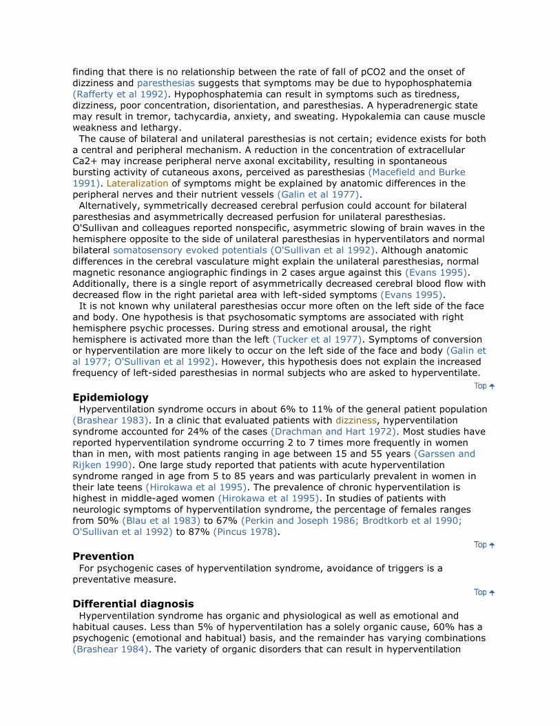
finding that there is no relationship between the rate of fall of pCO2 and the onset of
dizziness and paresthesias suggests that symptoms may be due to hypophosphatemia
(Rafferty et al 1992). Hypophosphatemia can result in symptoms such as tiredness,
dizziness, poor concentration, disorientation, and paresthesias. A hyperadrenergic state
may result in tremor, tachycardia, anxiety, and sweating. Hypokalemia can cause muscle
weakness and lethargy.
The cause of bilateral and unilateral paresthesias is not certain; evidence exists for both
a central and peripheral mechanism. A reduction in the concentration of extracellular
Ca2+ may increase peripheral nerve axonal excitability, resulting in spontaneous
bursting activity of cutaneous axons, perceived as paresthesias (Macefield and Burke
1991). Lateralization of symptoms might be explained by anatomic differences in the
peripheral nerves and their nutrient vessels (Galin et al 1977).
Alternatively, symmetrically decreased cerebral perfusion could account for bilateral
paresthesias and asymmetrically decreased perfusion for unilateral paresthesias.
O'Sullivan and colleagues reported nonspecific, asymmetric slowing of brain waves in the
hemisphere opposite to the side of unilateral paresthesias in hyperventilators and normal
bilateral somatosensory evoked potentials (O'Sullivan et al 1992). Although anatomic
differences in the cerebral vasculature might explain the unilateral paresthesias, normal
magnetic resonance angiographic findings in 2 cases argue against this (Evans 1995).
Additionally, there is a single report of asymmetrically decreased cerebral blood flow with
decreased flow in the right parietal area with left-sided symptoms (Evans 1995).
It is not known why unilateral paresthesias occur more often on the left side of the face
and body. One hypothesis is that psychosomatic symptoms are associated with right
hemisphere psychic processes. During stress and emotional arousal, the right
hemisphere is activated more than the left (Tucker et al 1977). Symptoms of conversion
or hyperventilation are more likely to occur on the left side of the face and body (Galin et
al 1977; O'Sullivan et al 1992). However, this hypothesis does not explain the increased
frequency of left-sided paresthesias in normal subjects who are asked to hyperventilate.
Epidemiology
Hyperventilation syndrome occurs in about 6% to 11% of the general patient population
(Brashear 1983). In a clinic that evaluated patients with dizziness, hyperventilation
syndrome accounted for 24% of the cases (Drachman and Hart 1972). Most studies have
reported hyperventilation syndrome occurring 2 to 7 times more frequently in women
than in men, with most patients ranging in age between 15 and 55 years (Garssen and
Rijken 1990). One large study reported that patients with acute hyperventilation
syndrome ranged in age from 5 to 85 years and was particularly prevalent in women in
their late teens (Hirokawa et al 1995). The prevalence of chronic hyperventilation is
highest in middle-aged women (Hirokawa et al 1995). In studies of patients with
neurologic symptoms of hyperventilation syndrome, the percentage of females ranges
from 50% (Blau et al 1983) to 67% (Perkin and Joseph 1986; Brodtkorb et al 1990;
O'Sullivan et al 1992) to 87% (Pincus 1978).
Prevention
For psychogenic cases of hyperventilation syndrome, avoidance of triggers is a
preventative measure.
Differential diagnosis
Hyperventilation syndrome has organic and physiological as well as emotional and
habitual causes. Less than 5% of hyperventilation has a solely organic cause, 60% has a
psychogenic (emotional and habitual) basis, and the remainder has varying combinations
(Brashear 1984). The variety of organic disorders that can result in hyperventilation

include: (1) effects of salicylate, caffeine, topiramate (Laskey et al 2000), and other
drugs; (2) cirrhosis and hepatic coma; (3) acute pain such as that accompanying a
myocardial infarction; (4) splenic flexure syndrome, cholecystitis, fever, and sepsis; and
(5) dissecting aortic aneurysm, respiratory dyskinesia, pulmonary embolism,
pneumothorax, interstitial lung disease, asthma, and heat and altitude acclimatization
(Brashear 1983; Block and Szidon 1994).
Neurologic disorders that may cause hyperventilation include Rett syndrome, Joubert
syndrome, Reye syndrome, pyruvate dehydrogenase deficiency, biotin-dependent
multiple carboxylase deficiency, malignant hyperthermia, brainstem tumor, primary
cerebral lymphoma, encephalitis, brainstem strokes, thalamic hemorrhage, syringobulbia,
and neurogenic pulmonary edema due to intracranial hypertension (Beumer and Bruyn
1993). Severe hyperventilation can occur in the absence of psychiatric, respiratory,
neurologic, or other organic abnormalities (Bass and Gardner 1985).
In patients diagnosed with hyperventilation syndrome by neurologists, the
misdiagnoses of referring physicians have included epilepsy, migraine, multiple sclerosis,
arteriovenous malformation, cerebrovascular disease, vertebrobasilar insufficiency,
brachial neuritis, angina, malingering, vasovagal attacks, functional illness,
hypoglycemia, and cerebral tumor (Blau et al 1983; Perkin and Joseph 1986).
Hyperventilation might also contribute to some of the manifestations of migraine such as
syncope.
Cardiac abnormalities should certainly be considered in those patients with chest pain.
The distinction between angina and hyperventilation syndrome at times is difficult
because hyperventilation can produce electrocardiogram changes including T-wave
inversions, systolic time-segment depression, and systolic time-segment elevation in
patients without coronary artery disease (Heckerling and Hanashiro 1985). Some
patients with angina may hyperventilate in response to their pain and anxiety. The
symptoms of mitral valve prolapse and hyperventilation syndrome overlap. In some
patients, symptoms may be due to hyperventilation (Tavel 1990).
The symptoms of panic attacks greatly overlap with hyperventilation syndrome, and the
differential diagnosis is quite similar (Tesar and Rosenbaum 1993; Stahl and Soefje
1995). The pathophysiology of panic attacks is controversial; hyperventilation syndrome
has been suggested as a cause (Ley 1985; Nutt and Lawson 1992; Folgering 1999).
Patients diagnosed with panic attacks that report brief episodic unilateral paresthesias or
weakness may have the neurologic symptoms reproduced with the hyperventilation test
(Coyle and Sterman 1986).
Tonic spasms (paroxysmal dystonia) of multiple sclerosis can be somewhat similar to
the muscle spasms, tetany, and paresthesias of hyperventilation syndrome. The episodes
consist of brief, recurrent, often painful abnormal posturing of 1 or more extremities
without alteration of consciousness, loss of sphincter control, or clonic movements. They
last for 10 seconds to 3 minutes and recur as often as 30 times daily. Some patients may
experience brief sensory disturbances in the involved extremity before or during the
attack. Because the episodes can be the initial presentation of multiple sclerosis (Heath
and Nightingale 1982) and can be triggered by hyperventilation (Shibasaki and Kuroiwa
1974), diagnostic confusion might result.
Diagnostic workup
The acute form of hyperventilation syndrome is easily recognized. However, the chronic
form is less easily recognized because the breathing rate is not reported as rapid or does
not appear rapid and because the symptoms may appear to be atypical. For example, a
respiration rate of 18 per minute combined with an increased tidal volume of 750 mL per
minute may lead to overbreathing that is not easily detectable. Because the chronic
disorder is intermittent, spot arterial pCO2 or end tidal volume pCO2 results can be
normal. The diagnosis depends on reproducing some or all of the symptoms with the
hyperventilation provocation test and excluding other possible causes by either clinical
reasoning or laboratory testing when indicated. Patients frequently report only 1 or 2
symptoms but, on performing the hyperventilation provocation test, report other
symptoms that appear during their typical episodes that they had forgotten.
The hyperventilation provocation test can be performed with either an increased
ventilation rate of up to 60 per minute or simply deep breathing for 3 minutes (Lum
1987). Based on a study of healthy subjects, a minimum duration of 3 minutes and end-
tidal pCO2 decreasing to at least 1.9 kPa or dropping well over 50% of baseline should
elicit symptoms in most people (Hornsveld et al 1995). Dizziness, unsteadiness, and
blurred vision commonly develop within 20 to 30 seconds, especially with the patient in
the standing position; paresthesias start later (Lum 1987). Chest pain is reported by
50% of patients after 3 minutes of hyperventilation and by all by 20 minutes (Evans and
Lum 1977). For clinical purposes, measurement of end tidal volume pCO2 is not
necessary. In addition, there is no clear correlation between paCO2 and neurologic signs
(Stoop et al 1986). The hyperventilation provocation test should not be performed in
patients with ischemic heart disease, cerebrovascular disease, pulmonary insufficiency,
hyperviscosity states, significant anemia, sickle cell disease, or uncontrolled hypertension
(Brashear 1983).
For some patients with hyperventilation syndrome, symptoms cannot be reliably
reproduced during the hyperventilation provocation test or even on consecutive tests
(Hirokawa et al 1995). In some cases, the hyperventilation provocation test lacks test-
retest reliability (Lindsay et al 1991). For others, antecedent anxiety and stress, not
present during the test, may predispose to symptom formation, perhaps because of a
hyperadrenergic state (Magarian 1982; Perkin and Joseph 1986). Different patterns of
hyperventilation with different respiratory rates, tidal volumes, and durations may induce
different symptoms (Grossman and De Swart 1984; Hornsveld et al 1995). Finally, as a
response to a change in body position from supine to standing, patients with
hyperventilation syndrome have an accentuated increase in ventilation that can be
calculated with noninvasive measurements of pulmonary gas exchange, and that
distinguishes them from healthy subjects (Malmberg et al 2000).
A double-blind, placebo-controlled study found the hyperventilation provocation test to
be invalid (Hornsveld et al 1996). Because hyperventilation seemed a negligible factor in
the experience of spontaneous symptoms, Hornsveld and colleagues even recommended
avoiding the term "hyperventilation syndrome." However, the study may be flawed
because of the method of patient selection. Patients were referred because of a suspicion
of hyperventilation syndrome and not based on reproduction of symptoms on a
hyperventilation provocation test. Moreover, the presenting symptoms of subjects were
not provided. Because the symptoms of hyperventilation syndrome can indeed be vague
and nonspecific and because symptoms sometimes occur only with certain types of
anxiety or stress, their conclusion may not apply to different population subsets (Naschitz
et al 1996). The subjects' symptoms may have been due to chest wall pain or panic
attacks.
Alternatively, the Nijmegen questionnaire (a list of 16 symptoms rated on a 5-point
scale from never to very often) can be used to screen for hyperventilation syndrome. The
efficacy of this questionnaire has been evaluated, revealing the sensitivity to be 91% and
the specificity to be 95% (when using the clinical diagnosis based on the pattern of
complaints as the gold standard) (van Dixhoorn and Duivenvoorden 1985). In a
consecutive series of 100 patients in a neuro-otology practice, 23% of patients seen for
vestibular assessment were diagnosed with hyperventilation syndrome; 74% of these
would have potentially remained undetected had the Nijmegen questionnaire not been
used (Humphriss et al 2004).
From my experience with patients with predominantly neurologic complaints, the
concept of hyperventilation syndrome is valuable and the hyperventilation provocation

test, despite its recognized shortcomings (Gardner 2000), is useful. In the individual
case, if the hyperventilation provocation test fails to reproduce the symptoms but clinical
suspicion persists, treatment such as breath holding, slow breathing, or breathing into a
paper bag can certainly be suggested on a trial basis.
Prognosis and complications
In a followup study of children and adolescents, 40% were still hyperventilating as
adults and many suffered from chronic anxiety (Herman et al 1981). One half of patients
with acute hyperventilation recover without treatment (Hirokawa et al 1995). In 10% of
those with chronic hyperventilation, symptoms may persist for more than 3 years
(Hirokawa et al 1995). With proper management, about 70% to 90% of adults become
symptom-free (Lum 1976; Campernolle et al 1979).
Management The various treatments for hyperventilation that have been proposed include patient
reassurance and education, instructions to hold the breath, breathe more slowly, or
breathe into a paper bag, along with breathing exercises and diaphragmatic retraining,
biofeedback, hypnosis, psychological and psychiatric treatment, and medications such as
beta blockers, benzodiazepines, and antidepressants (Beumer and Bruyn 1993). A study
of nonpharmacologic treatments found efficacy for educational sessions, breathing
techniques and retraining, and progressive relaxation; the greatest improvement
occurred in the group given an explanation and 8 sessions of breathing retraining
(Monday et al 1995). A small pilot study found benefit from acupuncture (Gibson et al
2007). There is a lack of well-controlled treatment trials comparing these approaches
(Herxheimer 1991).
In my experience, most patients respond to reassurance, education, and instructions to
hold the breath, breathe more slowly, or breathe into a paper bag. Providing the patient
with written materials such as those used by Lance may be worthwhile (Lance and
Goadsby 2005). If significant symptoms of stress, anxiety, or depression are present, use
of appropriate medication and psychological or psychiatric referral may be helpful.
Pregnancy
Although hyperventilation syndrome has often been seen during pregnancy, studies on
the incidence are not available.
Anesthesia
Not applicable.
ICD codes
ICD-9:
Hyperventilation: 306.1
ICD-10:
Hyperventilation: R06.4
Associated disorders
Acute hyperventilation
Anxiety
Chronic hyperventilation
Distal symmetrical paresthesias
Panic attacks
Raynaud phenomenon
Related summaries
Dizziness
Psychophysiological vertigo (psychogenic vertigo)
Syncope
Tension-type headache
Differential diagnosis
epilepsy
migraine
arteriovenous malformation
cerebrovascular disease
vertebrobasilar insufficiency
brachial neuritis
angina
malingering
vasovagal attacks
functional illness
hypoglycemia
cerebral tumor
cardiac abnormalities
mitral valve prolapse
panic attacks
multiple sclerosis
Demographics
For more specific demographic information, see the Epidemiology, Etiology, and
Pathogenesis and pathophysiology sections of this clinical summary.
Age
06-12 years
13-18 years
19-44 years
45-64 years
65+ years
Population
None selectively affected.
Occupation
None selectively affected.
Sex
female>male, >2:1
female>male, >1:1
Family history
None
Heredity
None
References cited
Allen TE, Agus B. Hyperventilation leading to hallucinations. Am J Psychiatry
1968;125:632-7.
Bass C, Gardner WN. Respiratory and psychiatric abnormalities in chronic symptomatic
hyperventilation. BMJ 1985;290:1387-90.
Beumer HM, Bruyn GW. Hyperventilation syndrome. In: Goetz CG, Tanner CM, Aminoff
MJ, editors. Handbook of clinical neurology. Vol. 19. Amsterdam: Elsevier, 1993:429-
48.**
Blau JN, Wiles CM, Solomon FS. Unilateral somatic symptoms due to hyperventilation.
BMJ 1983;286:1108.
Block M, Szidon P. Hyperventilation syndromes. Comp Ther 1994;20:306-11.
Brashear RE. Hyperventilation syndrome. Lung 1983;161:257-73.
Brashear RE. Hyperventilation syndrome: managing elderly patients. Geriatrics
1984;39:114-25.
Brautbar N, Leibovici H, Finlander P, et al. Mechanism of hypophosphatemia during acute
hyperventilation. Clin Res 1980;28:387A.
Brodtkorb E, Gimse R, Antonaci F, et al. Hyperventilation syndrome: clinical, ventilatory,
and personality characteristics as observed in neurological practice. Acta Neurol Scand
1990;81:307-13.
Campernolle T, Hoogduin K, Joele L. Diagnosis and treatment of the hyperventilation
syndrome. Psychosomatics 1979;20:612-25.
Coyle PK, Sterman AB. Focal neurologic symptoms in panic attacks. Am J Psychiatry
1986;143:648-9.
Da Costa JM. On irritable heart; a clinical study of a form of functional cardiac disorder
and its consequences. Am J Med Sci 1871;71:2-52.
Drachman DA, Hart CW. An approach to the dizzy patient. Neurology 1972;22:323-34.
Evans DW, Lum LC. Hyperventilation: an important cause of pseudoangina. Lancet
1977;22(1):155-7.
Evans RW. Neurologic aspects of hyperventilation syndrome. Semin Neurol 1995;15:115-
25.**
Evans RW. Hyperventilation syndrome. In Kaplan PW, Fisher RS, editors. Imitators of
epilepsy. New York: Demos, 2005:241-53.
Folgering H. The pathophysiology of hyperventilation syndrome. Monaldi Arch Chest
1999:54:365-72.

Galin D, Dimond R, Braff D. Lateralization of conversion symptoms more frequent on the
left. Am J Psychiatry 1977;134:578-80.
Gardner W. Orthostatic increase of respiratory gas exchange in hyperventilation
syndrome. Thorax 2000;55:257-9.
Garssen B, Rijken H. Hyperventilation syndrome. In: Kaptein AA, van der Ploeg HM,
Schreurs PJG, Beunderman R, editors. Behavioural medicine. John Wiley and Sons,
1990:159-72.
Gibson D, Bruton A, Lewith GT, Mullee M. Effects of acupuncture as a treatment for
hyperventilation syndrome: a pilot, randomized crossover trial. J Altern Complement Med
2007;13(1):39-46.
Goldman A. Clinical tetany by forced respiration. JAMA 1922;78:1193-5.
Gotoh F, Meyer JS, Takagi Y. Cerebral effects of hyperventilation in man. Arch Neurol
1965;12:410-23.
Gowers WR. The border-land of epilepsy. Faints, vagal attacks, vertigo, migraine, sleep
symptoms, and their treatment. London: J and A Churchill, 1907.**
Grossman P, de Swart JC. Diagnosis of hyperventilation syndrome on the basis of
reported complaints. J Psychosom Res 1984;28:97-104.
Heath PD, Nightingale S. Clusters of tonic spasms as an initial manifestation of multiple
sclerosis. Ann Neurol 1982;12:494-5.
Heckerling PS, Hanashiro PK. ST segment elevation in hyperventilation syndrome. Ann
Emerg Med 1985;14:126-7.
Herman SP, Stickler GB, Lucas AR. Hyperventilation syndrome in children and
adolescents: long-term follow up. Pediatrics 1981;67:183-7.
Herxheimer A. How much drug in the tablet? [letter; comment]. Lancet
1991;337(8748):1042.
Hirokawa Y, Kondo T, Ohta Y, Kanazawa O. Clinical characteristics and outcome of 508
patients with hyperventilation syndrome. Nippon Kyobu Shikkan Gakkai Zasshi
1995;33:940-6.
Hornsveld HK, Garssen B, Dop MJ, van Spiegel PI, de Haes JC. Double-blind placebo-
controlled study of the hyperventilation provocation test and the validity of the
hyperventilation syndrome. Lancet 1996;348:154-8.
Hornsveld H, Garssen B, van Spiegel P. Voluntary hyperventilation: the influence of
duration and depth on the development of symptoms. Biol Psychol 1995;40:299-312.
Humphriss RL, Baguley DM, Andersson G, Wagstaff S. Hyperventilation in the vestibular
clinic: use of the Nijmegen Questionnaire. Clin Otolaryngol 2004;29:232-7.
Jibiki I, Kurokawa K, Matsuda H, Fukushima T, Yamaguchi N, Hisada K. Widespread
reduction of regional cerebral blood flow during hyperventilation-induced EEG slowing

('buildup'). Observation from subtraction of brain imaging with single photon emission
computed tomography using technetium-99m hexamethyl-propyleneamine oxime.
Neuropsychobiology 1992;26(3):120-4.
Kerr WJ, Dalton JW, Gliebe PA. Some physical phenomena associated with the anxiety
states and their relation to hyperventilation. Ann Int Med 1937;11:961-91.
Kerr WJ, Gliebe PA, Dalton JW. Physical phenomena associated with anxiety states: the
hyperventilation syndrome. Calif Western Med 1938;48:12-6.
Kraaier V, Van Huffelen AC, Wieneke GH, Van der Worp HB, Bar PR. Quantitative EEG
changes due to cerebral vasoconstriction. Indomethacin versus hyperventilation-induced
reduction in cerebral blood flow in normal subjects. Electroencephalogr Clin Neurophysiol
1992;82(3):208-12.
Lance JW, Goadsby PJ. Appendix A. An explanation of hyperventilation. In: Mechanism
and management of headache. 7th ed. Oxford: Butterworth Heinemann, 2005.
Laskey AL, Korn DE, Moorjani BI, Patel NC, Tobias JD. Central hyperventilation related to
administration of topiramate. Pediatr Neurol 2000;22:305-8.
Levine SA. The origin of the term neurocirculatory asthenia. N Engl J Med 1965;273:604-
5.
Lewis BI. The hyperventilation syndrome. Ann Int Med 1953;38:918-27.
Lewis RA, Howell JB. Definition of the hyperventilation syndrome. Bull Eur Physiopathol
Respir 1986;22:201-4.
Lewis T. The soldier's heart and the effort syndrome. New York: Paul B. Hoeber, 1919.
Ley R. Agoraphobia, the panic attack and the hyperventilation syndrome. Behav Res Ther
1985;1:79-81.
Lindsay S, Saqi S, Bass C. The test-retest reliability of the hyperventilation provocation
test. J Psychosom Res 1991;35:155-62.
Lum LC. Hyperventilation: the tip and the iceberg. J Psychosom Res 1975;19:375-83.
Lum LC. The syndrome of chronic habitual hyperventilation. In: Hill OW, editor. Modern
trends in psychosomatic medicine. London: Buttersworth, 1976:196-230.
Lum LC. Hyperventilation syndromes in medicine and psychiatry: a review. J R Soc Med
1987;80:229-31.**
Macefield G, Burke D. Paraesthesiae and tetany induced by voluntary hyperventilation.
Increased excitability of human cutaneous and motor axons. Brain 1991;114:527-40.
Magarian GJ. Hyperventilation syndromes: infrequently recognized common expressions
of anxiety and stress. Medicine 1982;61:219-36.
Malmberg LP, Tamminen K, Sovijarvi AR. Orthostatic increase of respiratory gas
exchange in hyperventilation syndrome. Thorax 2000;55:295-301.
Monday J, Gautrin D, Cartier A. Chronic hyperventilation syndrome. The role of
respiratory re-training. Rev Mal Respir 1995;12:291-8.
Naschitz JE, Yeshurun D, Hardoff D, Yaffe M. Hyperventilation syndrome. Lancet
1996;348:750.
Nutt D, Lawson C. Panic attacks. A neurochemical overview of models and mechanisms.
Br J Psychiatry 1992;160:165-78.
O'Sullivan G, Harvey I, Bass C, Sheehy M, Toone B, Turner S. Psychophysiological
investigations of patients with unilateral symptoms in the hyperventilation syndrome. Br
J Psychiatry 1992;160:664-7.**
Patel VM, Maulsby RI. How hyperventilation alters the electroencephalogram: a review of
controversial viewpoints emphasizing neurophysiological mechanisms. J Clin Neurophysiol
1987;4:101-20.
Pearson MG, Qadiri MR, Finn R. Hypokalaemia in the chronic hyperventilation syndrome.
Br J Clin Pract 1986;40:28-9.
Perkin GD, Joseph R. Neurological manifestations of the hyperventilation syndrome. J R
Soc Med 1986;79:448-50.
Pincus JH. Disorders of conscious awareness. Hyperventilation syndrome. Br J Hosp Med
1978;19:312-3.
Rafferty GF, Saisch SGN, Gardner WN. Relation of hypocapnic symptoms to rate of fall of
end-tidal PCO2 in normal subjects. Respir Med 1992;86:335-40.
Shibasaki H, Kuroiwa Y. Painful tonic seizure in multiple sclerosis. Arch Neurol
1974;30:47-51.
Stahl SM, Soefje S. Panic attacks and panic disorder: the great neurologic impostors.
Semin Neurol 1995;15:126-32.
Stoop A, de Boo T, Lemmens W, Folgering H. Hyperventilation syndrome: measurement
of objective symptoms and subjective complaints. Respiration 1986;49:37-44.
Tavel ME. Hyperventilation syndrome with unilateral somatic symptoms. JAMA
1964;187:301-2.
Tavel ME. Hyperventilation syndrome-hiding behind pseudonyms? Chest 1990;97:1285-
8.
Tesar GE, Rosenbaum JF. Recognition and management of panic disorder. Adv Int Med
1993;38:123-49.
Tucker DM, Roth RS, Arneson BA, Buckingham V. Right hemisphere activation during
stress. Neuropsychologia 1977;15:697-700.
van Dixhoorn J, Duivenvoorden HJ. Efficacy of Nijmegen Questionnaire in recognition of
the hyperventilation syndrome. J Psychosom Res 1985;29:199-206.

**References especially recommended by the author or editor for general reading.

The role of hyperventilation - hypocapnia in thepathomechanism of panic disorder
O papel da hiperventilação - a hipocapnia nopatomecanismo do distúrbio de pânico
Abst rac t
Objective: The authors present a profile of panic disorder based on and generalized from the effects of acute and chronichyperventilation that are characteristic of the respiratory panic disorder subtype. The review presented attempts to integrate threepremises: hyperventilation is a physiological response to hypercapnia; hyperventilation can induce panic attacks; chronichyperventilation is a protective mechanism against panic attacks. Method: A selective review of the literature was made using theMedline database. Reports of the interrelationships among panic disorder, hyperventilation, acidosis, and alkalosis, as well ascatecholamine release and sensitivity, were selected. The findings were structured into an integrated model. Discussion: Thepanic attacks experienced by individuals with panic disorder develop on the basis of metabolic acidosis, which is a compensatoryresponse to chronic hyperventilation. The attacks are triggered by a sudden increase in (pCO
2) when the latent (metabolic)
acidosis manifests as hypercapnic acidosis. The acidotic condition induces catecholamine release. Sympathicotonia cannot ariseduring the hypercapnic phase, since low pH decreases catecholamine sensitivity. Catecholamines can provoke panic whenhyperventilation causes the hypercapnia to switch to hypocapnic alkalosis (overcompensation) and catecholamine sensitivitybegins to increase. Conclusion: Therapeutic approaches should address long-term regulation of the respiratory pattern andelimination of metabolic acidosis.
Descriptors: Acidosis; Catecholamines; Hyperventilation; Hypocapnia; Panic disorder
Resumo
Objetivo: Os autores apresentam um modelo de transtorno do pânico que se baseia nos efeitos da hiperventilação aguda ecrônica, característicos do subtipo respiratório de transtorno do pânico. O modelo é generalizado a partir desses efeitos. Eleintegra três características da hiperventilação: a hiperventilação é uma resposta fisiológica à hipercapnia; a hiperventilação podeinduzir ataques de pânico; a hiperventilação crônica representa um mecanismo protetor contra os ataques de pânico. Método:Revisão seletiva da literatura a partir da base de dados Medline. Foram selecionados relatos referentes à inter-relação entretranstorno do pânico, hiperventilação, acidose, alcalose, liberação de catecolaminas e sensibilidade a catecolaminas, sendo osachados estruturados de modo a formar um modelo integrado. Discussão: Os ataques de pânico do transtorno do pânicodesenvolvem-se com base numa acidose metabólica, que é uma resposta compensatória à hiperventilação crônica. Os ataquessão desencadeados por um súbito aumento da pressão parcial de dióxido de carbono (pCO
2), quando a acidose (metabólica)
latente se manifesta pela acidose hipercápnica. A condição acidótica induz liberação de catecolaminas. A simpaticotonia nãopode manifestar-se durante a fase de hipercapnia, pois o baixo pH diminui a sensibilidade às catecolaminas. As catecolaminaspodem provocar pânico quando a hipercapnia comuta para uma alcalose hipocápnica devido à supercompensação pelahiperventilação, situação na qual a sensibilidade às catecolaminas liberadas começa a aumentar. Conclusão: As abordagensterapêuticas deveriam voltar-se para a regulação em longo prazo do padrão respiratório e a eliminação da acidose metabólica.
Descritores: Acidose; Catecolaminas; Hiperventilação; Hipocapnia; Transtorno de pânico
1 St. Rokus Hospital, Department of Internal Medicine, Budapest, Hungary2 National Institute for Psychiatry and Neurology, Budapest, Hungary3 Institute of Psychiatry, Universidade de São Paulo (USP) School of Medicine, São Paulo (SP), Brazil
Rev Bras Psiquiatr. 2007;29(4):375-9
Andras Sikter,1 Ede Frecska,2 Ivan Mario Braun,3
Xenia Gonda,2 Zoltan Rihmer2
CorrespondenceZoltan RihmerHûvösvölgyi út 116, 1021Budapest, HungaryPhone: 36 1 391 5353 Fax: 36 1 200 0770E-mail: [email protected]
Financing: NoneConflict of interests: NoneSubmitted: May 17, 2006Accepted: April 2, 2007
SPECIAL ARTICLE
375
Artigo13_V02.p65 28/11/2007, 16:20375

Hypocapnia and panic disorder 376
Rev Bras Psiquiatr. 2007;29(4):375-9
Int roduct ion
Although the correlation between the respiratory panic
disorder (PD) subtype and hyperventilation is well established
in the literature, the active role of hyperventilation in the
pathomechanism of PD is less understood. Although Wilhelm
et al. provided a thorough review of the three main theories
on the role of hyperventi lation in PD,1 they identif ied
contradictions among them and did not offer a synthesis of
the current concepts. The increase of t issue H+ ion
concentration, which is regulated promptly and effectively by
respiration-induced changes in pCO2, is a strong stimulus of
catecholamine release. However, acidotic conditions decrease
the catecholamine sensitivity of target organs (the inverse of
what occurs in alkalosis). In a panic attack, these events occur
in rapid succession. Therefore, understanding the timing and
the time constant (how quickly a change develops) of
physiological variables is crucial for resolving the apparent
contradictions and establishing a coherent model.
The role of hypocapnia in the pathomechanism of PD has
been largely overlooked in the literature.2 During the late
1930s, hyperventilation played a central role in the diagnostic
formulation of hyperventilation syndrome.3-4 Shortly after the
conceptualization of PD in the DSM III (1980), it became
evident that there had been an overlap between symptoms of
hyperventilation syndrome and PD.5 The article written by Klein
in 1993 represented an important step toward settling the
long-standing debate on the role of hyperventilation.6 Klein
denied the importance of acute or chronic hyperventilation in
the generation of panic attacks. Although he described a
positive correlation between chronic hyperventilation and panic
attacks, he used this observation in order to put forth the idea
that chronic hyperventilation is protective against panic.
The aim of this study was to review the literature on PD,
with a special focus on the role of hyperventilation and
hypocapnia in the pathomechanism of PD. Special attention
was paid to studies investigating the interrelationships between
panic, pH, pCO2, tissue catecholamine sensitivity, and
catecholamine el iminat ion, as wel l as their temporal
characteristics.
Method
We conducted a selective review of the literature in the
Medline database, limiting our searches to articles published
between 1937 and 2006. Our approach was to discuss the
available literature on PD in order to show that it is not only a
psychiatric disorder but also a regulatory disorder. Our first
search included the terms “panic” and “hyperventilation”, and/
or “hypocapnia” and/or “hypercapnia”. This search strategy
yielded a total of 317 articles, from which we selected 33 for
analysis. In selecting these articles, our principal aim was to
represent all major opinions and trends. Within this context,
we selected articles written by the most prominent authors.
Therefore, these articles and their main arguments present a
logical progression. In addition, we reviewed the most
controversial papers. Furthermore, 17 of the articles included
deal with the physiology and pathology of catecholamine
homeostasis in relation to panic. These articles were identified
by adding the search terms “acid-base disorder”, “acidosis”,
“alkalosis”, “hypocapnia”, “hypercapnia”, “catecholamine”,
“noradrenaline”, “adrenaline”, and “sympathetic nervous
system”. In this step of the selection process, we focused on
articles investigating the relationship between acidosis/alkalosis
and catecholamine product ion, as wel l as on those
investigating the catecholamine sensitivity of brain and other
tissues in relation to pH. We also included 10 articles on
psychiatric topics presenting borderline relationships with PD.
The search of the literature and the article selection were
carried out by András Sikter, who has been researching this
topic for decades and therefore has profound insight into the
issue. The author notes on the articles evaluated have been
arranged into a logical order so that they can be integrated
into a coherent model.
Discuss ion
Maintaining the homeostasis of intracellular and extracellular
pH is a crucial regulatory task for the organism. A couple of deep
breaths can substantially decrease CO2 concentration, increasing
extracellular pH to 7.4-7.7. Since CO2 readily passes through
cell membranes, intracellular pCO2 decreases to the same
degree. Therefore, altering respiration can produce marked
changes in intracellular pH, the consequences of which are
outlined below.7 Acute hypocapnia has a progressive effect on
membrane permeability, metabolism, oxygen consumption, and
cardiac function,8 as well as exciting the nervous system
through the hypopolarization of neurons.9-10 In contrast,
hypercapnia results in decreased cell membrane permeability,11
metabolic depression,12 reduced muscle contractility13, and
hyperpolarization of neurons. In short, acute hypocapnia elicits
effects similar to those seen when the sympathetic nervous
system is stimulated, whereas acute hypercapnia evokes
parasympathetic effects. The latter occurs despite the fact that
hypercapnia causes increases in the levels of serum
adrenaline and noradrenaline.14 There are conflicting data
in the l i t e ra tu re rega rd ing se rum adrena l ine and
noradrenaline levels in acute hypocapnia, some authors
repor t ing no a l te ra t ions 14 and o thers demonst ra t ing
significant decreases.15 Hypoxia is a disturbing variable in
most investigations.16 Catecholamine sensitivity decreases
in hypercapnic acidosis and increases in hypocapnic
alkalosis.7 These changes in catecholamine level and
sensitivity affect target organs. Lower intracellular pH is
the t r igger fo r noradrenal ine re lease f rom the locus
coeruleus during panic attacks.17-18
Chronic hypocapnia elicits a cascade of changes due to
compensatory mechanisms for the restoration of intracellular
and extracellular physiological pH. Renal reabsorption of
chlorine ions and excretion of bicarbonate is increased in
sustained hypocapnia.19 In an experimental model of
hyperventilation in dogs, it was shown to take five days for the
kidneys to reestablish the equilibrium.20 In humans, the role
of intracellular buffer mechanisms is more important, and
adaptation to chronic hyperventilation or hypoventilation is
therefore less dependent on renal function.21 The process of
adaptation is similarly slow: to regain equilibrium can take
five to seven days.7 It takes equally as long to reverse the
changes after the occurrence of eucapnia. Buffer mechanisms
include the release of hydrogen ions, which shifts pH toward
the physiological level, although this process also occurs only
after a delay.
Various authors have downplayed the role of hyperventilation
in PD and have recommended that the term ‘hyperventilation
syndrome’ no longer be used.21-24 However, in another review
article, Gardner argues for the preservation of the term.25 Other
authors have stated that chronic hyperventilation is a common
cause of both hyperventilation syndrome and PD.5,25-26 Recently,
Nardi addressed the role of hyperventilation in PD and tried to
Artigo13_V02.p65 28/11/2007, 16:20376

Rev Bras Psiquiatr. 2007;29(4):375-9
377 Sikter A et al.
of PD, and that similar irregularity can be found in
generalized anxiety disorder.40 The similarity can explain
the high comorbidity of these two conditions.26 In individuals
with generalized anxiety disorder, the irregularity of breathing
is less pronounced than in those with PD.41 Various studies
have shown that, in individuals with PD, there is a strong
correlation between the degree of respiratory irregularity
and the frequency of panic attacks, as well as between
such irregularity and CO2 vulnerability.1,41-42 Irregular
respiration occasionally causes elevated pCO2, which can
trigger a panic attack.
The hypothalamic-pituitary-adrenocortical axis model of
hypercapnia induced panic is generally accepted in the
literature,17,43 although it was recently questioned by
Gorman,44 who discovered inconsistencies and pointed out
that, during CO2 challenge, actual pCO
2 values correlate
negatively with signs and symptoms of panic. This indicates
that panic develops not during hypercapnia but during the
subsequent hypocapnic phase. The way Gorman puts it:
“...in panic disorder patients, we have found that elevated
cortisol, fear and hypocapnia are intercorrelated in the few
minutes before actually experiencing an acute attack.”
As previously mentioned, higher pCO2 leads to increased
noradrenaline release.18 However, in human plasma,
noradrenaline has a half-life of only a few minutes.45-46 In
the rebound phase of hypocapnia, cells present increased
sensi t iv i ty to residual catecholamines.7 Strong
catecholamine stimuli are known to induce panic attacks.47
Individuals with PD present normal catecholamine levels
between panic attacks.48 It is therefore possible that, in
addit ion to the hypercapnia-re lated increase in
catecholamine levels, the hypocapnia- induced
catecholamine sensitivity plays a significant role in the
induction of panic.49
Borelli et al. conducted electrophysiological studies in
animals50 and concluded that panic attacks represent a
pathological manifestation of ‘freezing behavior’ (low-arousal
condition), rather than the high-arousal condition of the
‘fight-or-flight’ response.51 ‘Freezing behavior ’ initially
manifests as immobility, bradycardia, and hypoventilation
but can transform into the flight response, which is
characterized by vigorous locomotion, tachycardia, and
hyperventilation.52 The sudden change in the respiratory
pattern that precedes the flight response indicates the
similarity with panic attacks. It is quite probable that the
role the brainstem plays in the pathomechanism of PD is
more important than previously suggested.
We can build a profile of PD that integrates the three
hyperventilation theories. Individuals with the respiratory
PD subtype present chronic hyperventilation. The chronic
hyperventilation results in a compensatory decrease in
intracellular and extracellular pH due to renal secretion of
bicarbonate and due to the tissue buffer mechanisms. A
balanced steady-state is established between the hypocapnic
alkalosis and the metabolic acidosis. Multiple factors can
lead to a sudden increase in CO2 levels. In individuals with
PD and presenting sustained hypoventilation episodes,
irregular breathing causes abrupt changes in pCO2. In the
prelude to a panic attack, an abrupt increase of pCO2 occurs,
which leads to unusual ly high intracel lular H+
concentrat ions, thereby t r igger ing the re lease of
noradrenaline in the locus coeruleus. This sudden increase
in intracellular acidosis elicits hypocapnia by compensatory
clarify it, stating that it is considered to be “...a cause, a correlate,
or a consequence of panic attacks.” According to Nardi, acute
hyperventilation might play a role in the pathomechanism of the
respiratory PD subtype.27
In a study using transcutaneous monitoring, no relationship
was found between PD and hyperventilation.28 This method is
outdated due to its high inertia (slow decay) in monitoring changes
in arterial pCO2.2 It is widely accepted that the respiratory PD
subtype, which accounts for approximately 50% of all PD cases,
is c losely re lated to hypervent i lat ion and represents a
hyperventilation syndrome comorbidity.2,5,25-26
There are three views in the literature regarding the role that
hyperventilation plays in the pathomechanism of PD. In the first,
panic is triggered by elevated CO2 levels, and hyperventilation
follows as a physiological response.1,29 In this model, the
hyperventilation is a consequence, an epiphenomenon observed
during naturally-occurring and drug-induced panic attacks. In
the view of other authors30-31 – a view shared by followers of the
cognitive-behavioral theory of PD1 – hyperventilation-induced
hypocapnia plays a central role. The opinion of psychotherapists
can be summarized as fol lows: individuals with PD can
misinterpret the bodily sensations caused by hyperventilation as
being indicative of life-threatening danger.32
However, the most widely accepted view is that detailed by
Klein6: hyperventilation is a protective mechanism against panic
reactions. In his thorough study, Klein demonstrated that patients
with PD are hypersensitive to increases in pCO2, and that panic
attacks are triggered by a relative increase in the level of CO2.
Such individuals present chronic hyperventilation as a means of
avoiding the panic-inducing increase in CO2 levels.
We agree with Klein that chronic hyperventilation has some
defensive effects against panic attacks in individuals with PD,
since a sudden increase of pCO2 (e.g. CO
2 challenge) has been
shown to provoke such at tacks.33 However, chronic
hyperventilation is always accompanied by compensatory metabolic
acidosis. In an individual with chronic hyperventilation, life events
(i.e., relaxation, sleep, premenstrual phase, etc.) can cause pCO2
to rise to the normal baseline or above.6 The latent metabolic
acidosis then appears, and the elevated concentration of H+
increases CO2 sensitivity of the respiratory center. It can be
assumed that the chronic hyperventilation itself is responsible for
the increased CO2 sensitivity observed in individuals with PD.
The best supporting evidence is provided by Klein himself, who
found chronic hyperventilation to correlate positively with lactate-
induced panic and CO2 sensitivity.6 Hypophosphatemia, which is
an indicator of chronic hyperventilation, has been found to be
predictive of lactate-induced panic attacks.34 Decreased plasma
bicarbonate is also a marker of chronic hyperventilation and
sensitizes to the onset of panic attacks.35 However, chronic
hypercapnia accompanied by metabolic alkalosis has been shown
to correlate negatively with the development of panic attacks.6
Various researchers have been successful in using
hyperventilation to provoke panic attacks.27,36-39 Nardi addressed
the role of hypocapnia and hypercapnia in PD with equal focus
and equal importance in the pathogenesis.27,38-39 However, the
overall effect of hyperventilation on panic was significantly less
than that of CO2 inhalation. Respiratory challenge tests (CO
2 and
breath-holding) can provoke panic attacks in individuals presenting
the respiratory PD subtype.39 Cerebral hypoxia, chronic
hyperventilation, and anxiety persist in the interim between panic
attacks.26 Although individuals with PD are prone to continue
hyperventiling,1,32 the hyperventilation facilitates panic attacks.
Caldirola stated that an irregular breathing pattern is predictive
Artigo13_V02.p65 28/11/2007, 16:20377
Hypocapnia and panic disorder 378
Rev Bras Psiquiatr. 2007;29(4):375-9
hypervent i lat ion. In addit ion, individuals with PD
overcompensate for hypercapnia.53 The consequence is severe
sympathicotonia, since the higher catecholamine level resulting
from the previous hypercapnia overlaps with the increased
catecholamine sensitivity caused by the hypocapnic alkalosis.
The adrenergic/noradrenergic tonus results in fear mediated
by the limbic system, and the expectation of threat can create
a vicious circle. Long after the chemical component phase of
a panic attack is over, the cortical excitation persists, leaving
the individual with a lingering, subjective feeling of anxiety.
Hyperventilation pushes the individual toward progression of
the panic attack.
The profile is supported by various observations. Gorman
noted that, in the case of CO2 challenge, signs and symptoms
of panic correlated inversely with pCO2.44 One dose of biperiden
(an antimuscarinic agent) can prevent panic attacks induced
by CO2 inhalat ion, s ince i t e l iminates the rebound
hyperventilation response mediated via the muscarinic receptors
of the ventral medulla.54 Therapies proven to be effective in
PD, such as cognitive therapy, biofeedback, and antidepressant
pharmacotherapy, seem to also be useful in hyperventilation
syndrome.55-57 Successful pharmacotherapy of panic attacks
normal izes blood gas parameters, i .e. i t e l iminates
hyperventilation as well as the increased CO2 sensitivity
characteristic to PD.58
Conc lus ion
Taken separately, each of the three hyperventilation theories
can correctly interpret one step in the pathological dynamics
of panic attacks. Chronic hyperventilation predisposes an
individual to PD, since compensatory mechanisms (such as
alterations in renal function and tissue buffer capacity) lead
to chronic metabolic acidosis, which remains latent until it is
activated by chronic hypocapnia. The acidosis manifests when
hyperventilation decreases or hypercapnia develops6 (metabolic
+ respiratory acidosis). Acidosis induces catecholamine release,
and the activity of locus coeruleus increases. Therefore, panic
attacks can be triggered by the mitigation or elimination of
chronic hyperventilation, with a consequent elevation of pCO2.
Due to the decreased tissue catecholamine sensitivity developed
during acidosis, the catecholamine release does not cause an
abrupt increase in sympathetic activity during this phase.
Hypercapnia is compensated for – frequently overcompensated
for – by acute hyperventilation.29 Sympathicotonia develops
when the acute hypocapnia makes the tissues sensitive to the
circulating catecholamines, the elimination of which has a
time lag measured in minutes. Patients with PD react with
panic to extreme sympathicotonia, a reaction that involves
cognitive mechanisms.59 When chronic hyperventilation is
followed by acute hypoventilation (hypercapnia) and this
hypovent i lat ion is fo l lowed by (over)compensatory
hyperventilation (hypocapnia), acute hyperventilation can
provoke panic.
The pathogenesis of panic attacks can include defensive
mechanisms resembling ‘freezing behavior’, since prolonged
hypercapnia induces strong catecholamine release. However,
but the sympathetic response arises when tissue catecholamine
sensitivity increases due to subsequent hyperventilation.50
The respiratory PD subtype can be explained by the different
time constants of chemical processes: pCO2 and pH changes
are immediate, the elimination of catecholamines from blood
takes only minutes, and the clearance of metabolic acids can
require several days. Chronic hyperventilation, together with
the corresponding metabolic acidosis, is a predisposing factor
for PD. Therefore, therapeutic approaches should address long-
term regulation of respiratory patterns60 and elimination of
metabolic acidosis.
References
1. Wilhelm FH, Gerlach AL, Roth WT. Slow recovery from voluntary
hyperventilation in panic disorder. Psychosom Med.
2001;63(4):638-49.
2. Wilhelm FH, Gevirtz R, Roth WT. Respiratory dysregulation in anxiety,
functional cardiac, and pain disorders. Assessment, phenomenology,
and treatment. Behav Modif. 2001;25(4):513-45.
3. Kerr J, Dalton JW, Gliebe PA. Some physical phenomena associated
with anxiety states and their relation to hyperventilation. Ann Intern
Med. 1937;11:961-92.
4. Soley MH, Shock NW. The etiology of effort syndrome. Am J Med
Sci. 1938;196:840-51.
5. Cowley DS, Roy-Byrne PP. Hyperventilation and panic disorder.
Amer J Med. 1987;83(5):929-37.
6. Klein DF. False suffocation alarms, spontaneous panics, and related
conditions. An integrative hypothesis. Arch Gen Psychiatry.
1993;50(4):306-17.
7. Tenney SM. The effect of carbon dioxide on neurohumoral and
endocrine mechanisms. Anesthesiology. 1960;21:674-85.
8. Laffey JG, Kavanagh BP. Hypocapnia. N Engl J Med.
2002;347(1):43-53.
9. Macefield G, Burke D. Paraesthesiae and tetany induced by voluntary
hyperventilation. Increased excitability of human cutaneous and
motor axons. Brain. 1991;114(Pt 1B): 527-40.
10. Stenkamp K, Palva JM, Uusisaari M, Schuchmann S, Schmitz D,
Heinemann U, Kaila K. Enhanced temporal stability of cholinergic
hippocampal gamma oscillation following respiratory alkalosis in
vitro. J Neurophysiol. 2001;85(5):2063-9.
11. Klockner U, Isenberg G. Calcium channel current of vascular smooth
muscle cells: extracellular protons modulate gating and single
channel conductance. J Gen Physiol. 1994;103(4):665-78.
12. Portner HO, Bock C, Reipschlager A. Modulation of the cost of pHi
regulation during metabolic depression: a 31P-NMR study in
invertebrate (Sipunculus nudus) isolated muscle. J Exp Biol.
2000;203(Pt 16):2417-28.
13. Orchard CH, Kentish JC. Effects of changes of pH on the
contractile function of cardiac muscle. Am J Physiol. 1990;258(6
Pt 1):C967-81.
14. Low JM, Gin T, Lee TW, Fung K. Effect of respiratory acidosis and
alkalosis on plasma catecholamine concentrations in anaesthetized
man. Clin Sci (Lond.)1993;84(1):69-72.
15. Suzuki H, Hishida A, Ohishi K, Kimura M, Honda N. Role of hormonal
factors in plasma K alterations in acute respiratory and metabolic
alkalosis in dogs. Am J Physiol. 1990;258(2 Pt 2):F305-10.
16. Calbet JA. Chronic hypoxia increases blood pressure and
noradrenaline spillover in healthy humans. J Physiol. 2003;551(Pt
1):379-86.
17. Bailey JE, Argyropoulos SV, Lightman SL, Nutt DJ. Does the brain
noradrenaline network mediate the effects of the CO2 challenge? J
Psychopharmacol. 2003;17(3):252-9.
18. Filosa JA, Dean JB, Putnam RW. Role of intracellular and extracellular
pH in the chemosensitive response of rat locus coeruleus neurones.
J Physiol. 2002;541(Pt 2):493-509.
19. Stanbury SW, Thomson AE. The renal response to respiratory
alkalosis. Clin Sci. 1952;11(4):357-74.
20. Gennari FJ, Goldstein MB, Schwartz WB. The nature of the renal adaptation
to chronic hypocapnia. J Clin Invest. 1972;51(7):1722-30.
21. Cohen JJ, Madias NE, Wolf CJ, Schwartz WB. Regulation of acid-
base equilibrium in chronic hypocapnia. Evidence that the response
of the kidney is not geared to the defense of extracellular [H+]. J
Clin Invest. 1976;57(6):1483-9.
Artigo13_V02.p65 28/11/2007, 16:20378
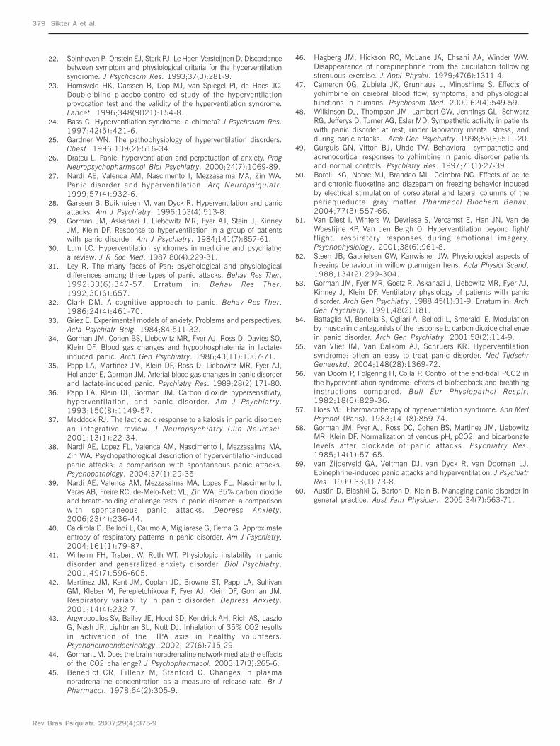
Rev Bras Psiquiatr. 2007;29(4):375-9
379 Sikter A et al.
22. Spinhoven P, Onstein EJ, Sterk PJ, Le Haen-Versteijnen D. Discordance
between symptom and physiological criteria for the hyperventilation
syndrome. J Psychosom Res. 1993;37(3):281-9.
23. Hornsveld HK, Garssen B, Dop MJ, van Spiegel PI, de Haes JC.
Double-blind placebo-controlled study of the hyperventilation
provocation test and the validity of the hyperventilation syndrome.
Lancet. 1996;348(9021):154-8.
24. Bass C. Hyperventilation syndrome: a chimera? J Psychosom Res.
1997;42(5):421-6.
25. Gardner WN. The pathophysiology of hyperventilation disorders.
Chest. 1996;109(2):516-34.
26. Dratcu L. Panic, hyperventilation and perpetuation of anxiety. Prog
Neuropsychopharmacol Biol Psychiatry. 2000;24(7):1069-89.
27. Nardi AE, Valenca AM, Nascimento I, Mezzasalma MA, Zin WA.
Panic disorder and hyperventilation. Arq Neuropsiquiatr.
1999;57(4):932-6.
28. Garssen B, Buikhuisen M, van Dyck R. Hyperventilation and panic
attacks. Am J Psychiatry. 1996;153(4):513-8.
29. Gorman JM, Askanazi J, Liebowitz MR, Fyer AJ, Stein J, Kinney
JM, Klein DF. Response to hyperventilation in a group of patients
with panic disorder. Am J Psychiatry. 1984;141(7):857-61.
30. Lum LC. Hyperventilation syndromes in medicine and psychiatry:
a review. J R Soc Med. 1987;80(4):229-31.
31. Ley R. The many faces of Pan: psychological and physiological
differences among three types of panic attacks. Behav Res Ther.
1992;30(6):347-57. Erratum in: Behav Res Ther.
1992;30(6):657.
32. Clark DM. A cognitive approach to panic. Behav Res Ther.
1986;24(4):461-70.
33. Griez E. Experimental models of anxiety. Problems and perspectives.
Acta Psychiatr Belg. 1984;84:511-32.
34. Gorman JM, Cohen BS, Liebowitz MR, Fyer AJ, Ross D, Davies SO,
Klein DF. Blood gas changes and hypophosphatemia in lactate-
induced panic. Arch Gen Psychiatry. 1986;43(11):1067-71.
35. Papp LA, Martinez JM, Klein DF, Ross D, Liebowitz MR, Fyer AJ,
Hollander E, Gorman JM. Arterial blood gas changes in panic disorder
and lactate-induced panic. Psychiatry Res. 1989;28(2):171-80.
36. Papp LA, Klein DF, Gorman JM. Carbon dioxide hypersensitivity,
hyperventilation, and panic disorder. Am J Psychiatry.
1993;150(8):1149-57.
37. Maddock RJ. The lactic acid response to alkalosis in panic disorder:
an integrative review. J Neuropsychiatry Clin Neurosci.
2001;13(1):22-34.
38. Nardi AE, Lopez FL, Valenca AM, Nascimento I, Mezzasalma MA,
Zin WA. Psychopathological description of hyperventilation-induced
panic attacks: a comparison with spontaneous panic attacks.
Psychopathology. 2004;37(1):29-35.
39. Nardi AE, Valenca AM, Mezzasalma MA, Lopes FL, Nascimento I,
Veras AB, Freire RC, de-Melo-Neto VL, Zin WA. 35% carbon dioxide
and breath-holding challenge tests in panic disorder: a comparison
with spontaneous panic attacks. Depress Anxiety.
2006;23(4):236-44.
40. Caldirola D, Bellodi L, Caumo A, Migliarese G, Perna G. Approximate
entropy of respiratory patterns in panic disorder. Am J Psychiatry.
2004;161(1):79-87.
41. Wilhelm FH, Trabert W, Roth WT. Physiologic instability in panic
disorder and generalized anxiety disorder. Biol Psychiatry.
2001;49(7):596-605.
42. Martinez JM, Kent JM, Coplan JD, Browne ST, Papp LA, Sullivan
GM, Kleber M, Perepletchikova F, Fyer AJ, Klein DF, Gorman JM.
Respiratory variability in panic disorder. Depress Anxiety.
2001;14(4):232-7.
43. Argyropoulos SV, Bailey JE, Hood SD, Kendrick AH, Rich AS, Laszlo
G, Nash JR, Lightman SL, Nutt DJ. Inhalation of 35% CO2 results
in activation of the HPA axis in healthy volunteers.
Psychoneuroendocrinology. 2002; 27(6):715-29.
44. Gorman JM. Does the brain noradrenaline network mediate the effects
of the CO2 challenge? J Psychopharmacol. 2003;17(3):265-6.
45. Benedict CR, Fillenz M, Stanford C. Changes in plasma
noradrenaline concentration as a measure of release rate. Br J
Pharmacol. 1978;64(2):305-9.
46. Hagberg JM, Hickson RC, McLane JA, Ehsani AA, Winder WW.
Disappearance of norepinephrine from the circulation following
strenuous exercise. J Appl Physiol. 1979;47(6):1311-4.
47. Cameron OG, Zubieta JK, Grunhaus L, Minoshima S. Effects of
yohimbine on cerebral blood flow, symptoms, and physiological
functions in humans. Psychosom Med. 2000;62(4):549-59.
48. Wilkinson DJ, Thompson JM, Lambert GW, Jennings GL, Schwarz
RG, Jefferys D, Turner AG, Esler MD. Sympathetic activity in patients
with panic disorder at rest, under laboratory mental stress, and
during panic attacks. Arch Gen Psychiatry. 1998;55(6):511-20.
49. Gurguis GN, Vitton BJ, Uhde TW. Behavioral, sympathetic and
adrenocortical responses to yohimbine in panic disorder patients
and normal controls. Psychiatry Res. 1997;71(1):27-39.
50. Borelli KG, Nobre MJ, Brandao ML, Coimbra NC. Effects of acute
and chronic fluoxetine and diazepam on freezing behavior induced
by electrical stimulation of dorsolateral and lateral columns of the
periaqueductal gray matter. Pharmacol Biochem Behav.
2004;77(3):557-66.
51. Van Diest I, Winters W, Devriese S, Vercamst E, Han JN, Van de
Woestijne KP, Van den Bergh O. Hyperventilation beyond fight/
fl ight: respiratory responses during emotional imagery.
Psychophysiology. 2001;38(6):961-8.
52. Steen JB, Gabrielsen GW, Kanwisher JW. Physiological aspects of
freezing behaviour in willow ptarmigan hens. Acta Physiol Scand.
1988;134(2):299-304.
53. Gorman JM, Fyer MR, Goetz R, Askanazi J, Liebowitz MR, Fyer AJ,
Kinney J, Klein DF. Ventilatory physiology of patients with panic
disorder. Arch Gen Psychiatry. 1988;45(1):31-9. Erratum in: Arch
Gen Psychiatry. 1991;48(2):181.
54. Battaglia M, Bertella S, Ogliari A, Bellodi L, Smeraldi E. Modulation
by muscarinic antagonists of the response to carbon dioxide challenge
in panic disorder. Arch Gen Psychiatry. 2001;58(2):114-9.
55. van Vliet IM, Van Balkom AJ, Schruers KR. Hyperventilation
syndrome: often an easy to treat panic disorder. Ned Tijdschr
Geneeskd. 2004;148(28):1369-72.
56. van Doorn P, Folgering H, Colla P. Control of the end-tidal PCO2 in
the hyperventilation syndrome: effects of biofeedback and breathing
instructions compared. Bull Eur Physiopathol Respir.
1982;18(6):829-36.
57. Hoes MJ. Pharmacotherapy of hyperventilation syndrome. Ann Med
Psychol (Paris). 1983;141(8):859-74.
58. Gorman JM, Fyer AJ, Ross DC, Cohen BS, Martinez JM, Liebowitz
MR, Klein DF. Normalization of venous pH, pCO2, and bicarbonate
levels after blockade of panic attacks. Psychiatry Res.
1985;14(1):57-65.
59. van Zijderveld GA, Veltman DJ, van Dyck R, van Doornen LJ.
Epinephrine-induced panic attacks and hyperventilation. J Psychiatr
Res. 1999;33(1):73-8.
60. Austin D, Blashki G, Barton D, Klein B. Managing panic disorder in
general practice. Aust Fam Physician. 2005;34(7):563-71.
Artigo13_V02.p65 28/11/2007, 16:20379


.Weexcludedcases of epistaxis associated](https://static.fdocuments.us/doc/165x107/60eaaf58fea34e421d6495a5/management-of-severe-epistaxis-during-pregnancy-a-case-few-cases-of-severe.jpg)


