2 Metallic Biomaterials - Darpublic · METALLIC BIOMATERIALS ... “ Biomedical Engineering...
21
Lecture Note 1/21 METALLIC BIOMATERIALS [Adopsi dari: Joseph D. Bronzino, “Biomedical Engineering Fundamentals”, CRC Press, third edition, 2006, Section V, CHAPTER 38, by Joon B. Park, Young Kon Kim.] 38.1 INTRODUCTION Metals are used as biomaterials due to their excellent electrical and thermal conductivity and mechanical properties. Since some electrons are independent in metals they can quickly transfer an electric charge and thermal energy. The mobile free electrons act as the binding force to hold the positive metal ions together. This attraction is strong, as evidenced by the closely packed atomic arrangement resulting in high specific gravity and high melting points of most metals. Since the metallic bond is essentially nondirectional, the position of the metal ions can be altered without destroying the crystal structure resulting in a plastically deformable solid. Some metals are used as passive substitutes for hard tissue replacement such as total hip and knee joints, for fracture healing aids as bone plates and screws, spinal fixation devices, and dental implants because of their excellent mechanical properties and corrosion resistance. Some metallic alloys are used for more active roles in devices such as vascular stents, catheter guide wires, orthodontic archwires, and cochlea implants. The first metal alloy developed specifically for human use was the “vanadium steel” which was used to manufacture bone fracture plates (Sherman plates) and screws. Most metals such as iron (Fe), chromium (Cr), cobalt (Co), nickel (Ni), titanium (Ti), tantalum (Ta), niobium (Nb), molybdenum (Mo), and tungsten (W), that were used to make alloys for manufacturing implants can only be tolerated by the body in minute amounts. Sometimes those metallic elements, in naturally occurring forms, are essential in red blood cell functions (Fe) or synthesis of a vitamin 812 (Co), but cannot be tolerated in large amounts in the body [Black, 1992]. The biocompatibility of the metallic implant is of considerable concern because these implants can corrode in an in vivo environment [Williams. 1982]. The consequences of corrosion are the disintegration of the implant material per se, which will weaken the implant, and the harmful effect of corrosion products on the surrounding tissues and organs. 38.2 STAINLESS STEELS The first stainless steel utilized for implant fabrication was the 18-8 (type 302 in modern classification), which is stronger and more resistant to corrosion than the vanadium steel. Vanadium steel is no longer used in implants since its corrosion resistance is inadequate in vivo. Later 18-8sMo stainless steel was introduced which contains a small percentage of molybdenum to improve the corrosion resistance in chloride solution (salt water). This alloy became known as type 316 stainless steel. In the l950s the carbon content of 316 stainless steel was reduced from 0.08 to a maximum amount of 0.03% (all are weight percent unless specified) for better corrosion resistance to chloride solution and to minimize the sensitization and hence, became known as type 316L stainless steel. The minimum effective concentration of chromium is 11% to impart corrosion resistance in stainless steels. The chromium is a reactive element, but it and its alloys can be passivated by 30% nitric acid to give excellent corrosion resistance. The austenitic stainless steels, especially type 316 and 316L, are most widely used for implant fabrication. These cannot be hardened by heat treatment but can he hardened by cold-working. This group of stainless steels is nonmagnetic and possesses better corrosion resistance than any others. The inclusion of molybdenum enhances resistance
Transcript of 2 Metallic Biomaterials - Darpublic · METALLIC BIOMATERIALS ... “ Biomedical Engineering...

Lecture Note
1/21
METALLIC BIOMATERIALS
[Adopsi dari: Joseph D. Bronzino, “Biomedical Engineering Fundamentals”, CRC Press, third edition, 2006, Section V,
CHAPTER 38, by Joon B. Park, Young Kon Kim.]
38.1 INTRODUCTION
Metals are used as biomaterials due to their excellent electrical and thermal conductivity and mechanical properties. Since some electrons are independent in metals they can quickly transfer an electric charge and thermal energy. The mobile free electrons act as the binding force to hold the positive metal ions together. This attraction is strong, as evidenced by the closely packed atomic arrangement resulting in high specific gravity and high melting points of most metals. Since the metallic bond is essentially nondirectional, the position of the metal ions can be altered without destroying the crystal structure resulting in a plastically deformable solid.
Some metals are used as passive substitutes for hard tissue replacement such as total hip and knee joints, for fracture healing aids as bone plates and screws, spinal fixation devices, and dental implants because of their excellent mechanical properties and corrosion resistance. Some metallic alloys are used for more active roles in devices such as vascular stents, catheter guide wires, orthodontic archwires, and cochlea implants.
The first metal alloy developed specifically for human use was the “vanadium steel” which was used to manufacture bone fracture plates (Sherman plates) and screws. Most metals such as iron (Fe), chromium (Cr), cobalt (Co), nickel (Ni), titanium (Ti), tantalum (Ta), niobium (Nb), molybdenum (Mo), and tungsten (W), that were used to make alloys for manufacturing implants can only be tolerated by the body in minute amounts. Sometimes those metallic elements, in naturally occurring forms, are essential in red blood cell functions (Fe) or synthesis of a vitamin 812 (Co), but cannot be tolerated in large amounts in the body [Black, 1992]. The biocompatibility of the metallic implant is of considerable concern because these implants can corrode in an in vivo environment [Williams. 1982]. The consequences of corrosion are the disintegration of the implant material per se, which will weaken the implant, and the harmful effect of corrosion products on the surrounding tissues and organs.
38.2 STAINLESS STEELS
The first stainless steel utilized for implant fabrication was the 18-8 (type 302 in modern classification), which is stronger and more resistant to corrosion than the vanadium steel. Vanadium steel is no longer used in implants since its corrosion resistance is inadequate in vivo. Later 18-8sMo stainless steel was introduced which contains a small percentage of molybdenum to improve the corrosion resistance in chloride solution (salt water). This alloy became known as type 316 stainless steel. In the l950s the carbon content of 316 stainless steel was reduced from 0.08 to a maximum amount of 0.03% (all are weight percent unless specified) for better corrosion resistance to chloride solution and to minimize the sensitization and hence, became known as type 316L stainless steel. The minimum effective concentration of chromium is 11% to impart corrosion resistance in stainless steels. The chromium is a reactive element, but it and its alloys can be passivated by 30% nitric acid to give excellent corrosion resistance.
The austenitic stainless steels, especially type 316 and 316L, are most widely used for implant fabrication. These cannot be hardened by heat treatment but can he hardened by cold-working. This group of stainless steels is nonmagnetic and possesses better corrosion resistance than any others. The inclusion of molybdenum enhances resistance
Lecture Note
2/21
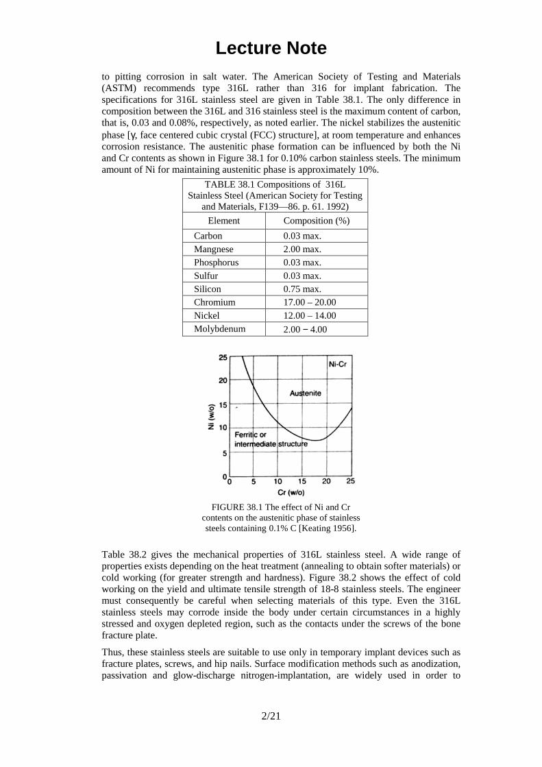
to pitting corrosion in salt water. The American Society of Testing and Materials (ASTM) recommends type 316L rather than 316 for implant fabrication. The specifications for 316L stainless steel are given in Table 38.1. The only difference in composition between the 316L and 316 stainless steel is the maximum content of carbon, that is, 0.03 and 0.08%, respectively, as noted earlier. The nickel stabilizes the austenitic phase [γ, face centered cubic crystal (FCC) structure], at room temperature and enhances corrosion resistance. The austenitic phase formation can be influenced by both the Ni and Cr contents as shown in Figure 38.1 for 0.10% carbon stainless steels. The minimum amount of Ni for maintaining austenitic phase is approximately 10%.
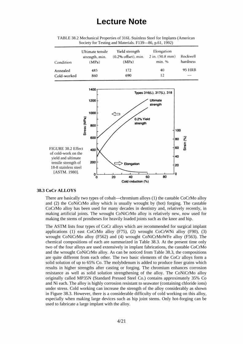
Table 38.2 gives the mechanical properties of 316L stainless steel. A wide range of properties exists depending on the heat treatment (annealing to obtain softer materials) or cold working (for greater strength and hardness). Figure 38.2 shows the effect of cold working on the yield and ultimate tensile strength of 18-8 stainless steels. The engineer must consequently be careful when selecting materials of this type. Even the 316L stainless steels may corrode inside the body under certain circumstances in a highly stressed and oxygen depleted region, such as the contacts under the screws of the bone fracture plate.
Thus, these stainless steels are suitable to use only in temporary implant devices such as fracture plates, screws, and hip nails. Surface modification methods such as anodization, passivation and glow-discharge nitrogen-implantation, are widely used in order to
TABLE 38.1 Compositions of 316L Stainless Steel (American Society for Testing
and Materials, F139—86. p. 61. 1992)
Element Composition (%)
Carbon 0.03 max. Mangnese 2.00 max. Phosphorus 0.03 max. Sulfur 0.03 max. Silicon 0.75 max. Chromium 17.00 – 20.00 Nickel 12.00 – 14.00 Molybdenum 2.00 − 4.00
FIGURE 38.1 The effect of Ni and Cr contents on the austenitic phase of stainless steels containing 0.1% C [Keating 1956].

Lecture Note
3/21
improve corrosion resistance, wear resistance, and fatigue strength of 316L stainless steel [Bordiji et al., 1996].
Lecture Note
4/21
38.3 CoCr ALLOYS
There are basically two types of cobalt—chromium alloys (1) the castable CoCrMo alloy and (2) the CoNiCrMo alloy which is usually wrought by (hot) forging. The castable CoCrMo alloy has been used for many decades in dentistry and, relatively recently, in making artificial joints. The wrought CoNiCrMo alloy is relatively new, now used for making the stems of prostheses for heavily loaded joints such as the knee and hip.
The ASTM lists four types of CoCr alloys which are recommended for surgical implant applications (1) east CoCrMo alloy (F75), (2) wrought CoCrWNi alloy (F90), (3) wrought CoNiCrMo alloy (F562) and (4) wrought CoNiCrMoWFe alloy (F563). The chemical compositions of each are summarized in Table 38.3. At the present time only two of the four alloys are used extensively in implant fabrications, the castable CoCrMo and the wrought CoNiCrMo alloy. As can be noticed from Table 38.3, the compositions are quite different from each other. The two basic elements of the CoCr alloys form a solid solution of up to 65% Co. The molybdenum is added to produce finer grains which results in higher strengths after casting or forging. The chromium enhances corrosion resistance as well as solid solution strengthening of the alloy. The CoNiCrMo alloy originally called MP35N (Standard Pressed Steel Co.) contains approximately 35% Co and Ni each. The alloy is highly corrosion resistant to seawater (containing chloride ions) under stress. Cold working can increase the strength of the alloy considerably as shown in Figure 38.3. However, there is a considerable difficulty of cold working on this alloy, especially when making large devices such as hip joint stems. Only hot-forging can be used to fabricate a large implant with the alloy.
TABLE 38.2 Mechanical Properties of 316L Stainless Steel for Implants (American Society for Testing and Materials. F139—86, p.61, 1992)
FIGURE 38.2 Effect of cold-work on the yield and ultimate tensile strength of 18-8 stainless steel
[ASTM. 1980].

Lecture Note
5/21
TABLE 38.3 Chemical Compositions of Co-Cr Alloys (American Society for Testing and Materials, F75—87, p. 42; 90-87, p.47; F562—84, p.150. 1992)
FIGURE 38.3 Relationship between ultimate tensile and the amount of cold-work for CoNiCrMo alloy [Devine and Wulff, 1975].
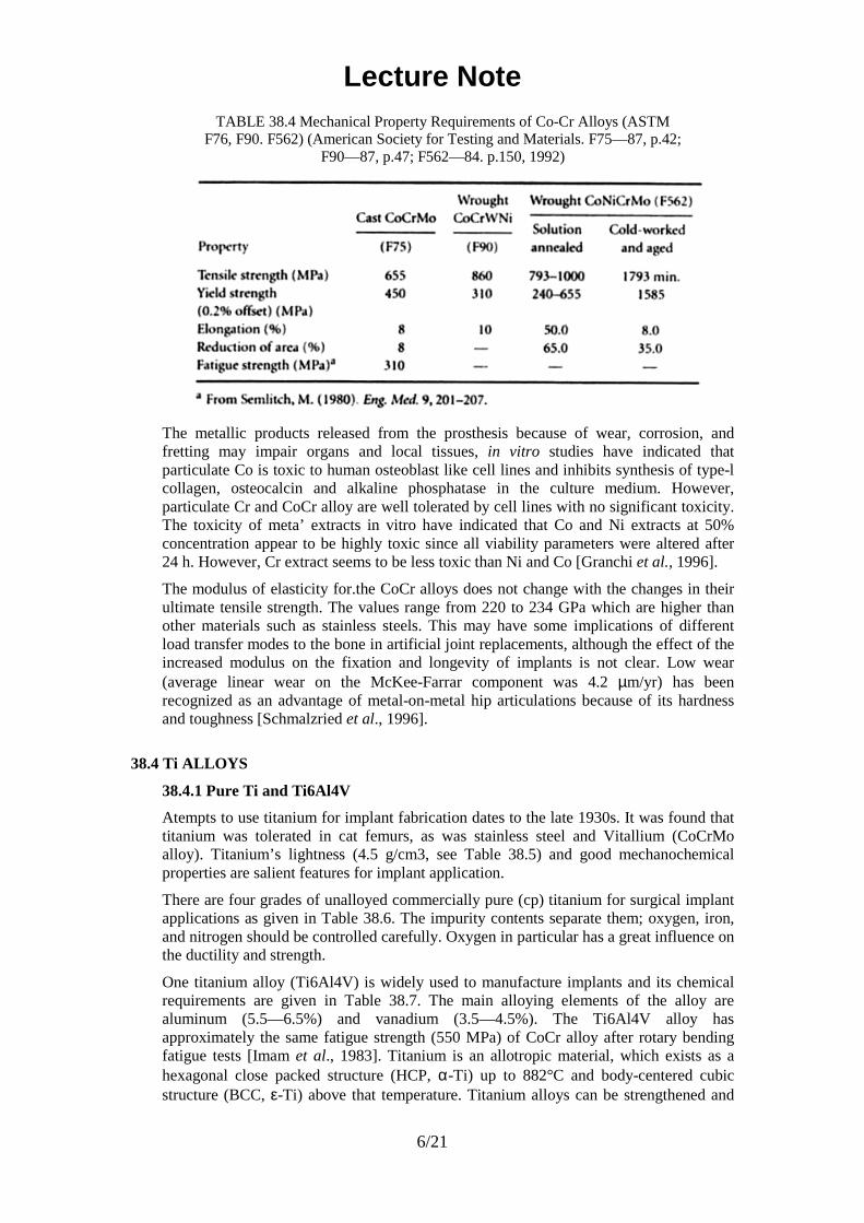
The abrasive wear properties of the wrought CoNìCrMo alloy are similar to the cast CoCrMo alloy (about 0.14 mm/yr in joint simulation tests with ultra high molecular weight polyethylene acetabular cup); however, the former is not recommended for the bearing surfaces of joint prosthesis because of its poor frictional properties with itself or other materials. The superior fatigue and ultimate tensile strength of the wrought CoNiÇrMo alloy make it suitable for the applications which require long service life without fracture or stress fatigue. Such is the case for the stems of the hip joint prostheses. This advantage is better appreciated when the implant has to be replaced, since it is quite difficult to remove the failed piece of implant embedded deep in the femoral medullary canal. Furthermore, the revision arthroplasty is usually inferior to the primary surgery in terms of its function due to poorer fixation of the implant. The mechanical properties required for CoCr alloys are given in Table 38.4. As with the other alloys, the increased strength is accompanied by decreased ductility. Both the cast and wrought alloys have excellent corrosion resistance.
Experimental determination of the rate of nickel release from the CoNiCrMo alloy and 316L stainless steel in 37°C Ringer’s solution showed an interesting result. Although the cobalt alloy has more initial release of nickel ions into the solution, the rate of release was about the same (3 x 10⁻¹⁰ g/cm2/day) for both alloys [Richards-Mfg-Company, 1980]. This is rather surprising since the nickel content of the CoNiCrMo alloy is about three times that of 316L stainless steel.
Lecture Note
6/21
TABLE 38.4 Mechanical Property Requirements of Co-Cr Alloys (ASTM F76, F90. F562) (American Society for Testing and Materials. F75—87, p.42;
F90—87, p.47; F562—84. p.150, 1992)
The metallic products released from the prosthesis because of wear, corrosion, and fretting may impair organs and local tissues, in vitro studies have indicated that particulate Co is toxic to human osteoblast like cell lines and inhibits synthesis of type-l collagen, osteocalcin and alkaline phosphatase in the culture medium. However, particulate Cr and CoCr alloy are well tolerated by cell lines with no significant toxicity. The toxicity of meta’ extracts in vitro have indicated that Co and Ni extracts at 50% concentration appear to be highly toxic since all viability parameters were altered after 24 h. However, Cr extract seems to be less toxic than Ni and Co [Granchi et al., 1996].
The modulus of elasticity for.the CoCr alloys does not change with the changes in their ultimate tensile strength. The values range from 220 to 234 GPa which are higher than other materials such as stainless steels. This may have some implications of different load transfer modes to the bone in artificial joint replacements, although the effect of the increased modulus on the fixation and longevity of implants is not clear. Low wear (average linear wear on the McKee-Farrar component was 4.2 µm/yr) has been recognized as an advantage of metal-on-metal hip articulations because of its hardness and toughness [Schmalzried et al., 1996].
38.4 Ti ALLOYS
38.4.1 Pure Ti and Ti6Al4V
Atempts to use titanium for implant fabrication dates to the late 1930s. It was found that titanium was tolerated in cat femurs, as was stainless steel and Vitallium (CoCrMo alloy). Titanium’s lightness (4.5 g/cm3, see Table 38.5) and good mechanochemical properties are salient features for implant application.
There are four grades of unalloyed commercially pure (cp) titanium for surgical implant applications as given in Table 38.6. The impurity contents separate them; oxygen, iron, and nitrogen should be controlled carefully. Oxygen in particular has a great influence on the ductility and strength.
One titanium alloy (Ti6Al4V) is widely used to manufacture implants and its chemical requirements are given in Table 38.7. The main alloying elements of the alloy are aluminum (5.5—6.5%) and vanadium (3.5—4.5%). The Ti6Al4V alloy has approximately the same fatigue strength (550 MPa) of CoCr alloy after rotary bending fatigue tests [Imam et al., 1983]. Titanium is an allotropic material, which exists as a hexagonal close packed structure (HCP, α-Ti) up to 882°C and body-centered cubic structure (BCC, ε-Ti) above that temperature. Titanium alloys can be strengthened and
Lecture Note
7/21
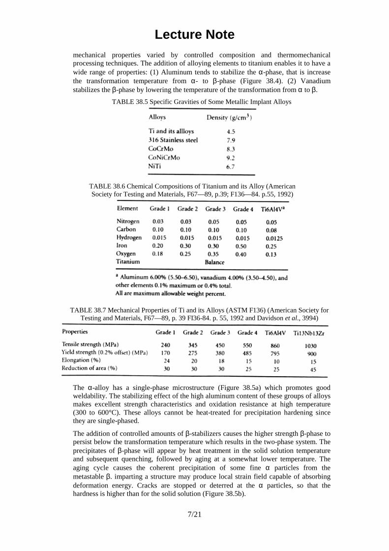
mechanical properties varied by controlled composition and thermomechanical processing techniques. The addition of alloying elements to titanium enables it to have a wide range of properties: (1) Aluminum tends to stabilize the α-phase, that is increase the transformation temperature from α- to β-phase (Figure 38.4). (2) Vanadium stabilizes the β-phase by lowering the temperature of the transformation from α to β.
TABLE 38.5 Specific Gravities of Some Metallic Implant Alloys
TABLE 38.6 Chemical Compositions of Titanium and its Alloy (American Society for Testing and Materials, F67—89, p.39; F136—84. p.55, 1992)
TABLE 38.7 Mechanical Properties of Ti and its Alloys (ASTM F136) (American Society for Testing and Materials, F67—89, p. 39 FI36-84. p. 55, 1992 and Davidson et al., 3994)
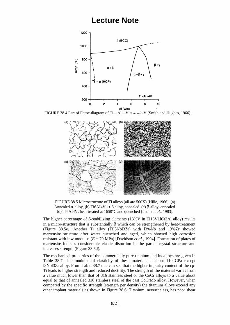
The α-alloy has a single-phase microstructure (Figure 38.5a) which promotes good weldability. The stabilizing effect of the high aluminum content of these groups of alloys makes excellent strength characteristics and oxidation resistance at high temperature (300 to 600°C). These alloys cannot be heat-treated for precipitation hardening since they are single-phased.
The addition of controlled amounts of β-stabilizers causes the higher strength β-phase to persist below the transformation temperature which results in the two-phase system. The precipitates of β-phase will appear by heat treatment in the solid solution temperature and subsequent quenching, followed by aging at a somewhat lower temperature. The aging cycle causes the coherent precipitation of some fine α particles from the metastable β. imparting a structure may produce local strain field capable of absorbing deformation energy. Cracks are stopped or deterred at the α particles, so that the hardness is higher than for the solid solution (Figure 38.5b).
Lecture Note
8/21
FIGURE 38.4 Part of Phase-diagram of Ti—Al—V at 4 w/o V [Smith and Hughes, 1966].
FIGURE 38.5 Microstructure of Ti alloys (all are 500X) [Hille, 1966]. (a) Annealed α-alloy, (b) Ti6AI4V. α-β alloy, annealed. (c) β-alloy, annealed.
(d) TI6AI4V. heat-treated at 1650°C and quenched [Imam et al., 1983].
The higher percentage of β-stabiIizing elements (13%V in Ti13V1lCr3Al alloy) results in a micro-structure that is substantially β which can be strengthened by heat-treatment (Figure 38.5e). Another Ti alloy (Til3Nbl3Zr) with l3%Nb and 13%Zr showed martensite structure after water quenched and aged, which showed high corrosion resistant with low modulus (E = 79 MPa) [Davidson et al., 1994]. Formation of plates of martensite induces considerable elastic distortion in the parent crystal structure and increases strength (Figure 38.5d).
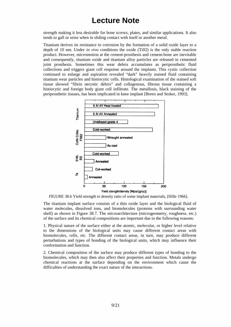
The mechanical properties of the commercially pure titanium and its alloys are given in Table 38.7. The modulus of elasticity of these materials is about 110 GPa except l3Nbl3Zr alloy. From Table 38.7 one can see that the higher impurity content of the cp-Ti leads to higher strength and reduced ductility. The strength of the material varies from a value much lower than that of 316 stainless steel or the CoCr alloys to a value about equal to that of annealed 316 stainless steel of the cast CoCrMo alloy. However, when compared by the specific strength (strength per density) the titanium alloys exceed any other implant materials as shown in Figure 38.6. Titanium, nevertheless, has poor shear
Lecture Note
9/21
strength making it less desirable for bone screws, plates, and similar applications. It also tends to gall or seize when in sliding contact with itself or another metal.
Titanium derives its resistance to corrosion by the formation of a solid oxide layer to a depth of 10 nm. Under in vivo conditions the oxide (Ti02) is the only stable reaction product. However, micromotion at the cement-prosthesis and cement-bone are inevitable and consequently, titanium oxide and titanium alloy particles are released in cemented joint prosthesis. Sometimes this wear debris accumulates as periprosthetic fluid collections and triggers giant cell response around the implants. This cystic collection continued to enlarge and aspiration revealed “dark” heavily stained fluid containing titanium wear particles and histiocytic cells. Histological examination of the stained soft tissue showed “fibrin necrotic debris” and collagenous, fibrous tissue containing a histiocytic and foreign body giant cell infiltrate. The metallosis, black staining of the periprosthetic tissues, has been implicated in knee implant [Breen and Stoker, 1993].
FIGURE 38.6 Yield strength to density ratio of some implant materials, [Hille 1966].
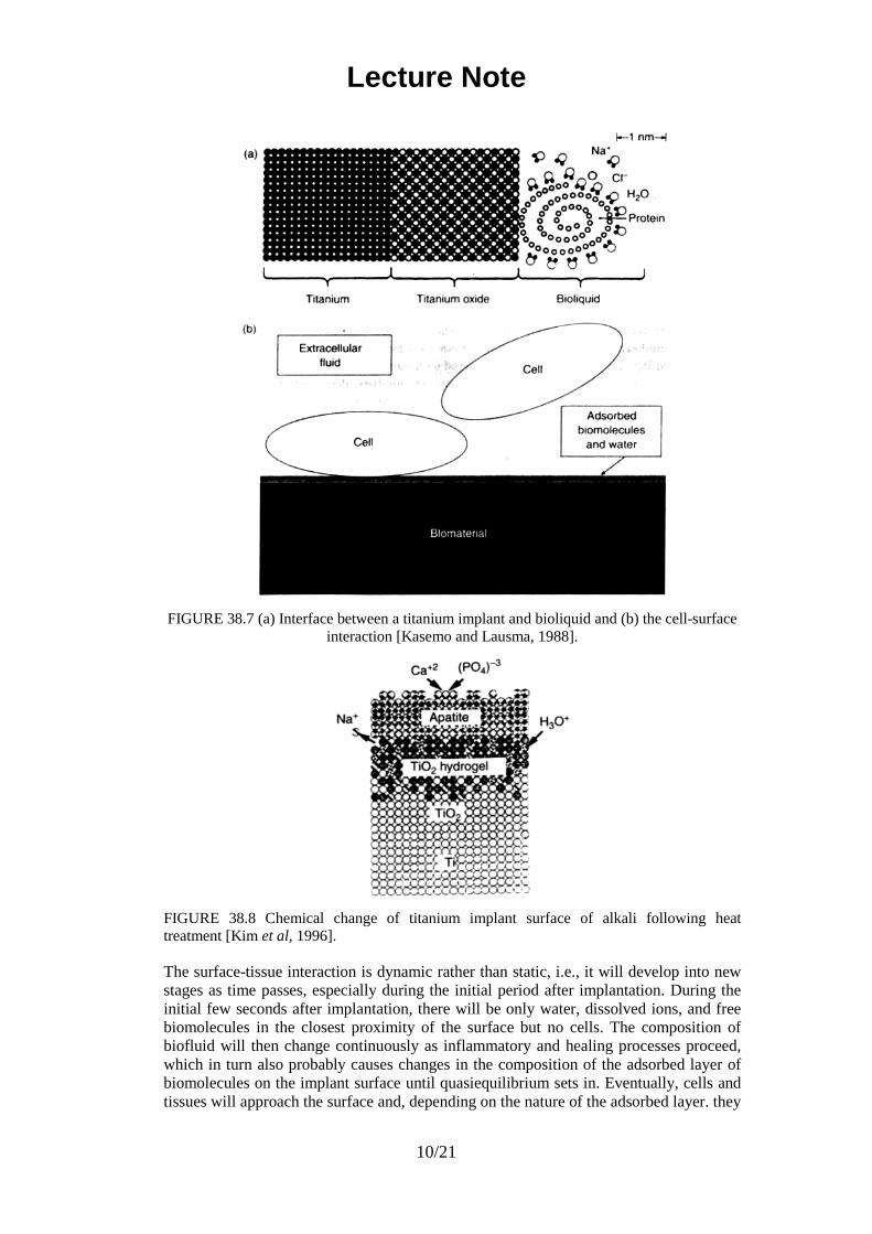
The titanium implant surface consists of a thin oxide layer and the biological fluid of water molecules, dissolved ions, and biomolecules (proteins with surrounding water shell) as shown in Figure 38.7. The microarchitecture (microgeometry, roughness. etc.) of the surface and its chemical compositions are important due to the following reasons:
1. Physical nature of the surface either at the atomic, molecular, or higher level relative to the dimensions of the biological units may cause different contact areas with biomolecules, cells, etc. The different contact areas, in turn, may produce different perturbations and types of bonding of the biological units, which may influence their conformation and function.
2. Chemical composition of the surface may produce different types of bonding to the biomolecules, which may then also affect their properties and function. Metals undergo chemical reactions at the surface depending on the environment which cause the difficulties of understanding the exact nature of the interactions.
Lecture Note
10/21
FIGURE 38.7 (a) Interface between a titanium implant and bioliquid and (b) the cell-surface interaction [Kasemo and Lausma, 1988].
FIGURE 38.8 Chemical change of titanium implant surface of alkali following heat treatment [Kim et al, 1996].
The surface-tissue interaction is dynamic rather than static, i.e., it will develop into new stages as time passes, especially during the initial period after implantation. During the initial few seconds after implantation, there will be only water, dissolved ions, and free biomolecules in the closest proximity of the surface but no cells. The composition of biofluid will then change continuously as inflammatory and healing processes proceed, which in turn also probably causes changes in the composition of the adsorbed layer of biomolecules on the implant surface until quasiequilibrium sets in. Eventually, cells and tissues will approach the surface and, depending on the nature of the adsorbed layer. they
Lecture Note
11/21
will respond in specific ways that may further modify the adsorbed biomolecules. The type of cells closest to the surface and their activities will change with time. For example, depending on the type of initial interaction, the final results may be fibrous capsule formation or tissue integration [Kasemo and Lausma, 1988; Hazan et al., 1993; Takatsuka et al, 1995; Takeshita et al., 1997; Yan et al, 1997].
Osseointegration is defined as direct contact without intervening soft tissue between viable remodeled bone and an implant. Surface roughness of titanium alloys have a significant effect on the bone apposition to the implant and on the bone implant interfacial pull out strength. The average roughness increased from 0.5 to 5.9 µm and the interfacial shear strength increased from 0.48 to 3.5 MPa [Feighan et al., 1995].
Highest levels of osteoblast cell attachment are obtained with rough sand blast surfaces where cells differentiated more than those on the smooth surfaces [Keller et al, 1994]. Chemical changes of the titanium surface following heat treatment is thought to form a TiO2 hydrogel layer on top of the TiO2 layer as shown in Figure 38.8. The TiO2 hydrogel layer may induce the apatite crystal formation [Kim et al, 1996].
In general, on the rougher surfaces there are lower cell numbers, decreased rate of cellular proliferation, and increased matrix production compared to smooth surface. Bone formation appears to be strongly related to the presence of transforming growth factor in the bone matrix [Kieswetter et al., 1996].
38.4.2 TiNi Alloys
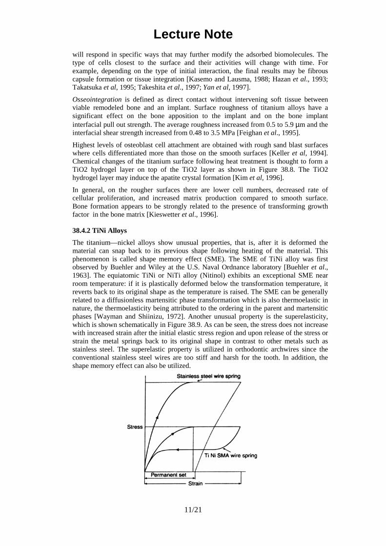
The titanium—nickel alloys show unusual properties, that is, after it is deformed the material can snap back to its previous shape following heating of the material. This phenomenon is called shape memory effect (SME). The SME of TiNi alloy was first observed by Buehler and Wiley at the U.S. Naval Ordnance laboratory [Buehler et al., 1963]. The equiatomic TiNi or NiTi alloy (Nitinol) exhibits an exceptional SME near room temperature: if it is plastically deformed below the transformation temperature, it reverts back to its original shape as the temperature is raised. The SME can be generally related to a diffusionless martensitic phase transformation which is also thermoelastic in nature, the thermoelasticity being attributed to the ordering in the parent and martensitic phases [Wayman and Shiinizu, 1972]. Another unusual property is the superelasticity, which is shown schematically in Figure 38.9. As can be seen, the stress does not increase with increased strain after the initial elastic stress region and upon release of the stress or strain the metal springs back to its original shape in contrast to other metals such as stainless steel. The superelastic property is utilized in orthodontic archwires since the conventional stainless steel wires are too stiff and harsh for the tooth. In addition, the shape memory effect can also be utilized.

Lecture Note
12/21
FIGURE 38.9 Schematic illustration of the stainless steel wire and TiNi SMA wire springs for orthodontic archwire behavior. (Modified from Wayman C.M. and
Duerig T.W. (1990), London: Butterworth-Heinemann, pp. 3-20.)
Lecture Note
13/21
Some possible applications of shape memory alloys are orthodontic dental archwire, intracranial aneurysm clip, vena cava filter, contractile artificial muscles for an artificial heart, vascular stent, catheter guide wire, and orthopedic staple [Duerig et al., 1990].
In order to develop such devices, it is necessary to understand fully the mechanical and thermal behavior associated with the martensitic phase transformation. A widely known NiTi alloy is 55-Nitinol (55 weight% or 50 atomic % Ni), which has a single phase and the mechanical memory plus other properties. for example, high acoustic damping, direct conversion of heat energy into mechanical energy, good fatigue properties, and low temperature ductility. Deviation from the 55-Nitinol (near stoichiometric NiTi) in the Ni-rich direction yields a second group of alloys which are also completely nonmagnetic but differ from 55-Nitinol in their ability to be thermally hardened to higher hardness levels. Shape recovery capability decreases and heat treatability increases rapidly as the Ni content approaches 60%. Both 55 and 60-Nitinols have relatively low modulus of elasticity and can be tougher and more resilient than stainless steel, NiCr, or CoCr alloys.
Efficiency of 55-Nitinol shape recovery can be controlled by changing the final annealing temperatures during preparation of the alloy device [Lee et al., 1988]. For the most efficient recovery, the shape is fixed by constraining the specimen in a desired configuration and heating to 482 to 510°C. If the annealed wire is deformed at a temperature below the shape recovery temperature, shape recovery will occur upon heating, provided the deformation has not exceeded crystallographic strain limits (—8% strain in tension). The NiTi alloys also exhibit good biocompatibilily and corrosion resistance in vivo. There is no significant difference between titanium and NiTi in the inhibition of mitosis in human fibroblasts. NiTi showed lower percentage bone and bone contact area than titanium and the Ti6AI4V alloy [Takeshita et al., 1997].
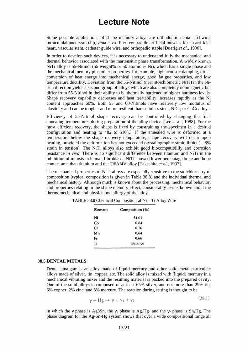
The mechanical properties of NiTi alloys are especially sensitive to the stoichiornetry of composition (typical composition is given in Table 38.8) and the individual thermal and mechanical history. Although much is known about the processing. mechanical behavior, and properties relating to the shape memory effect, considerably less is known about the thermomechanical and physical metallurgy of the alloy.
TABLE 38.8 Chemical Composition of Ni—Ti Alloy Wire
38.5 DENTAL METALS
Dental amalgam is an alloy made of liquid mercury and other solid metal particulate alloys made of silver, tin, copper, etc. The solid alloy is mixed with (liquid) mercury in a mechanical vibrating mixer and the resulting material is packed into the prepared cavity. One of the solid alloys is composed of at least 65% silver, and not more than 29% tin, 6% copper. 2% zinc, and 3% mercury. The reaction during setting is thought to be
in which the γ phase is Ag3Sn, the γ1 phase is Ag2Hg3 and the γ2 phase is Sn7Hg. The phase diagram for the Ag-Sn-Hg system shows that over a wide compositional range all
Lecture Note
14/21
three phases are present. The final composition of dental amalgams typically contain 45% to 55% mercury. 35% to 45% silver, and about 15% tin after fully set in about one day.
Gold and gold alloys are useful metals in dentistry as a result of their durability, stability, and corrosion resistance [Nielsen, 1986]. Gold fillings are introduced by two methods: casting and malleting. Cast restorations are made by taking a wax impression of the prepared cavity, making a mold from this impression in a material such as gypsum silica, which tolerates high temperature, and casting molten gold in the mold. The patient is given a temporary filling for the intervening time. Gold alloys are used for cast restorations, since they have mechanical properties which are superior to those of pure gold. Corrosion resistance is retained in these alloys provided they contain 75% or more of gold and other noble metals. Copper, alloyed with gold, significantly increases its strength. Platinum also improves the strength. But no more than about 4% can be added, or the melting point of the alloy is elevated excessively. Silver compensates for the color of copper. A small amount of zinc may be added to lower the melting point and to scavenge oxides formed during melting. Gold alloys of different composition are available. Softer alloys containing more than 83% gold are used for inlays which are not subjected to much stress. Harder alloys containing less gold are chosen for crowns and cusps which are more heavily stressed.
Malleted restorations are built up in the cavity from layers of pure gold foil. The foils are welded together by pressure at ambient temperature. In this type of welding, the metal layers are joined by thermal diffusion of atoms from one layer to the other. Since intimate contact is required in this procedure, it is particularly important to avoid contamination. The pure gold is relatively soft, so this type of restoration is limited to areas not subjected to much stress.
38.6 OTHER METALS
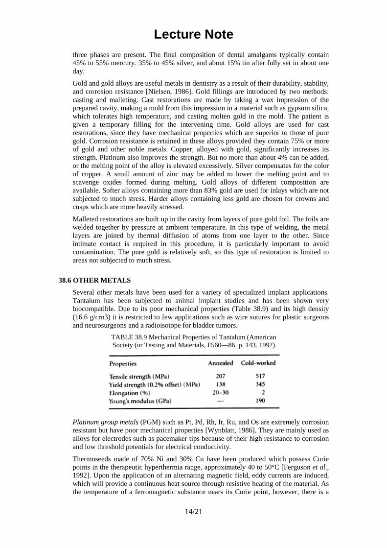
Several other metals have been used for a variety of specialized implant applications. Tantalum has been subjected to animal implant studies and has been shown very biocompatible. Due to its poor mechanical properties (Table 38.9) and its high density (16.6 g/crn3) it is restricted to few applications such as wire sutures for plastic surgeons and neurosurgeons and a radioisotope for bladder tumors.
TABLE 38.9 Mechanical Properties of Tantalum (American Society (or Testing and Materials, F560—86. p. 143. 1992)
Platinum group metals (PGM) such as Pt, Pd, Rh, Ir, Ru, and Os are extremely corrosion resistant but have poor mechanical properties [Wynblatt, 1986]. They are mainly used as alloys for electrodes such as pacemaker tips because of their high resistance to corrosion and low threshold potentials for electrical conductivity.
Thermoseeds made of 70% Ni and 30% Cu have been produced which possess Curie points in the therapeutic hyperthermia range, approximately 40 to 50°C [Ferguson et al., 1992]. Upon the application of an alternating magnetic field, eddy currents are induced, which will provide a continuous heat source through resistive heating of the material. As the temperature of a ferromagnetic substance nears its Curie point, however, there is a

Lecture Note
15/21
loss of ferromagnetic properties and a resulting loss of heat output. Thus, self-regulation of temperature is achieved and can be used to deliver a constant hyperthermic temperature extracorporeally at any time and duration.
Surface modifications of metal alloys such as coatings by plasma spray, physical or chemical vapor deposition, ion implantation, and fluidized bed deposition have been used in industry [Smith, 1993]. Coating implants with tissue compatible materials such as hydroxyapatite, oxide ceramics, Bioglass*, and pyrolytic carbon are typical applications in implants. Such efforts have been largely ineffective if the implants are permanent and particularly if the implants are subjected to a large loading. The main problem is the delamination of the coating or eventual wear of the coating. The added cost of coating or ion implanting hinders the use of such techniques unless the technique shows unequivocal superiority compared to the non-treated implants.
38.7 CORROSION OF METALLIC IMPLANTS
Corrosion is the unwanted chemical reaction of a metal with its environment, resulting in its continued degradation to oxides, hydroxides, or other compounds. Tissue fluid in the human body contains water, dissolved oxygen, proteins, and various ions such as chloride and hydroxide. As a result, the human body presents a very aggressive environment for metals used for implantation. Corrosion resistance of a metallic implant material is consequently an important aspect of its biocompatibility.
38.7.1 Electrochemical Aspects
The lowest free energy state of many metals in an oxygenated and hydrated environment is that of the oxide. Corrosion occurs when metal atoms become ionized and go into solution, or combine with oxygen or other species in solution to form a compound which flakes off or dissolves. The body environment is very aggressive in terms of corrosion since it is not only aqueous but also contains chloride ions and proteins. A variety of
chemical reactions occur when a metal is exposed to an aqueous environment, as shown in Figure 38.10. The electrolyte, which contains ions in solution, serves to complete the electric circuit. In the human body, the required ions are plentiful in the body fluids. Anions are negative ions which migrate toward the anode, and cations are positive ions which migrate toward the cathode. At the anode, or positive electrode, the metal oxidizes by losing valence electrons as in the following:
−+ +→ nenMM (38.2)
At the cathode, or negative electrode, the following reduction reactions are important:
The tendency of metals to corrode is expressed most simply in the standard electrochemical series of Nernst potentials, shown in Table 38.10. These potentials are obtained in electrochemical measurements in which one electrode is a standard hydrogen
FIGURE 38.10 Electrochemical cell

Lecture Note
16/21
electrode formed by bubbling hydrogen through a layer of finely divided platinum black. The potential of this reference electrode is defined to be zero. Noble metals are those which have a potential higher than that of a standard hydrogen electrode; base metals have lower potentials.
If two dissimilar metals are present in the same environment, the one which is most negative in the galvanic series will become the anode, and bimetallic (or galvanic) corrosion will occur. Galvanic corrosion can be much more rapid than the corrosion of a single metal, consequently, implantation of dissimilar metals (mixed metals) is to be avoided. Galvanic action can also result in corrosion within a single metal, if there is inhomogeneity in the metal or in its environment, as shown in Figure 38.11.
The potential difference, E, actually observed depends on the concentration of the metal ions in solution according to the Nernst equation,
in which R is the gas constant, E0 is the standard electrochemical potential, T is the absolute temperature, F is Faraday’s constant (96,487 C/mol), and n is the number of moles of ions.
FIGURE 38.11 Micro-corrosion cells. (a) Grain boundaries are anodic with respect to the grain interior. (b) Crevice corrosion due to oxygen deficient zone in metal’s environment.
The order of nobility observed in actual practice may differ from that predicted thermodynamically. The reasons arc that some metals become covered with a passivating film of reaction products which protects the metal from further attack The dissolution reaction may be strongly irreversible so that a potential barrier must be overcome. In this case, corrosion may be inhibited even though it remains energetically favorable. The kinetics of corrosion reactions are not determined by the thermodynamics alone.
38.7.2 Pourbaix Diagrams in Corrosion
The Pourbaix diagram is a plot of regions of corrosion, passivity, and immunity as they depend on electrode potential and pH [Pourbaix, 1974]. The Pourbaix diagrams are derived from the Nernst equation and from the solubility of the degradation products and the equilibrium constants of the reaction. For the sake of definition, the corrosion region is set arbitrarily at a concentration of greater than 10−6 g atom/l (molar) or more of metal in the solution at equilibrium. This corresponds to about 0.06 mg/l for metals such as iron and copper, and 0.03 mg/l for aluminum. Immunity is defined as equilibrium between metal and its ions at less than 10−6 M. In the region of immunity, the corrosion
TABLE 38.10 Standard Electrochemical Series
Lecture Note
17/21
is energetically impossible. Immunity is also referred to as cathodic protection. In the passivation domain, the stable solid constituent is an oxide, hydroxide, hydride, or a salt of the metal. Passivity is defined as equilibrium between a metal and its reaction products (oxides, hydroxides, etc.) at a concentration of 10−6 M or less. This situation is useful if reaction products are adherent. In the biomaterials setting, passivity may or may not be adequate; disruption of a passive layer may cause an increase in corrosion. The equilibrium state may not occur if reaction products are removed by the tissue fluid. Materials differ in their propensity to re-establish a passive layer which has been damaged. This layer of material may protect the underlying metal if it is firmly adherent and nonporous; in that case further corrosion is prevented. Passivation can also result from a concentration polarization due to a buildup of ions near the electrodes. This is not likely to occur in the body since the ions are continually replenished. Cathodic depolarization reactions can aid in the passivation of a metal by virtue of an energy barrier which hinders the kinetics. Equation 38.5 and Equation 38.6 are examples.
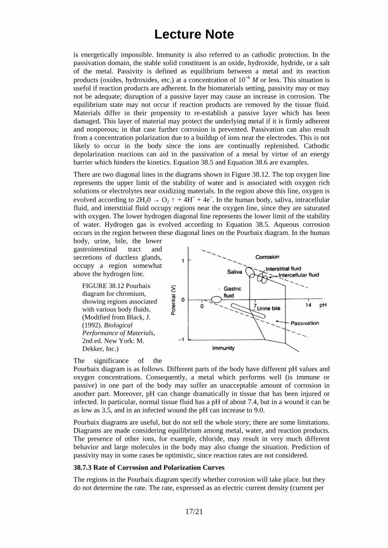
There are two diagonal lines in the diagrams shown in Figure 38.12. The top oxygen line represents the upper limit of the stability of water and is associated with oxygen rich solutions or electrolytes near oxidizing materials. In the region above this line, oxygen is evolved according to 2H20 → O2 ↑ + 4H+ + 4e−. In the human body, saliva, intracellular fluid, and interstitial fluid occupy regions near the oxygen line, since they are saturated with oxygen. The lower hydrogen diagonal line represents the lower limit of the stability of water. Hydrogen gas is evolved according to Equation 38.5. Aqueous corrosion occurs in the region between these diagonal lines on the Pourbaix diagram. In the human body, urine, bile, the lower gastrointestinal tract and secretions of ductless glands, occupy a region somewhat above the hydrogen line.
FIGURE 38.12 Pourbaix diagram for chromium, showing regions associated with various body fluids. (Modified from Black, J. (1992). Biological Performance of Materials, 2nd ed. New York: M. Dekker, Inc.)
The significance of the Pourbaix diagram is as follows. Different parts of the body have different pH values and oxygen concentrations. Consequently, a metal which performs well (is immune or passive) in one part of the body may suffer an unacceptable amount of corrosion in another part. Moreover, pH can change dramatically in tissue that has been injured or infected. In particular, normal tissue fluid has a pH of about 7.4, but in a wound it can be as low as 3.5, and in an infected wound the pH can increase to 9.0.
Pourbaix diagrams are useful, but do not tell the whole story; there are some limitations. Diagrams are made considering equilibrium among metal, water, and reaction products. The presence of other ions, for example, chloride, may result in very much different behavior and large molecules in the body may also change the situation. Prediction of passivity may in some cases be optimistic, since reaction rates are not considered.
38.7.3 Rate of Corrosion and Polarization Curves
The regions in the Pourbaix diagram specify whether corrosion will take place. but they do not determine the rate. The rate, expressed as an electric current density (current per
Lecture Note
18/21
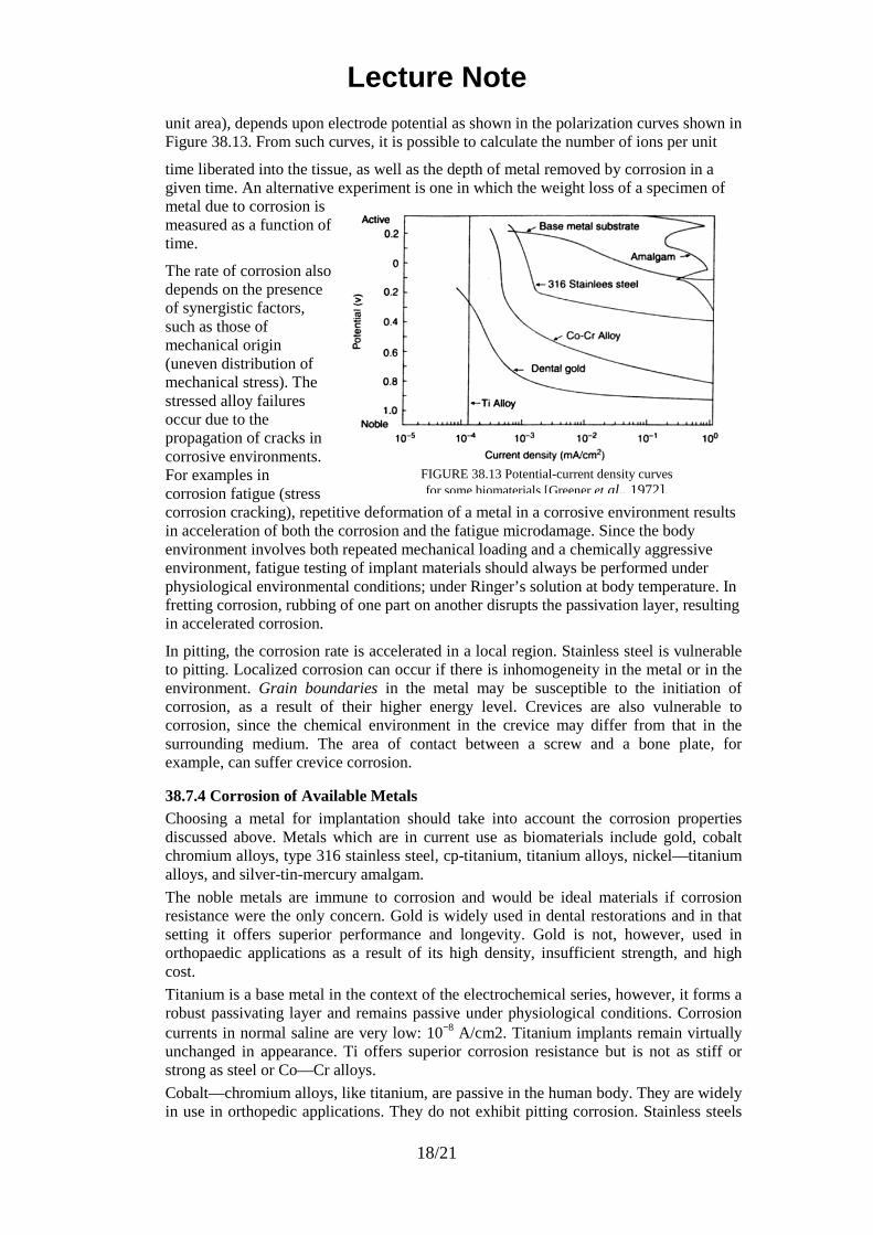
unit area), depends upon electrode potential as shown in the polarization curves shown in Figure 38.13. From such curves, it is possible to calculate the number of ions per unit
time liberated into the tissue, as well as the depth of metal removed by corrosion in a given time. An alternative experiment is one in which the weight loss of a specimen of metal due to corrosion is measured as a function of time.
The rate of corrosion also depends on the presence of synergistic factors, such as those of mechanical origin (uneven distribution of mechanical stress). The stressed alloy failures occur due to the propagation of cracks in corrosive environments. For examples in corrosion fatigue (stress corrosion cracking), repetitive deformation of a metal in a corrosive environment results in acceleration of both the corrosion and the fatigue microdamage. Since the body environment involves both repeated mechanical loading and a chemically aggressive environment, fatigue testing of implant materials should always be performed under physiological environmental conditions; under Ringer’s solution at body temperature. In fretting corrosion, rubbing of one part on another disrupts the passivation layer, resulting in accelerated corrosion.
In pitting, the corrosion rate is accelerated in a local region. Stainless steel is vulnerable to pitting. Localized corrosion can occur if there is inhomogeneity in the metal or in the environment. Grain boundaries in the metal may be susceptible to the initiation of corrosion, as a result of their higher energy level. Crevices are also vulnerable to corrosion, since the chemical environment in the crevice may differ from that in the surrounding medium. The area of contact between a screw and a bone plate, for example, can suffer crevice corrosion.
38.7.4 Corrosion of Available Metals Choosing a metal for implantation should take into account the corrosion properties discussed above. Metals which are in current use as biomaterials include gold, cobalt chromium alloys, type 316 stainless steel, cp-titanium, titanium alloys, nickel—titanium alloys, and silver-tin-mercury amalgam.
The noble metals are immune to corrosion and would be ideal materials if corrosion resistance were the only concern. Gold is widely used in dental restorations and in that setting it offers superior performance and longevity. Gold is not, however, used in orthopaedic applications as a result of its high density, insufficient strength, and high cost.
Titanium is a base metal in the context of the electrochemical series, however, it forms a robust passivating layer and remains passive under physiological conditions. Corrosion currents in normal saline are very low: 10−8 A/cm2. Titanium implants remain virtually unchanged in appearance. Ti offers superior corrosion resistance but is not as stiff or strong as steel or Co—Cr alloys.
Cobalt—chromium alloys, like titanium, are passive in the human body. They are widely in use in orthopedic applications. They do not exhibit pitting corrosion. Stainless steels
FIGURE 38.13 Potential-current density curves for some biomaterials [Greener et al., 1972].
Lecture Note
19/21
contain enough chromium to confer corrosion resistance by passivity. The passive layer is not as robust as in the case of titanium or the cobalt chrome alloys.
Only the most corrosion resistant of the stainless steels are suitable for implants. These are the austenitic types—316, 316L, and 317, which contain molybdenum. Even these types of stainless steel are vulnerable to pitting and to crevice corrosion around screws.
The phases of dental amalgam are passive at neutral pH, the transpassive potential for the γ2 phase is easily exceeded, due to interphase galvanic couples or potentials due to differential aeration under dental plaque. Amalgam. therefore, often corrodes and is the most active (corrosion prone) material used in dentistry.
Corrosion of an implant in the clinical setting can result in symptoms such as local pain and swelling in the region of the implant, with no evidence of infection; cracking or flaking of the implant as seen on x-ray films, and excretion of excess metal ions. At surgery, gray or black discoloration of the surrounding tissue may be seen and flakes of metal may be found in the tissue. Corrosion also plays a role in the mechanical failures of orthopaedic implants. Most of these failures are due to fatigue, and the presence of a saline environment certainly exacerbates fatigue. The extent to which corrosion influences fatigue in the body is not precisely known.
38.7.5 Stress Corrosion Cracking When an implant is subjected to stress, the corrosion process could be accelerated due to the mechanical energy. If the mechanical stress is repeated then fatigue stress corrosion takes place such as in the femoral stern of the hip joint and hip nails made of stainless steels [Dobbs and Scales, 1979; Sloter and Pichler, 1979]. However, other mechanisms of corrosion such as fretting may also be involved at point of contact such as in the counter-sink of the hip nail or bone fracture plate for the screws.
38.8 MANUFACTURING OF IMPLANTS
38.8.1 Stainless Steels The austenitic stainless steels work-harden very rapidly as shown in Figure 38.2 and therefore, cannot be cold-worked without intermediate heat treatments. The heat treatments should not induce, however, the formation of chromium carbide (CCr4) in the grain boundaries; this may cause corrosion. For the same reason, the austenitic stainless steel implants are not usually welded.
The distortion of components by the heat treatments can occur but this problem can be solved by controlling the uniformity of heating. Another undesirable effect of the heat treatment is the formation of surface oxide scales which have to be removed either chemically (acid) or mechanically (sand-blasting). After the scales are removed the surface of the component is polished to a mirror or mat finish. The surface is then cleaned, degreased, and passivated in nitric acid (ASTM Standard F86). The component is washed and cleaned again before packaging and sterilizing.
38.8.2 Co—Cr Alloys The CoCrMo alloy is particularly susceptible to work-hardening so that the normal fabrication procedure used with other metals cannot be employed. Instead, the alloy is cast by a lost wax (or investment casting) method which involves making a wax pattern of the desired component. The pattern is coated with a refractory material, first by a thin coating with a slurry (suspension of silica in ethyl silicate solution) followed by complete investing after drying (1) the wax is then melted out in a furnace (100—150°C), (2) the mold is heated to a high temperature burning out any traces of wax or gas forming materials, (3) molten alloy is poured with gravitational or centrifugal force, and (4) the mold is broken after cooled. The mold temperature is about 800—1000°C and the alloy is at 1350—1400°C.

Lecture Note
20/21
Controlling the mold temperature will have an effect on the grain size of the final cast; coarse ones are formed at higher temperatures which will decrease the strength. However, high processing temperature will result in larger carbide precipitates with greater distances between them resulting in a less brittle material. Again there is a complementary (trade off) relationship between strength and toughness.
38.8.3 Ti and Its Alloys Titanium is very reactive at high temperature and burns readily in the presence of oxygen. Therefore, it requires an inert atmosphere for high temperature processing or is processed by vacuum melting. Oxygen diffuses readily in titanium and the dissolved oxygen embrittles the metal. As a result, any hot working or forging operation should be carried out below 925°C. Machining at room temperature is not the solution to all the problems since the material also tends to gall or seize the cutting tools. Very sharp tools with slow speeds and large feeds are used to minimize this effect. Electrochemical machining is an attractive means.
DEFINING TERMS
Amalgam: An alloy obtained by mixing silver tin ailoy with mercury.
Anode: Positive electrode in an electrochemical cell.
Cathode: Negative electrode in an electrochemical cell.
Corrosion: Unwanted reaction of metal with environment. In a Pourbaix diagram, it is the region in which the metal ions are present at a concentration of more than 10−6 M.
Crevice corrosion: A form of localized corrosion in which concentration gradients around pre-existing crevices in the material drive corrosion processes.
Curie temperature: Transition temperature of a material from ferromagnetic to paramagnetic.
Galvanic corrosion: Dissolution of metal driven by macroscopic differences in electrochemical potential, usually as a result of dissimilar metals in proximity.
Galvanic series: Table of electrochemical potentials (voltage) associated with the ionization of metal atoms. These are called Nernst potentials.
Hyperthermia : Application of high enough thermal energy (heat) to suppress the cancerous cell activities. Above 41.5°C (but below 60°C) is needed to have any effect.
Immunity : Resistance to corrosion by an energetic barrier. In a Pourbaix diagram, it is the region in which the metal is in equilibrium with its ions at a concentration of less than 10—6 M. Noble metals resist corrosion by immunity.
Martensite: A metastable structure formed by quenching of austenite (g) structure in alloys such as steel and Ti alloys. They are brittle and hard, and therefore, are further treated with heat to make tougher.
Nernst potential: Standard electrochemical potential measured with respect to a standard hydrogen electrode.
Noble: Type of metal with a positive standard electrochemical potential.
Passivation: Production of corrosion resistance by a surface layer of reaction products (Normally oxide layer which is impervious to gas and water.)
Passivity: Resistance to corrosion by a surface layer of reaction products. In a Pourbaix diagram, it is the region in which the metal is in equilibrium with its reaction products at a concentration of less than 10−6 molar.

Lecture Note
21/21
Pitting : A form of localized corrosion in which pits form on the metal surface.
Pourbaix diagram: Plot of electrical potential vs. pH for a material in which the regions of corrosion, passivity, and immunity are identified.
Shape memory effect (SME): Thermoelastic behavior of some alloys which can revert back to their original shape when the temperature is greater than the phase transformation temperature of the alloy.
Superelasticity: Minimal stress increase beyond the initial strain region resulting in very low modulus in the region for some shape memory alloys.