2 Dr. hanan jafar - JU Medicine€¦ · main lipids are cholesterol and phospholipids. Lipid domain...
15
22 Hanan Jafar (2017) Dr. hanan jafar 2
Transcript of 2 Dr. hanan jafar - JU Medicine€¦ · main lipids are cholesterol and phospholipids. Lipid domain...

22
Hanan Jafar (2017)
Dr. hanan jafar
2

22
Hanan Jafar (2017)
Hematopoietic stem cell transplantation
This is a therapeutic procedure in which normal hematopoietic stem cells
(CD34+) from an appropriate donor are transferred to the patient having
defective or diseased marrow to re-constitute normal hematopoiesis.
Types of hematopoietic stem cell transplantation (HSCT)
1. Allogenic HSCT
Hematopoietic stem cells are obtained from a donor following HLA (human
leukocyte antigen) typing.
2. Autologous HSCT
Stem cells previously collected from the recipient are infused back to the
same patient.
Sources of hematopoietic stem cells
1. Peripheral blood stem cells mobilization and harvesting
Although HSCs are known to circulate in peripheral blood, their very
small number preclude their use for transplantation. Administration of
recombinant hematopoietic growth factor (mainly G-CSF) mobilises
HSCs from bone marrow and enhances their subsequent yield from
peripheral blood. The yield can be evaluated by mononuclear cell count
and CD34+ cells count.
2. Bone marrow harvesting
Up to 100 ml of bone marrow can be aspirated from the donor from
several sites in the iliac crest. Aspirated marrow is harvested in a bag (or
tubes) containing acid citrate dextrose solution.
3. Umbilical cord blood
Umbilical cord blood is an important and readily available source of
hematopoietic stem cells (Stem cell banks). However the number of stem
cells in the cord blood is limited, and therefore, most of the successful
cord blood transplantations have been done in small children.

22
Hanan Jafar (2017)
Normal Bone Marrow Histology (In lab, refer to book chapter)
Mesenchymal Stem (Stromal) Cells (MSCs)
Mesenchymal stromal cells are plastic adherent multipotent stem cells that
express CD105, CD90, and CD73 but that lack the expression of pan-leukocyte
(CD45), endothelial, or primitive hematopoietic (CD34), monocytic (CD14 or
CD11b), or B cell (CD79a or CD19) markers, as well as lack the expression of
HLA class II (HLA-DR) surface antigen. These cells exhibit a fibroblast-like
(spindle) shaped morphology and tri-lineage differentiation into osteoblasts,
adipocytes, and chondroblasts. They were first isolated from bone marrow, but
have been also identified from multiple sources such as adipose tissue, dental
tissues, cord tissue, and others.

22
Hanan Jafar (2017)
Erythrocytes (Red Blood Cells)
The erythrocytes (which are normally present in the millions) have the
important function of transport O2 and CO2 throughout the body. The vehicle
for this transportation is a protein-iron pigment called hemoglobin.
Traditionally, an erythrocyte was viewed as "an outer plasma membrane
enclosing hemoglobin and a limited number of enzymes necessary for
maintenance of plasma membrane integrity and gas transport function".
However, it has been recently shown that during their lifetime, like all other cell
types, red blood cells (RBCs) release both exosomes and plasma membrane
derived EVs (ectosomes). RBC exosomes are formed only during the
development of RBCs in bone marrow, and are released following the fusion of
microvesicular bodies (MVB) with the plasma membrane. On the other hand,
RBC EVs are generated during normal aging of RBCs in circulation by budding
of the plasma membrane due to complement-mediated calcium influx, followed
by vesicle shedding. Interestingly, researchers demonstrated that exosomes from
red blood cell units bind to monocytes and induce proinflammatory cytokines,
boosting T-cell responses in vitro. (Article).
Transmission electron microscopy (TEM) of RBCs showing regular membrane (C) and
ectosome release (D)
RBCs normally have a biconcave, disc-like shape, which means their centers are
thinner than their edges. Red blood cells are the only human cells without
nuclei. Typically, an erythrocyte's diameter is 7-8 um. The biconcave shape
provides around 30% more surface area, compared to spherical form. The larger
surface area greatly enhances gas exchange. Additionally, this shape allows
22
Hanan Jafar (2017)
RBCs to "squeeze" themselves through the smallest capillaries (around 3 um in
diameter).
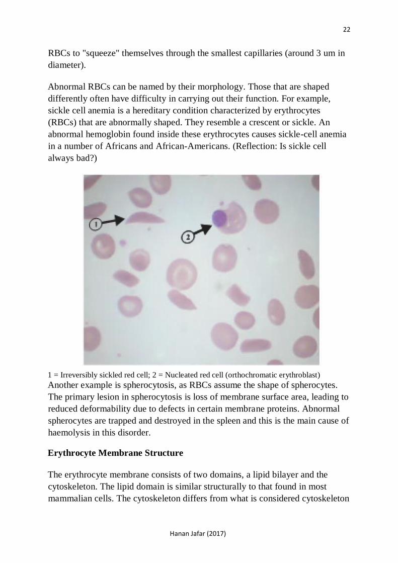
Abnormal RBCs can be named by their morphology. Those that are shaped
differently often have difficulty in carrying out their function. For example,
sickle cell anemia is a hereditary condition characterized by erythrocytes
(RBCs) that are abnormally shaped. They resemble a crescent or sickle. An
abnormal hemoglobin found inside these erythrocytes causes sickle-cell anemia
in a number of Africans and African-Americans. (Reflection: Is sickle cell
always bad?)
1 = Irreversibly sickled red cell; 2 = Nucleated red cell (orthochromatic erythroblast) Another example is spherocytosis, as RBCs assume the shape of spherocytes.
The primary lesion in spherocytosis is loss of membrane surface area, leading to
reduced deformability due to defects in certain membrane proteins. Abnormal
spherocytes are trapped and destroyed in the spleen and this is the main cause of
haemolysis in this disorder.
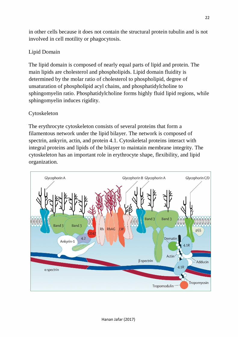
Erythrocyte Membrane Structure
The erythrocyte membrane consists of two domains, a lipid bilayer and the
cytoskeleton. The lipid domain is similar structurally to that found in most
mammalian cells. The cytoskeleton differs from what is considered cytoskeleton
22
Hanan Jafar (2017)
in other cells because it does not contain the structural protein tubulin and is not
involved in cell motility or phagocytosis.
Lipid Domain
The lipid domain is composed of nearly equal parts of lipid and protein. The
main lipids are cholesterol and phospholipids. Lipid domain fluidity is
determined by the molar ratio of cholesterol to phospholipid, degree of
unsaturation of phospholipid acyl chains, and phosphatidylcholine to
sphingomyelin ratio. Phosphatidylcholine forms highly fluid lipid regions, while
sphingomyelin induces rigidity.
Cytoskeleton
The erythrocyte cytoskeleton consists of several proteins that form a
filamentous network under the lipid bilayer. The network is composed of
spectrin, ankyrin, actin, and protein 4.1. Cytoskeletal proteins interact with
integral proteins and lipids of the bilayer to maintain membrane integrity. The
cytoskeleton has an important role in erythrocyte shape, flexibility, and lipid
organization.

22
Hanan Jafar (2017)
Hemoglobin
Hemoglobin consists of the protein globin and heme pigment. Globin Consists
of two and two subunits. Each subunit binds to a heme group. Each heme
group bears an atom of iron, which binds reversibly with one molecule of
oxygen. So, each hemoglobin molecule carries four molecules of oxygen. On
the other hand, carbon monoxide competes with oxygen for heme binding with
a much higher affinity. (Smoking and Hct)
Erythropoiesis
It is the pathway through which an erythrocyte matures from a hemocytoblast
(hematopoietic stem cell) into a full-blown erythrocyte. The rate of production
is very rapid (estimated at several million new RBCs per second) and a major
regulating factor is oxygen. If the body is in a state of hypoxia or lack of
oxygen, the kidneys produce a hormone called erythropoietin, which stimulates
the red bone marrow to increase the rate of RBC production. This will occur
following hemorrhage, or if a person stays for a time at a higher altitude. As a
result of the action of erythropoietin, more RBCs will be available to carry
oxygen and correct the hypoxic state.

22
Hanan Jafar (2017)
Erythrocyte differentiation takes place in several stages over a period of 1 week.
They all take place within the bone marrow (in the adult). Within the bone
marrow, erythroid progenitors are found in the form of "islands", called
erythroid colonies. An erythroid colony is composed of erythroblasts
surrounding a central macrophage. The more immature precursors are present
close to the macrophage and maturing forms are towards the periphery. The
macrophage has dendritic processes which extend between erythroid
progenitors, support them, and supply iron for hemoglobin synthesis.
The figure below shows Micrographs of erythroblastic islands. (A)
Transmission electron micrograph of an erythroblastic island isolated from bone
marrow. Note the extensive cell-cell contact. (B) Scanning electron micrograph
of an isolated erythroblastic island. The inset shows an optical microscopic
image of the same structure. Note the presence of an enucleating erythroblast
(➔) and a multilobulated reticulocyte (➤). (C) Confocal immunofluorescence
image of an island reconstituted from freshly harvested mouse bone marrow
cells stained with erythroid-specific marker (red), macrophage marker (green)
and DNAprobe (blue). Central macrophage is indicated by an arrow and a
multilobulated reticulocyte by an arrowhead.

22
Hanan Jafar (2017)

Hanan Jafar (2017)
23
With each stage of erythrocyte differentiation, cell size and nuclear size become
smaller and chromatin clumping increases. Color of cytoplasm changes from
basophilic to orange-red due to increased accumulation of hemoglobin.
The earliest morphologically identifiable erythroid cell in the bone marrow is
the proerythroblast (pronormoblast), a large (15-20 um) cell with a fine,
uniform chromatin pattern, one or more nucleoli, and dark blue cytoplasm. The
next cell in the maturation process is the basophilic (early) normoblast. This
cell is smaller in size (12-16 um) and has a coarser nuclear chromatin with
barely visible nucleoli. The cytoplasm is deeply basophilic.
The more differentiated erythroid cell is the polychromatic (intermediate)
normoblast (size 12-15 um). The nuclear size is smaller and the chromatin
becomes clumped. Polychromasia of cytoplasm results from admixture of blue
RNA and pink hemoglobin. This is the last erythroid precursor capable of
mitotic division. The orthochromatic (late) normoblast is 8 to 12 um in size.
The nucleus is small, dense and pyknotic and commonly eccentrically-located.
The cytoplasm stains mostly pink due to hemoglobinisation. It is called
orthochromatic because cytoplasmic staining is largely similar to that of
erythrocytes.
When the late normoblast ejects its nucleus, it becomes a reticulocyte, so called
because of a reticular (mesh-like) network of ribosomal RNA visible under a
microscope with certain stains such as new methylene blue and Romanowsky
stains. The reticulocyte is released into the blood stream, where it then matures
1-2 days later into an erythrocyte. Reticulocytes make up 1% of RBCs in
peripheral blood. Large numbers of reticulocytes or normoblasts in the
circulating blood mean that the number of mature RBCs is not sufficient to
carry the oxygen needed by the body. Such situations include hemorrhage, or
when mature RBCs have been destroyed, as in Rh disease of the newborn, and
malaria.
In summary, erythrpoeisis stages are as follows:
1. Hemocytoblast, or hematopoietic stem cell, which is a multipotent stem
cell.
2. Common myeloid progenitor, a multipotent stem cell.
3. Megakaryocyte and erythroid precursor, a committed precursor cell
4. Erythroid progenitor cell, a unipotent stem cell or lineage committed cell
5. Pronormoblast, or proerythroblast

22
Hanan Jafar (2017)
6. Basophilic or early normoblast, also called an erythroblast.
7. Polychromatophilic or intermediate normoblast
8. Orthochromatic or late normoblast
9. Reticulocyte 23
The maturation of red blood cells requires many nutrients. Protein and iron are
necessary for the synthesis of hemoglobin and become part of hemoglobin
molecules. The vitamins folic acid and B12 are required for DNA synthesis in

Hanan Jafar (2017)
the stem cells of the red bone marrow. As these cells undergo mitosis they must
continually produce new sets of chromosomes. Vitamin B12 is also called the
extrinsic factor because its source is external, our food. Parietal cells of the
stomach lining produce the intrinsic factor, a chemical that combines with the
vitamin B12 in food to prevent its digestion and promote its absorption in the
small intestines. A deficiency of either vitamin B12 or in the intrinsic factor
results in a type of anemia called pernicious anemia.
Life span
Red blood cells live for approximately 120 days. As they reach this age they
become fragile and are removed from circulation by cells of the tissue
macrophage system (formerly called the reticuloendothelial or RE system).
The organs that contain macrophages are the liver, spleen, and red bone
marrow. The old RBCs are phagocytized and digested by macrophages, and the
iron they contained is put into the blood to be returned to the red bone marrow
to be used for the synthesis of new hemoglobin. If not needed immediately for
this purpose, excess iron is stored in the liver. The iron of RBCs is actually
recycled over and over again.
Another part of the hemoglobin molecule is the heme portion, which cannot be
recycled and is a waste product. The heme is converted to bilirubin by
macrophages. The liver removes bilirubin from circulation and excretes it into
bile; bilirubin is called a bile pigment. Bile is secreted by the liver into the
duodenum and passes through the small intestine and colon, so bilirubin is
eliminated in feces, and gives feces their characteristic brown color. If bilirubin
is not excreted properly, perhaps because of liver disease such as hepatitis, it
remains in the blood. This may cause jaundice, a condition in which the whites
of the eyes appear yellow. This yellow color may also be seen in the skin of
light-skinned people.

22
Hanan Jafar (2017)

22
Hanan Jafar (2017)
Blood Groups
Human blood is divided into four major different types: A, B, Ab, and O. The
differences are due to antigens present on the surface of the blood cells.
Antigens are substances that produce an immune reaction by their nature of
being perceived as foreign to the body. In response, the body produces
substances called antibodies that nullify or neutralize the antigens. In blood,
these antigens are called agglutinogens because their presence can cause the
blood to clot.
The antibody is termed an agglutinin. For example, type A blood has A
antigen, type B has B antigen, type AB has both A and B antigens, and type O
has neither A nor B antigens. Following the logic of each of these
antigenantibody reactions, an individual with type AB blood is a universal
recipient, and an individual with type O blood is a universal donor.
The Rh factor is another antigen (often called D) that may be present on RBCs.
People whose RBCs have the Rh antigen are Rh positive, those without the
antigen are Rh negative. Rh negative people do not have natural antibodies to
the Rh antigen, and for them this antigen is foreign. If an Rh negative person
receives Rh positive blood by mistake, antibodies will be formed just as they
would be to bacteria or viruses.

22
Hanan Jafar (2017)
The Rh factor is important in pregnancy because a mismatch between the fetus
and the mother can cause erythroblastosis fetalis, or hemolytic disease of the
newborn. In this disorder, a mother with a negative Rh factor will develop
antibodies to an RH + fetus during the first pregnancy. If another pregnancy
occurs with an Rh + fetus, the antibodies will destroy the fetal blood cells.
Recent Advances
1. Bacterial glycosidases for the production of universal red blood cells
2. Generation of red blood cells from human embryonic/induced
pluripotent stem cells for blood transfusion