![Coronectomy of Impacted Mandibular Third Molars - Method ... · partial odontectomy is proposed by Ecuyer and Debien in 1984 [20]. Coronectomy has been defined as a method of removing](https://static.fdocuments.us/doc/165x107/5fa0f0dec8ee260c8e61afc9/coronectomy-of-impacted-mandibular-third-molars-method-partial-odontectomy.jpg)
2- A Preferable Technique for Protecting the Inferior Alveolar Nerve Coronectomy
5
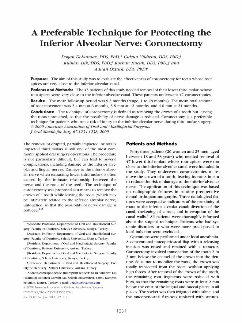
J Oral Maxillofac Surg 67:1234-1238, 2009 A Preferable Technique for Protecting the Inferior Alveolar Nerve: Coronectomy Dogan Dolanmaz, DDS, PhD,* Gulsun Yildirim, DDS, PhD,† Kubilay Isik, DDS, PhD,‡ Korhan Kucuk, DDS, PhD,§ and Adnan Ozturk, DDS, PhD¶ Purpose: The aim of this study was to evaluate the effectiveness of coronectomy for teeth whose root apices are very close to the inferior alveolar canal. Patients and Methods: The 43 patients of this study needed removal of their lower third molar, whose root apices were very close to the inferior alveolar canal. These patients underwent 47 coronectomies. Results: The mean follow-up period was 9.3 months (range, 1 to 48 months). The mean total amount of root movement was 3.4 mm at 6 months, 3.8 mm at 12 months, and 4.0 mm at 24 months. Conclusions: The technique of coronectomy is defined as removing the crown of a tooth but leaving the roots untouched, so that the possibility of nerve damage is reduced. Coronectomy is a preferable technique for patients who run a risk of injury to the inferior alveolar nerve during third molar surgery. © 2009 American Association of Oral and Maxillofacial Surgeons J Oral Maxillofac Surg 67:1234-1238, 2009 The removal of erupted, partially impacted, or totally impacted third molars is still one of the most com- monly applied oral surgery operations. The procedure is not particularly difficult, but can lead to several complications, including damage to the inferior alve- olar and lingual nerves. Damage to the inferior alveo- lar nerve when extracting lower third molars is often caused by the intimate relationship between the nerve and the roots of the teeth. The technique of coronectomy was proposed as a means to remove the crown of a tooth while leaving the roots (which may be intimately related to the inferior alveolar nerve) untouched, so that the possibility of nerve damage is reduced. 1,2 Patients and Methods Forty-three patients (20 women and 23 men, aged between 18 and 38 years) who needed removal of 47 lower third molars whose root apices were too close to the inferior alveolar canal were included in the study. They underwent coronectomies to re- move the crown of a tooth, leaving its roots in situ to reduce the risk of damage to the inferior alveolar nerve. The application of this technique was based on radiographic features in routine preoperative dental orthopantomographs. Three radiological fea- tures were accepted as indicators of the proximity of roots to the inferior alveolar canal: diversion of the canal, darkening of a root, and interruption of the canal walls. 3 All patients were thoroughly informed about the surgical technique. Patients who had sys- temic disorders or who were more predisposed to local infection were excluded. Operations were performed under local anesthesia. A conventional mucoperiosteal flap with a releasing incision was raised and retained with a retractor. Coronectomy involved transsection of the tooth 2 to 3 mm below the enamel of the crown into the den- tine. So as not to mobilize the roots, the crown was totally transected from the roots, without applying high forces. After removal of the crown of the tooth, the remaining root fragments were reduced with burs, so that the remaining roots were at least 2 mm below the crest of the lingual and buccal plates in all places. The socket was then irrigated with saline, and the mucoperiosteal flap was replaced with sutures. *Associate Professor, Department of Oral and Maxillofacial Sur- gery, Faculty of Dentistry, Selcuk University, Konya, Turkey. †Assistant Professor, Department of Oral and Maxillofacial Sur- gery, Faculty of Dentistry, Selcuk University, Konya, Turkey. ‡Resident, Department of Oral and Maxillofacial Surgery, Faculty of Dentistry, Baskent University, Ankara, Turkey. §Resident, Department of Oral and Maxillofacial Surgery, Faculty of Dentistry, Selcuk University, Konya, Turkey. ¶Professor, Department of Oral and Maxillofacial Surgery, Fac- ulty of Dentistry, Ankara University, Ankara, Turkey. Address correspondence and reprint requests to Dr Yildirim: Dis ¸ Hekimlig ˘i Fakültesi Cerrahi AD, Selçuk Universitesi, 42080 Kampüs Selçuklu, Konya, Turkey; e-mail: [email protected] © 2009 American Association of Oral and Maxillofacial Surgeons 0278-2391/09/6706-0013$36.00/0 doi:10.1016/j.joms.2008.12.031 1234
-
Upload
lbferreira -
Category
Documents
-
view
293 -
download
1
Transcript of 2- A Preferable Technique for Protecting the Inferior Alveolar Nerve Coronectomy
J6
Timicolcnccbur
g
g
o
o
u
H
S
©
0
d
Oral Maxillofac Surg7:1234-1238, 2009
A Preferable Technique for Protecting theInferior Alveolar Nerve: Coronectomy
Dogan Dolanmaz, DDS, PhD,* Gulsun Yildirim, DDS, PhD,†
Kubilay Isik, DDS, PhD,‡ Korhan Kucuk, DDS, PhD,§ and
Adnan Ozturk, DDS, PhD¶
Purpose: The aim of this study was to evaluate the effectiveness of coronectomy for teeth whose rootapices are very close to the inferior alveolar canal.
Patients and Methods: The 43 patients of this study needed removal of their lower third molar, whoseroot apices were very close to the inferior alveolar canal. These patients underwent 47 coronectomies.
Results: The mean follow-up period was 9.3 months (range, 1 to 48 months). The mean total amountof root movement was 3.4 mm at 6 months, 3.8 mm at 12 months, and 4.0 mm at 24 months.
Conclusions: The technique of coronectomy is defined as removing the crown of a tooth but leavingthe roots untouched, so that the possibility of nerve damage is reduced. Coronectomy is a preferabletechnique for patients who run a risk of injury to the inferior alveolar nerve during third molar surgery.© 2009 American Association of Oral and Maxillofacial Surgeons
J Oral Maxillofac Surg 67:1234-1238, 2009P
b4ctmtnodtrccatl
AiC3tthtbbp
he removal of erupted, partially impacted, or totallympacted third molars is still one of the most com-
only applied oral surgery operations. The procedures not particularly difficult, but can lead to severalomplications, including damage to the inferior alve-lar and lingual nerves. Damage to the inferior alveo-
ar nerve when extracting lower third molars is oftenaused by the intimate relationship between theerve and the roots of the teeth. The technique oforonectomy was proposed as a means to remove therown of a tooth while leaving the roots (which maye intimately related to the inferior alveolar nerve)ntouched, so that the possibility of nerve damage iseduced.1,2
*Associate Professor, Department of Oral and Maxillofacial Sur-
ery, Faculty of Dentistry, Selcuk University, Konya, Turkey.
†Assistant Professor, Department of Oral and Maxillofacial Sur-
ery, Faculty of Dentistry, Selcuk University, Konya, Turkey.
‡Resident, Department of Oral and Maxillofacial Surgery, Faculty
f Dentistry, Baskent University, Ankara, Turkey.
§Resident, Department of Oral and Maxillofacial Surgery, Faculty
f Dentistry, Selcuk University, Konya, Turkey.
¶Professor, Department of Oral and Maxillofacial Surgery, Fac-
lty of Dentistry, Ankara University, Ankara, Turkey.
Address correspondence and reprint requests to Dr Yildirim: Dis
ekimligi Fakültesi Cerrahi AD, Selçuk Universitesi, 42080 Kampüs
elçuklu, Konya, Turkey; e-mail: [email protected]
2009 American Association of Oral and Maxillofacial Surgeons
278-2391/09/6706-0013$36.00/0
toi:10.1016/j.joms.2008.12.031
1234
atients and Methods
Forty-three patients (20 women and 23 men, agedetween 18 and 38 years) who needed removal of7 lower third molars whose root apices were toolose to the inferior alveolar canal were included inhe study. They underwent coronectomies to re-ove the crown of a tooth, leaving its roots in situ
o reduce the risk of damage to the inferior alveolarerve. The application of this technique was basedn radiographic features in routine preoperativeental orthopantomographs. Three radiological fea-ures were accepted as indicators of the proximity ofoots to the inferior alveolar canal: diversion of theanal, darkening of a root, and interruption of theanal walls.3 All patients were thoroughly informedbout the surgical technique. Patients who had sys-emic disorders or who were more predisposed toocal infection were excluded.
Operations were performed under local anesthesia.conventional mucoperiosteal flap with a releasing
ncision was raised and retained with a retractor.oronectomy involved transsection of the tooth 2 tomm below the enamel of the crown into the den-
ine. So as not to mobilize the roots, the crown wasotally transected from the roots, without applyingigh forces. After removal of the crown of the tooth,he remaining root fragments were reduced withurs, so that the remaining roots were at least 2 mmelow the crest of the lingual and buccal plates in alllaces. The socket was then irrigated with saline, and
he mucoperiosteal flap was replaced with sutures.
Agafamfsplfpdtwa
ctT
wett
R
1ompaabmam0prgmar5
D
actatppcct
Fls
DN
D
DOLANMAZ ET AL 1235
ntibiotics were prescribed postoperatively. Radio-raphs were taken preoperatively and immediatelyfter an operation. All patients were invited to returnor appointments at 6, 12, and 24 months for clinicalnd radiographic assessment of the retained root frag-ents. The root remnants were clinically examined
or infection, and the amount of movement was mea-ured. For this purpose, a line parallel to the occlusallane was extended to the ramus, and a longitudinal
ine in the middle of the root remnant was drawnrom the tooth apex (Fig 1). If the tooth had 2 com-letely separate apices, the longitudinal line wasrawn in the middle of them. The distance betweenhe root apex and the intersection point of the 2 linesas measured at 6, 12, and 24 months, to obtain the
mount of movement of the root remnants.In one patient (female, 25 years old), the crown
ould not be totally separated from the roots, and theooth was displaced as we tried to remove the crown.hus, the coronectomy was abandoned, and the tooth
IGURE 1. View of measuring method of root movement. Dashedines represent the occlusal plane and the arrow shows the mea-ured distance.
olanmaz et al. Coronectomy for Protection of Inferior Alveolarerve. J Oral Maxillofac Surg 2009.
0
1
2
3
4
5
6
7
8
9
10
2.0 - 2.4 mm 2.5 - 2.9 mm 3.0 - 3.4
Num
ber o
f the
teet
h
FIGURE 2. Distribution of root moveme
olanmaz et al. Coronectomy for Protection of Inferior Alveolar Nerve
as extracted in the usual way. The patient experi-nced moderate paresthesia for 5 months. This pa-ient was excluded from the study, and her parame-ers are not included in the above-mentioned values.
esults
The mean follow-up period was 9.3 months (range,to 48 months). The mean total amount of movementf the root remnants of 47 teeth was found to be 3.4m in 6 months (range, 2.0 to 4.8 mm) (Fig 2). Nineatients could not return for their appointments at 12nd 24 months for various reasons, and thus themount of movement of the root remnants could note calculated for those 9 patients at 12 and 24onths. For the remaining 38 patients, the mean total
mount of movement at 12 and 24 months was 3.8m (range, 0.1 to 0.9 mm) (Fig 3) and 4.0 mm (range,
.0 to 0.3 mm) (Fig 4), respectively. None of theatients reported any problems associated with theoot fragments. In all cases, an examination of radio-raphs taken at 6 months revealed that the root frag-ents showed various amounts of movement farther
way from the inferior alveolar nerve. None of theetained roots required a second operation (FigsA,B).
iscussion
Removal of third molar teeth is a common practice,nd inferior alveolar nerve damage is a well-knownomplication of this procedure, particularly for teethhat have an intimate relationship with the inferiorlveolar canal. Risk factors for nerve injury are knowno include radiographic proximity, the surgeon’s ex-erience, surgical procedures, the patient’s age, andreexisting disease.4 Although the risk of nerve injuryan be reduced with careful surgical technique, itannot be absolutely avoided. Since the beginnings ofhird molar surgery, surgeons have been aware of the
3.5 - 3.9 mm 4.0 - 4.4 mm 4.5 - 4.8 mm
ing postoperative month 6 for 47 teeth.
mm
nts dur
. J Oral Maxillofac Surg 2009.

rtpmiratt
rrctwdtcnTbmhaosn
tcitrl
D Nerve
Fm
DN
Fom
1236 CORONECTOMY FOR PROTECTION OF INFERIOR ALVEOLAR NERVE
isk of inferior alveolar and lingual nerve injury.4 Al-hough subject to disagreement, the incidence of tem-orary injury to the inferior alveolar nerve after thirdolar surgery is reported to be up to 8%, and the
ncidence of permanent injury, to 3.6%.5,6 However, aate of 5% is usually accepted.7-10 Techniques thatppear to reduce these risks naturally gain the atten-ion of patients and surgeons contemplating electivehird molar removal.
The technique of coronectomy, or deliberate vitaloot retention, avoids the nerve canal by ensuringetention of the roots when they are close to theanal, as estimated using radiographs.11 It is knownhat broken fragments of vital teeth generally healithout complications.2,12 Coronectomy is a proce-ure based on this idea. If one third of an uninfectedooth can be left in its socket in certain situations, thisan be valid for a “larger” part of a similar tooth; butot all third molar teeth are suitable for coronectomy.eeth with acute infection and mobile teeth shoulde excluded, because root remnants of those teethay act like foreign bodies. In addition, teeth that areorizontally impacted along the course of the inferiorlveolar canal may be unsuitable, because sectioningf a tooth could endanger the nerve.13 We tried toeparate the crown completely, so that no high forceseeded to separate the crown and the root, because
0
1
2
3
4
5
6
7
0.1 mm 0.2 mm 0.3 mm 0.4 mm
Num
ber o
f the
teet
h
FIGURE 3. Distribution of root movemen
olanmaz et al. Coronectomy for Protection of Inferior Alveolar
0
2
4
6
8
10
12
14
16
0.0 mm 0.1 mm 0.2 mm 0.3 mm
Num
ber o
f the
teet
h
IGURE 4. Distribution of root movements during postoperativeonth 24 for 38 teeth.
olanmaz et al. Coronectomy for Protection of Inferior Alveolarerve. J Oral Maxillofac Surg 2009.
DN
hose forces could also mobilize the roots. This con-ern is shared by other authors.13 However, if a roots broken during extraction of a regular uninfectedooth, and if this root fragment is too difficult toemove or somehow exhibits a risk, it can safely beeft.2,12 Because these “mobilized” root fragments
0.6 mm 0.7 mm 0.8 mm 0.9 mm
g postoperative month 12 for 38 teeth.
. J Oral Maxillofac Surg 2009.
IGURE 5. A, Preoperative radiographic view of case 1 (croppedrthopantomograph). B, Radiographic view of case 1 after 24onths (cropped orthopantomograph).
0.5 mm
ts durin
olanmaz et al. Coronectomy for Protection of Inferior Alveolarerve. J Oral Maxillofac Surg 2009.

mttc
ntAncrfnaoqagtcc
loieam
tr
asbdi
mmmtwr
cbatlttrtoo
nnttt
Wwwn
tarRncgaIalrfiwttll
R
1
DOLANMAZ ET AL 1237
ostly heal uneventfully, it may not be essential toransect the crown totally during intentional root re-ention, to decrease the risk of perforating the lingualortex and damaging the lingual nerve.When making a decision about a coronectomy, it is
ecessary to determine the correct relationship be-ween the root apices and the inferior alveolar canal.t this point, different radiological imaging tech-iques can be used. Dental computerized tomographyan give very precise information about the root-canalelationship, and would probably be the best choiceor this purpose, but it is also rather expensive andot available at every clinic. Plain dental radiographsre more advantageous, and seem more suitable. Theyffer an opportunity to alter the extraction techni-ue to minimize risk to the nerve. In our study, wessessed lower third molars with a panoramic radio-raph, and then made the decision for a coronec-omy. The paresthesia that occurred in our “unsuc-essful coronectomy” supports the validity of ourriteria.Although some authors used preoperative prophy-
actic antibiotics,13 we did not prescribe any pre-perative antibiotics, and did not encounter any
nfections in our study group. We prescribed postop-rative antibiotics, but this is not unique to this oper-tion: we routinely prescribe antibiotics after thirdolar surgery.There is no need to treat the exposed pulp of the
ooth in a coronectomy. Animal studies show that rootsemain vital with minimal degenerative changes.14,15
The technique of leaving the retained root fragmentt least 2 to 3 mm inferior to the crest of the boneeems appropriate, and does appear to encourageone formation over the retained root fragment. Thisistance of 2 to 3 mm was validated in animal stud-
es.15
The movement of root remnants reached its maxi-um in the first postoperative 6 months (mean, 3.4m). The amount of root movement decreased to 0.4m between 6 and 12 months, and to 0.2 mm be-
ween 12 and 24 months. Probably this slowdownas attributable to new bone formation above the
oots over time.In agreement with Pogrel et al,13 we do not advo-
ate seeing the patient after 6 months unless he or sheecomes symptomatic. On the other hand, some16
ssert that a follow-up period of 25 months is requiredo evaluate the incidence of nerve injury, but not ofate eruption, which can occur up to 10 years afterhe initial operation. A longer review period mayherefore show that a proportion of these retainedoots do eventually erupt, and may cause a late infec-ion or require removal.11 One possible disadvantagef a coronectomy is the possible need for a second
peration, but in the procedure’s favor, root rem-ants can then be removed easily without risk to theerve.3 In our series, all root remnants showed migra-ion away from the inferior alveolar nerve, and hencehe potential of nerve injury during a second opera-ion is reduced.
We did not establish a “control group” in this study.hen the roots were too close to the canal, and ife believed that a routine impacted-tooth operationould jeopardize the inferior alveolar nerve, it wouldot have been ethical to perform a coronectomy.This study dealt with teeth that were very close to
he inferior alveolar nerve, a situation that can createhigh risk of paresthesia after their removal. Ethical
easons made the use of a control group impossible.oot movement was observed in all teeth after coro-ectomy, and all roots moved away from the inferioranal. We conclude that a coronectomy can be sug-ested for teeth that are very close to the inferiorlveolar nerve, with the risk of a secondary operation.f a second operation is needed for the remnant rootsfter a coronectomy, the roots can be removed with aow risk of paresthesia, because the roots would haveeceded from the inferior alveolar canal. Based on thendings in our patients, we agree with others3,11,13,17
ho described the coronectomy as an acceptableechnique, and we conclude that it is a valuable op-ion in selected cases. We also plan to examine theong-term clinical outcomes of coronectomies in aarger sample.
eferences1. Freedman GL: Intentional partial odontectomy: Review of
cases. J Oral Maxillofac Surg 55:524, 19972. Knutsson K, Lysell L, Rohlin M: Postoperative status after par-
tial removal of the mandibular third molar. Swed Dent J 13:15,1989
3. Drage NA, Renton T: Inferior alveolar nerve injury related tomandibular third molar surgery: An unusual case presentation.Oral Surg Oral Med Oral Pathol Oral Radiol Endod 93:358, 2002
4. Assael LA: Coronectomy: A time to ponder or a time to act?J Oral Maxillofac Surg 62:1445, 2004
5. Black CG: Sensory impairment following lower third molarsurgery: A prospective study in New Zealand. N Z Dent J 93:68,1997
6. Smith AC, Barry SE, Chiong AY, et al: Inferior alveolar nervedamage following removal of mandibular third molar teeth. Aprospective study using panoramic radiography. Aust Dent J42:149, 1997
7. Kipp DP, Goldstein BH, Weiss WW Jr: Dysesthesia after man-dibular third molar surgery: A retrospective study and analysisof 1377 surgical procedures. J Am Dent Assoc 100:185, 1980
8. Carmichael FA, McGowan DA: Incidence of nerve damagefollowing third molar removal: A West of Scotland Oral SurgeryResearch Group study. Br J Oral Maxillofac Surg 30:78, 1992
9. Sisk AL, Hammer WB, Shelton DW, et al: Complications follow-ing removal of impacted third molars: The role of the experi-ence of the surgeon. J Oral Maxillofac Surg 44:855, 1986
0. Rood JP: Lingual split technique. Damage to inferior alveolarand lingual nerves during removal of impacted mandibular
third molars. Br Dent J 154:402, 1983
1
1
1
1
1
1
1
1238 CORONECTOMY FOR PROTECTION OF INFERIOR ALVEOLAR NERVE
1. Renton T, Hankins M, Sproate C, et al: A randomized con-trolled clinical trial to compare the incidence of injury to theinferior alveolar nerve as a result of coronectomy and re-moval of mandibular third molars. Br J Oral Maxillofac Surg43:7, 2005
2. Fareed K, Khayat R, Salins P: Vital root retention: A clinicalprocedure. J Prosthet Dent 62:430, 1989
3. Pogrel MA, Lee JS, Muff DF: Coronectomy: A technique toprotect the inferior alveolar nerve. J Oral Maxillofac Surg 62:
1447, 20044. Cook RT, Hutchens LH, Burkes EJ: Periodontal osseous defectsassociated with vitally submerged roots. J Periodontol 48:249,1977
5. Plata RL, Kelln EE, Linda L: Intentional retention of vital submergedroots in dogs. Oral Surg Oral Med Oral Pathol 42:100, 1976
6. Zola MB: Avoiding anesthesia by root retention. J Oral Maxil-lofac Surg 50:954, 1992
7. O’Riordan BC: Coronectomy (intentional partial odontectomyof lower third molars). Oral Surg Oral Med Oral Pathol Oral
Radiol Endod 98:274, 2004


![Case Report Coronectomy of Mandibular Third Molar: Four ......mandibular third molar extraction is lower in coronectomy compared to complete extraction surgery [3,4]. Nevertheless,](https://static.fdocuments.us/doc/165x107/60e1df1257eec93cc26c791e/case-report-coronectomy-of-mandibular-third-molar-four-mandibular-third.jpg)