2 3 filethe ELISA method, offer good detection of anti-lipopolysaccharide (LP S) agglutinating...
29
1 BRUCELLACAPT vs. CLASSICAL TESTS IN THE SEROLOGICAL DIAGNOSIS AND MANAGEMENT OF HUMAN BRUCELLOSIS Aurora Casanova 1 , Javier Ariza 2 , Manuel Rubio 3 , Cristina Masuet 4 and Ramón Díaz 3 1 Department of Microbiology. Hospital Universitari de Bellvitge. Universidad de Barcelona. IDIBELL. L’Hospitalet de Llobregat. Barcelona. Spain 2 Department of Infectious Diseases. Hospital Universitari de Bellvitge. Universidad de Barcelona. IDIBELL. L’Hospitalet de Llobregat. Barcelona. Spain 3 Department of Microbiology. Clinica Universitaria de Navarra. Pamplona, Spain 4 Epidemiology Unit. Hospital Universitari de Bellvitge. IDIBELL. L’Hospitalet de Llobregat. Barcelona. Spain Running title: Serological Diagnosis of Human Brucellosis Keywords: Brucellosis, brucellacapt, serological Copyright © 2009, American Society for Microbiology and/or the Listed Authors/Institutions. All Rights Reserved. Clin. Vaccine Immunol. doi:10.1128/CVI.00348-08 CVI Accepts, published online ahead of print on 15 April 2009 on April 7, 2019 by guest http://cvi.asm.org/ Downloaded from
Transcript of 2 3 filethe ELISA method, offer good detection of anti-lipopolysaccharide (LP S) agglutinating...

1
BRUCELLACAPT vs. CLASSICAL TESTS IN THE SEROLOGICAL
DIAGNOSIS AND MANAGEMENT OF HUMAN BRUCELLOSIS
Aurora Casanova1, Javier Ariza
2, Manuel Rubio
3, Cristina Masuet
4 and Ramón
Díaz3
1Department of Microbiology. Hospital Universitari de Bellvitge. Universidad de
Barcelona. IDIBELL. L’Hospitalet de Llobregat. Barcelona. Spain
2Department of Infectious Diseases. Hospital Universitari de Bellvitge. Universidad de
Barcelona. IDIBELL. L’Hospitalet de Llobregat. Barcelona. Spain
3Department of Microbiology. Clinica Universitaria de Navarra. Pamplona, Spain
4Epidemiology Unit. Hospital Universitari de Bellvitge. IDIBELL. L’Hospitalet de
Llobregat. Barcelona. Spain
Running title: Serological Diagnosis of Human Brucellosis
Keywords: Brucellosis, brucellacapt, serological
Copyright © 2009, American Society for Microbiology and/or the Listed Authors/Institutions. All Rights Reserved.Clin. Vaccine Immunol. doi:10.1128/CVI.00348-08 CVI Accepts, published online ahead of print on 15 April 2009
on April 7, 2019 by guest
http://cvi.asm.org/
Dow
nloaded from

2
Corresponding author: A. Casanova. Department of Microbiology. Hospital
Universitari de Bellvitge. c/Feixa Llarga s/n. 08907, L’Hospitalet de LLobregat,
Barcelona, Spain
e-mail: [email protected]
on April 7, 2019 by guest
http://cvi.asm.org/
Dow
nloaded from

3
ABSTRACT
BrucellaCapt is an immunocapture agglutination test suggested as a possible substitute
for the Coombs test in the diagnosis of human brucellosis. Here it is compared with
classical tests in 321 samples from 48 patients with brucellosis (6.9 ± 1.7 samples per
patient), 20 of them with focal disease and 9 relapse episodes (mean follow-up: 18
months). BrucellaCapt was used according to the manufacturer’s instructions, while we
also applied a variant of BrucellaCapt in which the microtiter plates were not coated
with anti-total human immunoglobulin (BCAPV). The correlation between
BrucellaCapt and BCAPV was 0.982 (p<.001), with 260 coincident pairs of titers
(81%). Areas under the ROC curve for BrucellaCapt and BCAPV with respect to
Coombs were 0.969 and 0.960 respectively. BrucellaCapt, BCAPV, Coombs and
microagglutination (MAT) tests upon admission were positive in all cases: BrucellaCapt
1/2560, BCAPV 1/2560, Coombs 1/1280, MAT 1/320. The decrease in BrucellaCapt
and BCAPV titers over time was pronounced in comparison with Coombs. Cumulative
probabilities of persistence 12 months after therapy were: BrucellaCapt 80%, BCAPV
80%, Coombs test 87% and MAT 35%. Serological changes during relapse were
detected in 7 cases (88%) by the Coombs test, in 5 cases by BrucellaCapt and BCAPV,
and in 3 cases by MAT. BrucellaCapt is a sensitive, specific and simple test for routine
use in human brucellosis. Similar results were obtained with BCAPV. However, in
some cases of relapse and chronic forms of the disease the slight changes observed in
low-affinity antibodies alone are better detected by the Coombs test.
on April 7, 2019 by guest
http://cvi.asm.org/
Dow
nloaded from

4
INTRODUCTION
Human brucellosis continues to be a major health problem worldwide. Although this
endemic disease is limited to some areas of the Mediterranean basin and developing
countries in Asia, Africa and Latin America, sporadic cases may develop in any non-
endemic area; thus, the illness is nowadays included among traveling diseases (20).
The definite diagnosis of the disease is based on the isolation of Brucella in blood
cultures (11,32) but in certain clinical conditions the microorganism cannot be finally
recovered. In the last decade, PCR has emerged as a promising alternative method to
confirm the presence of the microorganism (15,23), although its use has not been
standardized. Thus, serological tests continue to play a relevant role in the diagnosis and
management of patients with brucellosis (11,31). Most classical tests used, along with
the ELISA method, offer good detection of anti-lipopolysaccharide (LPS) agglutinating
and/or non-agglutinating antibodies. However, problems in serological diagnosis
continue, mainly in chronic forms of the disease and in the course of follow-up, when
the meaning of persistent titers could be difficult to interpret and no definite criteria of
cure have yet been established (2,11,12,17,24,25).
In recent years the new immunocapture agglutination anti-Brucella test (BrucellaCapt)
has been reported to detect agglutinating and non-agglutinating antibodies with very
high sensitivity (1, 5, 6, 9, 14, 16, 17, 22, 27, 29). It has been suggested as a possible
substitute for the anti-human globulin (Coombs) test and, perhaps, as a better marker of
disease activity (1, 16, 27, 29). The aim of the present study was to investigate the basis
and mechanisms of the high sensitivity of BrucellaCapt (BCAP) and to conduct an
accurate evaluation of its behavior in comparison with classical tests in a large group of
brucellosis patients with several forms of the disease and across a prolonged follow-up
period.
on April 7, 2019 by guest
http://cvi.asm.org/
Dow
nloaded from

5
MATERIAL and METHODS
Patients and serum samples
Serum samples from 48 patients diagnosed with brucellosis at Bellvitge Hospital, a
tertiary teaching hospital in Barcelona, Spain, were included. These patients were part
of a series of cases with brucellosis who were treated and prospectively followed up in
our hospital over a period of years and reported elsewhere (2,3). In all patients with
positive blood cultures Brucella melitensis was identified. The criteria use to select the
48 cases were: 1. availability of frozen serum samples; 2. cases with complicated
brucellosis, focal disease or relapse; and 3. prolonged follow-up. The diagnosis of
brucellosis was based on clinical findings and positivity of blood cultures for Brucella
or a microagglutination test (MAT) titer of ≥1/160. Routine blood cultures and
serological and clinical evaluations were performed upon admission, at the end of
treatment, and at the first, second, third, sixth, ninth, twelfth and eighteenth month after
therapy. In cases with clinical relapse an additional sample was obtained for
bacteriological and serological studies. Twenty mL blood samples were frozen at -30ºC
until processing for specific serological studies. Relapse was defined as the
reappearance of signs or symptoms of the disease and/or a positive blood culture after
therapy. Focal disease was defined as the persistence of signs or symptoms of infection
at a particular anatomic site for >7 days.
Serological methods
Serological studies were carried out in the microbiology laboratories of the Bellvitge
Hospital and the Clinica Universitaria of Navarra. The Rose Bengal test (RB), MAT,
Coombs test in microtiter plate, BCAP and a variant of BCAP without microtiter plates
coated with anti-total human immunoglobulin (BCAPV) were performed for each
sample. All samples from the same patient were processed simultaneously. Patients
on April 7, 2019 by guest
http://cvi.asm.org/
Dow
nloaded from

6
whose serum antibody titers upon admission or at 12-18 months after therapy were two
or more times higher than the median titers for the whole group of patients at these
periods were arbitrarily considered to have high initial or high final titers respectively.
For analysis of the probability of persistent significant positivity, titers <1/40 for MAT
and titers <1/160 for Coombs, BCAP and BCAPV were considered negative.
RB was performed according to standard procedures (11) and using antigen supplied by
the Laboratorio Estatal de Sanidad Animal. Santa Fe (Granada), Spain. MAT was
performed on microtiter plates by a double dilution method from an initial 1/20 dilution
of the serum sample of phosphate–buffered saline pH 7.2 and using the milk ring test
antigen (B. abortus, Central Veterinary Laboratory, Weybridge, Surrey, UK) diluted
1/70 in PBS (7, 8, 11, 13) The plates were incubated for 18-24 hours at 37ºC. The
Coombs test was performed by microtitration following the method described by Otero
et al. (18), with some modifications (14,26,28), and using anti-IgG human
immunoglobulins (Operon, Zaragoza) diluted 1:1000 in PBS. BCAP was carried out
according to manufacturer’s instructions (Vircell SL, Santa Fe, Granada, Spain). The
test is a single-step immunocapture-agglutination assay and consists of microtiter plates
coated with anti-human immunoglobulins (IgG and IgA). After addition and dilution of
serum in an acid buffer, the antigen suspension included in the Brucellacapt kit (colored
B. abortus bacteria killed by formaldehyde treatment) was added and the strips were
sealed and incubated at 37ºC for 24 h in a dark humid chamber. Positive reactions show
agglutination over the bottom of the well. Negative reactions are indicated by a pellet at
the center of the bottom of the well. BCAPV (a variant of BCAP) was performed in an
identical way and with the same cell antigen suspension, but using microtiter plates that
were not coated with the anti-human immunoglobulin included in the Brucellacapt kit.
Statistical analysis
on April 7, 2019 by guest
http://cvi.asm.org/
Dow
nloaded from

7
All data were analyzed by SPSS 12.0 for Windows, except those on Bland-Altmann
which were analyzed by MedCalc. The Bland-Altman approach was calculated using
the method of differences. The data presented were: mean difference, standard vs. test
and 95% limits of agreement; and graphical display of difference vs. mean, standard vs.
test, not the difference with respect to the standard method. The plot was used to inspect
whether the difference and its variance were constant as a function of the average, this
being achieved via the correlation of the difference vs. the average; a value near zero
implied concordance. Serological data were expressed as median and range of
reciprocal titers. Sensitivity, specificity and likelihood ratio (LR) for positive and
negative results were calculated. LR values of >10 for positives and <0.1 for negatives
were considered conclusive. The kappa statistic was calculated for assessment of
agreement between data titers as categorical ratings. Spearman’s correlation coefficient
was calculated for the correlation analysis between titers obtained with BCAP, BCAPV,
MAT and the Coombs test. These correlations were separately evaluated in samples
from the three periods: 1) admission to first month post-therapy; 2) second to sixth
month post-therapy; and 3) thereafter. The results for sensitivity and specificity
obtained with BCAP and BCAPV titers versus a gold standard titer of Coombs (≥1/160)
and MAT (≥1/80) for different cut-off points were represented by a curve of diagnostic
efficiency (receiver operating characteristic curve or ROC curve). The area under the
curve was calculated with a corresponding 95% CI. Survival curves were constructed
with the Kaplan-Meier method and were compared by using the log-rank test. A P-value
<0.05 was considered to be statistically significant.
RESULTS
Characteristics of patients
on April 7, 2019 by guest
http://cvi.asm.org/
Dow
nloaded from
8
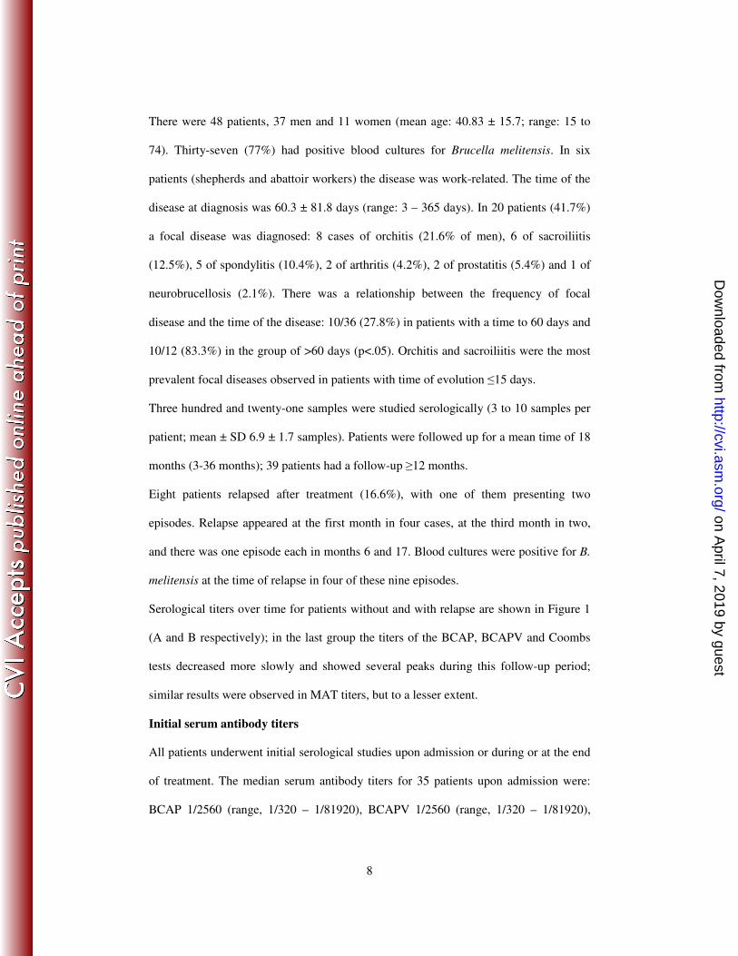
There were 48 patients, 37 men and 11 women (mean age: 40.83 ± 15.7; range: 15 to
74). Thirty-seven (77%) had positive blood cultures for Brucella melitensis. In six
patients (shepherds and abattoir workers) the disease was work-related. The time of the
disease at diagnosis was 60.3 ± 81.8 days (range: 3 – 365 days). In 20 patients (41.7%)
a focal disease was diagnosed: 8 cases of orchitis (21.6% of men), 6 of sacroiliitis
(12.5%), 5 of spondylitis (10.4%), 2 of arthritis (4.2%), 2 of prostatitis (5.4%) and 1 of
neurobrucellosis (2.1%). There was a relationship between the frequency of focal
disease and the time of the disease: 10/36 (27.8%) in patients with a time to 60 days and
10/12 (83.3%) in the group of >60 days (p<.05). Orchitis and sacroiliitis were the most
prevalent focal diseases observed in patients with time of evolution ≤15 days.
Three hundred and twenty-one samples were studied serologically (3 to 10 samples per
patient; mean ± SD 6.9 ± 1.7 samples). Patients were followed up for a mean time of 18
months (3-36 months); 39 patients had a follow-up ≥12 months.
Eight patients relapsed after treatment (16.6%), with one of them presenting two
episodes. Relapse appeared at the first month in four cases, at the third month in two,
and there was one episode each in months 6 and 17. Blood cultures were positive for B.
melitensis at the time of relapse in four of these nine episodes.
Serological titers over time for patients without and with relapse are shown in Figure 1
(A and B respectively); in the last group the titers of the BCAP, BCAPV and Coombs
tests decreased more slowly and showed several peaks during this follow-up period;
similar results were observed in MAT titers, but to a lesser extent.
Initial serum antibody titers
All patients underwent initial serological studies upon admission or during or at the end
of treatment. The median serum antibody titers for 35 patients upon admission were:
BCAP 1/2560 (range, 1/320 – 1/81920), BCAPV 1/2560 (range, 1/320 – 1/81920),
on April 7, 2019 by guest
http://cvi.asm.org/
Dow
nloaded from

9
Coombs 1/1280 (range, 1/160 – 1/20480), MAT 1/320 (range, 1/40 – 1/20480). RB,
MAT, Coombs, BCAP and BCAPV tests were positive in all cases, but in two patients
with positive blood cultures for B. melitensis and a disease duration before admission of
90 days, the MAT titer was 1/40.
Eighteen patients had high initial titers of serum antibodies: 9 had MAT titers of
≥1/1280, 14 had Coombs titers of ≥1/5120, 14 had BCAP titers of ≥1/10240 and 12 had
BCAPV titers of ≥1/10240.
No correlation was observed between the duration of disease before hospitalization and
titers upon admission (p>0.05), although MAT titers did show a tendency to decrease in
cases with prolonged disease.
Relationship between results from classical serological tests and Brucellacapt
The concordance between the MAT, Coombs, BCAP and BCAPV tests is shown in
Figure 2. The Spearman Rho coefficient between BCAP and BCAPV was 0.982
(p<.001), between BCAP and the Coombs and MAT tests it was 0.878 and 0.866,
respectively (p<.001), and between BCAPV and the Coombs and MAT tests it was
0.876 and 0.869, respectively (p<.001). The correlation between the Coombs and MAT
tests was 0.696 (p<.001). Multiple regression analysis indicated an independent
relationship between BCAP or BCAPV (the dependent variables) and the Coombs
(p<.005) and MAT (p<.001) tests. No significant differences were observed when these
correlations were evaluated separately in samples from different periods of disease
evolution.
When coincidence of pairs was evaluated in BCAP and BCAPV, titers were coincident
in 260 cases (81%) and non-coincident in 61 (19%) (weighted Kappa 0.926). These
non-coincident pairs were: higher BCAP, one dilution in 39 and two dilutions in 3; and
on April 7, 2019 by guest
http://cvi.asm.org/
Dow
nloaded from

10
higher BCAPV, one dilution in 19 cases (Table 1). Higher BCAP titers prevailed up to
titers of ≥1/640 (p<0.05).
Areas under the ROC curve (AUC) when Coombs was considered as the gold standard
ranged between 0.850 and 0.969, the difference between MAT and BCAP or BCAPV
being statistically significant (p<0.001); however, no significant difference was
observed between BCAP and BCAPV (difference: 0.009; p=0.078). Areas under the
ROC curve when MAT was considered as the gold standard were similar in BCAP
(0.942) and BCAPV (0.945), but were lower in Coombs (0.853), the difference between
BCAP or BCAPV and Coombs being statistically significant (p<0.001) (see Figure 3).
The sensitivity and specificity of BCAP, BCAPV and MAT titers with respect to a gold-
standard Coombs titer of ≥1/160 and those of BCAP, BCAPV and Coombs with respect
to a gold-standard MAT titer of ≥1/80 are shown in Tables 2 and 3.
Evolution of serological results over time in 40 patients without relapse
Antibody titers in serum samples from the 40 patients without relapse decreased over
time. In one patient who was in regular contact with sheep a transitory increase of ≥ 2
fold titers was detected in all tests, though he remained symptom-free; an isolated and
transitory increased was observed in BCAPV (one sample) and Coombs titers (one
sample) in patients who remained well.
The decrease of BCAP and BCAPV titers was very pronounced in comparison with
Coombs titers. Thus, if initial BCAP and BCAPV titers were two or more times higher
than those of Coombs, by the end of treatment they were already lower. Over the
following months these test titers decreased slowly in a similar way, although Coombs
titers showed a tendency to persist at higher levels than those of BCAP and BCAPV.
Differences in the evolution of BCAP and Coombs titers were further illustrated when
coincidence of pairs was analyzed: before therapy there were 5 coincident pairs, while
on April 7, 2019 by guest
http://cvi.asm.org/
Dow
nloaded from

11
in 22 cases BCAP titers were higher and in 7 they were lower than Coombs titers. At
the end of therapy or in the first month post-therapy there were 29 coincident pairs,
while in 22 cases BCAP titers were higher than Coombs and in 23 they were lower.
Over the following months there were 56 coincident pairs, while 30 BCAP titers were
higher and 70 lower than Coombs titers.
When this evolution of serological results in patients without relapse was compared
between the group of 26 patients without focal disease and the 14 patients with focal
disease, a slower decrease in titers in patients with focal disease for all tests was
observed; the differences in mean log10 titers upon admission and at 12th
month of
follow-up were: BCAP, 1.13 vs. 1.59; BACPV, 1.08 vs. 1.53; Coombs, 0.92 vs. 1.17;
and MAT, 0.73 vs. 1.32.
The time to negativization was 13 months for MAT, 30 months for BCAP, 29 months
for BCAPV and 30 months for the Coombs test. Kaplan-Meier analysis indicated the
following cumulative probabilities of persistence of serum antibody titers 12 months
after therapy: MAT 35%, BCAP 80%, BCAPV 80% and the Coombs test 87% (Figure
4).
Serological outcome in 8 patients with relapse
Of the 9 episodes among the 8 patients with relapse, serological results were available
for eight of them. Serological changes during relapse were detected in 7 cases (88%) by
the Coombs test (4 cases with two or more dilutions, 3 cases with one dilution only), in
5 cases by BCAP (2 with two or more dilutions, 3 with one dilution only) and BCAPV
(1 with two or more dilutions, 4 with one dilution only) and in 3 cases by MAT (1 with
two or more dilutions, 2 with one dilution only) (Table 4). These changes were more
evident in bacteremic relapses occurring at least three months after therapy (Figure 5).
In patients 1 and 2 (Table 4) clinical reactivation of spondylitis and orchitis was
on April 7, 2019 by guest
http://cvi.asm.org/
Dow
nloaded from

12
observed one month after the end of treatment and a concomitant increase in Coombs’
titers from 1/1280 to 1/5120 and from 1/1280 to 1/2560 respectively was detected.
Persistence of high titers 12-18 months after therapy
Thirty-three of the 40 patients who did not relapse were able to be followed up
clinically and serologically at least 12-18 months after therapy. Eleven of these patients
had high final serum antibody titers as follows: BCAP ≥1/320 (7 cases), BCAPV
≥1/320 (8 cases), Coombs ≥1/640 (7 cases) and MAT ≥1/80 (5 cases). The remaining 22
patients had low final serum antibody titers. The clinical evaluation was satisfactory
both for patients with high final titers and for those with low final titers. There was a
good correlation between serum antibody titers upon admission and those at 12-18
months; thus, among 15 patients with high initial titers, 9 had high final titers compared
to only two of the 18 patients who did not have high initial titers (P=0.003) . The
influence of focal disease in this persistence of high titers was less relevant.
on April 7, 2019 by guest
http://cvi.asm.org/
Dow
nloaded from

13
DISCUSSION
The results of our study are in accordance with those reported in previous series
regarding the high sensitivity and specificity of BCAP in the diagnosis of human
brucellosis (1, 9, 14, 16, 27, 29).
To date, BCAP has been considered an immunocapture-agglutination test whose ability
to detect agglutinant and non-agglutinant antibodies successfully has been mostly
related to the use of plates coated with anti-human immunoglobulins (1,9,14, 16, 27).
However, the present study demonstrates that performing a modified but similar test
using the same antigen and technique, but with plates that are not coated with
immunoglobulins (BCAPV), yields similar results to those of the standard BCAP. Thus,
the high ability of BCAP to detect anti-Brucella antibodies is not related specifically to
the mechanism of immunocapture, but rather is probably due to the characteristics of the
antigen and the acid pH conditions of the test (23,24). This is a very important finding,
not only as regards understanding the mechanisms involved in this test but also because
it may be simplified and its cost reduced.
In this study, BCAP had a sensitivity of 100% for the diagnosis of initial disease, given
that all patients had titers ≥1/320 at admission (median of 1/2560), which often were
several times above the suggested diagnostic threshold of 1/160-1/320.
When the evolution of serological titers was evaluated over time in patients with a good
clinical outcome we observed, as reported by others (9, 14, 16, 27), a more pronounced
and rapid decrease in BCAP titers in comparison with Coombs, which was already
evident in samples at the end of therapy or soon after treatment. The high sensitivity of
BCAP, coupled with this rapid decrease in its titers with treatment, suggests that the test
mainly detects high affinity antibodies. Furthermore, these findings indicate that BCAP
could be a better marker of infection activity and, therefore, a promising substitute for
on April 7, 2019 by guest
http://cvi.asm.org/
Dow
nloaded from

14
the Coombs test in the follow-up of patients with brucellosis. However, some additional
points should be taken into consideration before such a conclusion is drawn.
After the rapid decrease of BCAP titers detected during the first few months a slower
reduction occurs thereafter. Thus, BCAP/BCAPV and Coombs titers remained positive
at 12 months post-therapy in 80% and 87% of our patients, respectively, the decrease in
titers being particularly slow in patients with focal disease. It should be noted that about
20-25% of patients with a good clinical outcome had BCAP titers of ≥1/320 in samples
obtained 12-18 months after therapy, a finding that was mainly related to the detection
of very high titers for this test at the initial disease point, as was observed for the
Coombs test. Overall, BCAP seems to be more specific than the Coombs test as a
marker of disease activity; the detection of a titer <1/160 makes present or future
activity of the disease very unlikely.
At the time of relapse a serological increase in titers should be observed in order to
confirm this diagnosis (30). We previously reported an increase of IgG antibodies in
more than 80% of relapsing patients, as detected by IgG ELISA and Coombs tests (2,
21), and it has been suggested that BCAP may have a similar sensitivity (9, 16, 29).
However, in the present study, while all patients had BCAP titers ≥1/320 during relapse,
serological movement was observed only in 62% of cases with BCAP compared to 87%
of cases with Coombs, this change being of ≥two dilutions in 25% and 50% of cases,
respectively. The fact that an increase in these titers was detected only in exceptional
cases in the group of patients without relapse confers a high specific value on the
increase of Coombs titers as markers of relapse. Thus, the sensitivity of Coombs could
be higher than that of BCAP, because the Coombs test detects agglutinating and non-
agglutinating antibodies, including the ones with high- and low-affinity (4,10,19).
Taken together, these findings seem to indicate that a movement of low-affinity
on April 7, 2019 by guest
http://cvi.asm.org/
Dow
nloaded from

15
antibodies alone may be detected in some patients with active disease; in this regard, we
recently reported similar findings in the setting of late hepatosplenic reactivated
brucellosis (4, 10).
We conclude that BCAP is a very sensitive, specific and simple test for the routine
diagnosis and management of human brucellosis. However, its efficacy is not due to an
immune-capture effect, because similar results could be obtained with the same
technique but using non-coated anti-immunoglobulin plates; rather, it probably depends
on the characteristics of antigen used and the acid pH conditions of the test. This is
relevant in terms of cost reduction, because it means that the test can be used with non-
coated anti-immunoglobulin plates. As regards detecting mainly high affinity
antibodies, BCAP is more specific than the Coombs test. However, if the use of BCAP
test as a possible substitute for the Coombs test is considered, it should be taken into
account that in some cases of relapse and chronic forms of the disease slight changes of
low-affinity antibodies alone are observed, and these are better detected by the Coombs
test.
on April 7, 2019 by guest
http://cvi.asm.org/
Dow
nloaded from

16
ACKNOWLEDGMENTS
This work was supported by Ministerio de Sanidad y Consumo, Instituto de Salud
Carlos III- Spanish Network for Research in Brucellosis (G03/204).
We are grateful to Sonia Berenguer for her technical assistance.
We also thank Dr. F. Garrido and col. (Laboratorio Estatal de Sanidal Animal. Santa Fé.
Granada) for providing us with Rose Bengal.
on April 7, 2019 by guest
http://cvi.asm.org/
Dow
nloaded from

17
REFERENCES
1. Ardic, N., M. Ozyurt, O. Sezer, A. Erdemoglu, and T. Haznedaroglu. 2005.
Comparison of Coombs and immunocapture-agglutination tests in the diagnosis of
brucellosis. Chin. Med. J. 118: 252-4
2. Ariza, J., T. Pellicer, R. Pallarés, A. Foz, and F. Gudiol. 1992. Specific antibody
profile in human brucellosis. Clin. Infect. Dis. 14:131-140.
3. Ariza, J., J. Corredoira, R. Pallarés, P.F. Viladrich, G. Rufi, M. Pujol, and F.
Gudiol. 1995. Characteristics of and risk factor for relapse in human brucellosis.
Clin. Infect. Dis. 20:1241-1249
4. Ariza, J., C. Pigrau, C. Cañas, A. Marrón, F. Martínez, B. Almirante, J.M.
Corredoira, A. Casanova, J. Fabregat, and A. Pahissa. 2001. Current
understanding and management of chronic hepatosplenic suppurative brucellosis.
Clin. Infect. Dis. 32: 1024-1033
5. Benito, R., E. Durán, J. Gil, and C. Rubio. 2000. Brucelosis osteoarticular:
utilidad diagnóstica de las técnicas de inmunocaptura. Med. Clin; 114:639
6. Benito, R., E. Durán, J. Gil, and C. Rubio. 2001.Bacteriemia por Brucella con
serología convencional negativa. Enf. Infecc. Microbiol. Clin. 19:348-349
7. Brown, S. L., G. C. Klein, F. T. McKinney, and W.L. Jones. 1981. Safranin O-
stained antigen microagglutination test for detection of brucella antibodies. J. Clin.
Microbiol. 13:398-400.
8. Conrath, T.B. (Ed.). 1972. Handbook of microtiter procedures. Dynatech
Corporation, Cambridge, Mass .
9. Casao, M. A., E. Navarro, and J. Solera. 2004. Evaluation of Brucellacapt for the
diagnosis of human brucellosis. J. Infect. 49:102-108
on April 7, 2019 by guest
http://cvi.asm.org/
Dow
nloaded from

18
10. Díaz, R., J. Ariza, I. Alberola, A. Casanova, and M. Rubio. 2006. Secondary
serological response of patients with chronic hepatosplenic suppurative brucellosis.
Clinical and Vaccine Immunology. 13;1190-6
11. Diaz, R., and I. Moriyón. 1992. Laboratory techniques in the diagnosis of human
brucellosis. In: E.J. Young, and M.J. Corbel (ed). Brucellosis: Clinical and
laboratory aspects. Boca Ratón, Florida CRS Press, Inc.
12. Foz, A., and S. Garriga. 1954. Relation entre la fixation du complement et les
anticorps incomplets (test de Coombs) dans la brucellose humaine. Revue Immunol.
Thér. Antimicrob. 18:288-289.
13. Gaultney, J. B, R. D. Wende, and R. P. Williams. 1971. Microagglutination
procedures for febrile agglutination test. Appl. Microbiol. 22:635-640
14. Gómez, M.C., C. Rosa, P. Geijo, and M.A. Escribano. 1999. Estudio
comparativo del test Brucellacapt con el test de Coombs para Brucella. Enf. Infecc.
Microbiol. Clin. 17:283-285
15. Morata, P., M.I. Queipo-Ortuño, J.M. Reguera, M.A. García-Ordoñez, C.
Pichardo, and J.D. Colmenero. 1999. Posttreatment follow-up of brucellosis by
PCR assay. J. Clin. Microbiol. 37:4163-4166
16. Orduña, A., A. Almaraz, A. Prado, M.P. Gutiérrez, A. García-Pascual, A.
Dueñas, M. Cuervo, R. Abad, B. Hernandez, B. Lorenzo, M.A. Bratos, and A.
Rodríguez Torres. 2000. Evaluation of an immunocapture-agglutination test
(Brucellacapt) for serodiagnosis of human brucellosis. J. Clin. Microbiol. 38:4000-
5.
17. Ortega, M., A. Lara, M. J. Pérez, V. Díaz, A. Ruiz, and M. Rodríguez. 2001.
Bacteriemia por Brucella sp con serología convencional negativa. Enf. Infecc.
Microbiol. Clin. 19:34
on April 7, 2019 by guest
http://cvi.asm.org/
Dow
nloaded from

19
18. Otero, J.R., A. Fuertes, E. Palenque, and A.R. Noriega. 1982. Microtiter-adapted
method that facilitates the Coombs test for brucellosis. J. Clin. Microbiol. 16: 737-8.
19. Paul,W.E. 1993. Fundamental Inmunology. Raven Press. (Chapter 12). Antigen-
antibody interactions and monoclonal antibodies, pp.425-433.
20. Pappas, G., P. Papadimitriou, N. Akritidis, L. Christou, and E. Tsianos. 2006.
The new global map of human brucellosis. Lancet. Infect. Dis. 6:91-99
21. Pellicer, T., J. Ariza, A. Foz, R. Pallarés, and F. Gudiol. 1988. Specific
antibodies detected during relapse of human brucellosis. J. Infect. Dis. 157: 918-
924.
22. del Pozo, J. L., M. Rubio, and R. Díaz. 2002. Brucellacapt. ¿Tiene alguna utilidad
utilizar pocillos tapizados con antiinmunoglobulinas humanas?. Abstr. nº 599. X
Congreso Nacional de la Sociedad Española de Enfermedades Infecciosas y
Microbiología Clínica (SEIMC) Sevilla, Spain.
23. Queipo-Ortuño, M. I., P. Morata, P. Ocón, P. Manchado, and J. D. Colmenero.
1997. Rapid diagnosis of human brucellosis by peripheral-blood PCR assay. J. Clin.
Microbiol. 35: 2927-2930.
24. Rubio, M., J. L Pozo, J. M. Hernandez-Molina, I. Dorronsoro, T. Marrodan,
and R. Díaz. 2002. Diagnóstico de la brucelosis humana. Influencia del pH en la
prueba de seroaglutinación y sobre la actividad aglutinante de los anticuerpos IgM,
IgG e IgA. Enfer. Infecc. Microbiol. Clin. 20:144-149
25. Rubio, M., B. Barrio, and R. Díaz. 2001. Valor de las pruebas de rosa de bengala,
Coombs y contrainmunoelectroforesis para diagnosticar los casos de brucelosis
humana en los que la seroaglutinación es negativa. Enf. Infecc. Microbiol. Clin.; 19:
406-407
on April 7, 2019 by guest
http://cvi.asm.org/
Dow
nloaded from

20
26. Sanchez-Sousa, A., C. Torres, M. G. Campello, C. García, F. Parras, E.
Cercenado, and F. Baquero . 1990. Serological diagnosis of neurobrucellosis. J.
Clin. Pathol. 43:79-81
27. Serra, J., J. Velasco, P. Godoy, and J. Mendoza. 2001.Can the Brucellacapt test
be sustituted for the Coombs test in the diagnosis of human brucellosis ?. Enf.
Infecc. Microbiol. Clin. 19: 202-205
28. Torres, C., M. G. Campello, C. García, F. Parras, E. Cercenado, F. Baquero
and A. Sánchez-Sousa. 1986. Serología de la neurobrucelosis . Rev. Esp.
Microbiol. Clin. 1:117-120
29. Velasco, J., T. Marrodán, and R. Díaz. 1998. Estudio comparativo entre el test de
Coombs y Brucellacapt en el diagnóstico de la brucelosis humana. VIII Congreso
SEIMC. Palma de Mallorca
30. White, R.G. 1978. Immunoglobulin profiles of the chronic antibody response:
discussion in the relation to brucellosis infections. Postgrad. Med. J. 54: 595-602
31. Young, E.J. 1991. Serologic diagnosis of human brucellosis: analysis of 214 cases
of agglutination tests and review of the literature. Rev. Infect. Dis. 13: 359-372.
32. Young, E. 1995. Brucellosis: Current epidemiology, diagnosis and management. In
Current Clinical Topics in Infectious Diseases. J.S. Remington, and M.N. Swartz
(Ed.), Boston, Blackwell Scientific Publications; 15: 115-128.
on April 7, 2019 by guest
http://cvi.asm.org/
Dow
nloaded from

Table 1. Coincident pairs between BCAP titers and BCAPV titers
Reciprocal BCAP titers
Reciprocal BCAPV titers 20 40 80 160 320 640 1280 2560 5120 10240 20480 40960 81920 %
20 0 1 0 0 0 0 0 0 0 0 0 0 0 0.3
40 0 23 2 0 0 0 0 0 0 0 0 0 0 7.7
80 0 1 37 1 0 0 0 0 0 0 0 0 0 12.1
160 0 0 5 40 5 0 0 0 0 0 0 0 0 15.6
320 0 0 0 3 46 6 0 0 0 0 0 0 0 17.0
640 0 0 0 0 7 42 7 0 0 0 0 0 0 17.4
1280 0 0 0 0 0 0 23 2 3 0 0 0 0 8.7
2560 0 0 0 0 0 0 1 14 9 0 0 0 0 7.4
5120 0 0 0 0 0 0 0 0 16 5 0 0 0 6.5
10240 0 0 0 0 0 0 0 0 1 12 1 0 0 4.3
20480 0 0 0 0 0 0 0 0 0 1 3 0 0 1.2
40960 0 0 0 0 0 0 0 0 0 0 0 3 0 0.9
81920 0 0 0 0 0 0 0 0 0 0 0 0 1 0.3
% 0.0 7.7 13.7 13.7 18.0 14.9 9.7 5.0 9.0 5.6 1.2 0.9 0.3
on April 7, 2019 by guest
http://cvi.asm.org/
Dow
nloaded from

Table 2. Sensitivity, specificity, LR and PV of BCAP, BCAPV and MAT titers with respect to gold standard titers of Coombs of ≥1/160
Serological tests
Titer
Sensitivity
Specificity
+ LR
- LR
+ PV
- PV
BCAP
≥ 160
91.6
95.9
22.4
0.09
99.2
67.1
BCAPV
≥ 160
91.6
87.8
7.5
0.1
97.7
65.2
MAT
≥ 40
71.9
95.9
17.6
0.29
99
37.9
on April 7, 2019 by guest
http://cvi.asm.org/
Dow
nloaded from

Table 3. Sensitivity, specificity, LR and PV of BCAP, BCAPV and Coombs titers with respect to gold standard titers of MAT of ≥1/80
Serological tests
Titer
Sensitivity
Specificity
+ LR
- LR
+ PV
- PV
BCAP
≥ 640
86.7
87.9
7.14
0.15
86.1
88.4
BCAPV
≥ 640
88.7
89
8.07
0.13
87.5
90.1
Coombs
≥ 640
84.7
69.4
2.76
0.22
70.6
83.9
on April 7, 2019 by guest
http://cvi.asm.org/
Dow
nloaded from

Table 4. Antibody titers of BCAP, BCAPV, Coombs and MAT tests in pre-relapse and post-relapse serum samples from 8 cases of relapse
Case Time of relapse
(months)
Blood cultures at
relapse
Focal disease
Initial episode
Focal disease
at relapse
BCAP
Pre Post
BCAPV
Pre Post
Coombs
Pre Post
MAT
Pre Post
1
1st
Neg
Spondylitis
Spondylitis
2560 2560
2560 2560
1280 5120
320 160
2
1st
Neg
Orchitis
Orchitis
10240 10240
5120 5120
1280 2560
640 320
3
1st
Neg
Orchitis
Orchitis
640 1280
640 1280
640 1280
80 80
4
1st
Neg
Coxitis
Orchitis
320 640
320 640
640 1280
40 40
5
3rd
Pos
Sacroiliitis
Orchitis
640 10240
640 10240
320 2560
80 640
6
3rd
Pos
No
No
1280 2560
1280 2560
1280 5120
160 320
7
6th
Neg
Spondylitis
Arthritis
Orchitis
Arthritis 5120 2560
5120 2560
10240 5120
160 160
8
>12th
Pos
Sacroiliitis
No
1280 5120
640 1280
640 2560
80 160
Mean
--
--
--
--
1280 2560
960 2560
1280 2560
120 160
In bold type: cases with ≥2 increased dilutions; in italics: cases with only one increased dilution
on April 7, 2019 by guest
http://cvi.asm.org/
Dow
nloaded from

0640
128019202560320038404480512057606400
Adm
issio
n
Enf o
f the
rapy
1mon
th
2 m
onth
3 m
onth
6 m
onth
9 m
onth
12-1
8 m
onth
s
BCAP
BCAPV
Coombs
MAT
A.
B.
0
320
640
960
1280
1600
1920
2240
2560
2880
Adm
issio
n
Enf o
f the
rapy
1mon
th
2 m
onth
3 m
onth
6 m
onth
9 m
onth
12-1
8 m
onth
s
BCAP
BCAPV
Coombs
MAT
FIG. 1. Serological titers of MAT, BCAP, BCAPV and Coombs tests over time. (A) For patients without relapse and (B) with
relapse. The BCAP line is often hidden behind the BCAPV line
on April 7, 2019 by guest
http://cvi.asm.org/
Dow
nloaded from

A.
1,
0
1,5 2,0 2,5 3,0 3,5 4,0 4,5 5,0
0,8
0,6
0,4
0,2
0,0
-0,2
-0,4
AVERAGE of Log 10 BCAP and Log 10 BCAPV
Log
10 B
CA
P -
Log
10
BC
AP
V
Mean
0,02
-1.96 SD
-0,25
+1.96 SD
0,30
B.
1,0 1,5 2,0 2,5 3,0 3,5 4,0 4,5
1,0
0,5
0,0
-0,5
-1,0
AVERAGE of Log 10 BCAP and Log 10 Coombs
Log
10 B
CA
P -
Log
10
Coom
bs
Mean
-0,04
-1.96 SD
-0,74
+1.96 SD
0,66
C.
1,0 1,5 2,0 2,5 3,0 3,5 4,0 4,5 5,0
1,0
0,5
0,0
-0,5
-1,0
AVERAGE of Log 10 BCAPV and Log 10 Coombs
Log 1
0 B
CA
PV
-Log 1
0 C
oom
bs
Mean
-0,07
-1.96 SD
-0,73
+1.96 SD0,60
D. E. F.
1,0 1,5 2,0 2,5 3,0 3,5 4,0
2,5
2,0
1,5
1,0
0,5
0,0
AVERAGE of Log 10 BCAP and Log 10 MAT
Log 1
0 B
CA
P -
Log 1
0 M
AT
Mean
0,87
-1.96 SD
0,15
+1.96 SD
1,59
1,0 1,5 2,0 2,5 3,0 3,5 4,0 4,5
2,5
2,0
1,5
1,0
0,5
0,0
AVERAGE of Log 10 BCAPV and Log 10 MAT
Log 1
0 B
CA
PV
-Log 1
0 M
AT
Mean
0,85
-1.96 SD
0,16
+1.96 SD
1,53
1,0 1,5 2,0 2,5 3,0 3,5 4,0
2,5
2,0
1,5
1,0
0,5
0,0
-0,5
AVERAGE of Log 10 Coombs and Log 10 MAT
Log 1
0 C
oom
bs
-Log 1
0 M
AT
Mean
0,92
-1.96 SD
-0,06
+1.96 SD
1,89
FIG. 2. Bland-Altmann graphs showing the level of concordance of BCAP, BCAPV, Coombs and MAT titers. (A) BCAP-BCAPV,
(B) BCAP- Coombs, (C) BCAPV- Coombs , (D) BCAP- MAT, (E) BCAPV- MAT, (F) Coombs-MAT.
on April 7, 2019 by guest
http://cvi.asm.org/
Dow
nloaded from

A. B.
MATBCAPBCAPV
0 20 40 60 80 100
100
80
60
40
20
0
100-Specificity
Sen
siti
vit
y BCAPBCAP VCOOMBS
0 20 40 60 80 100
100
80
60
40
20
0
100-Specificity
Sen
siti
vit
y
FIG. 3. (A) Areas under the ROC curve (AUC) of BCAP, BCAPV and MAT when Coombs was considered as the gold
standard: BCAP and BCAPV were very similar (0.969 and 0.960), but higher than MAT (0.850) (p<0.001). (B) Areas under
the ROC curve (AUC) of BCAP, BCAPV and Coombs when MAT was considered as the gold standard: BCAP and BCAPV
were very similar (0.942 and 0.945), but higher than Coombs (0.853) (p<0.001).
on April 7, 2019 by guest
http://cvi.asm.org/
Dow
nloaded from

BCAP
BCAPV
Coombs
MAT
Time to negativisation (months)
403020100
1,0
,8
,6
,4
,2
0,0
Cu
mula
tive
surv
ival
FIG. 4. Probability of persistence of positive titers over time for BCAP, BCAPV, Coombs and MAT test. Kaplan-Meier
curves of BCAP, BCAPV, Coombs and MAT showing a similar decrease of BCAP and BCAPV titers, both earlier than
Coombs; MAT titers decreased even earlier than BCAP and BCAPV.
on April 7, 2019 by guest
http://cvi.asm.org/
Dow
nloaded from

A.
0
640
1280
1920
2560
3200
3840
4480
5120
5760
Adm
issio
n
Enf o
f the
rapy
1mon
th
2 m
onth
3 m
onth
6 m
onth
BCAP
BCAPV
Coombs
MAT
0
1280
2560
3840
5120
6400
7680
8960
10240
Adm
issio
n
Enf o
f the
rapy
1mon
th
3 m
onth
6 m
onth
BCAP
BCAPV
Coombs
MAT
B.
FIG. 5. (A) Patient with relapse 3 months after the end of therapy (nº6 table 4): note the increase in Coombs titers, while BCAP,
BCAPV and MAT titers remained unchanged. (B) Patient with relapse 3 months after the end of therapy (nº5 table 4): note the
increase of BCAP, BCAPV and Coombs titers, while MAT titers remained unchanged; note that the increase of BCAP and BCAPV
titers was much more pronounced than that of Coombs titers. In both figures the BCAP line is hidden behind the BCAPV line
on April 7, 2019 by guest
http://cvi.asm.org/
Dow
nloaded from