Brucella abortus Invasion of Synoviocytes Inhibits Apoptosis and ...

1199tthh WWoorrlldd CCoonnggrreessss oonn IIVVFF
in conjunction with
66tthh SSoocciieettyy ooff RReepprroodduuccttiivvee MMeeddiicciinnee aanndd SSuurrggeerryy CCoonnggrreessss
(October 4-8, 2017, Regnum Carya Resort Hotel, Antalya)
ORAL PRESENTATION

[[AAbbssttrraacctt:: 00110066]] [[OOPP--0011]] [[AAcccceepptteedd:: OOrraall PPrreesseennttaattiioonn]]FFrroozzeenn EEmmbbrryyoo TTrraannssffeerr--iiss IItt OOuurr FFuuttuurree?? ““TThheeoorryy AAbboouutt EEmmbbrryyoo CCrryyoo--ttrreeaattmmeenntt””IIaavvoorr KK VVllaaddiimmiirroovv,, DDeessiissllaavvaa TTaacchheevvaaIVF unit, SBALAGRM-Sofia, Sofia, Bulgaria; Faculty of Biology, Sofia University "St. Kliment Ohridski", Bulgaria
OObbjjeeccttiivvee:: If we examine the statistics and compare the progression of cryopreservation technology during recent years, wewill find that over of the last decade, there has been an over 2.5 times increase in treatment of frozen-thawed embryo trans-fer (FET), while the rate of fresh embryo transfers (ET) that was completed was the same. This increase in the usage of FETcorrelated with a quick growth in the number of live births, compared to fresh ET. The reason is that embryo freezing andthawing efficiency has improved dramatically with widespread use of vitrification vs. the older slow freezing method.MMaatteerriiaallss--MMeetthhooddss:: We consider the advantages and disadvantages of the “freeze all” strategy in assisted reproduction. Fromthe analysis of multiple results in assisted reproductive technology, we created the first ever theory that provides a scien-tific explanation of the causes and mechanism needed for higher success rates of FET. RReessuullttss:: We found evidence to support a ‘therapeutic’ effect of the freezing/thawing of embryos during the process of re-covery of the embryo and its subsequent implantation. Freezing/thawing can be seen as a way to provoke the endogenoussurvival and improve responses in developing preimplantation embryos. Different molecular mechanisms can explain thehigher success rate of FET compared to fresh ET in women of advanced reproductive age, including the higher miscarriagerate in cases of blastocyst FET compared to FET at early cleavage embryo, and the higher perinatal parameters of born chil-dren after frozen embryo transfer.CCoonncclluussiioonn:: We suggest that the method of cryopreservation is not only a technology for storing embryos, but it is also amethod of embryo treatment that can potentially improve the IVF procedure success rate. We believe that, through pre-senting the advantages arising from the freezing and thawing process described, as well as the theoretical rationale given inour theory, researchers should rethink the position of cryobiology in reproductive medicine.
KKeeyywwoorrddss:: Cryopreservation, FET, fresh ET, mitochondria, pregnancy
DOI: 10.24074/tjrms.01032017OP TJRMS 2017;1(3):125-90
127

[[AAbbssttrraacctt:: 00116644]] [[OOPP--0022]] [[AAcccceepptteedd:: OOrraall PPrreesseennttaattiioonn]]EEvvaalluuaattiioonn ooff CChhrroommoossoommaall AAnnaallaayyssiiss RReessuullttss ooff FFeemmaallee PPaattiieennttss wwiitthh RReeccuurrrreenntt MMiissccaarrrriiaaggeeEEttiioollooggyy wwhhoo AAddmmiitttteedd ttoo DDuuzzccee UUnniivveerrssiittyy RReesseeaarrcchh aanndd AApppplliiccaattiioonn HHoossppiittaallHHüüsseeyyiinn YYüüccee11,, RReecceepp EErröözz11,, MMuussttaaffaa DDooğğaann11,, AAşşkkıı EElllliibbeeşş KKaayyaa22,, AAllppeerr BBaaşşbbuuğğ22
1Duzce University Medical Faculty, Department of Medical Genetics, Turkey2Duzce University Medical Faculty, Department of Obstetrics and Gynecology, Turkey
Recurrent miscarriage or habitual abortus is an important human health problem and described as at least 2 or more con-secutive pregnancy losses. Etiologic causes of the disease are uterus anomalies, maternal system disorders, genetic anomaliesand autoimmune disorders. The chromosomes were obtained from peripheral blood samples of 356 females patients with ha-bitual abortus etiology who admitted to Duzce University Research and analaysed for each patients. According to chromo-somal analysis results; 327 females have normal karyotype (46,XX) (91.85%), 5 females have 46,XX,inv(9)(p11q13) (1.40%),4 females have 46,XX,1qh+ (1.12%), 3 females have 46,XX,16qh+ (0.84%), 2 females have 46,XX,9qh+ (0.56%), 1 female has46,XX,15cenh+ (0.28%), 1 female has 46,XX,21ps+ (0.28%), 1 female has 46,XX,13pstk+ps+ (0.28%), 1 female has46,XX,del(15)(p11.2) (0.28%), 3 females have 45,X /46,XX) (0.84%), 2 females have 45,X/47,XXX/46,XX (0.56%), 2 femaleshave 47,XXX /46,XX (0.56%), 1 female has 47,XXX/46,XX/45,X (0.28%), 1 female has 46,XX,t(18;20)(q22;p11.2) (0.28%),1females has 46,XX,t(14;16)(q23;q22) (0.28%) and 1 females has 46,XX,t(13;16)(q34;q12) (0.28%). Abnormal karyotypes weredetected in 29 (8.15%) from 356 females with habitual abortus etiology.
KKeeyywwoorrddss:: Habitual abortus, chromosomal analaysis
TJRMS 2017;1(3):125-90 DOI: 10.24074/tjrms.01032017OP
128
[[AAbbssttrraacctt:: 00116655]] [[OOPP--0033]] [[AAcccceepptteedd:: OOrraall PPrreesseennttaattiioonn]]TThhee FFaaccttoorr VV LLeeiiddeenn ((GG11669911AA)),, PPrroottrroommbbiinn IIII aanndd MMeetthhyylleenneetteettrraahhyyddrrooffoollaattee RReedduuccttaassee ((CC667777TT,, AA11229988CC)),, FFVV CCaammbbrriiddggee aanndd PPllaassmmiinnooggeenn AAccttiivvaattoorr IInnhhiibbiittoorr GGeennee AAnnaallyyssiiss RReessuullttss ooff FFeemmaallee PPaattiieennttss wwiitthh HHaabbiittuuaall AAbboorrttuuss EEttiioollooggyyRReecceepp EErröözz11,, HHüüsseeyyiinn YYüüccee11,, MMuussttaaffaa DDooğğaann11,, AAllppeerr BBaaşşbbuuğğ22,, AAşşkkıı EElllliibbeeşş KKaayyaa22
1Duzce University Medical Faculty, Department of Medical Genetics, Duzce, Turkey2Duzce University Medical Faculty, Department of Obstetrics and Gynecology, Duzce, Turkey
Recurrent miscarriage or habitual abortus is described as at least 2 or more consecutive pregnancy losses. For maintanenceof successful gestation, A successful uteroplacental circulation is a crucial and important condition. Therefore, the maternaltrombophilias {(Factor V Leiden (FVL), Prothrombin (PTm), MTHFR (methylenetetrahydrofolate reductase)} and PAI (Plas-minogen Activator Inhibitor) are crucial for gestation. The molecular analysis of FVL (G1691A), PTm (G20210A) andMTHFR (C677T) of 351 females patients and FV Cambridge, MTHFR (A1298C) and PAI of 102 females patients with ha-bitual abortus etiology who admitted to Duzce University School of Medicine were analysed for each patients. Accordingto analysis results; When the FVL (G1691A) to be taken into consideration, while 305 female have normal (GG) genotype(86,89%), 46 female have the altered genotype (13,11%) (GA or AA). When the PTm (G20210A) to be taken into consider-ation, while 326 female have normal genotype (92.88%) (GG), 25 female have the altered genotype (7.12%) (GA or AA).When the MTHFR (C677T) to be taken into consideration, while 158 female have normal genotype (45.01%) (CC), 193 fe-male have the altered genotype (CT or TT) (54.99%). When the MTHFR (A1298C) to be taken into consideration, while 50female have normal genotype (49.02%) (AA), 52 female have the altered genotype (AC or CC) (50.98%). When the PAI tobe taken into consideration 22 (21.57%) female have 5G/5G genotype, 58 female have 4G/5G (56,86%) genotype and 22 fe-male have 4G/4G (21.57%) genotype. For FV Cambridge, all females have normal genotype.
KKeeyywwoorrddss:: Habitual abortus, thrombophilia
DOI: 10.24074/tjrms.01032017OP TJRMS 2017;1(3):125-90
129

TJRMS 2017;1(3):125-90 DOI: 10.24074/tjrms.01032017OP
130
[[AAbbssttrraacctt:: 00116688]] [[OOPP--0044]] [[AAcccceepptteedd:: OOrraall PPrreesseennttaattiioonn]]TThhee RRoollee ooff SSeerruumm mmiiccrrooRRNNAA--113322 aass aa NNoovveell BBiioommaarrkkeerr ffoorr tthhee DDiiaaggnnoossiiss ooff PPoollyyccyyssttiicc OOvvaarryy SSyynnddrroommeeZZeeyynneepp SSooyymmaann11,, SSiinneemm DDuurrmmuuşş22,, SSeeddaa AAtteeşş33,, HHaaffiizzee UUzzuunn22,, RReemmiissee GGeelliişşggeenn22,, VVeeyysseell ŞŞaall44
1Department of Obstetrics and Gynecology, Istanbul Education and Research Hospital, Istanbul, Turkey2Department of Biochemistry, Cerrahpasa School of Medicine, Istanbul University, Istanbul, Turkey3Department of Obstetrics and Gynecology, School of Medicine, Bezmialem Vakif Gureba University4Department of Obstetrics and Gynecology, Hamidiye Sisli Etfal Education and Research Hospital
OObbjjeeccttiivveess:: Polycystic ovary syndrome (PCOS) is a heterogeneous disorder, and the underlying molecular mechanisms are notclear. In PCOS, abnormal regulation of relevant genes is required for follicular development. MicroRNAs (miRNAs) are small,noncoding RNA sequences that negatively regulate gene expression at the post-transcriptional level. However, few studieshave been conducted on circulating miRNAs in PCOS. The aim of this study was to investigate the role of the serum miRNAsas a biomarker for the diagnosis of PCOS and examine the relationship between sex hormones and the miRNAs expression. MMaatteerriiaall--MMeetthhooddss:: Fifty women with PCOS and age- and body mass index (BMI)- matched 50 healthy controls were re-cruited from a tertiary care center between January 2016 and June 2017. The expression of circulating miRNA-222, miRNA-146a, miRNA-132, and miRNA-320 were measured from serum samples of subjects.RReessuullttss:: Insulin, homeostasis model assessment of insulin resistance (HOMA-IR), free testosterone, sex hormone-bindingglobulin (SHBG), dehydroepiandrosterone sulfate (DHEAS), and androstenedione levels were significantly higher in thePCOS group than in the control group (p ˂ 0.05, Table 1). Serum miR-132, miR-146a, and miR-222 were significantly lowerin women with PCOS compared with controls (p ˂ 0.05). However, only the ratio of patients who had downregulated mir-132 was significantly higher in PCOS group than in the control group (p ˂ 0.05). There was no statistically significant dif-ference between the PCOS and control groups in terms of miR-320 expression (p > 0.05). Serum miR-132 was negativelycorrelated with HOMA-IR and insulin and positively correlated with SHBG (all p < 0.05).DDiissccuussssiioonn:: This is the first study indicated that serum miRNA-132 was significantly downregulated in women with PCOScompared with controls. Serum miRNA-132 may be a novel candidate as a molecular biomarker in the diagnosis of PCOS.This relationship may yield important insight into novel treatment modalities by revealing the pathogenesis of PCOS. Fur-ther larger studies are needed to evaluate the role of miRNA as a novel biomarker for the diagnosis of PCOS and to deter-mine physiopathological roles of miRNAs in PCOS.
KKeeyywwoorrddss:: Biomarker, miRNA, polycystic ovary syndrome
Control group (n=50) PCOS Group (n=50)
Mean±SD Mean±SD p
Age (years) 23.9±4.1 22.7±3.5 0.101 m
BMI (kg/m²) 24.3±4.7 25.5±5.2 0.306 m
Fasting glucose (mg/dl) 90.2±7.1 89.1±8.2 0.469 t
Fasting insulin (µU/mL) 7.5±4.8 9.9±5.3 0.012 m
HOMA-IR 1.7±1.2 2.2±1.3 0.030 m
FSH (mlU/mL) 6.7±2.1 6.3±1.9 0.221 m
LH (mlU/mL) 8.4±5.7 10.4±6.9 0.100 m
Estradiol (pg/mL) 83.6±91.9 50.4±33.7 0.121 m
Total testosterone (ng/dL) 56.4±21.2 63.8±23.0 0.148 m
SHBG (µg/dL) 87.5±73.1 49.6±24.8 0.000 m
DHEAS (ng/mL) 241.2±99.1 281.6±101.6 0.047 t
Androstenedione(ng/mL) 1.4±0.6 1.8±0.6 0.002 m
17-OH progesterone 0.9±0.8 0.8±0.6 0.975 m
AMH 5.2±3.1 9.6±5.0 0.000 m
TABLE 1: Demographics and biochemical characteristics of women with PCOS and control subjects.
m Mann-whitney U test, t t test, x² square test Values are expressed as means ± standard deviations. PCOS, polycystic ovary syndrome; BMI, body mass index; HOMA-IR, homeos-
tasis model assessment of insulin resistance; SHBG, sex hormone-binding globulin; DHEAS, dehydroepiandrosterone sulfate; AMH, antimullerian hormone.

DOI: 10.24074/tjrms.01032017OP TJRMS 2017;1(3):125-90
131
[[AAbbssttrraacctt:: 00117788]] [[OOPP--0055]] [[AAcccceepptteedd:: OOrraall PPrreesseennttaattiioonn]]CCaarryyoottyyppee EEvvoolluuttiioonn iinn PPaattiieennttss wwiitthh PPrreemmaattuurree OOvvaarriiaann FFaaiilluurreeAAssllıı AAkkddöönneerr11,, MMuurraatt CCeelliillooğğlluu22,, EErrkkaann ÇÇaağğllııyyaann33,, CCeerreenn AAyyddıınn44
1Dokuz Eylül University Faculty of Medicine, Department of Surgical Medical Sciences, Department of Gynecology and Obstetrics, İzmir2Dokuz Eylül University Faculty of Medicine, Department of Surgical Medical Sciences, Department of Gynecology and Obstetrics, Division ofReproductive Endocrinology, İzmir
OObbjjeeccttiivvee:: Chromosome anomalies are one of the major causes of premature ovarian failure(POF)(1). The objective of thisstudy is to evaluate the frequency and type of chromosomal anomalies in the patients with POF admitted to our clinic andto discuss the findings in the light of current literature and to provide guidance to new studies. MMaatteerriiaall--MMeetthhooddss:: The study was conducted between 01.03.2017–10.04.2017 in the Reproductive Endocrinology and In-fertility Policlinic in the Department of Obstetrics and Gynecology at Dokuz Eylul University. The files of the patients, whowere diagnosed with POF, between 2002-2017, were screened from the archives. 65 patients were included in the study. In-formation about age, smoking, alcohol use, age at menarche, HRT usage, additional disease, obstetric history, age of parentsat birth, FSH, LH, estradiol, prolactin, TSH, fT3, fT4, Anti-TPO, Anti-TG, TRAB, cortisol, ANA, Insulin, fasting blood glu-cose, LDL, HDL, triglyceride, total cholesterol, AMH levels, DEXA, mammogram and karyotype results were recorded fromfile information. Family history of POF, menopause age of mother, grandmother, aunt, sister and family history of mentallyretarded males were recorded. The Statistical Program for Social Sciences (SPSS, version 15) was used for statistical analy-sis. The Mann-Whitney U test, Fisher’s exact test, Pearson and Spearman correlation analysis were used in the analysis. P-values <=0.05 were considered to be statistically significant.RReessuullttss:: The results of 65 cases were examined. There was no statistically significant difference between the cases with nor-mal and abnormal karyotypes in terms of the smoking, alcohol use, history of additional disease, family history of mentallyretarded males, history of pregnancy after diagnosis, gravida, parity, abortus, HRT usage, DEXA and mammogram results(p>0.05). There was no statistically significant difference between the groups, in terms of the laboratuary results. Only fT3was significantly lower in the cases with abnormal karyotypes (p:0.019). There was no statistically significant difference be-tween the groups, in terms of the age of parents at birth and the age of menopause of mother, grandmother, aunt, and sis-ter. 5 cases (%7,7) were found to have chromosome anomalies (46+XX/45+X(4) and 46+XY/45+X(1)).CCoonncclluussiioonn:: Turner syndrome/mosaic Turner syndrome is the most frequent genetic anomaly related to POF. However, therelation between caryotype and ovarian function is not clear(2,3). 46+XY female is rare and these cases have female sekon-der sex characteristics and related to SRY gene mutation. 45+X/46+XY phenotypic interval can change from Turner Syn-drome to mix gonadal dysgenesis and normal male phenotype(4). Previous studies showed the importance of X chromosomein POF ethiology(5). POF cases which have low rate X chromosome mosacism can be important especially in ethiology ofidiopatic group(5). In conclusion, cytogenetic researches should be evalute routinely in cases with POF.
KKeeyywwoorrddss:: Premature ovarian failure, caryotype, genetics, ovarian aging
Mean Min Max Median
Age at Diagnose 32,6±5,5 18 39 34
VKİ(kg/m²) 23,4±2,6 18 30 23
FSH(mIU/mL) 76,3±26,8 42 155 69,2
LH(mIU/mL) 38,4±14,05 16 80,2 33
E2(pg/mL) 27,4±16,8 10 122 20
TSH(µIU/mL) 1,65±0,74 0,02 3,2 1,47
fT3(pg/mL) 2,7±0,73 0,21 4,48 2,78
fT4(ng/dL) 0,98±0,31 0,60 2,14 0,88
TRAB 1,04±0,18 0,31 1,8 1
Anti-TPO(IU/mL) 45,07±141,3 0,02 737 1,1
Anti-TG(IU/mL) 7,1±28,1 0,90 207 0,9
ANA(titre) 0,03±0,17 0 1 0
Kortizol(µg/dL) 10,2±4,9 2,5 28,8 9,29
İnsulin(µIU/mL) 8,7±9,48 1,44 56 6,01
AMH 0,47±0,075 0,01 0,31 0,01
Prolaktin(ng/mL) 9,4±3,9 3,05 21 8,6
FBG(mg/dL) 83,8±13,8 60 140 84
LDL(mg/dL) 117,2±37,1 59 212 106
HDL(mg/dL) 57,9±13,8 32 105 59
Trigliserit(mg/dL) 120,1±80,05 42 405 96
Total Kolesterol(mg/dL) 178,5±45,5 112 308 173
TABLE 1: BMI, age at diagnose, biochemical and hormonal parameters
Caryotype N N
46+XX 60(%92,3)
46+XX/45+X 4(%6,1)
46+XY(16)/45+X(36) 1(%1,5)
TABLE 2: Range of caryotype results.
TJRMS 2017;1(3):125-90 DOI: 10.24074/tjrms.01032017OP
132
[[AAbbssttrraacctt:: 00119933]] [[OOPP--0066]] [[AAcccceepptteedd:: OOrraall PPrreesseennttaattiioonn]]EExxoommee SSeeqquueenncciinngg UUnnrraavveellss NNoovveell MMeecchhaanniissmm iinn UUnneexxppllaaiinneedd IInnffeerrttiilliittyySSeerrddaarr CCooşşkkuunn11,, SSaatteeeesshh MMaaddddiirreevvuullaa11,, SSaaaadd AAllhhaassssaann11,, AAttiiff EEllnnoouurr22,, HHeessssaa AAllssaaiiff11,, NNiieemmaa IIbbrraahhiimm11,, FFiirrddoouuss AAbbdduullwwaahhaabb11,, SStteeffaann AArroolldd33,, FFoowwzzaann AAllkkuurraayyaa11
1King Faisal Specialist Hospital and Research Center, Riyadh2Sulaiman AlHabib Medical Group3King Abdullah University of Science and Technology
OObbjjeeccttiivvee:: About a quarter of infertile couples seeking infertility treatment are diagnosed as having unexplained infertilitythat defies diagnosis following standard investigations. These patients have been suggested to have immunological distur-bances or molecular defects due to mutations in the genes which have role in reproductive physiology. The objective of thisstudy was to utilize advanced genetic testing to identify possible genetic causes of unexplained infertility.Design: ProspectiveMMaatteerriiaallss--MMeetthhooddss:: A total of seven couples diagnosed as unexplained infertility and having immature oocytes following theiruneventful controlled ovarian stimulation and ovulation triggering were asked to participate IRB approved genetic testingstudy. All had failed intrauterine insemination cycles followed by in vitro fertilization treatment. They had ovarian stimu-lation with short or long agonist, or antagonist stimulation protocols followed by hCG/agonist induction for final follicularmaturation. Oocyte retrieval was performed and the obtained cumulus oocyte complexes either inseminated or denuded fromcumulus cells according to their treatment protocol. Once the patients was confirmed to have all immature oocytes they werecounselled for genetic testing and pedigree analysis. If agreeable, patients’ DNA samples were collected for positional map-ping and whole-exome sequencing.RReessuullttss:: Seven patients underwent a total of 22 oocyte retrievals and 215 oocytes were collected. There were only 6 oocyteswith a polar body and remaining 209 oocytes were either at germinal vesicle or metaphase 1 stage. None of these 6 oocytereached to embryo following sperm injection suggesting incomplete maturation. Twenty two oocytes were also cultured inin vitro maturation medium for 48 hours with none was resuming meiosis. Positional mapping examination of runs of ho-mozygosity revealed only one overlapping region in two patients. Exome sequencing revealed 63,890 variants in the indexof couple 1 and 72,370 variants in the index of couple 2. However, only one rare homozygous variant was identified withinthe candidate locus in each family and both variants involved mutations in the same gene. These variants were completelyabsent in 2,379 control exomes. The novel candidate gene has been reported to be involved in the oocyte maturation ofXenopus suggesting also possible involvement in human oocyte maturation. CCoonncclluussiioonnss:: Data suggest that next generation genetic testing uncover the reason behind unexplained infertility and mu-tation in the candidate gene causes meiotic arrest and leads to female infertility.
KKeeyywwoorrddss:: Keywords: Oocyte, maturation arrest, next generation sequencing, unexplained infertility
DOI: 10.24074/tjrms.01032017OP TJRMS 2017;1(3):125-90
133
[[AAbbssttrraacctt:: 00224444]] [[OOPP--0077]] [[AAcccceepptteedd::OOrraall PPrreesseennttaattiioonn]]TTeekk GGeenn HHaassttaallıığğıı vvee HHLLAA UUyyuummuunnddaa PPrreeiimmppllaannttaassyyoonn GGeenneettiikk TTaannıı ((PPGGTT)):: DDıışşkkaappıı DDeenneeyyiimmiiFFeerrddaa AAllppaassllaann PPıınnaarrllıı11,, İİsskkeennddeerr KKaappllaannooğğlluu22,, HHaanniiffee SSaaaatt11,, İİnnccii KKaahhyyaaooğğlluu22,, HHiillaall YYııllddıızz11,, SSoonnggüüll HHaarrşşııtt11,, SSeerrddaarr DDiillbbaazz22
1Sağlık Bilimleri Üniversitesi, Dışkapı Yıldırım Beyazıt EAH, Genetik Tanı Merkezi, Ankara,Türkiye2Sağlık Bilimleri Üniversitesi, Etlik Zübeyde Hanım Kadın Hastalıkları EAH, Tüp Bebek Kliniği, Ankara, Türkiye
AAmmaaçç:: Preimplantasyon Genetik Tanı (PGT)’da tek gen hastalıkları taraması ile kombine HLA doku tiplemesi tayini Ta-lasemi Majör gibi yaşamı tehtid eden ve kordon kanı ve /veya kemik iliği nakli ile tam olarak tedavi edilebilen genetikhastalıklara sahip çocuklar için etkin bir tedavi yöntemidir. Bu çalışmada, Sağlık Bilimleri Üniversitesi Dışkapı YıldırımBeyazıt Eğitim Araştırma Hastanesi Genetik Tanı Merkezi Preimplantasyon Genetik Tanı Labaratuarında 2014-2017 Mayısdöneminde çalışılan vakaların verileri sunulmuştur. GGeerreeçç--YYöönntteemm:: Labaratuarımıza çoğunluğu Sağlık Bilimleri Üniversitesi Etlik Zübeyde Hanım Kadın Hastalıkları Tüp BebekMerkezinden gelen 6-8 blastomer aşamasındaki 3. gün embriyolarından alınan biopsi örneklerinde preimplantasyon genetiktanı uygulamaları yapılan 91 vakada genetik analiz sonuçları, implantasyon ve gebelik başarısı ile canlı doğum oranlarıdeğerlendirildi. Biopsi örneklerinden DNA izolasyonu gerçekleştirildikten sonra mutasyonu taşıyan DNA fragmentlerinin ve HBB geni ileilişkili 11 markerın belirlenebilir seviyeye kadar polimeraz zincir reaksiyonu (PCR) ile çoğaltılması yapıldı. Amplifikasyondansonra DNA, normal DNA fragmentlerini mutasyonu taşıyan fragmentlerden ayırt etmeye olanak sağlayan mini-sekanslamatekniği ile kapiller elektroforez kullanılarak analiz edildi. Seçilmiş Linked Markerlar mutasyonun tanısı için bir destek oluş-turma ve bazı DNA kontaminantlarının belirlenmesi amacıyla kullanıldı. HLA tipleme analizlerinde uyumlu HLA genoti-plerinin belirlenmesi için HLA kompleksi ile ilişkilendirilen 32 marker, multipleks polimeraz zincir reaksiyonu (PCR) ileçalışılarak DNA fragmentleri kapiller elektroforez kullanılarak analiz edildi.Bulgular: 86 vaka Beta Talasemi (HBB GENİ), 1 vaka Orak Hücreli Anemi (HBB GENİ),1 vaka Blackfan Diamond Anemisi(RPS19 GENİ), 1 vaka ALL, 1 vaka Ağır Konjental Nötropeni (HAX1 GENİ) ve 1 vaka FankoniAplastik Anemisi (FANCAGENİ) olmak üzere toplamda 91 vaka çalışıldı. Toplam çalışılan blastomer sayısı 328, transferi yapılan vaka sayısı 41, gebe-lik gerçekleşen vaka sayısı 12, gerçekleşen canlı doğum sayısı 8 ve kemik iliği nakil işlemi yapılan vaka sayısı 3 olarak sap-tandı. SSoonnuuçç vvee TTaarrttıışşmmaa:: Bu sonuçlar kemik iliği nakli ile tam kür sağlan hastalıkların tedavisinde ailede sağlıklı ve HA uyumluçocuk varlığının önemi açısından oldukça önemlidir. Diğer preimplentasyon genetik test uygulanan hastalıklarlakarşılaştırıldığında transfer edilebilir blastomer bulma oranın düşük olduğu görülmüş ancak bunun nedeninin blastomerdeiki farklı genetik seçilim yapılması gerekliğinden kaynaklandığı düşülmüştür.
AAnnaahhttaarr KKeelliimmeelleerr:: Preimplantasyon genetik tanı, HLA uyumu, tek gen hastalığı

TJRMS 2017;1(3):125-90 DOI: 10.24074/tjrms.01032017OP
134
[[AAbbssttrraacctt:: 00111177]] [[OOPP--0088]] [[AAcccceepptteedd:: OOrraall PPrreesseennttaattiioonn]]VVaaggiinnaall PPrrooggeesstteerroonnee GGeell VVeerrssuuss IInnttrraammuussccuullaarr PPrrooggeesstteerroonnee ffoorr LLuutteeaall SSuuppppoorrtt iinnSSuubbooppttiimmaall RReessppoonnddeerr WWoommeenn UUnnddeerrggooiinngg AAnnttaaggoonniisstt IIVVFF//IICCSSII CCyycclleessHHaassaann UUlluubbaaşşooğğlluu11,, KKaaddiirr BBaakkaayy11,, AAllii YYaavvuuzzccaann22,, DDaavvuutt GGüüvveenn11
1Department of Obstetrics and Gynecology, Medical Faculty, Ondokuz Mayis University, Samsun, Turkey2Department of Obstetrics and Gynecology, Medical Faculty, Düzce University, Düzce, Turkey
OObbjjeeccttiivvee:: The aim of the study was to investigate the effects of two different vaginal progesterone forms (vaginal proges-terone gel versus intramuscular progesterone) administered for luteal phase support, on pregnancy outcomes in supoptimalresponder women aged <= 40, who underwent antagonist in IVF/ICSI-ET cycles. Although luteal phase support has beenshown not to affect the outcome of normosponder patients, there is no study of the route of administration of luteal phasesupport ( LPS ) in patients with suboptimal responder. MMaatteerriiaall MMeettoodd:: This is a retrospective study conducted using the files of patients who were admitted to the assisted repro-duction department of department of obstetrics and gynecology, medical faculty, Ondokuz Mayis University due to the de-sire to have children, between January 2010 and December 2015 we were evaluated that data of 189 patients with suboptimalresponder who underwent IVF / ICSI treatment and reached to file data. The cycles were categorized into two groups: prog-esterone vaginal gel 180 mg/day (Group 1, n = 76),progesterone ampoule 50 mg/ml intramusculer 1x2 (Group 2, n = 113).Clinical pregnancy, ongoing pregnancy rates and spontanous abortus were analyzed.Clinical pregnancy was defined as fetalcardiac activity observed by vaginal ultrasonography 4 or 5 weeks after oocyte retrieval and ongoing pregnancy was de-fined as ultrasound check of the embryo after 9 weeks of gestation. Luteal support commenced after oocyte retrieval and con-tinued until the day of pregnancy testing. If the test was positive, progesterone treatment was continued for up 9 weeks ofgestation. Stimulation was performed according to the disease-fix antagonist protocol. The dosage is adjusted according tothe ovarian response. Induction was started at a daily dose of FSH. RReessuullttss:: Patient and treatment characteristics such as patient age, antral follicle count (AFC) on day 3, FSH,TSH and estra-diol level on day 3, infertility diagnosis were similar in both groups (Table 1). Hovewer,In terms of infertile duration,sig-nificant differences were observed between groups (P=0,043). There were no significant differences in stimulationcharacteristics such as mean total gonadotropin doses, E2 levels on hCGday, total number of oocytes, number of fertilizedoocyte between the groups. Intra-cytoplasmic sperm injection outcomes did not show statistically significant differences be-tween the two groups regarding clinical pregnancy rate/cycle, livebirth rate and the spontaneous abortion (Table 2).DDiissccuussssiioonn:: According to our findings, we conclude that there were no statistically significant differences between the useof 50 mg progesterone ampoule 1x2 /day and 90 mg progesterone gel 2x1 /day for LPS treatment, in terms of clinical preg-nancy rate, and the live birth rate, the spontaneous abortion in suboptimal responder women undergoing antagonist IVF/ICSIcycles. prospec¬tive,more comprehensive, randomized studies are required in order to better define the optimum daily drugdoses, administration route, as well as the initiation and cessation time of the drug.
KKeeyywwoorrddss:: Progesterone vaginal gel, in vitro fertilization, antagonist protocol
progesterone gel progesterone
(n:76) ampoule (n:113)
Age (years) 30(19:40) 32(21:40) 0,323a
Duration of infertility (years) 6(2:8) 5(1:8) 0,043a**
Number of antral follicles on day 3 8(5:14) 8(5:12) 0,743a
D3 FSH [IU/L] 6(4:10) 6(4:10) 0,573a
D3 Estradiol [pg/mL] 36,5(5:80) 36,0(8:80) 0,594a
D3 Tsh [pg/mL] 2,4(1:6) 2,3(1:5) 0,982a
Infertility diagnosis, n (%)
Unexplained 40(52,6) 75(66,4)
Male factor 32(42,1) 34(30,1) 0,323b
Tubal 4(5,3) 4(3,5)
TABLE 1: Demographic characteristics of the patients in the groups.
Data are presented as median (min: max) and number (percent). a: Mann-Whitney U test, b: χ2 test.
FSH - follicle stimulating hormone, hCG - human chorionic gonadotropin, E2 - estradiol, TSH-thyroid
stimulating hormone **Statistically significant.
progesterone gel Progesterone
(n:76) ampoule(n:113)
Average gonadotropin dose 3125(2500:4250) 3200(2250:4900) 0,412a
Endometrial thickness on the day of hCG 9(7:16) 10(7:14) 0,535a
Total Duration of induction (day) 10(8,13) 10(8,14) 0,626a
E2 on the day of hCG 2000(1600:3100) 2000(1750:4300) 0,444a
Number of total oocytes retrieved 7(4:9) 7(4:9) 0,775a
Number of the fertilized oocytes 6(2:10) 6(2:10) 0,937a
Number of MII oocytes 6(1:9) 5(1:9) 0,260a
Clinical pregnancy rate/cycle (%) 73,7 69,9 0,573b
Live birth rate/cycle (%) 58,1 59,8 0,816b
Spontaneous abortion(%) 17,3 15,3 0,714b
TABLE 2: Stimulation characteristics and treatment outcomesof 198 cycles
Data are presented as median (min: max) and number (percent). a: Mann-Whitney U test, b: χ2 test.
hCG - human chorionic gonadotropin, E2 - estradiol, MII:metaphase-II oocytes
DOI: 10.24074/tjrms.01032017OP TJRMS 2017;1(3):125-90
135
[[AAbbssttrraacctt:: 00114411]] [[OOPP--0099]] [[AAcccceepptteedd:: OOrraall PPrreesseennttaattiioonn]]rrLLHH VVeerrssuuss hhMMGG iinn TTeerrmmss ooff PPrrooppoorrttiioonn ooff EETTss aatt tthhee BBllaassttooccyysstt SSttaaggee aanndd CCyycclleesswwiitthh EEmmbbrryyoo CCrryyoopprreesseerrvvaattiioonn iinn PPoooorr RReessppoonnddeerrssNNuurr DDookkuuzzeeyyllüüll GGüünnggöörr11,, AAyynnuurr EErrşşaahhiinn22,, FFeerrhhaatt CCeennggiizz11,, YYaasseemmiinn ÖÖzzddeemmiirr11,, DDuuyygguu KKüüttüükk11
1IVF Unit, Göztepe Medicalpark Hospital, Istanbul, Türkiye2Bahçeşehir University, Istanbul,Türkiye
The study question is the treatment with rLH more effective than hMG in terms of proportion of ETs at the blastocyst stageand proportion of cycles with embryo cryopreservation in poor responders.Although previous studies have shown benefi-cial effects of the addition of LH activity to FSH, in terms of PR in patients,no studies have compared two different go-nadotrophin preparations containing LH activity in the same group of poor responder women.A single-centre retrospective study was performed between January 2015 and December 2016 with 30 women <=39 yearsof age with diminished ovarian reserve undergoing ICSI cycles. Both rFSH in combination with rLH or hMG, were used inthe same patient group.30 patients with unexplained infertility,tubal factor or male factor undergoing ICSI cycles were recruited in a private hos-pital setting. The ovarian stimulation cycle started on the third day of the menstrual cycle and the starting gonadotrophindoses used were 225 IU/day of rFSH plus 75IU/day of rLH or 75 IU/day of u-hMG.Flexible antagonist protocol was used forall cycles. Oocyte pick up was performed 35-36 h after hCG injection.rLH addition was associated with a significant in-crease in the mean number of COC and MII oocytes.(p<0.001)The proportion of ETs at the blastocyst stage and cycles withembryo cryopreservation were not significantly different in poor responders treated with rLH or hMG.,Further studies are required to verify if rLH addition produces better results than HMG in young poor responder women.It is obvious that interventions used in poor responders require properly designed large randomized studies, because untilnow there is no evidence-based treatment for that particular group of patients.
KKeeyywwoorrddss:: rLH, hMG, ICSI, Blastocyst

TJRMS 2017;1(3):125-90 DOI: 10.24074/tjrms.01032017OP
136
[[AAbbssttrraacctt:: 00009955]] [[OOPP--1100]] [[AAcccceepptteedd:: OOrraall PPrreesseennttaattiioonn]]MMiittoocchhoonnddrriiaall CCoonnttrriibbuuttiioonn iinn EEgggg RReejjuuvveennaattiioonn aanndd PPrree--iimmppllaannttaattiioonn ooff EEmmbbrryyooSSaannaa AAbbbbaass11,, SShhaahhzzaadd BBhhaattttii22,, MMuuhhaammmmaadd AAssllaammkkhhaann33,, HHiikkmmaatt HHaakkaann AAyyddiinn44,, GGeerraarrddoo RRooddrriigguueezz GGoonnzzaalleezz55
1Hameed Latif Hospital, Lahore Institute of infertility and endocrinology2University of Health Sciences Lahore, University of Lahore Pakistan3University of Health Sciences Lahore4Medical Biochemistry, Ege University School of Medicine, Izmir, Turkey5Jara CIBO, IMSS, Universidad De Guadala, Guadalajara, Jalisco, Mexico
BBaacckkggrroouunndd:: Mitochondria are integral part of a cell known as “power house” of the cell, manifested by highly multifacetedand entirely organized intricate biological processes. Traditionally, it portrayed two essential physiological liabilities: peri-odic release of energy during the cell cycle and activation of specific integral proteins. Several studies predicted its manifoldfunctions by producing heterogeneous proteins that are unequivocally involved in a plethora of phosphorylation pathways,calcium signaling, cellular respiration, embryonic development and sequential programmed cell death. However, in earlyoogenesis and embryogenesis mitochondria has a nexus of developmental regulations and overwhelming role in infertilitytreatments. The oocyte quality is directly related with ovarian aging and devastating mitochondrial function which is im-portant for infertility and age related ovarian dysfunction.OObbjjeeccttiivvee:: Nearly 23% patients visiting the infertility centers consulting for infertility depicted signs of premature ovariansenescence (POS) and quality compromise oocyte. This study emphasize on the role of mitochondria in ovarian aging andon the expected effects in the next generation.SSuubbjjeeccttss aanndd MMeetthhooddss:: PubMed was employed to search the MEDLINE database for peer-reviewed original research papersand reviews about mitochondria and ovarian ageing, in human species from 2002 to 2017.The patients are divided into two groups, premature ovarian senescence (POS) group and normal ovarian senescence group(NOPS). Statistically, we apply orthogonal partial least squares discriminant analysis (OPLS-DA). In order to compute thevalue of oocyte mtDNA content we used OPLS model.RReessuullttss:: Our data showed a correlative capability (Q2 = 0.769) when OPLS-DA has employed among two mtDNA content(0.82), groups. The POS group showed three important variables by using projection matric i.e. oocyte the cumulus cellmtDNA content (0.85) and peroxisome active receptor γ co activator factor (1.02) all are lower in POS group than NOPSgroups of patients. While OPLS model predict oocyte mtDNA content only in the NOPS group (Q2 = 0.634). Moreover, wefound four new positively correlated variables that are directly linked with mitochondrial inner mass of quality compromisedoocytes (0.56), the cumulus cell mtDNA contents (1.032), expression of polymerase gamma (1.85) and mitochondrial DNAtranscription factor (1.50).
KKeeyywwoorrddss:: Egg Rejuvenation, pre-implantation, IVF, Mitochondrial DNA
DOI: 10.24074/tjrms.01032017OP TJRMS 2017;1(3):125-90
137
[[AAbbssttrraacctt:: 00111188]] [[OOPP--1111]] [[AAcccceepptteedd:: OOrraall PPrreesseennttaattiioonn]]VVaarriiccoocceelleeccttoommyy IImmpprroovveess tthhee SSppeerrmm CCaappaacciittyy ttoo IInndduuccee OOppttiimmaall EEmmbbrryyoonniicc DDeevveellooppmmeenntt PPoosstt--ffeerrttiilliizzaattiioonn iinn SSuurrrrooggaaccyy PPrrooggrraammIIooaannnniiss GGiiaakkoouummaakkiiss11,, DDiiaammaannttiiss DDaapphhnniiss11,, NNiikkoollaaooss SSooffiikkiittiiss22
1Mediterranean Fertility Institute, Chania, Greece2Medical School of the University of Ioannina, Ioannina, Greece
OObbjjeeccttiivvee:: Left varicocele has a detrimental effect (European Urology Supplements 13:89-99,2014) on the sperm ability toinduce optimal early embryonic development post-fertilization. Our objective was to evaluate the effects of varicocelec-tomy on early embryonic development of oocytes recovered from female donors.PPaarrttiicciippaannttss aanndd MMeetthhooddss:: ICSI procedures using female donor oocytes were performed with spermatozoa from 13 men(group A) with left varicoceles. The female partners of the latter men could not produce oocytes of appropriate quality forparticipating in assisted reproductive technology (ART) programs. Injected oocytes were cultured for 96 hours. The fertil-ization rate (FR) (100χ fertilized oocytes / injected oocytes) and the blastocyst development rate (BDR) (100χdeveloped blas-tocysts/fertilized oocytes) were recorded. Developed blastocysts were transferred to surrogate females. Another group ofnine men with left varicoceles (group B) whose wives could not produce oocytes of appropriate quality for ART programsunderwent microsurgical left varicocelectomy. Six to 12 months later ICSI cycles were performed using female donor oocytes.FR and BDR were recorded, as well. Developed blastocysts were transferred to surrogate females.RReessuullttss:: BDR was significantly larger (P<0.05) in group B than in group A (Chi-Square test -Yates correction).CCoonncclluussiioonnss:: Microsurgical left varicocelectomy improves the ability of the male gametes to trigger the cascade of ooplas-mic events that lead to early embryonic development up to the blastocyst stage. In the current study the injected donoroocytes had been recovered from normal young females thus any female infertility factor is excluded. It appears that leftvaricocelectomy improves the DNA integrity, the male gamete nucleus protein matrix quality and/or the centrosomic in-tegrity/function allowing more optimal embryonic development. The current study indicates an important role of the malegamete beyond fertilization. Such results have clinical significance in a Surrogacy Motherhood Program whereby globallegislation requires either of the intended parents to provide (in most cases the male partner) their own genetic material. Con-sequently, it is imperative to increase quality of the sperm in order to achieve high pregnancy rates.
KKeeyywwoorrddss:: Varicocelectomy, sperm capacity, embryonic development, surrogacy

TJRMS 2017;1(3):125-90 DOI: 10.24074/tjrms.01032017OP
138
[[AAbbssttrraacctt:: 00113366]] [[OOPP--1122]] [[AAcccceepptteedd:: OOrraall PPrreesseennttaattiioonn]]DDiiffffeerreennttiiaall GGeennee EExxpprreessssiioonn AAnnaallyyssiiss ooff HHuummaann CCuummuulluuss CCeellllss aass aa PPrreeddiiccttoorr ffoorr PPrreeggnnaannccyy aanndd LLiivvee BBiirrtthhŞŞiirriinn BBaakkttıı DDeemmiirraayy11,, EEggee NNaazzaann TTaavvmmeerrggeenn GGöökkeerr22,, EErrooll TTaavvmmeerrggeenn22,, ÖÖzzlleemm YYııllmmaazz33,, NNiillüüffeerr ÇÇaallıımmllııooğğlluu22,, HHüüsseeyyiinn OOkkaann SSooyykkaamm44,,GGüüllppeerrii ÖÖkktteemm33,, UUğğuurr SSeezzeerrmmaann55
1Tepecik Education and Research Hospital, Department of Obstetrics and Gynecology, Assisted Reproduction Unit, Izmir, Turkey2Ege University Faculty of Medicine, Department of Obstetrics and Gynecology, Izmir, Turkey3Ege University Faculty of Medicine, Department of Histology and Embryology, Izmir, Turkey4Epigenetiks Genetics Bioinformatics Software Inc. Istanbul, Turkey5Faculty of Medicine, Acibadem University, Department of Biostatistics and Medical Informatics, Istanbul, Turkey
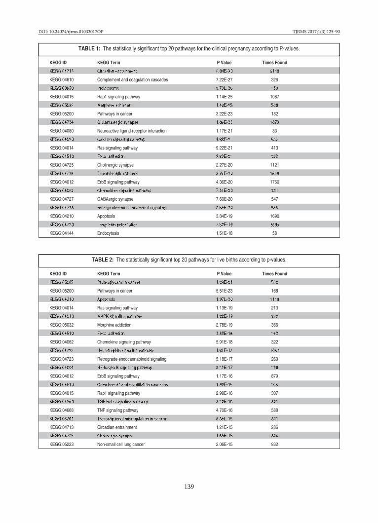
OObbjjeeccttiivvee:: The objective of this study was to provide valuable insights into the determination of clinical pregnancy and livebirth outcomes in the context of differential gene expression analysis of cumulus cells as predictors of clinical pregnancyand/or live birth after single embryo transfers. This study was performed with the consideration that each oocyte and eachgroup of cumulus cells surrounding different oocytes might have different genetic expression patterns that might affecthuman reproduction. MMaatteerriiaall:: This prospective case study included 10 women (aged 23-35 years) who were referred to a 3rd level reference cen-ter university hospital for idiopathic infertility and implemented Assisted Reproductive Technologies. Differential gene ex-pression analysis of cumulus cells was performed in 10 clusters of cumulus cells obtained from 10 cumulus-oocyte complexesof 10 patients.MMeetthhoodd:: Cumulus cells gene expressions were individually evaluated by microarrays using a NimbleGen Human Gene Ex-pression 12x135K Microarray Kit (NimbleGen, Roche, Germany). Total RNA was isolated using an RNeasy Kit (Qiagen,Hilden, Germany) and cDNA synthesis from total RNA was carried out using a Superscript Double Chain cDNA SynthesisKit (Invitrogen Life Technologies, Carlsbad, CA, USA) according to the manufacturer’s instructions. Same procedures relatedto the oocyte maturation and microinjection, and microarray analyses were applied to the group of cumulus cells, individ-ually. Two differential gene expression analyses were performed, one for outcome of clinical pregnancy and one for outcomeof live birth. Significant genes resulting from these analyses with p<0.05 were selected and given as input to PANOGA toolfor pathway analysis and it was run 20 times for each case and the resulting KEGG pathways were summarized across all runsfor clinical pregnancy and live birth. We report KEGG pathway id, KEGG pathway name, the significance of each pathwaythat are found in original PANOGA input, and the times they are found in our analysis. RReessuullttss:: Clinical pregnancy rates and live birth rates were used to assess the success of ICSI. Clinical pregnancy was ob-served in 3 patients, and live birth occurred in 2 patients from the total 10 patients in this study. 4371 significant genes fromclinical pregnancy and 1975 significant genes for live birth were enriched to 152 and 151 KEGG pathways by 20 PANOGAruns, respectively. Top 20 affected pathways in clinical pregnancy (Table 1) and live birth (Table 2) are analyzed and theseanalyses determined that circadian entrainment was the most affected pathway for clinical pregnancy and proteoglycans inthe cancer pathway was the most affected pathway for live birth. Circadian entrainment has both lower p-value of 6,04.e-30 and higher number of times count of 2142 than proteoglycans in pathways pathway which has a p value of 1,29e-24 and532 number of times count. There are 11 commonly affected pathways at different significances in top 20 affected pathwaysand circadian entrainment pathway is one of these commonly affected pathways. CCoonncclluussiioonn:: The results of this study provide new insights into human cumulus cells, and provide new information on thecrosstalk between different signalling pathways during pregnancy and clinical pregnancy.
KKeeyywwoorrddss:: Microarray, cumulus cell, pregnancy, live birth, circadian entrainment
DOI: 10.24074/tjrms.01032017OP TJRMS 2017;1(3):125-90
139
KEGG ID KEGG Term P Value Times Found
KEGG:04713 Circadian entrainment 6.04E-30 2142
KEGG:04610 Complement and coagulation cascades 7.22E-27 326
KEGG:03050 Proteasome 8.73E-26 159
KEGG:04015 Rap1 signaling pathway 1.14E-25 1087
KEGG:05032 Morphine addiction 1.48E-25 580
KEGG:05200 Pathways in cancer 3.22E-23 182
KEGG:04724 Glutamatergic synapse 1.06E-22 1073
KEGG:04080 Neuroactive ligand-receptor interaction 1.17E-21 33
KEGG:04020 Calcium signaling pathway 4.89E-21 656
KEGG:04014 Ras signaling pathway 9.22E-21 413
KEGG:04510 Focal adhesion 9.83E-21 233
KEGG:04725 Cholinergic synapse 2.27E-20 1121
KEGG:04728 Dopaminergic synapse 3.76E-20 1568
KEGG:04012 ErbB signaling pathway 4.36E-20 1750
KEGG:04062 Chemokine signaling pathway 7.46E-20 241
KEGG:04727 GABAergic synapse 7.60E-20 547
KEGG:04723 Retrograde endocannabinoid signaling 9.58E-20 653
KEGG:04210 Apoptosis 3.84E-19 1690
KEGG:04720 Long-term potentiation 7.52E-19 3695
KEGG:04144 Endocytosis 1.51E-18 58
TABLE 1: The statistically significant top 20 pathways for the clinical pregnancy according to P-values.
KEGG ID KEGG Term P Value Times Found
KEGG:05205 Proteoglycans in cancer 1.29E-24 532
KEGG:05200 Pathways in cancer 5.51E-23 168
KEGG:04210 Apoptosis 1.97E-20 1119
KEGG:04014 Ras signaling pathway 1.13E-19 213
KEGG:04010 MAPK signaling pathway 1.22E-19 283
KEGG:05032 Morphine addiction 2.78E-19 366
KEGG:04510 Focal adhesion 2.32E-18 162
KEGG:04062 Chemokine signaling pathway 5.91E-18 322
KEGG:04722 Neurotrophin signaling pathway 1.81E-17 1087
KEGG:04723 Retrograde endocannabinoid signaling 5.18E-17 260
KEGG:04064 NF-kappa B signaling pathway 8.13E-17 190
KEGG:04012 ErbB signaling pathway 1.17E-16 879
KEGG:04610 Complement and coagulation cascades 1.93E-16 105
KEGG:04015 Rap1 signaling pathway 2.99E-16 307
KEGG:04350 TGF-beta signaling pathway 3.12E-16 321
KEGG:04668 TNF signaling pathway 4.70E-16 588
KEGG:05202 Transcriptional misregulation in cancer 8.38E-16 341
KEGG:04713 Circadian entrainment 1.21E-15 286
KEGG:04725 Cholinergic synapse 1.85E-15 344
KEGG:05223 Non-small cell lung cancer 2.06E-15 932
TABLE 2: The statistically significant top 20 pathways for live births according to p-values.

TJRMS 2017;1(3):125-90 DOI: 10.24074/tjrms.01032017OP
140
[[AAbbssttrraacctt:: 00225599]] [[OOPP--1133]] [[AAcccceepptteedd:: OOrraall PPrreesseennttaattiioonn]]EEnnddoommeettrriiaall IInnjjuurryy RReevviissiitteedd:: LLaarrggee RRaannddoommiizzeedd aanndd CCoonnttrroolllleedd SSttuuddyy ttoo IInnvveessttiiggaattee iittss EEffffeeccttiivveenneessss iinn RRIIFFZZiiyyaa KKaalleemm11,, MMüübbeerrrraa NNaammllıı KKaalleemm22,, HHaalliill RRuussoo11,, TTiimmuurr GGüürrggaann11
1Gürgan Clinic IVF and Women Health Center, Ankara, Turkey2Liv Hospital Ankara, Ankara, Turkey
Despite the advances in assisted reproductive technologies (ART), a substantial percentage of patients still suffer from re-current implantation failure (RIF). It is evident from the scientific literature that local inflammation of the endometrium isrequired for embryo implantation. Thus, there are numerous studies investigating the effect of endometrial scarring in orderto increase the odds of implantation though increasing the inflammation of the endometrium, especially for RIF patients.However, these studies are lacking the necessary standard method for coming to a conclusion for the usefulness of en-dometrial scarring. Therefore, we present a new method that can be applied standardly on RIF patients in order to increasethe pregnancy rates of RIF patients.PPuurrppoossee:: To investigate the effect of hysteroscopic symmetrical endometrial injury for RIF patients.MMeetthhoodd:: This is a prospective and randomized controlled trial for RIF patients investigating the effect of systemic and sym-metric endometrial injury using office hysteroscopy. Endometrial injury was performed on the follicular phase of the men-strual cycle under general anesthesia. The main variant analyzed was the clinical pregnancy rates. Statistical calculations wereperformed on GraphPad Prism version 6.0 and p<0.05 were considered statistically significant.FFiinnddiinnggss:: Following randomized controlled trial we propose that the systemic endometrial injury model can be standard-ized to increase the odds of implantation in RIF patients to achieve clinical pregnancy.RReessuullttss:: The average maternal age for the control and endometrial injury group was 31.89 and 33.48 (n=72 and n=75, p>0.05,respectively). No statistical differences were found between the BMI of the female (23.88 kg/m2 and 24.13 kg/m2, p>0.05,respectively), previous cycles (3.74 and 3.84, p>0.05, respectively), cumulative embryo transfer (6.13 and 6.25, p>0.05, re-spectively), average number of MII eggs (75.61% and 77.82%, p>0.05, respectively), average ET (embryo transfer) day (3.18and 3.11, p>0.05, respectively), average embryos transferred (1.9 and 1.9, p>0.05, respectively), and endometrial thicknesson hCG day (10.28mm and 9.43mm, p>0.05, respectively) when two groups were compared. On average, the endometrialinjury was performed on the follicular phase of the menstrual cycle (average day: 10.76). The clinical pregnancy rates werefound to be significantly higher in the endometrial injury group when compared with the control group (38.82% and 18.50%,p<0.05, respectively).
KKeeyywwoorrddss:: Office hysteroscopy, recurrent implantation failure, RIF, embryo implantation

DOI: 10.24074/tjrms.01032017OP TJRMS 2017;1(3):125-90
141
[[AAbbssttrraacctt:: 00114433]] [[OOPP--1144]] [[AAcccceepptteedd:: OOrraall PPrreesseennttaattiioonn]]PPrreevvaalleennccee ooff LLooww VViittaammiinn DD LLeevveellss iinn IInnffeerrttiillee PPaattiieennttss -- AA SSiinnggllee CCeenntteerr PPiilloott SSttuuddyyBBuurrççiinn KKaarraammuussttaaffaaooğğlluu BBaallccıı,, BBüülleenntt EErrgguunnIstanbul University Istanbul Faculty of Medicine, Department of Obstetrics and Gynecology, Istanbul, Turkey
OObbjjeeccttiivvee:: Our objective was to evaluate serum levels of vitamin D in patients who presented with infertility to Istanbul Uni-versity, Istanbul Faculty of Medicine, Department of Obstetrics and Gynecology, Department of Reproductive Endocrinol-ogy and Infertility.MMaatteerriiaall:: This study used a retrospective cross-sectional design. We examined medical records of all infertile patients whovisited our clinic between March 2017 and August 2017. The inclusion criteria were voluntariness to participate in the study,being aged 18-49 years, and being required to give blood for serum 25-OH vitamin D testing. Patients who were admittedfor recurrent miscarriages or other symptoms such as hirsutism, and abnormal uterine bleeding were not included. Addi-tional exclusion criteria included current pregnancy, chronic diseases, celiac disease or other causes of malabsorption, dis-orders that may impact calcium or vitamin D metabolism, kidney diseases, medications affecting bone metabolism, vitaminD or calcium supplementation. MMeetthhoodd:: Demographic data were collected from medical records. Predictors for hypovitaminosis D, such as anticonvulsantuse, renal and cardiovascular disease, preexisting diabetes mellitus (type 1 or 2), malabsorption, gastrectomy, active liver dis-ease, acute myocardial infarction, alcoholism, anorexia nervosa, and steroid dependency were investigated.The normal value for vitamin D (25-OH vitamin D concentration in plasma) is considered as >= 30 ng/mL [75 nmol/L]. Se-vere deficiency is considered as <10 ng/mL [25nmol/L]; deficiency < 20 ng/mL [50 nmol/L]; and insufficiency 21-29 ng/mL[51-74 nmol/L]. First, the prevalence of vitamin D severe deficiency, deficiency, insufficiency, and sufficiency was calcu-lated. Then, all patients were grouped according to their BMI (normal, overweight, and obese) and age (younger than 35years, between 35-40, and older than 40 years) and whether there was a statistically significant difference for vitamin D lev-els between the groups was investigated.RReessuullttss:: During the study period (spring and summer seasons in Turkey), 711 consecutive outpatients, all Caucasians, agedbetween 18 and 49 years (mean ± SD, 30.6 ± 5.49 years) were enrolled for this study. Sixty-one patients did not give bloodfor vitamin D, 11 were actively taking vitamins including vitamin D, and 60 had medical problems that could influence vi-tamin D status, and were excluded. A total of 579 infertile women met the inclusion criteria. The mean serum 25-OH vita-min D concentration was 16.28 ng/mL±11.58 (range, 1-79.5 ng/mL); 220 patients were severely vitamin D deficient; 192 werevitamin D deficient; 95 were vitamin D insufficient; and only 72 patients were vitamin D sufficient. According to age groups,vitamin D deficiency was more frequent in the younger patients (<35 years) than in older patients (>35 years) and the dif-ference was statistically significant. No difference was seen when the patients were grouped according to BMI.CCoonncclluussiioonn:: This study showed that among persons presenting with infertility, more than three quarters of patients youngerthan 35 years and more than half of patients older than 35 years are vitamin D deficient and need treatment with vitaminD.
KKeeyywwoorrddss:: Infertility, 25-OH vitamin D, body mass index, age

TJRMS 2017;1(3):125-90 DOI: 10.24074/tjrms.01032017OP
142
[[AAbbssttrraacctt:: 00117799]] [[OOPP--1166]] [[AAcccceepptteedd:: OOrraall PPrreesseennttaattiioonn]]GGeennee EExxpprreessssiioonn iinn GGrroowwiinngg HHuummaann FFoolllliicclleess:: IIddeennttiiffyyiinngg MMaarrkkeerrss ooff FFoolllliiccllee AAccttiivvaattiioonnSSuussaannnnee EElliissaabbeetthh PPoorrss,, SSttiinnee GGrryy KKrriisstteennsseenn,, CCllaauuss YYddiinngg AAnnddeerrsseennLaboratory of Reproductive Biology, University Hospital of Copenhagen, Rigshospitalet, Copenhagen, Denmark
OObbjjeeccttiivvee:: Women receiving gonadotoxic treatment are of high risk of becoming infertile. An option for these women is topreserve fertility by ovarian tissue cryopreservation. In vitro activation is a novel method to increase the chances for preg-nancy after cryopreservation and autotransplantation by targeting cell signaling pathways controlling growth. Especially thePI3K-Akt-mTOR pathway and Hippo pathway have attracted much attention recently. Using gene expression data obtainedfrom nine different time points of follicle growth and maturation, the objective of this study was, to elucidate on the impactof these pathways and identify other potential mediators of human follicle recruitment and maturation. MMaatteerriiaallss:: Microarray analysis was performed on human pre-antral follicles in five distinct size categories plus granulosa cellsfrom small antral and pre-ovulatory follicles as previously published (Borgbo et al., 2013; Kristensen et al., 2014; Wissing etal., 2014, Petersen et al., 2015). The pre-antral follicles and the granulosa cells were obtained from donated ovarian tissuefrom women undergoing ovarian tissue cryopreservation for fertility preservation. Furthermore, granulosa cells from pre-ovulatory follicles were obtained from women undergoing fertility treatment. MMeetthhooddss:: In one study, human pre-antral follicles were isolated enzymatically from medullary tissue, divided into four dif-ferent size groups: <60 µm, 60-75 µm, 75-100 µm, 100-150 µm, >150 µm and snap frozen. In the other studies, granulosa cellsfrom antral follicles including 6mm antral, before hCG treatment and post-hCG treatment, were isolated from follicle flu-ids and snap frozen. RNA was isolated from all the samples. Microarray and q-PCR was performed as described previously(Kristensen et al., 2014; Petersen et al., 2015). RReessuullttss:: For PI3K-Akt-mTOR pathway high expression was found for FOXO1 and GSK3 in both preantral and antral folli-cles. For genes in the Hippo pathway; YAP1 and the target gene CTGF were highly expressed in preantral, but only mod-erately expressed in antral follicles. The remaining genes of both pathways were moderately or less expressed throughoutthe different follicle stages. Proteins of possible interest to follicle activation and maturation including MDK, MAPK8, S1PR1and HTRA1 were found with a distinct amplified expression in the early phases of growth and with low expression in theantral stages. CCoonncclluussiioonn:: The results confirm that a complex network of signaling pathways is present and contributing to growth andmaturation of follicles. Mapping the key players in activation and maturation of follicles will contribute to new therapiesfor infertility. Further verification with description of the proteins present including localization and phosphorylation sta-tus is needed to further describe their involvement.
KKeeyywwoorrddss:: Fertility preservation, gene expression, Hippo pathway, in vitro activation, PI3K-Akt-mTOR pathway
ReferencesBorgbo et al., 2013. Fertil. Steril. 100, 994–1001.
Kristensen et al., 2014. Mol. Hum. Reprod 20, 293–308.
Petersen et al., 2015 Mol Cell Endocrinol 403, 10-20.
Wissing et al., 2014 Hum. Reprod 29, 997–1010.
DOI: 10.24074/tjrms.01032017OP TJRMS 2017;1(3):125-90
143
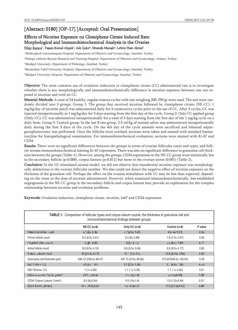
[[AAbbssttrraacctt:: 00118800]] [[OOPP--1177]] [[AAcccceepptteedd:: OOrraall PPrreesseennttaattiioonn]]EEffffeeccttss ooff NNiiccoottiinnee EExxppoossuurree oonn CClloommiipphheennee CCiittrraattee IInndduucceedd RRaattss:: MMoorrpphhoollooggiiccaall aanndd IImmmmuunnoohhiissttoocchheemmiiccaall AAnnaallyyssiiss iinn tthhee OOvvaarriieessNNiillaayy KKaarraaccaa11,, YYaaşşaamm KKeemmaall AAkkppaakk22,, AAssllıı ÇÇaakkıırr33,, MMuussttaaffaa MMaarraaşşllıı44,, LLeebbrriizz HHaallee AAkkttuunn55
1Medicalpark Gaziosmanpasa Hospital, Department of Obstetric and Gynaecology, Istanbul, Turkey2Dıskapı yildirim Beyazit Research and Training Hospital, Department of Obstetric and Gynaecology, Ankara, Turkey3Medipol University, Department of Pathology, Istanbul, Turkey4Bezmialem Vakif University Hospital, Department of Obstetric and Gynaecology, Istanbul, Turkey5Medipol University Hospital, Department of Obstetric and Gynaecology, Istanbul, Turkey
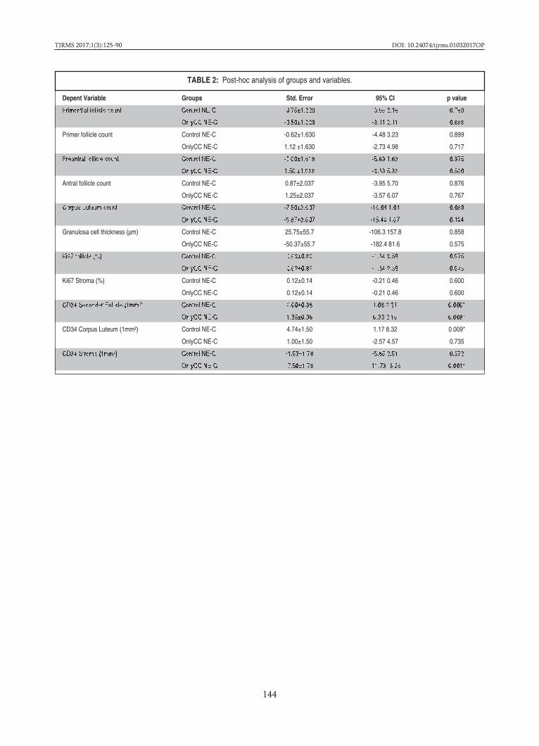
OObbjjeeccttiivvee:: The most common use of ovulation induction in clompihene citrate (CC) administered rats is to investigatewhether there is any morphologically and immunohistochemically difference in nicotine exposure between rats not ex-posed to nicotine and with no CC.MMaatteerriiaall--MMeetthhooddss:: A total of 24 healthy, regular eustrus cycles with rats weighing 200-250 gr were used. The rats were ran-domly divided into 3 groups. Group 1: The group that received nicotine followed by clomiphene citrate (NE-CC): 1mg/kg/day of nicotine patch was administered daily for 3 consecutive cycles prior to the use of CC. After 3 cycles, CC wasinjected intraperitoneally as 1 mg/kg/day for 3 days starting from the first day of the cycle. Group 2: Only CC applied group(Only-CC): CC was administered intraperitoneally for a total of 3 days starting from the first day of the 1 mg/kg cycle on adaily basis. Group 3: Control group: In the last 8 rats group, 2.5 ml/kg of normal saline was administered intraperitoneallydaily during the first 3 days of the cycle. On the 4th day of the cycle animals were sacrificed and bilateral salpin-gooophorectomy was performed. Once the follicles were overlaid, sections were taken and stained with standard haema-toxyline for histopathological examination. For immunohistochemical evaluation, sections were stained with Ki-67 andCD34.RReessuullttss:: There were no significant differences between the groups in terms of ovarian follicular count and types, and folli-cle-stroma immunohistochemical staining Ki-67 expression. There was also no significant difference in granulosa cell thick-ness between the groups (Table-1). However, among the groups, CD34 expressions in the NE-CC group were statistically lessin the secondary follicle (p=0.000), corpus luteum (p=0.012) but more in the overian storm (0.001) (Table-2).CCoonncclluussiioonn:: In the CC-stimulated animal model, we did not observe that transdermal nicotine exposure was morphologi-cally deleterious to the ovarian follicular number. We also could not detect the negative effect of nicotine exposure on thethickness of the granulose cell. Perhaps the effect on the ovarian stimulation with CC may be less than expected, depend-ing on the route or the dose of nicotine administered. However, when examined immunohistochemically, less establishedangiogenesis in the NE-CC group in the secondary follicle and corpus luteum may provide an explanation for the complexrelationship between nicotine and ovulation problems.
KKeeyywwoorrddss:: Ovulation induction, clomiphene citrate, nicotine, ki67 and CD34 expression
NE-CC (n=8) Only-CC (n=8) Control (n=8) P value
Primordial follicle count 5.2 (5± 3.05) 4.7 (4.5± 1.82) 4.5 (4± 2.32) 0.83
Primer follicle count 8.2 (8.5± 2.81) 9.3 (9± 3.88) 7.6 (7.5± 2.97) 0.56
Preantral follicle count 7.5 (8± 3.02) 9 (9± 3.77) 5.5 (4.5± 2.82) 0.12
Antral follicle count 8.6 (8.5± 4.10) 9.8 (9.5± 3.94) 9.5 (8.5± 4.17) 0.82
Corpus Luteum count 23 (22.5± 6.67) 16.1 (14± 9.3) 15.5 (15.5± 4.98) 0.09
Granulosa cell thickness (µm) 548.12 (536.5± 98.47) 497.75 (513± 69.90) 573.87(546.5± 150.54) 0.39
Ki67 Follicle (%) 4.5 (4± 1.41) 5.1 (5.5± 1.95) 5.1 (4.5± 1.55) 0.69
Ki67 Stroma (%) 1 (1± 0.00) 1.1 ( 1± 0.35) 1.1 ( 1± 0.35) 0.61
CD34 Seconder Follicle (1mm²) 0.87 (1±0.64) 2.1 (2±0.64) 2.8 (3±0.99) 0.00*
CD34 Corpus Luteum (1mm²) 8.5 (8±3.54) 9.5 (10±1.6) 13.2 (13±3.49) 0.01*
CD34 Stroma (1mm²) 14.1 (14.5±5.54) 6.6 (6.5±1.4) 12.5 (12.5±2.39) 0.00*
TABLE 1: Comparison of folliculer types and corpus luteum counts, the thickness of granulosa cell and immunohistochemical findings between groups
TJRMS 2017;1(3):125-90 DOI: 10.24074/tjrms.01032017OP
144
Depent Variable Groups Std. Error 95% CI p value
Primordial follicle count Control NE-C -0.75±1.229 -3.66 2.16 0.769
OnlyCC NE-C -0.50±1.229 -3.41 2.41 0.888
Primer follicle count Control NE-C -0.62±1.630 -4.48 3.23 0.899
OnlyCC NE-C 1.12 ±1.630 -2.73 4.98 0.717
Preantral follicle count Control NE-C -2.00±1.618 -5.83 1.83 0.375
OnlyCC NE-C 1.50 ±1.618 -2.33 5.33 0.560
Antral follicle count Control NE-C 0.87±2.037 -3.95 5.70 0.876
OnlyCC NE-C 1.25±2.037 -3.57 6.07 0.767
Corpus Luteum count Control NE-C -7.50±3.607 -16.04 1.04 0.089
OnlyCC NE-C -6.87±3.607 -15.42 1.67 0.124
Granulosa cell thickness (µm) Control NE-C 25.75±55.7 -106.3 157.8 0.858
OnlyCC NE-C -50.37±55.7 -182.4 81.6 0.575
Ki67 follicle (%) Control NE-C 0.62±0.82 -1.34 2.59 0.675
OnlyCC NE-C 0.62±0.82 -1.34 2.59 0.675
Ki67 Stroma (%) Control NE-C 0.12±0.14 -0.21 0.46 0.600
OnlyCC NE-C 0.12±0.14 -0.21 0.46 0.600
CD34 Seconder Follicle (1mm²) Control NE-C 2.00±0.38 1.08 2.91 0.000*
OnlyCC NE-C 1.25±0.38 0.33 2.16 0.008*
CD34 Corpus Luteum (1mm²) Control NE-C 4.74±1.50 1.17 8.32 0.009*
OnlyCC NE-C 1.00±1.50 -2.57 4.57 0.735
CD34 Stroma (1mm²) Control NE-C -1.62±1.78 -5.86 2.61 0.572
OnlyCC NE-C -7.50±1.78 -11.73 -3.26 0.001*
TABLE 2: Post-hoc analysis of groups and variables.
DOI: 10.24074/tjrms.01032017OP TJRMS 2017;1(3):125-90
145
[[AAbbssttrraacctt:: 00119900]] [[OOPP--1188]] [[AAcccceepptteedd:: OOrraall PPrreesseennttaattiioonn]]MMoonnooccyyttee ttoo HHDDLL CChhoolleesstteerrooll RRaattiioo iinn PPaattiieennttss wwiitthh PPoollyyccyyssttiicc OOvvaarryy SSyynnddrroommeeAAkkıınn UUssttaa11,, EEyyüüpp AAvvccıı22,, ÇÇaağğllaa BBaahhaarr BBüüllbbüüll11,, EErrttaann AAddaallıı11
1Department of Obstetrics and Gynecology, School of Medicine, Balikesir University, Balikesir, Turkey2Department of Cardiology, School of Medicine, Balikesir University, Balikesir, Turkey
OObbjjeeccttiivvee:: Polycystic ovary syndrome (PCOS) is one of the most common endocrinopathy among women of reproductiveage. It is characterized by oligo and/or anovulation, clinical and/or biochemical sign hyperandrogenism, and the appearanceof polycystic ovaries on ultrasound. Women with PCOS are more likely than other women to have increased in obesity, in-sulin resistance, hyperandrogenism, and chronic low-grade inflammation. PCOS affects 5-10 % of women in reproductiveage, and long-term complications include type 2 diabetes mellitus (DM), cardiovascular diseases (CVD), and infertility. Thereare various biomarker alterations associated with insulin resistance and low-grade inflammation in PCOS. Monocyte to HDLratio (MHR) is a recently emerged inflammation-based marker and recent studies have shown that MHR is a new predictorand prognostic indicator of cardiovascular diseases. Therefore, we aimed to investigate the MHR alteration and its useful-ness for prediction of cardiovasculary disease risk in PCOS.MMaatteerriiaall--MMeetthhooddss:: In the study population, we evaluated 26 consequtive patients with PCOS and 30 age and BMI-matchedcontrols. PCOS was diagnosed with the Rotterdam criteria. MHR was compared between PCOS and control group. Rela-tionship between MHR and the clinicopathological variables of PCOS were also evaluated. MedCalc Statistical SoftwareProgram version 17.2 (MedCalc, Belgium) was used for statistical analysis. A P-value of <0.05 was considered statistically sig-nificant.RReessuullttss:: MHR was higher in PCOS group than the age and BMI matched non-PCOS subjects (p<0.05). MHR were signifi-cantly correlated with HOMA-IR and CRP levels in the PCOS group. There was no correlation between MHR and patientsage.CCoonncclluussiioonnss:: Our study demonstrated that patients with PCOS have higher MHR than those of women in the control group.Increased MHR may be related to the future cardiovascular disease risk in PCOS patients. Further research is needed toevaluate the association between MHR and PCOS.
KKeeyywwoorrddss:: Monocyte to HDL cholesterol ratio, cardiovascular disease, PCOS

TJRMS 2017;1(3):125-90 DOI: 10.24074/tjrms.01032017OP
146
[[AAbbssttrraacctt:: 00008866]] [[OOPP--1199]] [[AAcccceepptteedd:: OOrraall PPrreesseennttaattiioonn]]TThhee EEffffeeccttss ooff VViittaammiinn DD LLeevveellss oonn TTuurrkkiisshh WWoommeenn''ss IIVVFF OOuuttccoommeess:: AA PPrroossppeeccttiivvee CCoohhoorrtt SSttuuddyyİİllkkaayy BBoozz11,, GGaammzzee TTeesskkeerreeccii11,, MMuurraatt ÖÖzzeekkiinnccii22
1Nursing Faculty, Akdenız Unıversıty, Antalya, Turkey2Medicine Faculty, Akdenız Unıversıty, Antalya, Turkey
OObbjjeeccttiivvee:: It is known that women with infertility have low vitamin D levels. There are conflicting results about potentialimpact of vitamin D deficiency on IVF outcomes. This study aimed to determine for the first time the vitamin D levels ofTurkish women who underwent embryo transfer (ET), and to demonstrate the correlation between vitamin D levels and invitro fertilization (IVF) results.MMaatteerriiaall--MMeetthhoodd:: This is the first prospective cohort study from Turkey. The duration of the study was June 15, 2015, andApril 1, 2016. A total of 208 infertile women who underwent IVF-ET were enrolled in the study. The vitamin D levels ofwomen with ET were measured by assessing the circulating levels of 25-hydroxyvitamin D (25(OH)D). Pregnancy was de-fined by serially increasing serum β-hCG titers to at least 25 IU/L, within 12 days after the cleavage stage. Correlation analy-sis was used to evaluate the correlation of vitamin D levels with socio-demographic variables and infertility treatment results,and chi-square analysis was used to compare the pregnancy rates. RReessuullttss:: The study found that 4.3% of women had replete vitamin D level (>30 ng/mL), 23.6% of women had insufficientvitamin D levels (20-29.99 ng/mL), and 72.1% of women had deficient vitamin D levels (<20ng/mL). Vitamin D levels ofwomen decreased as their body mass index (BMI), infertility diagnosis and duration of infertility treatment increased. Nostatistically significant difference was found between chemical and clinical pregnancy rates and vitamin D levels (p values:0.132 and 0.303, respectively). CCoonncclluussiioonn:: The women who underwent ET in Turkey had very high rates of vitamin D deficiency, and there was no cor-relation between their vitamin D deficiencies and pregnancy results. It is suggested that the effects of vitamin D levels onpregnancy outcomes should be analyzed after applying a vitamin D support program at the beginning of ET.
KKeeyywwoorrddss:: IVF Outcomes, Turkish Women, Vitamin D
DOI: 10.24074/tjrms.01032017OP TJRMS 2017;1(3):125-90
147
[[AAbbssttrraacctt:: 00009966]] [[OOPP--2200]] [[AAcccceepptteedd:: OOrraall PPrreesseennttaattiioonn]]SSuurrvviivvaall ooff CChhrroommoossoommaallllyy AAbbnnoorrmmaall EEmmbbrryyooss ttoo BBllaassttooccyysstt SSttaaggeeEEnnvveerr KK DDiirriiccaann11,, BBaattuu AAyyddıınnuurraazz22,, ÖÖzzlleemm AAkkssüünnggeerr33
1Department of Obstetrics and Gynecology, Center for Reproductive Endocrinology and Assisted Reproduction, Akdeniz University, Faculty of Medicine, Antalya, Turkey2Center for Assisted Reproduction, Private Gelecek IVF Center, Antalya, Turkey3Center for Assisted Reproduction, Memorial Antalya Hospital, Antalya, Turkey
IInnttrroodduuccttiioonn:: Several authors have suggested that many embryos arrest at the cleavage or morula stage during embryo cul-ture. Previous studies proposed that embryo arrest at the morula stage may act as a selection against chromosomal abnor-malities. The aim of this study was to evaluate the overall developmental outcome of embryos according to preimplantationgenetic screening (PGS) results.MMaatteerriiaall--MMeetthhooddss:: Here we report the outcome of 21 intracytoplasmic sperm injection (ICSI) cases with PGS. All patientsundergoing an ICSI treatment for male factor infertility and PGS with a day 5 embryo transfer schedule in 2013-2014 wereenrolled in this study. PGS was performed to 15 cases due to multiple implantation failures (MIF), 4 cases due to MIF to-gether with advanced maternal age (AMA) and 2 cases due to Robertsonian translocations. All cases were counseled by ahuman genetics specialist and informed written consent was obtained from each couple before PGS procedures. All cases un-derwent single blastomere biopsy on day 3 by laser zona dissection and aspiration. The survival and quality of chromoso-mally normal and abnormal embryos were analyzed retrospectively.RReessuullttss:: The mean female age was found to be 31,71 ± 4,36 at all cases. Embryo transfer was not performed to 2 cases as allembryos were abnormal and clinical pregnancy results could not be obtained from 2 cases. The overall clinical pregnancyrate was 23,53% with an implantation rate of 15,79%. A total number of 121 blastomeres were analyzed, 52 were found tobe normal, 65 blastomeres were abnormal and no result was obtained from 4 blastomeres. Complex aneuploidy was the mostfrequent abnormality (n=15). Other abnormalities were monosomies (n=21), trisomies (n=13), monosomy-trisomy combi-nations (n=3), haploidy (n=3), triploidy (n=2), tetraploidy (n=1), polyploidy (n=1), XXY (n=1), chaotic (n=2) and multinu-clear fragmentation (n=3). Of the 52 normal embryos, 7 were arrested at the cleavage stage and 16 were arrested at themorula stage. Of the 65 abnormal embryos, 30 were arrested at the cleavage stage and 21 were arrested at the morula stage.Arrested development at the cleavage stage was 13,46% for the normal embryos and 46,15% for the abnormal embryos(P<0,001). The overall embryo development rate of normal and abnormal embryos to blastocyst stage was 55,77% and 21,54%respectively (P<0,001).CCoonncclluussiioonnss:: 13% of the normal embryos and 46% of the abnormal embryos may arrested at the cleavage stage during ex-tended culture to the blastocyst stage. Our study shows that culture to blastocyst stage may further select against some chro-mosomal abnormalities, but it should be clarified that, although abnormal embryos have limited potential of developmentto the blastocyst stage, day 5 embryo morphology could not be used to select chromosomally normal embryos for transferdue to our observations that abnormal embryos-although less than the normal ones-may develop into blastocysts. Ongoingpregnancy rates and expanded genetic investigations are needed to clarify the developmental potential of normal and ab-normal embryos to the viable offspring.
KKeeyywwoorrddss:: Preimplantation genetic screening, assisted reproduction, blastocyst
TJRMS 2017;1(3):125-90 DOI: 10.24074/tjrms.01032017OP
148
[[AAbbssttrraacctt:: 00009988]] [[OOPP--2211]] [[AAcccceepptteedd:: OOrraall PPrreesseennttaattiioonn]]TThhee EEffffeecctt ooff EEssttrrooggeenn RReeppllaacceemmeenntt iinn AAddddiittiioonn ttoo PPrrooggeesstteerroonn ffoorr LLuutteeaall PPhhaassee SSuuppppoorrtt iinn IIVVFF--IICCSSII AAnnttaaggoonniisstt CCyycclleessCCeemmrree AAllaann11,, HHüüsseeyyiinn GGöörrkkeemmllii22
1Afyon Dinar State Hospital2Konya Necmettin Erbakan University Meram Medical Faculty
SSttuuddyy qquueessttiioonn:: To find out the effectiveness of the estrogen replacement for luteal phase support in antagonist cycles.SSuummmmaarryy aannsswweerr:: There were no significant differences between groups in terms of clinical pregnancy, biochemical preg-nancy, ongoing pregnancy and abortus ratios.WWhhaatt iiss kknnoowwnn aallrreeaaddyy:: There are little studies about estrogen replacement in luteal phase support. Studies related with ag-onist cycles gave very little effectiveness of estrogen replacement in luteal phase support but these are not proved by meta-analyses. Studies related with IVF-ICSI antagonist cycles are newly published and we don't have any metaanalyses relatedwith this procedure. In order to find out the effectiveness of estradiol in antagonist cycles for luteal phase support was ourmain aim in the study. SSttuuddyy ddeessiiggnn,, ssiizzee,, dduurraattiioonn:: Retrospectively two groups were designed in our study. In group one only progesteron hasbeen used both vaginally and intramuscularly. In group two we added estradiol TTS 7.8 mg daily. In each group we have 64patients and we have followed the patients until clinical pregnancies. Luteal phase support in each group has been admin-istered until ten weeks of gestational age. In group two estradiol replacement has been stopped at the day of βhCG result.PPaarrttiicciippaannttss//mmaatteerriiaallss,, sseettttiinngg,, MMeetthhooddss:: In this retrospective study, patient files were scanned and group which had GnRHantagonist protocol, having daily transdermal (7.8 mg) E2 supplementation in luteal phase support was compared with con-trol group in terms of pregnancy outcomes. The study and control group consists of 64 patients. β-hCG levels of all patientswere measured 12 days after embryo transfer. If the result is positive, estradiol is discontinued and progesterone support iscontinued until the 10th week of gestation. MMaaiinn rreessuullttss aanndd tthhee rroollee ooff cchhaannccee:: In our study, the use of estrogen in the GnRH antagonist protocol luteal phase supportdid not show any positive or negative effects on pregnancy outcomes. Although pregnancy rates are not statistically signif-icant in the control group, they are higher than in the study group. The statistically insignificant tendency may be due tolimited sample size. In IVF, if the main success criterion is expressed by the number of live births, the results of long termpregnancy registration of the patients are needed to express this. For a more objective assessment, there is a need for prospec-tive studies comparing the numbers of live births with larger sample sizes, with a more homogeneous distribution of de-mographic characteristicsLLiimmiittaattiioonnss,, rreeaassoonnss ffoorr ccaauuttiioonn:: No limitations.WWiiddeerr iimmpplliiccaattiioonnss ooff tthhee FFiinnddiinnggss:: For a more objective assessment, there is a need for prospective studies comparing thenumbers of live births with larger sample sizes, with a more homogeneous distribution of demographic characteristics.
KKeeyywwoorrddss:: Luteal phase support, estradiol, antagonist protocol

DOI: 10.24074/tjrms.01032017OP TJRMS 2017;1(3):125-90
149
[[AAbbssttrraacctt:: 00110044]] [[OOPP--2222]] [[AAcccceepptteedd:: OOrraall PPrreesseennttaattiioonn]]FFoolliikkuulleerr SSııvvıı GGeellaattiinnaazz DDüüzzeeyyiinniinn OOoossiitt KKaalliitteessii vvee FFeerrttiilliizzaassyyoonnaa EEttkkiissiiEEssrraa NNuurr TToollaa11,, EErrddaall BBiilleenn11,, HHiillmmii BBaahhaa OOrraall11,, DDuuyygguu KKuummbbuull DDooğğuuçç22,, İİllkkeerr GGüünnyyeellii11,, İİlltteerr İİllhhaann22
1Süleyman Demirel Üniversitesi Tıp Fakültesi, Kadın Hastalıkları ve Doğum AD, Isparta, Türkiye2Süleyman Demirel Üniversitesi Tıp Fakültesi, Biyokimya AD, Isparta, Türkiye
AAmmaaçç:: Matriks metalloproteinazlar (MMP) ve bunların aktivitesini dengeleyen doku inhibitörleri (TIMP) tarafından düzen-lenen doku remodelingi folikulogenez, ovulasyon, korpus luteum formasyonu ve regresyonu ve implantasyon gibi pek çokreproduktif süreçte gereklidir. Gelatinazlar, MMP ailesinin bir üyesi olup çinko bağımlı proteolitik enzimlerdir ve MMP-2ve MMP-9 olmak üzere 2 tipi vardır. İnsanda granuloza hücrelerinde, preimplante embriyolarda, servikal fibroblastlar, tro-foblastlar ve endometrial stromal hücrelerde gelatinaz ekspresyonu gösterilmiştir. Preovulatuar folikullerde gelatinaz ak-tivitesinin arttığı ve ovulasyonda folikul duvarının kollajen içeriğinin gelatinazlar tarafından parçalanarak azaldığı raporedilmiştir. Çalışmamızın amacı açıklanamayan infertilitesi olan hastaların folikul sıvısında MMP-2, MMP-9 ve TIMP se-viyeleri ile oosit kalitesi ve fertilizasyon arasındaki ilişkiyi değerlendirmek ve farklı iki invitro fertilizasyon (IVF) protokoluarasındaki folikuler sıvı gelatinaz ve TIMP seviyelerini karşılaştırmaktır.MMaatteerryyaall MMeettoodd:: Açıklanamayan infertilite nedeniyle IVF tedavisine giden 60 hasta çalışmaya alındı. Sistemik hastalık vekronik ilaç kullanımı olan hastalar çalışma dışında bırakıldı. 30 hastaya gonadotropin releasing hormon (GnRH) agonisti ileuzun, 30 hastaya ise GnRH antagonisti ile kısa protokol uygulanarak rekombinant ve/veya üriner folikul stimulan hormon(FSH) ile kontrollu overyan stimulasyon yapıldı. Oosit pick-up işlemi sırasında her folikulden ayrı ayrı folikul sıvısı topla-narak folikuler sıvı gelatinaz seviyeleri ticari kitle, ELISA yöntemiyle (Boster MMP-2 ve MMP-9 ELISA Kit, California,USA) ölçüldü. Alınan oositlerin kalitesi ışık mikroskobunda bakılan 4 parametreye göre değerlendirildi (Tablo 1). Parame-trelerden toplam 3-4 puan alan oositler grade A (iyi kalite), 2-3 puan alan grade B, 0-1 puan alan oositler grade C (kötükalite) olarak kabul edildi. Hastalar toplanan metafaz II (MII) oosit sayısına göre iki gruba ayrıldı. MII<=5 ise ‘poor respon-der’, MII>5 ise ‘normoresponder’ olarak adlandırıldı. Tüm oositlere intrastoplazmik sperm injeksiyonu yapıldı ve 2 pronuk-leus görülen oositler fertilize olarak kabul edildi.SSoonnuuççllaarr:: Agonist protokol uygulanan gruptaki hastaların yaş ortalaması (32.8±4.8) antagonist gruba göre (29.7±4) anlamlıolarak daha yüksekti (p=0.01). Vücut kitle indeksi, 3. gün bakılan hormon seviyeleri, toplanan oosit sayısı ve fertilize oositsayısı açısından agonist ve antagonist grup arasında fark yoktu. MMP-2 ve MMP-9 seviyesi agonist grupta (231.0±13.3 ng/mL,5.671±1.582 pg/mL, sırayla) antagonist gruba göre (179.7±23.2 ng/mL, 4.060.1±1.965 pg/mL, sırayla) daha yüksekti (p=0.001)(Tablo 2). Fertilize oositlerdeki MMP-2 seviyesiyle (208.2±31.6 ng/mL) fertilize olmayan oositlerdeki MMP-2 seviyesi(186.6±29.1 ng/mL) farklılık göstermezken (p=0.5), MMP-9 seviyesi fertilize grupta (5.095±1.850 pg/mL) nonfertilize grubagöre (3.374±2.012 pg/mL) anlamlı olarak artmıştı (p=0.02). Toplanan oosit sayısına göre poor responder ve normorespondergrup karşılaştırıldığında her iki grup arasında folikuler sıvı MMP-2 ve MMP-9 seviyeleri homojen olarak dağılmıştı (p=0.9,p=0.5, sırasıyla). Oosit kalitesi ve gelatinaz seviyeleri arasındaki ilişkiye bakıldığında folikuler sıvı MMP-2 seviyeleri gradeA, B ve C oositler arasında homojen olarak dağılmıştı (Şekil 1). MMP-9 seviyesi ise grade A oositlerde grade C oositlere göre(p=0.01), grade B oositlerde ve grade C oositlere göre anlamlı olarak dha yüksek bulundu (p=0.03) (Şekil 2).SSoonnuuçç:: Folikuler sıvıda artmış MMP-9 seviyesi açıklanamayan infertilitesi olan hastalarda yüksek oosit kalitesi ve fertiliza-syon potansiyeli ile ilişkilidir. Daha yüksek doz gonadotropin uygulaması, GnRH agonistin flare-up etkisi ve hasta yaşlarıarasındaki farklılık GnRH agonist protokolu uygulanan gruptaki yüksek gelatinaz seviyelerinin nedenleri olabilir. GnRH ag-onist protokol uygulamasının gelatinaz seviyesi üzerindeki pozitif etkisi ileri araştırmalarla araştırılmalıdır.
AAnnaahhttaarr KKeelliimmeelleerr:: Gelatinaz, in vitro fertilizasyon, oosit kalitesi
ŞEKİL 1: Oosit kalitesi ve folikuler sıvı MMP-2 se-viyesi arasındaki ilişki.
ŞEKİL 2: Oosit kalitesi ve folikuler sıvı MMP-9 se-viyesi arasındaki ilişki.
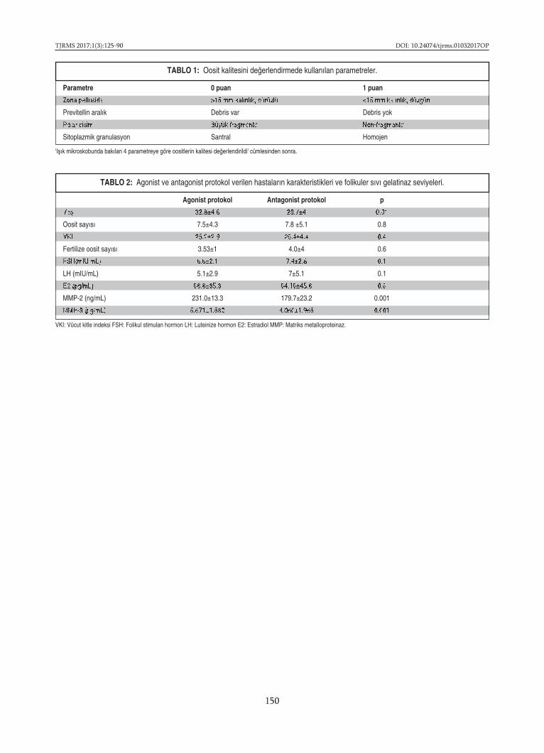
TJRMS 2017;1(3):125-90 DOI: 10.24074/tjrms.01032017OP
150
TABLO 1: Oosit kalitesini değerlendirmede kullanılan parametreler.
'Işık mikroskobunda bakılan 4 parametreye göre oositlerin kalitesi değerlendirildi' cümlesinden sonra.
Parametre 0 puan 1 puan
Zona pellusida >15 mm kalınlık, pürüzlü <15 mm kalınlık, düzgün
Previtellin aralık Debris var Debris yok
Polar cisim Büyük fragmente Non-fragmante
Sitoplazmik granulasyon Santral Homojen
TABLO 2: Agonist ve antagonist protokol verilen hastaların karakteristikleri ve folikuler sıvı gelatinaz seviyeleri.
VKI: Vücut kitle indeksi FSH: Folikul stimulan hormon LH: Luteinize hormon E2: Estradiol MMP: Matriks metalloproteinaz.
Agonist protokol Antagonist protokol p
Yaş 32.8±4.8 29.7±4 0.01
Oosit sayısı 7.5±4.3 7.8 ±5.1 0.8
VKI 25.2±2.9 26.4±4.4 0.4
Fertilize oosit sayısı 3.53±1 4.0±4 0.6
FSH(mIU/mL) 6.6±2.1 7.4±2.8 0.1
LH (mIU/mL) 5.1±2.9 7±5.1 0.1
E2 (pg/mL) 68.8±35.3 64.16±45.8 0.6
MMP-2 (ng/mL) 231.0±13.3 179.7±23.2 0.001
MMP-9 (pg/mL) 5.671±1.582 4.060±1.965 0.001

DOI: 10.24074/tjrms.01032017OP TJRMS 2017;1(3):125-90
151
[[AAbbssttrraacctt:: 00117766]] [[OOPP--2233]] [[AAcccceepptteedd:: OOrraall PPrreesseennttaattiioonn]]BBllaassttooccyysstt VViittrriiffiiccaattiioonn DDeessppiittee PPoooorr OOooccyyttee MMaattuurraattiioonn iinn aa PPaattiieenntt wwiitthh LLyymmpphhoommaa,, WWhhoossee PPrreeggnnaannccyy wwaass RReeccooggnniizzeedd DDuurriinngg RRaannddoomm SSttaarrtt OOvvaarriiaann SSttiimmuullaattiioonnMMuurraatt SSöönnmmeezzeerr11,, YYaavvuuzz EEmmrree ŞŞüükküürr11,, SSiinnaann ÖÖzzkkaavvuukkççuu22,, FFaattmmaa CCeeyyllaann IIllhhaann33,, CCooşşkkuunn ŞŞiimmşşiirr44,, CCeemm AAttaabbeekkooğğlluu11
1Ankara University School of Medicine, Department of Obstetrics and Gynecology2Ankara University School of Medicine, Department of Obstetrics and Gynecology, Centre for Assisted reproduction3Yenimahalle Training and Research Hospital, Department of Obstetrics and Gynecology4Liv Ankara Hospital, Department of Obstetrics and Gynecology
BBaacckkggrroouunndd:: Embryo/oocyte cryopreservation seems among the best options where possible for fertility preservation inwomen suffering from malignant diseases. However, in those who need to start a cytotoxic treatment very soon, one of thebiggest challenges in fertility preservation is the time required to complete the ovarian stimulation. To preclude this prob-lem random start controlled ovarian hyperstimulation (RS-COH) protocols have come into use. Here, we present a patientwith diffuse large B cell lymphoma whose pregnancy was recognized during treatment.CCaassee:: A 28-year-old woman who had a newly diagnosed diffuse large B cell lymphoma (active B cell type) was referred toour Fertility Preservation Unit. She was scheduled for chemotherapy plus rituximab therapy (R-CHOP) soon. At admissionshe was on 19th day of her menstrual cycle. Ultrasonography revealed 10 mm endometrial thickness and normal sizedovaries with 10 antral follicles. Her hormonal profile was as follows; estradiol: 202 pg/ml, progesterone: 11.2 ng/ml, FSH: 0.3IU/L, LH: 0.1 IU/L. Hence, RS-COH was started immediately by letrozole 5mg/day plus recombinant follicle stimulating hor-mone (rFSH) 200 IU/day. On day 4, estradiol was 58 pg/ml and progesterone was 9.4 ng/ml. Starting at day 5 rFSH dose wasset to 150 IU/day and 75 IU/day hMG was added to protocol. On day 6, an 8 mm of gestational sac was recognized and ß-hCG was 7867 IU/L. However, because of the time constraints COH was continued. On day 9 GnRH antagonist was addedto the protocol. On day 12 estradiol was 591 pg/ml and progesterone was 16.1 ng/ml. After 12 days of stimulation 40 mi-crogram of leuprolide acetate was administered when the largest three follicles attained a mean diameter of 18 mm, whilegeneral cohort follicles >13 mm. Dilatation and curettage was also performed under general anesthesia immediately afteroocyte retrieval. Among 10 retrieved oocytes, only three were evaluated as matured with intensely granulated cytoplasm.Two metaphase-I and 5 GV oocytes were incubated overnight for in vitro maturation in embryo culture medium and nextmorning ICSI was performed on 3 metaphase-II oocytes. All of the inseminated, in vitro matured oocytes were fertilized andcleaved normally in successive days. On the 5th day of insemination, 2 early blastocysts were vitrified. DDiissccuussssiioonn:: Progressive rise in progesterone or LH levels on the day of ovulation induction may result in poor oocyte qual-ity and IVF outcome. Premature luteinization in granulose cells of growing follicles may result in compromised follicle de-velopment and oocyte maturation. High levels of ß-hCG in presented case resulted in high progesterone levels and secondaryluteinizing effect. We believe that diminished maturation rate (30%) and arrested fertilization of in vivo matured oocytesmay due to premature luteinization and incomplete cytoplasmic competence. On the other hand, avoiding suboptimal en-docrine microenvironment by incubating in vitro, may trigger meiosis and remove suppressive effect of high progesteroneand ß-hCG. Patients with high progesterone levels on the day of ovulation induction may benefit from overnight in vitromaturation of oocytes despite low maturation rates.
KKeeyywwoorrddss:: Pregnancy, progesterone, random start controlled ovarian hyperstimulation
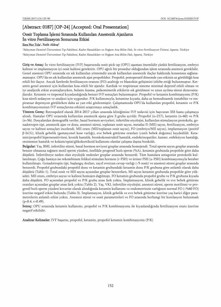
TJRMS 2017;1(3):125-90 DOI: 10.24074/tjrms.01032017OP
152
[[AAbbssttrraacctt:: 00118877]] [[OOPP--2244]] [[AAcccceepptteedd:: OOrraall PPrreesseennttaattiioonn]]OOoossiitt TTooppllaammaa İİşşlleemmii SSıırraassıınnddaa KKuullllaannııllaann AAnneesstteezziikk AAjjaannllaarrıınn İİnn vviittrroo FFeerrttiilliizzaassyyoonn SSoonnuuccuunnaa EEttkkiissiiEEssrraa NNuurr TToollaa11,, FFaattiihh AAkkkkuuşş22
1Süleyman Demirel Üniversitesi Tıp Fakültesi, Kadın Hastalıkları ve Doğum Ana Bilim Dalı, İn vitro Fertilizasyon Ünitesi, Isparta, Türkiye2Süleyman Demirel Üniversitesi Tıp Fakültesi, Kadın Hastalıkları ve Doğum Ana Bilim Dalı, Isparta, Türkiye
GGiirriişş vvee AAmmaaçç:: İn vitro fertilizasyon (IVF) başarısında oosit pick-up (OPU) aşaması önemlidir çünkü fertilizasyon, embryokalitesi ve implantasyon iyi oosit kalitesi gerektirir. OPU ağrılı bir prosedur olduğundan işlem sırasında anestezi gereklidir.Genel anestezi OPU sırasında en sık kullanılan yöntemdir ancak kullanılan anestezik ilaçlar hakkında konsensus sağlana-mamıştır. OPU’da en sık kullanılan anestezik ajan propofoldur. Propofol, postoperatif dönemde yan etkinin az görüldüğü kısaetkili bir ilaçtır. Ancak farelerde fertilizasyon oranını (FO) azalttığı ve blastokist gelişimini inhibe ettiği bulunmuştur. Ket-amin genel anestezi için kullanılan kısa etkili bir ajandır. Kardiak ve respiratuar sisteme minimal depresif etkili olması veiyi analjezik etkisi avantajlarıyken, bulantı kusma, psikomimetik etkilerin sık görülmesi ve uzun ayılma süresi dezavanta-jlarıdır. Ketamin ve tiopental kıyaslandığında benzer IVF sonuçları bulunmuştur. Propofol ve ketamin kombinasyonu (P/K)kısa süreli sedasyon ve analjezi için uygundur. P/K kullanımıyla, ketamine kıyasla, daha az hemodinamik instabilite ve res-piratuar depresyon görülürken daha az yan etki gözlenmiştir. Çalışmamızda OPU’da kullanılan propofol, ketamin ve P/Kkombinasyonunun IVF sonuçlarına etkisini araştırmayı amaçladık.YYöönntteemm GGeerreeçç:: Retrospektif olarak 2014-2017 yılları arasında kliniğimize IVF tedavisi için başvuran 333 hasta çalışmayaalındı. Hastalar OPU sırasında kullanılan anestezik ajana göre 3 gruba ayrıldı: Propofol (n=217), ketamin (n=60) ve P/K(n=56). Dosyalardan demografik veriler, bazal hormon seviyeleri, infertilite etyolojisi, kullanılan stimulasyon protokolu, go-nadotropin tipi, anestezik ajan ve dozu, anestezi süresi, toplanan oosit sayısı, metafaz II (MII) sayısı, fertilizasyon, embriyosayısı ve kalitesi sonuçları incelendi. MII oranı (MII/toplanan oosit sayısı), FO (embriyo/MII sayısı), implantasyon (pozitifβ-hCG), klinik gebelik (gestasyonel kese varlığı), eve bebek götürme oranları (canlı bebek doğumu) kaydedildi. Keta-min/propofol hipersensitivitesi, kronik hastalık, bronkokonstruktif hastalık, endokrinopatiler, kanser, enfeksiyon hastalığı,otoimmun hastalık ve kokain/opiat/glikokortikoid kullanımı olanlar çalışma dışına bırakıldı. BBuullgguullaarr:: Yaş, BMI, infertilite süresi, bazal hormon seviyesi gruplar arasında homojendi. Total sperm sayısı gruplar arasındabenzer olmasına rağmen motil sperm yüzdesi, özellikle progresif hızlı sperm (%A), ketamin grubunda propofole göre dahadüşüktü. İnfertiliteye neden olan etyolojik nedenler gruplar arasında benzerdi. Tüm hastalara antagonist protokolü kul-lanılmıştı. Çoğu hastaya ise rekombinant folikul stimulan hormon (r-FSH) ve üriner FSH (u-FSH) kombinasyonuyla beraberkullanılmıştı. Gonadotropin tipi, başlangıç dozları, zayıf overyan cevap varlığı (<5 oosit) ve anestezi süresi gruplar arasındabenzerdi. Propofol grubundaki propofol dozu ve ketamin grubundaki ketamin dozu P/K grubuna göre anlamlı olarak dahadüşüktü (Tablo 1). Total oosit ve MII sayısı açısından gruplar benzerken, MI sayısı ketamin grubunda propofole göre yük-sekti. MII oranı, embriyo sayısı ve kalitesi homojen dağılmıştı. FO ketamin grubunda propofol grubu ve P/K grubuna kıyasladaha düşüktü. FO açısından propofol ve P/K grubu arası fark yoktu. İmplantasyon, klinik gebelik ve eve bebek götürmeoranları açısından gruplar arası fark yoktu (Tablo 2). Yaş, VKI, infertilite etyolojisi, anestezi süresi, sperm motilitesi ve pro-gresif hızlı sperm yüzdesi kovariat olarak alındığında ketamin kullanımı ve endometriozis varlığının normal FO (>%60 FO)üzerine negatif etkisi bulundu (Tablo 3). İmplantasyon, klinik gebelik ve eve bebek götürme üzerine yaş harici diğer para-metrelerin anlamlı etkisi yoktu. Anestezi süresi ve oosit parametreleri ve FO arasında herhangi bir korelasyon bulunmadı(p=0.4, r=0.49).SSoonnuuçç:: OPU sırasında ketamin kullanımı, propofol ve P/K kombinasyonu ile kıyaslandığında fertilizasyon oranı üzerinenegatif etkilidir.
AAnnaahhttaarr KKeelliimmeelleerr:: IVF başarısı, propofol, ketamin, propofol ketamin kombinasyonu (P/K)

DOI: 10.24074/tjrms.01032017OP TJRMS 2017;1(3):125-90
153
Propofol grubu n=217 Ketamin grubu n=60 P/K grubu n=56 p değeriYaş (yıl) 31.94±5.91 31.73±4.81 30.58±5.19 0.2VKİ (kg/m2) 25.8±4.79 25.12±4.5 25.61±4.7 0.6İnfertilite süresi (yıl) 6.46±4.58 7.13±3.65 5.9±3.93 0.3FSH (mIU/ml) 8.99±6.3 7.52±2.38 8.57±4.51 0.1LH (mIU/ml) 5.98±4.08 5.20±3.04 6.43±8.83 0.4E2 (pg/ml) 58.43±65.88 56.86±43.54 60.79±86.43 0.9PG (ng/ml) 1.01±1.37 0.85±0.90 0.81±0.38 0.4Sperm parametreleri
Total sperm sayısı (milyon) 60.52±60.81 65.87±88.23 66.03±69.78 0.7Motilite (%) 45.88±25.13 33.08±24.21 44.26±26.14 0.002*A (%) 12.98±13.74 7.71±10.55 11.89±14.06 0.026**B (%) 32.3±20.44 25.68±18.92 32.5±21.66 0.07C (%) 12.23±9.77 14.33±12.8 12.25±9 0.3
İnfertilite etyolojisiPKOS 34/217 (15.7%) 6/60 (10%) 8/56 (14.3%)Açıklanamayan 60/217 (27.6%) 17/60 (28.3%) 14/56 (25%)DOR 50/217 (23%) 10/60 (16.7%) 14/56 (25%) 0.16Tubal 19/217 (8.8%) 0/60 (0%) 2/56 (3.6%)Endometriozis 3/217 (1.4%) 1/60 (1.7%) 1/56 (1.8%)Erkek faktörü 51/217 (23.5%) 26/60 (43.3%) 17/56 (30.4%)
Sigara kulllanımı (%) 16/217 (7.4%) 5/60 (8.3%) 8/56 (14.3%) 0.2Alkol kullanımı (%) 1/217 (0.5%) 0/60 (0%) 0/56 (0%) 0.7Gonadotropin tipi
r-FSH (%) 64/217 (29.5%) 15/60 (25%) 15/56 (26.8%) 0.7u-FSH+r-FSH 153/217 (70.5%) 45/60 (75%) 41/56 (73.2%)
r-FSH başlama dozu 249.02±62.40 232.91±64.35 238.39±55.58 0.1u-FSH başlama dozu 132.18±42.46 140.55±39.27 143.75±26.4 0.1Zayıf overyan cevap (<5 oosit) 72/217 (33.3%) 18/60 (30.5%) 17/56 (30.4%) 0.8Anestezi süresi (dk) 27.28±12.15 24.16±8.49 28.83±12.32 0.8Anestezik ilaçların ortalama dozu (mg) 156.08±47.37 88.03±28.34 P=139.28±50.37 <0.0001
K=44.55±18.93
TABLO 1: Bazal demografik veriler, overyan stimulasyon karakteristikleri ve anestezik parametreler.
VKI: Vücut kite indeksi; FSH: Folikul stimulan hormon; LH: Luteinizan hormon; E2: Estradiol; PG: Progesteron; TSH: Troid stimulan hormon; PRL: Prolaktin; A: Progresif hızlı spermyüzdesi; B: Progresif yavaş sperm yüzdesi; C: Nonprogressive sperm yüzdesi; PKOS: Polikistik over sendromu; DOR: Düşük overyan rezerv; r-FSH: Rekombinan folikul stimulan hor-mon; u-FSH: Üriner folikul stimulan hormon. OHSS: Overyan hiperstimulasyon sendromu. *: p değeri Bonferroni testi ile propofol ve ketamin grubu arasında anlamlı (p=0.002), pro-pofol ve P/K grubu (p=1) ve ketamin ve P/K grubu arasında (p=0.052) anlamlı değil. **: p değeri Bonferroni testi ile propofol ve ketamin grubu arasında anlamlı (p=0.02), propofol veP/K grubu (p=1) ve ketamin ve P/K grup arasında anlamlı değil (p=0.2).
Propofol grubu Ketamin grubu P/K grubu p
n=217 n=60 n=56 değeri
Total oosit sayısı 8.62±6.7 9.8±7.57 8.83±6.93 0.4
MII sayısı 5.86±4.66 6.55±4.97 5.89±4.49 0.6
MI sayısı 1.21±1.5 1.94±2.3 1.26±1.51 0.011*
MII oranı 68.71±24.4 70.98±20.47 70.54±22.94 0.7
Embriyo sayısı 3.56±3.03 3.1±3.34 3.92±2.82 0.3
Embriyo kalitesi
Grade 1 (n%) 141/161 (87.6%) 37/41 (90.2%) 42/44 (95.5%)
Grade 2 (n%) 17/161 (10.5%) 3/41 (7.3%) 1/44 (2.3%) 0.6
Grade 3 (n%) 3/161 (1.9%) 1/41 (2.4%) 1/44 (2.3%)
FO (%) 54.65±32.73 40.49±32.89 59.62±29.82 0.005**
İmplantasyon (n%) 36/158 (22.8%) 4/40 (10%) 10/43 (23.3%) 0.1
Klinik gebelik (n%) 27/158 (17.1%) 4/40 (10%) 9/43 (22.5%) 0.3
Eve bebek götürme (n%) 25/158 (15.8%) 3/40 (7.5%) 6/43 (14%) 0.4
TABLO 2: Bazal demografik veriler, overyan stimulasyon karakteristikleri ve anestezik parametreler.
MII: Metafaz II; MI: Metafaz I; FO: Fertilizasyon oranı. *: p değeri Bonferroni testi ile propofol ve keta-
min grubu arası anlamlı (p=0.009), propofol ve P/K grubu (p=1) ve ketamin ve P/K grubu arası anlamlı
değil (0.09). **p değeri Bonferroni testi ile propofol ve ketamin grubu (p=0.013) ve ketamin ve P/K
grubu arası anlamlı (p=0.008), propofol ve P/K grubu arası anlamlı değil (p=1).
β p değeri OR 95% CI for OREndometriozis 0.028PKOS 0.11 0.91 1.12 0.14-8.72AçıklanamayanKetamin 0.19 0.85 1.21 0.16-9.07
DOR 0.67 0.52 1.96 0.25-15.35Tubal 0.67 0.54 1.962 0.22-17.22Erkek -0.54 0.59 0.58 0.07-4.4P/K 0.028Propofol -0.35 0.28 0.7 0.36-1.34Ketamin -1.08 0.01 0.33 0.14-0.77
TABLO 3: Anestezik ilaçların fertilizasyon oranı üzerine prediktif etkisi
Kovariatlar: Yaş, VKI, infertilite etyolojisi, motil sperm yüzdesi, progresif hızlı sperm yüz-desi, kullanılan anestezik ilaç, anestezi süresi. PKOS: Polikistik over sendromu; DOR:Düşük overyan rezerv; P/K: Propofol ketamin kombinasyonu.

TJRMS 2017;1(3):125-90 DOI: 10.24074/tjrms.01032017OP
154
[[AAbbssttrraacctt:: 00221111]] [[OOPP--2255]] [[AAcccceepptteedd:: OOrraall PPrreesseennttaattiioonn]]EEffffeecctt ooff FFrreezzee AAllll EEmmbbrryyooss oonn PPrreeggnnaannccyy RRaatteess,, IIff nnoott SSuuiittaabbllee ffoorr EEmmbbrryyoo TTrraannssffeerrAAyynnuurr AAddeevviiyyee EErrşşaahhiinn11,, NNuurr DDookkuuzzeeyyllüüll GGüünnggöörr22,, FFeerrhhaatt CCeennggiizz22
1Bahcesehir University Medical Faculty, Department of Obstetrics and Gynecology, Istanbul, Turkey2Medicalpark Göztepe Hospital, IVF Center, Istanbul, Turkey
OObbjjeeccttiivvee:: The aim of the study is to analyse pregnancy rates in the first transfer cycle of couples whose was performed sin-gle fresh embryo transfer compared to those whose single frozen embryo transfer was done after vitrification of availableembryos due to any clinical and personal conditions.Elective frozen embryo transfer has been yield better pregnancy ratesthan fresh embryo transfer. Optimization of the implantation environment and endometrial receptivity are important. Freshembryo transfer with supra-physiological levels of hormones has been suggested to have an adverse effect on implantation.Freeze all embryos technique will not be detrimental for pregnancy rates and it is a good choice when there is no suitablecondition for fresh embryo transfer. Some ICSI cycles are encountered with high progesterone levels in the late follicularphase, risk of ovarian hyperstimulation syndrome, uterine factors(polyps,fibroids, inadequate endometrium,etc) personalconditions,etc. In such cases, freze all embryos can be a good option to improve pregnancy rates by deferring the transfer toa later naturally or medically assisted cycle.MMaatteerriiaall--MMeetthhooddss:: A retrospective case control study including all the consecutive 108 couples with elective freze all em-bryos and the 260 couples with single fresh transference in first cycle performed between May 2015 and September 2016,was done. Only women with a good prognosis under 35 years of age in their first ICSI cycle with at least two good qualityembryos. A Fisher’s test (p < 0.05) was used for statistic analysis.RReessuullttss:: 72.83% of them had positive beta-hCG results,70.65% had clinical pregnancy rates in 108 elective freze all groupin which the single embryo was transferred. 147 out of the 260 good prognosis women receiving single fresh embryo trans-fer had positive beta-hCG results (57.65%.).Clinical pregnancy rates of fresh ET group were found to be 55.69%. Both bio-chemical and clinical pregnancy rates of women undergoing frozen ET increased significantly (p<0.012 and p<0.013,respectively). Significantly higher live birth rates were observed in frozen ET group. (59.87% vs. 42.63% p<0.05)CCoonncclluussiioonn:: Embryo vitrification yields a high embryo viability in good quality conditions and produce comparable resultsin terms of pregnancy rates in subsequent cycles, permitting the correction or avoidance of clinical and personal conditions.Frozen single embryo transfer was associated with higher pregnancy and live birth rates when compared to fresh single em-bryo transfer.
KKeeyywwoorrddss:: Single embryo transfer, Fresh embryo transfer, Frozen embryo transfer, Pregnancy rate

DOI: 10.24074/tjrms.01032017OP TJRMS 2017;1(3):125-90
155
[[AAbbssttrraacctt:: 00222255]] [[OOPP--2266]] [[AAcccceepptteedd:: OOrraall PPrreesseennttaattiioonn]]DDooeess SSppeerrmm SSeelleeccttiioonn TTeecchhnniiqquuee IImmpprroovvee EEaarrllyy MMoorrpphhookkiinneettiicc PPaarraammeetteerrss aanndd CClliinniiccaall OOuuttccoommeess iinn CCoouupplleess wwiitthh HHiissttoorryy ooff PPrreevviioouuss IIVVFF FFaaiilluurreess,, PPrreevviioouuss LLooww FFeerrttiilliizzaattiioonn RRaattee oorr LLooww EEmmbbrryyoo QQuuaalliittyy??SSüülleeyymmaann AAkkaarrssuu11,, SSeeyyhhaann SSöönnmmeezz33,, İİbbrraahhiimm PPaallaa11,, FFeerrddaa BBuurrccuu TTaammeerr11,, SSeeyyiitt TTeemmeell CCeeyyhhaann22,, AAhhmmeett ZZeekkii IIşşııkk11
1Medikalpark Izmir Hospital IVF Center, Izmir, Turkey2Department of Obstetrics and Gynecology, Division of Reproductive Endocrinology and Infertility, S.B.U. Gülhane Training and Research Hospital, Ankara, Turkey3Department of Obstetrics and Gynecology, Infertility Unit, Denizli State Hospital, Denizli, Turkey
OObbjjeeccttiivvee:: Sperm quality has direct influence on fertilization and embryonic development. It was suggested that sperm thatbinds to hyaluronic acid (HA) have normal shape, minimal DNA fragmentation and low frequency of chromosomal aneu-ploidies. Physiological Intracytoplasmic Sperm Injection (PICSI) is a technique that use HA for selecting the best possiblesperm for fertilisation in the IVF protocol. The aim of this study was to investigate the effect of sperm selection using PICSIon morphokinetic parameters of time-lapse monitored cleavage stage embryos and pregnancy outcomes in couples with his-tory of previous IVF failures, previous low fertilization rate or low embryo quality. We used the conventional ICSI patientswho underwent their first IVF cycles and are assumed to have better outcomes as a control group. DDeessiiggnn:: Retrospective, single-center clinical study.MMaatteerriiaallss--MMeetthhooddss:: Between October 2014 and March 2017, a total of 235 IVF cycles were included in this retrospectivestudy. 85 cycles were carried out by PICSI (patients with history of multiple IVF failures, previous low fertilization rate orlow embryo quality) and 150 cycles were carried out by conventional ICSI (patients who underwent their first IVF cycles).All female participants were under 40 years of age having no endocrinological disease. Embryos were cultured in an incu-bator with time lapse technology (Embryoscope®). Cleavage timing from insemination to day 3 was studied and all mor-phokinetic parameters have been taken into account. Single, day 3 fresh embryo was transferred in all IVF cycles.Fertilization rates, Grade A embryos rates, chemical pregnancy, clinical pregnancy and live birth rates were studied andcompared among the two groups.RReessuullttss:: Mean female age was 31.94±4.55 in ICSI group and 32.25±4.1 in PICSI group. There was no significant differencebetween groups in terms of age, body mass index (BMI), retrived oocytes, MII oocytes and number of transferred embryos.There was no statistically significant difference in the fertilization rates and Grade A embryos rates between the ICSI andPICSI groups (56% vs 57%, p =0.568 and 64% vs 55% p =0.567). The clinical outcome measures; chemical pregnancy, clin-ical pregnancy and live birth rates and abortion rates were similar between groups (62%, 45%,39% and 8% in ICSI groupand 58%, 38%,29% and 7% in PICSI group, respectively; p=>0.05 for all).Regarding morphokinetic embryo parameters forIVF cycles; the times from insemination to tPnf, t2, t4, t3-t2, t4-t3 and t5-t3 were comparable between two groups but tpNa,t3, t5, t6, t7, t8 parameters were significantly shorter in the PICSI group. CCoonncclluussiioonn:: Our study showed that PICSI improves embryo morphokinetic parameters and clinical outcomes when it isused in couples with history of previous IVF failures, low fertilization rate and low embryo quality andthese patients havesimilar pregnancy outcomes compared to conventional ICSI patients who underwent first IVF cycles.
KKeeyywwoorrddss:: ICSI, PICSI, infertility, sperm, selection
TJRMS 2017;1(3):125-90 DOI: 10.24074/tjrms.01032017OP
156
[[AAbbssttrraacctt:: 00226600]] [[OOPP--2277]] [[AAcccceepptteedd:: OOrraall PPrreesseennttaattiioonn]]TTrreeaattmmeenntt ooff AAzzoooossppeerrmmiicc PPaattiieennttss wwiitthh MMaattuurraattiioonn AArrrreesstt UUssiinngg RRoouunndd SSppeerrmmaattiiddssHHaalliill RRuussoo,, ZZiiyyaa KKaalleemm,, EErrkkiinn KKeenntt,, TTiimmuurr GGüürrggaannGürgan Clinic IVF and Women Health Center, Ankara, Turkey
PPuurrppoossee:: Is to report a case of a non-obstructive azoospermic case with maturation arrest at round spermatid stage and roundspermatid injection to fertilise oocytes.DDeessiiggnn:: Case reportSSeettttiinngg:: Private IVF CentrePPaattiieenntt:: A 36 year old male patient with non-obstructive azoospermia.IInntteerrvveennttiioonn:: TESE with no sperm retrieval, round spermatid injection (ROSI), artificial oocyte activation (AOA), FISH.MMaaiinn oouuttccoommeess:: Fertilisation, cleavage, policy of the embryo, pregnancy outcome.RReessuullttss:: The fertilisation rate of the cycle were found to be 66% with 100% cleavage rate. One of the embryos that was fer-tilised were found to be euploid (with FISH with blastomere on day 3) and the other was found to be 46XXXX. One euploidembryo was transferred with negative pregnancy outcome.CCoonncclluussiioonnss:: In the light of this case and the present literature, it is possible fertilise human oocytes with the aid of AOAand to have normal embryo development with euploid chromosomal status.
KKeeyywwoorrddss:: Azoospermia, male factor infertility, round spermatid, maturation arrest, ICSI

DOI: 10.24074/tjrms.01032017OP TJRMS 2017;1(3):125-90
157
[[AAbbssttrraacctt:: 00009999]] [[OOPP--2288]] [[AAcccceepptteedd:: OOrraall PPrreesseennttaattiioonn]]PPoollyyccyyssttiicc OOvvaarriiaann SSyynnddrroommee ((PPCCOOSS)) tthhiiss MMyysstteerriioouuss DDiisseeaasseeMMoouussttaaffaa KKaammeell EEiissssaaCyberjaya University College of Medical Sciences, Faculty of Medicine, Department of Obstetrics and Gynaecology, Cyberjaya, 63000, Selangor,Malaysia
The pathophysiology of PCOS is not completely understood. PCOS is not a reproductive pathology but a systemic diseasethat starts early in intrauterine life. There is a great advancement in diagnosis and prevention of PCOS including hormonalcontraceptives, antiandrogen drugs, metformin and inositols. Hyperinsulinaemia and insulin resistance are as important asobesity and hirsutism. Research in this field may shed more light on this condition.
KKeeyywwoorrddss:: PCOS, pathophysiology, infertility
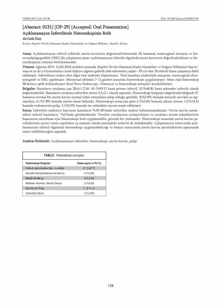
TJRMS 2017;1(3):125-90 DOI: 10.24074/tjrms.01032017OP
158
[[AAbbssttrraacctt:: 00113311]] [[OOPP--2299]] [[AAcccceepptteedd:: OOrraall PPrreesseennttaattiioonn]]AAççııkkllaannaammaayyaann İİnnffeerrttiilliitteeddee HHiisstteerreesskkooppiinniinn RRoollüüAAllii CCeennkk ÖÖzzaayyKonya Akşehir Devlet Hastanesi Kadın Hastalıkları ve Doğum Bölümü, Akşehir, Konya
AAmmaaçç:: Açıklanamayan infertil çiftlerde uterin kavitenin değerlendirilmesinde ilk basamak transvaginal ultrason ve his-terosalpingografidir (HSG).Bu çalışmanın amacı açıklanamayan infertile olgularda uterin kavitenin değerlendirilmesi ve his-tereskopinin rolünün belirlenmesidir. YYöönntteemm:: Ağustos 2015- Eylül 2016 tarihleri arasında Akşehir Devlet Hastanesi Kadın Hastalıları ve Doğum bölümüne başvur-muş en az iki yıl korunmasız cinsel ilişkiye rağmen gebelik elde edememiş yaşları <35 yıl olan 76 infertil hasta çalışmaya dahiledilmiştir. İnfertiliteye neden olan diğer tüm nedenler dışlanmıştır. Tüm hastalara jinekolojik muayene, transvaginal ultra-sonografi ve HSG yapılmıştır. Menstrual siklüsün 7-12.günleri arasında histereskopi uygulanmıştır. 4mm rijid histereskop30 derece optik kullanılmıştır (Karl Storz Endoscopy, Almanya) ve histereskopi sonuçları kaydedilmiştir. BBuullgguullaarr:: Hastaların ortalama yaşı 28.6±1.2’idi. 45 (%59.2) hasta primer infertil, 31(%40.8) hasta sekonder infertile olarakdeğerlendirildi. Hastaların ortalama infertilite süresi 4.3±2.1 olarak saptandı. Histereskopi bulguları değerlendirildiğinde 51hastanın normal bir uterin kavite normal tubal ostiumlara sahip olduğu görüldü. 3(%3.95) hastada stenotik servikal os sap-tanırken, 6 (%7.89) hastada uterin sineşi bulundu. Histereskopi sonucuna göre 2 (%2.63) hastada arkuat uterus, 11(%14.5)hastada endometrial polip, 3 (%3.95) hastada ise submüköz myom tespit edilmiştir. SSoonnuuçç:: İnfertilite nedeniye başvuran hastaların %10-20’sinde infertilite nedeni bulunamamaktadır. Uterin kavite anom-alileri infertil hastaların ~%3’ünde görülmektedir. Fertilite oranlarının iyileştirilmesi ve yardımcı üreme tekniklerininbaşarısının artırılması için histereskopi hızlı uygulanabilir, güvenli bir yöntemdir. Histereskopi sırasında uterin kavite pa-tolojilerinin ayırıcı tanısı yapılırken eş zamanlı olarak patolojinin tedavisi de mümkündür. Çalışmamızın sonucunda açık-lanamayan infertil olgularda histereskopi uygulanabileceği ve bunun sonucunda uterin kavite patolojilerinin saptanaraktedavi edilebileceğini saptadık.
AAnnaahhttaarr KKeelliimmeelleerr:: Açıklanamayan infertilite, histereskopi, uterin kavite, polip
Histereskopi Bulguları Hasta sayısı n=76 (%)
Normal uterin kavite+ tubal ostiumlar 51 (%67.1)
Stenotik internal/eksternal servikal os 3 (%3.95)
İntrauterin Sineşi 6 (%7.89
Müllerian Anomali ( Arkuat Uterus) 2 (%2.63)
Endometrial Polip 11 (%14.5)
Submüköz Myom 3 (%3.95)
TABLO: Histereskopi sonuçları.
DOI: 10.24074/tjrms.01032017OP TJRMS 2017;1(3):125-90
159
[[AAbbssttrraacctt:: 00114444]] [[OOPP--3300]] [[AAcccceepptteedd:: OOrraall PPrreesseennttaattiioonn]]PPrreeggnnaannccyy SSuucccceessss RRaattee aatt RReeccuurrrreenn IImmppllaannttaattiioonn FFaaiilluurree PPaattiieennttss AAfftteerr HHyysstteerroossccooppiicc EEnnddoommeettrriiaall IInnjjuurryyEElliiff GGaanniimmee AAyyddeenniizz,, UUmmuutt SSaarrııAcıbadem University Atakent Hospital
AAiimm:: Aim of this study is improving embryo implantation after hysterescopy fundal endometrial injury. In repeated im-plantation failure patients, we have to choice another way.OObbjjeeccttiivvee:: Infertility has been a big problem in human population entirely the history and it may be related to be part ofthe variant medical problems that has increased up to 50% since 1955 in the world. Perfect implantation is related on thedevelopment of high-quality embryos and to obtain of endometrial receptivity. However much the fertilization rate is rel-atively high and embryo culture conditions continue to improve, implantation is still the limiting step in the success of invitro fertilization-embryo transfer (IVF-ET). Endometrial injury is an intentional trauma to the endometrium by biopsy orcurettage.In our study, we have 30 patients totally and at the ovarian stimulation we had good responses from this patientsalthough we dont have positive results. It means we have good embryo quality. At this point we must discuss the problem.First step we need explore uterin cavity with hysterescopy. If there is a problem at the hysterescopy we have to solve en-dometrial problem but there is no problem at the endometrium we just impuls fundal endometrium for a local reaction. En-dometrium environment changes is the most important for implantation the embryo. For instance; thin endometrium, poorreceptivity, immunologic factors. Each of these factors are very important for implantation embryo. The woman’s age, theindication for IVF, uterine pathology, the treatment protocol employed, ovarian reserve, immunological factors, number ofembryos transferred, number of prepared embryos, embryo quality, embryo transfer technique, sperm quality and lutealphase support were determined as meddling with perfect implantation and protecting to recurrent failure. But reduced en-dometrium receptivity and low embryo quality are idea to be the most important factors. Another important question is tim-ing endometrial injury. Sometimes small endometrial injury with pipelle biopsy or hysterescopy makes a good reaction atthe endometrium. Furthermore endometrial injury in subfertile women induced through hysterescopy improves pregnancyand livebirth rates. Relationship between endometrial injury and increased pregnancy rates in next ART procedures hasbeen described in last publications of variable quality. This extensive literature revision, so aims to find the best availableoption on the influence and safety of endometrial injury for women with recurrent implantation failure undergoing ARTprocedures. Impairment of endometrial receptivity may be related with unexplain infertility. We believe that endometrialinjury facilitate the preparation of receptive endometrium.DDiissccuussssiioonn:: Our results suggest that quality of transferred embryos is the most important prognostic factor for conceptionand that blastocyst transfer and local endometrial injury should be recommended to patients with repeated IVF failure inorder to improve the pregnancy rate. At last larger prospective multicenter studies are needed to confirm these findings.
KKeeyywwoorrddss:: Endometrial injury, hysteroscopy, pregnancy rate
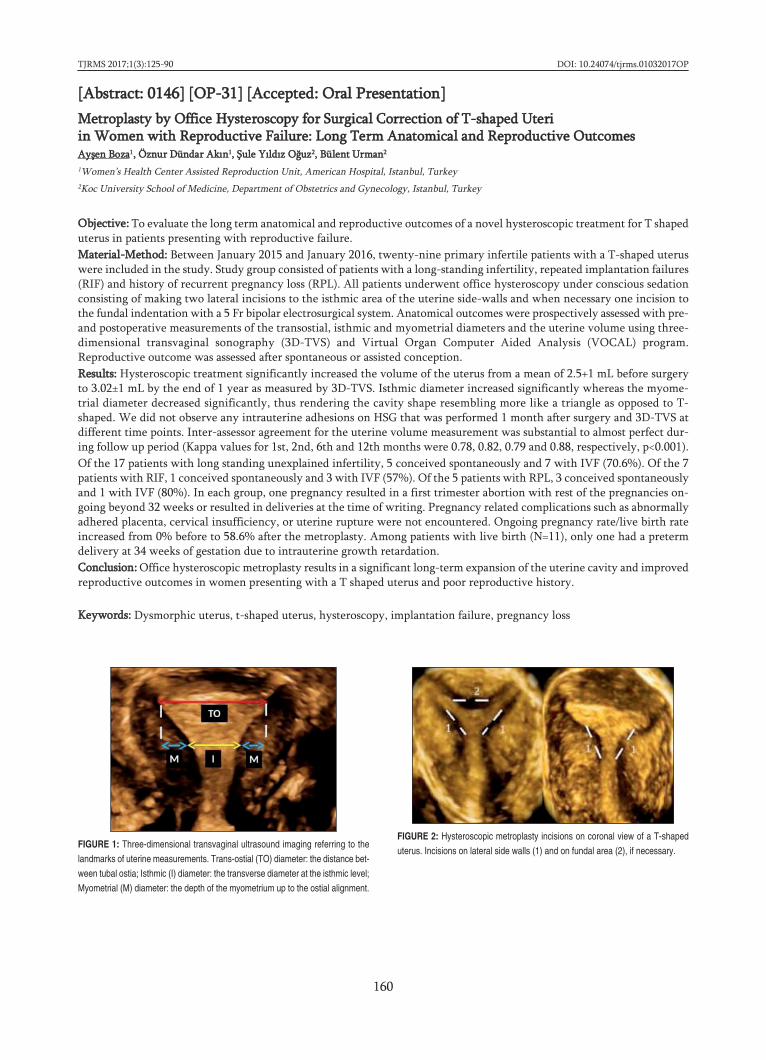
TJRMS 2017;1(3):125-90 DOI: 10.24074/tjrms.01032017OP
160
[[AAbbssttrraacctt:: 00114466]] [[OOPP--3311]] [[AAcccceepptteedd:: OOrraall PPrreesseennttaattiioonn]]MMeettrrooppllaassttyy bbyy OOffffiiccee HHyysstteerroossccooppyy ffoorr SSuurrggiiccaall CCoorrrreeccttiioonn ooff TT--sshhaappeedd UUtteerrii iinn WWoommeenn wwiitthh RReepprroodduuccttiivvee FFaaiilluurree:: LLoonngg TTeerrmm AAnnaattoommiiccaall aanndd RReepprroodduuccttiivvee OOuuttccoommeessAAyyşşeenn BBoozzaa11,, ÖÖzznnuurr DDüünnddaarr AAkkıınn11,, ŞŞuullee YYııllddıızz OOğğuuzz22,, BBüülleenntt UUrrmmaann22
1Women’s Health Center Assisted Reproduction Unit, American Hospital, Istanbul, Turkey2Koc University School of Medicine, Department of Obstetrics and Gynecology, Istanbul, Turkey
OObbjjeeccttiivvee:: To evaluate the long term anatomical and reproductive outcomes of a novel hysteroscopic treatment for T shapeduterus in patients presenting with reproductive failure. MMaatteerriiaall--MMeetthhoodd:: Between January 2015 and January 2016, twenty-nine primary infertile patients with a T-shaped uteruswere included in the study. Study group consisted of patients with a long-standing infertility, repeated implantation failures(RIF) and history of recurrent pregnancy loss (RPL). All patients underwent office hysteroscopy under conscious sedationconsisting of making two lateral incisions to the isthmic area of the uterine side-walls and when necessary one incision tothe fundal indentation with a 5 Fr bipolar electrosurgical system. Anatomical outcomes were prospectively assessed with pre-and postoperative measurements of the transostial, isthmic and myometrial diameters and the uterine volume using three-dimensional transvaginal sonography (3D-TVS) and Virtual Organ Computer Aided Analysis (VOCAL) program.Reproductive outcome was assessed after spontaneous or assisted conception. RReessuullttss:: Hysteroscopic treatment significantly increased the volume of the uterus from a mean of 2.5+1 mL before surgeryto 3.02±1 mL by the end of 1 year as measured by 3D-TVS. Isthmic diameter increased significantly whereas the myome-trial diameter decreased significantly, thus rendering the cavity shape resembling more like a triangle as opposed to T-shaped. We did not observe any intrauterine adhesions on HSG that was performed 1 month after surgery and 3D-TVS atdifferent time points. Inter-assessor agreement for the uterine volume measurement was substantial to almost perfect dur-ing follow up period (Kappa values for 1st, 2nd, 6th and 12th months were 0.78, 0.82, 0.79 and 0.88, respectively, p<0.001).Of the 17 patients with long standing unexplained infertility, 5 conceived spontaneously and 7 with IVF (70.6%). Of the 7patients with RIF, 1 conceived spontaneously and 3 with IVF (57%). Of the 5 patients with RPL, 3 conceived spontaneouslyand 1 with IVF (80%). In each group, one pregnancy resulted in a first trimester abortion with rest of the pregnancies on-going beyond 32 weeks or resulted in deliveries at the time of writing. Pregnancy related complications such as abnormallyadhered placenta, cervical insufficiency, or uterine rupture were not encountered. Ongoing pregnancy rate/live birth rateincreased from 0% before to 58.6% after the metroplasty. Among patients with live birth (N=11), only one had a pretermdelivery at 34 weeks of gestation due to intrauterine growth retardation. CCoonncclluussiioonn:: Office hysteroscopic metroplasty results in a significant long-term expansion of the uterine cavity and improvedreproductive outcomes in women presenting with a T shaped uterus and poor reproductive history.
KKeeyywwoorrddss:: Dysmorphic uterus, t-shaped uterus, hysteroscopy, implantation failure, pregnancy loss
FIGURE 1: Three-dimensional transvaginal ultrasound imaging referring to thelandmarks of uterine measurements. Trans-ostial (TO) diameter: the distance bet-ween tubal ostia; Isthmic (I) diameter: the transverse diameter at the isthmic level;Myometrial (M) diameter: the depth of the myometrium up to the ostial alignment.
FIGURE 2: Hysteroscopic metroplasty incisions on coronal view of a T-shapeduterus. Incisions on lateral side walls (1) and on fundal area (2), if necessary.

DOI: 10.24074/tjrms.01032017OP TJRMS 2017;1(3):125-90
161
FIGURE 3: Transostial, isthmic and myometrial diameter at three-dimensional transvagi-nal ultrasound at enrollment and after metroplasty (number of patients are given in pa-renthesis).
Indication Clinical pregnancy Spontaneous abortion Ongoing pregnancy/Live birth
Long standing unexplained infertility (N=17) 12(71) 1(6) 11(65)
Recurrent implantation failure (N=7) 4(57) 1(14) 3(43)
Recurrent pregnancy loss (N=5) 4(80) 1(20) 3(60)
TABLE 1: Reproductive outcomes after metroplasty according to the main indication to perform surgery.
TJRMS 2017;1(3):125-90 DOI: 10.24074/tjrms.01032017OP
162
[[AAbbssttrraacctt:: 00117744]] [[OOPP--3322]] [[AAcccceepptteedd:: OOrraall PPrreesseennttaattiioonn]]LLaappaarroossccooppiicc EExxcciissiioonn ooff aa LLaarrggee UUtteerriinnee CCyyssttiicc AAddeennoommyyoommaa iinn aann IInnffeerrttiillee PPaattiieennttYYaavvuuzz EEmmrree ŞŞüükküürr11,, FFaattmmaa CCeeyyllaann İİllhhaann22,, BBaattuuhhaann ÖÖzzmmeenn11,, CCeemm AAttaabbeekkooğğlluu11,, MMuurraatt SSöönnmmeezzeerr11
1Ankara University School of Medicine, Department of Obstetrics and Gynecology2Yenimahalle Training and Research Hospital, Department of Obstetrics and Gynecology
BBaacckkggrroouunndd:: Adenomyosis is defined as the presence of endometrial glands and stroma within the uterine musculature.Heavy menstrual bleeding, dysmenorrhea and diffuse uterine enlargement are the components of classical clinical manifes-tation. It has been believed that adenomyosis is a condition that generally affects multiparous and elderly women. Althoughthe exact mechanism remains unclear, a causative relationship between adenomyosis and infertility has been suggested inthe last decade. Here, we describe a rare form of adenomyosis, large cystic adenomyoma, in an adolescent patient who suf-fer from intractable dysmenorrhea.CCaassee:: An 17-year-old adolescent girl who suffered from intractable dysmenorrhea for 2 years that was unresponsive toNSAID applied for further investigation. Ultrasound examination revealed a cystic mass with a thick wall located in theright fundal part of the uterus measuring 4 cm in diameter. It was previously misdiagnosed as right adnexal mass. Transvaginalultrasonography revealed a 7.5x6.1x5.7 cm uterus. As the uterine mass was suspicious for cystic adenomyoma, laparoscopicexcision was considered. The laparoscope was inserted into the abdominal cavity through an 11 mm umbilical trocar. Threeadditional trocars were inserted under direct vision. The right anterior uterine wall close to fundus was incised with monopo-lar hook cautery. The cystic mass dissected from the myometrium by using scissor. During dissection the cyst was rupturedand endometriotic content drained. Following removal of the cyst the uterine defect was closed with interrupted 1-monocrylsutures. The specimen was easily removed by using endo-bag. The patient was discharged on the postoperative day 1 with-out complication. During 3 years of flow up the patient was completely free of dysmenorrhea or pelvic pain without usingNSAID.DDiissccuussssiioonn:: The pathophysiology of adenomyosis appears to be very similar to endometriosis and the underlying mechanismsof infertility can also be the same. Evidence suggests negative impact of adenomyosis on assisted reproductive technology(ART) cycle outcome. Although the exact pathogenesis is unknown the two generally accepted theories are myometrial in-vagination of endometrium or development from müllerian rests. Cystic adenomyoma is a rare form and more common inyoung patients. It’s generally classified as juvenile and adult type. While juvenile cystic adenomyoma starts within 5 yearsafter menarche or <= 18 years of age, adult cystic adenomyoma most commonly represents over the age 30. It might be ben-eficial to remove cystic adenomyoma to treat intractable dysmenorrhea or prior to ART treatment. In contrast to leiomy-omas, where the cleavage plane is clear, removal of cystic adenomyoma is difficult and necessitates sharp dissection. Inconclusion, laparoscopic excision of cystic adenomyoma is feasible in infertile patients.
KKeeyywwoorrddss:: Adenomyosis, infertility, laparoscopy, uterine cystic adenomyoma
DOI: 10.24074/tjrms.01032017OP TJRMS 2017;1(3):125-90
163
[[AAbbssttrraacctt:: 00118844]] [[OOPP--3333]] [[AAcccceepptteedd:: OOrraall PPrreesseennttaattiioonn]]IImmaacctt ooff CCoommbbiinnaattiioonn ooff HHyysstteerroossccooppiicc CCooaagguullaattiioonn ooff EEnnddoommeettrriioottiicc LLeessssiioonnss aanndd DDiieennooggeesstt ((VViissaannnnee 22 mmgg)) oonn PPrreeggnnaannccyy RRaattee iinn RRIIFF PPaattiieennttssNNaattaalliiaa KKhhoonneelliiddzzee,, KKeetteevvaann KKaannttaarriiaa
In Vitro Clinic, Center for Reproductive Health and In Vitro Fertilization, Tbilisi Georgia
OObbjjeeccttiivvee:: Repeated implantation failure (RIF) is a clinical entity affecting couples undergoing assisted reproductive tech-nology (ART). Variuos intrauterine pathologies contribute to RIF. Nevertheless, ultrasound or hysterospingography, whichare the common diagnostic tools for initial follow-up, have limited sensitivities. On the other hand, numerous research stud-ies have suggested a connection with adenomyosis and embryo implementation failure,but this could not be confirmed be-cause of the lack of consistence in diagnosis of adenomyosis (local or diffuse). In this context, we aimed to evaluate theimpact of combination of hysteroscopic coagulation of small endometryomas followed by medical treatment with Dienoest(Visanne 2 mg) on clinical pregnancy rate in subsequent ART cycles in women with history of RIF.MMaatteerriiaall--MMeetthhooddss:: The database of an assisted reproductive clinic was retrospectively reviewed to detect eligible cases. Atotal of 9 infertile women, aged 29-37 with history of two failed transfers (fresh and frozen) of good quality blastocyst, thethickness of endometrium between 8-11.5 mm on the day of progesterone initiation, with symptoms of dysmenorrhea andno 3 D ultrasound signs of adenomyosis, underwent hysteroscopic coagulation of endometriotic lessions. All of them weretalking Visanne 2 mg, 1 tab once a day continuously during 3 months. Afterwards, frozen HR-ET transfer of single blasto-cyst was performed in subsequent cycle.RReessuullttss:: 6 out of 9 women (66.7%) got pregnant (p= 0.41).CCoonncclluussiioonn:: Unrecognized uterine pathologies can be easily detected during diagnostic hysteroscopy. In RIF patients preg-nancy occurred in 66.7% (p= 0.41)after combination of hysteroscopic coagulation of endometriotic lessions and medicaltreatment with Dienogest (Visanne 2 mg) during 3 monthes.
KKeeyywwoorrddss:: Repeated implantation failure, hysteroscopy, adenomyosis

TJRMS 2017;1(3):125-90 DOI: 10.24074/tjrms.01032017OP
164
[[AAbbssttrraacctt:: 00221177]] [[OOPP--3344]] [[AAcccceepptteedd:: OOrraall PPrreesseennttaattiioonn]]EEffffeeccttiivveenneessss ooff HHyysstteerroossccooppyy iinn tthhee MMaannaaggeemmeenntt ooff UUtteerriinnee PPaatthhoollooggyy iinn WWoommeenn wwiitthh aann IInnttaacctt HHyymmeennAAiisshhaa MM EEllbbaarreeggAl-Amal Hospital for Obstetrics & Gynecology/Infertility Treatments and Genetic Research;Faculty of Medicine, Misurata University, Libya
BBaacckkggrroouunndd && OObbjjeeccttiivveess:: Virginity, defined as an intact hymen, is considered a sign of sexual purity in many societies, es-pecially Islamic and represents the honor of a women and her family, therefore, damage to the hymen carries with it seri-ous social implications for the patient. Based on these issues, and because of hysteroscopy being the only technique thatprovides direct visualization of the uterine cavity, our aim of this study was to assess role of hysteroscopy in the diagnosisand treatment of uterine pathology causing abnormal vaginal bleeding (AVB) in 17 unmarried patients with an intact hymen.MMaatteerriiaallss && MMeetthhooddss:: Medical records of 17 Referred patients with AVB, and intact hymen for whom hysteroscopies wereperformed during a period of 11 months, between (January 2016 through November 2016) at AL-Amal Hospital, were stud-ied retrospectively. Patients age ranged from 17 to 39 years, with average weights; 8 patients with menorrhagia, 5 womenwith intermenstrual bleeding, while the remaining 4 with US showed echogenicity in the uterine cavity and with metror-rhagia, all not responding to medical treatments. Preoperatively, necessary lab investigations done for all including CBC, co-agulation & hormonal profiles, misoprostol of 2 tablets (400μgm) were inserted PR 4 hrs. before intervention. All underwentdiagnostic hysteroscopy & operative (when necessary), 5 days after period, by means of a 3.9 mm Olympus continuous flow.Tissue samples removed sent for histopathological examination (HPE). Outcomes: treatment success (neither hymen norcervix were injured), menstrual flow reduction, degree of pain, need of second operation, patient satisfaction and compli-cations. Treatment was considered as a failure if hymen or cervix was injured or with repeated attacks of AVB. Consent wasobtained from each patient or her parents.RReessuullttss:: Mean time for performing hysteroscopies was 18 minutes, all went smoothly, neither hymen nor cervix were in-jured, also no uterine perforations were encountered. 9 patients had simple endometrial hyperplasia, and 3 women showedevidence of endometritis on HPE, given proper antibiotics, all of these patients had a clinical diagnosis of DUB. 5 patientswere diagnosed with endometrial polyps on HPE. No pathological findings indicating cancer or a precancerous lesions werefound. No evidence of bleeding disorders. Mean follow-up period was 3 months, none of the patients were lost to follow up.All patients but one(treatment failure, with repeated attacks of AVB) were satisfied and showed menstrual improvement.This patient went surgical re-intervention, and a missed sub-mucous myoma was diagnosed, and resected successfully.CCoonncclluussiioonnss:: Hysteroscopy, a minimally invasive procedure, found to be safe and effective in the management of women withintact hymen and with AVB due to uterine pathology, by avoiding trauma to the hymen and cervix, reducing the monthlyblood loss significantly and with a high satisfaction rate.
KKeeyywwoorrddss:: Hysteroscopy, intact hymen, uterine pathology, abnormal vaginal bleeding

DOI: 10.24074/tjrms.01032017OP TJRMS 2017;1(3):125-90
165
[[AAbbssttrraacctt:: 00223355]] [[OOPP--3355]] [[AAcccceepptteedd:: OOrraall PPrreesseennttaattiioonn]]İİnnttrraauutteerriinn SSiinneeşşii LLookkaalliizzaassyyoonnllaarrıınnıınn İİnnffeerrttiilliittee ÜÜzzeerriinnee EEttkkiissiiRRuukkiiyyee AAddaa BBeennddeerr11,, CCaannaann ÖÖzzccaann22,, RReeyyhhaann AAssllaannccaann33,, NNiiddaa BBaayyııkk33,, SSeevvttaapp HHaammddeemmiirr KKııllııçç33,, EErraayy ÇÇaallıışşkkaann33
1İstinye Üniversitesi Sağlık Bilimleri Fakültesi2VM Medicalpark Kocaeli Hastanesi3Bahçeşehir Üniversitesi Tıp Fakültesi
Bu çalışmada uterin sineşisi olan kadınlarda hangi lokalizasyonlardaki sineşiler infertiliteye neden oluyor ve müdahaleedilmeli sorularını cevaplamayı amaçladık.İnfertil olup histeroskopi ile uterin adhezyonları tespit edilen 39 hasta ile gebelikte yapılan ultrasonografi'de intrauterinsineşi tespit edilen 37 hasta sineşilerin lokalizasyonu açısından kıyaslandı. Sineşi lokalizasyonları olarak isthmus, fundus, cor-pus ve cornu belirlendi.Fundal sineşiler infertil grupta anlamlı olarak fazla olup (p<0.05), istmik sineşiler gebe grupta daha fazla (p<0.05) izlenmiştir.Corpustaki dens adhezyonlar ise infertil grupta gebe gruba göre daha fazladır (p<0.05).Adhezyonların yerine göre, özellikle fundal adhezyonlar, önemli infertilite nedenleri arasında bulunur. Bu adhezyonlara his-teroskopik yaklaşım ile uygun tedaviler gebelik oranını artırmaktadır.
AAnnaahhttaarr KKeelliimmeelleerr:: İntrauterin sineşi, infertilite
[[AAbbssttrraacctt:: 00226622]] [[OOPP--3366]] [[AAcccceepptteedd:: OOrraall PPrreesseennttaattiioonn]]TThhee EEffffeeccttss ooff CCaaffeetteerriiaa DDiieett IInndduucceedd MMaatteerrnnaall OObbeessiittyy oonn ooffff--SSpprriinnggss FFeerrttiilliittyyMMüübbeerrrraa NNaammllıı KKaalleemm11,, ZZiiyyaa KKaalleemm22,, EEllvvaann AAnnaaddooll33,, CCaannaann YYııllmmaazz33,, CCiiğğddeemm EEllmmaass33,, PPeerriihhaann YYaallççıınnkkaayyaa33,, HHaalliill RRuussoo22,, TTiimmuurr GGüürrggaann22
1Liv Hospital Ankara, Department of Obstetrics and Gynecology, Ankara,Turkey2Gürgan Clinic IVF and Women Health Center, Ankara, Turkey3Gazi University, Faculty of Medicine, Department of Hystology and Embryology, Ankara, Turkey
OObbjjeeccttiivvee:: In this study we aimed to investigate the effect of induced obesity nutrition in prior to pregnancy, during the preg-nancy, and in lactational period on the fertility of the offspring.MMaatteerriiaall--MMeetthhooddss:: In our study, 24 virgin female Wistar rats (120-125 gr) were obtained from Harlan Laboratories (TheNetherlands) and maintained with a 12-h light/dark schedule and fed standard rat diet ad libitum. Animals were randomlydivided into four nutritional groups; group 1a (n=6) and 1b (n=6) was fed a standart diet and group 2a (n=6) and 2b (n=6) fedcafeteria diet. After 10 weeks of standart or cafeteria feeding, all rats were paired with a Wistar stud male. Group 1a and 2adams fed standart diet whereas, group 1b and 2b dams fed cafeteria diet throughout pregnancy and lactation. After birth, alloffsprings weighed and litter size designed to twelve offsprings per litter (6 male and 6 female) and fed standart diet afterlactation until 8 weeks. All of the adult offsprings were sacrificed under ethical conditions. Serum FSH, LH, AMH, testos-teron and estradiol levels were estimated by ELISA methods. Ovarian tissues from female rats were analysed in terms of theirfollicular structures using hematoxylin-eosin and PTEN primary antibodies. Testicles from the male rats, on the other hand,were dissected and both motility and concentration of the sperm were analysed. SSttaattiissttiiccaall AAnnaallyyssiiss:: Statistical analysis was carried out using SPSS version 13.0 software program (SPSS Inc., Chicago, Illi-nois USA). All statistical tests were two-tailed, and P<0.05 was considered statistically significant. RReessuullttss:: There was a statistically significant difference in the primordial follicles count between 1A (55.62±9.35), 1B(47.17±7.48), 2A (15.62±5.92), and 2B (12.67±4.56) groups (p<0.001). However, there was no statistical difference betweenthe antral follicle count or the AMH levels from the aforementioned groups (p=0.691 and p=0.358 respectively). PTEN ex-pression was found to be present in every step of follicular development compartmentalizing in the oocyte, granulosa cells,and theca cells. However, as the follicles continue to develop there is a pattern that shows increase in PTEN accumulationin the granulosa cells and a reduction in PTEN in the oocyte cytoplasm. The PTEN expression in the oocyte cytoplasm wasfound to be decreased in the groups 2A and 2B when compared to groups 1A and 1B in conjunction with reduced primor-dial follicle count. There was no statistically significant difference in sperm count, motility, and progressively motile spermcount between groups (p>0.05).CCoonncclluussiioonn:: This study shows that in rats although there was no changes in the AMH levels and antral follicle count therewas a significantly decrease primordial follicles in female rats whoch were born from mothers that were fed with a cafete-ria diet. The male rats from the same mothers showed a reduced testicular mass and increased gonad fat tissues although thesedifferences seems to be not effecting the testicular function. Therefore, high maternal nutrition prior in pregnancy, in preg-nancy, and in lactational period is concluded to be potentially detrimental in female rats.
KKeeyywwoorrddss:: Maternal nutrition, cafeteria diet, primordial follicle, PTEN, sperm motility
TJRMS 2017;1(3):125-90 DOI: 10.24074/tjrms.01032017OP
166
[[AAbbssttrraacctt:: 00110011]] [[OOPP--3377]] [[AAcccceepptteedd:: OOrraall PPrreesseennttaattiioonn]]HHooww iiss aa SSuucccceessssffuull IIVVFF TTrreeaattmmeenntt RReellaatteedd ttoo MMiiccrroobbiioommee??PPeemmbbee SSaavvaaşşEastern Mediterranean University, Department of Biological Sciences
While Human Microbiome Project (HMP) has been sequencing the microbiome and identifying the vast communities of mi-crobiota that inhibit our bodies, it has already been hypothesized that microbes are involved in the physiology and patho-physiology of assisted reproduction since before the first success in in-vitro fertilization (IVF) and this relationship demandsincreased focus especially considering that up to forty percent of patients undergoing IVF have abnormal flora somewherealong the reproductive tract. Therefore the microbiome of the female genital tract should become an important considera-tion in IVF treatments and the aim of this study is to explore the current literature outlining the contribution of importantbacteria on reproductive health and outlines gaps in current research in order to highlight future areas of research. This relationship is evident based on the expansive literature available to date importantly after the sequencing data fromthe 16S rRNA subunit, we will explore the current literature and review the microbiome of the female reproductive tractfocusing on the influence of the reproductive microbiome in the assisted reproductive technology. A transition in the microbial flora (lactobacilli species) occurred in most of the women during the course of IVF treatmentsdue to hormonal changes (particularly with respect to variations in estradiol), or inflammation and progesterone resistanceor pharmacological interventions. Hence, the vaginal microbiome has a statistically significant relationship to the live birthrate. In parallel, this study will provide a platform to discuss what a common clinical practice should include for women un-dergoing IVF such as screening of the vaginal microbiome and to develop a model for useful screening tools and imple-mentation of microbial intervention strategies into modern day medicine. Although significant correlations appear to exist between the reproductive tract microbiome, hormone status and IVF suc-cess rates, a more comprehensive study is needed to better understand the mechanisms involved in a possible relationshipin reproductive assisted technologies. Therefore the future research needs to validate the hypothesis generated in these stud-ies in functional experiments and evaluate true impact on clinical practice.In this regard, the human microbiome research should focus on the possible solutions to mitigate the various factors affect-ing microbiome flora, implantation and delivery rates and this research will reflect the practice of modern biomedical re-search.
KKeeyywwoorrddss:: Microbiota, sequencing, reproduction, IVF
DOI: 10.24074/tjrms.01032017OP TJRMS 2017;1(3):125-90
167
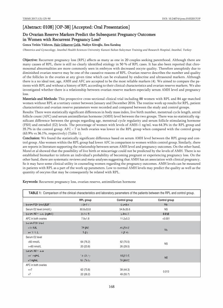
[[AAbbssttrraacctt:: 00110088]] [[OOPP--3388]] [[AAcccceepptteedd:: OOrraall PPrreesseennttaattiioonn]]DDoo OOvvaarriiaann RReesseerrvvee MMaarrkkeerrss PPrreeddiicctt tthhee SSuubbsseeqquueenntt PPrreeggnnaannccyy OOuuttccoommeess iinn WWoommeenn wwiitthh RReeccuurrrreenntt PPrreeggnnaannccyy LLoossss??GGoonnccaa YYeettkkiinn YYııllddıırrıımm,, HHaallee GGöökksseevveerr ÇÇeelliikk,, NNaaddiiyyee KKöörrooğğlluu,, EEssrraa KKaarraakkuuşşObstetrics and Gynecology, Istanbul Health Sciences University Kanuni Sultan Suleyman Training and Research Hospital, Istanbul, Turkey
OObbjjeeccttiivvee:: Recurrent pregnancy loss (RPL) affects as many as one in 20 couples seeking parenthood. Although there aremany causes of RPL, there is still no clearly identified etiology in 50 % of RPL cases. It has also been reported that chro-mosomal abnormalities are more commonly seen in embryos with decreased oocyte quality. Therefore aneuploidy due todiminished ovarian reserve may be one of the causative reasons of RPL. Ovarian reserve describes the number and qualityof the follicles in the ovaries at any given time which can be evaluated by endocrine and ultrasound markers. Althoughthere is a no ideal test, age, AMH and AFC are accepted to be the most reliable markers (4). We aimed to compare the pa-tients with RPL and without a history of RPL according to their clinical characteristics and ovarian reserve markers. We alsoinvestigated whether there is a relationship between ovarian reserve markers especially serum AMH level and pregnancyloss. MMaatteerriiaallss aanndd MMeetthhooddss:: This prospective cross-sectional clinical trial including 88 women with RPL and 84 age-matchedwomen without RPL at a tertiary center between January and December 2016. The routine work up results for RPL, patientcharacteristics and ovarian reserve parameters were recorded and compared between the study and control groups. Results: There were statistically significant differences in body mass index, live birth number, menstrual cycle length, antralfollicle count (AFC) and serum antimüllerian hormone (AMH) level between the two groups. There was no statistically sig-nificant difference between the groups regarding age, menstrual cycle regularity and serum follicle stimulating hormone(FSH) and estradiol (E2) levels. The percentage of women with levels of AMH<1 ng/mL was 64.3% in the RPL group and35.7% in the control group. AFC < 7 in both ovaries was lower in the RPL group when compared with the control group(63.9% vs 36.1%, respectively) (Table 1). CCoonncclluussiioonn:: We found the statistically significant difference based on serum AMH level between the RPL group and con-trol group. Also women within the RPL group had lower AFC in comparison to women within control group. Similarly, thereare reports in literature supporting the relationship between serum AMH level and pregnancy outcomes. On the other hand,Morel et al showed that the possibility of live birth or miscarriage could not be predicted by the levels of AMH. There is noestablished biomarker to inform an individual’s probability of becoming pregnant or experiencing pregnancy loss. On theother hand, there are systematic reviews and meta-analyses suggesting that AMH has an association with clinical pregnancy.So it may have some clinical utility in counseling women regarding the pregnancy outcomes. AMH levels can be measuredin patients with RPL as a part of the work up parameters. Low to normal AMH levels may predict the quality as well as thequantity of oocytes that may be consequently be related with RPL.
KKeeyywwoorrddss:: Recurrent pregnancy loss, ovarian reserve, antimüllerian hormone
TJRMS 2017;1(3):125-90 DOI: 10.24074/tjrms.01032017OP
168
RPL group Control group Control group
Serum FSH level (IU/L) 7.3±2.7 6.5±3.4 NS
Serum E2 level (nmol/L) 60.8±53.8 54.8±35.6 NS
Serum AMH level (ng/mL) 3.7±2.9 5.4±4.1 0.030
AFC in both ovaries 7.9±1.8 11.2±5.3 <0.001
Serum FSH level
<11 IU/L 79 (94) 82 (93.2)NS
>=11 IU/L 5 (6) 6 (6.8)
Serum E2 level
<60 nmol/L 64 (76.2) 62 (70.5)NS
>=60 nmol/L 20 (23.8) 26 (29.5)
Serum AMH level
<=1 ng/mL 18 (21.4) 10 (11.4)NS
>1 ng/mL 66 (78.6) 78 (88.6)
AFC in both ovaries
<=7 62 (73.8) 39 (44.3)0.013
>7 22 (26.2) 49 (55.7)
TABLE 1: Comparison of the clinical characteristics and laboratory parameters of the patients between the RPL and control group.
[[AAbbssttrraacctt:: 00119988]] [[OOPP--3399]] [[AAcccceepptteedd:: OOrraall PPrreesseennttaattiioonn]]AAnn UUnnuussuuaall IInnddiiccaattiioonn ooff IIVVFF:: KKaarrttaaggeenneerr''ss SSyynnddrroommeeVVeehhbbii YYaavvuuzz TTookkggöözz11,, LLeevveenntt DDiikkbbaaşş22,, BBaattuuhhaann ÖÖzzmmeenn33,, CCeemm SSoommeerr AAttaabbeekkooğğlluu33,, MMuurraatt SSöönnmmeezzeerr33
1Department of Obstetrics and Gynecology, School of Medicine, Giresun University, Giresun, Turkey2Department of Obstetrics and Gynecology, School of Medicine, Aksaray University, Aksaray, Turkey3Department of Obstetrics and Gynecology, Reproductive Endocrinology and Infertility Unit, School of Medicine, Ankara University, Ankara, Turkey
IInnttrroodduuccttiioonn:: Kartagener’s syndrome (KS) is a rare disorder characterized by the classic triad of chronic sinusitis, bronchiec-tasis and situs inversus. KS is a subgroup of primary ciliary dyskinesia. Ciliary defect may affect the ciliary activity of tubauterina. Some women with this syndrome have successfully conceived, although some of them are infertile. To our knowl-edge only 4 female patients with KS who were treated with in vitro fertilization/intracytoplasmic sperm injection(IVF/ICSI)and one of them was unsuccessful.CCaassee RReeppoorrtt:: CCaassee 11.. A 34 year-old woman presented with primary infertility of 6 years duration. She was diagnosed as KSsince childhood. Infertility work-up was performed in another infertility clinic; semen analysis, hysterosalpingogram andovulatory function were normal. She had intrauterine insemination cycles for 4 years in outside clinic and did not conceive.She referred to our infertility clinic and we considered the patient as primary infetility due to tubal ciliary dysfunction sec-ondary to KS. We planned IVF treatment. Controlled ovarian hyperstimulation was performed by antagonist protocol withhMG and r-FSH. Follicular development was assesed by transvaginal ultrasound examination. Choriogonadotropin alfa wasadministered for ovulation trigger and ovum retrieval was performed 36 hours after the hCG administration. Eight metaphaseII oocytes retrieved. Two blastocyst were transferred and resulted in singleton pregnancy. CCaassee 22.. The patient came to ourinfertility clinic at the age of 24 because of primary infertility for 3 years. She had a diagnosis of KS from childhood. The in-fertility examination included basal hormone assays, hysterosalpingogram and semen analysis. All these results were in nor-mal range. Primarily IVF treatment was planned for her and controlled ovarian hyperstimulation was started with r-FSH200 U/day on the 3rd day of the cycle by antagonist protocol and hMG 75 U/day was added later. When at least two folli-cles reached >18 mm, hCG was administered for ovulation trigger. Oocyte retrieval was performed and 6 oocytes were as-pirated. One embryo was transferred and singleton clinical pregnancy was achieved in the first IVF cycle. DDiissccuussssiioonn:: It is believed that immotility of tuba uterina due to lack of dynein arms in KS patients causes infertility. On theother hand, it was determined that ciliary motility in the female reproductive tract is not essential for fertility. It was sug-gested that muscular contractions of the fallopian tubes are the primary force for the transport of eggs and zygotes. Womenwith KS have a variable degree of infertility and our two patients did not become pregnant before being referred to our in-fertility clinic. One of them have experienced unsuccessful intrauterine insemination cycles before. In the literature, fourIVF-embryo transfer were reported for women with KS. Three of them conceived and two or three embryos were transferredin that patients. Different from the current literature our patients conceived in the first IVF-embryo transfer attempt andonly one or two embryo transfer were performed. In conclusion, women with KS who have infertility should be encour-aged by the possibility of conception, and IVF-embryo transfer is a reasonable treatment approach to try.
KKeeyywwoorrddss:: Kartagener's syndrome, infertility, in vitro fertilization, embryo transfer
DOI: 10.24074/tjrms.01032017OP TJRMS 2017;1(3):125-90
169
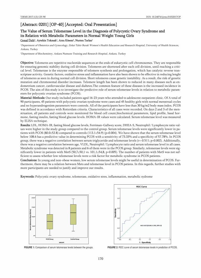
[[AAbbssttrraacctt:: 00220011]] [[OOPP--4400]] [[AAcccceepptteedd:: OOrraall PPrreesseennttaattiioonn]]TThhee VVaalluuee ooff SSeerruumm TTeelloommeerraassee LLeevveell iinn tthhee DDiiaaggnnoossiiss ooff PPoollyyccyyssttiicc OOvvaarryy SSyynnddrroommee aanndd iittss RReellaattiioonn wwiitthh MMeettaabboolliicc PPaarraammeetteerrss iinn NNoorrmmaall WWeeiigghhtt YYoouunngg GGiirrllssCCeemmaall ÜÜnnllüü11,, AAyytteekkiinn TTookkmmaakk11,, AArrzzuu KKöösseemm22,, NNuurrtteenn TTaarrllaann11
1Department of Obstetrics and Gynecology, Zekai Tahir Burak Women’s Health Education and Research Hospital, University of Health Sciences,Ankara, Turkey2Department of Biochemistry, Ankara Numune Training and Research Hospital, Ankara, Turkey
OObbjjeeccttiivvee:: Telomeres are repetitive nucleotide sequences at the ends of eukaryotic cell chromosomes. They are responsiblefor ensuring genomic stability during cell division. Telomeres are shortened after each cell division, until reaching a criti-cal level. Telomerase is the enzyme responsible of telomere synthesis and prolongation, which has catalytic reverse tran-scriptase activity. Genetic factors, oxidative stress and inflammation have also been shown to be effective in reducing lenghtof telomeres as seen in during normal cell division. Short telomeres cause genetic instability. As a result, the risk of geneticmutation and chromosomal disorder increases. Telomere length has been shown to reduced in many diseases such as en-dometrium cancer, cardiovascular disease and diabetes.The common feature of these diseases is the increased incidence inPCOS. The aim of this study is to investigate the predictive role of serum telomerase levels in relation to metabolic param-eters for polycystic ovarian syndrome (PCOS).MMaatteerriiaall--MMeetthhooddss:: Our study included patients aged 16-23 years who attended to adolescent outpatient clinic. Of A total of90 participants, 45 patients with polycystic ovarian syndrome were cases and 45 healthy girls with normal menstrual cyclesand no hyperandrogenism parameters were controls. All of the participants have less than 30 kg/m2 body mass index. PCOSwas defined in accordance with Rotterdam criteria. Characteristics of all cases were recorded. On days 2 and 3 of the men-struation, all patients and controls were monitored for blood cell count,biochemical parameters, lipid profile, basal hor-mone, fasting insulin, fasting blood glucose levels. HOMA-IR values were calculated. Serum telomerase level was measuredby ELISA technique. RReessuullttss:: LDL, HOMA-IR, fasting blood glucose levels, Ferriman–Gallwey score, DHEA-S, Neutrophil / Lymphocyte ratio val-ues were higher in the study group compared to the control group. Serum telomerase levels were significantly lower in pa-tients with PCOS (80.0±52.8) compared to controls (113.1±54.9) (p=0.004). We have shown that the serum telomerase levelbelow 108.6 has a predictive value in determining PCOS with a sensitivity of 73.33% and a specificity of 57.78%. In PCOSgroup, there was a negative correlation between serum triglyceride and telomerase levels (r=-0.511; p=0.002). Additionally,there was a negative correlation between age, VLDL, Neutrophil / Lymphocyte ratio and serum telomerase level in all cases.Metabolic syndrome was detected in 8 patients and 6 of them were in the PCOS group. Similarly, telomerase levels were sig-nificantly lower in patients with MetS (50,7±50,1 vs. 101,1±54,8, p=0.005). The number of patients with MetS was not suf-ficient to assess whether low telomerase levels were a risk factor for metabolic syndrome in PCOS patients.CCoonncclluussiioonnss:: In young and non-obese women, low serum telomerase levels might be useful in determination of PCOS. Fur-thermore, there may be a relation between Mets and telomerase level in PCOS patiens. In this regards, further studies withmore participants are needed to justify and improve our results.
KKeeyywwoorrddss:: Polycystic ovary syndrome, telomerase, oxidative stres, inflammation, metabolic sydrome
TJRMS 2017;1(3):125-90 DOI: 10.24074/tjrms.01032017OP
170
FIGURE 1: Comparison of serum telomerase levels between the groups. FIGURE 2: ROC curve of serum telomerase levels in prediction of PCOS.
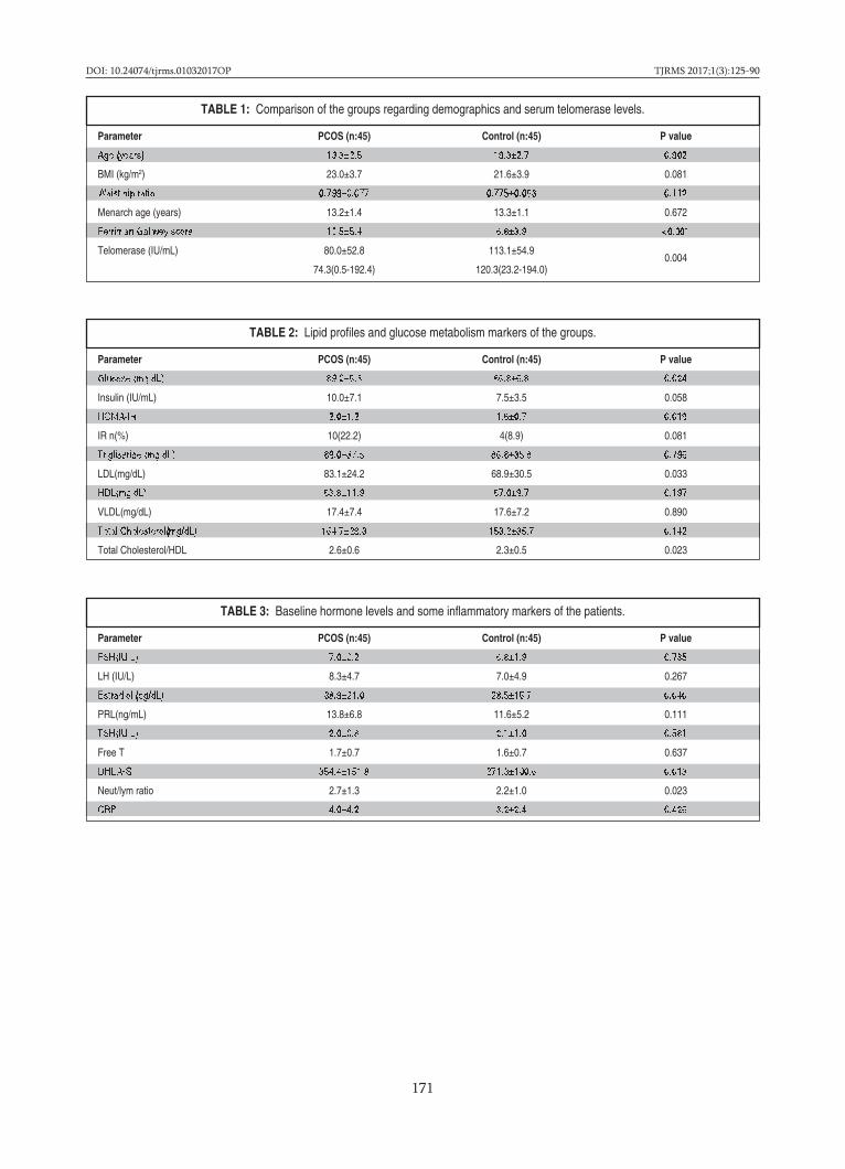
DOI: 10.24074/tjrms.01032017OP TJRMS 2017;1(3):125-90
171
Parameter PCOS (n:45) Control (n:45) P value
Age (years) 19.3±2.5 19.3±2.7 0.902
BMI (kg/m2) 23.0±3.7 21.6±3.9 0.081
Waist/hip ratio 0.799±0.077 0.775±0.063 0.112
Menarch age (years) 13.2±1.4 13.3±1.1 0.672
Ferriman Gallwey score 12.5±5.4 6.8±3.9 <0.001
Telomerase (IU/mL) 80.0±52.8 113.1±54.90.004
74.3(0.5-192.4) 120.3(23.2-194.0)
TABLE 1: Comparison of the groups regarding demographics and serum telomerase levels.
Parameter PCOS (n:45) Control (n:45) P value
Glucose (mg/dL) 89.2±6.3 86.8±6.8 0.024
Insulin (IU/mL) 10.0±7.1 7.5±3.5 0.058
HOMA-IR 2.0±1.2 1.6±0.7 0.019
IR n(%) 10(22.2) 4(8.9) 0.081
Trigliseride (mg/dL) 89.0±37.5 86.8±35.8 0.796
LDL(mg/dL) 83.1±24.2 68.9±30.5 0.033
HDL(mg/dL) 63.8±11.9 67.0±9.7 0.197
VLDL(mg/dL) 17.4±7.4 17.6±7.2 0.890
Total Cholesterol(mg/dL) 164.7±29.3 153.2±35.7 0.142
Total Cholesterol/HDL 2.6±0.6 2.3±0.5 0.023
TABLE 2: Lipid profiles and glucose metabolism markers of the groups.
Parameter PCOS (n:45) Control (n:45) P value
FSH(IU/L) 7.0±2.2 6.8±1.9 0.735
LH (IU/L) 8.3±4.7 7.0±4.9 0.267
Estradiol (pg/dL) 38.9±21.0 28.5±16.7 0.040
PRL(ng/mL) 13.8±6.8 11.6±5.2 0.111
TSH(IU/L) 2.0±0.8 2.1±1.0 0.581
Free T 1.7±0.7 1.6±0.7 0.637
DHEA-S 354.4±151.8 271.3±100.6 0.013
Neut/lym ratio 2.7±1.3 2.2±1.0 0.023
CRP 4.0±4.2 3.2±2.4 0.426
TABLE 3: Baseline hormone levels and some inflammatory markers of the patients.

[[AAbbssttrraacctt:: 00220077]] [[OOPP--4411]] [[AAcccceepptteedd:: OOrraall PPrreesseennttaattiioonn]]PPrreevvaalleennccee aanndd PPrreeddiiccttoorrss ooff tthhee UUssaaggee ooff CCoommpplleemmeennttaarryy AAlltteerrnnaattiivvee MMeeddiicciinnee AAmmoonngg IInnffeerrttiillee PPaattiieennttssSSeeyyhhaann SSöönnmmeezz11,, MMuussttaaffaa ÖÖzzttüürrkk22,, FFeerrhhaann SSöönnmmeezz11,, EErrsseenn EErraayyddıınn11,, MMeehhmmeett CCaanneerr ÖÖzzeerr11,, MMuurraatt SSeerrkkaanntt ÜÜnnaall11,, SSeeyyiitt TTeemmeell CCeeyyhhaann22
1Department of Obstetrics and Gynecology, Infertility Unit, Denizli State Hospital, Denizli, Turkey2Department of Obstetrics and Gynecology, Division of Reproductive Endocrinology and Infertility, S.B.Ü. Gülhane Training and Research Hospital, Ankara, Turkey
OObbjjeeccttiivvee:: The use of Complementary and Alternative Medicine (CAM) has grown considerably over the past few decadesin Western countries. Parallel to this increase, different CAM modalities are becoming more widespread among infertile pa-tients. The primary objective of this study was to evaluate the prevalence, characteristics and the most frequently used CAMmethods among infertile couples. A secondary aim was to assess predictors, information sources and documentation attitudeof CAM among patients seeking assisted reproduction treatment. DDeessiiggnn:: This study was designed as a cross-sectional survey. MMaatteerriiaallss--MMeetthhooddss:: The study was conducted on patients admitted infertility clinics of two hospitals (one university and an-other state hospital) which are located in the cities of Ankara and Denizli, Turkey. A total of 324 patients (162 couples) whohad been diagnosed with an infertility and had sufficient intellectual ability to understand all questions were enrolled in thisstudy. CAM prevalence, CAM modalities and factors associated with CAM use among infertile couples were investigated.RReessuullttss:: The overall prevalence of CAM among 324 participants was 25.6 % (n=83). One half (48%) of the infertile coupleshad experience with at least one type of CAM method. Of those using CAM, the majority of subjects reported taking herbalproducts (84%). The frequency of CAM tended to be higher among females compared to males (33% versus 17%, respec-tively; p=0.02). Failure to conceive with previous medical therapies was found to be significantly associated with CAM inan infertility population. These patients were more inclined to use CAM. (p=0.01) After logistic regression analyses, only gen-der and previous unsuccessful ART treatment were found to be associated independently with CAM use. CCoonncclluussiioonn:: CAM use is common among infertile population. CAM users were more likely to be women at any age who hadfailed to achieve a pregnancy with previous artificial reproductive treatment(s), mainly used herbal products and mostly re-luctant to report the use of CAM.
KKeeyywwoorrddss:: Complementary, alternative, medicine, infertility
TJRMS 2017;1(3):125-90 DOI: 10.24074/tjrms.01032017OP
172

[[AAbbssttrraacctt:: 00221144]] [[OOPP--4422]] [[AAcccceepptteedd:: OOrraall PPrreesseennttaattiioonn]]MMaalliiggnnaanntt OOvvaarriiaann IImmmmaattuurree TTeerraattoommaass:: OOnnccoollooggiicc aanndd FFeerrttiilliittyy OOuuttccaammeessHHaanniiffii ŞŞaahhiinn11,, EEddaa KKooccaammaann22,, İİbbrraahhiimm YYaallççıınn11,, MMuussttaaffaa EErrkkaann SSaarrıı11,, AAllii AAyyhhaann22
1Ankara Zekai Tahir Burak Women’s Health Training and Research Hospital, University of Health Sciences. Department of Gynecologic Oncology2Başkent University Faculty of Medicine Department of Gynecologic Oncology
AAiimm:: Malignant Ovarian immature teratomas are rare neoplasms. This study aimed to evaluate the oncologic and fertilityoutcome in women with all stage malignant ovarian immature teratomas (MOIT) MMeetthhooddss:: Case records of 24 patients from 2007-2017 with (MOIT) were reviewed. Histology was based on WHO classifi-cation. Tumours were staged according to International Federation of Gynecology and Obstetrics (FIGO) system. Data wascollected on age, grade, stage, Tumor size, peritoneal involvement,Residual tumor volume, Lymphovascular space invasion,treatment, recurrence overall survival (OS) and Disease-free survival (DFS). OS and DFS was determined by Kaplan-Meiermethod.RReessuullttss:: Median age of our patients was 23 (15-47) years. Seventeen patients were in grade I, seventeen in III. Eight patientswere in stage I, sixteen in grade II-III. Median follow up was 49 months (10-105). All patients underwent initial surgery. Fer-tility sparing procedures were performed in 83.3 % patients. In univariate analysis, peritoneal involvement, stage and resid-ual tumor volume were significant on DFS and OS (P< 0.005). There was no independent prognostic factor for DFS and OSin multivariate analysis. 5-year OS rate 89.3%. 5-year DFS rate 72%. The Survival status was 91.7% and Recurrence rate was20.8%. Nine patients had pregnancy. Five patients had live pregnancy. Three patients underwent embryo freezing. Two pa-tients had spontaneous pregnancy.CCoonncclluussiioonnss:: MOIT has good prognosis with conservative surgery and chemotherapy. Fertility sparing surgery has becomea standard in MOIT.
KKeeyywwoorrddss:: Immature teratoma, Fertility
DOI: 10.24074/tjrms.01032017OP TJRMS 2017;1(3):125-90
173
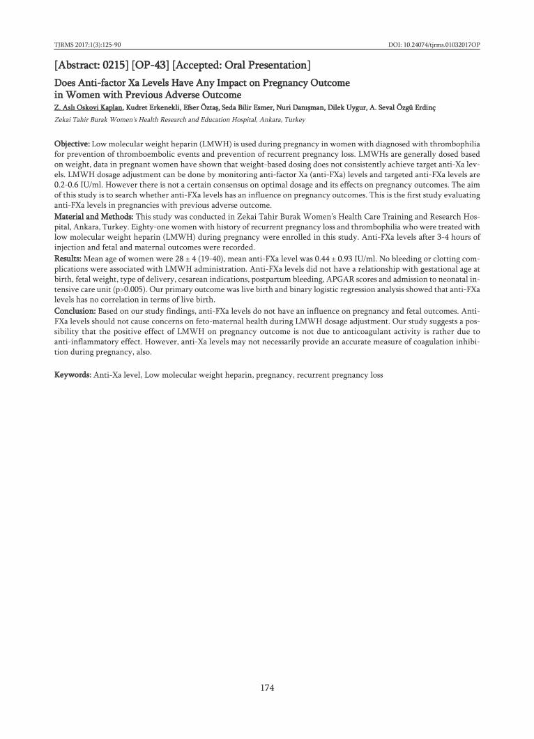
[[AAbbssttrraacctt:: 00221155]] [[OOPP--4433]] [[AAcccceepptteedd:: OOrraall PPrreesseennttaattiioonn]]DDooeess AAnnttii--ffaaccttoorr XXaa LLeevveellss HHaavvee AAnnyy IImmppaacctt oonn PPrreeggnnaannccyy OOuuttccoommee iinn WWoommeenn wwiitthh PPrreevviioouuss AAddvveerrssee OOuuttccoommeeZZ.. AAssllıı OOsskkoovvii KKaappllaann,, KKuuddrreett EErrkkeenneekkllii,, EEffsseerr ÖÖzzttaaşş,, SSeeddaa BBiilliirr EEssmmeerr,, NNuurrii DDaannıışşmmaann,, DDiilleekk UUyygguurr,, AA.. SSeevvaall ÖÖzzggüü EErrddiinnççZekai Tahir Burak Women's Health Research and Education Hospital, Ankara, Turkey
OObbjjeeccttiivvee:: Low molecular weight heparin (LMWH) is used during pregnancy in women with diagnosed with thrombophiliafor prevention of thromboembolic events and prevention of recurrent pregnancy loss. LMWHs are generally dosed basedon weight, data in pregnant women have shown that weight-based dosing does not consistently achieve target anti-Xa lev-els. LMWH dosage adjustment can be done by monitoring anti-factor Xa (anti-FXa) levels and targeted anti-FXa levels are0.2-0.6 IU/ml. However there is not a certain consensus on optimal dosage and its effects on pregnancy outcomes. The aimof this study is to search whether anti-FXa levels has an influence on pregnancy outcomes. This is the first study evaluatinganti-FXa levels in pregnancies with previous adverse outcome.MMaatteerriiaall aanndd MMeetthhooddss:: This study was conducted in Zekai Tahir Burak Women’s Health Care Training and Research Hos-pital, Ankara, Turkey. Eighty-one women with history of recurrent pregnancy loss and thrombophilia who were treated withlow molecular weight heparin (LMWH) during pregnancy were enrolled in this study. Anti-FXa levels after 3-4 hours ofinjection and fetal and maternal outcomes were recorded.RReessuullttss:: Mean age of women were 28 ± 4 (19-40), mean anti-FXa level was 0.44 ± 0.93 IU/ml. No bleeding or clotting com-plications were associated with LMWH administration. Anti-FXa levels did not have a relationship with gestational age atbirth, fetal weight, type of delivery, cesarean indications, postpartum bleeding, APGAR scores and admission to neonatal in-tensive care unit (p>0.005). Our primary outcome was live birth and binary logistic regression analysis showed that anti-FXalevels has no correlation in terms of live birth. CCoonncclluussiioonn:: Based on our study findings, anti-FXa levels do not have an influence on pregnancy and fetal outcomes. Anti-FXa levels should not cause concerns on feto-maternal health during LMWH dosage adjustment. Our study suggests a pos-sibility that the positive effect of LMWH on pregnancy outcome is not due to anticoagulant activity is rather due toanti-inflammatory effect. However, anti-Xa levels may not necessarily provide an accurate measure of coagulation inhibi-tion during pregnancy, also.
KKeeyywwoorrddss:: Anti-Xa level, Low molecular weight heparin, pregnancy, recurrent pregnancy loss
TJRMS 2017;1(3):125-90 DOI: 10.24074/tjrms.01032017OP
174

[[AAbbssttrraacctt:: 00222244]] [[OOPP--4444]] [[AAcccceepptteedd:: OOrraall PPrreesseennttaattiioonn]]AAzzaallmmıışş OOvveerryyaann YYaannııttllıı OOllgguullaarrddaa İİnn VViittrroo FFeerrttiilliizzaassyyoonn PPrroottookkoollüünnee EEkklleenneenn BBüüyyüümmee HHoorrmmoonnuunn SSoonnuuççllaarraa EEttkkiissiiRRuukkiiyyee AAddaa BBeennddeerr11,, CCaannaann ÖÖzzccaann33,, RReeyyhhaann AAssllaannccaann22,, BBeerrttaann AAkkaarr11,, AAyynnuurr EErrşşaahhiinn22,, EErraayy ÇÇaallıışşkkaann22
1İstinye Üniversitesi Sağlık Bilimleri Fakültesi, 2Bahçeşehir Üniversitesi Tıp Fakültesi, 3VM Medicalpark Kocaeli Hastanesi, İstanbul, Türkiye
İn vitro fertilizasyon (İVF) denenen infertil kadınların %9-24'ü Bologna kriterleri ile azalmış overyan yanıtlı olgular olaraktanımlanmıştır. Bu kadınlarda gebelik oranını artırmak adına farklı yöntemler denenmektedir.Çalışmamıza Bologna kriterlerine uygun olarak belirlenen 93 azalmış overyan yanıtlı infertil kadını dahil ettik. Bunlardan47 tanesinde büyüme hormonu (BH) İVF protokolüne eklendi. Sonuçlar gebelik oranı, canlı doğum oranı, iptal edilen sik-lus, implantasyon oranı,fertilizasyon oranı, toplanan oosit sayısı ile değerlendirildi.Gebelik oranı, canlı doğum oranı, iptal edilen siklus, implantasyon oranı,fertilizasyon oranı, toplanan oosit sayısı gibi sonuçlardeğerlendirildiğinde BH eklenen İVF protokollerinin eklenmeyenlere istatistiksel açıdan anlamlı bir üstünlüğü buluna-mamıştır.Çalışmamızda da görüldüğü üzere, BH'nun İVF sikluslarında tedavi protokolüne eklenmesinin gebelik oranını artırmakadına katkısı yoktur.
KKeeyywwoorrddss:: Büyüme hormonu, in vitro fertilizasyon, azalmış ovaryen yanıt
DOI: 10.24074/tjrms.01032017OP TJRMS 2017;1(3):125-90
175

[[AAbbssttrraacctt:: 00225544]] [[OOPP--4455]] [[AAcccceepptteedd:: OOrraall PPrreesseennttaattiioonn]]HHyysstteerroossaallppiinnggooggrraapphhyy PPrriioorr ttoo tthhee GGoonnaaddoottrrooppiinn SSttiimmuullaatteedd IInnttrraauutteerriinnee IInnsseemmiinnaattiioonn IImmpprroovveessCClliinniiccaall PPrreeggnnaannccyy RRaatteess iinn WWoommeenn wwiitthh UUnneexxppllaaiinneedd IInnffeerrttiilliittyyEErrkkaann ÇÇaağğllııyyaann,, EEmmrree OOkkyyaayy,, TTaayyllaann BBoodduurr,, ÇÇaağğllaann EErrttuuğğrruull,, EEllvvaann KKooyyuunn,, MMüüggee KKoovvaallıı,, EErrbbiill DDooğğaann,, BBüülleenntt GGüülleekklliiDokuz Eylul University Faculty of Medicine, Obstetrics and Gynecology Department, Izmir, Turkey
OObbjjeeccttiivvee:: Gonadotropin stimulated intrauterine insemination (IUI) cycles performed following one month after hysteros-alpingography (HSG) are associated with improvement in clinical pregnancy rates in unexplained infertile couples.MMaatteerriiaallss aanndd MMeetthhoodd:: A retrospective cohort study was performed between 2008 and 2014. A total of 92 unexplained in-fertile couples undergoing their first cycle IUI stimulated by gonadotropins were included in the analysis. Participants wereclassified into two groups according to IUI cycles performed one month (Group A, n = 25 cycles) or longer than one month(Group B, n = 67 cycles) after the HSG procedure.RReessuullttss:: The overall clinical pregnancy rate was found as 25% (23 clinical pregnancies / 92 cycles). Clinical pregnancy ratewas 44 % (11/25) for Group A and 17.9 % (12/67) for Group B. In Group A, there were significantly higher clinical preg-nancy rates compared to Group B (OR: 3.6, 95% CI, 1.3–9.8; p = 0.012).CCoonncclluussiioonn:: It has been demonstrated that fertility improving effect of HSG was most prominent in the first six monthsafter procedure. Likewise, in gonadotropin stimulated IUI cycles performed following one month after HSG, there seems tobe an improvement in pregnancy rates in unexplained couples. In unexplained cases, it may be a reasonable approach to planIUI cycles in the first month after HSG in clinical practice.
KKeeyywwoorrddss:: Ovarian stimulation, Infertility, Hysterosalpingography
TJRMS 2017;1(3):125-90 DOI: 10.24074/tjrms.01032017OP
176
[[AAbbssttrraacctt:: 00226644]] [[OOPP--4466]] [[AAcccceepptteedd:: OOrraall PPrreesseennttaattiioonn]]FFrreesshh VVeerrssuuss FFrroozzeenn--tthhaawweedd BBllaassttooccyysstt TTrraannssffeerr iinn HHiigghh RReessppoonnddeerrss wwiitthh aanndd wwiitthhoouutt CCaallcciiuumm IInnffuussiioonnZZiiyyaa KKaalleemm11,, MMüübbeerrrraa NNaammllıı KKaalleemm22,, HHaalliill RRuussoo11,, TTiimmuurr GGüürrggaann11
1Gurgan Clinic IVF and Women Health Center,Department of IVF,Ankara,Turkey2Liv Hospital Ankara, Department of Obstetrics and Gynecology, Ankara,Turkey
OObbjjeeccttiivvee:: In this study, we aimed to investigate the pregnancy and live birth rates in frozen-thawed embryo transfer thatcarried out by embryo transfer in fresh cycle and all the embryos obtained from the fresh cycle were frozen transferring tothe artificially prepared endometrium in another cycle for the group with high OHSS risk. MMaatteerriiaallss--MMeetthhooddss:: This is a retrospective study including a total of 254 patients who underwent ICSI in a private IVF cen-ter between 2013 and 2016. The study group consisted of women with high response to controlled ovarian hyperstimula-tion (high responder). Patients with a total follicle count of 15 or more and/or estradiol (E2) value of over 3000 pg/mL onthe day of ovulation induction were considered as high responders and with a high risk of OHSS. Patients over 40 years old,male factor, recurrent pregnancy loss stories, those with uterine pathologies, chronic systemic medical problems, systemicdrug use and those induced with agonist were excluded from the study. The patients who received embryo transfer in freshcycle and calcium infusion referred as Fresh Ca+ group, whereas those without calcium therapy were called as fresh Ca-group. In the frozen group, those receiving calcium infusions were called as frozen Ca+ group, whereas those without cal-cium treatment were called as frozen Ca- group. In this study, gonadotropin-releasing hormone (GnRH) agonist and GnRHantagonist protocols were administered in patients during COH. RReessuullttss:: A total of 254 high-responder were included in this study. Fresh cycles were performed to 181 (71.2%) patients, whilefrozen cycles were performed to 73 (28.7%) patients. Calcium treatment was administered to 109 (60%) patients in freshcycle, while it was administered to 45 (61.6%) of them in frozen cycle. The patients’ age, body mass index (BMI), hormonalvalues at the beginning of the cycle, infertility duration and the IVF cycle are shown in Table 1. The characteristics of thecycle were shown in Table 2 in comparison between four groups. In Table 3, the results of the cycle were also given com-paratively between four groups. Implantation rates, clinical pregnancy rates, and live birth rates did not show statisticallysignificant difference between groups.CCoonncclluussiioonn:: In this study, there was no statistically significant difference between fresh and frozen-thawed cycles in patientswith high OHSS risk in terms of implantation, clinical pregnancy, and live birth rates. Furthermore, these rates were notdifferent in the cycles with or without calcium treatment. The OHSS rates were not different between fresh and thaw cy-cles; however, these rates were significantly less in the Ca+ group than in the Ca- group. OHSS was not developed in the totalfreeze group with Ca treatment. Thus, freeze-all policy can be applied more reliably in order to be protected from OHSS.We also conclude that calcium infusion is beneficial in preventing OHSS without altering pregnancy rates.
KKeeyywwoorrddss:: OHSS, fresh embryo transfer, frozen-thawed transfer, calcium, pregnancy rate
DOI: 10.24074/tjrms.01032017OP TJRMS 2017;1(3):125-90
177
[[AAbbssttrraacctt:: 00009911]] [[OOPP--4477]] [[AAcccceepptteedd:: OOrraall PPrreesseennttaattiioonn]]TThhee RRaattiioo ooff aannttii--MMüülllleerriiaann HHoorrmmoonnee,, IInnhhiibbiinn--BB,, aanndd IInnssuulliinn GGrroowwtthh FFaaccttoorr--22 aass PPrreeddiiccttoorr ffoorr tthhee QQuuaalliittyy ooff OOooccyytteess iinn tthhee IInn VViittrroo FFeerrttiilliizzaattiioonn CCyycclleessAAgguuss SSuupprriiyyaaddiiAgus Supriyadi, Women and Childrent Harapankita Hospital, Jakarta, Indonesia
BBaacckkggrroouunndd:: The outcome of an in vitro fertilization (IVF) cycle depends on a num¬ber of factors including oocyte quality.However, assessment of oocyte quality using morphological evaluation is considerably difficult. The aim of this study is tofind out the accurate compre¬hen¬sive pre¬dic¬tor for the quality of oocyte by respectively mea-suring serum and follicu-lar levels of AMH, inhibin-B, IGF-2. MMaatteerriiaallss--MMeetthhooddss:: Selected 38 IVF cycles from infertile couples between Sep-tember 2013 and August 2014 from MelatiIVF Clinic Harapan Kita Mother and Child Hospital, Jakarta, Indonesia were included to this study, from which at least oneoocyte was needed. The age of the infertile IVF subject was 26 to 42 years old. Three serum and follicular fluids biomark-ers (AMH, inhibin-B, IGF-2) were measured three times at the IVF cycles (Day-2, Day-trigger, and Day-ovum pick up).Several models were in-ves¬ti¬ga¬ted which was expected able to predict: (1) the number of mature oocytes (MO), (2) thefertilizable oocyte (F) and (3) the transferable embryo. The data was analyzed using multivariate linear regression.RReessuullttss:: Among 72 recruited subjects, only 43 of them were eligible for further eva-luation, out of which 38 subjects had com-plete data. MO, FO, and E were positively correlated with serum inhibin-B at Day-ovum pick-up (r=0.54; 0.33, 0.33, re-specti¬ve¬ly), basal anthral follicle (BAF) count (r=0.69; 55; 52, respectively), and inhibin3 ratio (r=0.54; 0.33, 0.33,respectively). Prediction of MO, FO, and E was improved by com-bining those three factors (r=0.72; 0.63; 0.60, respectively)in our regression mo¬dels. CCoonncclluussiioonnss:: Inhibin-B level at Day-ovum pick-up was independently correlated with the number of mature oocytes, fer-tilizable oocyte, and transferable embryos in IVF cycles. Prediction models that were obtained from the equation of linearregression involving the combination of BAF count, inhibin-B at ovum pick-up day and its form of ratios can be used as pa-rameters to predict the quality of oocyte.
KKeeyywwoorrddss:: AMH, Inhibin-B, IGF II, Quality oocyte
TJRMS 2017;1(3):125-90 DOI: 10.24074/tjrms.01032017OP
178

[[AAbbssttrraacctt:: 00115511]] [[OOPP--4488]] [[AAcccceepptteedd:: OOrraall PPrreesseennttaattiioonn]]DDiimmiinniisshheedd OOvvaarriiaann RReesseerrvvee iinn WWoommeenn wwiitthh TTrraannssffuussiioonn--ddeeppeennddeenntt BBeettaa--tthhaallaasssseemmiiaa MMaajjoorr:: IIss IIrroonn GGoonnaaddoottooxxiicc??AAyysseell UUyyssaall11,, GGüüll AAllkkaann11,, OOnnuurr EErrooll11,, AAyyşşeeggüüll KKuurrttooğğlluu22,, EErrddaall KKuurrttooğğlluu33
1Health Science University, Training and Research Hospital, Department of Obstetrics and Gynecology, Antalya2Health Science University, Antalya Training and Research Hospital, Department of Biochemistry3Health Science University, Training and Research Hospital, Department of Hematology, Antalya
OObbjjeeccttiivvee:: Iron accumulation in the endocrine glands has been implicated in the aetiopathogenesis of decreased reproduc-tive capacity in patients with beta-thalassemia major (β-TM). ]. Hypogonadism may result from iron deposition in the hy-pothalamus or most commonly in the pituitary gonadotrophs. However, whether ovarian function is intact or impaired bya direct effect of iron (primary hypogonadism) is not clearly understood yet.The aim of the current study was to investigatethe serum concentration of anti-Müllerian hormone (AMH), a marker of ovarian reserve, in women with transfusion-de-pendent β-TM.SSttuuddyy DDeessiiggnn:: In this case-control study, we recruited 43 women with transfusion-dependent TM and 44 age-matchedhealthy controls. Hormonal and haematological parameters, serum level of AMH, antral follicle count, and ovarian volumewere assessed.RReessuullttss:: Twenty-two of the 43 women were hypogonadotropic, 8 with primary amenorrhea and 14 with secondary amen-orrhea. The normal cycle group was significantly younger than the amenorrhea group (p = 0.034). The duration of repeatedtransfusion and chelation therapy was longer in women with amenorrhea compared to those without. FSH, LH, estradiol,prolactin, and AMH levels; antral follicle count; and ovarian volume were significantly lower in women with TM comparedwith the control group (p < 0.05 for all). Although serum FSH, LH, estradiol levels and ovarian reserve parameters (ovarianvolume, AFC, and AMH levels) were significantly lower in the amenorrheic group (p < 0.001 for all) compare to normal cyclegroup, these variables are the same range between the control group and TM subgroup without amenorrhea. Conclusion: Serum AMH levels were significantly diminished in thalassemic women with amenorrhea. Although hypogo-nadotropic hypogonadism was present with significantly depressed FSH and LH levels and lower ovarian volume in thesepatients, decreased AMH levels suggested that iron overload might have a direct effect on the ovary. Furthermore, we spec-ulate that the observed association between AMH levels and ferritin concentration, the hopeful results of intensive chela-tion programs on gonadal functions, and the toxic effect of endometriotic cyst fluid on ovarian functions all indicate thatiron might be gonadotoxic.
KKeeyywwoorrddss:: Ovarian reserve; iron overload; thalassemia major
DOI: 10.24074/tjrms.01032017OP TJRMS 2017;1(3):125-90
179
Thalassemia major Control group
Characteristics (n=43) (n=44) p
Age (years) 23.4±5.1 23.2±5.3 0.800
BMI (kg/m2) 20.9±2.5 22.7±4.4 0.025*
Ferritin (mg/L) 2287 (3676) 13.0 (13.7) <0.001*
FSH (mIU/mL) 4.21±3.14 6.21±1.94 <0.001*
LH (mIU/mL) 2.47 (4.39) 4.56 (2.55) <0.001*
Estradiol (pg/mL) 25 (62) 34 (18) <0.001*
AMH (ng/mL) 1.77 (3.29) 3.52 (3.53) 0.002*
AFC (n) 3 (4) 11 (6) <0.001*
Diabetes mellitus (n, %) 6 (13.9) 0 0.001*
Cardiac iron overload (n, %) 10 (23.3) 0 0.001 *
TABLE 1: Characteristics of study groups.
Variables r p
Age -0.081 0.462
Hypogonadism -0.510 <0.001*
FSH 0.482 <0.001*
Ferritin -0.331 0.002*
AFC 0.598 <0.001*
Frequency of transfusion 0.305 <0.05*
Chelation type 0.121 0.446
Diabetes mellitus 0.025 0.726
Cardiac iron overload 0.025 0.877
TABLE 2: Correlations of AMH levels with other clinical andbiochemical parameters
[[AAbbssttrraacctt:: 00116600]] [[OOPP--4499]] [[AAcccceepptteedd:: OOrraall PPrreesseennttaattiioonn]]FFiirrsstt SStteeppss TToowwaarrddss CCrreeaattiinngg aa BBiioopprroosstthheettiicc HHuummaann OOvvaarryySSttiinnee GGrryy KKrriisstteennsseenn,, SSuussaannnnee EElliissaabbeetthh PPoorrss,, MMaajjaa RRaammlløøssee,, KKaaddiiggee HHaarrbb,, CCllaauuss YYddiinngg AAnnddeerrsseennLaboratory of Reproductive Biology, University Hospital of Copenhagen, Copenhagen, Denmark
OObbjjeeccttiivvee:: Decellularization is a novel strategy for organ replacement, where the cells of an organ are removed, leaving anextracellular matrix (ECM) for recellularization. In this study we aim to produce a decellularization protocol for humanovarian tissue and evaluate its biocompatibility and biofunctionality using isolated preantral follicles.MMaatteerriiaall:: Donated human ovarian tissue and isolated preantral follicles from women undergoing ovarian tissue cryopreser-vation for fertility preservation. MMeetthhooddss:: Pieces of human ovarian cortical and medullary tissue (approximately 7x7x2mm) were decellularized using 0.1%sodium dodecyl (SDS) for 3, 6 and 24 hours followed by 24 hours DNase treatment (1mg/mL). The level of decellularizationand the preservation of the ECM composition were characterized quantitatively and qualitatively by DNA quantification,collagen quantification using Picrosirius Red, Periodic Acid-Schiff (PAS) staining, and immunoflourescence detecting Col-lagen IA and the nuclear counterstain 4',6-diamidino-2-phenylindole (DAPI). Biofunctionality of the decellularized humanovarian tissue (DCT) was evaluated by in vitro andin vivo studies. Human granulosa cells (GCs) were reseeded on the DCTand cultured in vitro for 12 days. Murine (n=120) and human (n=20) preantral follicles (83-177µm and 54-162 µm in diam-eter, respectively) were isolated mechanically and/or enzymatically, reseeded on the DCT and grafted subcutaneously to im-munodeficient mice for 3 weeks.RReessuullttss:: Incubation in 0.1% SDS for 6-24 hours efficiently decellularized both human ovarian cortical and medullary tissueby eliminating all cells and leaving the ECM intact. DNA quantities in the DCT were significantly lower compared tomatched native samples, and contained less than 50ng DNA/mg tissue wet weight. Histological examination using PAS stain-ing confirmed that the ovarian tissues were completely decellularized, and no visible nuclear material was found within thedecellularized sections. DCT also stained positive for collagen I, and collagen quantities in DCT constituted 88-98% of theindividual baselines for native samples (n=4). Mature human GCs were able to recellularize the DCT in vitro by successfullyrepopulating and migrating into the scaffold. Xenotransplantation experiments showed that the DCT was able to support sur-vival of isolated human follicles and growth of isolated murine follicles of which several grew to antral stages. The follicu-lar recovery rates after 3 weeks grafting were similar for both human (25%) and murine follicles (26-32.5%). The averagediameter of isolated murine follicles increased from 114 ±26µm to 195 ±143µm following 3 weeks grafting. Murine follicleswere located inside the DCT with an average distance of 880 ±512µm from the scaffold edge, whereas, healthy human fol-licles were found at the edge of the scaffold probably due to the relatively short grafting period.CCoonncclluussiioonn:: We have demonstrated an effective protocol for decellularization of human ovarian tissues and successful re-cellularization with isolated preantral follicles. This is the first time isolated human follicles have survived in a decellular-ized human scaffold. Therefore, this proof-of-concept could be a potential new strategy, to eliminate the risk of malignantcell reoccurrence in former cancer patients having cryopreserved ovarian tissue transplanted for fertility restoration.
KKeeyywwoorrddss:: Bioprosthetic ovary, Decellularization, Extracellular matrix, Fertility restoration, Isolated follicles
TJRMS 2017;1(3):125-90 DOI: 10.24074/tjrms.01032017OP
180
[[AAbbssttrraacctt:: 00116677]] [[OOPP--5500]] [[AAcccceepptteedd:: OOrraall PPrreesseennttaattiioonn]]PPCCOOSS OOllgguullaarrıınnddaa KKoonnttrroolllluu OOvvaarriiaann SSttiimmuullaassyyoonn vvee İİnnttrraauutteerriinn İİnnsseemmiinnaassyyoonn SSiikklluussllaarrıınnddaa GGnnRRHH AAnnttaaggoonniisstt KKuullllaannıımmıınnıınn EEttkkiissiiRRuunnaa ÖÖzzeellççii,, SSeerrddaarr DDiillbbaazz,, BBeerrnnaa DDiillbbaazz,, DDeerryyaa CCıırrııkk,, SSaayynnuurr YYııllmmaazzAnkara Etlik Zübeyde Hanım Eğitim ve Araştırma Hastanesi
AAmmaaçç:: Kontrollü ovarian stimulasyon(KOH) ve intrauterin inseminasyon (IUI) sikluslarında GnRH antagonist kullanımıyoluyla premature LH yükselmesinin engellenmesinin gebelik sonuçlarına olan etkisinin araştırılması.YYöönntteemm:: Ankara Etlik Zübeyde Hanım Eğitim ve Araştırma Hastanesinde 1 Ocak 2015- 31 Aralık 2016 tarihleri arasında İn-fertilite polkliniğine başvuran infertil hastalardan Rotterdam kriterlerine göre PCOS tanısı almış olan ve kontrollu ovu-lasyon indüksiyonu ve intrauterin inseminasyon uygulanan olgular çalışmaya dahil edildi. Olgular 2 gruba ayrıldı; KOHesnasında prematur LH yükselişini engellemek amacıyla antagonist uygulanan hastalar 1. grup (çalışma grubu), antagonistuygulanmayanlar ise 2. grup (kontrol grubu)olarak belirlendi.Çalışma grupları; PCOS tanısı almış, 18-40 yaş arasında vevucut kitle indeksi ( VKI )40kg/m2 altında olan histerosalfingografide (HSG) bilateral tubal geçişi gösterilen, tiroid ve pro-laktin değerleri normal olan ve en az 3 siklus başarısız klomen /IUI siklusu geçirmiş hastalardan oluşturuldu. Her iki gruptada rekombinant FSH (rFSH) tedavisi standart 50-75 IU olarak menstruasyonun 3. günü başlandı. Antagonist kullanılan 1.grupta ultrasonografide 2 veya daha fazla 13-14 mm ve üzeri follikül izlendiğinde 0.25 mg Cetrorelix başlandı. Ultrasono-grafide 18 mm ve üzeri follikül izlendiğinde hCG uygulandı. Premature LH yükselişi, LH: 10IU/L, premature luteinizasyonise LH seviyesinin 10 IU/L ve progesteron düzeyinin 1ng/ml üzerinde ölçülmesi olarak kabul edildi, hCG uygulamasındanyaklaşık 36 saat sonra IUI yapıldı.BBuullgguullaarr:: Toplam 175 hasta çalışmaya dahil edildi.İncelenen hastaların yaş ortalaması 29.73±4.48 yıl, ortalama infertilitesüresi 4 (min:1-maks:16) yıldı. Hastaların VKİ ortalaması 27.39±3.62 (min:18.0-maks:36.9) kg/m2’ydi. 175 hastanın 88’ine(%50.3) “FSH+Antagonist” tedavisi uygulanırken geriye kalan 87’sine (%49.7) sadece “FSH” tedavisi uygulandı. Tedavi gru-pları arasında sadece “FSH” tedavisi uygulanan hastalarla “FSH+Antagonist” tedavisi uygulananlar arasında yaş, VKİ ve in-fertilite süresi açısından istatistiksel olarak anlamlı bir fark saptanmadı (p>0.05). Tedavi grupları arasında bazal FSH, LH, E2,TSH ve PRL açısından da istatistiksel olarak anlamlı bir fark saptanmadı (p>0.05). Siklus başına kullanılan FSH dozu ve stim-ulasyon süresi açısından gruplar arasında fark izlenmedi (814.06±337.28 vs 704.75±271.20 IU, p= 0.074 ve 9.33±2.59 vs9.44±2.08 gün,p= 0.563) (Tablo 1). Ortalama antagonist kullanım süresi 2.97±1.31 gün olarak bulundu.Ultrasonografide 16mm üzeri follikül sayısı çalışma grubunda istatistiksel olarak anlamlı şekilde yüksek bulundu (1.37±0.69 vs1.64±0.77, p=0,008)ancak 11-16 mm arasındaki ortalama follikül sayısı açısından gruplar arasında anlamlı fark bulunamadı, p=0.393. Prematureluteinizasyon, çalışma grubunda sadece 1 (%1.1) vakada izlenirken kontrol grubunda 15 (%17.2) vakada görüldü, iki gruparasında istatistiksel olarak anlamlı fark bulundu, p=0.001. Çalışma grubunda 4 vakada, kontrol grubunda ise 3 vakada hiper-stimulasyon riski nedeniyle siklus iptal edildi. Monofolliküler gelişim çalışma grubuda anlamlı olarak daha yüksek bulundu(%72.6 vs %56.8, p=0.006) ancak klinik gebelik oranları açısından her iki grup arasında anlamlı fark görülmedi(p=0,096)(Tablo 2).SSoonnuuçç:: PCOS tanısı almış hastalarda rFSH tedavi uygulamasına GnRH antagonist eklenmesi, monofolliküler gelişim eldeedilmesine ve premature luteinizasyon gelişimininin azalmasına ve hCG günü LH, P ve E2 seviyelerinde iyileşmeye nedenolmuş ancak OHSS gelişimini azaltmamıştır ayrıca antagonist kullanımı klinik gebelik oranlarında istatistiksel olarak anlamlıolmamakla beraber, kabul edilebilir bir artışa neden olmuştur bu nedenle antagonist uygulaması PCOS olgularında alternatifbir tedavi yöntemi olarak kullanılabilir.
AAnnaahhttaarr KKeelliimmeelleerr:: GnRh antagonist, ovulasyon indüksiyonu, PCOS
DOI: 10.24074/tjrms.01032017OP TJRMS 2017;1(3):125-90
181

TJRMS 2017;1(3):125-90 DOI: 10.24074/tjrms.01032017OP
182
FSH+Antagonist (n=88) Ort±SD FSH (n=87) Ort±SD P
Toplam FSH Dozu IU 814.06±337.28 704.75±271.20 0.074
FSH Tedavi Süresi (gün 9.33±2.59 9.44±2.08 0.563
hCG günü LH (IU/L) 4.09±3.13 7.83±4.28 <0.001
hCG günü P (IU/L) 0.67±0.45 1.26±1.51 0.002
hCG günü E2 (pg/mL) 593.07±458.43 758.15±595.89 0.004
IUI Yapılma Günü 11.86±2.28 11.33±1.81 0.229
TABLO 1: Tedavi gruplarının stimulasyon özellikleri.
rFSH + Antagonist n=88 n (%) rFSH n=87 n (%) P
Premature luteinizasyon
Var 1 (1.1) 15 (17.2)0.001
Yok 87 (98.9) 72 (82.8)
OHSS
Var 1 (1.1) 01.000
Yok 87 (98.9) 87 (100)
Siklus iptali
Var 3 (3.4) 4 (4.6)0.720
Yok 85 (96.6) 83 (95.4)
Klinik Gebelik
Var 22 (25.0) 13 (14.9)0.096
Yok 66 (75.0) 74 (85.1)
hCG günü 16 mm’den büyük follikül sayısı, 1.37±0.69 1.64±0.77 0.008
Monofolliküler gelişim
Var 61 (72.6) 43 (51.8)0.006
Yok 23 (27.4) 40 (48.2)
TABLO 2: Tedavi gruplarının siklus özellikleri.
[[AAbbssttrraacctt:: 00117777]] [[OOPP--5511]] [[AAcccceepptteedd:: OOrraall PPrreesseennttaattiioonn]]RReeccoovveerryy ooff HHoorrmmoonnaall SSttaattuuss aanndd RReegguullaarr MMeennssttrruuaall PPaatttteerrnn iinn TTwwoo PPaattiieennttss FFoolllloowwiinngg RRee--iimmppllaannttaattiioonn ooff CCrryyoopprreesseerrvveedd--tthhaawweedd OOvvaarriiaann TTiissssuueeMMuurraatt SSöönnmmeezzeerr11,, SSiinnaann ÖÖzzkkaavvuukkççuu22,, YYaavvuuzz EEmmrree ŞŞüükküürr11,, SSüühheeyyllaa İİşşbbaaccaarr22,, EEbbrruu İİbbiişş22
1Ankara University School of Medicine, Department of Obstetrics and Gynecology2Ankara University School of Medicine, Department of Obstetrics and Gynecology, Centre for Assisted Reproduction, Ankara, Turkey
BBaacckkggrroouunndd:: Ovarian tissue cryopreservation (OTC) before gonadotoxic treatments and subsequent transplantation of frozenthawed tissue fragments is a method of fertility preservatio. The first live birth by OTC was reported in 2004 and since thenthis method has resulted in approximately 100 live births worldwide. As a pilot center in Turkey, our IVF Unit has startedfertility preservation program firstly and been performing OTC since 2008 using slow freezing. Here, we present endocrinefollow up of two cases, who had autologous orthotopic ovarian retransplantation recently by laparoscopy.CCaassee 11:: A 20-year-old, gravida 0 woman suffered from Hodgkin’s lymphoma in 2006. Thereafter she received ABVDchemotherapy plus radiotherapy. In 2011 her disease recurred and she was scheduled for preconditioning high dosechemotherapy before hematopoietic stem cell transplantation (HSCT). Her pre-operative ultrasonography revealed a nor-mal sized uterus and normal ovaries. She had regular menstrual pattern. Laparoscopic ovarian wedge resection was per-formed with subsequent OTC of 15 cortical strips before HSCT. After HSCT she experienced amenorrhea and climactericsymptoms. In February 2017, ultrasonography revealed bilateral small sized ovaries. Her FSH and AMH values were 90.4mIU/ml and 0.01 ng/ml, respectively. Prior to surgery, pathologic evaluation was conducted on 1 thawed piece, in order toavoid malignant reintroduction back to patient with the tissue. Ovarian re-implantation was performed laparoscopicallyafter thawing of 9 cortical strips into the retroperitoneal area of right pelvic wall in February 2017. Menstrual bleedings oc-curred 34, 61 and 95 days after re-implantation. Her FSH value was 6.2 mIU/ml and AMH value also increased. CCaassee 22:: A 24-year-old, gravida 0 woman suffered from acute myeloid leukemia M4 in 2012. Thereafter she receivedchemotherapy. In 2015 her disease recurred. Her pre-operative ultrasonography revealed a normal sized uterus and normalovaries. She had regular menstrual pattern. Laparoscopic ovarian wedge resection was performed with subsequent OTC of20 cortical strips before BMT. After BMT she experienced amenorrhea and climacteric symptoms. In February 2017, ultra-sonography revealed bilateral small sized ovaries. Her FSH and AMH values were 33.9 mIU/ml and 0.02 ng/ml, respectively.One of her cryopreserved tissue pieces has also been evaluated prior to surgery and no malignant contamination was found.Her cryopreserved tissue was thawed and laparoscopically re-implanted into the peritoneum between remaining right ovaryand its tuba uterina in March 2017. Menstrual bleedings occurred 55 and 90 days after re-implantation. Her FSH and AMHvalues were 22.6 mIU/ml and ng/ml, respectively. DDiissccuussssiioonn:: Here we present the second and third re-implantation procedures performed in our center. Re-implantation ofcryopreserved ovarian tissue recovers menopausal situation of cancer patients who received gonadotoxic treatments. Al-though we cannot completely rule out the possibility of native ovarian function, fast recovery of menstrual pattern follow-ing re-implantation confirms hormonal function of grafts. In addition, a detailed pathologic evaluation of ovarian strips isrecommended before freezing or prior to re-implantation in order to avoid malignant reintroduction.
KKeeyywwoorrddss:: Gonadotoxic treatment, fertility preservation, menopause, ovarian tissue cryopreservation, re-implantaiton
DOI: 10.24074/tjrms.01032017OP TJRMS 2017;1(3):125-90
183

[[AAbbssttrraacctt:: 00118833]] [[OOPP--5522]] [[AAcccceepptteedd:: OOrraall PPrreesseennttaattiioonn]]IImmppaacctt ooff tthhee DDuurraattiioonn ooff AAuuttooiimmmmuunnee TThhyyrrooiiddiittiiss oonn OOvvaarriiaann DDyyssffuunnccttiioonn TTyyppeeAAyysseell UUyyssaall11,, OOnnuurr EErrooll11,, AAhhmmeett TTuurrpp22,, SSüühheeyyllaa GGöörraarr33,, FFeeyyzzaa TTeekkeellii44,, SSeelleenn BBoozzkkuurrtt55
1Health Science University, Antalya Training and Research Hospital, Department of Obstetrics and Gynecology, Antalya, Turkey2Harran University, School of Medicine, Department of Obstetrics and Gynecology, Şanlıurfa, Turkey3Health Science University, Antalya Training and Research Hospital, Department of Endocrinology, Antalya, Turkey4Health Science University, Antalya Training and Research Hospital, Department of Biochemistry, Antalya, Turkey5Akdeniz University, School of Medicine, Department of Biostatistics and Medical Informatics, Antalya, Turkey
OObbjjeeccttiivvee:: The human ovary is a common target of autoimmune attack, leading to ovarian dysfunction. Polycystic ovary syn-drome (PCOS) or premature ovarian failure (POF) can result from anti-ovarian autoimmunity. We investigated whether theduration of autoimmune immune attack in women with Hashimoto’s thyroiditis (HT) impacted the type of ovarian dys-function that developed.SSttuuddyy DDeessiiggnn:: In this case-control study, we recruited 81 women (aged 18-35 years) diagnosed with HT. Hormonal param-eters, serum levels of anti-Müllerian hormone (AMH), antral follicle count (AFC), and ovarian volume were assessed. Womenwith HT were evaluated for any evidence of PCOS by the 2003 revised Rotterdam criteria. Diminished ovarian reserve(DOR) was defined by AFC <7 follicles or serum AMH <1 ng/mL.RReessuullttss:: A total of 21 (26%) of the 81 women had DOR, 26 (32%) had a normal ovarian reserve, and 34 (42%) had PCOS.Both duration of autoimmune thyroiditis and duration of overt hypothyroidism were significantly shorter in the PCOSgroup than in the normal reserve and DOR groups. Kaplan–Meier curves illustrated the time to event of different ovarianfunctions (PCOS vs. normal ovarian function vs. DOR) according to HT duration, indicating that PCOS was more commonduring the early years of HT, whereas cases of normal ovarian reserve or DOR increased with disease duration.CCoonncclluussiioonn:: This is the first study to investigate the effects of the duration of autoimmune thyroiditis on ovarian reserve andfunction. We showed that in the early years of HT, ensuing ovarian dysfunction often resulted in PCOS, but in later years,the ratios of both overt hypothyroidism and DOR increased.
KKeeyywwoorrddss:: Autoimmune thyroiditis; anti-Müllerian hormone; hypothyroidism; ovarian reserve; polycystic ovary syndrome
TJRMS 2017;1(3):125-90 DOI: 10.24074/tjrms.01032017OP
184
FIGURE 1: Kaplan–Meier curves of event of different ovarian functions (PCOS vs. normal ovarian function vs. DOR)

DOI: 10.24074/tjrms.01032017OP TJRMS 2017;1(3):125-90
185
Characteristic Diminished ovarian reserve (n =21 Normal ovarian functions (n = 26) PCOS (n =34) p
Age (years) 32.2 ± 3.1 30.0 ± 2.5 27.2 ± 3.3 <0.001a
BMI (kg/m2) 25.2± 4.1 24.6 ± 3.9 27.6 ± 4.6 0.02a
Gravidity (n) 2 (1) 1 (1) 1 (2) 0.058
Smoking (n, %) 10 (47.6) 14 (53.8) 16 (47.1) 0.909
Family history of thyroiditis (n, %) 16 (76.2) 14 (53.8) 19 (55.9) 0.174
Duration of HT (years) 3 (8) 4.5 (8) 1 (3) 0.029a
Duration of hypothyroidism (years) 2.7 (6) 4 (5) 0 (2) <0.001a
Overt hypothyroidism (n, %) 19 (90.5) 23 (88.5) 16 (47.1) 0.001a
Infertility (n, %) 3 (14.2) 5 (19.2) 23 (67.6) <0.001a
Duration of infertility (years) 0 (0) 0 (1) 2 (3) <0.001a
TABLE 1: Clinical characteristics of the patients.
Values are given as mean ±SD or median (interquartile range) as indicated. PCOS, polycystic ovary syndrome; BMI, body mass index; HT, Hashimoto’s thyroiditis a Significant diffe-
rence.
Parameters Diminished ovarian reserve (n=21) Normal ovarian functions (n = 26) PCOS (n =34) p
AMH ( ng/mL) 0.7 (1) 3.6 (2.5) 9.1 (8.3) <0.001a
Ovarian volume (mm3) 6 (3) 8 (2) 14 (5) <0.001a
AFC (n) 6 (3) 8 (2) 12 (5) <0.001a
FSH (mIU/mL) 9.4 (8.8) 7.1 (3.2) 5.7 (1.6) 0.002a
LH ( mIU/mL) 4.2 (4.7) 5.3 (3.9) 6.5 (4.8) 0.035a
Estradiol (pg/mL) 54 (49) 60 (48) 52 (39) 0.814
TSH (mIU/L) 3 (3.6) 1.4 (2.7) 2 (2) 0.187
fT4 ( ng/L) 0.8 (0.2) 0.9 (0.3) 0.8 (0.3) 0.062
Positive anti-TPO (n,%) 20 (95.2) 22 (84.6) 30 (88.2) 0.499
Positive anti-TG (n,%) 10 (46.7) 11 (42.3) 22 (64.7) 0.163
25-OH vitamin D (ng/mL) 15 (12) 19 (11) 16 (10) 0.035a
TABLE 2: Biochemical and ultrasonographic parameters of the patients.
Values are given median (interquartile range) PCOS, polycystic ovary syndrome; AMH, anti-Müllerian hormone; AFC, antral follicle count; FSH, follicle stimulating hormone; LH, lu-
teinizing hormone; SHBG, sex hormone-binding globulin; TSH, thyroid stimulating hormone; fT4, free thyroxine; anti-TPO, anti-thyroid peroxidase; anti-TG, antithyroglobulin a Signi-
ficant difference.

[[AAbbssttrraacctt:: 00119955]] [[OOPP--5533]] [[AAcccceepptteedd:: OOrraall PPrreesseennttaattiioonn]]EEffffeecctt ooff NNiiggeellllaa SSaattiivvaa oonn RReepprroodduuccttiivvee SSyysstteemm iinn EExxppeerriimmeennttaall RRaatt MMooddeellMMeellaahhaatt AAttaasseevveerr11,, ZZüülleeyyhhaa EErriişşkkiinn22,, AAllppeerr BBaaşşbbuuğğ33,, SSüülleeyymmaann AAkkaarrssuu44,, ÇÇiiğğddeemm SSöönnmmeezz55,, ÖÖzzddeenn AAkkaarr DDiizzaakkaarr66,, ÇÇiiğğddeemm ÖÖzzeerr77
1Department of Obstetrics and Gynecology, Giresun University Faculty of Medicine, Giresun, Turkey2Department of Histology, Giresun University Faculty of Medicine, Giresun, Turkey3Department of Obstetrics and Gynecology, Düzce University Faculty of Medicine, Düzce, Turkey4IVF Unit, İzmir Medical Park Hospital, İzmir, Turkey5Department of Clinical Chemistry, University of Health Sciences, Dr Abdurrahman Yurtarslan Oncology Training and Research Hospital,Ankara, Turkey.6Department of Histology and Embryology, Gazi University Faculty of Medicine, Ankara, Turkey7Department of Physiology, Gazi University Faculty of Medicine, Ankara, Turkey
OObbjjeeccttiivvee:: This study aims to evaluate the anti-oxidative effects of Nigella sativa oil (NSO), on ovarian and uterine tissuesin a model of ischemia/reperfusion related with adnexal torsion.MMeetthhooddss:: Thirty wistar albino rats were randomly allocated to the following five groups. Group 1 (n=6), laparotomy only;Group 2 (n=6), laparotomy and ovarian ischemia for 3 h using vascular clips; Group 3 (n=6), laparotomy, followed by 3 h ofovarian ischemia and 3 h of reperfusion; Group 4 (n=6), NSO (2 ml/kg) was administered 1 h before laparotomy, followedby 3 h of ovarian ischemia; Group 5 (n=6), NSO (2 ml/kg) was administered 1 h before laparotomy, followed by 3 h of ovar-ian ischemia and 3 h of reperfusion. Uterus and both ovaries were examined histopathologically. Serum anti-Müllerian hor-mone (AMH), and tissue malondialdehyde (MDA) and glutathione (GSH) levels were measured.RReessuullttss:: The AMH levels were significantly lower in Groups 2 and Group 3 than Group 1 (p=0.028). The AMH levels ofGroup 5 were significantly higher than those of Group 3 (p=0.028). As shown by the histopathological examination, therewere no statistically significant differences among the follicle counts of the groups. Uterine tissue MDA levels were signif-icantly decreased and GSH levels were significantly increased in Group 4 and Group 5 when compared with Group 2 andGroup 3. Administration of NSO significantly decreased the leukocyte accumulation and cell degeneration in uterine tissues. CCoonncclluussiioonn:: Adnexal torsion affects both the uterus and ovaries. NSO exerts protective effects on ischemia/reperfusion in-jury induced by adnexal torsion in uterine and ovarian tissues. The protective role of NOS in ischemia/reperfusion injurymay be attributed to its ability to scavenge oxygen free radicals.
KKeeyywwoorrddss:: Ischemia/reperfusion injury, Nigella sativa oil, oxidative stress
TJRMS 2017;1(3):125-90 DOI: 10.24074/tjrms.01032017OP
186
FIGURE 1: AMH levels of the rat groups. FIGURE 2: Follicular reserve levels of the rat groups.
AMH pg/ml*
Group 1 (control) 303 ± 79
Group 2 (ischemia) 108 ± 15
Group 3 (I/R) 111 ± 9
Group 4 (NSO and ischemia) 228 ± 141
Group 5 (NSO and I/R) 176 ± 15
TABLE 1: AMH levels of the groups.

DOI: 10.24074/tjrms.01032017OP TJRMS 2017;1(3):125-90
187
Group 1 Group 2 Group 3 Group 4 Group 5
Epithelial degeneration + ++++ +++ ++ ++
Stromal edema - ++++ +++ ++ ++
Mononuclear cell infiltration - ++++ +++ ++ ++
Extravasation - ++++ +++ +++ +++
Congestion - ++++ +++ +++ ++
TABLE 2: Semi-quantitative scoring of histopathological alterations in uterine tissue.
Note: - absent, +: minimal; ++: modarate; +++ severe, ++++ very severe.
Group1 Group 2 Group 3 Group 4 Group 5
MDA levels (nmol/gr) 30.8 ± 3.1 45.0± 2.3 103.1± 6.5 35.9± 2.9 56.9 ± 2.2
GSH levels (µmol/gr) 4.49± 0,07 4.43±0.05 3.49±0.13 4.18±0.03 3.98±0.12
TABLE 3: MDA and GSH levels in uterine tissue.
The values are means ± SE MDA Levels.; p< 0.05: G2/G4 p< 0.01: G1/G2, G2/G5 p< 0.001:G1/G3, G1/G5, G2/G3, G3/G4, G3/G5, G4/G5. GSH Levels.; p< 0.05: G2/G5, G3/G5, p<
0.01: G2/G4, G1/G5, G1/G4, p< 0.001:G1/G3, G2/G3, G3/G4

[[AAbbssttrraacctt:: 00225533]] [[OOPP--5544]] [[AAcccceepptteedd:: OOrraall PPrreesseennttaattiioonn]]AAnn EEvvaalluuaattiioonn ooff FFrroozzeenn TThhaawweedd CCyycclleess OOuuttccoommeess ttoo PPrreevveenntt OOHHSSSS AAccccoorrddiinngg ttoo TThhoossee ooff EElleeccttiivveellyy PPeerrffoorrmmeedd FFrroozzeenn TThhaawweedd OOnneess AAfftteerr FFrreesshh CCyycclleessEErrkkaann ÇÇaağğllııyyaann,, EEmmrree OOkkyyaayy,, SSeeddaa DDooğğaann,, MMüüggee KKoovvaallıı,, OOnnuurr YYaavvuuzz,, EErrbbiill DDooğğaann,, BBüülleenntt GGüülleekklliiDokuz Eylul University, Obstetrics and Gynecology Department, Izmir, Turkey
OObbjjeeccttiivvee:: To compare results of frozen/thawed cycles (FTCs) to prevent OHSS with those of electively performed FTCs afterfresh IVF/ICSI and embryo transfer (ET) cycles MMaatteerriiaallss aanndd MMeetthhoodd:: A total of 320 FTCs performed at Dokuz Eylul University-IVF Centre between January 2012 and May2017 were included. These are divided as Group A (n=226, FTCs which were performed to prevent OHSS) and Group B(n=94, electively performed FTC group). In all FTCs, vitrification was used as cryopreservation method and endometriumpreparation was performed by artificial protocol. SPSS Version 22 was used for statistical analysis.RReessuullttss:: In Group A, primary infertility rate and basal antral follicle count is significantly higher than in Group B, while otherdemographic characteristics is similar between the groups (Table 1). Group A has significantly higher number of retrievedoocyte, MII oocyte and obtained embryo as well as increased transferred blastocyst rate compared to Group B (Table 2).However, clinical pregnancy rates are not different between the groups (Table 3). CCoonncclluussiioonn:: Although the number of retrieved MII oocytes, obtained embryos and transferred blastocyst rates are signifi-cantly higher in FTCs to prevent OHSS, clinical pregnancy rates are not improved compared to electively performed FTCsafter fresh IVF/ICSI and embryo transfer (ET) cycles.
KKeeyywwoorrddss:: OHSS, frozen thawed cycles, clinical pregnancy rate
TJRMS 2017;1(3):125-90 DOI: 10.24074/tjrms.01032017OP
188

[[AAbbssttrraacctt:: 00225588]] [[OOPP--5555]] [[AAcccceepptteedd:: OOrraall PPrreesseennttaattiioonn]]VVaarriiccoocceellee aann oolldd SSttoorryy wwiitthh NNoovveell FFiinnddiinnggss UUssiinngg AAnniimmaall MMooddeellss;; MMoolleeccuullaarr CChhaannggeess ooff TTeessttiiccuullaarr TTiissssuuee,, SSeemmeenn QQuuaalliittyy aanndd PPrree--iimmppllaannttaattiioonn EEmmbbrryyoo DDeevveellooppmmeennttMMaazzddaakk RRaazzii11,, SSaannaa MMoosshhaarrii22
1Department of Basic Sciences, Faculty of Veterinary Medicine, Urmia University, Urmia, Iran2Department of Histology and Embryology, Faculty of Basic Sciences, Urmia University, Urmia, Iran
OObbjjeeccttiivvee:: VCL (Varicocele), is known as main male-related infertility disorder in more than 15-20% of infertile male in-dividuals. Suppressed endocrine and antioxidant statuses of testicles, progressive germ cells depletion, developmental in-flammation and reduced semen quality have been illustrated as VCL-induced pathophysiology. Present study was done touncover the role of Leydig-Sertoli cells niche on SSCs (spermatogonial stem cells) self-renewal, SSCs cell cycle machineryand to uncover the role of oxidative stress and inflammatory reactions on germ cells development and pre-implantation em-bryo development after experimental VCL induction. MMaatteerriiaallss--MMeetthhooddss:: 20 mature wistar rats were divided into two control-sham and test groups. The experimental varicocelewas induced in test group. Following 60 days, the Leydig cells distribution and steroidogenesis as well as Sertoli cells distri-bution were assessed using special immunohistochemical and immunofluorescent staining. The Sertoli cells-related growthfactor GDNF (Glial cell-derived neurotrophic factor) and SSCs-restricted C-ret, Gfrα1, Etv5 and Bcl-6b mRNA and proteinsexpression/synthesis were evaluated using RT-PCR, Western blotting and immunohistochemical techniques. To estimate thegrowth factor-related cell cycle checkpoints expression, the mRNA and protein levels of cyclin D1, Cdk4, p53 and p21 wereanalyzed. Serum levels of testosterone and inhibin B, tissue total antioxidant capacity, GSH-px, SOD and MDA levels wereestimated. The tissue DNA fragmentation was analyzed using DNA ladder and TUNEL staining. Moreover to estimate therole of inflammation in SSCs proliferation, the testicular nitric oxide and COX II contents and activities were investigatedusing ELISA, RT-PCR and immunohistochemical methods. Finally, the sperm quality and pre-implantation embryo devel-opment were analyzed. RReessuullttss:: The VCL significantly (P<0.05) diminished Leydig and Sertoli cells number/one mm2 of tissue, diminished Leydigcells steroidogenesis, reduced serum levels of testosterone and inhibin B, down-regulated testicular antioxidant potential, sup-pressed GDNF, C-ret and Gfrα1, Etv5 and Bcl-6b expression and synthesis. Moreover, VCL-induced animals exhibited di-minished expression/synthesis of cyclin D1 and Cdk4 and represented enhanced p21 and p53 expression/synthesis. SevereDNA fragmentation as well as enhanced testicular nitric oxide, COX II levels were revealed in VCL-induced animals. More-over, the VCL-induced animals exhibited decreased sperm count, motility and viability, and represented enhanced DNA frag-mentation. Decreased percentage of zygote formation, 2-cell, 4-cell embryos and blastocyst formation were revealed inVCL-induced group versus control animals. CCoonncclluussiioonn:: The VCL by inducing sever oxidative stress and by initiating/progressing inflammatory reactions suppressesthe testicular antioxidant and endocrine statuses, which in turn adversely affects the Leydig-Sertoli cells network. The dis-rupted network interactions suppress the Sertoli cells-related niche through down-regulating GDNF and its receptors ex-pression. On the other hand, the VCL adversely affects the growth factor-related cell cycle machinery by inducing severeDNA damage and reducing cyclin D1 and Cdk4 expression, which in turn results in cyclin D1 degradation through protea-some pathway, p21 interaction with Cdk4. All these molecular derangements lead to SSCs depletion, which is able to de-crease sperm production/count. Moreover, we showed that, oxidative stress along with inflammatory reactions individuallyand simultaneously affect the sperm quality during late sperm maturation process, which in term negatively affects the pre-implantation embryo development.
KKeeyywwoorrddss:: Varicocele, spermatogonial stem cell, spermatogenesis, self-renewal, embryo development
DOI: 10.24074/tjrms.01032017OP TJRMS 2017;1(3):125-90
189