1954 777 BIOCHEMISTRY
7
BIOCHEMISTRY: SCHEINBERG ET AL. 12 Z. Dische and E. Borenfreund, J. Biol. Chem., 184, 517, 1950. 13 D. H. Brown, J. Biol. Chem., 204, 877, 1953. 4 R. P. Harpur and J. H. Quastel, Nature, 164, 693, 1949. 11 R. K. Crane and A. Sols., J. Biol. Chem., 203, 273, 1953. 16 H. Ruppender and E. Bueding, unpublished observations. 17 J. H. Quastel and A. Cantero, Nature, 171, 252, 1953. DIFFERENTIAL TI TRA TION BY MEANS OF PAPER ELECTROPHORESIS AND THE STRUCTURE OF HUMAN HEMOGLOBINS* BY I. HERBERT SCHEINBERG, RUTH S. HARRIS, AND JOAN L. SPITZER NEW YORK STATE PSYCHIATRIC INSTITUTE, AND DEPARTMENT OF MEDICINE, COLLEGE OF PHYSICIANS AND SURGEONS, COLUMBIA UNIVERSITY Communicated by J. T. Edsall, July 19, 1954 Patients suffering from sickle-cell anemia possess hemoglobin (hemoglobin b) which differs from normal adult hemoglobin (hemoglobin a). A molecular basis for this difference was first demonstrated experimentally by Pauling, Itano, Singer, and Wells in 1949 by means of free electrophoresis in the Tiselius apparatus. 1 They showed that the hemoglobin b molecule possesses a net charge about three units more positive than hemoglobin a, within a pH range of about 1.5 units on either side of neutrality. Since that time, three additional abnormal hemoglobins, hemoglobin c,2 with an even greater net positive charge than hemoglobin b, hemo- globin d1, and hemoglobin e4 have been described. The present paper describes a new experimental method of differential titration by means of which information was obtained concerning the chemical basis of the difference in charge between hemoglobins a, b, and c. As the pH of a protein solution is increased, various groups within the protein molecule lose protons. Neutral terminal carboxyl groups and the neutral free carboxyl groups of aspartic and glutamic acid residues become negatively charged in the pH range from 2 to 5.5; imidazole groups of histidine residues lose their positive charges in the range from 5.5 to 8.5; terminal amino groups lose their posi- tive charges from 6.5 to 9.0; epsilon amino groups of lysine residues lose their posi- tive charges in the range from 9 to 11.5; neutral tyrosine hydroxyl groups become negatively charged in the range from 9 to 12; and guanidinium groups of arginine residues lose their positive charges above-often considerably above-pH 11.5 In some instances groups may be tied off so that they are completely unreactive in the native protein,6' 7 and the characteristic pH ranges within which the groups ion- ize may be modified by neighboring substituents.5 Suppose that hemoglobin b possesses three more net positive charges than hemo- globin a by virtue of possessing three more positively charged nitrogen atoms such as, for example, those in lysine residues. In solutions with a pH of about 11.5 or higher the difference in charge between the two hemoglobins should disappear, since all epsilon amino groups are uncharged. On the other hand, if we assume that hemoglobin b possesses its greater net positivity by possessing three less free car- boxyl groups than hemoglobin a, then the difference in charge between the two VOL. 40, 1954 777 Downloaded by guest on February 24, 2022
Transcript of 1954 777 BIOCHEMISTRY

BIOCHEMISTRY: SCHEINBERG ET AL.
12 Z. Dische and E. Borenfreund, J. Biol. Chem., 184, 517, 1950.13 D. H. Brown, J. Biol. Chem., 204, 877, 1953.4 R. P. Harpur and J. H. Quastel, Nature, 164, 693, 1949.
11 R. K. Crane and A. Sols., J. Biol. Chem., 203, 273, 1953.16 H. Ruppender and E. Bueding, unpublished observations.17 J. H. Quastel and A. Cantero, Nature, 171, 252, 1953.
DIFFERENTIAL TITRATION BYMEANS OF PAPER ELECTROPHORESISAND THE STRUCTURE OF HUMAN HEMOGLOBINS*
BY I. HERBERT SCHEINBERG, RUTH S. HARRIS, AND JOAN L. SPITZER
NEW YORK STATE PSYCHIATRIC INSTITUTE, AND DEPARTMENT OF MEDICINE, COLLEGE OF PHYSICIANS
AND SURGEONS, COLUMBIA UNIVERSITY
Communicated by J. T. Edsall, July 19, 1954
Patients suffering from sickle-cell anemia possess hemoglobin (hemoglobin b)which differs from normal adult hemoglobin (hemoglobin a). A molecular basis forthis difference was first demonstrated experimentally by Pauling, Itano, Singer,and Wells in 1949 by means of free electrophoresis in the Tiselius apparatus.1 Theyshowed that the hemoglobin b molecule possesses a net charge about three unitsmore positive than hemoglobin a, within a pH range of about 1.5 units on eitherside of neutrality. Since that time, three additional abnormal hemoglobins,hemoglobin c,2 with an even greater net positive charge than hemoglobin b, hemo-globin d1, and hemoglobin e4 have been described. The present paper describes anew experimental method of differential titration by means of which informationwas obtained concerning the chemical basis of the difference in charge betweenhemoglobins a, b, and c.As the pH of a protein solution is increased, various groups within the protein
molecule lose protons. Neutral terminal carboxyl groups and the neutral freecarboxyl groups of aspartic and glutamic acid residues become negatively chargedin the pH range from 2 to 5.5; imidazole groups of histidine residues lose theirpositive charges in the range from 5.5 to 8.5; terminal amino groups lose their posi-tive charges from 6.5 to 9.0; epsilon amino groups of lysine residues lose their posi-tive charges in the range from 9 to 11.5; neutral tyrosine hydroxyl groups becomenegatively charged in the range from 9 to 12; and guanidinium groups of arginineresidues lose their positive charges above-often considerably above-pH 11.5In some instances groups may be tied off so that they are completely unreactive inthe native protein,6' 7 and the characteristic pH ranges within which the groups ion-ize may be modified by neighboring substituents.5Suppose that hemoglobin b possesses three more net positive charges than hemo-
globin a by virtue of possessing three more positively charged nitrogen atoms suchas, for example, those in lysine residues. In solutions with a pH of about 11.5 orhigher the difference in charge between the two hemoglobins should disappear, sinceall epsilon amino groups are uncharged. On the other hand, if we assume thathemoglobin b possesses its greater net positivity by possessing three less free car-boxyl groups than hemoglobin a, then the difference in charge between the two
VOL. 40, 1954 777
Dow
nloa
ded
by g
uest
on
Feb
ruar
y 24
, 202
2

BIIOCHEMAISTRY: SCHEINBERG ET AL.
hemoglobins should disappear in a solution with a pH of about 3.5 or less, sincemost carboxyl groups are uncharged around this pH. Therefore, comparisons ofthe charges on hemoglobins a, b, and c at selected pH values may be expected toyield information as to the nature of the groups responsible for the differences incharge when the comparisons are considered in the light of the pK values of thevarious groups.
Paper electrophoresis is a simple technique for comparing relative mobilities' andtherefore for comparing relative charges of proteins which are as similar in size andshape as hemoglobins a, b, and, presumably, c." 9 If paper electrophoretic experi-ments are made on several paper strips, each with the three hemoglobins but eachin a buffer of different pH, the differences in the distances which the hemoglobinsmigrate on a given strip should bear a close relation to the differences in charge be-tween the hemoglobins at that pH. If electro-osmotic flow can be measured8and if, as seems reasonable, the hemoglobins do not bind different numbers of anysmall ions other than protons,' the ratio of the distances which the hemoglobinsmigrate should equal the ratio of the numbers of protons bound by each hemoglobinat that pH. Such experiments can be considered to be differential titrations of thethree forms of hemoglobin and are similar to experiments performed by Durrum"°in which histidine could be distinguished from arginine and lysine as well as from theneutral amino acids.
MATERIALS AND METHODS
Hemoglobins.-Blood was drawn into acid-citrate-dextrose solution from sub-jects who were homozygous for hemoglobins a, b, or c.1' Hemoglobin solutionswere prepared according to the method of Drabkin,'2 and were converted to car-bonmonoxyhemoglobin.Hemoglobin solutions for use in paper electrophoretic experiments were generally
made isoionic by passage over columns of ion-exchange resins, according to themethod of Dintzis."3 For determination of the isoionic points, 5-7 per cent solu-tions of carbonmonoxyhemoglobin flowing from the columns were collected undermineral oil, and the pH was measured without added salt. All pH measurementswere made in a Beckman Model G pH-meter at room temperature.Paper Electrophoresis.-Electrophoretic experiments on paper were carried out
essentially as described by Kunkel and Tiselius.8 Two spots of hemoglobin a weregenerally used, and these, together with two spots of a 10 per cent dextran solution,provided evidence regarding the uniformity of the electric field and an estimate ofthe electro-osmotic flow. Runs were made, at least in duplicate, at room temper-ature, for 1-4 hours, with a potential of 350 volts. A current of 7-10 milliamperesflowed in most experiments. Papers were dried at 1200 C. and stained with alco-holic bromphenol blue.8The pH of the buffers was measured at room temperature (22-27° C.) before, and
after, most runs. The change in pH of the buffer during a run was seldom morethan 0.03 pH units. The ionic strength of all buffers was 0.06.
RESULTS AND DISCUSSION
The isoionic points'4 of the three hemoglobins, measured, as described above, inthe absence of salt, were as follows: hemoglobin a, 7.11; hemoglobin b, 7.21;
778 PROC. N. A. S.
Dow
nloa
ded
by g
uest
on
Feb
ruar
y 24
, 202
2
BIOCHEMISTRY: SCHEINBERG ET AL.
hemoglobin c, 7.42. If the hemoglobins bind no other small ions present in thesolution except hydrogen ions, each isoionic point should be equal to the isoelectricpoint as measured electrophoretically, except for the electrostatic change of iso-ionic point due to the ionic strength of the buffers used in electrophoretic experi-ments.'4 In a solution somewhat more acid than the lowest pH of the three iso-ionic points, all three hemoglobins will move as cations in an electrophoretic experi-ment. Hemoglobin c, with the greatest positive charge, will move faster thanhemoglobin b, while hemoglobin a, with the smallest positive charge, will move asthe slowest of the three. In a solution somewhat more alkaline than the highestpH of the three isoionic points, the relative mobilities of the hemoglobins, now allanions, will be reversed, and hemoglobin c, with the greatest positive charge, orsmallest negative charge, will move as the slowest.
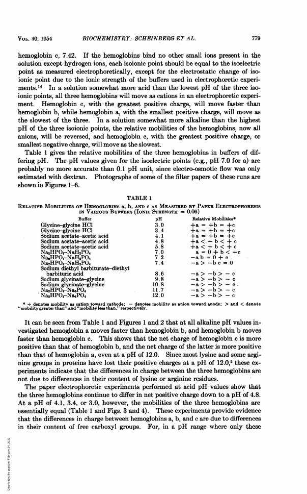
Table 1 gives the relative mobilities of the three hemoglobins in buffers of dif-fering pH. The pH values given for the isoelectric points (e.g., pH 7.0 for a) areprobably no more accurate than 0.1 pH unit, since electro-osmotic flow was onlyestimated with dextran. Photographs of some of the filter papers of these runs areshown in Figures 1-6.
TABLE 1RELATIVE MOBILITIES OF HEMOGLOBINS a, b, AND C AS MEASURED BY PAPER ELECTROPHORESIS
IN VARIOUS BUFFERS (IONIC STRENGTH = 0.06)Buffer pH Relative Mobilities*
Glycine-glycine HCl 3.0 +a = +b= +cGlycine-glycine HCl 3.4 +a = +b= +cSodium acetate-acetic acid 4.1 +a = +b= +cSodium acetate-acetic acid 4.8 +a < + b < + cSodium acetate-acetic acid 5.8 +a < + b < + cNa2HPO4-NaH2PO4 7.0 a = 0 + b < +cNa2HPO4-NaH2PO4 7.2 -a b = 0 + cNa2HPO4-NaH2PO4 7.4 -a> -b c = .0Sodium diethyl barbiturate-diethyl
barbituric acid 8.6 -a> -b> - cSodium glycinate-glycine 9.8 -a> -b> - cSodium glycinate-glycine 10.8 -a> -b> - c.Na2HPO4-NaPO4 11. 7 -a> -b> - cNa2HPO4-NasPO4 12.0 -a > -b> - c
* + denotes mobility as cation toward cathode; - denotes mobility as anion toward anode; > and < denote"mobility greater than" and "mobilityless than," respectively.
It can be seen from Table 1 and Figures 1 and 2 that at all alkaline pH values in-vestigated hemoglobin a moves faster than hemoglobin b, and hemoglobin b movesfaster than hemoglobin c. This shows that the net charge of hemoglobin c is morepositive than that of hemoglobin b, and the net charge of the latter is more positivethan that of hemoglobin a, even at a pH of 12.0. Since most lysine and some argi-nine groups in proteins have lost their positive charges at a pH of 12.0,5 these ex-periments indicate that the differences in charge between the three hemoglobins arenot due to differences in their content of lysine or arginine residues.The paper electrophoretic experiments performed at acid pH values show that
the three hemoglobins continue to differ in net positive charge down to a pH of 4.8.At a pH of 4.1, 3.4, or 3.0, however, the mobilities of the three hemoglobins areessentially equal (Table 1 and Figs. 3 and 4). These experiments provide evidencethat the differences in charge between hemoglobins a, b, and c are due to differencesin their content of free carboxyl groups. For, in a pH range where only these
VOL. 40, 1954 779
Dow
nloa
ded
by g
uest
on
Feb
ruar
y 24
, 202
2

BIOCHEMISTRY: SCHEINBERG ET AL.
groups are losing their charges, the differences in charge between the three proteinsdisappear.
It is necessary to prove that the equality of the mobilities of the three hemo-globins at a pH of about 4.1 and lower is not due to irreversible denaturation of theproteins. Solutions of the three hemoglobins, brought to a pH of 4.1 with hydro-chloric acid, were dialyzed at room temperature against an acetate buffer of pH 4.1for 197 minutes. The dialysis sacs were then placed in a veronal buffer of pH 8.6and dialyzed overnight. This procedure causes a portion of each hemoglobin toprecipitate, and the precipitates were removed by centrifugation. A paper electro-phoretic experiment was performed with the supernatant solutions in veronal bufferof pH 8.6. The result is pictured in Figure 5 and shows that the same differencesin mobility were observed as had been seen at this pH in experiments in which thethree hemoglobins had not been exposed to acid (Fig. 1). In a similar experimentthe precipitated hemoglobins were dissolved in a small amount of sodium hydroxideand electrophoresis was performed at a pH of 10.8. The result, seen in Figure 6, showsthat some of the denatured hemoglobin remained at the starting point but that theportions that migrated did so with the usual differences in mobility between hemo-globins a, b, and c, which are observed at alkaline pH. The exposure to a pH of4.1, therefore, has not irreversibly denatured the hemoglobins with respect to thedifferences in structure which are responsible for the differences in charge at higherpH values. Exposure of the hemoglobins for 2-3 hours to a buffer of pH 3.4, 3.0,or 11.8, with subsequent dialysis against a veronal buffer of pH 8.6, also yieldedsupernatant hemoglobins still possessing the characteristic differences in mobilityatpH8.6.Havinga and Itano have cited experiments in which electrophoretic analyses in
acidic buffers failed to disclose any significant differences in mobility between de-natured globins prepared from hemoglobins a and b.'5 They also report experi-ments in which the usual differences in mobility characteristic of the two hemo-globins were observed in the native globins of these proteins at a pH of 7.0 and ina 0.01 molar solution of Na2HPO4. They do not report experiments on the de-natured globins in alkaline buffers.On the basis of these results it would appear unlikely that ionizable groups
other than carboxyl groups could account for the differences in charge between theproteins. Since the imidazole nitrogen of histidine ionizes in a pH range of around5.5-8.5,5 such a nitrogen atom would be expected to be charged at pH 4.0 and un-charged at pH 11. Therefore, a difference in the histidine content of the hemo-globins would result in differences in mobility at acid pH values and in no differ-ences in mobility in alkaline buffers. This is the opposite of what was observed.Differences in tyrosine content should not contribute to differences in charge be-tween the hemoglobins, except at pH values near and above the pK value of thetyrosine hydroxyl group, which is about 10.8.5 The phosphorus content of hemo-globin a is not large enough for differences in the content of phosphate groups be-tween hemoglobins a, b, and c to account for their differences in charge.'6 Differ-ences in specific binding of buffer ions by the hemoglobins cannot account for the
780 P'ROC1. N. A. S.
Dow
nloa
ded
by g
uest
on
Feb
ruar
y 24
, 202
2

BIOCHEMISTRY: SCHEINBERG ET AL.
x i.,.:
a
c
b
K
_ +
x
a
b
a
C
Fig. I
.50
Fig. 2
a
b
a
C,
Fig. 3_ +
x i
b -4t
C -
X T
Fig. 5
+ -
K .a.
a 41A
4:
X
Fig. 4
x
a I - ,..b :A
X
Fi g. 6
FIGS. 1-6.-Paper electrophoresis of hemoglobins a, b, and c and dextran, x. The line is the lo-cus of the starting points for hemoglobins and dextran. The outlined circles are locations of dex-tran spots which stained too faintly to be photographed. The + and - signs indicate on whichside of the starting line the anode and cathode, respectively, are located.
Fig. 1, veronal buffer, pH = 8.6, r/2 = 0.06; run for 240 minutes at 400 volts. Fig. 2, phos-phate buffer, pH = 11.7, r/2 = 0.06; run for 126 minutes at 350 volts. Fig. 3, acetate buffer,pH = 4.08, r/2 = 0.06; run for 57 minutes at 400 volts and 140 minutes at 200 volts. Fig. 4,glycine buffer, pH = 3.35, r/2 = 0.06; run for 75 minutes at 350 volts. Fig. 5, veronal buffer,pH = 8.65, r/2 = 0.06; run for 225 minutes at 350 volts. Hemoglobin solutions, adjusted topH 4.1, were dialyzed against pH 4.08 acetate buffer for 197 minutes, then against pH 8.6 veronalbuffer overnight. Supernatant hemoglobin solutions from veronal dialysis were run in this experi-ment.
Fig. 6, phosphate buffer, pH = 10.8, r/2 = 0.06; run for 95 minutes at 350 volts. Hemoglobinsolutions, adjusted to pH 4.6, were dialyzed against pH 4.01 acetate buffer for 88 minutes, witha final pH of 4.05 of the hemoglobin solutions. Solutions were then dialyzed against veronal bufferof pH 8.6 overnight. The precipitated hemoglobins were centrifuged, and the precipitates, dis-solved in a minimum amount of 0.1 N NaOH, were run, in this experiment.
VOL. 40, 1954 -781
*a 6:*:.
Dow
nloa
ded
by g
uest
on
Feb
ruar
y 24
, 202
2

782BIOCHEMISTRY: SCHEINBERG ETAL.
differences in their charges and mobilities, for two reasons. First, and more im-portant, the isoionic points of the three hemoglobins are different in the absence of
any salt. Second, mobility differences have been observed in several different alka-line buffers, and the equal mobilities at low pH have been observed in glycine andacetate buffers. Finally, it may be mentioned that ordinary titration studies ofthese hemoglobins"7 have revealed that the total charge per molecule of hemoglobina at various pH values is as follows: pH 3.0, +88; pH 4.0, +47; pH 12, -67.These figures indicate that the disappearance of differences in mobility in acidbuffers is not due simply to a much greater total charge on the hemoglobin moleculein acid than is present in alkali.
Pauling and his co-workers calculated, on the basis of the difference in isoelectricpoints between hemoglobins a and b, that hemoglobin b possessed two to four morenet positive charges per molecule than did hemoglobin a.' Using this figure, our
experiments, therefore, provide evidence that hemoglobin b possesses two to fourfewer free carboxyl groups than does hemoglobin a. Since the difference betweenthe isoionic points of hemoglobins b and c is of the same order of magnitude as,
though somewhat larger than, the difference, between the isoionic points of hemo-
globins a and b, it seems probable that hemoglobin c contains about five to eightfewer free carboxyl groups than hemoglobin a. In order to calculate quantitativelythe differences in content of free carboxyl groups in hemoglobins a, b, and c, we
have attempted to combine paper electr~phoretic experiments, in which electro-osmotic flow is measured, with acid-base titration experiments. These results willbe reported in a subsequent paper.The structural basis for the apparent difference in the content of free carboxyl
groups of these hemoglobins remains obscure. The available analytical data'8do not indicate a smaller content of aspartic or glutamic acid residues, or a largernumber of carboxyl groups which exist as amides, in hemoglobin b than in hemo-globin a. No amino acid analyses of hemoglobinc have been published.
It has been suggested that hemoglobins a and b have identical polypeptide chainsbut that the folding of the chains is different in the two molecules, presumably re-
sulting in some charged groups being unreactive.15" "I An actual masking of acid-
binding groups, perhaps due to folding of the molecule, has recently been experi-
mentally demonstrated in native horse hemoglobin by Steinhardt and Zaiser.6,These workers have shown that thirty-six acid-binding groups are not available fortitration until the protein has been exposed briefly to a pH of about 4.0 or less.When these thirty-six groups are broken out, the hemoglobin is simultaneouslydenatured, as judged by spectroscopic changes and loss of solubility at the isoelectricpH. Renaturation, and presumably remasking of the thirty-six groups, is effected
by returning the protein to neutrality. Steinhardt and Zaiser consider these
thirty-six groups to be epsilon amino or guanidinium groups, since their pK valuesare above 5, presumably too high for carboxyl groups, and since all the histidineresidues of horse hemoglobin are titrated in the native protein.7 It is tempting toconsider whether the excess positive charge on hemoglobins b and c could be dueto an analogous masking of differing numbers of charged groups in the three hemo-
globins. The fact that the variations in charge persist at pH 12.0 indicates thatthese differences are not due to epsilon amino groups and are not very likely to bedue to guanidinium groups. There is the possibility that carboxyl groups may
782LYRIC.
Dow
nloa
ded
by g
uest
on
Feb
ruar
y 24
, 202
2

CHEMISTRY: M. DELBR UCK
also be masked in hemoglobins with more of them blocked in the abnormal hemo-globins. Other more, complicated masking possibilities may be imagined to ac-count for the differences in charge, but the evidence provided by the experimentsreported above makes it very probable that the three hemoglobins differ in theircontent of free carboxyl groups.The method of differential titration described in this paper should be applicable
to the study of other sets of closely related proteins.
* This work was supported in part by a grant (A-572) from the National Institute of Arthritisand Metabolic Diseases of the National Institutes of Health, United States Public Health Service.
It is a pleasure to thank Professors John T. Edsall and George Scatchard for their very helpfulcritical discussions. We are also indebted to Dr. Helen Ranney, Miss Grace Vanderhoff, andMrs. Catherine Holavko for their assistance in obtaining the hemoglobins studied and in constructing the paper electrophoresis apparatus.
1 L. Pauling, H. A. Itano, S. J. Singer, and I. C. Wells, Science, 110, 543-548, 1949.2 H. A. Itano and J. V. Neel, these PROCEEDINGS, 36, 613-617, 1950.3H. A. Itano, these PROCEEDINGS, 37, 775-784, 1951.4H. A. Itano, Eli Lilly Award in Biochemistry Address, 125th Meeting of the American Chemi-
cal Society, Kansas City, Missouri, March 23-April 1, 1954.5 E. J. Cohn and J. T. Edsall, Proteins, Amino Acids and Peptides (New York: Reinhold
Publishing Corp., 1943), chap. xx.6 J. Steinhardt and E. M. Zaiser, J. Biol. Chem., 190, 197-210,1951; E. M. Zaiser and J. Stein-
hardt, J. Am. Chem. Soc., 73, 5568-5572, 1951; J. Steinhardt and E. M. Zaiser, J. Am. Chem.Soc., 75, 1599-1605, 1953.
7E. M. Zaiser and J. Steinhardt, J. Am. Chem. Soc., 76, 1788-1791, 1954.8 H. G. Kunkel and A. Tiselius, J. Gen. Physiol., 35, 89-118, 1951.9 M. F. Perutz, A. M. Liquori, and F. Eirich, Nature, 167, 929-931, 1951.
10 E. L. Durrum, J. Am. Chem:'Soc., 72, 2943-2948, 1950.11 H. A. Itano, Science, 117, 89-94, 1953; H. M. Ranney, D. L. Larson, and G. H. McCormack,
Jr., J. Clin. Invest., 32, 1277-1284, 1953.12 D. L. Drabkin, J. Biol. Chem., 164, 703-723, 1946.18 H. M. Dintzis, Methods and Procedures Employed in the Preparation and Characterization of
Proteins. XVI (Cambridge, Mass.: Department of Biophysical Chemistry, Harvard Univer-sity, 1952; thesis, Harvard University, 1952).
14 G. Scatchard and E. S. Black, J. Phys. &-Colloid Chem., 53, 88-99, 1949.16 E. Havinga and H. A. Itano, these PROCEEDINGS, 39, 65-67, 1953.16 E. Havinga, these PROCEEDINGS, 39, 59-64, 1953.17 I. H. Scheinberg, R. S. Harris, and B. M. Lang (in preparation).18 W. A. Schroeder, L. M. Kay; and I. C. Wells, J. Biol. Chem., 187, 221-40, 1950.
ON THE REPLICATION OF DESOXYRIBONUCLEIC ACID (DNA)
BY M. DELBRPCK
KERCKHOFF LABORATORIES OF BIOLOGY, CALIFORNIA INSTITUTE OF TECHNOLOGY
Communicated May 18, 1954
The discoveries of Hershey and Chase' concerning the role ofDNA in transmittinggenetic information in phage and of Watson and Crick2 concerning the structure ofDNA have brought the problem of the replication of DNA into focus. The struc-ture proposed by Watson and Crick consists of two polynucleotide chains woundhelically around a common axis, tied together by hydrogen bonds between the
VOL. 40, 1954 783
Dow
nloa
ded
by g
uest
on
Feb
ruar
y 24
, 202
2


![8 Eigenvectors and the Anisotropic Multivariate Gaussian …jrs/189s17/lec/08.pdf · 2017. 2. 14. · 777 777 777 777 777 5 [diagonal matrix of eigenvalues] Defn. of “eigenvector”:](https://static.fdocuments.us/doc/165x107/61216a6db677231115104a22/8-eigenvectors-and-the-anisotropic-multivariate-gaussian-jrs189s17lec08pdf.jpg)