Complication and management of tooth extraction or exodontia
Upload
drnizar-chaarCategory
view
183download
7
Exodontia
Instructor – Dr.Jesus George1

Introduction
It is a procedure that incorporates principles of surgery, physics and mechanics.
Painless removal of the tooth or root with minimal injury to the surrounding soft tissue & bone
2

Cont.
Removal of tooth does not require large amount of force, but fine and controlled forced in such a manner that tooth is not pulled from bone but lifted gently from alveolar process
3

Pain and Anxiety control Local anesthesia
Profound local anesthesia results in loss of pain, temperature and touch but not pressure.
When the tooth has pulpitis or surrounding soft & hard tissues inflamed or infected, periodontal injection is given, that gives anesthesia for 15-20min. If it fails intra osseous injection can be given.
4

Sensory innervation of jaws
Inferior alveolar nerve all mandibular teeth, buccal soft tissues of PM, canine & incisors.
Lingual nerve; Lingual soft tissues of all teeth
Long buccal nerve: Buccal- soft tissues of molars.
5

Cont.
Anterior superior alveolar nerve; maxillary incisors and canine, buccal soft tissues of incisors and canines.
Middle superior alveolar nerve: Max. PM & MB root of 1st molar, Buccal soft tissue of PM.
6
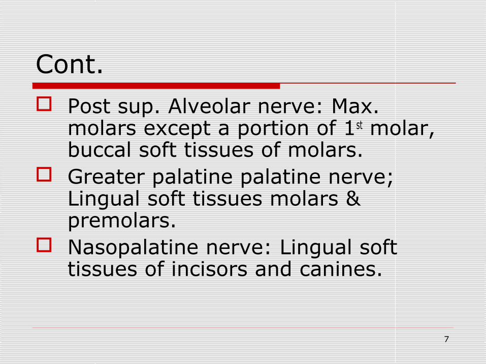
Cont. Post sup. Alveolar nerve: Max.
molars except a portion of 1st molar, buccal soft tissues of molars.
Greater palatine palatine nerve; Lingual soft tissues molars & premolars.
Nasopalatine nerve: Lingual soft tissues of incisors and canines.
7

Cont.
Mandibular PM region buccal soft tissue innervated primarily by mental branch of IAN and also by terminal branches of long buccal nerve.
8

Duration of Anesthesia
1. Local anesthesia with out vasoconstrictors: Max. teeth-10-20min Mand. teeth- 40-60min. Soft tissue- 2-3 HR
9

Cont.
2. Local anesthesia with vasoconstrictors Max. teeth 50-60 min Mand. teeth 90-100min Soft tissue 3-4 HR
10

Cont.
3. Long acting local anesthesia with vasoconstrictors Max. teeth 60-90 min. Mand teeth - 3HR Soft tissue 4-9 HR
11

Sedation
In case of mild anxiety- proper explanation of procedure; assurance that there will not be sharp pain, expression of concern caring, empathy will reduce anxiety.
In moderate anxiety: Preoperative oral diazepam provide rest at night before surgery and relieve anxiety in morning.
12

Cont.
Sedation by inhalation of nitrous oxide or IV sedation with diazepam can be given in severe anxiety.
13

Presurgical Medical Assessment
A proper medical history
14

Indications for removal of teeth Severe caries: that can not be
restored. Pulpal necrosis: if endodontic Rx can
not be performed becoz Pt declines, or root canal that is tortuous, calcified or endodontic failure.
Severe periodontal disease: excessive bone loss and irreversible tooth mobility.
15

Cont.
Mal - opposed teeth; if they traumatize the soft tissue or can not be repositioned by orthodontic Rx (Max. III M. in severe buccal version and causes ulceration & trauma on cheek or teeth that are hyper erupted becoz of loss of teeth in opposing arch.
16

Cont.
Orthodontic reasons: Max. & Mand PMs Mand. incisors are commonly extracted.
Cracked teeth: or with fractured root Preprosthetic extraction: teeth
interfering with design and placement of full dentures, partial dentures
17

Cont.
Impacted teeth: that is unable to erupt to functional occlusion.
Supernumerary teeth: Impacted, interfering with eruption of succedaneous teeth or causing resorption and displacement of adjacent teeth should be extracted.
18

cont.
Teeth associated with pathologic lesions: If maintaining the tooth compromises, complete surgical removal of lesion.
Pre - radiation therapy: remove teeth in line of radiation therapy.
Severe attrition, abrasion or erosion
19

Cont. Teeth involved in jaw #: If tooth is
severely luxated, tooth in # line should be removed.
Esthetics: Severely stained, malopposed or protruding teeth are removed.
Economics: inability of PT to pay or to take time from work may require the tooth to be extracted
20

Contraindications for removal of teeth
Systemic contraindications: Uncontrolled diabetes End stage renal disease with severe
uremia Uncontrolled leukemia Uncontrolled cardiac disease Unstable angina pectoris Recent MI
21

Cont. Severely uncontrolled hypertension Pregnancy 1st and last trimester Bleeding disorders like hemophilia Platelet disorders Patients on anticoagulants
22

Cont.
Local Contraindications: H/o therapeutic radiation- causes
osteoradio necrosis Tooth in area of tumour: disseminate
cells and cause metastasis A/c infection Central hemangioma
23
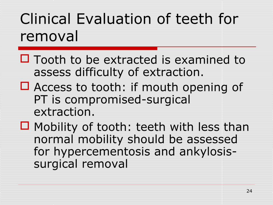
Clinical Evaluation of teeth for removal Tooth to be extracted is examined to
assess difficulty of extraction. Access to tooth: if mouth opening of
PT is compromised-surgical extraction.
Mobility of tooth: teeth with less than normal mobility should be assessed for hypercementosis and ankylosis- surgical removal
24

Cont.
Condition of crown: if large portion of crown is decayed by caries or tooth with large amalgam restoration, forceps is placed as far apical as possible.
25

Cont.
If large amount of calculus is present on tooth, it should be removed before extraction otherwise it will interfere with application of forceps or contaminate socket after extraction.
26

Cont.
If adjacent tooth has amalgam restoration or undergone endodontic therapy, care must be taken while using elevators.
27

Radiographic Examination of tooth for removal
IOPA shows portion of crown and root of tooth under consideration
If it is a I° tooth its relationship with a succedaneous tooth should be visible
Relationship of associated vital structures For Max. teeth relation with max. sinus.
28

Cont. For Mand. Molars inferior alveolar canal For Mand premolars relation with mental
foramen
Configuration of roots- If excess curvature surgical extraction
Length of roots Hypercementosis Root # more liable to #
29

Cont. Root resorption liable to # H/o endodontic Rx -tooth is brittle or
ankylosed -so surgical extraction. Condition of surrounding bone
If more radio opaque- condensing osteitis or sclerosis- so difficult to extract.
Periapical pathologies- should be removed after extraction.
30

Patient & surgeon Preparation
All patients should be considered as having blood born disease.
Surgeon should wear surgical gloves, mask, eyewear with side shield, long sleaving gowns.
If surgeon has long hair it should be covered with surgical CAP.
31

Order of extraction
Lower teeth are removed before the upper & posteriors are removed before anteriors to prevent bleeding from socket obscuring field of operation (prof.J.Moore)
32

Methods of extraction
Closed or intra-alveolar Open or transalveolar or surgical Stobie technique – extraction of
multiple mandibular anteriors by using elevators b/w teeth
33

Chair position for forceps extraction Best position is one that is most
comfortable to PT & to surgeon. Correct position allows surgeon to
deliver force with arm and shoulder and not with hand.
For Max. extraction,
34

Maxillary teeth
Position of chair Height of chair is such that height of
patient's mouth is at or slightly below operator's elbow.
Chair is tipped backward that Max.occlusal plane is 60° to floor
35

Cont. Position of patient
During procedures of Max. Right + left quadrant PT's head is turned towards operator.
For Max. Ant. Teeth, PT should be looking straight ahead.
Position of operator Front & right side of the patient for right
handed operator & reverse in left handed operator
36

Cont.
Position of left arm Left upper teeth, thumb supports the
palatal alveolar bone & index finger retract the buccal tissues
Right upper teeth – thumb retracts the buccal tissues & index finger supports the palatal alveolar bone
In left handed operator the reverse
37

Mandibular teeth
Position of chair Chair is positioned in such a way that,
Mand. occlusal plane is parallel to floor. Surgeon's arms are inclined downward at
an angle of 120° at elbow.
Position of patient In Mand. right post teeth-PT is turned
towards surgeon.
38

Cont. Position of operator
Mand. right post teeth, operator is behind the pt &
In Mand. left post region, surgeon is in front of PT.
Left handed operator the position is reverse
If surgeon chooses to sit, the PT is at a more lower level than standing and other position are similar
39

Cont. Position of left arm
Lower left teeth – thumb supports the mandible &index finger retracts the buccal soft tissues ,middle finger controls tongue
Lower right teeth – index finger retract the buccal tissues, thumb controls the tongue & other fingers supports the mandible.
Reverse for left handed operator
40

Mechanical Principles Involved in tooth extraction:
Elevators I°rly works on lever principle E.g straight elevator
Wedge principle is also used when elevator is used to luxate tooth.
Wheel and axle principle is used by triangular shaped elevatorsE.g Cryer's elevator
41

Principles of forceps use
Use of forceps: To expand bony socket To remove tooth
Forceps should be placed below CEJ Traction towards least resistance
42

Cont.
Alveolar purchase By Kruger For removal of anterior teeth or roots After detaching the labial gingiva the
labial beak is placed under the tissues in alveolar bone &apply pressure
43

Major Motions of forceps 1.Apical pressure: Tooth socket is
expanded by insertion of beaks down into periodontal ligament.
2. Buccal pressure: produces expansion of buccal plate and lingual apical pressure
Lingual pressure: Expands lingual cortical plate and buccal apical pressure.
44

Cont.
Rotational pressure: Teeth with single conical roots e.g. Max. incisors Mand. PM, But the roots should not be curved.
Tractional force: For delivering tooth out of socket.
45

Procedure for closed extraction:
Requirements for extraction Adequate access and visibility Unimpeded pathway of removal Use of controlled force.
46

General steps for closed extraction
Loosening of soft tissue attachment from tooth
Done by a Periosteal elevator Helps to assess anesthesia Allows extraction forceps to be placed
apically.
47

Cont.
Luxation of tooth with a dental elevator:
A straight elevator is inserted to the tooth into interdental space.
Strong, slow, forceful, turning of handle moves tooth in posterior direction causing expansion of bone
Tearing of periodontal ligament
48

Cont. Excess force can damage or displace
adjacent tooth especially if it has a large restoration or caries
Adaptation of forceps to tooth: Tips of forceps beaks should grasp root Lingual beak is seated first. Beaks must be parallel to long axis of
tooth Force should be applied with shoulder &
upper arm & not with wrist.
49

Cont. Sterile drape should be put across
Pt's chest Before Extraction, PT should
vigorously rinse mouth with antiseptic mouth rinse.
4X4 inch gauze can be placed in to back of mouth to prevent teeth or fragments falling into mouth
50

Cont.
Luxation of tooth with forceps: Major force should be directed towards
thinnest portion of bone. Slow steady force is used.
Removal of tooth from socket: Done by tractional force usually given
buccally
51

Role of opposite hand Reflect soft tissues of cheek, lips and
tongue, give visibility. Protect other teeth from forceps. Stabilize PT's head Supporting and stabilizing mand. during
mand. extraction. Supports alveolar process and provide
tactile information about expansion of alveolar process.
52

Role of assistant
Helps to visualize and gain access, by reflecting soft tissues and tongue
Suction away blood, saliva, irrigating solution
Stabilize mandible
53

Specific Technique for removal of Each tooth Maxillary incisor teeth:
They have conical roots. LI may have a distal curvature for root. Alveolar bone is thin over buccal side
and thick over palatal side. After apical Pre. the force is given
buccally, less palatal force followed by rotational force, no rotational force if there is curvature.
Tooth is delivered in labial direction
54

Cont.
Maxillary canine Longest tooth in mouth Root is oblong in C.S. Bone on labial aspect is thin. So a
fragment of bone usually fractures from buccal aspect when tooth is removed.
Buccal, palatal and a small amount of rotational movement and removed in labio - incisal direction.
55

Cont. If Bone is detached from periosteum, it
should be removed. If buccal bone is attached to periosteum,
it can be left, normal healing will occur.
56

Cont. Maxillary I PM
Single rooted with bifurcation to bucco- lingual roots at apical 1/3
Most common root # Buccal bone is thinner Tooth should be luxated as much as
possible. Apical, buccal, palatal movements,
palatal should be less
57

Cont.
Maxillary II PM Single rooted Thin bone buccally and thick palatally Buccal, palatal, bucco - occlusal
tractional force.
58

Cont.
Maxillary molar 3 roots,2 buccal roots are relatively
closer and palatal is divergent towards palate.
Buccal cortical plate is thinner than palatal.
Forceps have projection on buccal beak to fit buccal bifurcation.
59

Cont. Upper cowhorn forceps is used in teeth
with large caries or restoration. More buccal force, less palatal force
removed with bucco occlusal tractional force.
II M similar anatomy except less divergence for roots and removed in similar way.
Erupted III M. conical roots Easily extracted by elevators alone
60

Cont.
Mand. ANT. Teeth Incisor roots are thinner and shorter and
canine roots are longer and heavler. Bone on labial aspect of canine is
somewhat thicker. Equal movements labially, lingually &
tooth is luxated by a rotational force & extracted by labio-incisal tractional force
61

Cont.
Mand. PMs Roots are straight & conical Bone thinner on buccal & thicker on
lingual aspect. Buccal, less lingual, rotational and
occluso - buccal tractional force. If any root curvature rotation is avoided
62

Cont.
Mand. Molars 2 roots and widely divergent for IM Roots may converge at apical 1/3 Most difficult of all teeth to extract. Apical, buccal, lingual and bucco occlusal
tractional force. Lingual bone is thinner than buccal so
more lingual pressure
63

Cont. Lower cowhorn forceps is used by
squeezing the bifurcation, buccolingual movements can also be used.
Erupted mand. III M. Conical roots lingual plate is thinner, so more movements are given lingually and delivered in lingo occlusal direction.
64

Modification for extraction of I° teeth
Similar buccolingual movements Rotational movement is avoided for
multirooted teeth. Tooth is delivered in least resistant
path. If the roots embrace PMT crown,
sectioning of roots should be done
65

Post extraction care
If any periapical pathology in radiograph, and no granuloma removed with extracted tooth, periapical area is carefully curetted.
If any debris, calculus, amalgam, tooth fragment, in socket it is removed with curette.
Remnants of periodontal ligament & bleeding bony walls improves healing.
66

Cont.
Vigorous curettage delay healing by causing additional injury
Finger pressure is applied to buccal & lingual cortical plates to compress the socket, to prevent bony undercuts
If there is excess granulation tissue around gingival cuff, it should be removed with curette or hemostat.
67

Cont. Sharp bony projections should be
smoothed with bone file. Moistened 2x2 inch gauze is placed
over extraction socket and it should fit into the space that was previously occupied by tooth. So that biting force will give pressure, will cause hemostasis.
Larger gauze is placed if multiple teeth extracted of opposing tooth is missing.
68

OPEN EXTRACTION Indications
Failure to remove tooth by closed method
Unfavourable root pattern Fracture or caries extending to root Hypercementosis Ankylosis Impacted tooth Sclerosed bone
69

Steps in open extraction
Incision Raising mucoperiosteal flap Removal of bone around the tooth or
root Establishment of point of application
of elevator Removal of tooth from socket
70

Cont.
Trimming the bone Toileting the wound Control of bleeding Repositioning & suturing Packing
71

Planning of an incision
Def.of incision-a cut or wound deliberately made by an operator in skin or mucosa using a sharp instrument, so that the underlying structures can be exposed for surgical access.
Incision is placed parallel to structures without causing damage to vital structures
72

Cont.
Extraoral incisions are planned along the Langers lines of normal skin tension or creases, so that min. scar is formed.
Incision should be placed on sound bone.
Pen grasp (intraoral) or table knife (extra oral) grasp is used
73

Cont.
Skin or mucosa to be incised to be stabilized with finger pressure to guide the passage of blade.
A firm continuous stroke should be used.
Change in direction is accomplished by a gradual curve.
74

Incisions in oral cavity
Incise through attached gingiva over a healthy bone.
Incisions placed near teeth for extractions should be made in gingival sulcus.
Integrity of interdental papilla should be maintained.
75

Cont.
Incisions involving reflection of mucoperiosteal flap are direct, straight-line or curvilinear taking the shortest distance vertically through the tissues.
Blood supply to the incision should be adequate.
76

Contraindications for placement of incisions
Over canine prominence Vertical incision in mental nerve
region. Near greater palatine vessels in
palate. Through incisive papillae. Over bony lesions
77

Cont.
Over freni. Vertical incision on lingual side of
mandibular arch
78

Types of incisions
Horizontal:-given along the gingival margin either mesially or distally. e.g. Internal bevel incision & crevicular incision.
Vertical:-also called releasing incision Single vertical incision-triangular flap Double vertical incisions-trapezoidal flap
79

Cont. Incision should extend beyond
mucogingival line to alveolar mucosa. Vertical incisions should be placed at
obtuse angle to horizontal incision & should leave interdental papillae intact
80

Cont.
Semilunar (curved,elliptical) Used to maintain attached gingiva intact
& for endodontic surgery. Horizontal component rest on bone. 5mm gap is present from base of
gingival sulcus to incision.
81

Flap design
Complications of flap surgery Flap tearing Flap necrosis Flap dehiscence
82

Cont.
Flap tearing:-to prevent this Incision should be clean,sharp&should
penetrate entire mucoperiosteum. Flap should be reflected as one unit. Length of flap should not be more than
twice the width of base.
83

Cont. Flap necrosis:-to prevent this
Base of flap should be wider. Margins of flap should be either parallel
to each other or converge from base to apex.
Axial blood supply should be included in flap e.g.palatal flap based on greater palatine artery.
84

Cont. Flap dehiscence=separation of flap
margins or gaping of wound. Causes
Poor tissue handling Too tight suturing Hematoma formation Infection
Prevention Sutures are placed over healthy bone.
85

CONT.
Types of flaps A.1.Full thickness-mucoperiosteal flap 2.Partial thickness B.1.Envelop 2.Triangular 3.Rhomboid 4.Semilunar
86

CONT.
C.1.Labial, buccal 2.Palatal, lingual
87

CONT. Envelop flap Most common type Sulcular incision is made around the
tooth on buccal or lingual aspect including interdental papillae.
Entire mucoperiosteal flap is elevated.
Mainly used in surgical extraction of teeth.
88

CONT.
Triangular flap A vertical releasing incision is made
on one side of envelope flap diverging towards buccal vestibule.
Vertical incision is made in the interproximal area not on the facial aspect of tooth to avoid periodontal defect.
89

CONT.
Flap is reflected towards the base of the flap.
Rhomboid flap 2 vertical releasing incisions are
made on either side of envelope flap. Base of flap should be wider.
90

CONT.
Semilunar flap Used in periapical surgery. Suture line should not be on bony
defect.
91

Cont. Toileting the wound Irrigation Debridement of necrotic, foreign
bodies, severely injured tissues. Antibiotics Use of medicated mouthwashes after
every food intake.
92

Cont.
Hemostasis should be achieved To minimize blood loss. Increase visibility Reduces operating time Minimizes postsurgical trauma.
93

Cont.
it can be achieved by Intermittent pressure:-with cotton
or gauze sponges. pressure is applied for 20-30sec for smaller vessels&5-10 min. for larger vessels.
Electrocautery:-for this area around the vessel is dried thoroughly.Avoid unnecessary burning.
94

Cont.
Suture ligation:-when large vessel is severed it is grasped with hemostat. Nonabsorbable suture is used to ligate the vessel.
Vasoconstrictors:-epinephrine, thrombin or collagen gel foam
95

Cont.
Compression dressing over the wound:-if there is oozing over a large area a cotton pad or ribbon gauze is stabilized over the wound &secured in position with sutures & kept for 2-3 days.
96

Healing of extracted socket
Hematoma & Fibrin (clot) {0-4 days} Granulation tissue(3days – 3 weeks) Fibrous tissue – " Callus - " Calcification - " Bone remodeling (after 3 weeks)
97

Complications
# of crown or roots of the tooth being extracted
# of alveolar bone # of maxillary tuberosity #of adjacent or opposing tooth # of mandible Dislocation of TMJ
98

Cont. Displacement of root into soft tissues,
maxillary antrum Bleeding Injury to gums, lips, IAN & its
branches, lingual nerve, tongue, floor of mouth, greater palatine artery
Dry socket Osteomyelitis Infection
99

Cont.
Trismus Hematoma OAF
100

Dry socket or alveolar osteitis Causes
Undue trauma during extraction Pre existing infection Disturbance of clot due to vigorous
mouth wash or curettage Increased fibrinolytic activity Localized impaired vascular supply Smoking Use of OCP
101

CONT.
Clinical features Continuous throbbing & excruciating pain h/o extraction 48-72 hrs Alveolar socket is covered with grayish
necrotic tissues Denuded alveolar bone Halitosis
102

Cont. L.A. Irrigate with warm saline or
chlorhexidine for removal of dead bone or infected tissues
Do not curette Obtundant dressing (ZOE with cotton
to cover the denuded bone or whitehead varnish
Antibiotic, analgesic
103

Hematoma
Control bleeding prior to closure Apply ice extraorally Antibiotics to prevent infection Anti inflammatory drugs
104