19 1 139 151 2017 MATERIALS ENGINEERING … · Duitama, Colombia. ****Grupo de Investigación en...
13
139 Ingeniería y Competitividad, Volumen 19, No. 1, P. 139 - 151 (2017) MATERIALS ENGINEERING Preparation and characterization of scaffold nanofibers by electrospinning, based on chitosan and fibroin from Silkworm (Bombyx mori) INGENIERÍA DE MATERIALES Obtención y caracterización de andamios electrohilados a base de quitosano y fibroína del capullo (Bombyx mori) Yesica C. Cárdena-Pérez*,****, Ricardo Vera-Graziano**, Efrén d. J. Muñoz-Prieto*,**** Edwin Y. Gómez-Pachón***,****§ *Escuela de Ciencias Químicas, Facultad de Ciencias básicas, Universidad Pedagógica y Tecnológica de Colombia-UPTC. Tunja, Colombia. **Instituto de Investigaciones en Materiales, Universidad Nacional Autónoma de México. México D.F., México. ***Escuela de Diseño Industrial, Universidad Pedagógica y Tecnológica de Colombia-UPTC. Duitama, Colombia. ****Grupo de Investigación en desarrollo y nuevos materiales-DANUM, Universidad Pedagógica y Tecnológica de Colombia-UPTC. Tunja, Colombia. [email protected], [email protected], [email protected], §[email protected], (Recibido: Noviembre 25 de 2015 – Aceptado: Abril 27 de 2016) Abstract Here we present a study on the preparation of scaffolding for nanofibers composed of chitosan (QS) and fibroin from cocoon silkworm (Bombyx mori) (FGS) for its potential use in biomedicine. The scaffolds conformed by nanofibers were prepared from different mixtures of QS and FGS. They were characterized with analytical techniques such as scanning electron microscopy-SEM, infrared spectroscopy-FTIR, X-ray diffraction-XRD, Thermogravimetric Analysis-TGA, Differential Scanning Calorimetry-DSC and Citocompatibility tests, to determine its morphology, functional groups, thermal transitions, and cell viability of the scaffolding QS/FGS. Nanofibers of different diameters of QS/FGS (8-177 nm) were obtained and compared with the fibers of cocoons (22-36 µm) and fibroin (7-13 µm). The best scaffolds were achieved from a physical mixture with a weight ratio QS: FGS (1:3) dissolved in trifluoroacetic acid under the following electrospinning parameters. Voltage: 20 kV, injection flow: 0.5 ml/h, needle-collector distance: 12 cm, relative humidity: 28% Temperature: 25 ° C. It was determined that the scaffolding QS/FGS possessed fibers with nanometric diameters less than 200 nm, which allow optimal cell adhesion and thermal stability at the application site, with a view to control the degradation of the scaffold. Thermal analyses, supplemented with XRD studies reveal the internal structure of the fibers. Besides Cytocompatibility test on prepared cell scaffolds, it indicates that the integrity requirement was achieved and the necessity to control the pH to achieve an increased cell viability. The results show that nanofibers QS/FGS can be a good candidate for tissue engineering in biomedical applications such as scaffolds. Keywords: Antibacterial activity, chitosan, electrospinning, nanofibers. Resumen Aquí se presenta un estudio sobre preparación de andamios de nanofibras compuestos por quitosano (QS) y fibroína del capullo de gusano de seda (Bombyx mori ) (FGS) para su potencial uso en biomedicina. Los andamios conformados por nanofibras se prepararon a partir de diferentes mezclas de QS y FGS, se caracterizaron con apoyo de técnicas analíticas como microscopia electrónica de barrido- SEM, espectroscopia infrarroja-FTIR, difracción de rayos X- DRX, termogravimetría-TGA, calorimetría diferencial de barrido-DSC y pruebas de citocompatibilidad con el fin de determinar su morfología, grupos funcionales, transiciones térmicas, viabilidad y evaluación celular de los andamios QS/FGS, respectivamente. Se obtuvieron andamios de fibras de diámetros nanométricos de QS/ FGS (8-177 nm) y se compararon con las fibras de los capullos de seda de gusano (22-36 μm) y las fibras de la fibroína (7-13 μm). Los mejores andamios se lograron a partir de una mezcla física con relación en peso QS/FGS (1:3) disuelta en ácido trifluoroacético bajo los siguientes parámetros de electrohilado: voltaje: 20 kV, flujo de inyección: 0.5 mL/h, distancia aguja-colector: 12 cm, humedad relativa: 28% y temperatura: 25°C. Se determinó que los andamios QS/FGS poseen fibras con diámetros nanométricos inferiores a 200 nm, las cuales permitirán una óptima adhesión celular; una estabilidad térmica en el lugar de aplicación, con el fin de controlar la degradación del andamio. El análisis térmico, complementado con los estudios de DRX revela la estructura interna de las fibras. Además las pruebas citocompatibilidad sobre los andamios celulares preparados, indican que cumple el requisito de integridad, y la necesidad de controlar el pH para lograr una mayor viabilidad celular. Los resultados indican que las nanofibras de QS/FGS pueden ser un buen candidato para la ingeniería de tejidos en aplicaciones biomédicas como andamios celulares. Palabras clave: Actividad antibacteriana, electrohilado, nanofibras, quitosano
Transcript of 19 1 139 151 2017 MATERIALS ENGINEERING … · Duitama, Colombia. ****Grupo de Investigación en...
139
Ingeniería y Competitividad, Volumen 19, No. 1, P. 139 - 151 (2017)
134
Ingeniería Y Competitividad, Volumen 19, No. 1, P. 134 - 146 (2017)
1
Ingeniería Y Competitividad, Volumen 18, No. 2, P. 1 - 10 (2016)
MATERIALS ENGINEERING
Preparation and characterization of scaffold nanofibers by electrospinning, based on chitosan and fibroin from Silkworm (Bombyx mori)
INGENIERÍA DE MATERIALES
Obtención y caracterización de andamios electrohilados a base de quitosano y fibroína del capullo (Bombyx mori)
Yesica C. Cárdena-Pérez*,****, Ricardo Vera-Graziano**, Efrén d. J. Muñoz-Prieto*,**** Edwin Y. Gómez-Pachón***,****§
*Escuela de Ciencias Químicas, Facultad de Ciencias básicas, Universidad Pedagógica y Tecnológica de Colombia-UPTC. Tunja, Colombia.**Instituto de Investigaciones en Materiales, Universidad Nacional Autónoma de México. México D.F., México.
***Escuela de Diseño Industrial, Universidad Pedagógica y Tecnológica de Colombia-UPTC. Duitama, Colombia.****Grupo de Investigación en desarrollo y nuevos materiales-DANUM, Universidad Pedagógica y Tecnológica
de Colombia-UPTC. Tunja, [email protected], [email protected], [email protected], §[email protected],
(Recibido: Noviembre 25 de 2015 – Aceptado: Abril 27 de 2016)
AbstractHere we present a study on the preparation of scaffolding for nanofibers composed of chitosan (QS) and fibroin from cocoon silkworm (Bombyx mori) (FGS) for its potential use in biomedicine. The scaffolds conformed by nanofibers were prepared from different mixtures of QS and FGS. They were characterized with analytical techniques such as scanning electron microscopy-SEM, infrared spectroscopy-FTIR, X-ray diffraction-XRD, Thermogravimetric Analysis-TGA, Differential Scanning Calorimetry-DSC and Citocompatibility tests, to determine its morphology, functional groups, thermal transitions, and cell viability of the scaffolding QS/FGS. Nanofibers of different diameters of QS/FGS (8-177 nm) were obtained and compared with the fibers of cocoons (22-36 µm) and fibroin (7-13 µm). The best scaffolds were achieved from a physical mixture with a weight ratio QS: FGS (1:3) dissolved in trifluoroacetic acid under the following electrospinning parameters. Voltage: 20 kV, injection flow: 0.5 ml/h, needle-collector distance: 12 cm, relative humidity: 28% Temperature: 25 ° C. It was determined that the scaffolding QS/FGS possessed fibers with nanometric diameters less than 200 nm, which allow optimal cell adhesion and thermal stability at the application site, with a view to control the degradation of the scaffold. Thermal analyses, supplemented with XRD studies reveal the internal structure of the fibers. Besides Cytocompatibility test on prepared cell scaffolds, it indicates that the integrity requirement was achieved and the necessity to control the pH to achieve an increased cell viability. The results show that nanofibers QS/FGS can be a good candidate for tissue engineering in biomedical applications such as scaffolds.
Keywords: Antibacterial activity, chitosan, electrospinning, nanofibers.
ResumenAquí se presenta un estudio sobre preparación de andamios de nanofibras compuestos por quitosano (QS) y fibroína del capullo de gusano de seda (Bombyx mori) (FGS) para su potencial uso en biomedicina. Los andamios conformados por nanofibras se prepararon a partir de diferentes mezclas de QS y FGS, se caracterizaron con apoyo de técnicas analíticas como microscopia electrónica de barrido-SEM, espectroscopia infrarroja-FTIR, difracción de rayos X- DRX, termogravimetría-TGA, calorimetría diferencial de barrido-DSC y pruebas de citocompatibilidad con el fin de determinar su morfología, grupos funcionales, transiciones térmicas, viabilidad y evaluación celular de los andamios QS/FGS, respectivamente. Se obtuvieron andamios de fibras de diámetros nanométricos de QS/FGS (8-177 nm) y se compararon con las fibras de los capullos de seda de gusano (22-36 μm) y las fibras de la fibroína (7-13 μm). Los mejores andamios se lograron a partir de una mezcla física con relación en peso QS/FGS (1:3) disuelta en ácido trifluoroacético bajo los siguientes parámetros de electrohilado: voltaje: 20 kV, flujo de inyección: 0.5 mL/h, distancia aguja-colector: 12 cm, humedad relativa: 28% y temperatura: 25°C. Se determinó que los andamios QS/FGS poseen fibras con diámetros nanométricos inferiores a 200 nm, las cuales permitirán una óptima adhesión celular; una estabilidad térmica en el lugar de aplicación, con el fin de controlar la degradación del andamio. El análisis térmico, complementado con los estudios de DRX revela la estructura interna de las fibras. Además las pruebas citocompatibilidad sobre los andamios celulares preparados, indican que cumple el requisito de integridad, y la necesidad de controlar el pH para lograr una mayor viabilidad celular. Los resultados indican que las nanofibras de QS/FGS pueden ser un buen candidato para la ingeniería de tejidos en aplicaciones biomédicas como andamios celulares.
Palabras clave: Actividad antibacteriana, electrohilado, nanofibras, quitosano
140
Ingeniería y Competitividad, Volumen 19, No. 1, P. 139 - 151 (2017)
135
Ingeniería Y Competitividad, Volumen 19, No. 1, P. 134 - 146 (2017)
2
Ingeniería Y Competitividad, Volumen 18, No. 2, P. 1 - 10 (2016)
1. Introduction
Continued progress in medical science and technology have allowed surgical tissue or whole organs transplants to become possible options to restore the native functions of several damaged parts of the human body. Tissue engineering is aimed at a fundamental understanding of structure-function relationships in normal and pathological mammals, where the body uses its own system tissues, sometimes with the help of biological substitutes such as cellular scaffolds capable of regenerating tissues spontaneously mimicking the functions of natural extracellular matrix (ECM), which is the set of materials that are part of a fabric. The MEC is a physiological means of integration, of complex biochemical nature, in which the cells are "immersed" (Zhang et al., 2009; Gómez-Pachón et al., 2013)
There are different techniques for making cellular scaffolds. electrospinning is a simple method for the preparation of fibers at the nanoscale and microscale, due to its simplicity, versatility and low cost (Nair & Laurencin, 2007). Some natural polymers such as fibroin silkworm, collagen, elastin, fibronectin, chitosan, alginate, cellulose and hyaluronic acid have been successfully used as scaffold materials for tissue engineering (Dunne et al., 2014).
In particular, chitosan (QS) has been considered one of the most promising for creating scaffolds in tissue engineering and appropriate for wound dressing biopolymers because of its excellent biological properties such as biodegradability, biocompatibility and antibacterial activity ( Duan et al., 2006).
Within the broad spectrum of existing bio-polymers is fibroin, fibrous protein, used for the minimal inflammatory reaction, good cell adhesion, oxygen permeability, and high tensile strength (Zhang et al., 2013). An important source of fibroin is the cocoon of the silkworm (Bombyx mori), which is found in high proportion in Colombia and whose production area is about 400 thousand hectares in the departments of
Caldas, Quindío, Risaralda and northern Valle del Cauca (Mesa & Millán, 2008).
To develop cell scaffolds for biomedical appli-cations the nanofiber has been investigated, which must have the capacity of cell adherence, biocompatibility properties, biodegradability and to allow an interconnected porosity. For this reason, the nanofibers have to have diameters less than 300 nanometers.
Research has been carried out into the following: scaffolds of fibroin and chitosan, with the purpose of developing a compound for the regeneration of bone material since chitosan promotes osteogenic differentiation, finding cell scaffolds with dia-meters between 215-478 nm and ratio QS/FGS (1: 1) mixture (Chen et al., 2012), but several studies suggest that to mimic the extracellular matrix in the body there must be scaffolds with smaller diameters (50 to 150 nm), depending on the type of tissue, something that had not been developed with these materials until before the present study (Silvera & Barrios, 2012).
Therefore, in this paper QS/FGS scaffolding na-nofiber, has been prepared and evaluated, which included the study of various suitable physical mixtures of QS/FGS polymer proportions and the solubility of these in different solvents in order to optimize electrospinning and improve the quality nanofibers.
Taking advantage of its natural properties, scaffolds were prepared made up of nanofibers with diameters less than 200 nanometers. Thus, morphology, crystallinity, thermal stability, dimensions and cytotoxicity were studied in order to assess their potential in tissue regeneration, something that so far had not been fully realized in previous work.
These compounds are attractive because the silk protein nanofibers has applications in cell culture media and is compatible with human tissue. Silk protein, such as QS/FGS are composed of chemically reactive amino groups, such as amino
141
Ingeniería y Competitividad, Volumen 19, No. 1, P. 139 - 151 (2017)
136
Ingeniería Y Competitividad, Volumen 19, No. 1, P. 134 - 146 (2017)
3
Ingeniería Y Competitividad, Volumen 18, No. 2, P. 1 - 10 (2016)
acids lysine, arginine and histidine, important source of promising tissue engineering cells.
2.Materials and methods
2.1 Materials
The cocoons of the silkworm (Bombyx mori) were provided by the Center for Molecular Biology and Biotechnology at the Technological University of Pereira; the chitosan (QS) was provided by Sigma-Aldrich. Sigma-Aldrich reagents were used as solvents: formic acid (88%) (AF), dichloromethane (99.8%) (DCM), trifluoroacetic acid (99%) (TFA), 1,1,1,3,3 acid , 3-hexafluoro-2-propanol (99%) (HFIP), 2, 2,2-trifluoroethanol (99%) (TFE) glutaraldehyde solution (25% in water) (GTA).
2.2 Methods
2.2.1 Degumming the silkworm cocoon
The process of degumming of fibers was performed to extract sericin proteins that make up the glue, which constitutes the internal structure of the fibers of the silkworm cocoon (Bombyx mori); this was carried out by a thermochemical process that involves washing the silk fibers three times with water at 83°C for 5 hours and drying at room temperature for 2 hours. This procedure was proposed by Jiang et al. (2006) and is efficient in separating the sericin covering the fibroin; soluble globular protein is in alkaline solutions or water at high temperatures.
2.2.2 Solubility of silkworm cocoon fibroin and chitosan
Solvents used for the polymer solutions have a significant influence on the quality of the scaffolding to be formed homogeneous solutions of polymer blends for electrospinning. QS and FGS solubility tests in different organic solvents were performed: AF, DCM, TFA, 1,1,1,3,3,3 - HFIP and 2,2,2- TFE at temperature of 25 °C. The tests were conducted in 5 ml vials, wherein the polymers and solvents were placed on a hot plate
with magnetic stirring (IKA ® C-MAG HS7) at room temperature for 10 hours.
2.2.3 Preparation of solutions for electrospinning
In order to find an appropriate solution for the electrospinning process, a pilot scheme was pro-posed: three solutions were prepared by weight proportions of (3: 1), (1: 1) and (1: 3) QS/FGS, with a final concentration of 5% weight/volume of solute and solvent. A predetermined amount of FGS was weighed on an analytical balance (PioneerTM) in the vial, the solvent was added, the mixture was placed on the hot plate at 40°C for 12 hours. Once the FGS was dissolved, QS was added until dissolved.
2.2.4 Equipment and QS: FGS electrospinning parameters
Team used an electrospinning machine built at the Institute for Materials Research (IIM-UNAM), which has a high voltage source (GLASSMAN HIGH VOLTAGE, INC) and an injection pump (New Era Pump Systems, Inc NE- 4000). The equipment and guidance used for generating fibers are shown in Figure 1. The syringe with the solution injection pump was set, and the desired flow of polymer solution was programmed.
An aluminum embossing plate was installed (4 thousandths of a millimeter thick with a contact
Figure 1. Electrospinning equipment used to prepare scaffolds, developed at the Institute of Materials
Research IIM-UNAM, Mexico.
142
Ingeniería y Competitividad, Volumen 19, No. 1, P. 139 - 151 (2017)
137
Ingeniería Y Competitividad, Volumen 19, No. 1, P. 134 - 146 (2017)
4
Ingeniería Y Competitividad, Volumen 18, No. 2, P. 1 - 10 (2016)
area of 8cm x 8cm to collect the fibers), to a preset manifold - needle distance. The high voltage source was turned on and the electric potential was applied, pre-established between the needle and the collector, thereby starting the electro-spinning process. the fibers constituting the polymer scaffold is were deposited on the aluminum collector, the aluminum plate collector base was removed, labeled and stored for later characterization.
Different operating conditions were programmed: Weight ratio QS/FGS (3:1, 1:1 and 1:3), injection speed or flow (0.3; 0.5; 0.6 mL/h), voltage (18, 20, 22 kV), needle-collector distance (9, 12, 15cm), relative humidity (20 - 30%) and temperature (20- 28°C).
2.2.5 Chemical, physical and thermal analysis of silkworm cocoon (Bombyx mori), fibroin, chitosan nanofiber scaffolds and QS/FGS
The morphology was studied by scanning electron microscopy, SEM (JEOL JSM-7600 F), equipped with a tungsten filament (W). The difference in voltage supplied to the electron gun was 20.0 kV, the samples analyzed were silkworm cocoon, fibroin and nanofiber scaffolds, these were cut into pieces of 0.5cm x 0.5cm and fixed with adhesive tape on aluminum sample pans, were coated with gold by plasma-assisted powdering with a current of 30 mA for 5 min. With the above, an image processing with the digitization program "Image J" was carried out to determine the diameter of the fibers, the existence of agglomerations (clusters), breaks, defects of the material obtained and to make an estimate of the percentage of porosity, where the pores are formed from space between the fibers; To this end, the "Image J" software applies to the images one color to solid fibers and another to the spaces between fibers (pores) and the software calculates the ratio of the two colors and therefore the porosity of the area relative to the entire area of the scaffold.
For analysis of the functional groups of silkworm cocoon silk fibroin, chitosan and scaffolding a spectrometer Thermo Scientific Nicolet 6700 was used, which is coupled with an accessory of attenuated total reflectance (ATR-FTIR), in order to analyze the possible existence of chemical changes
in the scaffold material due to the electrospinning effect. Measurements of the spectra obtained from the samples ranged from 4000 to 400 cm-1, analyses were performed on samples directly without further preparation.
The existence of crystallinity was analyzed in fibers of silkworm cocoon silk, fibroin, chitosan and QS/FGS nanofiber scaffolds through X-ray diffraction in a Siemens D-500, using Cu Ka (λ = 0.1542 A °) radiation at different angles, with steps of 0.02°/sec. The thermal stability was determined by thermogravimetry for each of the samples (TGA Q5000 IR TA Instruments unit). Approximately 5 mg of samples were taken and subjected from 30 to 400 ° C at 10°C/min under nitrogen atmosphere. The thermal decomposition behavior was analyzed by means of the program TA Universal Analysis. Thermal transitions of the silkworm cocoon, fibroin, chitosan and scaffold QS/FGS, were studied by differential scanning calorimetry, DSC (TA Instruments Q2000 DSC unit) under nitrogen. The samples were heated from 0 ° C to 300 ° C at a heating rate of 10°C/min, to determine changes in its physical state, such as glass transition temperature (Tg), crystallization temperature (Tc) and melting temperature (Tf).
2.2.6 QS/FGS Crosslinking of nanofiber on scaffolding
Scaffolds containing chitosan and silk fibroin are partially soluble in water, so a glutaraldehyde cross-linking via binding of aldehyde groups with free amino groups of chitosan and amino acids silk fibroin was performed. For this purpose a sealed desiccator at room temperature was used, where the scaffold and 10 ml of 25% glutaraldehyde solution were deposited, the solvent was emptied to increase the bonding between the fibers. After crosslinking, the sample was dried at room temperature (Cai et al., 2010).
2.2.7 Determination of the in vitro cytocompatibility of QS/FGS scaffolds
Prior to study, cytocompatibility of QS/FGS scaf-folding with glutaraldehyde, the scaffold was im-
143
Ingeniería y Competitividad, Volumen 19, No. 1, P. 139 - 151 (2017)
138
Ingeniería Y Competitividad, Volumen 19, No. 1, P. 134 - 146 (2017)
5
Ingeniería Y Competitividad, Volumen 18, No. 2, P. 1 - 10 (2016)
mersed in a solution of 5 M in order to increase the pH to 7.5 (like the human body), since a lower value affects cell survival. In a LabCoco lyophilizer a sublimation process was conducted by lowering the pressure (about 50 mBar) at an initial temperature of -110°C and to then gradually increase it for 7 hours.
2.2.7.1 Seeding and cell cultivation in the QS/FGS scaffolding
On the QS/FGS scaffold mesenchymal cells from human umbilical cord were sown. Prior to that, the scaffold was sterilized for 20 minutes with UV light, being placed straight after in multiwell plates and seven microliters of DMEM/F12 culture medium was added supplemented (10% Fetal Bovine Serum and 1% antibiotic) where 7.500 cells were suspended. The cell suspension was left on the scaffold for 40 minutes in an incubator (37 ° C, 5% CO2 and humidity), promoting its absorption by the material, as well as cell adherence. Subsequently, culture medium was added to keep cells alive. Biological material evaluation was conducted at the Laboratory of Tissue Engineering of the Faculty of Medicine, UNAM-Mexico.
2.2.7.2 Cell viability by Live/Dead assay with Calcein - ethidium homodimer
This study was performed to develop tissue engineering scaffolds that may be biocompatible and biomimetic. The estimation of cell viability is important to evaluate the effect of drugs, environmental pollutants and potential biological modifiers. Traditionally the integrity of the cell membrane is used as an indicator of cell viability, the damage of this protection generally results in the loss of the cellular structure. There are many other factors that indicate cell viability such as growth, reproduction, maintenance of electrical potential of the membrane. Otherwise, the lack of any activity is indicative of cell death. The techniques for these assessments made use of: fluorescence microscopes, pH meters, spectrometry, spectroscopy and any standard method for determination can be used.
To perform the tests, working solutions of each reagent are combined in an appropriate culture medium, and
incubated at a temperature necessary to maintain healthy cells. Cells are incubated enough to allow reagents and optimal fluorescence intensities of the two colors should be reasonably marked. Following incubation with the reagents cells must be washed to reduce the possibility of background fluorescence from unspecific extracellular binding of ethidium homodimer or hydrolysis in aqueous solution of AM calcein. The fluorescence of cells alone or cell suspension or tissue can be detected in the fluorescence microscope, fluorescence spectroscopy or microplate reader using appropriate filters.
For this study, in order to estimate the feasibility of scaffold QS/FGS, the essay "Live/Dead" with homodimer calceína- ethidium was used, in which the medium was removed to evaluate and saline phosphate (PBS) was added; the cell sample was washed to remove or dilute any residue supplemented, the PBS was removed with care not to dislodge the cells, 0.25 mL of 0.5 mL of calcein and ethidium homodimer was added to Hanks to cover the cell culture. To the mixture mesenchymal cells were added and incubated for 45 min, removed and washed twice with PBS. Viral detection was performed using fluorescence microscopy on a Nikon microscope which uses a high intensity illumination to excite fluorescent molecules in the sample. The comparison was made with respect to the positive control cell, for which a scaffold of chitosan was used, which had been proven to possess living cells having had the same "Live/Dead" test applied.
3. Analysis of results
3.1 Solubility of silkworm cocoon fibroin and chitosan
After examining the solubility of chitosan and fibroin in various organic solvents, by direct visual observation to determine the homogeneity of the solution, it was confirmed that the fibroin is an insoluble hydrophobic protein in formic acid (FA), dichloromethane (DCM) 1,1,1,3,3,3-hexafluoro-2- propanol (HFIP) and 2,2,2-trifluoroethanol (TFE). It is soluble in trifluoroacetic acid (TFA) at room temperature to form a homogeneous solution
144
Ingeniería y Competitividad, Volumen 19, No. 1, P. 139 - 151 (2017)
139
Ingeniería Y Competitividad, Volumen 19, No. 1, P. 134 - 146 (2017)
6
Ingeniería Y Competitividad, Volumen 18, No. 2, P. 1 - 10 (2016)
that changes from white to brown. Chitosan is soluble in formic acid (FA), dichloromethane (DCM)1,1,1,3,3,3-hexafluoro-2-propanol (HFIP and trifluoroacetic acid (TFA) at room temperature, and insoluble in 2, 2, 2-trifluoroethanol (TFE).
It was found that the trifluoroacetic acid is the most suitable| for completely dissolving the two polymers, dissolution was achieved in two stages, the first is by protonation of the amino groups (-NH2) along both polymer chains; second was by ionic interaction between the protonated amino group (-NH 3) and the anionic groups of trifluoroacetic acid (Sangsanoh & Supaphol, 2006).
3.2 Morphological characterization by scanning electron microscopy SEM
3.2.1 Morphological characterization of silkworm cocoon and fibroin
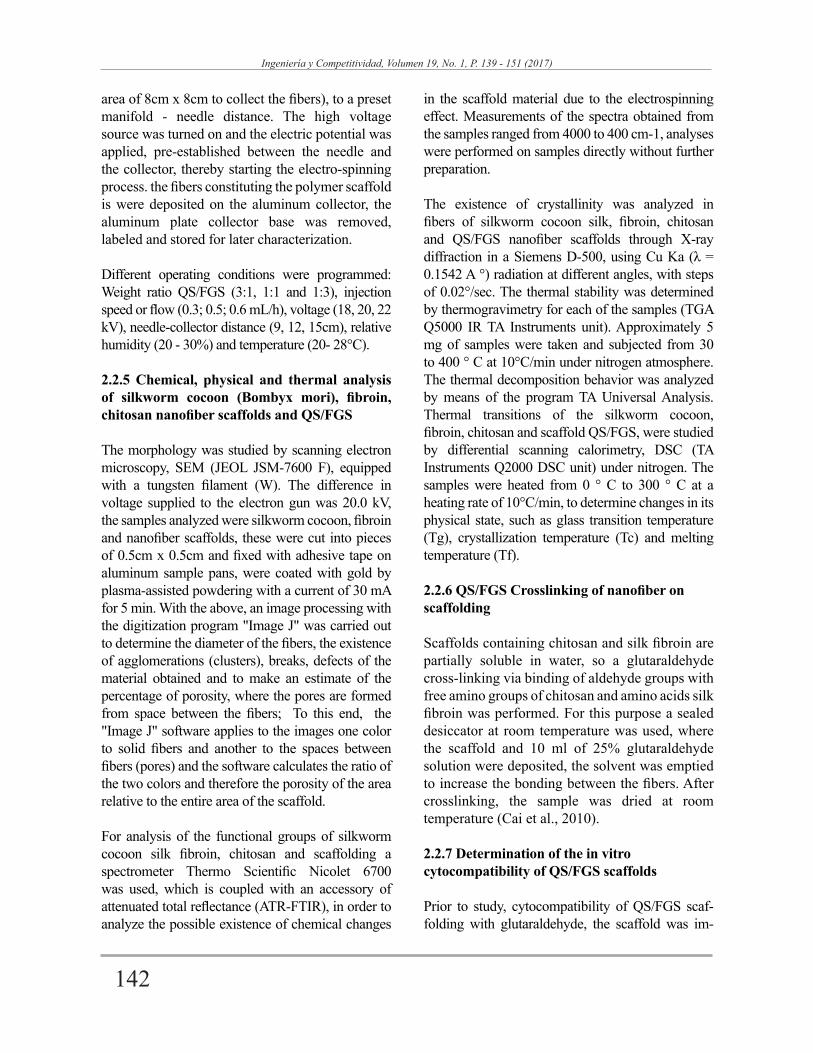
In Figure 2, the micrographs of the fibers are shown: (a) the silkworm cocoon (Bombyx mori) and (b) fibroin. Cocoon fibers are randomly oriented, are flat and rough, it appears that each fiber is coated with sericin. The average diameter of these cocoon fibers is 36 +/- 2 µm, which coincides with the results reported by Jiang et al. (2006). Figure 2 (b), cylindrical fibers are observed with diameters of 13 +/- 7μm, randomly oriented composite fibroin, because the thermal process degummed at 60°C removes the sericin coating; morphology and reduction in the value of the average fiber diameter are evidence of correct degumming.
Figure 2. SEM micrographs of silkworm cocoon and fibroin (a) 1000X and fibroin (b) 500X, taken at the IIM-UNAM.
It can be seen that the silkworm cocoon (Bombyx mori) has an average diameter of 36 +/- 22 (µm) and average porosity of 1 (%), while fibroin has an average diameter of 13 +/- 7 (µm) and an average porosity of 7.6 (%).
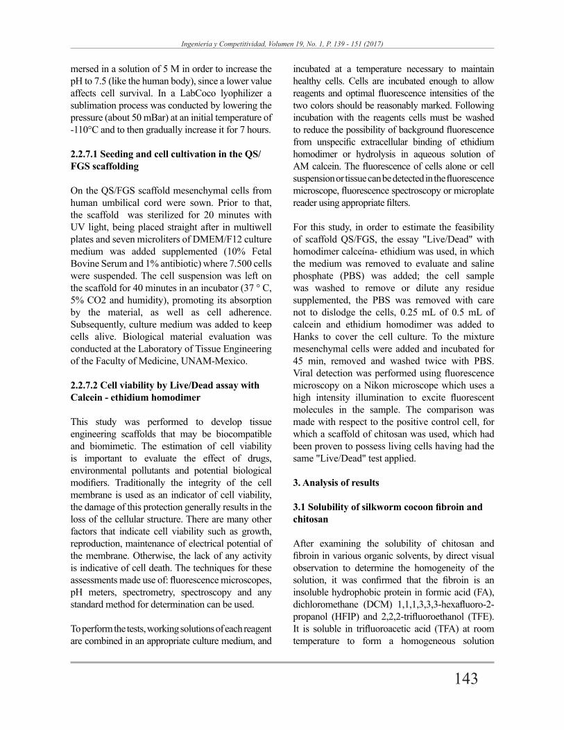
3.2.2 Effect of the QS/FGS weight ratios
After preparing scaffolds with different com-binations of electro-spinning parameters men-tioned in item 2.2.4, it was determined that the combination of factors which enable to obtain solid nanofibers were: injection flow 0.5 mL/h, voltage 20 kV, capillary distance to the collector of 12 cm, temperature of 25°C and relative humidity of 28%. Also, it was determined that the variable "weight
ratio of QS/FGS" has an important influence on the reduction of defects in the nanofibers.
In Figure 3, SEM micrographs of QS/FGS scaffolds, with an increase of 50000 X, under the electrospinning parameters shown above. Figure 3 (a) corresponds to the QS/FGS scaffold with a (3:1) ratio; continuous fibers are randomly oriented, with evidence of bulbs and crushed nanofibers. In Figure 3 (b) scaffold fibers (1:1) ratio are of both a flattened and a cylindrical shape, are randomly oriented and have breaks. In Figure 3 (c) scaffolding (1:3) ratio is shown, being observed that the fibers are randomly oriented, are homogeneous, continuous and have no bulbs or ruptures.
145
Ingeniería y Competitividad, Volumen 19, No. 1, P. 139 - 151 (2017)
140
Ingeniería Y Competitividad, Volumen 19, No. 1, P. 134 - 146 (2017)
7
Ingeniería Y Competitividad, Volumen 18, No. 2, P. 1 - 10 (2016)
Figure 3. SEM micrographs of QS:FGS scaffolds to 50000x different weight ratios (a) QS/FGS (3: 1), (b) QS:FGS (1: 1) and (c) QS:FGS (1:3). taken at IIM-UNAM.
The QS/FGS fiber diameters and estimated porosity of scaffolds are described as follows:
QS/FGS weight ratio (3:1) presents an average diameter of 104 +/- 14 (nm) and average porosity of 8 (%); while the QS/FGS weight ratio (1:1) presents an average diameter of 177 +/- 18 (nm) and average porosity 25 (%) and finally the weight ratio of QS/FGS (1: 3 ratio) has an average diameter of 155 +/- 8 (nm) and average porosity of 14 (%).
It shows that there was a decrease in pore size with increasing chitosan content in the mixtures. Mixed scaffolds having high chitosan show smaller pore size as compared to scaffolds silk fibroin. This may be due to hidrofibrosity of chitosan.
The QS/FGS scaffold (1: 3 ratio) has a diameter and intermediate porosity so it was chosen for
further characterization to determine whether their characteristics are suitable for biomedical applications.
3.3 Functional groups by Infrared spectroscopy with Fourier Transformation (FTIR)
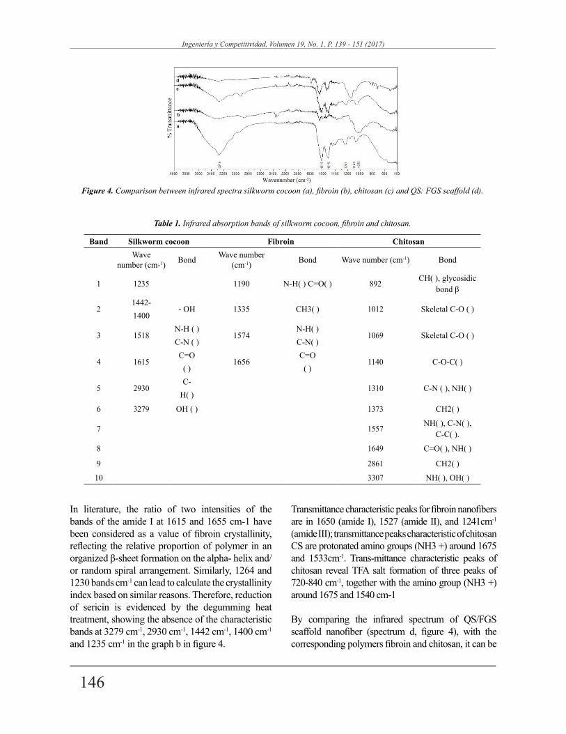
Figure 4 shows the infrared spectra of the cocoon, fibroin, chitosan and QS/FGS scaffold; Table 1 shows the allocation of the main absorption bands for silkworm cocoon (Mandal et al presented., 2011), fibroin (Koperska et al., 2014; Chen et al., 2012) and chitosan (Staroszczyk et al., 2014; Van de Velde & P. Kiekens, 2004; Winie & Arof, 2006). The results agree with those reported in references cited in the sericin silkworm cocoon, configured in a random coil, while fibroin, has a highly crystalline structure in β-sheet formation.
146
Ingeniería y Competitividad, Volumen 19, No. 1, P. 139 - 151 (2017)
141
Ingeniería Y Competitividad, Volumen 19, No. 1, P. 134 - 146 (2017)
8
Ingeniería Y Competitividad, Volumen 18, No. 2, P. 1 - 10 (2016)
Band Silkworm cocoon Fibroin ChitosanWave
number (cm-1) Bond Wave number (cm-1) Bond Wave number (cm-1) Bond
1 1235 1190 N-H( ) C=O( ) 892CH( ), glycosidic
bond β
21442-
- OH 1335 CH3( ) 1012 Skeletal C-O ( )1400
3 1518N-H ( )
1574N-H( )
1069 Skeletal C-O ( )C-N ( ) C-N( )
4 1615C=O
1656C=O
1140 C-O-C( )( ) ( )
5 2930C-
1310 C-N ( ), NH( )H( )
6 3279 OH ( ) 1373 CH2( )
7 1557NH( ), C-N( ),
C-C( ).
8 1649 C=O( ), NH( )
9 2861 CH2( )
10 3307 NH( ), OH( )
Figure 4. Comparison between infrared spectra silkworm cocoon (a), fibroin (b), chitosan (c) and QS: FGS scaffold (d).
Table 1. Infrared absorption bands of silkworm cocoon, fibroin and chitosan.
In literature, the ratio of two intensities of the bands of the amide I at 1615 and 1655 cm-1 have been considered as a value of fibroin crystallinity, reflecting the relative proportion of polymer in an organized β-sheet formation on the alpha- helix and/or random spiral arrangement. Similarly, 1264 and 1230 bands cm-1 can lead to calculate the crystallinity index based on similar reasons. Therefore, reduction of sericin is evidenced by the degumming heat treatment, showing the absence of the characteristic bands at 3279 cm-1, 2930 cm-1, 1442 cm-1, 1400 cm-1 and 1235 cm-1 in the graph b in figure 4.
Transmittance characteristic peaks for fibroin nanofibers are in 1650 (amide I), 1527 (amide II), and 1241cm-1 (amide III); transmittance peaks characteristic of chitosan CS are protonated amino groups (NH3 +) around 1675 and 1533cm-1. Trans-mittance characteristic peaks of chitosan reveal TFA salt formation of three peaks of 720-840 cm-1, together with the amino group (NH3 +) around 1675 and 1540 cm-1
By comparing the infrared spectrum of QS/FGS scaffold nanofiber (spectrum d, figure 4), with the corresponding polymers fibroin and chitosan, it can be
147
Ingeniería y Competitividad, Volumen 19, No. 1, P. 139 - 151 (2017)
142
Ingeniería Y Competitividad, Volumen 19, No. 1, P. 134 - 146 (2017)
9
Ingeniería Y Competitividad, Volumen 18, No. 2, P. 1 - 10 (2016)
seen that most of the signals of the most important functional groups coincide, as are amide I amide II, amide III and C = O polymergroups, showing no evidence of new functional groups, therefore, the process parameters (flow rate, applied voltage and distance from the needle-collector) not only are appropriate to electro-spin and evaporate the trifluoroacetic acid, but also do not affect the chemical structure of the materials. However, the band observed at 3270cm-1 has a lower intensity, which may be due to changes in the morphology of the nanofibers.
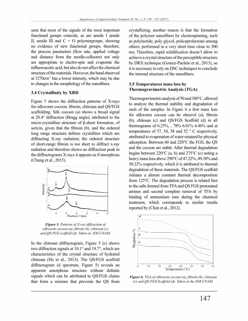
3.4 Crystallinity by XRD
Figure 5 shows the diffraction patterns of X-rays for silkworm cocoon, fibroin, chitosan and QS/FGS scaffolding. Silk cocoon (a) shows a broad signal at 20.4° diffraction (Bragg angle), attributed to the micro-crystalline structure of β-sheet formation, of sericin, given that the fibroin (b), and the ordered long range structure defines crystallites which are diffracting X-ray radiation; the ordered structure of short-range fibroin is too short to diffract x-ray radiation and therefore shows no diffraction peak in the diffractograms X-rays it appears as if amorphous (Chung et al., 2015).
In the chitosan diffractogram, Figure 5 (c) shows two diffraction signals at 10.1° and 19.7°, which are characteristics of the crystal structure of hydrated chitosan (He et al., 2013). The QS/FGS scaffold diffractogram (d spectrum, Figure 5) reveals an apparent amorphous structure without definite signals which can be attributed to QS/FGS chains that form a mixture that prevents the QS from
Figure 5. Patterns of X-ray diffraction of silkworm cocoon (a), fibroin (b), chitosan (c)
and QS:FGS scaffold (d). Taken at IIM-UNAM.
crystallizing; another reason is that the formation of the polymer nanofibers by electrospinning, such as polylactide, poly glycol, policaprolactone among others, performed in a very short time close to 500 ms; Therefore, rapid solidification doesn’t allow to achieve a crystal structure of the perceptible structure by DRX technique (Gomez-Pachón et al., 2013), so it is necessary to rely on DSC techniques to conclude the internal structure of the nanofibers.
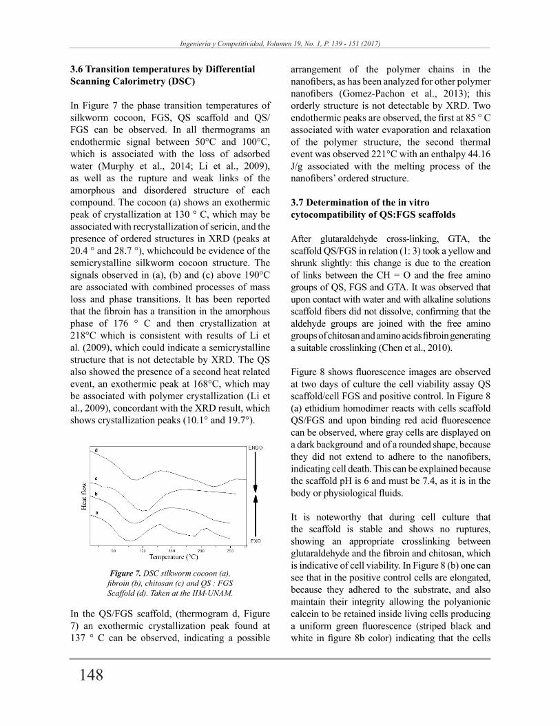
3.5 Temperatures mass loss by Thermogravímetric Analysis (TGA)
Thermogravimetric analysis of 50 and 380°C, allowed to analyze the thermal stability and degradation of each of the samples. In Figure 6 a first mass loss for silkworm cocoon can be observd (a), fibroin (b), chitosan (c) and QS/FGS Scaffold (d) in all thermograms of 6.25%, , 78% 6.01% 6.46% and at temperatures of 57, 54, 58 and 52 ° C respectively, attributed to evaporation of water retained by physical adsorption. Between 60 and 220°C the FGS, the QS and the cocoon are stable. After thermal degradation begins between 220°C (a, b) and 275°C (c) noting a heavy mass loss above 290°C of 47.22%, 49.38% and 50.22% respectively, which it is attributed to thermal degradation of these materials. The QS/FGS scaffold initiates a almost constant thermal decomposition from 125°C. The degradation process is related first to the salts formed from TFA and QS FGS protonated amines and second complete removal of TFA by binding of ammonium ions during the chemical treatment, which corresponds to similar results reported by (Chen et al., 2012).
Figure 6. TGA of silkworm cocoon (a), fibroin (b), chitosan (c) and QS:FGS Scaffold (d). Taken at the IIM-UNAM.
148
Ingeniería y Competitividad, Volumen 19, No. 1, P. 139 - 151 (2017)
143
Ingeniería Y Competitividad, Volumen 19, No. 1, P. 134 - 146 (2017)
10
Ingeniería Y Competitividad, Volumen 18, No. 2, P. 1 - 10 (2016)
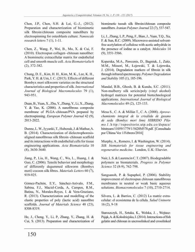
3.6 Transition temperatures by Differential Scanning Calorimetry (DSC)
In Figure 7 the phase transition temperatures of silkworm cocoon, FGS, QS scaffold and QS/FGS can be observed. In all thermograms an endothermic signal between 50°C and 100°C, which is associated with the loss of adsorbed water (Murphy et al., 2014; Li et al., 2009), as well as the rupture and weak links of the amorphous and disordered structure of each compound. The cocoon (a) shows an exothermic peak of crystallization at 130 ° C, which may be associated with recrystallization of sericin, and the presence of ordered structures in XRD (peaks at 20.4 ° and 28.7 °), whichcould be evidence of the semicrystalline silkworm cocoon structure. The signals observed in (a), (b) and (c) above 190°C are associated with combined processes of mass loss and phase transitions. It has been reported that the fibroin has a transition in the amorphous phase of 176 ° C and then crystallization at 218°C which is consistent with results of Li et al. (2009), which could indicate a semicrystalline structure that is not detectable by XRD. The QS also showed the presence of a second heat related event, an exothermic peak at 168°C, which may be associated with polymer crystallization (Li et al., 2009), concordant with the XRD result, which shows crystallization peaks (10.1° and 19.7°).
In the QS/FGS scaffold, (thermogram d, Figure 7) an exothermic crystallization peak found at 137 ° C can be observed, indicating a possible
arrangement of the polymer chains in the nanofibers, as has been analyzed for other polymer nanofibers (Gomez-Pachon et al., 2013); this orderly structure is not detectable by XRD. Two endothermic peaks are observed, the first at 85 ° C associated with water evaporation and relaxation of the polymer structure, the second thermal event was observed 221°C with an enthalpy 44.16 J/g associated with the melting process of the nanofibers’ ordered structure.
3.7 Determination of the in vitro cytocompatibility of QS:FGS scaffolds
After glutaraldehyde cross-linking, GTA, the scaffold QS/FGS in relation (1: 3) took a yellow and shrunk slightly: this change is due to the creation of links between the CH = O and the free amino groups of QS, FGS and GTA. It was observed that upon contact with water and with alkaline solutions scaffold fibers did not dissolve, confirming that the aldehyde groups are joined with the free amino groups of chitosan and amino acids fibroin generating a suitable crosslinking (Chen et al., 2010).
Figure 8 shows fluorescence images are observed at two days of culture the cell viability assay QS scaffold/cell FGS and positive control. In Figure 8 (a) ethidium homodimer reacts with cells scaffold QS/FGS and upon binding red acid fluorescence can be observed, where gray cells are displayed on a dark background and of a rounded shape, because they did not extend to adhere to the nanofibers, indicating cell death. This can be explained because the scaffold pH is 6 and must be 7.4, as it is in the body or physiological fluids.
It is noteworthy that during cell culture that the scaffold is stable and shows no ruptures, showing an appropriate crosslinking between glutaraldehyde and the fibroin and chitosan, which is indicative of cell viability. In Figure 8 (b) one can see that in the positive control cells are elongated, because they adhered to the substrate, and also maintain their integrity allowing the polyanionic calcein to be retained inside living cells producing a uniform green fluorescence (striped black and white in figure 8b color) indicating that the cells
Figure 7. DSC silkworm cocoon (a), fibroin (b), chitosan (c) and QS : FGS Scaffold (d). Taken at the IIM-UNAM.

149
Ingeniería y Competitividad, Volumen 19, No. 1, P. 139 - 151 (2017)
144
Ingeniería Y Competitividad, Volumen 19, No. 1, P. 134 - 146 (2017)
11
Ingeniería Y Competitividad, Volumen 18, No. 2, P. 1 - 10 (2016)
are alive. The results indicate that the proposed scaffold sample shows integrity and the study of
cell viability should continue by adjusting the pH of the scaffolding to 7.4.
Figure 8. Two color fluorescence cell viability assay taken two days of cell culture, QS / FGS scaffold (a) and positive control of cell formation (b).
4. Conclusions
The thermochemical treatment performed allowed the complete removal of the fibroin from the cocoon of the silkworm (Bombyx mori), as evidenced in the spectroscopic and morphological analysis. Homogeneous solutions of chitosan and fibroin in trifluoroacetic acid, suitable for processing by electrospinning were obtained. Electrospun QS/FGS scaffolding in proportion (1: 3) was obtained with physicochemical properties useful in tissue engineering. The optimal process parameters were: flow injection of 0.5 mL/h, applied voltage of 20 kV, capillary distance to the collector of 12 cm and weight ratio (1: 3) QS/FGS dissolved in TFA. The QS/FGS scaffold presents homogeneous fibers, randomly distributed without defects, with an average diameter of 155 nm, which represents a significant contribution of this investigation because that cell adhesion is given in below 200 nm diameter, as well as improved mechanical properties.
The dissolution of the polymers and the elec-trospinning process did not alter the chemical structure of chitosan or fibroin. Spectroscopic analysis indicated that primary, secondary and tertiary, as well as the C=O groups of the polymers were not altered. According to the studies of differential scanning calorimetry and thermogravimetry, scaffolds are stable at body temperature (36-38 ° C). They lose moisture above 50°C and do not degrade below
125°C. The in vitro cytocompatibility tests by cell viability assay suggest that QS/FGS scaffolding ratio (1: 3) are stable, have no breaks and although not yet any definitive cell growth tests due to pH crosslinked scaffold, these results show potential for application as cell tissue engineering scaffolds.
5. Acknowledgements
At the Pedagogical and Technological University of Colombia (UPTC) and the National Autonomous University of Mexico in which this study was carried out under the cooperation agreement in force UPTC - IIM-UNAM, which received support from projects UPTC-DIN under the SGI 1781 code , PAPIIT-UNAM/IN108930 and CONACYT/CNPq174247. We are also grateful to Dr. Andres Castell for the cytocompatibility tests performed.
6. References
Cai, Z.X., Mo, X.M., Zhang, K.H., Fan, L.P., Yin, A.L., He C.L. & Wang, H.S. (2010). Fabrication of chitosan/silk fibroin composite nanofibers for wound-dressing applications. International journal of molecular sciences 11 (9), 3529-3539.
Chen, F., Porter, D. & Vollrath, F. (2012). Silk cocoon (Bombyx mori): Multi-layer structure and mechanical properties. Acta Biomaterialia 8 (7), 2620-2627.
150
Ingeniería y Competitividad, Volumen 19, No. 1, P. 139 - 151 (2017)
145
Ingeniería Y Competitividad, Volumen 19, No. 1, P. 134 - 146 (2017)
12
Ingeniería Y Competitividad, Volumen 18, No. 2, P. 1 - 10 (2016)
Chen, J.P., Chen, S.H. & Lai, G.-J., (2012). Preparation and characterization of biomimetic silk fibroin/chitosan composite nanofibers by electrospinning for osteoblasts culture. Nanoscale research letters 7 (1), 1-11.
Chen, Z., Wang, P., Wei, B., Mo, X. & Cui, F. (2010). Electrospun collagen–chitosan nanofiber: A biomimetic extracellular matrix for endothelial cell and smooth muscle cell. Acta Biomaterialia 6 (2), 372-382.
Chung, D. E., Kim, H. H., Kim, M. K., Lee, K. H., Park, Y. H. & Um, I. C. (2015). Effects of different Bombyx mori silkworm varieties on the structural characteristics and properties of silk. International Journal of Biological Macromolecules 79 (1), 943-951.
Duan, B., Yuan, X., Zhu, Y., Zhang, Y., Li, X., Zhang, Y. & Yao, K. (2006). A nanofibrous composite membrane of PLGA–chitosan/PVA prepared by electrospinning. European Polymer Journal 42 (9), 2013-2022.
Dunne, L. W., Iyyanki, T., Hubenak, J. & Mathur, A. B. (2014). Characterization of dielectrophoresis-aligned nanofibrous silk fibroin–chitosan scaffold and its interactions with endothelial cells for tissue engineering applications. Acta Biomaterialia 10 (8), 3630-3640.
Jiang, P., Liu, H., Wang, C., Wu, L., Huang, J. & Guo, C. (2006). Tensile behavior and morphology of differently degummed silkworm (Bombyx mori) cocoon silk fibres. Materials Letters 60 (7), 919-925.
Gómez-Pachón, E.Y., Sánchez-Arévalo, F.M., Sabina, F.J., Maciel-Cerda, A, Campos, R.M., Batina, N., Morales-Reyes, I. & Vera-Graziano, R. (2013). Characterization and modelling of the elastic properties of poly (lactic acid) nanofibre scaffolds. Journal of Materials Science 48 (23), 8308-8319.
He, J., Cheng, Y., Li, P., Zhang, Y., Zhang, H. & Cui, S. (2013). Preparation and characterization of
biomimetic tussah silk fibroin/chitosan composite nanofibers. Iranian Polymer Journal 22 (7), 537-547.
Li, J., Zhang, L.P., Peng, F., Bian, J., Yuan, T.Q., Xu, F. & Sun, R.C. (2009). Microwave-assisted solvent-free acetylation of cellulose with acetic anhydride in the presence of iodine as a catalyst. Molecules 14 (9), 3551-3566.
Koperska, M.A., Pawcenis, D., Bagniuk, J., Zaitz, M.M., Missori, M., Łojewski, T. & Lojewska, J. (2014). Degradation markers of fibroin in silk through infrared spectroscopy. Polymer Degradation and Stability 105 (1), 185-196.
Mandal, B.B., Ghosh, B. & Kundu, S.C. (2011). Non-mulberry silk sericin/poly (vinyl alcohol) hydrogel matrices for potential biotechnological applications. International Journal of Biological Macromolecules 49 (2), 125-133.
Mesa S., C. A. & Millán T., C. A. (2008). Aprove-chamiento integral de la crisálida de gusano de seda (Bombyx mori linn) HIBRIDO Pila-mo 1 . h ttp://repositorio.utp.edu.co/dspace/bitstream/11059/1779/1/5428M578.pdf. [Consultado por Última Vez 1/Febrero/2016]
Murphy, A., Romero, I. & Washington, W. (2014). Silk biomaterials for tissue engineering and regenerative medicine. London, U.K: Elsevier.
Nair, L.S. & Laurencin C.T. (2007). Biodegradable polymers as biomaterials. Progress in Polymer Science 32 (8-9), 762-798.
Sangsanoh, P. & Supaphol, P. (2006). Stability improvement of electrospun chitosan nanofibrous membranes in neutral or weak basic aqueous solutions. Biomacromolecules 7 (10), 2710-2714.
Silvera, L. & Barrios, C. (2012) La matriz extra-celular: el ecosistema de la célula. Salud Uninorte 16 (2), 9-18
Staroszczyk, H., Sztuka, K., Wolska, J. , Wojtasz-Pająk, A. & Kołodziejska, I. (2014). Interactions of fish gelatin and chitosan in uncrosslinked and crosslinked
151
Ingeniería y Competitividad, Volumen 19, No. 1, P. 139 - 151 (2017)
146
Ingeniería Y Competitividad, Volumen 19, No. 1, P. 134 - 146 (2017)
13
Ingeniería Y Competitividad, Volumen 18, No. 2, P. 1 - 10 (2016)
with EDC films: FT-IR study. Spectrochimica. Acta Part A: Molecular and Biomolecular Spectroscopy 117 (1), 707- 712.
Van de Velde, K. & Kiekens, P. (2004). Structure analysis and degree of substitution of chitin, chitosan and dibutyrylchitin by FT-IR spectroscopy and solid state 13C NMR. Carbohydrate Polymers 58 (4), 409-416.
Winie T. & Arof, A. K. (2006). FT-IR studies on interactions among components in hexanoyl chitosan-
based polymer electrolytes. Spectrochimica Acta Part A: Molecular and Biomolecular Spectroscopy 63 (3), 677-684.
Zhang, Q., Li, M., Xu, W., Li, J. & Yan, S. (2013). A novel silk fibroin scaffolds with oriented mul-tichannels, Materials Letters 105, (1) 8-11.
Zhang, X., Reagan, M. R. & Kaplan, D. L. (2009). Electrospun silk biomaterial scaffolds for regenerative medicine. Advanced Drug Delivery Reviews 61 (12), 988-1006.
Revista Ingeniería y Competitividad por Universidad del Valle se encuentra bajo una licencia Creative Commons Reconocimiento - Debe reconocer adecuadamente la autoría, proporcionar un enlace a la licencia e indicar si se han realizado cambios. Puede hacerlo de cualquier manera razonable, pero no de una manera que sugiera que tiene el apoyo del licenciador o lo recibe por el uso que hace.