18F Labeling of a Near-Infrared Fluorophore Enables ...tsienlab.ucsd.edu/Publications/Ting 2010...
9
Fast 18 F Labeling of a Near-Infrared Fluorophore Enables Positron Emission Tomography and Optical Imaging of Sentinel Lymph Nodes Richard Ting, † Todd A. Aguilera, † Jessica L. Crisp, † David J. Hall, ‡ William C. Eckelman, ‡ David R. Vera, ‡ and Roger Y. Tsien* ,†,§ Department of Pharmacology 0647, Department of Radiology, and Howard Hughes Medical Institute, University of California, San Diego, La Jolla, California 92093. Received March 11, 2010; Revised Manuscript Received June 24, 2010 We combine a novel boronate trap for F - with a near-infrared fluorophore into a single molecule. Attachment to targeting ligands enables localization by positron emission tomography (PET) and near-infrared fluorescence (NIRF). Our first application of this generic tag is to label Lymphoseek (tilmanocept), an agent designed for receptor- specific sentinel lymph node (SLN) mapping. The new conjugate incorporates 18 F - in a single, aqueous step, targets mouse SLN rapidly (1 h) with reduced distal lymph node accumulation, permits PET or scintigraphic imaging of SLN, and enables NIRF-guided excision and histological verification even after 18 F decay. This embodiment is superior to current SLN mapping agents such as nontargeted [ 99m Tc]sulfur colloids and Isosulfan Blue, as well as the phase III targeted ligand [ 99m Tc]SPECT Lymphoseek counterpart, species that are visible by SPECT or visible absorbance separately. Facile incorporation of 18 F into a NIRF probe should promote many synergistic PET and NIRF combinations. INTRODUCTION Different molecular imaging techniques have complementary advantages and disadvantages in spatial and temporal resolution, depth penetration, sensitivity, and cost (1). A powerful way to combine synergistic advantages is to construct synthetic probes that can be imaged by two or more modalities (2-4). Radiotrac- ers and near-infrared fluorescence (NIRF) are particularly suitable combinations because both are sufficiently sensitive to enable direct visualization at receptor binding concentrations (nanomolar to picomolar). In contrast, magnetic resonance imaging (MRI) and X-ray contrast tomography require much higher elemental concentrations of the probe (1). The excellent penetration of γ-ray photons in positron emission tomography (PET) allows quantitative detection regardless of depth, making this technique ideal for whole-body scanning. NIRF offers much higher spatial and temporal resolution and cheaper instrumenta- tion, but it is largely limited to superficial targets, making it ideal for image-guided surgery and histology. Multimodality PET/optical probes under development cur- rently include 18 F/quantum dot (QD) (5), 18 F/nanoparticle (6), bis(thiosemicarbazonato) 64 Cu chelates (3, 7), and 64 Cu/cypate (4) conjugates. PET bioconjugate radiochemistry is often limited to the physically more common radionuclides 64 Cu (t 1/2 ) 762 min), 68 Ga (t 1/2 ) 68 min), and 18 F(t 1/2 ) 110 min), because they are easily manipulated and possess nuclear half-lives sufficiently long enough for chemical isotope manipulation and in vivo distribution. Unfortunately, each of these PET nuclides presents intrinsic complications. For example, 64 Cu (3, 4, 8) and 68 Ga (9, 10) chelates can suffer from impure isotope production as well as lowered specific activity because of impure isotope decay (11) and large chelation moieties that may alter ligand biodistribution and may also suffer from in vivo metal ion transchelation (12). Alternatively, traditional 18 F labeling methods (5, 13-17) relying on C-F bond formation are water- sensitive, multistep processes that often require harsh reaction conditions and long processing times that are poorly suited to the short half-life of 18 F. To simplify PET chemistry, there has been recent interest in developing rapid, one-step labeling procedures from shelf-stable final target precursors with aqueous 18 F - , while reducing the side products and chromatographic purifications associated with traditional C- 18 F labelings. Recent silica-based (18-21) and boron-based (22-24) aqueous 18 F capture technology allows direct preparation of isotopically pure PET compounds that are cleanly labeled, are easily purified, and have been shown to be stable in vivo (19, 25). We chose to generate [ 18 F]fluoroborates because they require less bulky hydrophobic substituents and potentially triple the F - incorporation stoichiometry and attain- able specific activity (22-24). A typical preparation of 18 F fluoride has a specific activity of ∼10 Ci/µmol even if no carrier was deliberately added and therefore contains 170 atoms of 19 F for each atom of 18 F(26). Incorporation of three fluorine atoms triples the probability that at least one will be radioactive. We combine this boron-based 18 F capture technology [t 1/2 of B-F solvolysis ) 5550 ( 1740 min (23)] with a NIR fluorophore to give a generally conjugatable PET/NIRF multimodality probe 1 (Figure 1A). Our first test of this probe is to help find and excise sentinel lymph nodes. The current method for sentinel lymph node mapping in melanoma and breast cancer involves injection of a combination of Isosulfan Blue (27) and a 99m Tc-labeled colloid radiopharma- ceutical (28, 29). This pair allows for lymphoscintigraphy and absorbance-guided intraoperative lymph node excision (30). With little affinity for the sentinel lymph node, 99m Tc-labeled colloids and Isosulfan Blue will travel past the sentinel node in a lymph chain restricting the surgeon to a small time-window within which to perform the mapping procedure. 99m Tc- radiolabeled colloids are large particles that are nonspecifically trapped within lymphoid tissue. These agents are inefficient at entering lymph channels and, over time, reach distal lymph nodes. Distal colloid or dye uptake leads to the surgical removal of more lymph nodes than necessary, which increases the extent of the dissection, forces the pathologist to review a greater * To whom correspondence should be addressed. † Department of Pharmacology 0647. ‡ Department of Radiology. § Howard Hughes Medical Institute. Bioconjugate Chem. 2010, 21, 1811–1819 1811 10.1021/bc1001328 2010 American Chemical Society Published on Web 09/27/2010
Transcript of 18F Labeling of a Near-Infrared Fluorophore Enables ...tsienlab.ucsd.edu/Publications/Ting 2010...
Fast 18F Labeling of a Near-Infrared Fluorophore Enables PositronEmission Tomography and Optical Imaging of Sentinel Lymph Nodes
Richard Ting,† Todd A. Aguilera,† Jessica L. Crisp,† David J. Hall,‡ William C. Eckelman,‡ David R. Vera,‡ andRoger Y. Tsien*,†,§
Department of Pharmacology 0647, Department of Radiology, and Howard Hughes Medical Institute, University of California,San Diego, La Jolla, California 92093. Received March 11, 2010; Revised Manuscript Received June 24, 2010
We combine a novel boronate trap for F- with a near-infrared fluorophore into a single molecule. Attachment totargeting ligands enables localization by positron emission tomography (PET) and near-infrared fluorescence (NIRF).Our first application of this generic tag is to label Lymphoseek (tilmanocept), an agent designed for receptor-specific sentinel lymph node (SLN) mapping. The new conjugate incorporates 18F- in a single, aqueous step,targets mouse SLN rapidly (1 h) with reduced distal lymph node accumulation, permits PET or scintigraphicimaging of SLN, and enables NIRF-guided excision and histological verification even after 18F decay. Thisembodiment is superior to current SLN mapping agents such as nontargeted [99mTc]sulfur colloids and IsosulfanBlue, as well as the phase III targeted ligand [99mTc]SPECT Lymphoseek counterpart, species that are visible bySPECT or visible absorbance separately. Facile incorporation of 18F into a NIRF probe should promote manysynergistic PET and NIRF combinations.
INTRODUCTION
Different molecular imaging techniques have complementaryadvantages and disadvantages in spatial and temporal resolution,depth penetration, sensitivity, and cost (1). A powerful way tocombine synergistic advantages is to construct synthetic probesthat can be imaged by two or more modalities (2-4). Radiotrac-ers and near-infrared fluorescence (NIRF) are particularlysuitable combinations because both are sufficiently sensitive toenable direct visualization at receptor binding concentrations(nanomolar to picomolar). In contrast, magnetic resonanceimaging (MRI) and X-ray contrast tomography require muchhigher elemental concentrations of the probe (1). The excellentpenetration of γ-ray photons in positron emission tomography(PET) allows quantitative detection regardless of depth, makingthis technique ideal for whole-body scanning. NIRF offers muchhigher spatial and temporal resolution and cheaper instrumenta-tion, but it is largely limited to superficial targets, making itideal for image-guided surgery and histology.
Multimodality PET/optical probes under development cur-rently include 18F/quantum dot (QD) (5), 18F/nanoparticle (6),bis(thiosemicarbazonato) 64Cu chelates (3, 7), and 64Cu/cypate(4) conjugates. PET bioconjugate radiochemistry is often limitedto the physically more common radionuclides 64Cu (t1/2 ) 762min), 68Ga (t1/2 ) 68 min), and 18F (t1/2 ) 110 min), becausethey are easily manipulated and possess nuclear half-livessufficiently long enough for chemical isotope manipulation andin vivo distribution. Unfortunately, each of these PET nuclidespresents intrinsic complications. For example, 64Cu (3, 4, 8) and68Ga (9, 10) chelates can suffer from impure isotope productionas well as lowered specific activity because of impure isotopedecay (11) and large chelation moieties that may alter ligandbiodistribution and may also suffer from in vivo metal iontranschelation (12). Alternatively, traditional 18F labelingmethods (5, 13-17) relying on C-F bond formation are water-
sensitive, multistep processes that often require harsh reactionconditions and long processing times that are poorly suited tothe short half-life of 18F.
To simplify PET chemistry, there has been recent interest indeveloping rapid, one-step labeling procedures from shelf-stablefinal target precursors with aqueous 18F-, while reducing theside products and chromatographic purifications associated withtraditional C-18F labelings. Recent silica-based (18-21) andboron-based (22-24) aqueous 18F capture technology allowsdirect preparation of isotopically pure PET compounds that arecleanly labeled, are easily purified, and have been shown to bestable in vivo (19, 25). We chose to generate [18F]fluoroboratesbecause they require less bulky hydrophobic substituents andpotentially triple the F- incorporation stoichiometry and attain-able specific activity (22-24). A typical preparation of 18Ffluoride has a specific activity of ∼10 Ci/µmol even if no carrierwas deliberately added and therefore contains 170 atoms of 19Ffor each atom of 18F (26). Incorporation of three fluorine atomstriples the probability that at least one will be radioactive. Wecombine this boron-based 18F capture technology [t1/2 of B-Fsolvolysis ) 5550 ( 1740 min (23)] with a NIR fluorophore togive a generally conjugatable PET/NIRF multimodality probe1 (Figure 1A). Our first test of this probe is to help find andexcise sentinel lymph nodes.
The current method for sentinel lymph node mapping inmelanoma and breast cancer involves injection of a combinationof Isosulfan Blue (27) and a 99mTc-labeled colloid radiopharma-ceutical (28, 29). This pair allows for lymphoscintigraphy andabsorbance-guided intraoperative lymph node excision (30).With little affinity for the sentinel lymph node, 99mTc-labeledcolloids and Isosulfan Blue will travel past the sentinel node ina lymph chain restricting the surgeon to a small time-windowwithin which to perform the mapping procedure. 99mTc-radiolabeled colloids are large particles that are nonspecificallytrapped within lymphoid tissue. These agents are inefficient atentering lymph channels and, over time, reach distal lymphnodes. Distal colloid or dye uptake leads to the surgical removalof more lymph nodes than necessary, which increases the extentof the dissection, forces the pathologist to review a greater
* To whom correspondence should be addressed.† Department of Pharmacology 0647.‡ Department of Radiology.§ Howard Hughes Medical Institute.
Bioconjugate Chem. 2010, 21, 1811–1819 1811
10.1021/bc1001328 2010 American Chemical SocietyPublished on Web 09/27/2010
number of lymph nodes, and increases the probability of locallymphedema, a complication of lymph node resection. Analternative probe is Lymphoseek (tilmanocept), a mannosylated16 kDa dextran conjugate, that can be labeled with 99mTc forSPECT-based imaging (31, 32) and is currently in phase IIIclinical trials. It binds with a high affinity to cell-surface lectins[KD ) 0.12 nM (33)] and was developed specifically for sentinellymph node mapping. 99mTc-labeled Lymphoseek has demon-strated rapid injection site clearance, a low level of distal nodeaccumulation, a lack of local systemic toxicity, and greaterspecific sentinel lymph node uptake than filtered [99mTc]sulfurcolloid (32, 34, 35). A version of Lymphoseek that can beviewed by both PET and NIRF would combine moleculartargeting, the higher spatial resolution of PET compared to 99mTcSPECT, and the greater sensitivity and depth penetration ofNIRF compared to the red absorbance of Isosulfan Blue.
Here we apply our generally conjugatable PET/NIRF probe1 (Figure 1A) to Lymphoseek to enable multimodality-guidedsentinel node visualization and excision. This [18F]boron/opticalmultimodality beacon ([18F]BOMB) is differentiated fromtraditional C-18F PET labeling strategies (5, 13-17) by its one-step [18F]fluoride wash-in labeling. [18F]BOMB Lymphoseek 3(Figure 1B) allows rapid, receptor-specific, positive identificationof the sentinel lymph node in both the NIRF and PET imagingmodes while maintaining little breakthrough to distal lymphnodes. This success predicts that [18F]BOMB should be gen-eralizable to other medically relevant targets.
EXPERIMENTAL PROCEDURES
Chemical Synthesis. PET/NIR Dual-Probe Reaction withLymphoseek: Synthesis of PET/NIR Lymphoseek Probe 2. Thegenerally conjugatable NHS ester of PET/NIR probe 1 (4 µmol)(see the Supporting Information for detailed synthesis) wasadded as a CH2Cl2 solution directly into a 1.5 mL glass vialcontaining 5.5 mg (0.34 µmol) of Lymphoseek bearing 5.8amine groups per dextran (Reliable Biopharmaceuticals). This
solution was concentrated to dryness and then resuspended in100 µL of DMF. This solution was sonicated for 1 h and left toreact for 18 h. The next day, 10 µL of diisopropylethylaminewas added and the reaction mixture was left for an additional2 h. The reaction mixture was then transferred to a polypropy-lene centrifuge tube containing 25 mL of CH2Cl2. A pellet of 2was isolated following centrifugation for 20 min at 3000 rcf,washed with two more portions of CH2Cl2, dried under highvacuum, and resuspended in 1 mL of water. This solution wascentrifuged for 2 min at 18000 rcf. Water-soluble 2 was decantedfrom the pellet and divided into four aliquots, which werelyophilized and stored at -78 °C. On the basis of fluorescencereadings, 115.9 nmol of fluorophore-conjugated Lymphoseek2 was isolated. On the basis of a hexose assay (36), 34-49nmol of Lymphoseek was isolated as 2. This corresponds to a10-14% yield of 2 from unconjugated Lymphoseek. Theincorporation ratio of fluorophore to Lymphoseek was 2.36 to3.41 fluorophores per molecule of Lymphoseek. Spectropho-tometric constants of a 1.5 µM solution in water were as follows:absorbance maximum ) 758 nm, ε758 ) 110000 cm-1 M-1 perfluorophore, excitation maximum ) 757 nm, emission maximum) 777 nm, and quantum yield ) 0.026.
Radiochemistry. 18F-Labeled Lymphoseek 3. A 300-500 µLaqueous solution containing 100 mCi of 18F- was transferredvia syringe to a 10 mL glass vial containing 0.4 µL of 0.05 MKHF2 (40 nmol) fluoride carrier. Note that acidic solutions ofH18F are volatile and radioactive, and therefore, the followingprocedures should be performed in a vented hot cell or a fumehood with radiation shielding. This sample was heated to drynessat 140 °C under a nitrogen flow. The clear dry solid wasresuspended in 5 µL of a 0.25 M HCl/25% MeOH/75% H2Osolution. This solution, containing 30 mCi, was transferred toa 600 µL microcentrifuge tube containing 10.6 nmol oflyophilized 2. This volume was sufficient to completely solu-bilize 3 and was reacted at 40 °C for 1 h. Following reaction,
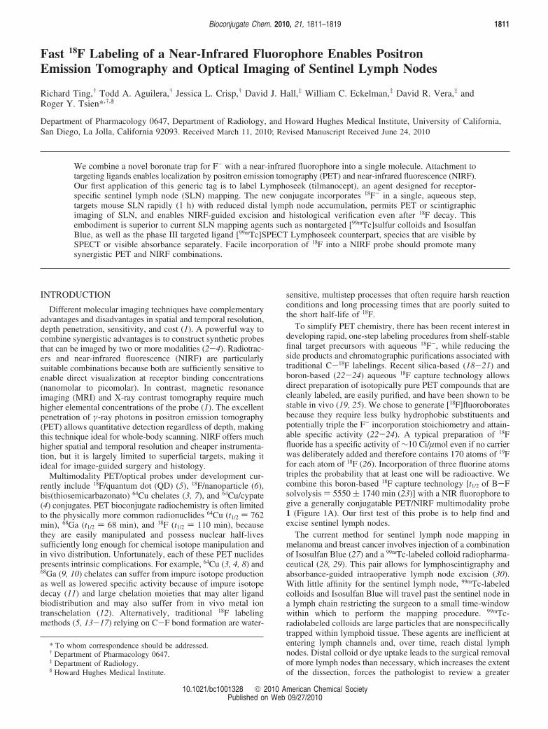
Figure 1. (A) Structure of the 18F boron trap/heptamethine cyanine (C7-Cy) PET/NIR probe, [18F]BOMB 1, activated as an NHS ester for generalbioconjugation. The boron-based 18F trap is colored red, while the NIR fluorophore is colored green. (B) Structure of PET/NIR dual probe Lymphoseek(DTPA-mannosyl-dextran) 2 and the aqueous 18F labeling scheme used to generate 18F-labeled [18F]BOMB Lymphoseek 3. (C) Excitation (blue)and emission (red) spectra of an aqueous solution of [18F]PET/NIR-Lymphoseek 3 (�f ) 0.027, excitation maximum ) 755 nm, emission maximum) 772 nm, 1.5 µM Lymphoseek solution in water). The amine (NN), mannose (NM), and DTPA (ND) densities on Lymphoseek are 5.8, 16.5, and4.2 mol/dextran, respectively. The average preconjugation molecular weight of Lymphoseek is 16122.
1812 Bioconjugate Chem., Vol. 21, No. 10, 2010 Ting et al.
the reaction was quenched with 40 µL of a 1 M MOPS bufferedsolution (pH 7.3).
To remove unreacted [18F]fluoride, 5 µL of 3.0 M KHF2 wasadded, and then the entire 45 µL was transferred to a MicroBio Spin 6 column [molecular weight cutoff of 6000 (Bio-Rad)]buffered with 10 mM Tris (pH 7.4) and centrifuged withoutadded eluant for 4 min at 1000 rcf. To the 40 µL of eluant wasadded an additional 5 µL of 3.0 M KHF2 (30 µmol) to preventthe nonspecific elution of [18F]fluoride, and the solution wasimmediately centrifuged. This described spin column purificationwas repeated three more times with a fresh spin column eachtime. Bio-Rad P-6 columns quantitatively remove small MWorganic molecule impurities (see the Supporting Information)and 93% of [18F]fluoride per column (99.998% of all [18F]fluo-ride over four columns). Chromatography took 30 min total,and the final pH of the 60 µL of Tris-buffered, eluted solutionwas 7-8. Fluorescence showed that 6.0 nmol of 18F-labeledPET/NIRF Lymphoseek 3 was eluted from the spin columns90 min following 18F- concentration. The volume was 60 µL,and the activity was 300 µCi [total preparation time (reactionand purification) of 90 min]. See the Supporting Informationfor final analyses.
This purified solution was divided into two 30 µL portions,each sufficient for 3 × 10 µL, 1.0 nmol injections of Lympho-seek. To one portion was added 1.0 µL of a 31 mM solution ofunconjugated Lymphoseek to give enough injectate for 3 × 10µL, 11.0 nmol injections. Injections with specific activities of0.05 Ci/µmol for 1 nmol Lymphoseek injections and 0.005 Ci/µmol for 11 nmol Lymphoseek injections were conducted for15-75 min following synthesis and purification (see theSupporting Information for specifics).
19F (nonradioactiVe) Labeling of Lymphoseek 3. A 10.6 nmolquantity of lyophilized 2 was dissolved in a 0.25 M HCl/25%MeOH/75% H2O solution in a 600 µL Eppendorf tube and leftto react at 40 °C for 1 h. This reaction was quenched and thesolution chromatographed under conditions similar to thosedescribed above for 18F labeling. Spectrophotometric constantsof a 1.5 µM solution of [19F]3 in water were as follows:absorbance maximum ) 756 nm, ε756 ) 110000 cm-1 M-1 perfluorophore, excitation maximum ) 755 nm, emission maximum) 772 nm, and quantum yield ) 0.027 (see the SupportingInformation for HPLC characterization).
In Vivo Experiments. The in vivo procedures in this studyhave all been approved by the University of California, SanDiego, Institutional Animal Care and Use Committee. A singlesubcutaneous injection of 3 was made into the right rear footpadof 20-25 g, 6-8-week-old athymic nude mice (Charles RiverLaboratories). These mice were anesthetized 20 min prior toinjection with isoflurane, taped to black cardboard, and placedin pairs on an imaging stage. Six pairs of injections were made.The injected mice received either a 1 nmol dose or a 11 nmoldose (10 µL each) of 10 mM Tris-buffered [18F]Lymphoseek 3(pH 7.3). The distribution of [19F]Lymphoseek 3 proceeded for1 h and 20 min before the mice were euthanized by isofluraneoverdose followed by cervical dislocation.
Imaging. PET and CT scanning were conducted on a eXploreVista PET scanner or eXplore Locus CT scanner from GEHealthcare. PET acquisition was conducted in a single blockin static emission mode with a 100-700 keV energy window.All PET scans were 20 min unless specified otherwise. Acquisi-tion and OSEM PET reconstruction were performed with GEExplore Vista 3.1/MMKS Image Software. This reconstructedimage was fused with computed tomography (CT) imagesprocessed with GE Medical Systems eXplore Utilities. CT Imageacquisition was performed at 93 µm resolution over 10 min.PET/CT image fusion was conducted with the open sourceprogram amide 0.9.1.
NIRF imaging was conducted using multiple cameras becauseof radioactive licensing regulations. IR image acquisition of thenonradioactive compositions in Figure 2 was performed on aMaestro small animal imaging instrument (CRI Inc.) with a 820nm emission filter and a 710-760 nm excitation wavelength.Data were collected over a 0.5-20 s exposure. Image processingwas performed with Maestro version 2.0.2 and Photoshop. TheMaestro, used for Figure 2, was the best instrument for real-time, fluorescence-guided node excision, although a customsystem optimized for real-time intraoperative imaging wouldbe yet more convenient. The success of sentinel lymph nodeexcision was 100% as determined by histology with the Maestro.Because animals containing 18F could not be transported to theMaestro, NIRF in vivo image acquisition of 18F-labeledcompounds was performed on two systems closer to the hotcell and PET scanner. The first was an eXplore Optix (ART,Advanced Research Technologies Inc.) optical imaging system.This is a point source-detector system with a 750 nm excitationlaser and a 780 nm long pass filter placed before the photo-multiplier tube for single-photon fluorescence detection. Theregions of interest were raster-scanned in 1.5 mm steps withlaser powers ranging from 200 to 800 µW and signal integrationtimes ranging from 200 to 1000 ms per point. Image processingwas performed with Optix version 2 and Photoshop. However,the slow raster scan of the Optix made real-time, image-guidednode excision difficult, so a custom full field system (37) wasemployed for faster NIRF imaging of 18F-labeled compounds(Figure 3C). This system used a pulsed laser tuned to 720 nm(Mai TaiSpectraPhysics) whose output was passed through anexpansion lens and diffuser for uniform area illumination. Areadetection of the fluorescence intensity was acquired in reflectionmode using a 780 nm fluorescence filter and standard 50 mmlens (Nikon) mounted to a microchannel plate (Picostar HRI,La Vision) for signal amplification, which was coupled to anelectron-multiplying CCD camera (Andor) to capture the image.Subsequent image processing was performed with Image J andPhotoshop. Both the Optix and custom systems can measurenanosecond decay kinetics to provide extra information aboutprobe lifetime and depth, but this dimension was not exploitedhere.
Lymph Node RemoVal and Dissection. Animals were eutha-nized by isoflurane overdose or CO2 inhalation followed bycervical dislocation. After the skin had been removed, fluores-cent images were taken to improve the visualization of thepositions of the fluorescent lymph nodes. With mice placed inprone positions, both right and left sets of lymph nodes weresurgically removed in the following order: the left popliteal,the left lumbar, the right lumbar, and the right popliteal.Following excision, nodes were weighted, submersed in OptimalCutting Temperature (OCT) Compound (Sakura Tissue-Tec #4585) cryogenic embedding medium, and replaced next to theirmouse of origin within the field of view, approximately half acentimeter below the surgical site in the corresponding mouse.In some instances where sentinel node excision was difficult,fluorescence guidance was utilized to guide surgical removal.
Following imaging, samples of blood (vena cava), liver,kidney, spleen, the hind feet (right and left), bone (contralateralfemur), and muscle from the contralateral femur were collected.
Histology. Immediately following excision, lymph nodes wereweighed and then coated in OCT Compound to prevent lymphnodes from becoming dehydrated during PET/CT/NIRF analysisand overnight scintillation counting. Lymph nodes were frozenat -80 °C for extended periods of time in this preservative.Frozen sections (10 µm) of the lymph nodes were imaged underNIRF conditions on a stereomicroscope (Zeiss, Lumar). Tissuewas stained with hematoxylin and eosin and subsequentlyimaged via light microscopy (Zeiss, Axiovert). Images were
PET and Optical Imaging of Sentinel Lymph Nodes Bioconjugate Chem., Vol. 21, No. 10, 2010 1813
processed using Image J. The fluorescent histology was acquiredusing 710/75 nm band-pass excitation and 810/90 nm band-pass emission filters.
Biodistributions. Tissue and injectate samples of knowndilutions were assayed (Gamma 9000, Beckman Instruments)for radioactivity using a 400-600 keV energy collectionwindow.
NonradioactiVe [19F]Lymphoseek 3 Dosing Experiments.Nonradioactive [19F]Lymphoseek 3 was prepared as describedfor its 18F counterpart. This preparation was divided into 10 µLaliquots of 10 mM Tris (pH 7.3) that contained 1.0 nmol of19F-labeled Lymphoseek 3 diluted with 2, 10, 29, or 300 nmolof unlabeled Lymphoseek.
RESULTS
Synthesis and Conjugation of a General PET/NIRFNHS Probe. We appended a sufficiently electron withdrawingboronic ester (23) to a heptamethine cyanine, a member of aclass of small-molecule dyes that possess an established safetyrecord (38, 39). Through chemistry pioneered by the Achilefugroup (40), we modified the inexpensive synthon IR-775(Sigma-Aldrich) into an N-hydroxysuccinimide (NHS)-activatedpre-PET/NIRF probe 1 (Figure 1A) in three steps with a finalyield of 50.7%. The resulting pre-PET/NIRF probe 1 (Figure1A) shows long wavelength NIR fluorescence and adequatequantum yield [excitation maximum ) 755 nm, emissionmaximum ) 772 nm, and �f ) 0.027 (Figure 1C)]. It wasconjugated to Lymphoseek in a ratio of two to three molecules
of fluorophore per molecule of Lymphoseek to give precursor2 (Figure 1B). Self-quenching on 2 or 3 was not observed (seethe Supporting Information).
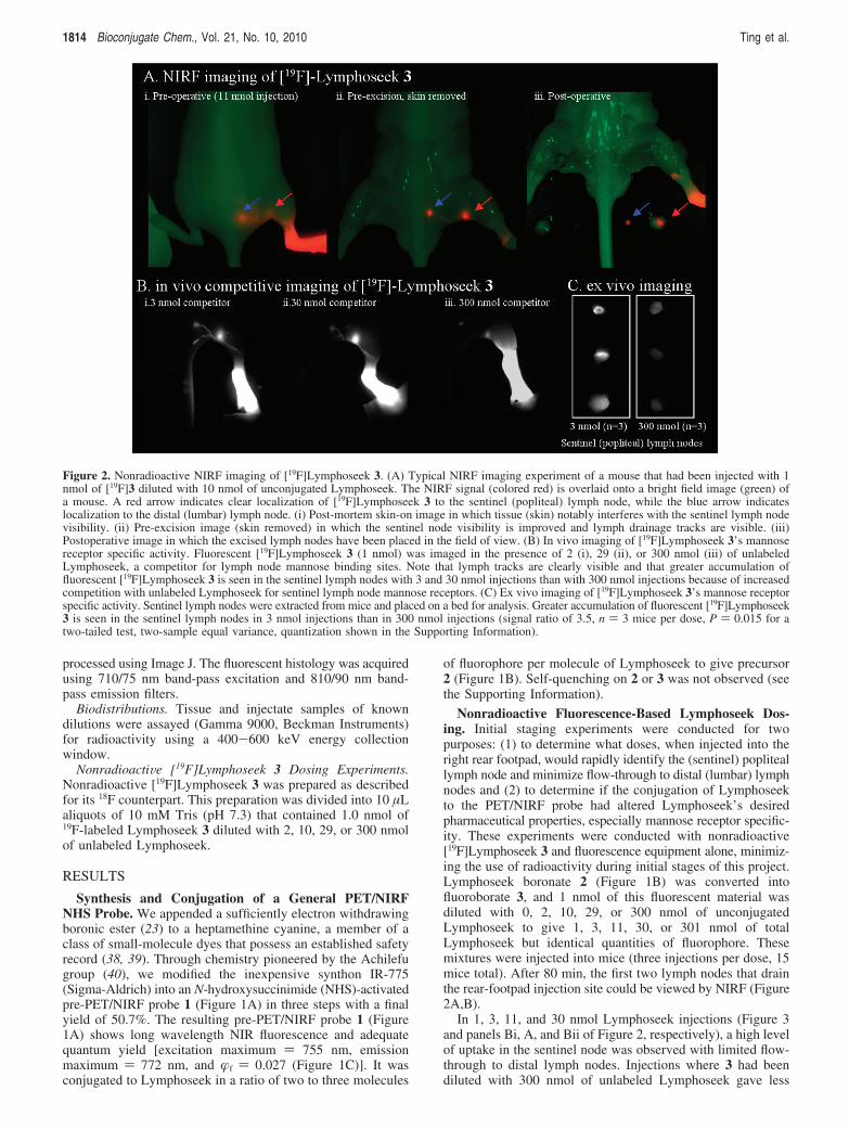
Nonradioactive Fluorescence-Based Lymphoseek Dos-ing. Initial staging experiments were conducted for twopurposes: (1) to determine what doses, when injected into theright rear footpad, would rapidly identify the (sentinel) popliteallymph node and minimize flow-through to distal (lumbar) lymphnodes and (2) to determine if the conjugation of Lymphoseekto the PET/NIRF probe had altered Lymphoseek’s desiredpharmaceutical properties, especially mannose receptor specific-ity. These experiments were conducted with nonradioactive[19F]Lymphoseek 3 and fluorescence equipment alone, minimiz-ing the use of radioactivity during initial stages of this project.Lymphoseek boronate 2 (Figure 1B) was converted intofluoroborate 3, and 1 nmol of this fluorescent material wasdiluted with 0, 2, 10, 29, or 300 nmol of unconjugatedLymphoseek to give 1, 3, 11, 30, or 301 nmol of totalLymphoseek but identical quantities of fluorophore. Thesemixtures were injected into mice (three injections per dose, 15mice total). After 80 min, the first two lymph nodes that drainthe rear-footpad injection site could be viewed by NIRF (Figure2A,B).
In 1, 3, 11, and 30 nmol Lymphoseek injections (Figure 3and panels Bi, A, and Bii of Figure 2, respectively), a high levelof uptake in the sentinel node was observed with limited flow-through to distal lymph nodes. Injections where 3 had beendiluted with 300 nmol of unlabeled Lymphoseek gave less
Figure 2. Nonradioactive NIRF imaging of [19F]Lymphoseek 3. (A) Typical NIRF imaging experiment of a mouse that had been injected with 1nmol of [19F]3 diluted with 10 nmol of unconjugated Lymphoseek. The NIRF signal (colored red) is overlaid onto a bright field image (green) ofa mouse. A red arrow indicates clear localization of [19F]Lymphoseek 3 to the sentinel (popliteal) lymph node, while the blue arrow indicateslocalization to the distal (lumbar) lymph node. (i) Post-mortem skin-on image in which tissue (skin) notably interferes with the sentinel lymph nodevisibility. (ii) Pre-excision image (skin removed) in which the sentinel node visibility is improved and lymph drainage tracks are visible. (iii)Postoperative image in which the excised lymph nodes have been placed in the field of view. (B) In vivo imaging of [19F]Lymphoseek 3’s mannosereceptor specific activity. Fluorescent [19F]Lymphoseek 3 (1 nmol) was imaged in the presence of 2 (i), 29 (ii), or 300 nmol (iii) of unlabeledLymphoseek, a competitor for lymph node mannose binding sites. Note that lymph tracks are clearly visible and that greater accumulation offluorescent [19F]Lymphoseek 3 is seen in the sentinel lymph nodes with 3 and 30 nmol injections than with 300 nmol injections because of increasedcompetition with unlabeled Lymphoseek for sentinel lymph node mannose receptors. (C) Ex vivo imaging of [19F]Lymphoseek 3’s mannose receptorspecific activity. Sentinel lymph nodes were extracted from mice and placed on a bed for analysis. Greater accumulation of fluorescent [19F]Lymphoseek3 is seen in the sentinel lymph nodes in 3 nmol injections than in 300 nmol injections (signal ratio of 3.5, n ) 3 mice per dose, P ) 0.015 for atwo-tailed test, two-sample equal variance, quantization shown in the Supporting Information).
1814 Bioconjugate Chem., Vol. 21, No. 10, 2010 Ting et al.

fluorescence in the sentinel lymph node of mice because ofcompetition for SLN mannose receptors (Figure 2Biii,C). Suchsaturable binding of [19F]Lymphoseek 3 to a limited number ofSLN mannose receptors (32, 34, 41) suggests that conjugationto 1 does not alter Lymphoseek’s mannose receptor specificity.This observation is confirmed quantitatively in separate biodis-tribution experiments (vide infra).
Image-guided surgical excisions of the SLN were thenconducted and confirmed (Figure 2C). Real-time imaging witha NIRF camera allows for rapid and simple removal of lymphnodes, especially because the lymph tracks connecting the nodesare fully visible (Figure 2Bi,Biii). Notably, lymph tracks in micehave not been previously observed with [99mTc]Lymphoseek.The observed lymph tracks connecting injection sites and nodesshould help identify the sentinel node in cases where thedistinctions between distal and sentinel nodes are ambiguous.
Radiochemistry. From NIRF experiments (Figure 2), wedetermined that total doses of Lymphoseek of up to 30 nmolwork well in imaging the SLN in mice. Using these doses, weincorporated different 18F activities into 3 to determine theoptimal level of radioactivity that would provide sufficient signalto image 1 nmol of Lymphoseek by PET, synthesized on a largemulti-injection scale starting with only 100 mCi of 18F- ion. Aspecific activity of 1-50 Ci/mmol at the time of injection provedto be adequate for imaging 18F-labeled 3 in a 20 min PET scan1 h after injection.
Size exclusion gel chromatography in spin columns was usedto remove small molecule impurities and free [18F]fluoride ionsfrom radiochemical preparations. The removal of [18F]fluoridewas evidenced by the absence of a bone signal in PET images(Figure 3A and Supporting Information) (25) and biodistributionstudies (Figure 4, bone). In mouse serum, the chemical releaseof F- from 18F-labeled Lymphoseek 3 had a half-life of 21 h(Supporting Information), 11.6-fold slower than the radioactivedecay of 18F.
Multimodality PET/CT/NIRF Imaging. [18F]Lymphoseek3 was tested for in vivo multimodal imaging in 12 mice.Injections containing 1 nmol and 10-50 µCi of fluorescent 3,with 0 or 10 nmol of added unlabeled Lymphoseek, were madeinto the right rear footpad of anesthetized nude mice positionedsecurely on trays for corroborative multimodality imaging. Onehour later, a 20 min PET/CT scan was acquired (Figure 3A,left). Mice were then sacrificed by CO2 inhalation followed bycervical dislocation; skin was removed, and NIRF images wereobtained before (Figure 3C, left) and after excision of nodes,which were placed below their location of origin on the imagingtray. The bright field image of this dissection is shown in Figure3B (before excision, left, and after excision, right). Successfulnode excision was confirmed by both NIRF (Figure 3C, right)and PET/CT imaging (Figure 3A, right, and SupportingInformation), showing the node signal outside the body.Following image acquisition, tissues were collected for scin-
Figure 3. Multimodality imaging of a mouse injected with a 10 µL, 1 nmol, 48.1 µCi dose of 18F-labeled Lymphoseek 3 (0.048 Ci/µmol). The redarrows indicates the location of the sentinel lymph node. (A) Pre-excision, live-mouse PET (red color table)/CT (blue color table) scan (left) andpostoperative PET/CT scan (right) of the mouse with excised nodes (right and left lumbar and popliteal) that are placed below the excision site inthe field of view. (B) Bright field images of the mouse with skin removed before (left) and after node excision (right). (C) NIRF images taken 90min postexcision on a custom full field IR camera before (left) and after (right) lymph node excision. The injection site was covered with blackcardboard to block intense signal from the foot and allow better visualization of the fluorescent lymph nodes. (D) H and E stain (top, color images)and NIRF (bottom, black and white images) histological verification of lymph node excision. A PET projection video of panel A is given asSupporting Information.
PET and Optical Imaging of Sentinel Lymph Nodes Bioconjugate Chem., Vol. 21, No. 10, 2010 1815

tigraphy and histological sectioning (Figure 3D). Little or noflow-through of [18F]Lymphoseek 3 to distal lymph nodes wasobserved in both NIRF and PET images taken under bothinjection conditions. PET and NIRF signals were confined tothe SLN, the injection site, and, in some cases, the bladder.
Biodistribution. Scintigraphy corroborated the qualitativedata observed in PET and IR images (Figure 3A,C). The[18F]Lymphoseek 3 tissue distribution is shown in Figure 4 asthe percent of injected dose (% ID) before (left) and after (right)normalization by tissue mass. In 1 and 11 nmol injections ofLymphoseek, the tissues of interest containing the most [18F]Ly-mphoseek 3 are the right foot (320 ( 60 and 390 ( 40% ID/gper foot, respectively) and the right popliteal (sentinel) lymphnode (170 ( 50 and 300 ( 140% ID/g per node, respectively).There is 4-7 times less [18F]Lymphoseek 3 in the ipsilateraldistal (lumbar) lymph nodes (22 ( 6 and 75 ( 30% ID/g,respectively) than in the sentinel lymph nodes. Much less[18F]Lymphoseek accumulation is observed in contralateralnodes (5 ( 3 and 9 ( 4% ID/g for contralateral popliteal lymphnodes and 8 ( 2 and 23 ( 10% ID/g for contralateral lumbarlymph nodes, for 1 and 11 nmol injections, respectively).Nonlymphatic tissue, i.e., blood, liver, kidney, spleen, and bone,contained not more than 4% ID/g of [18F]Lymphoseek 3.
Statistical analysis verified that [18F]Lymphoseek 3 retainsreceptor targeting properties consistent with [99mTc]Lymphoseekliterature (32, 41). In mice treated with 1 nmol of [18F]Lym-phoseek, the sentinel node extracted 87 ( 7% (Table 1) of thetotal lymph node accumulation, significantly greater than 74 (7% extracted at an 11 nmol dose. These values correspond to3 ( 0.9 and 40 ( 15 pmol of total Lymphoseek (3 ( 0.9 and3.6 ( 1 pmol of 18F-labeled Lymphoseek 3, respectively) inthe sentinel node. These are subsaturating doses of Lymphoseek.The Lymphoseek binding capacity from a rear footpad injectionin the popliteal node of mice is 49 ( 25 pmol (41).
Histology. NIRF histology (Figure 3D) showed that Lym-phoseek 3 indeed localized to SLNs (Figure 3D), though 18Factivity was no longer measurable. On the basis of the specificactivity of radiolabeling, the majority (>99%) of the fluorescentmaterial was nonradioactive [19F]3 even before the 18F haddecayed. Hematoxylin and eosin (H and E) frozen sections ofthe excised lymph nodes of mice were prepared and analyzed.Specifically, lymph nodes were confirmed through the observa-tion of an encapsulated dense mass of densely packed lympho-cytes. The dense packing of these mononuclear cells includesa high nucleus:cytoplasm ratio that gives a dense basophilic/purple staining under H and E staining conditions.
DISCUSSION
We describe the novel synthesis of a small molecule NHSester that confers both 18F PET and NIRF visibility ontotargeting ligands bearing free amines. Heterobifunctional adapt-ers could easily convert the selectivity toward other reactivegroups such as thiols. Because the 18F is appended to the cyaninedye, only one attachment site on the targeting ligand is required.If standard NIRF and PET labels were independently affixed(4, 7), at least two chemically reactive yet biologically tolerantsites on the ligand would be required. Unless regioselectivitycould be tightly controlled, greater combinatorial complexityof products could result, because each site could remainunmodified or receive a NIRF or PET label. Homogeneouslabeling happens not to be essential for Lymphoseek, whichalready contains a statistical distribution of amines, but Lym-phoseek had the advantages of large-scale availability andextensive preclinical and clinical characterization. PET/NIRF-labeled Lymphoseek 3 retains the desired pharmaceuticalproperties of [99mTc]Lymphoseek: rapid 1 h sentinel nodetargeting, low level of distal lymph node accumulation, andsentinel node extraction values (1 nmol dose, 87 ( 7%; 11 nmol
Figure 4. [18F]Lymphoseek 3 tissue biodistribution in mice that were sacrificed 1 h and 20 min following injection. Units of percent injected dose(ID, left) and percent injected dose per gram (right). Biodistribution data from 12 mice were used to compile this figure.
Table 1. Distribution of Radioactive [18F]Lymphoseek 3 in Mice That Were Sacrificed 80 min following Injectiona
percent [18F]3 activityextracted in the sentinel node
Lymphoseek in the sentinel orpopliteal lymph node (pmol)
(percent of detected nodal Lymphoseek)
Lymphoseek flow-through to thedistal or lumbar node (pmol)
(percent of detected nodal Lymphoseek)
1 nmol Lymphoseek injection 87 ( 7% 3.0 ( 0.9 (91%) 0.3 ( 0.1 (9%)11 nmol Lymphoseek injection 74 ( 7% 40 ( 15 (83%) 8 ( 3 (17%)t test (one-tailed two-sample
unequal variance)P ) 0.022 P ) 0.033
a Scintigraphy data from six mice were obtained per injected Lymphoseek dose (12 mice total). The percent activity extracted is defined as thedifference in counts between the sentinel and distal lymph nodes minus the sum of counts in the sentinel and distal lymph nodes [(countspopliteal -countslumbar)/(countspopliteal + countslumbar)]. The percent of detected nodal Lymphoseek is defined as the amount of Lymphoseek in the lumbar lymphnode divided by the sum of the Lymphoseek in both the popliteal and lumbar lymph nodes. Error ) σ/(n ) 6)1/2.
1816 Bioconjugate Chem., Vol. 21, No. 10, 2010 Ting et al.

dose, 74 ( 7%) that compare very well to [99mTc]Lymphoseekextraction ratios, 90 ( 10% in rabbit popliteal SLN (31, 32).
PET rapidly and noninvasively quantitates picomole amountsof 1 through deep tissue. An example of the superior tomo-graphic resolution conveyed by 18F PET in Figure 3A is includedas an .mpg file (Supporting Information). Notably, this high-resolution projection was generated from a 20 min scan on alive mouse using only 33 µCi of activity. PET is more reliablethan NIRF for imaging through deep tissue. This point isillustrated by comparing the skin-on NIRF image (Figure 2Ai)to the skin-off NIRF image (Figure 2Aii). If one were to useFigure 2Ai to evaluate Lymphoseek distribution based onrelative NIRF brightness, it would incorrectly appear that thesentinel node (red arrow), buried in muscle and fat, has lessLymphoseek than the lumbar node (blue arrow), which sits nextto the spine near the surface of the mouse. Following mousesacrifice followed by skin removal (Figure 2Aii), we see thatthe opposite is true and that the NIRF signal is biased to tissuedepth. Note that the lymph nodes seen in Figure 2 Ai would beentirely invisible by NIRF if wild-type, hair-bearing mice wereused. PET is superior to NIRF in that it allows for quantita-tive noninvasive deep tissue three-dimensional imaging. Boththe live mouse PET images shown in Figure 3 and the .mpgfile included as Supporting Information illustrate this. Additionaladvances in PET imaging may include better CT attenuationalgorithms for higher-resolution images and real-time PETreconstruction to guide surgery. If a PET scanner is notavailable, standard scintigraphic devices can still be used toregister the positron-generated γ-rays (42-44). Consistent withprevious studies of 18F-labeled aryltrifluoroborates, no signal isobserved in bone structure (Figures 3A and 4), demonstratingthe in vivo stability of the aryl trifluoroborate to solvolysis andrelease of free 18F (22, 23, 45).
The stable NIRF component on 1 also allows for NIRF-guided surgery as well as fluorescent histology long afterradiotracer decay. Histological resolution (Figure 3D) and theimaging of lymph tracks (Figure 2Bi-iii) are possible only byNIRF, as these structures fall below the spatial detection limitsof PET. The heterogeneous foci seen in NIRF histology (Figure3D) identify the most probable places to find micrometastaticdisease in the SLN (46), and their fluorescence might reducethe error in pathological evaluations. The heterogeneous dis-tribution of 3 also suggests that only a fraction of the SLNactually filters the afferent lymph channel draining the foot. Botha 1 nmol injection of pure [18F]Lymphoseek 3 and an 11 nmolinjection of 3 diluted with unlabeled Lymphoseek are useful inhighlighting sentinel popliteal nodes. Our use of a diluted 11nmol injection may seem superfluous as it requires an extra stepin preparation and is slightly inferior to the 1 nmol injection interms of unwanted flow-through to distal lymph nodes (Table1). However, the fact that the lymph node mannose receptorbinding of 3 can be diluted with unlabeled Lymphoseek suchthat distal node flow-through is increased serves to illustrate animportant trait of the dual probe, that the conjugation of targetsto NHS ester 1 does not alter its target’s desired diagnosticproperties, in this case Lymphoseek’s mannose receptor-specifictargeting properties.
This PET/NIRF version of Lymphoseek 3 may improvesentinel node biopsy, because a single injection would replacethe required multiple-injection combination (47, 48) of aradiotracer, such as [99mTc]Lymphoseek or [99mTc]sulfur colloid,with a nonspecific optical excision aid such as isosulfan bluedye. Attachment of both the positron emitter and fluorophoreto a single molecularly targeted agent diminishes the risk offlow-through to distal lymph nodes and ensures perfect coreg-istration of the two signals, allowing PET and NIRF tocomplement each other. PET images would noninvasively
identify sentinel nodes that drain a tumor and aid in preoperativeplanning. During surgery, Geiger counting of the γ-rays couldhelp locate deeply buried nodes. Once they were exposed,higher-resolution real-time NIRF imaging would guide precisionresection, including lymph tracks as well as nodes. A postopera-tive PET scan could confirm whether removal was complete,and NIRF would aid histological identification of metastaseswithin the resected tissue. Successful application of PET/NIRFNHS ester 1 to a well-established, clinically interesting systemsuggests extension to other medically relevant targets. Inprevious combinations of PET and NIRF, the positron emitterand fluorophore were independently attached (4, 7), whereas 1combines these capabilities in a single, versatile small moleculeonly slightly larger than the usual commercially available NIRFlabels. It is essential that 18F is incorporated into fluoroboratesunder mild conditions such as a weakly acidic methanol/watersolution at 40 °C, because heptamethine cyanine dyes wouldnot survive high-temperature reaction with anhydrous nucleo-philic fluoride as required for traditional 18F chemistries suchas fluorobenzyl labeling. Despite scope for further optimization,formation of aryl fluoborate from F- satisfies many of the criteriafor “click chemistry”, modularity, high yield, no offensivebyproducts, mild aqueous reaction conditions, orthogonality toother functional groups, and simple product isolation (49).Although the B-F bond is not as chemically stable as the usualC-F bonds, appropriately substituted aryl fluoborates are stableenough that hydrolysis is negligible within the radioactive half-life of 18F. Our new chemistry should facilitate the multimodalitylabeling of peptides, proteins, and polysaccharides, medicallyrelevant ligands previously difficult or impossible to label byprevious methods.
ACKNOWLEDGMENT
We thank Jacqueline Corbeil for excellent technical assistancewith PET, CT, and NIRF imaging studies and Larry Gross forhis assistance in obtaining high-resolution mass spectrometrydata. This work is supported by the Howard Hughes MedicalInstitute (HHMI) and Grant W81XWH-05-1-0183 from theDepartment of Defense Breast Cancer Research Program. R.T.is supported by a Canadian Institute of Health Research (CIHR)Post-Doctoral Fellowship (MFE 83832).
Supporting Information Available: PET/CT video; PET/CT reconstructed data accessible with amide 0.9.1 (http://amide.sourceforge.net); in vivo and synthetic experimentalprocedures; additional multimodality PET/CT/NIRF data; fluo-rophore absorption, excitation, and emission spectra; Lympho-seek certificate of analysis; and Lymphoseek in vivo stabilityand saturation assays. This material is available free of chargevia the Internet at http://pubs.acs.org.
LITERATURE CITED
(1) Tsien, R. Y. (2003) Imagining imaging’s future. Nat. ReV. Mol.Cell Biol. 9, s16–s21.
(2) Bhushan, K. R., Mistra, P., Liu, F., Mathur, S., Lenkinski, R. E.,and Frangioni, J. V. (2008) Detection of breast cancer micro-calcifications using a dual-modality SPECT/NIR fluorescentprobe. J. Am. Chem. Soc. 130, 17648–17649.
(3) Pascu, S. I., Waghorn, P. A., Conry, T. D., Betts, H. M.,Dilworth, J. R., Churchill, G. C., Pokrovska, T., Christlieb, M.,Aigbirhio, F. I., and Warren, J. E. (2007) Designing Zn(II) andCu(II) derivatives as probes for in vitro fluorescence imaging.Dalton Trans., 4988–4997.
(4) Edwards, W. B., Xu, B., Akers, W., Cheney, P. P., Liang, K.,Rogers, B. E., Anderson, C. J., and Achilefu, S. (2008) Agonist-antagonist dilemma in molecular imaging: Evaluation of amonomolecular multimodal imaging agent for the somatostatinreceptor. Bioconjugate Chem. 19, 192–200.
PET and Optical Imaging of Sentinel Lymph Nodes Bioconjugate Chem., Vol. 21, No. 10, 2010 1817
(5) Duconge, F., Pons, T., Pestourie, C., Herin, L., Theze, B.,Gombert, K., Mahler, B., Hinnen, F., Kuhnast, B., Dolle, F.,Dubertret, B., and Tavitian, B. (2008) Fluorine-18-labeledphospholipid quantum dot micelles for in vivo multimodalimaging from whole body to cellular scales. Bioconjugate Chem.19, 1921–1926.
(6) Devaraj, N. K., Keliher, E. J., Thurber, G. M., Nahrendorf,M., and Weissleder, R. (2009) F-18 Labeled Nanoparticles forin Vivo PET-CT Imaging. Bioconjugate Chem. 20, 397–401.
(7) Kimura, R., Miao, Z., Cheng, Z., Gambhir, S., and Cochran, J.(2010) A Dual-Labeled Knottin Peptide for PET and Near-Infrared Fluorescence Imaging of Integrin Expression in LivingSubjects. Bioconjugate Chem. 21, 436–444.
(8) Wu, A. M., Yazaki, P. J., Tsai, S. W., Nguyen, K., Anderson,A. L., McCarthy, D. W., Welch, M. J., Shively, J. E., Williams,L. E., Raubitschek, A. A., Wong, J. Y. C., Toyokuni, T., Phelps,M. E., and Gambhir, S. S. (2000) High-resolution microPETimaging of carcino-embryonic antigen-positive xenografts byusing a copper-64-labeled engineered antibody fragment. Proc.Natl. Acad. Sci. U.S.A. 97, 8495–8500.
(9) Dimitrakopoulou-Strauss, A., Hohenberger, P., Haberkorn, U.,Macke, H. R., Eisenhut, M., and Strauss, L. G. (2007) 68Ga-labeled bombesin studies in patients with gastrointestinal stromaltumors: Comparison with 18F-FDG. J. Nucl. Med. 48, 1245–1250.
(10) Meyer, G. J., Macke, H., Schuhmacher, J., Knapp, W. H.,and Hofmann, M. (2004) 68Ga-labelled DOTA-derivatised pep-tide ligands. Eur. J. Nucl. Med. Mol. Imaging 31, 1097–1104.
(11) Abbas, K., Kozempel, J., Bonardi, M., Groppi, F., Alfarano,A., Holzwarth, U., Simonelli, F., Hofman, H., Horstmann, W.,Menapace, E., Leseticky, L., and Gibson, N. (2006) Cyclotronproduction of Cu-64 by deuteron irradiation of Zn-64. Appl.Radiat. Isot. 64, 1001–1005.
(12) Boswell, C. A., Sun, X. K., Niu, W. J., Weisman, G. R., Wong,E. H., Rheingold, A. L., and Anderson, C. J. (2004) Comparativein vivo stability of copper-64-labeled cross-bridged and conven-tional tetraazamacrocyclic complexes. J. Med. Chem. 47, 1465–1474.
(13) Olasz, E. B., Lang, L., Seidel, J., Green, M. V., Eckelman,W. C., and Katz, S. I. (2002) Fluorine-18 labeled mouse bonemarrow-derived dendritic cells can be detected in vivo by highresolution projection imaging. J. Immunol. Methods 260, 137–148.
(14) Kuhnast, B., de Bruin, A., Hinnen, F., Tavitian, B., and Dolle,F. (2004) Design and synthesis of a new [F-18]fluoropyridine-based haloacetamide reagent for the labeling of oligonucleotides:2-Bromo-N-[3-(2-[F-18]fluoropyridin-3-yloxy)propyl]aceta-mide. Bioconjugate Chem. 15, 617–627.
(15) Ross, T. L., Ermert, J., Hocke, C., and Coenen, H. H. (2007)Nucleophilic F-18-fluorination of heteroaromatic iodonium saltswith no-carrier-added [F-18]fluoride. J. Am. Chem. Soc. 129,8018–8025.
(16) Flavell, R. R., Kothari, P., Bar-Dagan, M., Synan, M.,Vallabhajosula, S., Friedman, J. M., Muir, T. W., and Ceccarini,G. (2008) Site-specific F-18-labeling of the protein hormoneleptin using a general two-step ligation procedure. J. Am. Chem.Soc. 130, 9106–9112.
(17) Glaser, M., and Arstad, E. (2007) “Click labeling” with 2-[F-18]fluoroethylazide for positron emission tomography. Biocon-jugate Chem. 18, 989–993.
(18) Schirrmacher, R., Bradtmoller, G., Schirrmacher, E., Thews,O., Tillmanns, J., Siessmeier, T., Buchholz, H. G., Bartenstein,P., Wangler, B., Niemeyer, C. M., and Jurkschat, K. (2006) 18F-labeling of peptides by means of an organosilicon-based fluorideacceptor. Angew. Chem., Int. Ed. 45, 6047–6050.
(19) Hohne, A., Mu, L., Honer, M., Schubiger, P. A., Ametamey,S. M., Graham, K., Stellfeld, T., Borkowski, S., Berndorff, D.,Klar, U., Voigtmann, U., Cyr, J. E., Friebe, M., Dinkelborg, L.,and Srinivasan, A. (2008) Synthesis, 18F-labeling, and in vitroand in vivo studies of bombesin peptides modified with silicon-based building blocks. Bioconjugate Chem. 19, 1871–1879.
(20) Mu, L., Hohne, A., Schubiger, P. A., Ametamey, S. M.,Graham, K., Cyr, J. E., Dinkelborg, L., Stellfeld, T., Srinivasan,A., Voigtmann, U., and Klar, U. (2008) Silicon-based buildingblocks for one-step 18F-radiolabeling of peptides for PETimaging. Angew. Chem., Int. Ed. 47, 4922–4925.
(21) Schirrmacher, E., Wangler, B., Cypryk, M., Bradtmoller, G.,Schafer, M., Eisenhut, M., Jurkschat, K., and Schirrmacher, R.(2007) Synthesis of p-(di-tert-butyl[18F]fluorosilyl)benzaldehyde([18F]SiFA-A) with high specific activity by isotopic exchange:A convenient labeling synthon for the 18F-labeling of N-amino-oxy derivatized peptides. Bioconjugate Chem. 18, 2085–2089.
(22) Ting, R., Harwig, C. W., Lo, J., Li, Y., Adam, M. J., Ruth,T. J., and Perrin, D. M. (2008) Substituent effects on aryltrif-luoroborate solvolysis in water: Implications for Suzuki-Miyauracoupling and the design of stable F-18-labeled aryltrifluoroboratesfor use in PET imaging. J. Org. Chem. 73, 4662–4670.
(23) Ting, R., Lo, J., Adam, M. J., Ruth, T. J., and Perrin, D. M.(2008) Capturing aqueous [F-18]-fluoride with an arylboronicester for PET: Synthesis and aqueous stability of a fluorescent[F-18]-labeled aryltrifluoroborate. J. Fluorine Chem. 129, 349–358.
(24) Ting, R., Adam, M. J., Ruth, T. J., and Perrin, D. M. (2005)Arylfluoroborates and alkylfluorosilicates as potential PET imag-ing agents: High-yielding aqueous biomolecular F-18-labeling.J. Am. Chem. Soc. 127, 13094–13095.
(25) Ting, R., Harwig, C., auf dem Keller, U., McCormick, S.,Austin, P., Overall, C. M., Adam, M. J., Ruth, T. J., and Perrin,D. M. (2008) Toward [F-18]-labeled aryltrifluoroborate ra-diotracers: In vivo positron emission tomography imaging ofstable aryltrifluoroborate clearance in mice. J. Am. Chem. Soc.130, 12045–12055.
(26) Valk, P. E., Bailey, D. L., Townsend, D. W., and Maisey,M. N. (2003) Positron Emission Tomography: Basic Science andClinical Practice, Springer-Verlag, London.
(27) Hirsch, J. I., Tisnado, J., Cho, S. R., and Beachley, M. C.(1982) Use of Isosulfan Blue for Identification of LymphaticVessels: Experimental and Clinical Evaluation. Am. J. Roent-genol. Radium Ther. 139, 1061–1064.
(28) Mariani, G., Gipponi, M., Moresco, L., Villa, G., Bartolomei,M., Mazzarol, G., Bagnara, M. C., Romanini, A., Cafiero, F.,Paganelli, G., and Strauss, H. W. (2002) Radioguided sentinellymph node biopsy in malignant cutaneous melanoma. J. Nucl.Med. 43, 811–827.
(29) Mariani, G., Moresco, L., Viale, G., Villa, G., Bagnasco, M.,Canavese, G., Buscombe, J., Strauss, H. W., and Paganelli, G.(2001) Radioguided sentinel lymph node biopsy in breast cancersurgery. J. Nucl. Med. 42, 1198–1215.
(30) Weaver, D. L., Krag, D. N., Ashikaga, T., Harlow, S. P., andO’Connell, M. (2000) Pathologic analysis of sentinel andnonsentinel lymph nodes in breast carcinoma: A multicenterstudy. Cancer 88, 1099–1107.
(31) Vera, D. R., Wisner, E. R., and Stadalnik, R. C. (1997)Sentinel node imaging via a nonparticulate receptor-bindingradiotracer. J. Nucl. Med. 38, 530–535.
(32) Vera, D. R., Wallace, A. M., Hoh, C. K., and Mattrey, R. F.(2001) A synthetic macromolecule for sentinel node detection:Tc-99m-DTPA-mannosyl-dextran. J. Nucl. Med. 42, 951–959.
(33) Hoh, C. K., Wallace, A. M., and Vera, D. R. (2003) Preclinicalstudies of [Tc-99m]DTPA-mannosyl-dextran. Nucl. Med. Biol.30, 457–464.
(34) Vera, D. R., Wallace, A. M., and Hoh, C. K. (2001) [Tc-99m]MAG3-mannosyl-dextran: A receptor-binding radiophar-maceutical for sentinel node detection. Nucl. Med. Biol. 28, 493–498.
(35) Schoder, H., Glass, E. C., Pecking, A. P., Harness, J. K.,Wallace, A. M., Hirnle, P., Alberini, J. L., Vilain, D., Larson,S. M., Hoh, C. K., and Vera, D. R. (2006) Molecular targetingof the lymphovascular system for imaging and therapy. CancerMetastasis ReV. 25, 185–201.
1818 Bioconjugate Chem., Vol. 21, No. 10, 2010 Ting et al.

(36) Dubois, M., Gilles, K. A., Hamilton, J. K., Rebers, P. A., andSmith, F. (1956) Colorimetric Method for Determination ofSugars and Related Substances. Anal. Chem. 28, 350–356.
(37) Hall, D. J., Sunar, U., Farshchi-Heydari, S., and Han, S. H.(2009) In vivo simultaneous monitoring of two fluorophores withlifetime contrast using a full-field time domain system. Appl.Opt. 48, D74–D78.
(38) Fox, I. J., and Wood, E. H. (1960) Indocyanine Green:Physical and Physiologic Properties. Proc. Staff Meet. Mayo Clin.35, 732–744.
(39) Fox, I. J., Brooker, L. G. S., Heseltine, D. W., Essex, H. E.,and Wood, E. H. (1956) New Dyes for Continuous Recordingof Dilution Curves in Whole Blood Independent of Variationsin Blood Oxygen Saturation. Am. J. Physiol. 187, 599.
(40) Lee, H., Mason, J. C., and Achilefu, S. (2006) Heptamethinecyanine dyes with a robust C-C bond at the central position ofthe chromophore. J. Org. Chem. 71, 7862–7865.
(41) Limmer, K., and Vera, D. R. Manuscript in preparation.(42) Bax, J. J., Visser, F. C., van Lingen, A., Huitink, J. M., Kamp,
O., van Leeuwen, G. R., Visser, G. W., Teule, G. J., and Visser,C. A. (1993) Feasibility of assessing regional myocardial uptakeof 18F-fluorodeoxyglucose using single photon emission com-puted tomography. Eur. Heart J. 14, 1675–1682.
(43) Knesaurek, K., and Machac, J. (2006) Comparison of 18FSPECT with PET in myocardial imaging: A realistic thorax-cardiac phantom study. BMC Nucl. Med. 6, 5.
(44) Leichner, P. K., Morgan, H. T., Holdeman, K. P., Harrison,K. A., Valentino, F., Lexa, R., Kelly, R. F., Hawkins, W. G.,and Dalrymple, G. V. (1995) SPECT imaging of fluorine-18.J. Nucl. Med. 36, 1472–1475.
(45) Bohn, P., Deyine, A., Azzouz, R., Bailly, L., Fiol-Petit, C.,Bischoff, L., Fruit, C., Marsais, F., and Vera, P. (2009) Designof silicon-based misonidazole analogues and 18F-radiolabelling.Nucl. Med. Biol. 36, 895–905.
(46) Haigh, P. I., Lucci, A., Turner, R. R., Bostick, P. J., Krasne,D. L., Stern, S. L., and Morton, D. L. (2001) Carbon dyehistologically confirms the identity of sentinel lymph nodes incutaneous melanoma. Cancer 92, 535–541.
(47) Chen, S. L., Iddings, D. M., Scheri, R. P., and Bilchik, A. J.(2006) Lymphatic mapping and sentinel node analysis: Currentconcepts and applications. CasCancer J. Clin. 56, 292–309 (quiz316-317).
(48) Edwards, M. J., Martin, K. D., and McMasters, K. M. (1999)Lymphatic mapping and sentinel lymph node biopsy in thestaging of melanoma. Surg. Oncol., 51–57.
(49) Kolb, H. C., Finn, M. G., and Sharpless, K. B. (2001) Clickchemistry: Diverse chemical function from a few good reactions.Angew. Chem., Int. Ed. 40, 2004–2021.
BC1001328
PET and Optical Imaging of Sentinel Lymph Nodes Bioconjugate Chem., Vol. 21, No. 10, 2010 1819


![Beyond Responsive [18F 2015]](https://static.fdocuments.us/doc/165x107/55d137adbb61eb9f488b4756/beyond-responsive-18f-2015.jpg)