
16. Disinfectants and disinfectant by-products · per day for female and male rats, respectively)...
125
16. Disinfectants and disinfectant by-products Disinfectants 16.1 Introduction Disinfection is unquestionably the most important step in the treatment of water for public supply. The destruction of microbiological pathogens is essential and almost invariably involves the use of reactive chemical agents such as chlorine, which are not only powerful biocides but also capable of reacting with other water constituents to form new compounds with potentially harmful long-term health effects. Thus, an overall assessment of the impact of disinfection on public health must consider not only the microbiological quality of the treated water, but also the toxicity of the disinfectants and their reaction products. The paramount importance of microbiological quality requires some flexibility in the derivation of guideline values for these substances. Fortunately this is possible because of the substantial margin of safety incorporated into these values. Guideline values for carcinogenic disinfectant by-products are presented here for an excess lifetime cancer risk of 10 -5 . The conditions specified for disinfection vary not only according to water composition and temperature but also with available technology and socioeconomic factors in different parts of the world. Where local circumstances require that a choice must be made between meeting either microbiological guidelines or guidelines for disinfectants or disinfectant by- products, the microbiological quality must always take precedence, and where necessary, a chemical guideline value can be adopted corresponding to a higher level of risk. Efficient disinfection must never be compromised. Although not addressed with respect to the individual parameters presented below, it is noted that, in a number of epidemiological studies, positive associations between the ingestion of chlorinated drinking- water and mortality rates from cancer, particularly of the bladder, have been reported. The degree of evidence for this association is considered inadequate by IARC (1). The level of disinfection by-products can be reduced by optimizing the treatment process (see Volume 1, section 6.3). Removal of organic substances prior to disinfection reduces the formation of potentially harmful by-products. The following guidance is provided to help authorities decide which guideline values may be of greater or lesser importance for setting national standards: guideline values for chemicals of greater importance generally include those for chloramines and chlorine (when used as disinfectants); followed by those for bromoform, dibromochloromethane, bromodichloromethane, chloroform, and chloral hydrate; and chlorite, bromate, dichloroacetic acid, and trichloroacetic acid (provisional guideline values have been established for this last group). Guideline values for chemicals of lesser importance generally include those for 2,4,6- trichlorophenol, formaldehyde, dichloroacetonitrile, dibromoacetonitrile, trichloroacetonitrile, and cyanogen chloride. Although given less importance, it may be appropriate to measure their levels at least once. It should also be noted that a number of non-volatile, poorly characterized by-products may be formed as well, including those derived from humic substances. These recommendations are general, and local monitoring and surveillance capabilities must be considered in the setting of national standards. Reference 1. International Agency for Research on Cancer. Chlorinated drinking-water; chlorination by-products; some other halogenated compounds; cobalt and cobalt compounds. Lyon, 1991:45-359 (IARC Monographs on the Evaluation of Carcinogenic Risks to Humans, Volume 52).
Transcript of 16. Disinfectants and disinfectant by-products · per day for female and male rats, respectively)...

16. Disinfectants and disinfectant by-products
Disinfectants
16.1 Introduction
Disinfection is unquestionably the most important step in the treatment of water for public supply. Thedestruction of microbiological pathogens is essential and almost invariably involves the use of reactivechemical agents such as chlorine, which are not only powerful biocides but also capable of reacting withother water constituents to form new compounds with potentially harmful long-term health effects. Thus,an overall assessment of the impact of disinfection on public health must consider not only themicrobiological quality of the treated water, but also the toxicity of the disinfectants and their reactionproducts.
The paramount importance of microbiological quality requires some flexibility in the derivation of guidelinevalues for these substances. Fortunately this is possible because of the substantial margin of safetyincorporated into these values. Guideline values for carcinogenic disinfectant by-products are presentedhere for an excess lifetime cancer risk of 10-5. The conditions specified for disinfection vary not onlyaccording to water composition and temperature but also with available technology and socioeconomicfactors in different parts of the world. Where local circumstances require that a choice must be madebetween meeting either microbiological guidelines or guidelines for disinfectants or disinfectant by-products, the microbiological quality must always take precedence, and where necessary, a chemicalguideline value can be adopted corresponding to a higher level of risk. Efficient disinfection must never becompromised.
Although not addressed with respect to the individual parameters presented below, it is noted that, in anumber of epidemiological studies, positive associations between the ingestion of chlorinated drinking-water and mortality rates from cancer, particularly of the bladder, have been reported. The degree ofevidence for this association is considered inadequate by IARC (1).
The level of disinfection by-products can be reduced by optimizing the treatment process (see Volume 1,section 6.3). Removal of organic substances prior to disinfection reduces the formation of potentiallyharmful by-products.
The following guidance is provided to help authorities decide which guideline values may be of greater orlesser importance for setting national standards: guideline values for chemicals of greater importancegenerally include those for chloramines and chlorine (when used as disinfectants); followed by those forbromoform, dibromochloromethane, bromodichloromethane, chloroform, and chloral hydrate; and chlorite,bromate, dichloroacetic acid, and trichloroacetic acid (provisional guideline values have been establishedfor this last group). Guideline values for chemicals of lesser importance generally include those for 2,4,6-trichlorophenol, formaldehyde, dichloroacetonitrile, dibromoacetonitrile, trichloroacetonitrile, and cyanogenchloride. Although given less importance, it may be appropriate to measure their levels at least once. Itshould also be noted that a number of non-volatile, poorly characterized by-products may be formed aswell, including those derived from humic substances. These recommendations are general, and localmonitoring and surveillance capabilities must be considered in the setting of national standards.
Reference
1. International Agency for Research on Cancer. Chlorinated drinking-water; chlorination by-products;some other halogenated compounds; cobalt and cobalt compounds. Lyon, 1991:45-359 (IARCMonographs on the Evaluation of Carcinogenic Risks to Humans, Volume 52).

16.2 Chhloramines
16.2.1 General description
Identity
CAS no.: 10599-90-3Molecular formula: NH2Cl
Mono-, di-, and trichloramines are formed when water containing ammonia is chlorinated. Onlymonochloramine, the most abundant chloramine, will be considered here, as it has been the mostextensively studied.
Physicochemical properties (1-3)1
1 Conversion factor in air: 1 ppm = 2.1 mg/m3
Property ValueMelting point -66 °C Water solubility Soluble
Organoleptic properties
Most individuals are able to taste chlorine and its by-products, including chloramines, at concentrationsbelow 5 mg/litre, and some at levels as low as 0.3 mg/litre (1).
Major uses
The chloramines are used as intermediates in the manufacture of hydrazine; when formed in situ fromammonia and chlorine, they are also disinfectants for drinking-water (4).
Environmental fate
Monochloramine is persistent in the environment. Its rate of disappearance is primarily a function of pHand salinity: its half-life increases with increasing pH and decreases with increasing salinity. Itdecomposes more quickly if discharged into receiving waters containing bromide, presumably as a resultof the formation of bromochloramine and the decomposition of the dihalamine. Monochloramine isexpected to decompose via chlorine transfer to give organic nitrogen-containing compounds in receivingwaters (5).
16.2.2 Analytical methods
Chloramines can be determined by colorimetric methods; the detection limit is about 10 µg/litre (6,7).
16.2.3 Environmental levels and human exposure
Water
Inorganic chloramines are found as by-products of the chlorination of water. In one survey, mono- anddichloramines were found in secondary sewage effluents and cooling water at levels in the range of 0.03-1.0 and 0.002-0.70 mg/litre, respectively (8). Many water companies have begun to use chloramines fordisinfection instead of chlorine to prevent the formation of trihalomethanes. Typical chloramineconcentrations of 0.5-2 mg/litre are found in drinking-water supplies where chloramine is used as aprimary disinfectant or to provide a chlorine residual in the distribution system (9).
16.2.4 Kinetics and metabolism in laboratory animals and humans
Monochloramine administered by the oral route in the rat is readily absorbed from the gastrointestinaltract, the highest concentration 5 days after administration being found in plasma followed by whole blood,skin, testes, packed cells, bone marrow, kidney, lung, stomach, thyroid and thymus, carcass, liver, ileum,and fat. Monochloramine is metabolized to the chloride ion, which is excreted mainly in the urine and to alesser extent in the faeces (10).
16.2.5 Effects on laboratory animals and in vitro test systems
Short-term exposure
Male A/JAX mice (12 per dose) were exposed to monochloramine at concentrations of 0, 2.5, 25, 50, 100,or 200 mg/litre (approximately 0, 0.4, 4, 8, 15, or 30 mg/kg of body weight per day) for 30 days. Nosignificant adverse effects on various haematological parameters, including blood cell counts,haemoglobin, GSH levels, and glucose-6-phosphate dehydrogenase activity, were reported at any doselevel tested. The NOAEL in this study was 30 mg/kg of body weight per day (11).
Monochloramine was administered to Fischer 344 rats and B6C3F1 mice in drinking-water atconcentrations of 0, 25, 50, 100, 200, or 400 mg/litre (approximately 0, 2.5, 5, 10, 20, or 40 mg/kg of bodyweight per day in rats and 0, 4, 8, 15, 30, or 60 mg/kg of body weight per day in mice) for 13 weeks.Decreased body weight gain and liver damage (e.g. cellular hypertrophy) were reported in mice exposedat concentrations of 100, 200, and 400 mg/litre. The authors also reported decreased body weight gainand decreased relative liver weight in male and female rats and increased protein excretion in male ratsgiven 200 and 400 mg/litre. The NOAELs in this study were 50 mg/litre (8 mg/kg of body weight per day)for mice and 100 mg/litre (10 mg/kg of body weight per day) for rats (12).
Monochloramine at 0, 25, 50, 100, or 200 mg/litre was administered in drinking-water to male and femaleSprague-Dawley rats for 90 days, corresponding to 0, 1.8, 3.4, 5.8, and 9.0 mg/kg of body weight per dayfor males and 0, 2.6, 4.3, 7.7, and 12.1 mg/kg of body weight per day for females. The authors consideredthe highest dose a LOAEL for both sexes, based on the respective reductions in liver and spleen weights.In addition, overall reductions in body weight gain were observed at 50 mg/litre and higher, but significantreductions only at 200 mg/litre. The authors concluded that 100 mg/litre (7.7 and 5.8 mg/kg of body weightper day for female and male rats, respectively) can be considered a NOAEL (13).
Long-term exposure
Monochloramine was administered for 2 years to male and female F344/N rats at 0, 50, 100, or 200mg/litre in the drinking-water, corresponding to average doses of 2.9, 5.2, and 9.4 mg/kg of body weightper day in males and 3.1, 5.7, and 10.2 mg/kg of body weight per day in females. The authors failed tofind any clinical changes attributable to the consumption of chloraminated water. Mean body weights ofrats given the highest dose were lower than those of their respective control groups. Significant decreasesin liver and kidney weights in high-dose males and increases in brain- and kidney-weight-to-body-weightratios in high-dose rats of both sexes were related to the lower body weights in these groups. Based onthese considerations, the authors considered the NOAELs for this study to be 5.2 and 5.7 mg/kg of bodyweight per day for male and female rats, respectively. However, it is probable that the observed weightdecreases were a direct result of the unpalatability of the drinking-water (14).
In a second bioassay, B6C3F1 mice were exposed for 2 years to monochloramine in their drinking-waterat levels of 0, 50, 100, or 200 mg/litre, corresponding to average doses of 0, 5.4, 9.8, and 17.0 mg/kg ofbody weight per day for males and 0, 5.8, 10.6, and 19.0 mg/kg of body weight per day for females. Theauthors reported that there were no clinical changes attributable to the consumption of chloraminatedwater. Based on changes in body weight at the highest dose, the NOAELs were 9.8 and 10.6 mg/kg of

body weight per day for male and female mice, respectively (14).
Reproductive toxicity, embryotoxicity, and teratogenicity
Chloramine was administered by gavage at doses of 0, 2.5, 5.0, or 10 mg/kg of body weight per day tomale and female Long-Evans rats for 66-76 days before and during mating and throughout gestation andlactation. No significant differences were observed between controls and exposed rats in fertility, viability,litter size, mean weight of pups, or day of eye opening. There were no alterations in sperm count, directprogressive sperm movement, percentage mobility, or sperm morphological characteristics in adult males.The weights of male and female reproductive organs were not significantly different between the test andcontrol groups, and no significant anatomical changes were seen on tissue examination. A NOAEL of 10mg/kg of body weight per day was identified (15).
In a study in which monochloramine was administered to female Sprague-Dawley rats (6 per dose) at 0, 1,10, or 100 mg/litre daily (approximately 0, 0.1, 1, or 10 mg/kg of body weight per day) in drinking-waterbefore mating and throughout gestation, it was found not to be teratogenic or embryotoxic. The reliabilityof these findings is reduced because of the small number of dams exposed and the lack of data onmaternal toxicity (16).
Mutagenicity and related end-points
Monochloramine was reported to be weakly mutagenic at the trpC locus of Bacillus subtilis (17). It did notincrease the number of revertant colonies above the levels in untreated controls in assays employingSalmonella typhimurium strains TA97, TA100, and TA102 (18), nor did it significantly increase bonemarrow chromosomal aberrations or micronuclei in CD-1 mice, or cause sperm-head abnormalities inB6C3F1 mice (19).
Carcinogenicity
In 2-year bioassays, mice and rats were exposed to chloramine at 0, 50, 100, or 200 mg/litre in drinking-water, the highest doses being equivalent to 9.4 and 10.2 mg/kg of body weight per day for male andfemale rats, respectively, and 17.0 and 19.0 mg/kg of body weight per day for male and female mice,respectively. The studies provided equivocal evidence of the carcinogenic activity of chloraminateddrinking-water in female F344/N rats, as indicated by an increase in comparison with concurrent controls,in the incidence of mononuclear-cell leukaemia. This increase, however, was within the range observed inhistorical controls. There was no evidence of carcinogenic activity in male rats or male and female mice(14).
16.2.6 Effects on humans
Chloramine was administered at increasing doses (approximately 0.0001, 0.01, 0.11, 0.26, or 0.34 mg/kgof body weight per day) to five groups of 10 human subjects each, over a 16-day period. There were noadverse effects on clinical signs, urinalysis, haematology, and clinical chemistry in comparison withcontrols. In a second phase of the study, 10 healthy adult males were given a chloramine solution ofconcentration 5 mg/litre (0.04 mg/kg of body weight per day). There were no adverse effects on physicalcondition, urinalysis, or clinical chemistry and no serious objections to the taste of chloramine at the dosetested (20).
Acute haemolytic anaemia, characterized by the denaturation of haemoglobin and the lysis of red bloodcells, was reported in haemodialysis patients when tapwater containing chloramines was used for dialysis(21).
In epidemiological studies, no association was found between the ingestion of chloraminated drinking-water and increased mortality rates for bladder cancer (22,23).
16.2.7 Guideline value
IARC has not evaluated the carcinogenic potential of inorganic chloramines. In the National ToxicologyProgram (NTP) bioassay in two species (14), the incidence of mononuclear cell leukaemias in femaleF344/N rats was increased, but no other increases in tumour incidence were observed. Althoughmonochloramine has been shown to be mutagenic in some in vitro studies, it has not been found to begenotoxic in vivo.
The guideline value for monochloramine is 3 mg/litre (rounded figure), based on a TDI of 94 µg/kg of bodyweight, calculated from a NOAEL of 9.4 mg/kg of body weight per day (the highest dose administered tomales in the 2-year NTP rat drinking-water study, chosen because of the probability that the lower bodyweights were caused by the unpalatability of the drinking-water) (14). An uncertainty factor of 100 (forintra- and interspecies variation) is incorporated, and 100% of the TDI is allocated to drinking-water. Anadditional uncertainty factor for possible carcinogenicity was not applied because equivocal cancer effectsreported in the NTP study in only one species and in only one sex were within the range observed inhistorical controls.
Available data are insufficient for the establishment of guideline values for dichloramine and trichloramine.The odour thresholds of dichloramine and trichloramine are much lower than that for monochloramine.
References
1. National Institute for Occupational Safety and Health. Registry of Toxic Effects of Chemical Substances(RTECS). Cincinnati, OH, US Department of Health and Human Services, 1985-86.
2. Weast RC, ed. CRC handbook of chemistry and physics, 67th ed. Boca Raton, FL, CRC Press, 1986.
3. Hawley GG. The condensed chemical dictionary, 10th ed. New York, Van Nostrand Reinhold,1981:228.
4. Moore GS, Calabrese EJ. The health effects of chloramines in potable water supplies: a literaturereview. Journal of environmental pathology and toxicology, 1980, 4(1):257-263.
5. Jolley RL, Carpenter JH. A review of the chemistry and environmental fate of reactive oxidants inchlorinated water. In: Jolley RL et al., eds. Water chlorination: environmental impact and health effects.Vol. 4. Ann Arbor, MI, Ann Arbor Science, 1983:3-47.
6. International Organization for Standardization. Water quality. Determination of free chlorine and totalchlorine. Geneva, 1985 (ISO7393/1,2:1985).
7. International Organization for Standardization. Water quality. Determination of free chlorine and totalchlorine. Geneva, 1990 (ISO7393/3:1990).
8. Jolley RL et al. Chlorination of organics in cooling waters and process effluents. In: Jolley RL, ed. Waterchlorination: environmental impact and health effects. Vol. 1. Ann Arbor, MI, Ann Arbor Science,1978:105-138.
9. Bull RJ et al. Health effects of disinfectants and disinfection by-products. Denver, CO, American WaterWorks Association, 1991.
10. Abdel-Rahman MS, Waldron DM, Bull RJ. A comparative kinetics study of monochloramine andhypochlorous acid in rat. Journal of applied toxicology, 1983, 3(4):175-179.

11. Moore GS, Calabrese EJ, McGee M. Health effects of monochloramines in drinking water. Journal ofenvironmental science and health, 1980, A15(3):239-258.
12. Gulf South Research Institute. A subchronic study of chloramine generated in-situ in the drinking waterof F344 rats and B6C3F1 mice. Rockville, MD, Tracor-Jitco, 1981 (unpublished report).
13. Daniel FB et al. Comparative subchronic toxicity studies of three disinfectants. Journal of the AmericanWater Works Association, 1990, 82:61-69.
14. National Toxicology Program. NTP technical report on the toxicology and carcinogenicity studies ofchlorinated and chloraminated water in F344/N rats and B6C3F1 mice. Research Triangle Park, NC, 1990.
15. Bull RJ et al. Use of biological assay systems to assess the relative carcinogenic hazards ofdisinfection by-products. Environmental health perspectives, 1982, 46:215-227.
16. Abdel-Rahman MS, Berardi MR, Bull RJ. Effect of chlorine and monochloramine in drinking water onthe developing rat fetus. Journal of applied toxicology, 1982, 2(3):156-159.
17. Shih KL, Lederberg J. Chloramine mutagenesis in Bacillus subtilis. Science, 1976, 192:1141-1143.
18. Thomas EL et al. Mutagenic activity of chloramines. Mutation research, 1987, 188:35-43.
19. Meier JR et al. Evaluation of chemicals used for drinking water disinfection for production ofchromosomal damage and sperm-head abnormalities in mice. Environmental mutagenesis, 1985, 7:201-211.
20. Lubbers JR, Chauhan S, Bianchine JR. Controlled clinical evaluations of chlorine dioxide, chlorite andchlorate in man. Fundamental and applied toxicology, 1981, 1:334-338.
21. Kjellstrand CM et al. Hemolysis in dialyzed patients caused by chloramines. Nephron, 1974, 13:427-433.
22. Zierler S, Danley RA, Feingold L. Type of disinfectant in drinking water and pattern of mortality inMassachusetts. Environmental health perspectives, 1986, 69:275-279.
23. Zierler S et al. Bladder cancer in Massachusetts related to chlorinated and chloraminated drinkingwater: a case control study. Archives of environmental health, 1988, 43(2):195-200.
16.3 Chlorine
16.3.1 General description
Identity
Element or compound CAS no. Molecular formulaChlorine 7782-50-5 Cl2Hypochlorous acid 7790-92-3 HOClSodium hypochlorite 7681-52-9 NaOCl
Physicochemical properties of chlorine (1,2)1
1 Conversion factor in air: 1 ppm = 2.9 mg/m3
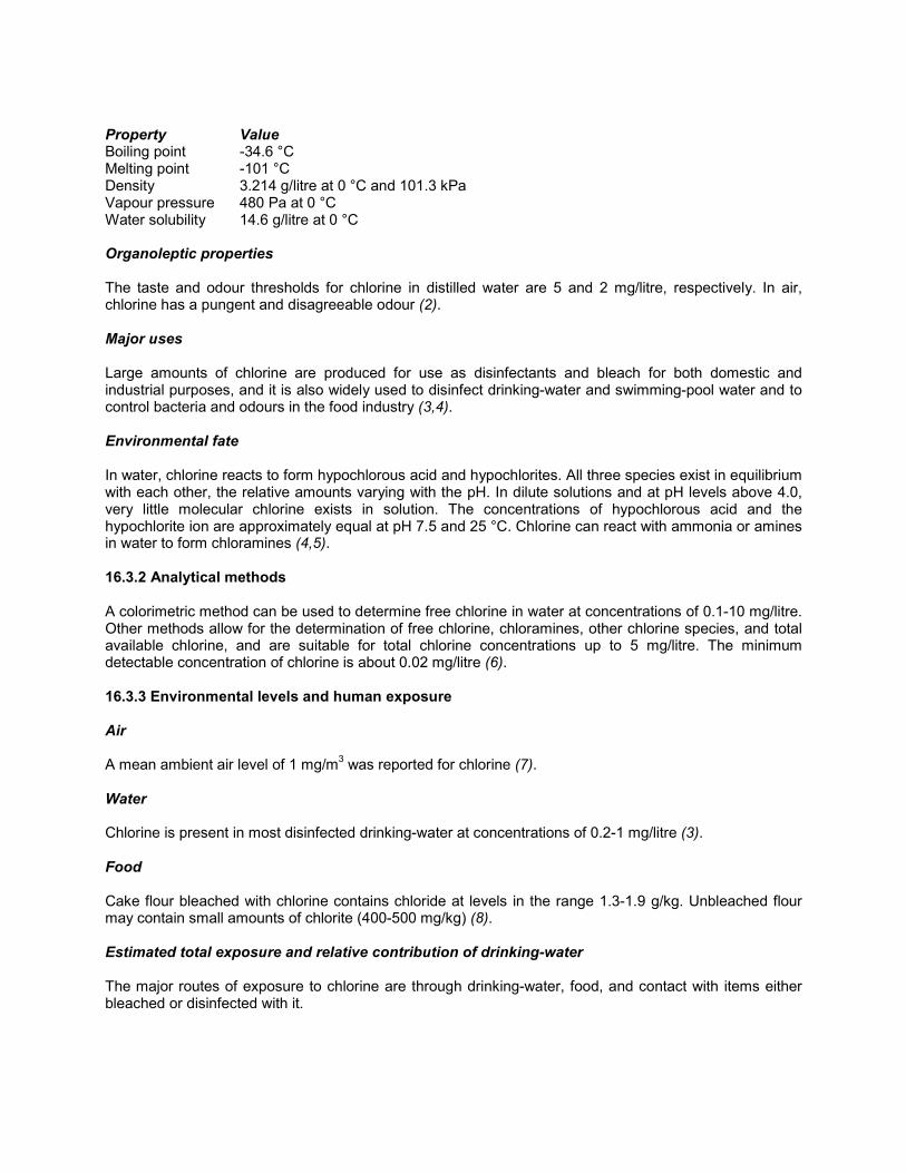
Property ValueBoiling point -34.6 °CMelting point -101 °CDensity 3.214 g/litre at 0 °C and 101.3 kPaVapour pressure 480 Pa at 0 °CWater solubility 14.6 g/litre at 0 °C
Organoleptic properties
The taste and odour thresholds for chlorine in distilled water are 5 and 2 mg/litre, respectively. In air,chlorine has a pungent and disagreeable odour (2).
Major uses
Large amounts of chlorine are produced for use as disinfectants and bleach for both domestic andindustrial purposes, and it is also widely used to disinfect drinking-water and swimming-pool water and tocontrol bacteria and odours in the food industry (3,4).
Environmental fate
In water, chlorine reacts to form hypochlorous acid and hypochlorites. All three species exist in equilibriumwith each other, the relative amounts varying with the pH. In dilute solutions and at pH levels above 4.0,very little molecular chlorine exists in solution. The concentrations of hypochlorous acid and thehypochlorite ion are approximately equal at pH 7.5 and 25 °C. Chlorine can react with ammonia or aminesin water to form chloramines (4,5).
16.3.2 Analytical methods
A colorimetric method can be used to determine free chlorine in water at concentrations of 0.1-10 mg/litre.Other methods allow for the determination of free chlorine, chloramines, other chlorine species, and totalavailable chlorine, and are suitable for total chlorine concentrations up to 5 mg/litre. The minimumdetectable concentration of chlorine is about 0.02 mg/litre (6).
16.3.3 Environmental levels and human exposure
Air
A mean ambient air level of 1 mg/m3 was reported for chlorine (7).
Water
Chlorine is present in most disinfected drinking-water at concentrations of 0.2-1 mg/litre (3).
Food
Cake flour bleached with chlorine contains chloride at levels in the range 1.3-1.9 g/kg. Unbleached flourmay contain small amounts of chlorite (400-500 mg/kg) (8).
Estimated total exposure and relative contribution of drinking-water
The major routes of exposure to chlorine are through drinking-water, food, and contact with items eitherbleached or disinfected with it.

16.3.4 Kinetics and metabolism in laboratory animals and humans
Most studies on the pharmacokinetics of chlorine, hypochlorous acid, or hypochlorites employ reactive36Cl-labelled compounds and probably reflect the fate of the chloride ion or other reaction productsgenerated from the parent molecules. In rats, hypochlorous acid was readily absorbed through thegastrointestinal tract, distribution being highest in the plasma; smaller amounts were found in bonemarrow, kidney, testes, lung, skin, duodenum, spleen, liver, and bone (9,10). In vivo, sodium hypochloritewas metabolized to trichloroethanoic acid, dichloroethanoic acid, chloroform, and dichloroacetonitrile (11).Hypochlorous acid administered to rats was excreted primarily in the urine and faeces, mostly in the formof chloride ion (10). None was excreted in expired air (9).
16.3.5 Effects on laboratory animals and in vitro test systems
Acute exposure
Calcium hypochlorite has an oral LD50 in the rat of 850 mg/kg of body weight (2).
Short-term exposure
No consistent effects on organ weights or histopathology of tissues were noted in Sprague-Dawley rats(10 per sex per dose) given chlorine in drinking-water at 0, 25, 50, 100, 175, or 200 mg/litre (males: 0, 2,7.5, 12.8, or 16.7 mg/kg of body weight per day; females: 0, 3.5, 12.6, 19.5, or 24.9 mg/kg of body weightper day) for 90 days (12) or in rats fed flour containing 1257 or 2506 mg of chlorine per kg (62.5 or 125mg/kg of body weight per day) for 28 days (13).
Enhanced weight gain was observed in all male rats (10 per dose) given drinking-water containingchlorine at 0, 20, 40, or 80 mg/litre (0, 4.1, 8.1, or 15.7 mg/kg of body weight per day) for 6 weeks (14).The results of a 4-week study in which female C57BL/6N mice were given hyperchlorinated tapwater (4.8-5.8 mg/kg of body weight per day) suggested an adverse effect on the macrophage defence mechanismsof mice. The LOAEL in this study was 4.8 mg/kg of body weight per day (15).
In a study in which male CR-1:CD-1 mice (30 per dose) received chlorinated drinking-water (0.02, 0.2, 2.9,or 5.8 mg/kg of body weight per day) for 120 days, none of the mice showed evidence of a statisticallysignificant change in humoral or cell-mediated immune response. A NOAEL of 5.8 mg/kg of body weightper day was identified (16).
Long-term exposure
F344 rats (50 per sex per dose) were administered sodium hypochlorite in drinking-water (males: 0.05%or 0.1%, 75 or 150 mg/kg of body weight per day; females: 0.1% or 0.2%, 150 or 300 mg/kg of bodyweight per day) for 2 years. Effects included a dose-related depression in body weight gain in all groups,depressed liver, brain, and heart weights in males given a 0.05% dose, decreased salivary gland weightsin both female groups, and decreased kidney weights in females given 0.2% (17).
In a 2-year bioassay, F344 rats and B6C3F1 mice were given chlorine in drinking-water at levels of up to275 mg/litre (up to 24 mg/kg of body weight per day for male rats and male mice, 15 mg/kg of body weightper day for female rats, and 22 mg/kg of body weight per day for female mice). There was a dose-relateddecrease in water consumption for both mice and rats. No effects on body weight or survival wereobserved in any of the treated animals (18).
Wistar rats were fed cake prepared from flour treated with 1250 or 2500 mg of chlorine per kg (males:12.8 or 25.3 mg/kg of body weight per day; females: 17.0 or 35.0 mg/kg of body weight per day) for 104weeks. A dose-related reduction in spleen weight was seen in females, and dose-related haematologicaleffects were observed in both sexes. A LOAEL of 12.8 mg/kg of body weight per day was identified in this

study (19).
Reproductive effects, embryotoxicity, and teratogenicity
C3H/HeJ and C57BL/6J mice administered drinking-water containing 10 mg of residual chlorine per litre(1.9 mg/kg of body weight per day) for 6 months showed no adverse reproductive effects (20). In a seven-generation study in which rats were given drinking-water chlorinated at 100 mg/litre (10 mg/kg of bodyweight per day), no treatment-related effects on fertility were found (21).
Oral administration of hypochlorite ion or hypochlorous acid at 100, 200, or 400 mg of chlorine per litre(1.6, 4.0, or 8.0 mg/kg of body weight per day) resulted, in the case of hypochlorite, in dose-relatedincreases in the amount of sperm-head abnormalities in male B6C3F1 mice. A NOAEL of 8.0 mg/kg ofbody weight per day was identified for hypochlorous acid and a LOAEL of 1.6 mg/kg of body weight perday for hypochlorite ion (22).
Mutagenicity and related end-points
Sodium hypochlorite has been found to be mutagenic in Salmonella typhimurium TA1530 and TA100 butnot TA1538 (23,24). Calcium and sodium hypochlorite both produced chromosomal aberrations inChinese hamster fibroblast cells without metabolic activation (24). Hypochlorite ion and hypochlorous acidwere negative in the in vivo erythrocyte micronucleus assay and in bone marrow aberration studies (22).
Carcinogenicity
F344 rats (50 per sex per dose) were given sodium hypochlorite in drinking-water (males: 0.05% or 0.1%,75 or 150 mg/kg of body weight per day; females: 0.1% or 0.2%, 150 or 300 mg/kg of body weight perday) for 2 years. Experimental groups did not differ from controls with respect to the total tumourincidences or mean survival times, and most of the tumours found were of types that commonly occurspontaneously in F344 rats. The authors concluded that sodium hypochlorite was not carcinogenic in rats(16).
In a seven-generation toxicity study, the incidence of malignant tumours in rats consuming drinking-waterwith a free chlorine level of 100 mg/litre (10 mg/kg of body weight per day) did not differ from that incontrols (21). The incidence of tumours in treated animals was not significantly elevated in F344 rats andB6C3F1 mice (50 per sex per dose) given solutions of sodium hypochlorite (70 or 140 mg/kg of bodyweight per day for male rats, 95 or 190 mg/kg of body weight per day for female rats, 84 or 140 mg/kg ofbody weight per day for male and female mice) in their drinking-water for 103-104 weeks (25).
In a 2-year bioassay, F344 rats and B6C3F1 mice were given chlorine in drinking-water at levels of 0, 70,140, or 275 mg/litre (8, 13, or 24 mg/kg of body weight per day for male rats; 5, 7, or 15 mg/kg of bodyweight per day for female rats; 8, 15, or 24 mg/kg of body weight per day for male mice; and 1, 13, or 22mg/kg of body weight per day for female mice). Although there was a marginal increase in mononuclear-cell leukaemia in the groups of female rats given 140 and 275 mg/litre, it was considered to be equivocalevidence of carcinogenic activity because the incidence was significantly elevated compared with controlsonly for the middle dose and the incidence of leukaemia in the concurrent controls was lower than themean in historical controls (18).
16.3.6 Effects on humans
Exposure to chlorine, hypochlorous acid, and hypochlorite ion through ingestion of household bleachoccurs most commonly in children. Intake of a small quantity of bleach generally results in irritation of theoesophagus, a burning sensation in the mouth and throat, and spontaneous vomiting. In these cases, it isnot clear whether it is the sodium hypochlorite or the extremely caustic nature of the bleach that causesthe tissue injury.

The effects of heavily chlorinated water on human populations exposed for varying periods weresummarized in a report that was essentially anecdotal in character and did not describe in detail the healtheffects observed (26). In a study on the effects of progressively increasing chlorine doses (0, 0.001, 0.014,0.071, 0.14, 0.26, or 0.34 mg/kg of body weight) on healthy male volunteers (10 per dose), there was anabsence of adverse, physiologically significant toxicological effects in all of the study groups (27). It hasbeen reported that asthma can be triggered by exposure to chlorinated water (28). Episodes of dermatitishave also been associated with exposure to chlorine and hypochlorite (29, 30).
In a study of 46 communities in central Wisconsin where chlorine levels in water ranged from 0.2 to 1mg/litre, serum cholesterol and low-density lipoprotein levels were higher in communities using chlorinatedwater. Levels of high-density lipoprotein (HDL) and the cholesterol/HDL ratio were significantly elevated inrelation to the level of calcium in the drinking-water, but only in communities using chlorinated water. Theauthors speculated that chlorine and calcium in drinking-water may interact in some way that affects lipidlevels (31).
An increased risk of bladder cancer appeared to be associated with the consumption of chlorinatedtapwater in a population-based, case-control study of adults consuming chlorinated or non-chlorinatedwater for half of their lifetimes (32).
16.3.7 Guideline value
In humans and animals exposed to chlorine in drinking-water, specific adverse treatment-related effectshave not been observed. IARC has concluded that hypochlorites are not classifiable as to theircarcinogenicity to humans (Group 3) (17).
The guideline value for free chlorine in drinking-water is derived from a NOAEL of 15 mg/kg of bodyweight per day, based on the absence of toxicity in rodents that received chlorine as hypochlorite indrinking-water for up to 2 years (18). Application of an uncertainty factor of 100 (for inter- and intraspeciesvariation) to this NOAEL gives a TDI of 150 µg/kg of body weight. With an allocation of 100% of the TDI todrinking-water, the guideline value is 5 mg/litre (rounded figure). It should be noted, however, that thisvalue is conservative, as no adverse effect level was identified in this study. Most individuals are able totaste chlorine or its by-products (e.g. chloramines) at concentrations below 5 mg/litre, and some at levelsas low as 0.3 mg/litre.
References
1. Sconce JS, ed. Chlorine: its manufacture, properties and uses. New York, Reinhold PublishingCorporation, 1962:1-45.
2. National Institute for Occupational Safety and Health. Criteria for a recommended standard foroccupational exposure to chlorine. Cincinnati, OH, US Department of Health, Education and Welfare,1976 (NIOSH Publication No. 760170; NTIS PB-266367/2).
3. White GC. Current chlorination and dechlorination practices in the treatment of potable water,wastewater and cooling water. In: Jolley RL, ed. Water chlorination: environmental impact and healtheffects. Vol. 1. Ann Arbor, MI, Ann Arbor Science, 1978:1-18.
4. Dychdala GR. Chlorine and chlorine compounds. In: Black SS, ed. Disinfection, sterilization andpreservation, 2nd ed. Philadelphia, PA, Lea and Febiger, 1977:167-195.
5. Chlorine and hydrogen chloride. Geneva, World Health Organization, 1982:1-95 (Environmental HealthCriteria, No. 21).

6. American Public Health Association. Standard methods for the examination of water and waste water,17th ed. Washington, DC, 1989:4-45-4-67.
7. National Academy of Sciences. Drinking water and health: disinfectants and disinfectant by-products.Vol. 7. Washington, DC, National Academy Press, 1987.
8. Sollars WF. Chloride content of cake flours and flour fractions. Cereal chemistry, 1961, 38:487-500.
9. Abdel-Rahman MS, Couri D, Bull RJ. Metabolism and pharmacokinetics of alternate drinking waterdisinfectants. Environmental health perspectives, 1982, 46:19-23.
10. Abdel-Rahman MS, Waldron DM, Bull RJ. A comparative kinetics study of monochloramine andhypochlorous acid in rat. Journal of applied toxicology, 1983, 3:175-179.
11. Mink FL et al. In vivo formation of halogenated reaction products following peroral sodiumhypochlorite. Bulletin of environmental contamination and toxicology, 1983, 30:394-399.
12. Daniel FB et al. Comparative subchronic toxicity studies of three disinfectants. Journal of the AmericanWater Works Association, 1990, 82:61-69.
13. Lehman A. Appraisal of the safety of chemicals in foods, drugs and cosmetics. Association of Foodand Drug Officials of the United States, quarterly bulletin, 1959.
14. Cunningham HM. Effect of sodium hypochlorite on the growth of rats and guinea pigs. Americanjournal of veterinary research, 1980, 41:295-297.
15. Fidler IJ. Depression of macrophages in mice drinking hyperchlorinated water. Nature, 1977, 270:735-736.
16. Hermann LM, White WJ, Lang CM. Prolonged exposure to acid, chlorine, or tetracycline in drinkingwater: effects on delayed-type hypersensitivity, hemagglutination titers, and reticuloendothelial clearancerates in mice. Laboratory animal science, 1982, 32:603-608.
17. International Agency for Research on Cancer. Chlorinated drinking-water; chlorination by-products;some other halogenated compounds; cobalt and cobalt compounds. Lyon, 1991:45-359 (IARCMonographs on the Evaluation of Carcinogenic Risks to Humans, Volume 52).
18. National Toxicology Program. Report on the toxicology and carcinogenesis studies of chlorinated andchloraminated water in F344/N rats and B6C3F1 mice (drinking water studies). Research Triangle Park,NC, US Department of Health and Human Services, 1992 (NTP TR 392).
19. Fisher N et al. Long-term toxicity and carcinogenicity studies of cake made from chlorinated flour. 1.Studies in rats. Food chemistry and toxicology, 1983, 21:427-434.
20. Les EP. Effect of acidified-chlorinated water on reproduction in C3H/HeJ and C57BL/6J mice.Laboratory animal care, 1968, 18:210-213.
21. Druckrey H. Chlorinated drinking water toxicity tests involving seven generations of rats. Food andcosmetics toxicology, 1968, 6:147-154.
22. Meier JR et al. Evaluation of chemicals used for drinking water disinfection for production ofchromosomal damage and sperm-head abnormalities in mice. Environmental mutagenesis, 1985, 7:201-211.
23. Wlodkowski TJ, Rosenkranz HS. Mutagenicity of sodium hypochlorite for Salmonella typhimurium.Mutation research, 1975, 31:39-42.
24. Ishidate M et al. Primary mutagenicity screening of food additives currently used in Japan. Foodchemistry and toxicology, 1984, 22:623-636.
25. Kurokawa Y et al. Long-term in vivo carcinogenicity tests of potassium bromate, sodium hypochlorite,and sodium chlorite conducted in Japan. Environmental health perspectives, 1986, 69:221-235.
26. Muegge OJ. Physiological effects of heavily chlorinated drinking water. Journal of the American WaterWorks Association, 1956, 48:1507-1509.
27. Lubbers JR, Chauan S, Bianchine JR. Controlled clinical evaluations of chlorine dioxide, chlorite andchlorate in man. Environmental health perspectives, 1982, 46:57-62.
28. Watson SH, Kibler CS. Drinking water as a cause of asthma. Journal of allergies, 1933, 5:197-198.
29. Environmental Criteria and Assessment Office. Ambient water quality criterion for the protection ofhuman health: chlorine. Washington, DC, Office of Water Regulations and Standards, US EnvironmentalProtection Agency, 1981.
30. Eun HC, Lee AY, Lee YS. Sodium hypochlorite dermatitis. Contact dermatitis, 1984, 11:45.
31. Zeighami EA, Watson AP, Craun GF. Serum lipid levels in neighboring communities with chlorinatedand nonchlorinated drinking water. Fundamental and applied toxicology, 1990, 6:421-432.
32. Cantor K et al. Bladder cancer, drinking water source and tap water consumption: a case-controlstudy. Journal of the National Cancer Institute, 1987, 79:1269-1279.
16.4 Chlorine dioxide, chlorite, and chlorate
16.4.1 General description
Identity
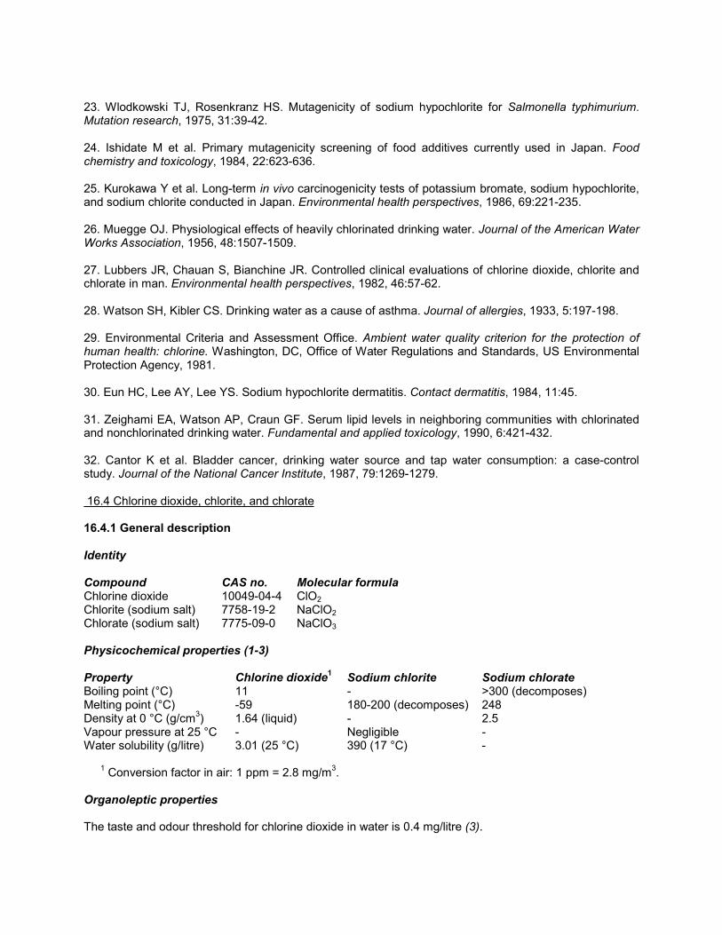
Compound CAS no. Molecular formulaChlorine dioxide 10049-04-4 ClO2Chlorite (sodium salt) 7758-19-2 NaClO2Chlorate (sodium salt) 7775-09-0 NaClO3
Physicochemical properties (1-3)
Property Chlorine dioxide1 Sodium chlorite Sodium chlorateBoiling point (°C) 11 - >300 (decomposes)Melting point (°C) -59 180-200 (decomposes) 248Density at 0 °C (g/cm3) 1.64 (liquid) - 2.5Vapour pressure at 25 °C - Negligible -Water solubility (g/litre) 3.01 (25 °C) 390 (17 °C) -
1 Conversion factor in air: 1 ppm = 2.8 mg/m3.
Organoleptic properties
The taste and odour threshold for chlorine dioxide in water is 0.4 mg/litre (3).

Major uses
Chlorine dioxide is used for disinfection and odour/taste control of water; as a bleaching agent forcellulose, paper pulp, flour, and oils; and for cleaning and detanning leather. Sodium chlorite is used in on-site production of chlorine dioxide; as a bleaching agent in production of paper, textiles, and strawproducts; and in the manufacture of waxes, shellacs, and varnishes. Sodium chlorate is used in thepreparation of chlorine dioxide; in the manufacture of dyes, matches, and explosives; for tanning andfinishing leather; and in herbicides and defoliants (1-3).
Environmental fate
Chlorine dioxide rapidly decomposes into chlorite, chlorate, and chloride ions in treated water, chloritebeing the predominant species. This reaction is favoured by alkaline conditions.
16.4.2 Analytical methods
Methods are available for the determination of chlorine dioxide, chlorite, and total available chlorine (4,5).The limits of detection for these methods are 8 µg/litre for chlorine dioxide, 4 µg/litre for total chlorine, and10 µg/litre for chlorite and chlorate.
16.4.3 Environmental levels and human exposure
Water
Chlorite occurs in drinking-water when chlorine dioxide is used for purification purposes. The levels ofchlorite in water reported in one study ranged from 3.2 to 7.0 mg/litre (6).
Food
Chlorine dioxide, chlorite, and chlorate may occur in foodstuffs as a result of their use in flour processing,as a decolorizing agent for carotenoids and other natural pigments (chlorine dioxide), as a bleaching agentin the preparation of modified food starch (sodium chlorite), as an indirect additive in paper andpaperboard products used for food packaging (sodium chlorite), and as a defoliant, desiccant, andfungicide in agriculture (sodium chlorate) (7-9).
Estimated total exposure and relative contribution of drinking-water
The major route of environmental exposure to chlorine dioxide, sodium chlorite, and sodium chlorate isthrough drinking-water.
16.4.4 Kinetics and metabolism in laboratory animals and humans
Chlorine dioxide is rapidly absorbed from the gastrointestinal tract. No particular organ appears toselectively concentrate the dose following exposure (10). Following oral ingestion by monkeys, chlorinedioxide was rapidly converted into chloride ion and, to a lesser extent, chlorite and chlorate (11). Excretionis mainly via the urine, smaller amounts being excreted in faeces (12).
Chlorite was readily absorbed when administered to rats, then randomly distributed throughout the tissues(12). It was transformed mainly into chloride in rats, smaller amounts appearing as unchanged chlorite.Excretion was mainly via the urine, followed by faeces (13).
Chlorate was readily absorbed and randomly distributed throughout the tissues of rats (12). It wasexcreted mainly in the form of chloride in the urine, smaller amounts appearing as chlorite and chlorate
(13).
16.4.5 Effects on laboratory animals and in vitro test systems
Chlorine dioxide
Short-term exposure
Drinking-water containing 0, 10, or 100 mg of chlorine dioxide per litre (equivalent to approximately 0, 1.5,or 15 mg/kg of body weight per day) was administered to mice (10 per dose) for 30 days with no apparenteffects on blood parameters. The NOAEL for this study was 15 mg/kg of body weight per day (14).
A total of 12 African green monkeys were exposed to water containing chlorine dioxide at concentrationsof 0, 30, 100, or 200 mg/litre (corresponding to measured doses of 0, 3.5, 9.5, or 11 mg/kg of body weightper day) using a rising dose protocol. Each dose was maintained for 30-60 days. A slight suppression ofthyroid function (decreased thyroxine) was observed in monkeys receiving the two highest doses. Noother effects were noted. The NOAEL was 3.5 mg/kg of body weight per day (11).
Six monkeys were treated for 8 weeks with drinking-water containing chlorine dioxide at 100 mg/litre,corresponding to an average measured dose of about 4.6 mg/kg of body weight per day. Thyroxine levelwas reduced after 4 weeks of treatment but rebounded after a further 4 weeks. In the same study,drinking-water containing chlorine dioxide at 0, 100, or 200 mg/litre was administered to male rats (12 perdose) (equivalent to 0, 10, or 20 mg/kg of body weight per day). A dose-dependent decrease in thyroxinelevels was observed after 8 weeks of treatment; there was no rebound. The exposure level of 100 mg/litre,equivalent to a dose of approximately 10 mg/kg of body weight per day, was the LOAEL in this study (15).
Sprague-Dawley rats (10 per sex per dose) were exposed to 0, 25, 50, 100, or 200 mg of chlorine dioxideper litre in drinking-water for 90 days (approximate dose levels of 0, 2, 4, 6, or 12 mg/kg of body weightper day for males and 0, 2, 5, 8, or 15 mg/kg of body weight per day for females). Water consumption wasdecreased in both sexes at the three highest dose levels, probably due because of its reduced palatability.Food consumption was decreased in males receiving the highest dose. Goblet-cell hyperplasia wassignificantly increased in the nasal turbinates of females given 100 or 200 mg/litre and males at all doses.Inflammation of the nasal cavity was observed in males at 25 mg/litre and in both sexes at higher doses.The authors concluded that the lowest dose (2 mg/kg of body weight per day) was a LOAEL (16).
Long-term exposure
In a drinking-water study, chlorine dioxide was administered to rats (7 per sex per dose) at concentrationsof 0, 0.5, 1, 5, 10, or 100 mg/litre (highest dose equivalent to about 13 mg/kg of body weight per day) for 2years. At the highest dose level, survival rate was substantially decreased in both sexes, and mean lifespan was reduced compared with that for control animals. No correlation was observed betweentreatment and histopathological findings. In this study, a NOAEL of 10 mg/litre (1.3 mg/kg of body weightper day) was identified (17).
Reproductive toxicity, embryotoxicity, and teratogenicity
Female rats were exposed to 0, 1, 10, or 100 mg of chlorine dioxide per litre in drinking-water (equivalentto 0, 0.1, 1, or 10 mg/kg of body weight per day) for 2.5 months before mating and throughout gestation.At the highest dose, there was a slight reduction in the number of implants and live births per pregnancy.No effects were observed at 1 mg/kg of body weight per day, which was identified as the NOAEL (18).
Female Sprague-Dawley rats (13-16 per dose) were supplied with drinking-water containing 0, 2, 20, or100 mg of chlorine dioxide per litre from 2 weeks before mating to gestation and lactation until pups wereweaned on postnatal day 21. No significant effect on the body weight of either the dams or the pups was
observed at any dose tested. At 100 mg/litre (14 mg/kg of body weight per day for the pregnant dam), asignificant depression of serum thyroxine and an increase in serum triiodothyronine were observed in thepups at weaning, but not in the dams. Neurobehavioural exploratory and locomotor activities weredecreased in pups born to dams exposed to 100 mg/litre but not to those exposed to 20 mg/litre (3 mg/kgof body weight per day), which was considered a NOAEL (19).
In a second experiment, rat pups were exposed directly (by gavage) to 14 mg of chlorine dioxide per kg ofbody weight per day (equivalent to the dose received by a pregnant dam drinking water containing 100 mgof chlorine dioxide per litre) on postnatal days 5-20. In this study, serum thyroxine levels were depressed,a somewhat greater and more consistent delay in the development of exploratory and locomotor activitywas seen, and pup body weight gain was reduced. The decrease in serum triiodothyronine levels was notstatistically significant. Based on decreased pup development and decreased thyroid hormone levels, aLOAEL of 14 mg/kg of body weight per day (the only dose tested) was identified (19).
Cell number was significantly depressed in the cerebellum of 21-day-old rat pups born to dams suppliedduring gestation and lactation with water containing 100 mg of chlorine dioxide per litre (about 14 mg/kg ofbody weight per day to the dam). A group of 12 rat pups dosed directly by gavage with 14 mg/kg of bodyweight per day had depressed cell numbers in both the cerebellum and forebrain at postnatal day 11 anddisplayed decreased voluntary running-wheel activity at postnatal days 50-60, despite the fact thatchlorine dioxide treatments were terminated at 20 days of age. These data suggest that chlorine dioxide iscapable of influencing brain development in neonatal rats. In this study, a LOAEL of 14 mg/kg of bodyweight per day, the only dose tested, was identified (20).
The developmental neurotoxic potential of chlorine dioxide was evaluated in a study in which it wasadministered to rat pups by oral intubation at 14 mg/kg of body weight per day on postnatal days 1-20.Forebrain cell proliferation was decreased on postnatal day 35, and there were decreases in forebrainweight and protein content on postnatal days 21 and 35. Cell proliferation in the cerebellum and olfactorybulbs was comparable to that in untreated controls, as were migration and aggregation of neuronal cells inthe cerebral cortex. Histopathological examination of the forebrain, cerebellum, and brain stem did notreveal any lesions or changes in these tissues. In this study, a LOAEL of 14 mg/kg of body weight per day(the only dose tested) was identified (18).
Mutagenicity and related end-points
Chlorine dioxide was mutagenic in Salmonella typhimurium strain TA100 in the absence of a metabolicactivation system (21). No sperm-head abnormalities were observed in male mice following chlorinedioxide gavage (22). No chromosomal abnormalities were seen in either the micronucleus test or acytogenetic assay in mouse bone marrow cells following gavage dosing with chlorine dioxide (22).
Carcinogenicity
Tumours were not observed in rats following 2-year exposures to chlorine dioxide in drinking-water (17).
Chlorite
Acute exposure
An oral LD50 of 105 mg/kg of body weight has been reported in rats (23). Quail were more resistant thanrats; the LD50 was 493 mg/kg of body weight (24).
Short-term exposure
Single doses of sodium chlorite administered orally to cats produced methaemoglobinaemia (25). A doseof 20 mg of chlorite per litre (equivalent to approximately 1.5 mg of chlorite per kg of body weight) caused
up to 32% of the haemoglobin to be in the methaemoglobin state and was considered to be the LOAEL. Adose-dependent increase in methaemoglobinaemia and anaemia was observed in 12 African greenmonkeys treated with sodium chlorite at 0, 25, 50, 100, or 400 mg/litre in drinking-water using a risingdose protocol. Doses of chlorite were approximately 0, 3, 6, 13, and 50 mg/kg of body weight per day, andeach dose level was maintained for 30-60 days (11).
Rats were exposed to chlorite ion at 0, 10, 50, 100, 250, or 500 mg/litre in drinking-water (equivalent to 0,1, 5, 10, 25, or 50 mg/kg of body weight per day) for 30-90 days. Haematological parameters weremonitored, and the three highest concentrations produced transient anaemia. At 90 days, red blood cellglutathione levels in the 100 mg/litre group were 40% below those of controls; there was at least a 20%reduction in the rats receiving 50 mg/litre. In this study, a NOAEL of 1 mg/kg of body weight per day wasidentified (25).
Long-term exposure
The effect of sodium chlorite in drinking-water at 0, 1, 2, 4, 8, 100, or 1000 mg/litre on the survival andpostmortem pathology of albino rats (7 per sex per dose) was examined in a 2-year study. The life span ofthe animals was not significantly affected at any dose. No effects were observed in animals exposed to 8mg/litre (0.7 mg/kg of body weight per day) or less. Animals exposed to 100 or 1000 mg/litre (9.3 or 81mg/kg of body weight per day) exhibited treatment-related renal pathology; the author concluded that thiswas the result of a nonspecific salt effect (17).
Reproductive toxicity, embryotoxicity, and teratogenicity
Female mice (10 per dose) were treated with sodium chlorite at 0 or 100 mg/litre in drinking-water(equivalent to 0 and 72 mg/kg of body weight per day) from day 1 of gestation and throughout lactation.Conception rates were 56% for controls and 39% for treated mice. The body weights of pups at weaningwere reduced in treated mice relative to controls, so that 72 mg/kg of body weight per day is the LOAELfor this study (14).
In a series of experiments, sodium chlorite was administered to male rats (12 rats per dose) in drinking-water for 66-76 days at concentrations of 0, 1, 10, 100, or 500 mg/litre (equivalent to 0, 0.1, 1, 10, or 50mg/kg of body weight per day). No compound-related abnormalities were observed on histopathologicalexamination of the reproductive tract. Abnormal sperm morphology and decreased sperm motility wereseen at the two highest dose levels, but no sperm effects were observed at 1 mg/kg of body weight perday, which can be identified as the NOAEL. In another part of the same study, male rats were bred withfemale rats treated at the same dose levels for 2 weeks before and throughout a 10-day breeding period.Females were exposed to sodium chlorite throughout gestation and lactation until the pups were weanedon day 21. There was no evidence of any adverse effects on conception rates, litter size, day of eyeopening, or day of vaginal opening. Based on reproductive effects, a NOAEL of 10 mg/kg of body weightper day, the highest dose tested, was identified (26).
Treatment of maternal mice with 100 mg of sodium chlorite per litre in drinking-water (equivalent to 14 mgof chlorite per kg of body weight per day) throughout gestation and lactation resulted in pups withdecreased body weights (14% below those of controls) at weaning. In this study, a LOAEL fordevelopmental effects of 14 mg/kg of body weight per day was identified (14).
Fetuses from maternal rats exposed to chlorite ion via drinking-water at levels of up to 10 mg/litre (about 1mg/kg of body weight per day) were examined. No compound-related skeletal or soft-tissue anomalieswere observed. A NOAEL of 1 mg/kg of body weight per day was identified (27).
Mutagenicity and related end-points
No chromosomal abnormalities were seen in either the micronucleus test or a cytogenetic assay in mouse

bone marrow cells following gavage dosing with chlorite (22).
Carcinogenicity
In a long-term study in which mice received sodium chlorite in drinking-water for 85 weeks, there was nosignificant increase in tumours as compared with controls at a dose of 250 mg/litre (about 36 mg ofchlorite ion per kg of body weight per day). Although treated male mice exhibited an increased incidenceof lung and liver tumours, tumour rates were within historical ranges for control mice, increases in the livertumours did not display a typical dose-response pattern, and significant increases were seen only forbenign tumours (28). Tumours were not observed in rats following 2-year exposures to sodium chlorite indrinking-water (17).
Chlorate
Acute exposure
An acute oral dosing study in dogs demonstrated lethality at levels of sodium chlorate as low as 600 mg ofchlorate ion per kg (29).
Short-term exposure
Beagle dogs (4 per sex per dose) were exposed by gavage to sodium chlorate at doses of 0, 10, 60, or360 mg/kg of body weight per day for 3 months. There was no significant effect at any dose level on bodyweight, food consumption, clinical chemistry, organ weights, ophthalmic effects, gross necropsy, or tissuehistopathology. Haematological changes were limited to a slight elevation in methaemoglobin level inhighest-dose animals, but this appeared to be within normal limits and was not judged to be treatment-related. In this study, a NOAEL of 360 mg/kg of body weight per day in dogs was identified (30).
Sprague-Dawley rats (14 per sex per dose) were exposed by gavage to sodium chlorate at doses of 0, 10,100, or 1000 mg/kg of body weight per day for up to 3 months. No treatment-related effects wereobserved on mortality, physical appearance or behaviour, body weight, food consumption, clinicalchemistry, gross necropsy, or organ histopathology. At the highest dose, haematological changesindicative of anaemia included decreases in erythrocyte count, haemoglobin concentration, anderythrocyte volume fraction (haematocrit). In this study, a NOAEL of 100 mg/kg of body weight per daywas identified (31).
Reproductive toxicity, embryotoxicity, and teratogenicity
Sodium chlorate was administered to pregnant CD rats by gavage at doses of 0, 10, 100, or 1000 mg/kgof body weight per day on days 6-15 of gestation. There were no maternal deaths in treated animals ortreatment-related effects on maternal body weight gain, food consumption, clinical observations, numberof implantations, or gross necropsy. Examination of fetuses on day 20 revealed no effects on fetal weightor sex ratio, and no external, visceral, or skeletal abnormalities were detected. In this study, adevelopmental NOAEL of 1000 mg/kg of body weight per day in rats was identified (32).
Mutagenicity and related end-points
No chromosomal abnormalities were seen in either the micronucleus test or a cytogenetic assay in mousebone marrow cells following gavage dosing with chlorate (22).
16.4.6 Effects on humans
Chlorine dioxide
Six different doses of chlorine dioxide (0.1, 1, 5, 10, 18, or 24 mg/litre) in drinking-water were administeredto each of 10 male volunteers using a rising dose protocol. Serum chemistry, blood count, and urinalysisparameters were monitored. A treatment-related change in group mean values for serum uric acid wasobserved, which the authors concluded was not physiologically detrimental. The highest dose tested, 24mg/litre (about 0.34 mg/kg of body weight per day), can be identified as a single-dose NOAEL (33).
The same male volunteers drank 0.5 litres of water containing 5 mg of chlorine dioxide per litre each dayfor approximately 12 weeks, and were then kept under observation for 8 weeks. Serum chemistry, bloodcounts, and urinalysis revealed no abnormalities, except for a slight change in blood urea nitrogen, whichthe authors concluded was of doubtful physiological or toxicological significance. This exposure,equivalent to 36 µg/kg of body weight per day, can be considered a NOAEL (33).
In a prospective study of 197 persons, a portion of the population of a rural village exposed for 12 weeksto a chlorine dioxide-treated water supply (containing 0.25-1.1 mg of chlorine dioxide per litre and 0.45-0.91 mg of free chlorine per litre) experienced no significant changes in haematological parameters,serum creatinine, or total bilirubin (6).
Chlorite
The effects of sodium chlorite on humans were evaluated in 10 male volunteers on a rising dose protocol.Single doses of 0.01, 0.1, 0.5, 1.0, 1.8, and 2.4 mg of chlorite ion per litre in 1 litre of drinking-water wereingested by each subject. Changes in group mean values for serum urea nitrogen, creatinine, and ureanitrogen/creatinine ratio were observed, which the authors concluded were not adverse physiologicaleffects. The highest dose tested, 2.4 mg/litre (0.034 mg/kg of body weight per day), can be identified as asingle-dose NOAEL (33).
The same volunteers ingested 0.5 litres of water per day containing 5 mg of sodium chlorite per litre forapproximately 12 weeks, and were then kept under observation for 8 weeks. Treatment was associatedwith a change in group mean corpuscular haemoglobin; however, as there was no trend over time for thischange and values were within the normal ranges, the authors were reluctant to attach physiologicalsignificance to the observation. The dose tested, equivalent to 36 µg/kg of body weight per day, wasidentified as the NOAEL (33).
Chlorate
Because of its use as a weed killer, a large number of cases of chlorate poisoning have been reported (3).Symptoms include methaemoglobinaemia, anuria, abdominal pain, and renal failure. For an adult human,the oral lethal dose is estimated to be as low as 20 g of sodium chlorate (230 mg of chlorate per kg ofbody weight) (34).
Ten male volunteers were given six separate doses of sodium chlorate following a rising dose protocol,single doses of 0.01, 0.1, 0.5, 1.0, 1.8, and 2.4 mg of chlorate ion per litre in 1 litre of drinking-water beingingested by each volunteer. Very slight changes in group mean serum bilirubin, iron, and methaemoglobinwere observed, but the authors concluded that they were not adverse physiological effects. The highestdose tested, 2.4 mg/litre (34 µg/kg of body weight per day), can be identified as a single-dose NOAEL(33).
The volunteers also ingested 0.5 litres of water per day containing 5 mg of sodium chlorate per litre (36µg/kg of body weight per day) for approximately 12 weeks, and were then kept under observation for 8weeks. Treatment was associated with slight changes in group mean serum urea nitrogen and mean

corpuscular haemoglobin, but the authors concluded that these were not physiologically significant asvalues remained within the normal range for each parameter. The NOAEL was 36 µg/kg of body weightper day (33).
16.4.7 Guideline values
Chlorine dioxide
Chlorine dioxide has been shown to impair neurobehavioural and neurological development in ratsexposed perinatally. Significant depression of thyroid hormones has also been observed in rats andmonkeys exposed to it in drinking-water studies.
A guideline value has not been established for chlorine dioxide because of its rapid breakdown andbecause the chlorite provisional guideline value (see below) is adequately protective for potential toxicityfrom chlorine dioxide. The taste and odour threshold for this compound is 0.4 mg/litre.
Chlorite
Chlorite affects the red blood cells, resulting in methaemoglobin formation in cats and monkeys. IARC hasconcluded that chlorite is not classifiable as to its carcinogenicity to humans (Group 3) (35).
The TDI for chlorite is 10 µg/kg of body weight, based on the NOAEL of 1 mg/kg of body weight per dayfor decreased red blood cell glutathione levels in a 90-day study in rats exposed to chlorite in theirdrinking-water (25) and applying an uncertainty factor of 100 (to account for inter- and intraspeciesvariation). Owing to the acute nature of the response and the existence of a 2-year rat study, an additionaluncertainty factor of 10 was not incorporated to account for the short duration of the key study. The TDIderived in this manner is consistent with the NOAEL (36 µg/kg of body weight per day) in a 12-weekclinical study in a small number of human volunteers (33).
On the assumption that drinking-water contributes 80% of the total exposure, the provisional guidelinevalue is 0.2 mg/litre (rounded figure). This guideline value is designated as provisional because use ofchlorine dioxide as a disinfectant may result in the chlorite guideline value being exceeded, and difficultiesin meeting the guideline value must never be a reason for compromising adequate disinfection.
Chlorate
Available data on the effects of chlorate in humans and experimental animals are considered insufficientto permit the development of a guideline value. Data on accidental poisonings indicate that the lethal doseto humans is about 230 mg/kg of body weight per day. This is of the same order of magnitude as theNOAELs identified from studies in rats and dogs. Although no effects were observed in a 12-week clinicalstudy in a small number of human volunteers ingesting 36 µg/kg of body weight per day, a guideline valuewas not derived from these results because no adverse effect level was determined.
Further research is needed to characterize the nonlethal effects of chlorate. Until data become available, itmay be prudent to try to minimize chlorate levels. However, adequate disinfection should not becompromised.
References
1. Budavari S, O’Neill M, Smith A, eds. The Merck index. An encyclopedia of chemicals, drugs andbiologicals, 11th ed. Rahway, NJ, Merck, 1989.
2. Meister R, ed. Farm chemicals handbook. Willoughby, OH, Meister Publishing Co., 1989.

3. National Academy of Sciences. Drinking water and health. Vol. 7. Washington, DC, National AcademyPress, 1987.
4. American Public Health Association. Method 4500, chlorine. In: Standard methods for the examinationof water and waste water, 17th ed. Washington, DC, 1989:4.45-4.67.
5. American Public Health Association. Method 4500, chlorine dioxide. In: Standard methods for theexamination of water and waste water, 17th ed. Washington, DC, 1989:4.75-4.83.
6. Michael GE et al. Chlorine dioxide water disinfection: a prospective epidemiology study. Archives ofenvironmental health, 1981, 36:20-27.
7. Chemical Manufacturers Association. A review of the uses, chemistry and health effects of chlorinedioxide and the chlorite ion. Washington, DC, 1989.
8. US Food and Drug Administration. Food and drugs, Vol. 21, Parts 170-179. Washington, DC, Office ofthe Federal Register, 1990.
9. US Environmental Protection Agency. Sodium chlorate: exemption from the requirement of a tolerance.Federal register, 1983, 48:19028.
10. Abdel-Rahman MS. Pharmacokinetics of chlorine obtained from chlorine dioxide, chlorine, chloramineand chloride. In: Jolley RL et al., eds. Water chlorination: environmental impact and health effects. Vol. 5.Chelsea, MI, Lewis Publishers, 1985:281-293.
11. Bercz JP et al. Subchronic toxicity of chlorine dioxide and related compounds in drinking water in thenonhuman primate. Environmental health perspectives, 1982, 46:47-55.
12. Abdel-Rahman MS, Couri D, Bull RJ. Metabolism and pharmacokinetics of alternate drinking waterdisinfectants. Environmental health perspectives, 1982, 46:19-23.
13. Abdel-Rahman MS, Couri D, Bull RJ. The kinetics of chlorite and chlorate in rats. Journal ofenvironmental pathology, toxicology and oncology, 1985, 6(1):97-103.
14. Moore GS, Calabrese EJ. Toxicological effects of chlorite in the mouse. Environmental healthperspectives, 1982, 46:31-37.
15. Harrington RM, Shertzer HG, Bercz JP. Effects of chlorine dioxide on thyroid function in the Africangreen monkey and the rat. Journal of toxicology and environmental health, 1986, 19:235-242.
16. Daniel FB et al. Comparative subchronic toxicity studies of three disinfectants. Journal of the AmericanWater Works Association, 1990, 82:61-69.
17. Haag HB. The effect on rats of chronic administration of sodium chlorite and chlorine dioxide in thedrinking water. Report to the Mathieson Alkali Works from the Medical College of Virginia, 1949.
18. Toth GP et al. Effects of chlorine dioxide on the developing rat brain. Journal of toxicology andenvironmental health, 1990, 31:29-44.
19. Orme J et al. Effects of chlorine dioxide on thyroid function in neonatal rats. Journal of toxicology andenvironmental health, 1985, 15:315-322.
20. Taylor DH, Pfohl RJ. Effects of chlorine dioxide on the neurobehavioral development of rats. In: JolleyRL et al., eds. Water chlorination: environmental impact and health effects. Vol. 5. Chelsea, MI, Lewis

Publishers, 1985:355-364.
21. Ishidate M et al. Primary mutagenicity screening of food additives currently used in Japan. Foodchemistry and toxicology, 1984, 22:623-636.
22. Meier JR et al. Evaluation of chemicals used for drinking water disinfection for production ofchromosomal damage and sperm-head abnormalities in mice. Environmental mutagenesis, 1985, 7:201-211.
23. Musil J et al. Toxicologic aspects of chlorine dioxide application for the treatment of water containingphenols. Technology of water, 1964, 8:327-346.
24. Fletcher D. Acute oral toxicity study with sodium chlorite in bobwhite quail. Industrial Bio-TestLaboratory’s report to Olin Corporation, 1973 (IBT No. J2119).
25. Heffernan WP, Guion C, Bull RJ. Oxidative damage to the erythrocyte induced by sodium chlorite, invivo. Journal of environmental pathology and toxicology, 1979, 2:1487-1499.
26. Carlton BD et al. Sodium chlorite administration in Long-Evans rats: reproductive and endocrineeffects. Environmental research, 1987, 42:238-245.
27. Suh DH, Abdel-Rahman MS, Bull RJ. Effect of chlorine dioxide and its metabolites in drinking water onfetal development in rats. Journal of applied toxicology, 1983, 3:75-79.
28. Kurokawa Y et al. Long-term in vivo carcinogenicity tests of potassium bromate, sodium hypochloriteand sodium chlorite conducted in Japan. Environmental health perspectives, 1986, 69:221-235.
29. Sheahan BJ, Pugh DM, Winstanley EW. Experimental sodium chlorate poisoning in dogs. Research inveterinary science, 1971, 12:387-389.
30. A subchronic (3-month) oral toxicity study in the dog via gavage administration with sodium chlorate.East Millstone, NJ, Bio/dynamics, Inc., 1987 (unpublished report no. 86-3114, for Sodium Chlorate TaskForce, Oklahoma City).
31. A subchronic (3-month) oral toxicity study of sodium chlorate in the rat via gavage. East Millston, NJ,Bio/dynamics, Inc., 1987 (unpublished report no. 86-3112, for Sodium Chlorate Task Force, OklahomaCity).
32. A teratogenicity study in rats with sodium chlorate. East Millston, NJ, Bio/dynamics, Inc., 1987(unpublished report no. 86-3117, for Sodium Chlorate Task Force, Oklahoma City).
33. Lubbers JR, Chauhan S, Bianchine JR. Controlled clinical evaluations of chlorine dioxide, chlorite andchlorate in man. Fundamental and applied toxicology, 1981, 1:334-338.
34. National Academy of Sciences. Drinking water and health. Vol. 4. Washington, DC, National AcademyPress, 1980.
35. International Agency for Research on Cancer. Chlorinated drinking-water; chlorination by-products;some other halogenated compounds; cobalt and cobalt compounds. Lyon, 1991:45-359 (IARCMonographs on the Evaluation of Carcinogenic Risks to Humans, Volume 52).

16.5 Iodine
16.5.1 General description
Identity
CAS no.: 7553-56-2Molecular formula: I2
Physicochemical properties (1,2)1,2
1 Also includes data from the Hazardous Substances Data Bank of the National Library of Medicine,Bethesda, MD
2 Conversion factor in air: 1 ppm = 10 mg/m3
Property ValueBoiling point 184.4 °CMelting point 113.5 °CDensity 4.93 g/cm3 at 25 °CVapour pressure 40 Pa at 25 °CWater solubility 0.34 g/litre at 25 °CLog octanol-water partition coefficient 2.49
Organoleptic properties
The taste and odour thresholds for iodine are 0.147-0.204 mg/litre in water and 9 mg/m3 in air (3).
Major uses
Iodine is used as an antiseptic for skin wounds, as a disinfecting agent in hospitals and laboratories, andfor the emergency disinfection of drinking-water in the field. Iodide is used in pharmaceuticals and inphotographic developing materials.
Environmental fate
Iodine occurs naturally in water in the form of iodide (I-), which is largely oxidized to iodine during watertreatment.
16.5.2 Analytical methods
Iodide in water is normally determined by a titrimetric procedure which can be used for solutionscontaining 2-20 mg of iodide per litre. A leuco crystal violet method may be used for the determination ofiodide or molecular iodine in water. This photometric method is applicable to iodide concentrations of 50-6000 µg/litre; the detection limit for iodine is 10 µg/litre (4,5).
16.5.3 Environmental levels and human exposure
Water
The mean concentration of total iodine in drinking-water in the USA is 4 µg/litre, and the maximumconcentration is 18 µg/litre (2). This is presumably predominantly iodide.

Food
The main natural sources of dietary iodide are seafood (200-1000 µg/kg) and seaweed (0.1-0.2% iodideby weight). Iodide is also found in cow’s milk (20-70 µg/litre) and may be added to table salt (100 µg ofpotassium iodide per gram of sodium chloride) to ensure an adequate intake of iodine (2,6). The estimateddietary iodine requirement for adults ranges from 80 to 150 µg/day (7).
Estimated total exposure
Exposure to iodine may occur through drinking-water, pharmaceuticals, and food. At a concentration of 4µg/litre in drinking-water, adult human daily intake will be 8 µg of iodine, on the assumption that 2 litres ofdrinking-water are consumed per day.
16.5.4 Kinetics and metabolism in laboratory animals and humans
Molecular iodine is rapidly converted into iodide following ingestion and this is efficiently absorbedthroughout the gastrointestinal tract (8). Molecular iodine vapour is converted into iodide before absorption(2). The highest concentration of iodine in the human body is found in the thyroid, which contains 70-80%of the total iodine content (15-20 mg). Muscle and eyes also contain high iodide concentrations (6,8).
Iodine is an essential element in the synthesis of the thyroid hormones thyroxine (T4) and triiodothyronine(T3) through the precursor protein thyroglobulin and the action of the enzyme thyroid peroxidase. Iodide isexcreted primarily by the kidneys and is partially reabsorbed from the tubules following glomerular filtration(8). Smaller amounts of iodine are excreted in saliva, sweat, bile, and milk (9).
16.5.5 Effects on laboratory animals and in vitro test systems
Acute exposure
The acute oral LD50 for potassium iodide in rats was 4340 mg/kg of body weight (3320 mg of iodide per kgof body weight), and the lowest oral lethal dose in mice was 1862 mg/kg of body weight (1425 mg ofiodide per kg of body weight) (9).
Short-term exposure
The effects of iodide on autoimmune thyroiditis were investigated in two strains of chickens (CS and OS)known to be genetically susceptible to this disease. Administration of iodide in drinking-water (20 or 200mg/litre, as potassium iodide) during the first 10 weeks of life increased the incidence of the disease, asdetermined by histological examination of the thyroid and measurement of T3, T4, and thyroglobulinantibodies. Excessive iodide consumption may increase the incidence of this disease in humans (10).
Reproductive toxicity, embryotoxicity, and teratogenicity
No effects were observed on ovulation rate, implantation rate, or fetal development in female rats givendoses of 0, 500, 1000, 1500, or 2000 mg of iodide (as potassium iodide) per kg of diet during gestationand lactation. The dose-related survival rate for pups ranged from 93% (controls) to 16% (2000 mg/kg).Milk secretion was absent or greatly diminished in females exposed to iodide and the high mortality inpups was attributed to the dams’ lactational failure (11).
The effects of iodide on brain enzymes in rat pups born to females given 1.1 mg of iodide per day aspotassium iodide (about 37 mg/kg of body weight per day) in drinking-water were studied. Transientincreases in glutamate dehydrogenase and decreases in succinate dehydrogenase were observed.Increases in phosphofructokinase and malate enzymes were noted, but no changes in hexokinase werereported. Serum T4 levels did not differ significantly from control values (12).

Metabolism was severely disturbed in foals born to mares receiving excess iodine (48-432 mg of iodineper day) in the diet during pregnancy and lactation. The long bones of the legs of foals showedosteopetrosis (abnormally dense bones); phosphorus and alkaline phosphatase levels in the blood wereelevated (13).
Carcinogenicity
In a study on the tumorigenic effects of iodide on the thyroid, groups of 20 rats were fed diets containing 0or 1000 mg of iodide per kg as potassium iodide (0 or 39 mg of iodide per kg of body weight per day) for19 weeks. No tumours were found on histopathological examination of the thyroid in either the treated oruntreated groups (14). The exposure period may have been too short for a carcinogenic effect to bedetected.
16.5.6 Effects on humans
Short-term exposure
Oral doses of 2000-3000 mg of iodine (about 30-40 mg/kg of body weight) are estimated to be lethal tohumans, but survival has been reported after ingestion of 10 000 mg. Doses of 30-250 ml of tincture ofiodine (about 16-130 mg of total iodine per kg of body weight) have been reported to be fatal. Acute oraltoxicity is primarily due to irritation of the gastrointestinal tract, marked fluid loss and shock occurring insevere cases. Exposure to iodine vapour results in lung, eye, and skin irritation, while high concentrationsrapidly lead to pulmonary oedema (2).
In rare instances, a hypersensitization reaction may occur immediately after or within several hours of oralor dermal exposure to iodide. The most striking symptoms are angio-oedema (acute, transitory swelling ofthe face, hands, feet, or viscera) and swelling of the larynx, which may cause suffocation (8). Iodide hasbeen used in the past as an expectorant in the treatment of asthma and related conditions at a typicaldose of 3.3 mg/kg of body weight (2).
Long-term exposure
Chronic iodide exposure results in iodism; the symptoms resemble those of a sinus cold but may alsoinclude salivary gland swelling, gastrointestinal irritation, acneform skin, metallic or brassy taste, gingivitis,increased salivation, conjunctival irritation, and oedema of eyelids (8). Chronic ingestion of 2 mg of iodideper day (0.03 mg/kg of body weight per day) is considered by some authors to be excessive, but dailydoses of 50-80 mg (0.8-1.3 mg/kg of body weight per day) are consumed by some Japanese without illeffect (6).
Chronic consumption of iodinated drinking-water has not been shown to cause adverse health effects inhumans, although some changes in thyroid status have been observed. In a 5-year study of prisoninmates consuming water containing iodine at a concentration of 1 mg/litre (approximately 0.03 mg/kg ofbody weight per day), no cases of hyper- or hypothyroidism, urticaria, or iodism were seen. However, asmall but statistically significant decrease in radioactive iodine uptake by the thyroid and an increase inprotein-bound iodine concentrations were reported (15). No adverse health effects were reported in menwho drank water providing iodide at doses of 0.17-0.27 mg/kg of body weight per day for 26 weeks (16).
In one study, the rate of radioactive iodide uptake by the thyroid was measured in 22 individuals withthyroid disease and 10 with normal thyroid function, before and after administration of 2.0 mg of iodide.Radioactive iodine uptake decreased by 54-99% in patients with thyroid disease but only by 8-54% innormal controls. These results suggest that iodide may aggravate certain pre-existing thyroid diseaseconditions (17).
Eight cases of congenital goitre and hypothyroidism in children were reported to be associated withmaternal ingestion of iodide (18). Estimates of maternal iodide exposure ranged from 12 to 1650 mg/day(about 0.02-27 mg/kg of body weight per day) in individuals taking iodide as an expectorant in thetreatment of asthma. No direct evidence of a cause-and-effect relationship between iodide exposure andhealth effects during pregnancy was reported.
Hypothyroidism has also been reported in infants of mothers receiving multiple topical applications ofpovidone-iodine (about 1% free iodine) during pregnancy and lactation (19).
16.5.7 Conclusions
In 1988, JECFA set a PMTDI for iodine of 1 mg/day (17 µg/kg of body weight per day) from all sources,based mainly on data on the effects of iodide (20). However, recent data from studies in rats indicate thatthe effects of iodine in drinking-water on thyroid hormone concentrations in the blood differ from those ofiodide (21,22).
Available data therefore suggest that derivation of a guideline value for iodine on the basis of informationon effects of iodide is inappropriate, and there are few relevant data on the effects of iodine. Becauseiodine is not recommended for long-term disinfection, lifetime exposure to iodine from water disinfection isunlikely. For these reasons, a guideline value for iodine has not been established at this time.
References
1. Budavari S, O’Neill M, Smith A, eds. The Merck index. An encyclopedia of chemicals, drugs, andbiologicals, 11th ed. Rahway, NJ, Merck, 1989.
2. National Academy of Sciences. Drinking water and health. Vol. 3. Washington, DC, National AcademyPress, 1980.
3. Ruth JH. Odor thresholds and irritation levels of several chemical substances: a review. Journal of theAmerican Industrial Hygiene Association, 1986, 47:A142-A151.
4. US Environmental Protection Agency. Methods for chemical analysis of water and wastes. Cincinnati,OH, 1983 (EPA-600/4-79-020).
5. American Public Health Association. Standard methods for the examination of water and wastewater,17th ed. Washington, DC, 1989.
6. Hetzel BS, Maberly GF. Iodine. In: Mertz W, ed., Trace elements in human and animal nutrition, 5th ed.New York, NY, Academic Press, 1986:139-208.
7. National Research Council. Recommended dietary allowances. Washington, DC, National AcademyPress, 1989:214.
8. Welt LG, Blythe WB. Anions: phosphate, iodide, fluoride and other anions. In: Goodman LD, Gilman A,eds. The pharmacological basis of therapeutics, 4th ed. New York, Macmillan, 1970.
9. Stokinger HE. The halogens and the nonmetals, boron and silicon. In: Clayton GD, Clayton FE, eds.Patty’s industrial hygiene and toxicology, Vol. 2B, 3rd rev. ed. New York, NY, John Wiley, 1981:2937-3043.
10. Bagchi N et al. Induction of autoimmune thyroiditis in chickens by dietary iodine. Science, 1985,230:325-327.
11. Ammerman CB et al. Reproduction and lactation in rats fed excessive iodine. Journal of nutrition,1964, 84:107-112.
12. Morales de Villalobos LM, Campos G, Ryder E. Effect of chronic ingestion of iodide during pregnancyand lactation on rat pup brain enzymes. Enzyme, 1986, 35:96-101.
13. Silva CA et al. Consequence of excess iodine supply on a thoroughbred stud in southern Brazil.Journal of reproduction and fertility, 1987, 35(Suppl.):529-533.
14. Hiasa Y et al. Potassium perchlorate, potassium iodide, and propylthiouracil: promoting effect on thedevelopment of thyroid tumors in rats treated with N-bis(2-hydroxypropyl)-nitrosamine. Japanese journalof cancer research, 1987, 78:1335-1340.
15. Thomas WC et al. Iodine disinfection of water. Archives of environmental health, 1969, 19:124-128.
16. Morgan DP, Karpen RJ. Test of chronic toxicity of iodine as related to the purification of water. U.S.Armed Forces medical journal, 1953, 4:725-728.
17. Paris J et al. The effect of iodides on Hashimoto’s thyroiditis. Journal of clinical endocrinology andnetabolism, 1961, 21:1037-1043.
18. Carswell F, Kerr MM, Hutchinson JH. Congenital goiter and hypothyroidism produced by maternalingestion of iodides. Lancet, 1970, i:1241-1243.
19. Danziger Y, Pertzelan A, Mimouni M. Transient congenital hypothyroidism after topical iodine inpregnancy and lactation. Archives of disease in childhood, 1987, 62:295-296.
20. Joint FAO/WHO Expert Committee on Food Additives. Toxicological evaluation of certain foodadditives and contaminants. Cambridge, Cambridge University Press, 1989 (WHO Food Additives Series,No. 24).
21. Thrall KD, Bull RJ. Differences in the distribution of iodine and iodide in the Sprague-Dawley rat.Fundamental and applied toxicology, 1990, 15:75-81.
22. Sherer TT, Thrall KD, Bull RJ. Comparison of toxicity induced by iodine and iodide in male and femalerats. Journal of toxicology and environmental health, 1991, 32(1):89-101.
Disinfectant by-products
16.6 Bromate
16.6.1 General description
Identity
Compound CAS no. Molecular formulaPotassium bromate 7758-01-2 KBrO3Sodium bromate 7789-38-0 NaBrO3
The bromate ion (BrO3-) may exist in a variety of salts, the most common of which are potassium and
sodium bromate.
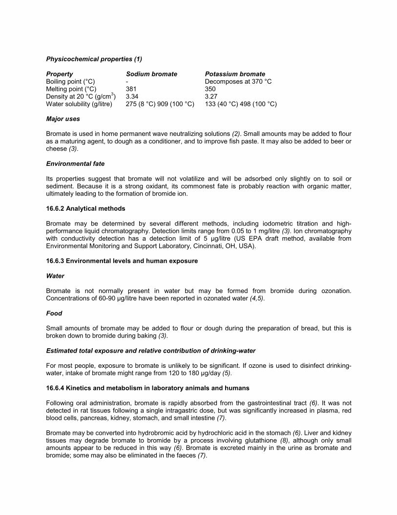
Physicochemical properties (1)
Property Sodium bromate Potassium bromateBoiling point (°C) - Decomposes at 370 °CMelting point (°C) 381 350Density at 20 °C (g/cm3) 3.34 3.27Water solubility (g/litre) 275 (8 °C) 909 (100 °C) 133 (40 °C) 498 (100 °C)
Major uses
Bromate is used in home permanent wave neutralizing solutions (2). Small amounts may be added to flouras a maturing agent, to dough as a conditioner, and to improve fish paste. It may also be added to beer orcheese (3).
Environmental fate
Its properties suggest that bromate will not volatilize and will be adsorbed only slightly on to soil orsediment. Because it is a strong oxidant, its commonest fate is probably reaction with organic matter,ultimately leading to the formation of bromide ion.
16.6.2 Analytical methods
Bromate may be determined by several different methods, including iodometric titration and high-performance liquid chromatography. Detection limits range from 0.05 to 1 mg/litre (3). Ion chromatographywith conductivity detection has a detection limit of 5 µg/litre (US EPA draft method, available fromEnvironmental Monitoring and Support Laboratory, Cincinnati, OH, USA).
16.6.3 Environmental levels and human exposure
Water
Bromate is not normally present in water but may be formed from bromide during ozonation.Concentrations of 60-90 µg/litre have been reported in ozonated water (4,5).
Food
Small amounts of bromate may be added to flour or dough during the preparation of bread, but this isbroken down to bromide during baking (3).
Estimated total exposure and relative contribution of drinking-water
For most people, exposure to bromate is unlikely to be significant. If ozone is used to disinfect drinking-water, intake of bromate might range from 120 to 180 µg/day (5).
16.6.4 Kinetics and metabolism in laboratory animals and humans
Following oral administration, bromate is rapidly absorbed from the gastrointestinal tract (6). It was notdetected in rat tissues following a single intragastric dose, but was significantly increased in plasma, redblood cells, pancreas, kidney, stomach, and small intestine (7).
Bromate may be converted into hydrobromic acid by hydrochloric acid in the stomach (6). Liver and kidneytissues may degrade bromate to bromide by a process involving glutathione (8), although only smallamounts appear to be reduced in this way (6). Bromate is excreted mainly in the urine as bromate andbromide; some may also be eliminated in the faeces (7).
16.6.5 Effects on laboratory animals and in vitro test systems
Acute exposure
Potassium bromate administered orally (9) and intraperitoneally (10) to mice gave LD50s of 223-363 and136 mg of bromate per kg of body weight, respectively. LD50s of 280-495 mg/kg of body weight wereobtained for rats, mice, and hamsters given potassium bromate by gavage (11).
Short-term exposure
Potassium bromate was administered to groups of F344 rats (10 per sex per dose) in water atconcentrations of 0, 150, 300, 600, 1250, 2500, 5000, or 10 000 mg/litre (approximately 16, 32, 63, 140,270, 540, or 1080 mg of bromate per kg of body weight per day) for 13 weeks. All animals exposed to2500 mg/litre or higher died within 7 weeks. Observed signs of toxicity included significant inhibition ofbody weight gain in males at 600 mg/litre or above and significant increases in serum parameters in bothsexes at 600 mg/litre (11).
Long-term exposure
Male Wistar rats were exposed to 0.04% potassium bromate in drinking-water (approximately 30 mg ofbromate per kg of body weight per day) for up to 15 months. Effects included markedly inhibited bodyweight gain in all exposed animals, karyopyknotic foci in tubules of the inner kidney medulla, increasedblood urea nitrogen (BUN), and marked structural abnormalities of the cortical tubules. Based on bodyweight and renal effects, a LOAEL of 30 mg of bromate per kg of body weight per day was identified in thisstudy (12).
Reproductive toxicity, embryotoxicity, and teratogenicity
The reproductive effects of potassium bromate were evaluated in a study in which rats and mice were fedflour treated with 15 mg of potassium bromate per kg over five and eight generations, respectively. Noeffects on reproductive performance or survival were observed in either species (11).
Mutagenicity and related end-points
Positive results were obtained for the mutagenicity of potassium bromate in Salmonella typhimurium strainTA100 using the Ames test and for chromosomal aberrations in cultured Chinese hamster fibroblast cells(13). Positive results were also obtained in an in vivo study of the acute cytogenetic effect of potassiumbromate on rat bone marrow cells (14) and in the mouse micronucleus test (9,10). Some evidence of DNAdamage in rats given potassium bromate has been observed (15).
Carcinogenicity
F344 rats (50 per sex per dose) were given drinking-water containing 0, 250, or 500 (reduced to 400 atweek 60) mg of potassium bromate per litre (average doses 0, 9.6, and 21.3 mg of bromate per kg of bodyweight per day in males and 0, 9.6 or 19.6 mg of bromate per kg of body weight per day in females) for110 weeks. The incidence of renal tumours in the three groups was 6%, 60%, and 88% in males and 0,56%, and 80% in females. The incidence of peritoneal mesotheliomas in dosed males was alsosignificantly elevated (11%, 33%, and 59%). The authors concluded that potassium bromate wascarcinogenic in both male and female rats (16).
In a subsequent study, male F344 rats were given water containing potassium bromate at 0, 15, 30, 60,125, 250, or 500 mg/litre (equivalent to 0, 0.7, 1.3, 2.5, 5.6, 12, or 33 mg of bromate per kg of bodyweight) for 104 weeks. The incidence of renal cell tumours in these dose groups was 0, 0, 0, 4%, 21%,

25%, and 45% and was significantly elevated at 12 mg/kg of body weight per day and above. Theincidence of dysplastic foci (considered to be preneoplastic lesions) was 0, 5%, 25%, 25%, 50%, 95%,and 95% and was significantly elevated at 5.6 mg/kg of body weight per day and above (17).
The carcinogenic potential of potassium bromate was investigated in B6C3F1 female mice (50 per dose)supplied with water containing 0, 500, or 1000 mg of potassium bromate per litre (average dose 0, 43.5, or91.6 mg of bromate per kg of body weight per day) for 78 weeks. Based on histological examination oftissues at week 104, no significant difference in tumour incidence between exposed and control animalswas apparent (16).
Male F344 rats were supplied with water containing 500 mg of potassium bromate per litre (average dose32.3 mg of bromate per kg of body weight per day) for up to 104 weeks to assess the time-course of renalcell tumour induction. Dysplastic foci and renal adenomas were first observed following 26 weeks oftreatment, but the incidence was not statistically significant. Renal dysplastic foci (62%) and adenomas(52%) were significantly increased as compared with controls by 52 weeks of treatment. After 104 weeks,renal adenocarcinomas were observed in 3 of 20 rats (15%) and adenomas in 6 of 20 (30%). Thecombined incidence of follicular adenomas and adenocarcinomas of the thyroid (7/35) was significantlyincreased in rats receiving treatment for 104 weeks. The authors concluded that the minimum inductiontime for renal adenoma development was 26 weeks (18).
In a related study, the incidence of renal cell tumours was investigated in F344 rats exposed to watercontaining 500 mg of potassium bromate per litre (29.6-35.5 mg of bromate per kg of body weight per day)for up to 104 weeks. The incidence of renal dysplastic foci was 65% in animals exposed for 1-13 weeksand increased to 100% in animals exposed for 39-52 weeks (0% in controls). The combined incidence ofadenomas and adenocarcinomas in rats exposed for 13-52 weeks ranged from 47% to 74%, which issimilar to or higher than that in animals exposed continuously for 104 weeks (45%). The authorsconcluded that the minimum total cumulative dose necessary for the induction of renal adenomas andadenocarcinomas was 4 g of potassium bromate per kg (3.1 g of bromate per kg) and the minimumtreatment period for the induction of these tumours was 13 weeks (18).
No significant differences were observed in the incidence of tumours in male or female newborn F344 ratsor ICR mice when potassium bromate was administered subcutaneously (19).
16.6.6 Effects on humans
Most cases of human poisoning from bromate are due to the accidental or intentional ingestion of homepermanent wave solutions, which can contain 2-10% bromate. In children, serious poisonings have beenreported following ingestion of 60-120 ml of 2% potassium bromate (equivalent to 46-92 mg of bromateper kg of body weight per day for a 20-kg child). Lethal oral doses of potassium bromate are estimated tobe 200-500 mg/kg of body weight (154-385 mg of bromate per kg of body weight) (2).
Toxic effects of bromate salts include nausea, vomiting, abdominal pain and diarrhoea, varying degrees ofcentral nervous system depression, seizures, respiratory depression, and pulmonary oedema, most ofwhich are reversible. Irreversible effects include renal failure and deafness, both of which have beenobserved following the ingestion of 240-500 mg of potassium bromate per kg of body weight (185-385 mgof bromate per kg of body weight) (2).
16.6.7 Provisional guideline value
JECFA evaluated bromate and recommended that there should be no residues in food when bromate isused in food processing (20). IARC has concluded that there is sufficient evidence for the carcinogenicityof potassium bromate in animals (3) and has classified it in Group 2B (possible human carcinogen).Bromate is mutagenic both in vitro and in vivo.

To estimate cancer risks, the linearized multistage model was applied to the incidence of renal tumours inmale rats given potassium bromate in drinking-water (16), although it was noted that, if the mechanism oftumour induction is oxidative damage in the kidney, application of the low-dose cancer risk model may notbe appropriate. The concentrations in drinking-water associated with excess lifetime cancer risks of 10-4,10-5, and 10-6 are 30, 3, and 0.3 µg/litre, respectively.
Because of limitations in available analytical and treatment methods, a provisional guideline value of 25µg/litre is recommended. This value is associated with a lifetime excess cancer risk of 7 × 10⁻⁵.
References
1. Weast RC, ed. CRC handbook of chemistry and physics, 67th ed. Boca Raton, FL, CRC Press, 1986.
2. Mack RB. Round up the usual suspects. Potassium bromate poisoning. North Carolina medical journal,1988, 49:243-245.
3. International Agency for Research on Cancer. Some naturally occurring and synthetic foodcomponents, furocoumarins and ultraviolet radiation: potassium bromate. Lyon, 1986:207-220 (IARCMonographs on the Evaluation of the Carcinogenic Risk of Chemicals to Humans, Volume 40).
4. Haag WR, Holgne J. Ozonation of bromide-containing waters: kinetics of formation of hypobromousacid and bromate. Environmental science and technology, 1983, 17:261-267.
5. McGuire MJ, Krasner SW, Gramith JT. Comments on bromide levels in state project water and impactson control of disinfectant by-products. Los Angeles, CA, Metropolitan Water District of Southern California,1990.
6. Kutom A et al. Bromate intoxication: hairdressers’ anuria. American journal of kidney disease, 1990,15:84-85.
7. Fujie M et al. Metabolism of potassium bromate in rats. I. In vivo studies. Chemosphere, 1984, 13:1207-1212.
8. Tanaka K et al. Metabolism of potassium bromate in rats. II. In vitro studies. Chemosphere, 1984,13:1213-1219.
9. Nakajima M et al. Effect of route of administration in the micronucleus test with potassium bromate.Mutation research, 1989, 223:399-402.
10. Hayashi M et al. Difference between intraperitoneal and oral gavage application in the micronucleustest: the 3rd collaborative study by CSGMT/JEMS-MMS. Mutation research, 1989, 223:329-344.
11. Kurokawa Y et al. Toxicity and carcinogenicity of potassium bromate-a new renal carcinogen.Environmental health perspectives, 1990, 87:309-335.
12. Nakano K et al. Renal changes induced by chronic oral administration of potassium bromate or ferricnitrilotriacetate in Wistar rats. Japanese archives of internal medicine, 1989, 36:41-47.
13. Ishidate M et al. Primary mutagenicity screening of food additives currently used in Japan. Foodchemistry and toxicology, 1984, 22:623-636.
14. Fujie K et al. Acute cytogenetic effects of potassium bromate on rat bone marrow cells in vivo.Mutation research, 1988, 206:455-458.

15. Kasai H et al. Oral administration of the renal carcinogen, potassium bromate, specifically produces 8-hydroxy-deoxyguanosine in rat target organ DNA. Carcinogenesis, 1987, 8:1959-1961.
16. Kurokawa Y et al. Long-term in vivo carcinogenicity tests of potassium bromate, sodium hypochloriteand sodium chlorite conducted in Japan. Environmental health perspectives, 1986, 69:221-235.
17. Kurokawa Y et al. Dose-response studies on the carcinogenicity of potassium bromate in F344 ratsafter long-term oral administration. Journal of the National Cancer Institute, 1986, 77:977-982.
18. Kurokawa Y et al. Relationship between the duration of treatment and the incidence of renal celltumors in male F344 rats administered potassium bromate. Japanese journal of cancer research, 1987,78:358-364.
19. Matsushima Y et al. Lack of carcinogenicity of potassium bromate after subcutaneous injection tonewborn mice and newborn rats. Science reports of the Research Institutes, Tohoku University, 1986,33:22-26.
20. Joint FAO/WHO Expert Committee on Food Additives. Toxicological evaluation of certain foodadditives and contaminants. Cambridge, Cambridge University Press, 1989 (WHO Food Additives Series,No. 24).
16.7 Chlorophenols
16.7.1 General description
Identity
Compound CAS no. Molecular formula2-Chlorophenol 95-57-8 ClC6H4OH2,4-Dichlorophenol 120-83-2 Cl2C6H3OH2,4,6-Trichlorophenol 88-06-2 Cl3C6H2OH
A total of 19 possible chlorinated phenols exist, but only 2-chlorophenol (2-CP), 2,4-dichlorophenol (2,4-DCP), and 2,4,6-trichlorophenol (2,4,6-TCP) will be evaluated here, as these are the most likely to occurin drinking-water as possible by-products of disinfection.
Physicochemical properties (1-3)
Property 2-CP1 2,4-DCP2 2,4,6-TCP3
Boiling point (°C) 175-176 210-211 246Melting point (°C) 8.7 43-44 68Density (g/cm3) 1.24 1.38 1.49Vapour pressure (kPa) 0.133 (12.1 °C) 0.133 (53 °C) 0.133 (76 °C)Water solubility (mg/litre) 28 000 4500 900Log octanol-water partition coefficient 2.15 3.06 -
1 Conversion factors in air: 2-CP, 1 ppm = 5.26 mg/m3.2 Conversion factors in air: 2,4-DCP, 1 ppm = 6.67 mg/m3.3 Conversion factors in air: 2,4,6-TCP, 1 ppm = 8.08 mg/m3.
Organoleptic properties
Chlorophenols generally have very low organoleptic thresholds. The taste thresholds in water for 2-CP,2,4-DCP, and 2,4,6-TCP are 0.1, 0.3, and 2 µg/litre, respectively. Odour thresholds are 10, 40, and 300

µg/litre, respectively (2).
Major uses
2-CP is used as a precursor in the production of higher chlorophenols and dyestuffs, and as apreservative. 2,4-DCP is used as a mothproofing agent, germicide and antiseptic, and in the production ofthe pesticide 2,4-D. 2,4,6-TCP is used in the production of 2,3,4,6-tetrachlorophenol andpentachlorophenol, and as a germicide, glue and wood preservative, and antimildew agent (4,5).
16.7.2 Analytical methods
EPA methods 604 (6,7), 525 (8), and 8270 (9) are used for the determination of chlorophenols. The mostsensitive technique involves the formation of the pentafluorobenzyl ether derivatives (an option in method604); the method has a detection limit of 0.5-5 µg/litre. Chlorophenols can also be determined by gaschromatography with an electron-capture detector. The detection limits are 1-10 µg/litre formonochlorophenols, 0.5 µg/litre for dichlorophenols, and 0.01 µg/litre for trichlorophenols (1).
16.7.3 Environmental levels and human exposure
Water
Chlorophenols are present in drinking-water as a result of the chlorination of phenols during disinfection,as by-products of the reaction of hypochlorite with phenolic acids, as biocides, or as degradation productsof phenoxy herbicides. Data from 40 Canadian treatment plants indicate that chlorophenol levels indrinking-water are generally quite low but vary considerably from one location to another (10). Chlorinationincreased the concentrations of 2-CP (maximum 65 ng/litre), 2,4-DCP (72 ng/litre), and 2,4,6-TCP (719ng/litre). Drinking-water from the Ruhr area of Germany contained 2,4-DCP at 3B6 ng/litre and 2,4,6-TCPat 1 ng/litre (1). Several chlorophenols were present in Finnish tapwater at levels roughly one order ofmagnitude higher than those found in Germany (11).
16.7.4 Kinetics and metabolism in laboratory animals and humans
Chlorophenols are well absorbed after oral administration (12), and they readily penetrate the skin (13).Chlorophenols do not appear to accumulate in body tissues in rats but are rapidly metabolized andeliminated from the body (14-16). The major metabolite is the glucuronide conjugate of the parentchlorophenol. Less abundant metabolites include sulfate conjugates and possibly chloromethoxyphenolisomers of the parent compounds (12,14,16,17). Chlorophenols are readily excreted as glucuronideconjugates in urine and, to a lesser extent, faeces (12,16,18).
16.7.5 Effects on laboratory animals and in vitro test systems
2-Chlorophenol
Acute exposure
The oral LD50 for 2-CP in mice was reported to be 670 mg/kg of body weight (19).
Long-term exposure
Immunological (e.g. humoral and cell-mediated immunity, macrophage function) and haematological (e.g.red and white blood cell count, haematocrit, haemoglobin) effects were assessed in groups of 12-20weanling female Sprague-Dawley rats exposed to 0, 5, 50, or 500 mg of 2-CP per litre in drinking-water (0,0.5, 5, or 50 mg/kg of body weight per day) in a reproductive study. Females were exposed from 3 weeksof age until breeding at 90 days, and throughout gestation to parturition. No treatment-related differences

were found. A NOAEL of 50 mg/kg of body weight per day can be identified (15,20).
Reproductive toxicity, embryotoxicity, and teratogenicity
Groups of 12-20 weanling female Sprague-Dawley rats were exposed to 0, 5, 50, or 500 mg of 2-CP perlitre in drinking-water (0, 0.5, 5, or 50 mg/kg of body weight per day) for 10 weeks, then bred. Treatmentwas continued during breeding, gestation, and weaning. Parameters evaluated included percentageconception, litter size, birth weight, number of stillbirths, weanling weight, and haematology in weanlingrats. A treatment-related increase in conception rate, an increase in the number of stillbirths, and adecrease in the size of the litters were observed at the highest dose (15,21).
Carcinogenicity
In a 24-month experiment, female Sprague-Dawley rats (12-22 per dose) were given 2-CP in drinking-water at 0, 5, 50, or 500 mg/litre (0, 0.5, 5, or 50 mg/kg of body weight per day) for 10 weeks, then bred.Ethylurea and nitrite, precursors of the transplacental carcinogen nitrosoethylurea (NEU), wereadministered to females on days 14-21 of pregnancy. The effects on tumour incidence and latency weremost evident in male progeny that received 2-CP with NEU, both pre- and postnatally. The lowest level of2-CP appeared to exert the greatest effect. The authors suggested that 2-CP is a co-carcinogen (21).
2,4-Dichlorophenol
Acute exposure
The acute oral LD50s for 2,4-DCP in rats ranged from 580 to 4000 mg/kg of body weight (22,23). Acuteoral LD50s were 1276 and 1352 mg/kg of body weight for male and female CD-1 mice, respectively (24).
Short-term exposure
CD-1 mice (20 per sex per dose) were exposed to 2,4-DCP in drinking-water for 90 days at concentrationsof 0.2, 0.6, or 2.0 g/litre (mean daily doses of 50, 143, and 491 mg/kg of body weight for females and 40,114, and 383 mg/kg of body weight for males). There were no significant differences in body weight gainand no differences in terminal organ weights or organ weight ratios. Haematological differences, namelyan increase in leukocytes (high dose) and in polymorphonuclear leukocytes (low dose), were observedonly in males. Changes in clinical chemistry parameters, namely a decrease in creatinine (low dose), anincrease in BUN/creatinine ratios (mid-dose), and an increase in alkaline phosphatase (high dose), weresignificant in females. These changes were not consistently dose-related, and a LOAEL cannot beestablished (24).
ICR mice of both sexes were fed 2,4-DCP in the diet at 0, 0.05%, 0.1%, or 0.2% (0, 45, 100, or 230 mg/kgof body weight per day) for 6 months. Hyperplasia of hepatic cells was reported in one of seven animalsreceiving 0.2%. There were no other significant differences in histopathology, organ or body weight gains,red or white blood cell counts, or alanine aminotransferase and aspartate aminotransferase activities atany dose. The authors identified a NOAEL of 100 mg/kg of body weight per day (23).
Pre- and postnatal treatment of rats with 300 mg of 2,4-DCP per litre of drinking-water for 147 dayssignificantly increased liver and spleen weights and enhanced humoral immune responsiveness. Cell-mediated immunity was depressed at 30 and 300 mg/litre. No histopathological changes were reported.Based on these findings, a NOAEL of 3 mg/litre (0.3 mg/kg of body weight per day) and a LOAEL of 30mg/litre (3 mg/kg of body weight per day) can be identified (25).
Long-term exposure
Investigations of the effects of long-term exposure to 2,4-D have been designed primarily to test its
carcinogenic properties and are described below.
Reproductive toxicity, embryotoxicity, and teratogenicity
Administration of 2,4-DCP (0, 50, 150, or 500 mg/kg of body weight per day) in drinking-water to male CD-1 mice for 90 days had no effect on sperm motility or ability to penetrate ova (26). Exposure of female ratsto 0, 3, 30, or 300 mg of 2,4-DCP per litre in drinking-water from 3 weeks of age throughout parturition andlactation had no significant effect on conception, litter size and weight, number of stillborn pups, or survivalof weanlings continued on treatment for 5 weeks (25).
Mutagenicity and related end-points
2,4-DCP did not show mutagenic potential in Salmonella typhimurium strains TA98, TA100, TA1535, andTA1537 with and without metabolic activation (27). In eukaryotic test assays, 2,4-DCP was not mutagenicin primary hepatocyte cultures, as shown by the absence of unscheduled DNA synthesis (28).
Carcinogenicity
F344 rats and B6C3F1 mice were given 2,4-DCP in feed for 2 years at dietary concentrations of 0, 5000,or 10 000 mg/kg (mice and male rats) and 0, 2500, or 5000 mg/kg (female rats) (male rats: 0, 210, or 440mg/kg of body weight per day; female rats: 0, 210, or 250 mg/kg of body weight per day; male mice, 0,800, or 1300 mg/kg of body weight per day; female mice, 0, 430, or 820 mg/kg of body weight per day).There was no evidence of carcinogenicity in either species. The maximum tolerated dose was probablyreached, judging from the lower body weight in the treated animals, especially at the high dose. Survivalwas not affected in either species (29).
2,4,6-Trichlorophenol
Acute exposure
The oral LD50 for 2,4,6-TCP has been reported as 820 mg/kg of body weight in rats (30).
Short-term exposure
2,4,6-TCP was mixed with corn oil and administered daily by gavage to Sprague-Dawley rats (10 per sexper dose) for 90 consecutive days at 0, 80, 240, or 720 mg/kg of body weight per day. At 240 mg/kg ofbody weight per day, liver weight increased in males and adrenal gland weight increased in females. Atthe highest dose, treatment-related effects included salivation, increased weights of the kidneys, liver,adrenal glands, and testes, and an increase in serum albumin, total protein, and serum alanineaminotransferase, as well as a decrease in urinary pH. No gross or histopathological changes were seen.In this study, a LOAEL of 240 mg/kg of body weight per day and a NOAEL of 80 mg/kg of body weight perday were identified (Bercz JP et al., unpublished data, 1989).
Long-term exposure
Female Sprague-Dawley rats (12-14 per dose) were exposed to 2,4,6-TCP in drinking-water at 0, 3, 30, or300 mg/litre from 3 weeks of age and throughout breeding, gestation, parturition, and lactation. Ten pupsfrom each dose group were weaned at 3 weeks and continued on treatment for 12-15 weeks. A dose-related increase in the liver weight of the pups reached statistical significance at 30 and 300 mg/litre. At300 mg/litre, the spleen weight of the pups also increased significantly. No treatment-related changes incell-mediated immunity, humoral immunity, or macrophage function were seen in the treated groups. Inthis study, a LOAEL of 30 mg/litre (3 mg/kg of body weight per day) and a NOAEL of 3 mg/litre (0.3 mg/kgof body weight per day) were identified (21).
F344 rats (50 per sex per dose) were given 2,4,6-TCP in their feed at 0, 5000, or 10 000 mg/kg (0, 250, or500 mg/kg of body weight per day) for 106-107 weeks. Mean body weights of both dosed groups werelower than those of corresponding controls and were dose-related throughout the study. Other clinicalsigns were common to both the dosed and the control groups. There was no significant dose-related trendin mortality. In a similar experiment in B6C3F1 mice, dose-related decreases in mean body weights wereseen in male and female mice. Other clinical signs were common to both dosed and control groups. Therewas no statistically significant dose-related trend in mortality in either sex (30).
Reproductive toxicity, embryotoxicity, and teratogenicity
Sprague-Dawley rats were exposed to 2,4,6-TCP at 0, 30, or 300 mg/litre in drinking-water from 3 weeksof age to parturition. There were no statistically significant treatment-related effects on percentageconception, litter size, percentage stillborn, birth weight, and percentage survival to weaning (21).
Male Long-Evans hooded rats were given 2,4,6-TCP at 0 or 1000 mg/kg of body weight in corn oil bygavage, 5 days per week for 11 weeks (average 0 or 714 mg/kg of body weight per day), then bred withuntreated females. No treatment-related effects were seen in copulatory behaviour, semen characteristics,organ weights, fertility, or fetal outcome. Female rats were given 0, 100, 500, or 1000 mg/kg of bodyweight by gavage, 5 days per week for 2 weeks prior to and during mating and up to day 21 of gestation.No treatment-related effects were reported in litter size or pup survival at the dose levels tested (31).
Mutagenicity and related end-points
Mutagenic activity was not detected in S. typhimurium strains TA98, TA100, TA1535, and TA1537 withand without metabolic activation (27). 2,4,6-TCP showed weak but significant mutagenic activity in theMP-1 strain of Saccharomyces cerevisiae (32). There was no effect on mitotic crossing-over or mitoticgene conversion. Pregnant mice injected with 2,4,6-TCP displayed a slightly increased frequency ofspotted coat in the offspring, indicative of weak mutagenic activity (32).
Carcinogenicity
Administration of 2,4,6-TCP to mice at 100 mg/kg of body weight for 72 weeks led to increases in theincidences of hepatomas and reticulum-cell sarcomas. However, the incidences were not statisticallysignificant if males and females are considered separately or if matched controls are considered (33).
F344 rats and B6C3F1 mice were administered 2,4,6-TCP (96-97% pure) in the feed for over 2 years.Rats and male mice received doses of 0, 5000, or 10 000 mg/kg of body weight and female mice receivedtime-weighted average doses of 0, 5214, or 10 428 mg/kg of body weight. A statistically significant dose-related increase in the incidence of lymphomas or leukaemias was observed in male rats (3/20, 23/50,and 29/50 for control, low-, and high-dose groups, respectively). In addition, the combined incidence ofhepatocellular carcinomas and adenomas was significantly increased as compared with controls in bothmale and female mice (30). The 2,4,6-TCP may have been contaminated with 1,3,6,8-tetrachlorodibenzo-p-dioxin (1,3,6,8-TCDD), which might also be capable of inducing liver tumours in mice but is not expectedto induce leukaemias in male rats, as the 2,3,7,8-TCDD isomer does not appear to do so (34).
16.7.6 Guideline values
2-Chlorophenol
Because of the limited database on the toxicity of 2-CP, no health-based guideline value has beenderived.
2,4-Dichlorophenol
Because the database for the toxicity of 2,4-DCP is limited, no health-based guideline value has beenderived.
2,4,6-Trichlorophenol
2,4,6-TCP has been reported to induce lymphomas and leukemias in male rats and hepatic tumours inmale and female mice. IARC has concluded that 2,4,6-TCP is possibly carcinogenic to humans (Group2B) (35). The compound has not been shown to be mutagenic in the Ames test but has shown weakmutagenic activity in other in vitro and in vivo assays.
A guideline value can be derived for 2,4,6-TCP by applying the linearized multistage model to leukaemiasin male rats observed in a 2-year feeding study (33). The hepatic tumours found in this study were notused for risk estimation, because of the possible role of contaminants in their induction. Theconcentrations of 2,4,6-TCP in drinking-water (and hence the guideline values) associated with 10-4, 10-5,and 10-6 excess lifetime cancer risks are 2000, 200, and 20 µg/litre, respectively.
The lowest reported taste threshold for 2,4,6-TCP is 2 µg/litre. If water containing this chlorophenol is freefrom taste, it is unlikely to present an undue risk to health.
References
1. Dietz F, Traud J. [Trace analysis of phenols, especially chlorophenols in water, by GC.] Vom Wasser,1978, 51:235-237 (in German).
2. Kozak VP et al. Reviews of the environmental effects of pollutants: XI. Chlorophenols. Washington, DC,US Environmental Protection Agency, 1979 (EPA 600/1-79-012).
3. Chlorophenols. Geneva, World Health Organization, 1989 (Environmental Health Criteria, No. 93).
4. Freiter ER. Chlorophenols. In: Kirk-Othmer encyclopedia of chemical technology. 3rd ed., Vol. 5, NewYork, John Wiley, 1979:864-872.
5. Doedens JD. Chlorophenols. In: Kirk-Othmer encyclopedia of chemical technology, 2nd ed. New York,John Wiley, 1963:325-338.
6. US Environmental Protection Agency. U.S. EPA Method 604-Phenols. Washington, DC, 1985 (40 CFRpart 136).
7. Kawahara FK. Analysis of mercaptans, phenols and organic acids in surface waters with use ofpentafluorobenzyl derivatives. Environmental science and technology, 1971, 5:235-239.
8. Environmental Monitoring and Support Laboratory. U.S. EPA Method 525-Detection of organiccompounds in drinking water by liquid-solid extraction and capillary column gas chromatography. Revision2.1. Cincinnati, OH, US Environmental Protection Agency, 1988.
9. Office of Solid Waste. U.S. EPA Method 8270-Gas chromatography/mass spectrometry for semi-volatileorganics, capillary column technique, test method for evaluating solid waste, 3rd ed. Washington, DC, USEnvironmental Protection Agency, 1986 (SW 846).
10. Sithole BB, Williams DT. Halogenated phenols in water at forty Canadian potable water treatmentfacilities. Journal of the Association of Official Analytical Chemists, 1986, 69(5):807-810.
11. Passivirta J et al. Polychlorinated phenols, guaiacols and catechols in the environment. Chemosphere,1985, 14(5):469-491.
12. Carpenter HM, Bender RC, Buhler DR. Absorption, metabolism and excretion of 2- and 4-chlorophenolin rats. Toxicology, 1985, 5:109 (abstract).
13. Roberts MS, Anderson RA, Swarbrick J. Permeability of human epidermis to phenolic compounds.Journal of pharmacy and pharmacology, 1977, 29:677-683.
14. Somani SM, Khalique A. Distribution and metabolism of 2,4-dichlorophenol in rats. Journal oftoxicology and environmental health, 1982, 9:889-897.
15. Exon JH, Koller LD. Effects of transplacental exposure to chlorinated phenols. Environmental healthperspectives, 1982, 46:137-140.
16. Bahig ME, Kraus A, Klein W. Excretion and metabolism of 2,4,6-trichlorophenol-14C in rats.Chemosphere, 1981, 10(3):323-327.
17. Somani SM, Smart T, Khalique A. Metabolism of 2,4-dichlorophenol by isolated perfused rat liver.Journal of toxicology and environmental health, 1984, 13:787-798.
18. Korte F et al. Ecotoxicologic profile analysis. Chemosphere, 1978, 7(1):79-102.
19. Bubnov WD, Jafizov FN, Ogryzkov SE. [The toxic properties of activated o-chlorophenol for white miceand blue foxes.] Trudy Vsesojuznogo Naucno-Issledovatel’skogo Instituta Veterinarnoj SanitarijEktoparazitologij, 1969, 33:258-263 (in Russian) (cited in ref. 2).
20. Exon JH, Koller LD. Effects of chlorinated phenols on immunity in rats. International journal ofimmunology and pharmacology, 1983, 5(2):131-136.
21. Exon JH, Koller LD. Toxicity of 2-chlorophenol, 2,4-dichlorophenol and 2,4,6-trichlorophenol. In: JolleyRL et al., eds. Water chlorination: chemistry, environmental impact and health effects. Vol. 5. Chelsea, MI,Lewis Publishers, 1985:307-330.
22. Deichmann WB, Mergard EG. Comparative evaluation of methods employed to express the degree oftoxicity of a compound. Journal of industrial hygiene and toxicology, 1948, 30:373.
23. Kobayashi S et al. [Chronic toxicity of 2,4-dichlorophenol in mice: a simple design for the toxicity ofresidual metabolites of pesticides.] Toho University, Japan, journal of the Medical Society, 1972, 19:356-362 (in Japanese).
24. Borzelleca JF et al. Acute and subchronic toxicity of 2,4-dichlorophenol in DC-1 mice. Fundamentaland applied toxicology, 1985, 5:478-486.
25. Exon JH et al. Toxicologic, pathologic and immunotoxic effects of 2,4-dichlorophenol in rats. Journal oftoxicology and environmental health, 1984, 14:723-730.
26. Seyler DE et al. The use of in vitro methods for assessing reproductive toxicity. Dichlorophenols.Toxicology letters, 1984, 20:309-315.
27. Rasanen L, Hattula ML, Arstila AU. The mutagenicity of MCPA and its soil metabolites, chlorinatedphenols, catechols and some widely used slimicides in Finland. Bulletin of environmental contaminationand toxicology, 1977, 18(5):565-571.
28. Probst GS et al. Chemically-induced unscheduled DNA synthesis in primary rat hepatocyte cultures: acomparison with bacterial mutagenicity using 218 compounds. Environmental mutagenesis, 1981, 3:11-32.
29. National Toxicology Program. Toxicology and carcinogenesis studies of 2,4-dichlorophenol in F344/Nrats and B6C3F1 mice. Research Triangle Park, NC, 1989 (NTP-TR-353).
30. National Cancer Institute. Bioassay of 2,4,6-trichlorophenol for possible carcinogenicity. Bethesda,MD, 1979 (NCI-CG-TR-155).
31. Blackburn K et al. Evaluation of the reproductive toxicology of 2,4,6-trichlorophenol in male and femalerats. Fundamental and applied toxicology, 1986, 6:233-239.
32. Fahrig R, Nilsson CA, Rappe C. Genetic activity of chlorophenols and chlorophenol impurities. In: RaoKR, ed. Pentachlorophenol: chemistry, pharmacology and environmental toxicology. New York, NY,Plenum Press, 1978:325-338.
33. Bionetics Research Laboratories. Evaluation of carcinogenic, teratogenic and mutagenic activities ofselected pesticides and industrial chemicals. Vol. 1. Carcinogenic studies. Bethesda, MD, National CancerInstitute, 1968.
34. Firestone D et al. Determination of polychlorodibenzo-p-dioxins and related compounds in commercialchlorophenols. Journal of the Association of Official Analytical Chemists, 1972, 55(1):85-92.
35. International Agency for Research on Cancer. Overall evaluations of carcinogenicity: an updating ofIARC Monographs volumes 1-42. Lyon, 1987:154-156 (IARC Monographs on the Evaluation ofCarcinogenic Risks to Humans, Suppl. 7).
16.8 Formaldehyde
16.8.1 General description
Identity
CAS no.: 50-00-00Molecular formula: CH2O
The IUPAC name for formaldehyde is methanal.
Physicochemical properties (1-4)1
1 Conversion factor in air: 1 ppm = 1.2 mg/m3 at 25 °C
Property ValuePhysical state Colourless gasBoiling point -19.2 °CMelting point -118 °CRelative density 1.04 (air = 1)Vapour pressure 52.6 kPa at -33 °CWater solubility Freely miscible at 25 °CLog octanol-water partition coefficient -1
Organoleptic properties
Formaldehyde has a pungent, suffocating, hay- or straw-like odour. Taste and odour thresholds are 50and 25, respectively (3,4).
Major uses
Formaldehyde’s main industrial use is in the production of urea-formaldehyde, phenolic, melamine,pentaerythritol, and polyacetal resins. Its second largest use is in the industrial synthesis of a number oforganic compounds. It is also used in cosmetics, fungicides, textiles, and embalming fluids (1).
16.8.2 Analytical methods
Formaldehyde in drinking-water is generally determined by a high-performance liquid chromatographicmethod following derivatization with 2,4-dinitrophenylhydrazine and liquid-solid extraction. The detectionlimit is 6.2 µg/litre (5).
16.8.3 Environmental levels and human exposure
Air
Formaldehyde is emitted into air from plastics and resin glues. Low levels in air may also result from thephoto-oxidation of hydrocarbons derived from fossil fuel. Typical levels in air are a few µg/m3. Smokersare exposed to high levels of formaldehyde (1,6,7).
Water
Formaldehyde in drinking-water is formed mainly by the oxidation of natural organic (humic) matter duringozonation (8) and chlorination (9). It also enters drinking-water via industrial effluents and leaching frompolyacetal plastic fittings. Concentrations of up to 30 µg/litre have been found in ozonated drinking-water(10,11).
Food
Concentrations of formaldehyde ranging from 3 to 23 mg/kg have been reported in a variety of foods (6).
Estimated total exposure and relative contribution of drinking-water
The general population is exposed to formaldehyde mainly by inhalation, smokers receiving about 0.38mg/day by this route (1,7). People are also exposed by ingesting contaminated drinking-water and food,and from the use of urea-formaldehyde foam in housing insulation, and of cosmetics containingformaldehyde.
16.8.4 Kinetics and metabolism in laboratory animals and humans
Ingested formaldehyde is readily absorbed by the gastrointestinal tract. In dermal studies, it was absorbedless readily in monkeys than in rats or guinea-pigs (12). It appears to be distributed mainly to muscle,lower levels being found in the intestines, liver, and other tissues (13).
Formaldehyde is rapidly oxidized to formic acid; the subsequent oxidation to carbon dioxide and water isslower in monkeys than in rats (14). Other metabolic products, such as N,N’-bis(hydroxymethyl)urea andN-(hydroxymethyl)urea, have been reported in rats (15). Metabolites are eliminated in the urine, faeces,and expired air, the relative amounts depending on the route of administration (1,16,17).

16.8.5 Effects on laboratory animals and in vitro test systems
Acute exposure
Oral LD50s of 800 and 260 mg/kg of body weight have been reported for the rat and guinea-pig,respectively (18).
Short-term exposure
In a 4-week study, Wistar rats (10 per sex per dose) received formaldehyde in drinking-water at doses of0, 5, 25, or 125 mg/kg of body weight per day. Rats receiving the highest dose showed lowered food andliquid intake, histopathological changes in the stomach (i.e., focal hyperkeratosis of the forestomach,moderate papillomatous hyperplasia), and, in males only, lowered total protein and albumin levels inplasma. The NOAEL was 25 mg/kg of body weight per day (1,19).
Oral doses of 0, 50, 100, or 150 mg/kg of body weight per day in rats and 0, 50, 75, or 100 mg/kg of bodyweight per day in dogs for 91 days had no effect on haematology, clinical chemistry, urinalysis, or grossmicroscopic pathology. Depression in body weight gain was observed in both species at the highest doselevels and in male rats given 100 mg/kg of body weight per day (20).
Long-term exposure
In a 2-year study, Wistar rats were exposed to formaldehyde in drinking-water at mean doses of 0, 1.2, 15,or 82 mg/kg of body weight per day for males and 0, 1.8, 21, or 109 mg/kg of body weight per day forfemales. Adverse effects were observed only in animals receiving the highest dose and included lowerfood and liquid intake, lower body weights, and pathological changes in the stomach, characterized bythickening of the mucosal wall. Relative kidney weights were increased in high-dose females, and anincreased incidence of renal papillary necrosis was found in both sexes. Exposure did not appear to affectsurvival, haematology, or clinical chemistry. The NOAEL was 15 mg/kg of body weight per day (21).
In a similar study, Wistar rats were given formaldehyde in drinking-water at 10, 50, or 300 mg/kg of bodyweight per day. At the end of 12 months, rats of both sexes in the high-dose group were observed to havegastric erosions, ulcers, squamous cell hyperplasia, hyperkeratosis, and basal cell hyperplasia. Only onemale and one female from the mid-dose group showed hyperkeratosis (1,22).
Reproductive toxicity, embryotoxicity, and teratogenicity
No teratogenic effects were reported in mice given formaldehyde at oral doses of 0, 74, 148, or 185 mg/kgof body weight per day on days 6-15 of gestation (23). Growth and viability of neonates from mice givenoral doses of 540 mg/kg of body weight per day on days 8-12 of gestation were unaffected (24). No effectson reproductive performance or on the health of offspring were observed in beagle dogs fed 0, 3.1, or 9.4mg of formaldehyde per kg of body weight per day in their diet on days 4-56 after mating (25). Spermabnormalities were observed in male rats given single oral doses of 100-200 mg/kg of body weight (26).Intraperitoneal injection of formaldehyde at 8 or 16 mg/kg of body weight per day for 10 days resulted indegeneration of testicular tissue, inhibition of spermatogenesis, and lowered male reproductive organweights in rats (27).
Mutagenicity and related end-points
Formaldehyde has shown evidence of mutagenicity in prokaryotic and eukaryotic cells in vitro. It has alsobeen shown to be genotoxic in Drosophila melanogaster. Formaldehyde binds readily to proteins, RNA,and single-stranded DNA to induce DNA-protein cross-links and breaks in single-stranded DNA. It reactsreadily with macromolecules in cells, mainly at the point of exposure (28). In vivo, formaldehyde increasesboth DNA synthesis in rats (29) and the number of micronuclei and nuclear anomalies in epithelial cells in
rats (30).
Carcinogenicity
There is little evidence that formaldehyde is carcinogenic by the oral route. In a 2-year study in whichWistar rats were exposed to formaldehyde in drinking-water at mean doses of 0, 1.2, 15, or 82 mg/kg ofbody weight per day for males and 0, 1.8, 21, or 109 mg/kg of body weight per day for females, exposuredid not appear to affect tumour incidence (21). In a 2-year study in which Sprague-Dawley rats wereexposed to formaldehyde in drinking-water at dose levels of 0, 1, 5, 10, 50, 100, or 150 mg/kg of bodyweight per day, a dose-dependent increase in the incidence of leukaemia (mainly lymphoblastic) andlymphosarcoma was reported at dose levels of 5 mg/kg of body weight per day or greater. The increase inthe incidence of gastrointestinal neoplasms was not dose-related. Tumours of this type were rare inhistorical controls and not detected in concurrent controls (31).
In a carcinogenicity study, a group of 10 rats was given drinking-water containing 0.5% formalin (0.2%formaldehyde) for 32 weeks. Histopathological changes were observed in the stomach, as well asneoplastic changes in the forestomach and papillomas. In addition, the authors reported evidence thatformaldehyde had tumour-promoting activity. However, because of the presence of high levels ofmethanol in formalin, the usefulness of this information is limited (32). In another study, formaldehydeinduced ornithine decarboxylase activity (an indication of tumour-promoting activity) in rats given a singleoral formaldehyde dose of up to 100 mg/kg of body weight (29). There is no evidence that formaldehydeacts as a carcinogen or promoter when applied to mouse skin (33).
There is some evidence that inhalation exposure to formaldehyde causes cancer in rats and mice byirritating the nasal epithelium. Rats exposed to 17 mg of formaldehyde per m3, 6 h per day, 5 days perweek for 2 years, exhibited an increased incidence of squamous cell carcinoma of the nasal cavity.Tumours were also noted in mice at the same level of exposure, but this species was less sensitive thanthe rat (34,35).
16.8.6 Effects on humans
Irritation and allergic contact dermatitis have been associated with exposure of the skin to formaldehyde atlevels higher than those encountered in drinking-water (36). Its presence in some types of water filters hasbeen associated with the occurrence of haemolytic anaemia in dialysis patients (1,37).
There is some evidence that formaldehyde is a carcinogen in humans exposed by inhalation.Epidemiological investigations of the mortality of factory workers following prolonged occupationalexposure to formaldehyde showed a slight excess of lung cancer that was not related to formaldehydeexposure (2,38). An increase in the incidence of nasopharyngeal cancer was also noted but again did notappear to be related to formaldehyde (39).
16.8.7 Guideline value
Rats and mice exposed to formaldehyde by inhalation exhibited an increased incidence of carcinomas ofthe nasal cavity at doses that caused irritation of the nasal epithelium (34,35). Ingestion of formaldehydein drinking-water for 2 years caused stomach irritation in rats (21,22). Papillomas of the stomachassociated with severe tissue irritation were observed in one study (32).
On the basis of studies in which humans and experimental animals were exposed to formaldehyde byinhalation, IARC has classified formaldehyde in Group 2A (40). The weight of evidence indicates thatformaldehyde is not carcinogenic by the oral route. A guideline value has been derived, therefore, on thebasis of a TDI. A TDI of 150 µg/kg of body weight was calculated based on the NOAEL of 15 mg/kg ofbody weight per day in a 2-year study in rats (21), incorporating an uncertainty factor of 100 (for intra- andinterspecies variation). No account was taken of potential carcinogenicity from the inhalation of
formaldehyde from various indoor water uses, such as showering. With an allocation of 20% of the TDI todrinking-water, the guideline value is 900 µg/litre.
References
1. Formaldehyde. Geneva, World Health Organization, 1989 (Environmental Health Criteria, No. 89).
2. Acheson ED et al. Formaldehyde process workers and lung cancer. Lancet, 1984, 1(8385):1066-1067(letter).
3. Bills TD, Marking LL, Chandler JH. Investigation in fish control. 73. Formalin: Its toxicity to nontargetaquatic organisms, persistence and counteraction. Washington, DC, US Department of the Interior, Fishand Wildlife Service, 1977:1-7.
4. Verschueren K. Handbook of environmental data on organic chemicals, 2nd ed. New York, VanNostrand Reinhold, 1983:678-679.
5. Environmental Monitoring Systems Laboratory. Method 554. Determination of carbonyl compounds indrinking water by DNPH derivatization and high performance liquid chromatography (HPLC). Cincinnati,OH, US Environmental Protection Agency, 1991.
6. International Agency for Research on Cancer. Some industrial chemicals and dyestuffs. Lyon,1982:345-389 (IARC Monographs on the Evaluation of the Carcinogenic Risk of Chemicals to Humans,Volume 29).
7. National Research Council. Formaldehyde: an assessment of its health effects. Washington, DC,National Academy of Sciences, 1980.
8. Glaze WH, Koga M, Cancilla D. Ozonation by-products. 2. Improvement of an aqueous-phasederivatization method for the detection of formaldehyde and other carbonyl compounds formed by theozonation of drinking water. Environmental science and technology, 1989, 23:838-847.
9. Becher G, Ovrum NM, Christman RF. Novel chlorination by-products of aquatic humic substances.Science of the total environment, 1992, 117/118:509-520.
10. Tomkins BA et al. Liquid chromatographic determination of total formaldehyde in drinking water.Journal of the Association of Official Analytical Chemists, 1989, 72:835-839.
11. Krasner SW et al. The occurrence of disinfection by-products in US drinking water. Journal of theAmerican Water Works Association, 1989, 81:41-53.
12. Jeffcoat AR. Percutaneous penetration of formaldehyde. Final report (July 1981-July 1983). ResearchTriangle Park, NC, Research Triangle Institute, 1983:59.
13. Bhatt HS, Lober SB, Combes B. Effect of glutathione depletion on aminopyrine and formaldehydemetabolism. Biochemical pharmacology, 1988, 37:1581-1589.
14. McMartin KE et al. Methanol poisoning. V. Role of formate metabolism in the monkey. Journal ofpharmacology and experimental therapeutics, 1977, 201:564-572.
15. Mashford PM, Jones AR. Formaldehyde metabolism by the rat: a re-appraisal. Xenobiotica, 1982,12:119-124.
16. Galli CL et al. Toxicological evaluation in rats and mice of the ingestion of a cheese made from milk

with added formaldehyde. Food chemistry and toxicology, 1983, 21:313-317.
17. Upreti RK et al. Toxicokinetics and molecular interaction of [14C]-formaldehyde in rats. Archives ofenvironmental contamination and toxicology, 1987, 16:263-273.
18. Smyth HF Jr, Seaton J, Fischer L. The single dose toxicity of some glycols and derivatives. Journal ofindustrial hygiene and toxicology, 1941, 23:259-268.
19. Til HP et al. Evaluation of the oral toxicity of acetaldehyde and formaldehyde in a 4-week drinking-water study in rats. Food chemistry and toxicology, 1988, 26:447-452.
20. Johannsen FR, Levinskas GJ, Tegeris AS. Effects of formaldehyde in the rat and dog following oralexposure. Toxicology letters, 1986, 30:1-6.
21. Til HP et al. Two-year drinking-water study of formaldehyde in rats. Food chemistry and toxicology,1989, 27:77-87.
22. Tobe M, Naito K, Kurokawa Y. Chronic toxicity study of formaldehyde administered orally to rats.Toxicology, 1989, 56:79-86.
23. Marks TA, Worthy WC, Staples RE. Influence of formaldehyde and Sonacide (potentiated acidgluturaldehyde) on embryo and fetal development in mice. Teratology, 1980, 22:51-58.
24. Seidenberg JM, Anderson DG, Becker RA. Validation of an in vivo developmental toxicity screen in themouse. Teratogenesis, carcinogenesis and mutagenesis, 1987, 6:361-374.
25. Hurni H, Ohder H. Reproduction study with formaldehyde and hexamethylenetetramine in beagledogs. Food and cosmetics toxicology, 1977, 11:459-462.
26. Cassidy SL, Dix KM, Jenkins T. Evaluation of a testicular sperm head counting technique using ratsexposed to dimethoxyethyl phthalate (DMEP), glycerol alpha-monochlorohydrin (GMCH), epichlorohydrin(ECH), formaldehyde (FA), or methyl methanesulphonate (MMS). Archives of toxicology, 1983, 53:71-78.
27. Shah BM et al. Formaldehyde-induced structural and functional changes in the testis of rats. Journal ofreproductive biology and comparative endocrinology, 1987, 7:42-52.
28. Ma TM, Harris MM. Review of the genotoxicity of formaldehyde. Mutation research, 1988, 196:37-59.
29. Overman DO. Absence of embryotoxic effects of formaldehyde after percutaneous exposure inhamsters. Toxicology letters, 1985, 24:107-110.
30. Migliore L et al. Micronuclei and nuclear anomalies induced in the gastrointestinal epithelium of ratstreated with formaldehyde. Mutagenesis, 1989, 4(5):327-334.
31. Soffritti M et al. Formaldehyde: an experimental multipotential carcinogen. Toxicology and industrialhealth, 1989, 5(5):699-730.
32. Takahashi M et al. Effects of ethanol, potassium metabisulfite, formaldehyde and hydrogen peroxideon gastric carcinogenesis in rats after initiation with N-methyl-N1-nitro-N-nitrosoguanidine. Japanesejournal of cancer research (Gann), 1986, 77:118-124.
33. Krivanek ND, Chromey NC, McAlack JW. Skin initiation-promotion study with formaldehyde in CD-1mice. In: Clary JJ, Gibson JE, Waritz RS, eds. Formaldehyde: toxicology, epidemiology, mechanisms.New York, Marcel Dekker, 1983:159-171.
34. Swenberg JA et al. Induction of squamous cell carcinomas of the rat nasal cavity by inhalationexposure to formaldehyde vapor. Cancer research, 1980, 40:3398-3402.
35. Kerns WD et al. Carcinogenicity of formaldehyde in rats and mice after long-term inhalation exposure.Cancer research, 1983, 43:4382-4392.
36. Cosmetic, Toiletry and Fragrance Association. Final report on the safety assessment of formaldehyde.Journal of the American College of Toxicologists, 1984, 3:157-184.
37. Beall JR. Formaldehyde in dialysis patients. A review. In: Turosk V, ed. Formaldehyde: analyticalchemistry and toxicology. Washington, DC, Chemical Society, 1985:275-287 (Advances in ChemistrySeries, Vol. 210).
38. Acheson ED et al. Formaldehyde in the British chemical industry. An occupational cohort study.Lancet, 1984, 1(8377):611-616.
39. Collins JJ et al. Formaldehyde exposure and nasopharyngeal cancer: re-examination of the NationalCancer Institute study and an update of one plant. Journal of the National Cancer Institute, 1988, 80:376-377.
40. International Agency for Research on Cancer. Overall evaluations of carcinogenicity: an updating ofIARC Monographs volumes 1-42. Lyon, 1987:211-216 (IARC Monographs on the Evaluation ofCarcinogenic Risks to Humans, Suppl. 7).
16.9 MX
16.9.1 General description
Identity
CAS no.: 77439-76-0Molecular formula: C5H3Cl3O3
MX is the common name for 3-chloro-4-dichloromethyl-5-hydroxy-2(5H)-furanone.
Major uses
MX does not have any commercial uses.
Environmental fate
In drinking-water at normal pH, MX exists in the open-ring form, i.e. as (Z)-2-chloro-3-(dichloromethyl)-4-oxobutenoic acid.
16.9.2 Analytical methods
MX in drinking-water can be determined by first concentrating organics using XAD resins, followed byhigh-pressure liquid chromatography, capillary-column gas chromatography, and mass spectroscopy (1-3).
16.9.3 Environmental levels and human exposure
Water
MX is formed by the reaction of chlorine with complex organic matter in drinking-water and is present inthe chlorinated effluents of pulp mills. It has been identified in chlorinated humic acid solutions anddrinking-water in Finland, the United Kingdom and the USA, and was found to be present in 37 watersources at levels of 2-67 ng/litre (2,4). Five drinking-water samples from different Japanese citiescontained MX at concentrations ranging from <3 to 9 ng/litre (5).
16.9.4 Kinetics and metabolism in laboratory animals and humans
At least 40% of a dose of MX administered by gavage to rats was absorbed, about 5% of it beingrecovered in the liver, muscle, skin, kidneys, and blood (6). It has been demonstrated that MX is asubstrate for direct conjugation with glutathione in vitro and that glutathione-S-transferase enhances thereaction (7). Cumulative excretion of label in 48 h by male rats given 14C-labelled MX by gavage wasabout 34% in the urine and 47% in faeces (6). No parent compound was excreted in the urine from rats(8).
16.9.5 Effects in laboratory animals and in vitro test systems
Acute exposure
An acute oral LD50 of 128 mg/kg of body weight per day has been estimated for mice (9).
Short-term exposure
MX in distilled water was administered to Swiss-Webster mice (5 per sex per dose) by gavage at 10, 20,42, 88, or 184 mg/kg of body weight per day for 2 days. At 184 mg/kg of body weight per day, all animalsdied within 1 day following the second dose; enlarged stomachs and haemorrhagic areas of theforestomach were observed. At lower doses, no deaths occurred, no effects on body weight were notedduring the 2-week observation period, and gross necropsy results were normal (9).
Mutagenicity and related end-points
MX was reported to be an extremely potent mutagen in Salmonella typhimurium strain TA100 withoutmetabolic activation by the S9 fraction of rat liver homogenate. The responses were also positive but notas strong in strains TA92, TA97, TA98, TA102, and TA1535. No mutagenic response was found withTA1537. The addition of the S9 fraction dramatically decreased the responses of TA100, TA98, andTA1535 (9,10).
MX has been examined for genotoxic activity in cultured mammalian cells. In Chinese hamster ovary cells(CHO-K1), it induced significant increases in structural chromosomal aberrations with and withoutmetabolic activation (9). It also induced DNA strand breaks in suspensions of rat hepatocytes, rattesticular cells and V79 Chinese hamster cells (11), but not micronuclei in mouse bone marrow in vivo,despite its relatively high clastogenic activity in mammalian cells in vitro (9).
Carcinogenicity
A skin tumour initiation-promotion assay was conducted in SENCAR mice (20-40 per dose). A single doseof MX was administered either topically in acetone or orally in distilled water at 5, 16, 28, or 50 mg/kg ofbody weight. Two weeks after treatment, 12-O-tetradecanoyl-phorbol-13-acetate (TPA) was appliedtopically three times per week for 30 weeks. At 24 weeks, no tumours were observed in animals receivingMX alone (topically or orally) without TPA promotion; results in topically treated animals with TPApromotion were similar to those in controls. In orally treated mice, both the tumour incidence and numberof tumours per mouse were significantly elevated at 16 mg of MX per kg of body weight and above.Because of disparities in the tumour incidences for the topical and oral control groups, the data were

reanalysed at 28 weeks, when tumour incidence was significantly higher than for controls only in the groupreceiving 16 mg/kg of body weight. The authors concluded that these results require confirmation beforeMX can be considered to possess tumour-initiating activity when administered orally (4).
16.9.6 Conclusions
There are very limited data on the toxicity of MX. 14C-labelled MX is rapidly absorbed, and most of theradioactivity is excreted in the urine within 24-48 h. It is unlikely to be absorbed as the parent compoundbecause of its high reactivity. MX is an extremely potent mutagen in some strains of Salmonellatyphimurium, but the addition of liver extract dramatically reduces the response. It is only weakly active orinactive in short-term tests for genotoxicity in vivo. Available data are inadequate to permit a guidelinevalue for MX to be established.
References
1. Munch JW et al. Determination of a chlorinated furanone (MX) in treated water. In: Proceedings of theAmerican Water Works Quality Technology Conference, Baltimore, MD, November 1987. Denver, CO,American Water Works Association, 1987:933-942.
2. Hemming J et al. Determination of the strong mutagen 3-chloro-4-(dichloromethyl)-5-hydroxy-2(5H)-furanone in chlorinated drinking and humic waters. Chemosphere, 1986, 15:549-556.
3. Kronberg L, Vartiainen T. Ames mutagenicity and concentration of the strong mutagen 3-chloro-4-(dichloromethyl)-5-hydroxy-2(5H)-furanone and of its geometric isomer (E)-2-chloro-3-(dichloromethyl)-4-oxobutenoic acid in chlorine-treated tapwater. Mutation research, 1988, 206:177-182.
4. Meier JR et al. Genotoxic and carcinogenic properties of chlorinated furanones-important by-products ofwater chlorination. In: Waters MD et al., eds. Genetic toxicology of complex mixtures: short-termbioassays in the analysis of complex environmental mixtures. New York, Plenum Press, 1989:185-195.
5. Suzuki N, Nakaniski J. The determination of strong mutagen, 3-chloro-4-(dichloromethyl)-5-hydroxy-2(5H)-furanone in drinking water in Japan. Chemosphere, 1990, 21:387-392.
6. Ringhand HP et al. Synthesis of 3-14C-3-chloro-4-(dichloromethyl)-5-hydroxy-2(5H)-furanone and itsuse in a tissue distribution study in the rat. Chemosphere, 1989, 18:2229-2236.
7. Meier JR et al. Importance of glutathione in the in vitro detoxification of 3-chloro-4-(dichloromethyl)-5-hydroxy-2(5H)-furanone, an important mutagenic by-product of water chlorination. In: Jolley RL et al., eds.Water chlorination: chemistry, environmental impact and health effects. Chelsea, MI, Lewis Publishers,1990.
8. Komulainen H et al. Pharmacokinetics in rat of 3-chloro-4-(dichloromethyl)-5-hydroxy-2(5H)-furanone(MX), a drinking water mutagen, after a single dose. Pharmacology and toxicology, 1992, 70:424-428.
9. Meier JR, Blazak WF, Knohl RB. Mutagenic and clastogenic properties of 3-chloro-4-(dichloromethyl)-5-hydroxy-2(5H)-furanone: a potent bacterial mutagen in drinking water. Environmental molecular mutation,1987, 10:411-424.
10. Holmbom B et al. Fractionation, isolation, and characterization of Ames mutagenic compounds in kraftchlorination effluents. Environmental science and technology, 1984, 18(5):333-337.
11. Brunborg G et al. Genotoxic effects of the drinking water mutagen 3-chloro-4-(dichloromethyl)-5-hydroxy-2[5H]-furanone (MX) in mammalian cells in vitro and rats in vivo. Mutation research, 1991,260:55-64.

16.10 Trihalomethanes
16.10.1 General description
Identity
Compound CAS no. Molecular formulaBromoform 75-25-2 CHBr3Dibromochloromethane (DBCM) 124-48-1 CHBr2ClBromodichloromethane (BDCM) 75-27-4 CHBrCl2Chloroform 67-66-3 CHCl3
Trihalomethanes are halogen-substituted single-carbon compounds with the general formula CHX3, whereX may be fluorine, chlorine, bromine, or iodine, or a combination thereof. From the point of view ofdrinking-water contamination, only four members of the group are important, namely bromoform, DBCM,BDCM, and chloroform, the last of these being the one most commonly found. The IUPAC names ofbromoform and chloroform are tribromomethane and trichloromethane, respectively.
Physicochemical properties (1-7)
Property Bromoform1 DBCM2 BDCM3 Chloroform4
Boiling point (°C) 149-150 119 90 61-62Melting point (°C) 8.3 - -57.1 -63.5Density at 20 °C (g/cm3) 2.90 2.38 1.98 1.48Vapour pressure (kPa) 0.75 (25 °C) 2.0 (10 °C) 6.67 (20 °C) 26.7 (25 °C)Water solubility (mg/litre) 3190 (30 °C) 1050 (30 °C) 3320 (30 °C) 7220 (25 °C)Log octanol - water partitioncoefficient
2.38 2.08 1.88 1.97
1 Conversion factor in air: 1 ppm = 10.34 mg/m3.2 Conversion factor in air: 1 ppm = 8.52 mg/m3.3 Conversion factor in air: 1 ppm = 6.70 mg/m3.4 Conversion factor in air: 1 ppm = 4.96 mg/m3.
Organoleptic properties
The odour threshold for bromoform in water is 0.3 mg/litre. Chloroform has a characteristic odour, withodour threshold values of 2.4 mg/litre in water and 420 mg/m3 in air (2, 7).
Major uses
Brominated trihalomethanes have been used as laboratory reagents, as chemical intermediates for thesynthesis of organic compounds, and as fluids for mineral ore separation. They were formerly used assolvents for fats, waxes, and resins and as flame retardants. Bromoform has been used as a sedative andcough suppressant (8).
Chloroform is used mainly as the starting material in the manufacture of the refrigerant fluorocarbon-22. Itis an important extraction solvent for resins, gums, and other products. Chloroform was previously used asan anaesthetic, but it has been replaced by safer materials.
Environmental fate
In air, brominated trihalomethanes may be degraded by photo-oxidative interaction with atmospheric
hydroxyl radicals; their typical atmospheric half-life is about 2 months (1, 9). Chloroform can be photo-oxidized in air with a half-life of 26-260 days (10).
Volatilization is a major transport process for trihalomethanes. Estimated volatilization half-lives from riversand streams are 1 h to 24 days for bromoform, 0.7 h to 16 days for DBCM, and 0.5-24 h for BDCM (11,12). Volatilization half-lives for chloroform are 1-2 days for ponds and rivers and 9-10 days for lakes (13,14).
Under anaerobic conditions, brominated trihalomethanes are readily bio-degraded within days in thepresence of methane-producing bacteria and under denitrifying and sulfate-reducing conditions (15, 16).Chloroform can be bio-degraded in groundwater with a half-life ranging from weeks to years (13).Hydrolysis of brominated trihalomethanes in aqueous media is very slow; estimated half-lives are 1000,274, and 686 years for BDCM, DBCM, and bromoform, respectively (17). Based on partition coefficients,bioaccumulation of trihalomethanes in aquatic organisms may occur, but only to a limited degree (8, 13).
Chloroform is not strongly absorbed by soil or sediments (13). Brominated trihalomethanes are expectedto be mobile in soil, based on their partition coefficients and data from percolation studies (8). Studies inaqueous media suggest that anaerobic biodegradation could be a major removal process in soil ifvolatilization is restricted. Chloroform can be biodegraded in soil with a half-life of 4-24 weeks (13).
16.10.2 Analytical methods
The preferred technique for the determination of trihalomethanes is gas chromatography, with detection byflame ionization, electron capture, or mass spectroscopy (18). The purge-and-trap gas chromatographicprocedure is well suited to biological and environmental samples that are soluble in water; it has adetection limit of approximately 0.5 µg/litre (19). The detection limit for chloroform in biological materials(e.g. blood, tissue, and food) when the purge-and-trap technique is used is in the range 0.1 - 1 µg/kg (13).
16.10.3 Environmental levels and human exposure
Air
Ambient air concentrations at several urban locations in the USA averaged 37 ng/m3 for bromoform, 32ng/m3 for DBCM, and 7.4 ng/m3 for BDCM (highest values reported were 0.73, 0.23, and 1.3 µg/m3,respectively). The maximum concentration of bromoform in the air sampled in Toronto (Canada) was 0.1µg/m3 (54 samples) (20, 21). Typical background levels of chloroform in outdoor air in rural/remote,urban/suburban, and source-dominated areas are 0.02-0.2, 0.2-3.4, and 0.2-13 µg/m3, respectively (13,22-24). Typical concentrations of chloroform in indoor air range from 0.07 to 3.6 µg/m3 (23, 25, 26).
Water
Trihalomethanes are generated principally as by-products of the chlorination of drinking-water.Hypochlorous acid oxidizes bromide ion to form hypobromous acid, which reacts with endogenous organicmaterials (e.g. humic or fulvic acids) to form brominated trihalomethanes (1). Chlorine reacts with thesame organic substances in water to form chloroform. The amount of each trihalomethane formeddepends on the temperature, pH, and chlorine and bromide ion concentrations (27). Trihalomethanes arerarely found in raw water but are often present in finished water (28, 29).
In a Canadian survey of the water supplies of 70 communities, the average concentrations of bromoform,DBCM, BDCM, and chloroform were 0.1, 0.4, 2.9, and 22.7 µg/litre, respectively (30). In a survey of 105systems in the USA using surface water, bromoform was found in 14 supplies at <1.0 - 5.7 µg/litre(median 1.3 µg/litre), DBCM in 70 supplies at <0.5-45 µg/litre (median 3.2 µg/litre), and BDCM in 99supplies at <0.5-62 µg/litre (median 8.2 µg/litre). In a survey of 315 systems using groundwater,bromoform was found in 81 supplies at <1.0-110 µg/litre (median 3.0 µg/litre), DBCM in 107 supplies at

<0.5-32 µg/litre (median 4.1 µg/litre), and BDCM in 104 supplies at <0.5-51 µg/litre (median 3.5 µg/litre)(31). Chloroform was found in 99.5% of finished drinking-water samples in two surveys in the USA atconcentrations ranging from the detection limit to 311 µg/litre; in most samples, concentrations werebetween 32 and 68 µg/litre (32, 33).
Trihalomethanes can be found in chlorinated swimming pools, total concentrations ranging from 120 to660 µg/litre (34). BDCM concentrations were about the same in saltwater as in freshwater pools (13-34µg/litre) (35).
Food
BDCM is not a common food contaminant but has been found in trace amounts in some samples: 1.2µg/kg in one dairy composite and 7 µg/kg in butter (36). Chloroform has been found in various foods,including seafood (3-180 µg/kg), dairy products (1-33 µg/kg), meat (1-4 µg/kg), oil and fat (2-10 µg/kg),beverages (0.4-18 µg/kg), fruit and vegetables (2-18 µg/kg), and bread (2 µg/kg) (37). Many drugs containresidual amounts of chloroform as a result of its use as a solvent or formation as a by-product in themanufacturing process (38).
Estimated total exposure and relative contribution of drinking-water
The major routes of exposure to trihalomethanes are via drinking-water and inhalation. If it is assumedthat the average human intake of air is 20 m3/day, the average daily intake of chloroform by inhalation inurban areas can be estimated to be 4 - 68 µg. Indoor air contamination from such sources as volatilizationfrom household uses of chlorinated water (e.g. showers, cleaning) probably contributes more to humanexposure than outdoor air. It has been estimated that the ingestion of 2 litres of drinking-water per day bythe average adult results in an exposure of 4-88 µg of chloroform (13).
16.10.4 Kinetics and metabolism in laboratory animals and humans
Available studies indicate that gastrointestinal absorption is high for all trihalomethanes, while chloroformis also rapidly and extensively absorbed through the lungs (39-42). Because of their high lipophilicity,accumulation is higher in tissues of high lipid content, including body fat, liver, and kidneys (41, 42).
In rats, trihalomethanes are oxidized by the hepatic cytochrome P-450 mixed-function oxidase system totrihalomethanols, which then decompose to yield highly reactive dihalocarbonyls (43, 44). The amountmetabolized depends on the species, being higher in mice than in rats (39, 42). Under anaerobicconditions, chloroform is reduced by cytochrome P-450 to yield the dichloromethyl radical (45). As thereactive metabolites of trihalomethanes may be responsible for their toxicity or carcinogenicity (14),interspecies differences in metabolic patterns should be taken into account in the extrapolation of toxicityor carcinogenicity data from experimental animals to humans (46).
Excretion of unchanged compounds and carbon dioxide occurs primarily in exhaled air, only smallamounts being excreted in urine (39, 42).
16.10.5 Effects on laboratory animals and in vitro test systems
Bromoform
Acute exposure
The oral LD50s for bromoform administered in an aqueous vehicle to male and female mice were 1400 and1550 mg/kg of body weight, respectively (47). For male and female rats given bromoform in corn oil, LD50swere 1388 and 1147 mg/kg of body weight, respectively (48).
Short-term exposure
Bromoform was administered in drinking-water at levels of 0, 5, 50, 500, or 2500 mg/litre (0, 0.6, 7, 52, or250 mg/kg of body weight per day) to Sprague-Dawley rats (20 per sex per dose) for 90 days. Mild tomoderate histological changes in the liver and thyroid and a significant increase in the severity of hepaticlesions were observed at the highest dose, and lactate dehydrogenase activity was significantly reduced.Based on the observed liver effect, the NOAEL was 52 mg/kg of body weight per day (49).
Young adult rats (10 per sex per dose) were given bromoform by gavage in corn oil at doses of 0, 12, 25,50, 100, or 200 mg/kg of body weight per day, 5 days per week for 13 weeks. Male and female mice weregiven doses of 0, 25, 50, 100, 200, or 400 mg/kg of body weight per day. Growth was not affected exceptat the highest dose in male mice, in which it was slightly suppressed. Male mice at the two highest doselevels showed “minimal to moderate” hepatocellular vacuolation in a few cells. Male rats showed a dose-related increase in hepatocellular vacuolation, which became statistically significant at 50 mg/kg of bodyweight per day. The NOAELs for hepatocellular vacuolation were 25 and 100 mg/kg of body weight perday in male rats and male mice, respectively (50).
Long-term exposure
The effect of feeding bromoform (microencapsulated and mixed in the diet) was evaluated in Wistar SPFrats (40 per sex) dosed for 2 years at 0.04%, 0.16%, or 0.65% (18, 71, or 480 mg/kg of body weight perday for males and 30, 120, or 870 mg/kg of body weight per day for females). Animals given the highestdose exhibited body weight depression; decreases in serum triglycerides, nonesterified fatty acids,glucose, and cholinesterase activity; elevated �-glutamyl transpeptidase activity; and yellowing androughening of the liver surface. Similar but less severe effects were seen in the mid-dose groups. Basedon body weight depression and serum enzyme changes, the authors considered the NOAELs to be 18and 30 mg/kg of body weight per day for male and female rats, respectively (51).
Rats of both sexes and female mice (50 per dose) were given bromoform by gavage in corn oil at doses of100 or 200 mg/kg of body weight per day, 5 days per week for 2 years. Male mice received 50 or 100mg/kg of body weight per day. Survival was reduced relative to controls in male rats in the high-dosegroup.
A dose-related suppression of growth was also noted in male rats, but female rats showed an adverseeffect on growth only at the high dose level. Male mice at the lower dose showed no effect on growth, butfemale mice showed a slight suppression that was not clearly related to dose. Rats of both sexes andfemale mice showed a dose-related increased incidence of fatty change (or vacuolation) in the liver. Anincreased incidence of mild fatty changes was noted in both low-dose and high-dose female mice but notin male mice. The LOAEL was 100 mg/kg of body weight per day for hepatocellular vacuolation andsuppression of weight gain in rats and female mice (50).
Reproductive toxicity, embryotoxicity, and teratogenicity
The effect of bromoform on fertility and reproduction was investigated in Swiss CD-1 mice (20 pairs perdose) dosed for 105 days at 0, 50, 100, or 200 mg/kg of body weight per day in corn oil by gavage. Noapparent effect on fertility or reproduction (e.g. litters per pair, live pups per litter, pup body weights) wasreported in either the parental or the F1 generation, and a reproductive NOAEL of 200 mg/kg of bodyweight per day was identified (52).
Mutagenicity and related end-points
Bromoform was positive in the Ames test in Salmonella typhimurium strain TA100 without activation (53,54) and negative or equivocal in strains TA1535 or TA1937 with and without activation (50). Bromoformgave positive results in the following assays: chromosomal aberration in CHO cells with activation (54)
and in mouse bone marrow cells in vivo (50), sister chromatid exchange in human lymphocytes (55), inCHO cells without activation (50), and in mouse bone marrow cells in vivo (50, 55), and gene mutation inmouse lymphoma cells (50). It was negative for sister chromatid exchange in CHO cells with activation(50), and results were equivocal in the micronucleus assay (50, 54).
Carcinogenicity
When bromoform (4, 48, or 100 mg/kg of body weight) was administered intraperitoneally to male strain Amice (20 per dose) 3 times per week for 8 weeks, and they were kept under observation for 16 additionalweeks, an increased incidence of lung tumours was seen at the intermediate dose (56).
Groups of 50 male B6C3F1 mice were given bromoform by gavage in corn oil at doses of 0, 50, or 100mg/kg of body weight per day, 5 days per week for 105 weeks. Females received doses of 0, 100, or 200mg/kg of body weight per day. No increase in tumours was reported in any tissue in any group. In a similarstudy, Fischer 344/N rats (50 per sex per dose) were also exposed to bromoform by gavage in corn oil atdoses of 0, 100, or 200 mg/kg of body weight per day, 5 days per week for 105 weeks. Adenomatouspolyps or adenocarcinoma (combined) of the large intestine (colon or rectum) were induced in three malerats given the highest dose and in eight female rats given the highest dose. The increase was consideredto be significant, as these tumours are rare in control animals. On the basis of these data, it wasconcluded that there was “some evidence” of carcinogenic activity in male rats and “clear evidence” infemale rats. There were no tumours in the control rats and one in a mid-dose female rat (50).
Dibromochloromethane
Acute exposure
The oral LD50s for DBCM administered in an aqueous vehicle to male and female mice were 800 and1200 mg/kg of body weight, respectively (47). LD50s for male and female rats given the compound in cornoil were 1186 and 848 mg/kg of body weight, respectively (48).
Short-term exposure
DBCM was administered in drinking-water at levels of 0, 5, 50, 500, or 2500 mg/litre (0, 0.6, 7, 52, or 250mg/kg of body weight per day) to Sprague-Dawley rats (20 per sex per dose) for 90 days. Mild tomoderate histological changes in the liver and thyroid and a significant increase in the severity of hepaticlesions were observed at the highest dose. Based on the observed liver effect, the NOAEL was 52 mg/kgof body weight per day (49).
Fischer 344/N rats and B6C3F1 mice (10 per sex per dose) were given DBCM by gavage in corn oil atdose levels of 0, 15, 30, 60, 125, or 250 mg/kg of body weight per day, 5 days per week for 13 weeks. Thefinal body weights of rats that received 250 mg/kg of body weight were depressed. A dose-dependentincrease in hepatic vacuolation was observed in male rats. Based on this hepatic effect, the NOAEL in ratswas 30 mg/kg of body weight per day. Kidney and liver toxicity were observed in male and female rats andmale mice at 250 mg/kg of body weight per day. Survival rates for treated animals and correspondingcontrols were comparable except in high-dose rats. Clinical signs in the treated animals and controls werecomparable. Based on the renal and hepatic lesions, a NOAEL of 125 mg/kg of body weight per day wasidentified in mice (57).
Long-term exposure
The effect of feeding DBCM (microencapsulated and mixed in the diet) was evaluated in Wistar SPF rats(40 per sex) dosed for 2 years at 0.022%, 0.088%, or 0.35% (10, 39, or 210 mg/kg of body weight per dayfor males and 17, 66, or 350 mg/kg of body weight per day for females). Animals receiving the highestdose exhibited depressed body weight; decreases in serum triglycerides, nonesterified fatty acids,

glucose, and cholinesterase activity; elevated �-glutamyl transpeptidase activity; and yellowing androughening of the liver surface. Similar but less severe findings were present in the mid-dose groups.Based on the body weight depression and serum enzyme changes, the authors considered the NOAELsto be 10 and 17 mg/kg of body weight per day for male and female rats, respectively (51).
Rats (50 per sex per dose) were given DBCM by gavage in corn oil at 0, 40, or 80 mg/kg of body weight, 5days per week for 104 weeks, and mice (50 per sex per dose) received 0, 50, or 100 mg/kg of body weightby gavage for 105 weeks. Survival in rats and female mice was comparable in all dose groups, whereas itwas decreased in high-dose male mice. An overdosing accident at week 58 killed 35 male mice in the low-dose group, so this group was not evaluated further. Mean body weights of high-dose male rats and high-dose mice of both sexes were lower than those of the vehicle controls. The incidence of fattymetamorphosis of the liver was increased in male and female rats and female mice at both low- and high-dose levels. Male mice showed liver effects at the high dose. There was an increased incidence of kidneynephrosis in female rats and in male mice but not in male rats or female mice. Follicular cell hyperplasia ofthe thyroid occurred at increased incidence in female mice but not in males. Based on the hepatic lesions,LOAELs of 50 and 40 mg/kg of body weight per day for mice and rats, respectively, were identified (57).
Reproductive toxicity, embryotoxicity, and teratogenicity
In a multigeneration reproductive study, groups of 10 male and 30 female ICR mice were treated withDBCM in emulphor at 0, 0.1, 1.0, or 4.0 g/litre (0, 17, 171, or 685 mg/kg of body weight per day) indrinking-water for 35 days, then mated; subsequent re-matings occurred 2 weeks after weaning. The F1mice were treated with the same test solution for 11 weeks after weaning and then mated; re-matingoccurred 2 weeks after weaning. At 17 mg/kg of body weight per day, there was only a slight depression inthe body weight of the newborn pups in the F2b, generation. At 171 mg/kg of body weight per day, therewas a significant decrease in female body weight and an increase in the occurrence of gross liverpathology of F0 and F1b mice; the lesions varied in severity from fat accumulation to distinct masses on theliver surface. Although not occurring in every generation, there were significant decreases in litter size,pup viability, postnatal body weight, and lactation index. At 685 mg/kg of body weight per day, the effectswere of the same types but more severe. Body weight gain was significantly reduced in both males andfemales at the highest dose (685 mg/kg of body weight per day) and in females at the middle dose (171mg/kg of body weight per day). Animals in both these groups exhibited enlarged livers with grossmorphological changes. In addition, the gestation index, fertility, and survival of the F1 generation weresignificantly reduced. Based on maternal toxicity and fetotoxicity, a NOAEL of 17 mg/kg of body weight perday was identified (58).
Mutagenicity and related end-points
DBCM was positive in the Ames test with S. typhimurium strain TA100 without activation (53, 54) butnegative in strains TA98, TA1535, and TA1537 with or without activation (58). It gave positive results forchromosomal aberration in CHO cells with activation (54) and for sister chromatid exchange in humanlymphocytes and mouse bone marrow cells in vivo (55); it was negative in the micro-nucleus assay (54).
Carcinogenicity
DBCM was administered to rats and mice (50 per sex per dose) in corn oil by gavage at doses of 0, 40, or80 mg/kg of body weight per day for rats and 0, 50, or 100 mg/kg of body weight per day for mice, 5 daysper week for 104-105 weeks. An overdose killed 35 of the 50 low-dose male mice, so that this group couldnot be used for the study of carcinogenicity. DBCM significantly increased the incidence of hepatocellularadenomas and the combined incidence of hepatocellular adenomas and carcinomas in high-dose femalemice. The incidence of hepatocellular carcinomas was significantly increased in high-dose male mice; thecombined incidence of hepatocellular adenomas and carcinomas was marginally significant by the life-table test but not by the incidental tumour test. DBCM did not produce an increased incidence of tumoursin treated rats. There was “no evidence” of carcinogenic activity in male or female rats, “equivocal
evidence” of carcinogenicity in male mice, and “some evidence” of carcinogenicity in female mice underthe conditions of this study (57).
Bromodichloromethane
Acute exposure
Oral LD50s for BDCM administered in an aqueous vehicle to mice were 450 and 900 mg/kg of body weightfor males and females, respectively (47). Male and female rats given the compound in corn oil had LD50sof 916 and 969 mg/kg of body weight, respectively (48).
Short-term exposure
BDCM was administered in drinking-water at levels of 0, 5, 50, 500, or 2500 mg/litre (0, 0.6, 7, 52, or 250mg/kg of body weight per day) to Sprague-Dawley rats (20 per sex per dose) for 90 days. Mild tomoderate histological changes in the liver and thyroid and a significant increase in the severity of hepaticlesions were observed at the highest dose. Based on the observed liver effect, the NOAEL was 52 mg/kgof body weight per day (49).
Fischer 344/N rats and B6C3F1 mice were given BDCM by gavage in corn oil 5 days per week for 13weeks. Rats (10 per sex per dose) were given 0, 19, 38, 75, 150, or 300 mg/kg of body weight per day.Male mice (10 per dose) were given 0, 6.3, 12.5, 50, or 100 mg/kg of body weight per day, and femalemice were given 0, 25, 50, 100, 200, or 400 mg/kg of body weight per day. Of the male and female ratsthat received the highest dose, 50% and 20% respectively died before the end of the study. None of themice died. Body weights decreased significantly in male and female rats given BDCM at 150 and 300mg/kg of body weight per day. Centrilobular degeneration of the liver was observed at 300 mg/kg of bodyweight per day in male and female rats and at 200 and 400 mg/kg of body weight per day in female mice.Degeneration and necrosis of the kidney were observed at 300 mg/kg of body weight per day in male ratsand at 100 mg/kg of body weight per day in male mice. The NOAELs in rats were 75 and 150 mg/kg ofbody weight per day for body weight reduction and for hepatic and renal lesions, respectively. The NOAELfor renal lesions in mice was 50 mg/kg of body weight per day (59).
Long-term exposure
The effect of feeding BDCM (microencapsulated and mixed in the diet) was evaluated in Wistar SPF rats(40 per sex) dosed for 2 years at 0.014%, 0.055%, or 0.22% (6, 24, or 130 mg/kg of body weight per dayfor males and 11, 41, or 220 mg/kg of body weight per day for females). Animals receiving the highestdose exhibited depressed body weight; decreases in serum triglycerides, nonesterified fatty acids,glucose, and cholinesterase activity; elevated �-glutamyl transpeptidase activity; and yellowing androughening of the liver surface. Similar but less severe findings were present in the mid-dose groups.Based on the body weight depression and serum enzyme changes, the authors considered the NOAELsto be 6 and 11 mg/kg of body weight per day for male and female rats, respectively (51).
Groups of Fischer 344/N rats (50 per sex per dose) were given 0, 50, or 100 mg of BDCM per kg of bodyweight per day in corn oil by gavage 5 days per week for 102 weeks. Male B6C3F1 mice (50 per dose)were given 0, 25, or 50 mg/kg of body weight per day, and female mice received 0, 75, or 150 mg/kg ofbody weight per day by gavage for 102 weeks. Renal cytomegaly was observed in male rats at 50 mg/kgof body weight per day and above and in male mice at 25 mg/kg of body weight per day and above. Fattymetamorphosis of the liver was observed in male and female rats at 50 mg/kg of body weight per day andabove and in male mice at 25 mg/kg of body weight per day and above. Compound-related follicular cellhyperplasia of the thyroid was also observed in male and female mice. Survival was decreased in femalemice only. Mean body weights were decreased at 100 mg/kg of body weight per day in rats and at 50 and150 mg/kg of body weight per day in male and female mice, respectively. Based on the observed renaland liver effects, a LOAEL of 50 mg/kg of body weight per day was identified for rats. Based on the
observed renal, liver, and thyroid effects in male mice and thyroid effects in female mice, a LOAEL of 25mg/kg of body weight per day was identified for mice (59).
Reproductive toxicity, embryotoxicity, and teratogenicity
A dose-related increased incidence in sternebral anomalies was reported in fetuses from groups of 9-15pregnant rats exposed to BDCM in corn oil by gavage at doses of 0, 50, 100, or 200 mg/kg of body weightper day on days 6-15 of gestation. The authors interpreted the sternebral anomalies as evidence of afetotoxic (rather than a teratogenic) effect. The LOAEL based on this fetotoxic effect was 50 mg/kg of bodyweight per day (60).
Mutagenicity and related end-points
BDCM was positive in the Ames test with S. typhimurium strain TA100 without activation (53, 54) butnegative in strains TA98, TA1535, and TA1537 with or without activation (59). It induced gene mutationsin mouse lymphoma cells with, but not without, activation (59). BDCM gave conflicting results forchromosomal aberration in CHO cells with and without activation (54, 59), positive results for sisterchromatid exchange in human lymphocytes and in mouse bone marrow cells in vivo (55), and negativeresults for the micronucleus assay (54) and sister chromatid exchange in CHO cells (59).
Carcinogenicity
When BDCM (20, 40, or 100 mg/kg of body weight) was administered intra-peritoneally to male strain Amice (20 per dose) 3 times per week for 8 weeks, and they were kept under observation for 16 additionalweeks, an increased incidence of lung tumours was seen at the highest dose (56).
Fischer 344/N rats (50 per sex per dose) were given BDCM in corn oil by gavage at 0, 50, or 100 mg/kg ofbody weight, 5 days per week for 102 weeks. Male B6C3F1 mice (50 per dose) were given 0, 25, or 50mg/kg of body weight per day by gavage, and females received 0, 75, or 150 mg/kg of body weight perday. BDCM caused a significant increase in tumours of the kidney in male mice, the liver in female mice,and the kidney and large intestine in male and female rats. In male mice, the incidences of tubular celladenomas and the combined incidences of tubular-cell adenomas and adenocarcinomas of the kidneyswere significantly increased at 50 mg/kg of body weight per day. In female mice, significant increases inhepatocellular adenomas occurred at 75 and 150 mg/kg of body weight per day, whereas hepatocellularcarcinomas were significantly increased at 150 mg/kg of body weight per day. In male and female rats, theincidence of tubular cell adenomas, adenocarcinomas, and the combined incidence of adenomas andadenocarcinomas of the kidneys were significantly increased only at 100 mg/kg of body weight per day.Adenosarcomas of the large intestine were increased in male rats at both doses and in high-dose femalerats. Adenomatous polyps were significantly increased in male rats in a dose-dependent manner but werepresent in females at the high dose only. Based on the data, it was concluded that, under the conditions ofthis study, there was “clear evidence” of carcinogenic activity for male and female mice and rats (59).
Chloroform
Acute exposure
Oral LD50s in rats and mice for chloroform range from 908 to 2000 mg/kg of body weight (14).
Short-term exposure
Six-week-old male Osborne-Mendel rats (30 per dose) were given chloroform in drinking-water at 0, 20,38, 58, 81, or 160 mg/kg of body weight per day (based on average weight and water intake) for 36, 60, or90 days (61). A decrease in body weight was observed at the highest dose. No effects on percentage ofkidney fat or serum biochemistry were observed, and gross and microscopic pathology findings were mild

and not dose-related.
A similar study was conducted on B6C3F1 female mice (30 per dose) given chloroform in drinking-water atapproximately 0, 32, 64, 97, 145, 290, or 436 mg/kg of body weight per day (61). Histologically,centrilobular fatty changes in the liver appeared to be treatment-related. There was a statisticallysignificant increase in the percentage of liver fat at the highest dose. Based on fatty changes in the liver, aNOAEL of 290 mg/kg of body weight per day and a LOAEL of 436 mg/kg of body weight per day wereidentified.
Chloroform was administered to weanling SD rats (20 per dose) in drinking-water at approximately 0, 0.7,6, 50, or 180 mg/kg of body weight per day (based on average body weight and water intake) (62).Increased mortality, decreased growth rate, and decreased food intake were reported at the highest dose.Mild to moderate liver and thyroid lesions were observed in all groups. However, after a 90-day recoveryperiod, these effects were not significantly different from controls, except for the thyroid lesions in thehighest-dose males. No significant changes were observed in the serum biochemical or haematologicalparameters. A NOAEL of 50 mg/kg of body weight per day was established, based on thyroid lesions anddecreased growth.
Chloroform was administered by gavage to B6C3F1 mice (10 per sex per dose) at doses of 60, 130, or270 mg/kg of body weight per day for 90 days, either in corn oil or in 2% emulphor (63). When chloroformwas given in corn oil, there was a significant increase in aspartate aminotransferase levels in both maleand female mice at 270 mg/kg of body weight per day, as well as a significant degree of diffuseparenchymal degeneration and mild to moderate early cirrhosis. Significant pathological lesions were notobserved in the mice receiving chloroform in 2% emulphor. The data suggest that administration ofchloroform by gavage in corn oil results in more marked hepatotoxicity than administration of chloroform inan aqueous suspension. The study identified 270 mg/kg of body weight per day as a LOAEL for serumenzyme elevation and diffuse liver pathology when chloroform is given in corn oil and the same level as aNOAEL when chloroform is given in an aqueous vehicle.
Long-term exposure
Male and female SD rats (50 per dose) were dosed by gavage with chloroform in a toothpaste-basedvehicle at 0 or 60 mg/kg of body weight per day, 6 days per week for 80 weeks. There was a marginal butconsistent and progressive retardation of weight gain in both sexes. A decrease in plasma cholinesteraseactivity and a significant decrease in relative liver weight were seen in treated female rats. No othersignificant effects on the liver or kidney were observed. In this study, a LOAEL of 60 mg/kg of body weightper day, based on decreases in body weight, liver weight, and plasma cholinesterase activity, wasidentified (64).
Drinking-water containing chloroform at 600 or 1800 mg/litre (86 or 258 mg/kg of body weight per day)was administered to B6C3F1 mice (35 per dose) for 24 or 52 weeks. The animals in the high-dose groupshowed a statistically significant decrease in mean body weight. Focal areas of cellular necrosis werefound in the kidneys and liver of treated mice, and focal areas of hepatic lipid accumulation were seen inthe high-dose mice. A NOAEL was not identified (65).
Chloroform was administered to Osborne-Mendel rats (50-330 per dose) and B6C3F1 mice (50-430 perdose) at concentrations of 0, 200, 400, 900, or 1800 mg/litre in drinking-water for 23 months. The time-weighted average doses were 0, 19, 38, 81, and 160 mg/kg of body weight per day for rats and 0, 34, 65,130, and 263 mg/kg of body weight per day for mice. Although various blood chemistry andhaematological parameters differed significantly from the control values at some time points, theseparameters were not significantly different from those of matched controls with identical waterconsumption, and the authors concluded that the effects of chloroform were probably secondary to thoseof reduced water and food consumption. There was increased mortality in the two highest dose groups formice. The percentage of fat in the mouse liver was significantly increased in the two highest dose groups

at 6 months. A NOAEL was not established (66).
Chloroform was administered to beagle dogs (12-15 per dose) in a toothpaste base in gelatin capsules atdose levels of 15 or 30 mg/kg of body weight per day for 7.5 years. The most significant effect was theformation of hepatic “fatty cysts” and nodules of altered hepatocytes at both doses. There was also amoderate rise in serum enzyme (e.g. alanine aminotransferase) levels, which reached a peak in the sixthyear of the study. Based on the increased alanine aminotransferase levels and increased frequency offatty cysts, a LOAEL of 15 mg/kg of body weight per day was identified in this study (67).
Reproductive toxicity, embryotoxicity, and teratogenicity
Rats were given chloroform at doses of 0, 20, 50, or 126 mg/kg of body weight per day by gavage in cornoil on days 6-15 of gestation. Dams (25 per dose) receiving 50 or 126 mg/kg of body weight per daydisplayed signs of maternal toxicity (decreased weight gain, mild fatty changes in the liver). Reduced bodyweights were seen in the fetuses from dams dosed at the highest level. There was, however, no evidenceof teratogenicity. The maternal and fetal NOAELs were 20 and 50 mg/kg of body weight per day,respectively (68).
In a similar study in rabbits, dams were dosed by gavage with chloroform at 0, 20, 35, or 50 mg/kg of bodyweight per day on days 6-18 of gestation. Maternal toxicity (decreased weight gain) was observed in damsgiven 50 mg/kg of body weight per day. Fetuses from dams (15 per dose) given 20 or 50 mg/kg of bodyweight per day had slightly reduced body weights. An increased incidence of incompletely ossified skullbones (usually parietals) was observed at 20 and 35 mg/kg of body weight per day. The authors did notconsider these effects to be evidence of teratogenicity or fetotoxicity. The maternal NOAEL identified inthis study was 35 mg/kg of body weight per day (68).
Mutagenicity and related end-points
Chloroform was not mutagenic in several Ames bacterial test systems with or without microsomalactivation (53, 69, 72). However, positive responses were observed in a host-mediated assay in malemice (71), a sex-linked recessive lethal test in Drosophila (70), an assay utilizing yeast (72), and a sperm-head abnormality assay in mice (73).
Carcinogenicity
Osborne-Mendel rats (50 per sex per dose) were treated with chloroform in corn oil, 5 days per week for78 weeks. Male rats received 90 or 180 mg/kg of body weight per day; females were treated with 125 or250 mg/kg of body weight per day for 22 weeks and 90 or 180 mg/kg of body weight per day thereafter.Lower body weight gain and survival were observed in all treated groups. The most significant observationwas a dose-related increase in renal epithelial tumours of tubular-cell origin in male rats. There was anincrease in thyroid tumours in female rats, but this was not considered biologically significant by theauthors (74).
B6C3F1 mice (50 per sex per dose) were given chloroform in corn oil by gavage, 5 days per week for 78weeks. For the first 18 weeks, dose levels were 100 or 200 mg/kg of body weight per day for males and200 or 400 mg/kg of body weight per day for females; they were then raised to 150 or 300 mg/kg of bodyweight per day for males and 250 or 500 mg/kg of body weight per day for females for the rest of thestudy. Survival rate and weight gain were similar in all treated groups, except for an increase in lesions(including tumours) that tended to shorten the lives of the high-dose female mice. Statistically significantdose-related increases in hepatocellular carcinomas were observed in all treated groups. Nodularhyperplasia of the liver was observed in many male mice that had not developed hepatocellularcarcinomas (74).
Male Osborne-Mendel rats (50 - 330 per dose) and female B6C3F1 mice (50 - 430 per dose) were given

chloroform in drinking-water at levels of 0, 200, 400, 900, or 1800 mg/litre (average dose 0, 19, 38, 81, or160 mg/kg of body weight per day in rats and 0, 34, 65, 130, or 263 mg/kg of body weight per day in mice)for 104 weeks. The incidence of renal tubular adenomas and adenocarcinomas in male rats wasincreased in a dose-related manner (14% in the highest-dose group compared with 2% in the controlgroup). In the female B6C3F1 mice, there was no statistically significant increase in the incidence ofhepatocellular carcinomas (66).
It is important to note that chloroform in corn oil at a dose of 250 mg/kg of body weight per day promotedthe development of liver tumours in B6C3F1 mice (74), whereas in drinking-water at 263 mg/kg of bodyweight per day it failed to induce such tumours in the same strain of mice (66). This may be due either tothe toxicokinetic difference between the administration of chloroform as a bolus dose by gavage in corn oilas compared with continuous dosing in water or, alternatively, a synergistic interaction betweenchloroform and corn oil (74).
16.10.6 Effects on humans
In the past, orally administered bromoform was used as a sedative for children with whooping cough.Typical doses were around 180 mg, given 3-6 times per day. Deaths as a result of accidental overdosewere occasionally reported. The clinical signs in fatal cases were central nervous system depressionfollowed by respiratory failure (75, 76). Based on these clinical observations, the estimated lethal dose fora child weighing 10-20 kg is probably about 300 mg/kg of body weight, and the LOAEL for mild sedation isaround 54 mg/kg of body weight per day.
Chloroform is a central nervous system depressant and can also affect liver and kidney function. Based oncase reports, the mean lethal oral dose for humans was estimated at approximately 44 g (77), but a fataldose may be as small as 211 mg/kg of body weight, death being caused by respiratory or cardiac arrest(14). Workers exposed to chloroform by inhalation at levels of 112-1158 mg/m3 for one or more yearscomplained of nausea, lassitude, dry mouth, flatulence, thirst, depression, irritability, and scaldingurination (78). Workers inhaling chloroform at levels of 10-1000 mg/m3 for 1-4 years had an increasedincidence of viral hepatitis and enlarged liver (79).
In several epidemiological studies (80, 81), associations between the ingestion of chlorinated drinking-water (which typically contains trihalomethanes) and increased cancer mortality rates have been reported.In one study (82), there was an apparent association between bladder cancer and trihalomethanes, and ahigher degree of correlation was noted with the brominated trihalomethanes than with chloroform.However, as chlorinated water contains many by-products, it is not possible from these epidemiologicalstudies to conclude that brominated trihalomethanes are human carcinogens. In another study, noapparent association was found between chloroform ingested in drinking-water and risk of colorectalcancer (83).
16.10.7 Guideline values
The trihalomethanes may act as an indicator for the presence of other chlorination by-products. Control ofthe four most commonly occurring trihalomethanes in drinking-water should help to reduce levels of otheruncharacterized chlorination by-products.
Because these four compounds usually occur together, it has been the practice to consider totaltrihalomethanes as a group, and a number of countries have set guidelines or standards on this basis. Inthe first edition of the Guidelines for drinking-water quality, a guideline value was established forchloroform only; few data existed for the remaining trihalomethanes and, for most water supplies,chloroform was the most commonly encountered member of the group. In this edition, no guideline valuehas been set for total trihalomethanes; however, guideline values have been established separately for allfour trihalomethanes.

For authorities wishing to establish a total trihalomethane standard to account for additive toxicity, thefollowing fractionation approach could be taken:
1����
chloroform
chloroform
BDCM
BDCM
DBCM
DBCM
bromoform
bromoform
GVC
GVC
GVC
GVC
where C = concentration and GV = guideline value.
Authorities wishing to use a guideline value for total trihalomethanes should not simply add up theguideline values for the individual compounds in order to arrive at a standard, because the fourcompounds are basically similar in toxicological action.
In controlling trihalomethanes, a multistep treatment system should be used to reduce organictrihalomethane precursors, and primary consideration should be given to ensuring that disinfection isnever compromised.
Bromoform
In a bioassay carried out by the National Toxicology Program (NTP) in the USA, bromoform induced asmall increase in relatively rare tumours of the large intestine in rats of both sexes but did not inducetumours in mice. Data from a variety of assays on the genotoxicity of bromoform are equivocal. IARC hasclassified bromoform in Group 3.
A TDI was derived on the basis of a NOAEL of 25 mg/kg of body weight per day for the absence ofhistopathological lesions in the liver in a well-conducted and well-documented 90-day study in rats (50).This NOAEL is supported by the results of two long-term studies. The TDI is 17.9 µg/kg of body weight,correcting for exposure on 5 days per week and using an uncertainty factor of 1000 (100 for intra- andinterspecies variation and 10 for possible carcinogenicity and the short duration of the study). With anallocation of 20% of the TDI to drinking-water, the guideline value is 100 µg/litre (rounded figure).
Dibromochloromethane
In an NTP bioassay, DBCM induced hepatic tumours in female and possibly in male mice but not in rats.The genotoxicity of DBCM has been studied in a number of assays, but the available data are consideredinconclusive. IARC has classified DBCM in Group 3.
A TDI was derived on the basis of a NOAEL of 30 mg/kg of body weight per day for the absence ofhistopathological effects in the liver in a well-conducted and well-documented 90-day study in rats (57).This NOAEL is supported by the results of long-term studies. The TDI is 21.4 µg/kg of body weight,correcting for exposure on 5 days per week and using an uncertainty factor of 1000 (100 for intra- andinterspecies variation and 10 for the short duration of the study). An additional uncertainty factor forpotential carcinogenicity was not applied because of the questions regarding mouse liver tumours fromcorn oil vehicles and inconclusive evidence of genotoxicity. With an allocation of 20% of the TDI todrinking-water, the guideline value is 100 µg/litre (rounded figure).
Bromodichloromethane
In an NTP bioassay, BDCM induced renal adenomas and adenocarcinomas in both sexes of rats andmale mice, rare tumours of the large intestine (adenomatous polyps and adenocarcinomas) in both sexesof rats, and hepatocellular adenomas and adenocarcinomas in female mice. BDCM has given bothpositive and negative results in a variety of in vitro and in vivo genotoxicity assays. IARC has classifiedbromodichloromethane in Group 2B (84).
Cancer risks have been estimated on the basis of increases in incidence of kidney tumours in male miceobserved in the NTP bioassay (59), as these tumours yield the most protective value. Hepatic tumours infemale mice were not considered owing to the possible role of the corn oil vehicle in their induction,although the estimated risks are within the same range. Using the linearized multistage model, the rangeof concentrations of BDCM in drinking-water associated with excess lifetime cancer risks of 10-4, 10-5, and10-6 for kidney tumours are 600, 60, and 6 µg/litre, respectively. These values are supported by a recentlypublished feeding study in rats that was not available for full evaluation.
Chloroform
IARC has classified chloroform in Group 2B as a possible human carcinogen (85). In long-term studies,chloroform has been shown to induce hepatocellular carcinomas in mice when administered by gavage inoil-based vehicles but not in drinking-water; it has been reported to induce renal tubular adenomas andadenocarcinomas in male rats regardless of the carrier vehicle. Chloroform has been studied in a widevariety of genotoxicity assays and has been found to give both positive and negative results.
The guideline value is based on extrapolation of the observed increase in kidney tumours in male ratsexposed to chloroform in drinking-water for 2 years (66), although it is recognized that chloroform mayinduce tumours through a non-genotoxic mechanism. Using the linearized multistage model,concentrations of chloroform in drinking-water of 2000, 200, and 20 µg/litre were calculated to correspondto excess lifetime cancer risks of 10-4, 10-5, and 10-6, respectively. The guideline value associated with anexcess lifetime cancer risk of 10-5 is also supported by a 7.5-year study in dogs, in which a LOAEL of 15mg/kg of body weight per day was observed for liver effects (applying an uncertainty factor of 1000 (100for inter- and intraspecies variation and 10 for the use of a LOAEL) and allocating 50% of the TDI todrinking-water).
References
1. Office of Water Regulations and Standards. An exposure and risk assessment for trihalomethanes.Washington, DC, US Environmental Protection Agency, 1980.
2. Budavari S, O’Neill M, Smith A, eds. The Merck index. An encyclopedia of chemicals, drugs, andbiologicals, 11th ed. Rahway, NJ, Merck, 1989.
3. Verschueren K. Handbook of environmental data on organic chemicals. New York, Van NostrandReinhold, 1977.
4. Hawley GG. The condensed chemical dictionary, 10th ed. New York, Van Nostrand Reinhold,1981:241.
5. Office of Research and Development. Health assessment document for chloroform. Final report.Research Triangle Park, NC, US Environmental Protection Agency, 1985 (EPA-600/8-84-004F).
6. Montgomery JH, Welkom LM. Groundwater chemicals desk reference. Chelsea, MI, Lewis Publishers,1990.
7. Hansch C, Leo AJ. Medchem Project Issue No. 26. Claremont, CA, Pomona College, 1985.
8. Agency for Toxic Substances and Disease Registry. Toxicological profile for bromoform andchlorodibromomethane. Atlanta, GA, US Public Health Service, 1989.
9. Radding SB, Liu DH, Johnson HL. Review of the environmental fate of selected chemicals. Washington,DC, US Environmental Protection Agency, Office of Toxic Substances, 1977:69-72 (EPA 560/5-77-003).

10. Office of Toxic Substances and Environmental Criteria and Assessment Office. Chemical fate rateconstants for SARA Section 313 chemicals and Superfund health evaluation manual chemicals.Washington, DC, US Environmental Protection Agency, 1989 (EPA-68-02-4254).
11. Kaczmar SW, D’Itri FM, Zabik MJ. Volatilization rates of selected haloforms from aqueousenvironments. Environmental toxicology and chemistry, 1985, 3(l):31-35 (cited in ref. 8).
12. Mackay DM et al. Vapor pressure corrections for low-volatility environmental chemicals. Environmentalscience and technology, 1982, 16:645-649.
13. Agency for Toxic Substances and Disease Registry. Toxicological profile for chloroform. Atlanta, GA,US Department of Health and Human Services, 1989.
14. Office of Research and Development. Health assessment document for chloroform. Final report.Research Triangle Park, NC, US Environmental Protection Agency, 1985 (EPA-600/8-84-004F).
15. Bouwer EJ, Rittman BE, McCarty PL. Anaerobic degradation of halogenated 1- and 2-carbon organiccompounds. Environmental science and technology, 1981, 15(5): 596-599.
16. Bouwer EJ, McCarty PL. Transformation of halogenated organic compounds under denitrificationconditions. Applied environmental microbiology, 1983, 45:1295-1299.
17. Mabey WR et al. Aquatic fate process data for organic priority pollutants. Washington, DC, USEnvironmental Protection Agency, Office of Water Regulation and Standards, 1982 (EPA 4014-81-PB87-16909).
18. Fishbein L. A survey of the analysis of halogenated alkanes and alkenes in biological samples. In:Fishbein L, O’Neil ID, eds. Environmental carcinogens, selected methods of analysis, Vol. 7. Some volatilehalogenated hydrocarbons. Lyon, International Agency for Research on Cancer, 1985:141-168 (IARCScientific Publications No. 68).
19. Environmental Monitoring and Support Laboratory. Method 501.1. The analysis of trihalomethanes infinished waters by the purge and trap method. Cincinnati, OH, US Environmental Protection Agency,1979.
20. Environment Canada, Pollutant Management Division. Ambient air concentrations of volatilecompounds in Toronto and Montreal. Ottawa, 1986.
21. Department of National Health and Welfare (Canada). Draft review on trihalomethanes. Ottawa, 1990.
22. Brodzinsky R, Singh HB. Volatile organic chemicals in the atmosphere: an assessment of availabledata. Menlo Park, CA, Atmospheric Science Center, SRI International, 1982 (Contract 68-02-3452).
23. Wallace LA. Personal exposure, indoor and outdoor air concentrations, and exhaled breathconcentrations of selected volatile organic compounds measured for 600 residents of New Jersey, NorthDakota, North Carolina, and California. Toxicology and environmental chemistry, 1986, 612:215-236.
24. Office of Research and Development. The Total Exposure Assessment Methodology (TEAM) study:summary and analysis. I. Final report. Washington, DC, US Environmental Protection Agency, 1986.
25. Andelman JB. Human exposures to volatile halogenated organic chemicals in indoor and outdoor air.Environmental health perspectives, 1985, 62:313-318.
26. Andelman JB. Inhalation exposure in the home to volatile organic contaminants of drinking water.
Science of the total environment, 1985, 47:443-460.
27. Aizawa T, Magara Y, Musashi M. Effects of bromide ions on trihalomethane (THM) formation in water.Aqua, 1989, 38:165-175.
28. Boland PA. National screening program for organics in drinking water. Menlo Park, CA, SRIInternational, 1981 (report submitted to US Environmental Protection Agency, Office of Drinking Water,Washington, DC; contract no. 68-01-4666).
29. Rook JJ. Formation of haloforms during chlorination of natural waters. Journal of the Society for WaterTreatment and Examination, 1974, 23:234-243.
30. Department of National Health and Welfare (Canada). National survey for halomethanes in drinkingwater. Ottawa, 1977.
31. Brass HJ, Weisner MJ, Kingsley BA. Community water supply survey: sampling and analysis forpurgeable organics and total organic carbon. Paper presented at the American Water Works AssociationAnnual Meeting, Water Quality Division, 9 June 1981.
32. Brass HJ et al. The National Organic Monitoring Survey: sampling and analysis for purgeable organiccompounds. In: Pojasek RB, ed. Drinking water quality enhancement through source protection. AnnArbor, MI, Ann Arbor Science Publishers, 1977:393-416.
33. Symons JM, Bellar TA, Carswell JK. National Organic Reconnaissance Survey for halogenatedorganics. Journal of the American Water Works Association, 1975, 67:634-647.
34. Beech JA. Estimated worst case trihalomethane body burden of a child using a swimming pool.Medical hypotheses, 1980, 6:303-307.
35. Agency for Toxic Substances and Disease Registry. Toxicological profile for bromodichloromethane.Draft for public comment. Atlanta, GA, US Public Health Service, 1989.
36. Entz RC, Thomas KW, Diachenko EW. Residues of volatile halocarbons in foods using headspacegas chromatography. Journal of agricultural chemistry, 1982, 30:846-849.
37. McConnell G, Ferguson DM, Pearson CR. Chlorinated hydrocarbons and the environment.Endeavour, 1975, 34:13-18 (reviewed in ref. 14).
38. International Agency for Research on Cancer. Some halogenated hydrocarbons. Chloroform. Lyon,1979:401-427 (IARC Monographs on the Evaluation of the Carcinogenic Risk of Chemicals to Humans,Volume 20).
39. Fry BJ, Taylor T, Hathway DE. Pulmonary elimination of chloroform and its metabolite in man.Archives Internationales de pharmacodynamie et de thérapie, 1972, 196:98-111.
40. Lehman KB, Hasegawa O. Studies of the absorption of chlorinated hydrocarbons in animals andhumans. Archives of hygiene, 1910, 72:327-342.
41. Brown DM et al. Metabolism of chloroform. I. The metabolism of 14C-chloroform by different species.Xenobiotica, 1974, 4:151-163 (reviewed in ref. 14).
42. Mink FL, Brown J, Rickabaugh J. Absorption, distribution and excretion of 14C-trihalomethanes in miceand rats. Bulletin of environmental contamination and toxicology, 1986, 37:752-758.

43. Ahmed AE, Kubic VL, Anders MW. Metabolism of haloforms to carbon monoxide. I. In vitro studies.Drug metabolism and disposition, 1977, 5:198-204.
44. Gopinath C, Ford EJ. The role of microsomal hydroxylases in the modification of chloroformhepatotoxicity in rats. British journal of experimental pathology, 1975, 56:412-522.
45. Wolf CR et al. The reduction of polyhalogenated methane by liver microsomal cytochrome P-450.Molecular pharmacology, 1977, 13:698-705.
46. Reitz RH, Gehring PJ, Park CN. Carcinogenic risk estimation for chloroform: an alternative to EPA’sprocedures. Food and cosmetics toxicology, 1978, 16:511-514.
47. Bowman FJ, Borzelleca JF, Munson AE. The toxicity of some halomethanes in mice. Toxicology andapplied pharmacology, 1978, 44:213-215.
48. Chu I et al. The acute toxicity of four trihalomethanes in male and female rats. Toxicology and appliedpharmacology, 1980, 52:351-353.
49. Chu I et al. Toxicity of trihalomethanes. I. The acute and subacute toxicity of chloroform,bromodichloromethane, chlorodibromomethane and bromoform in rats. Journal of environmental scienceand health, 1982, B17:205-224.
50. National Toxicology Program. Toxicology and carcinogenesis studies of tribromomethane (bromoform)in F344/N rats and B6C3F1 mice (gavage studies). Research Triangle Park, NC, US Department of Healthand Human Services, 1989 (TR 350).
51. Tobe M et al. Studies on the chronic oral toxicity of tribromomethane, dibromochloromethane andbromodichloromethane. Tokyo, Tokyo Medical and Dental University, 1982 (unpublished interagencyreport to the National Institute of Hygienic Sciences).
52. National Toxicology Program. Bromoform - reproduction and fertility assessment in Swiss CD-1 micewhen administered by gavage. Research Triangle Park, NC, National Institute of Environmental HealthSciences, 1989 (NTP-89-068).
53. Simmon VF, Kauhanen K, Tardiff RG. Mutagenic activity of chemicals identified in drinking water.Developments in toxicology and environmental science, 1977:249-258.
54. Ishidate M et al. Studies on the mutagenicity of low boiling organohalogen compounds. Tokyo, TokyoMedical and Dental University, 1982 (unpublished interagency report to the National Institute of HygienicSciences).
55. Morimoto K, Koizumi A. Trihalomethanes induce sister chromatid exchanges in human lymphocytes invitro and mouse bone marrow cells in vivo. Environmental research, 1983, 32(1):72-79.
56. Theiss JC et al. Test for carcinogenicity of organic contaminants of United States drinking waters bypulmonary tumor response in strain A mice. Cancer research, 1977, 37:2717-2720.
57. National Toxicology Program. Toxicology and carcinogenesis studies of chlorodibromomethane inF344/N rats and B6C3F1 mice (gavage studies). Research Triangle Park, NC, US Department of Healthand Human Services, 1985 (TR 282).
58. Borzelleca JF, Carchman RA. Effects of selected organic drinking water contaminants on malereproduction. Research Triangle Park, NC, US Environmental Protection Agency, 1982 (EPA 600/1-82-009; NTIS PB82-259847; Contract No. R804290).
59. National Toxicology Program. Toxicology and carcinogenesis studies of bromodichloromethane inF344/N rats and B6C3F1 mice (gavage studies). Research Triangle Park, NC, US Department of Healthand Human Services, 1987 (TR 321).
60. Ruddick JA, Villeneuve DC, Chu I. A teratological assessment of four trihalomethanes in rats. Journalof environmental science and health, 1983, B18:333-349.
61. Jorgenson TA, Rushbrook CJ. Effects of chloroform in the drinking water of rats and mice: ninety-daysubacute toxicity study. Menlo Park, CA, SRI International, 1980 (prepared for US EnvironmentalProtection Agency, contract no. 68-03-2626; publication no. EPA-600/1-80-030).
62. Chu I et al. Toxicity of trihalomethanes: II. Reversibility of toxicological changes produced bychloroform, bromodichloromethane, chlorodibromomethane and bromoform in rats. Journal ofenvironmental science and health, 1982, B17:225-240.
63. Bull RJ et al. Enhancement of the hepatotoxicity of chloroform in B6C3F1 mice by corn oil: implicationsfor chloroform carcinogenesis. Environmental health perspectives, 1986, 69:9-58.
64. Palmer AK et al. Safety evaluation of toothpaste containing chloroform. II. Long-term studies in rats.Journal of environmental pathology and toxicology, 1979, 2:821-833.
65. Klaunig JE, Ruch RJ, Pereira MA. Carcinogenicity of chlorinated methane and ethane compoundsadministered in drinking water to mice. Environmental health perspectives, 1986, 69:89-95.
66. Jorgenson TA, Meierhenry EF, Rushbrook CJ. Carcinogenicity of chloroform in drinking water to maleOsborne-Mendel rats and female B6C3F1 mice. Fundamental and applied toxicology, 1985, 5:760-769.
67. Heywood R et al. Safety evaluation of toothpaste containing chloroform. III. Long-term study in beagledogs. Journal of environmental pathology and toxicology, 1979, 2:835-851.
68. Thompson DJ, Warner SD, Robinson VB. Teratology studies on orally administered chloroform in therat and rabbit. Toxicology and applied pharmacology, 1974, 29:348-357.
69. Uehleke H et al. Metabolic activation of haloalkanes and tests in vitro for mutagenicity. Xenobiotica,1977, 7:393-400 (reviewed in ref. 14).
70. Gocke E et al. Mutagenicity of cosmetics ingredients licensed by the European communities. Mutationresearch, 1981, 90:91-109 (reviewed in ref. 14).
71. Agustin JS, Lim-Syliano CY. Mutagenic and clastogenic effects of chloroform. Bulletin of thePhiladelphia Biochemical Society, 1978, 1:17-23.
72. Callen DF, Wolf CR, Philpot RM. Cytochrome P-450 mediated genetic activity and cytotoxicity ofseven halogenated aliphatic hydrocarbons in Saccharomyces cerevisiae. Mutation research, 1980, 77:55-63.
73. Land PC, Owen EL, Linde HW. Morphologic changes in mouse spermatogen after exposure toinhalational anesthetics during early spermatogenesis. Anesthesiology, 1981, 54:53-56.
74. National Cancer Institute. Report on carcinogenesis bioassay of chloroform. Springfield, VA, 1976(NTIS PB-264018).
75. Burton-Fanning FW. Poisoning by bromoform. British medical journal, 1901, May 18:1202-1203.
76. Dwelle EH. Fatal bromoform poisoning. Journal of the American Medical Association, 1903, 41:1540.
77. Gosselin RE et al. Clinical toxicology of commercial products. Acute poisoning, 4th ed. Baltimore, MD,Williams and Wilkins, 1976.
78. Challen P Jr, Hickish DE, Bedford J. Chronic chloroform intoxication. British journal of industrialmedicine, 1958, 15:243-249.
79. Bomski H, Sobolewska A, Strakowski A. [Toxic damage of the liver by chloroform in chemical industryworkers.] Archiv für Gewerbepathologie und Gewerbehygiene, 1967, 24:127-134 (in German).
80. Brenniman GR et al. Case-control study of cancer deaths in Illinois communities served by chlorinatedor non-chlorinated water. In: Jolley R et al., eds. Water chlorination: environmental impact and healtheffects. Vol. 3. Ann Arbor, MI, Ann Arbor Science Publishers, 1980:1043-1057.
81. Cragle DL et al. A case-control study of colon cancer and water chlorination in North Carolina. In:Jolley RL et al., eds. Water chlorination: chemistry, environmental impact and health effects. Vol. 5.Chelsea, MI, Lewis Publishing, 1985:153-157.
82. US Environmental Protection Agency. Preliminary assessment of suspected carcinogens in drinkingwater. Report to Congress. Washington, DC, 1975 (EPA-56014-75-005, PB 260961).
83. Lawrence CE et al. Trihalomethanes in drinking water and human colorectal cancer. Journal of theNational Cancer Institute, 1984, 72:563-568.
84. International Agency for Research on Cancer. Chlorinated drinking-water; chlorination by-products;some other halogenated compounds; cobalt and cobalt compounds. Lyon, 1991:45-359 (IARCMonographs on the Evaluation of Carcinogenic Risks to Humans, Volume 52).
85. International Agency for Research on Cancer. Overall evaluations of carcinogenicity: an updating ofIARC Monographs volumes 1-42. Lyon, 1987:152-154 (IARC Monographs on the Evaluation ofCarcinogenic Risks to Humans, Suppl. 7).
Other chlorination by-products
A number of oxidation by-products are formed when chlorine reacts with organic materials, such as humicor fulvic acids, present in water as a result of the degradation of animal or plant matter. The followingchlorination by-products are dealt with in this section: chlorinated acetic acids, trichloroacetaldehyde,chloroacetones, halogenated acetonitriles, cyanogen chloride, and chloropicrin.
16.11 Chlorinated acetic acids
16.11.1 General description
Identity
Compound CAS no. Molecular formulaMonochloroacetic acid 79-11-8 ClCH2COOHDichloroacetic acid 79-43-6 Cl2CHCOOHTrichloroacetic acid 76-03-9 Cl3CCOOH
The IUPAC names for these compounds are mono-, di- and trichloroethanoic acid, respectively.
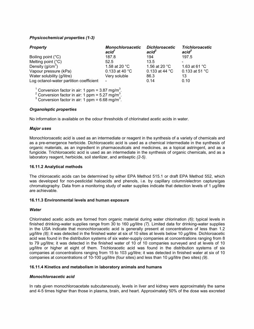
Physicochemical properties (1-3)
Property Monochloroaceticacid1
Dichloroaceticacid2
Trichloroaceticacid3
Boiling point (°C) 187.8 194 197.5Melting point (°C) 52.5 13.5 -Density (g/cm3) 1.58 at 20 °C 1.56 at 20 °C 1.63 at 61 °CVapour pressure (kPa) 0.133 at 40 °C 0.133 at 44 °C 0.133 at 51 °CWater solubility (g/litre) Very soluble 86.3 13Log octanol-water partition coefficient - 0.14 0.10
1 Conversion factor in air: 1 ppm = 3.87 mg/m3.2 Conversion factor in air: 1 ppm = 5.27 mg/m3.3 Conversion factor in air: 1 ppm = 6.68 mg/m3.
Organoleptic properties
No information is available on the odour thresholds of chlorinated acetic acids in water.
Major uses
Monochloroacetic acid is used as an intermediate or reagent in the synthesis of a variety of chemicals andas a pre-emergence herbicide. Dichloroacetic acid is used as a chemical intermediate in the synthesis oforganic materials, as an ingredient in pharmaceuticals and medicines, as a topical astringent, and as afungicide. Trichloroacetic acid is used as an intermediate in the synthesis of organic chemicals, and as alaboratory reagent, herbicide, soil sterilizer, and antiseptic (2-5).
16.11.2 Analytical methods
The chloroacetic acids can be determined by either EPA Method 515.1 or draft EPA Method 552, whichwas developed for non-pesticidal haloacids and phenols, i.e. by capillary column/electron capture/gaschromatography. Data from a monitoring study of water supplies indicate that detection levels of 1 µg/litreare achievable.
16.11.3 Environmental levels and human exposure
Water
Chlorinated acetic acids are formed from organic material during water chlorination (6); typical levels infinished drinking-water supplies range from 30 to 160 µg/litre (7). Limited data for drinking-water suppliesin the USA indicate that monochloroacetic acid is generally present at concentrations of less than 1.2µg/litre (8); it was detected in the finished water at six of 10 sites at levels below 10 µg/litre. Dichloroaceticacid was found in the distribution systems of six water-supply companies at concentrations ranging from 8to 79 µg/litre; it was detected in the finished water of 10 of 10 companies surveyed and at levels of 10µg/litre or higher at eight of them. Trichloracetic acid was found in the distribution systems of sixcompanies at concentrations ranging from 15 to 103 µg/litre; it was detected in finished water at six of 10companies at concentrations of 10-100 µg/litre (four sites) and less than 10 µg/litre (two sites) (9).
16.11.4 Kinetics and metabolism in laboratory animals and humans
Monochloroacetic acid
In rats given monochloroacetate subcutaneously, levels in liver and kidney were approximately the sameand 4-5 times higher than those in plasma, brain, and heart. Approximately 50% of the dose was excreted
in the urine within 17 h. Monochloroacetic acid is metabolized to oxalate and glycine. It can also beconjugated with glutathione, phospholipids, and cholesterol. The glutathione conjugate degrades tothiodiacetic acid (10,11).
Dichloroacetic acid
Plasma dichloroacetic acid concentrations peaked in rats 30 min after dosing by gavage, suggesting rapidintestinal absorption (12). Levels in liver and muscle increased following administration (13). In rats, dogs,and humans given sodium dichloroacetate intravenously, average half-lives of the parent compound in theplasma were 2.97, 20.8, and 0.43 h, respectively; the apparent dose dependence of plasma clearancesuggests that metabolic transformation becomes rate-limiting at high doses (14). In the rat,dichloroacetate is rapidly metabolized by dechlorination to glyoxalate, which in turn is metabolized tooxalate (15). In humans, urinary excretion of unchanged dichloroacetate was negligible after 8 h, andcumulative excretion was less than 1% of the total dose in all subjects (14).
Trichloroacetic acid
Trichloroacetic acid appears to be rapidly absorbed from the intestinal tract, metabolism occurring mainlyin the liver. It can be converted into carbon dioxide and chloride ion or reduced to the aldehyde. Acomparatively small proportion of trichloroacetic acid is metabolized, much of this compound beingexcreted unchanged in the urine (16,17).
16.11.5 Effects on laboratory animals and in vitro test systems
Monochloroacetic acid
Acute exposure
The acute oral LD50s for monochloroacetic acid in mice, male rats, and male guinea-pigs were estimatedto be 255, 76, and 80 mg/kg of body weight, respectively (18). In other studies, oral LD50s in mice werefound to be 165 mg/kg of body weight (19) and 260 mg/kg of body weight (20). In rats, an oral LD50 of2820 mg/kg of body weight in males and a dermal LD50 of 8068 mg/kg of body weight were reported (21).
Short-term exposure
Mice (20 per sex per dose) received monochloroacetic acid by gavage at 0, 25, 50, 100, 150, or 200mg/kg of body weight per day for 13 weeks. Mortality was increased at the highest dose, and females inthis group experienced decreased weight gain and increased absolute and relative liver weights.Cholinesterase levels were decreased in females at 150 and 200 mg/kg of body weight per day. TheNOAEL was 100 mg/kg of body weight per day for decreased cholinesterase levels (22).
In rats (20 per sex per dose) that received monochloroacetic acid by gavage at 0, 30, 60, 90, 120, or 150mg/kg of body weight per day for 13 weeks, effects were seen at every dose level. At 90 mg/kg of bodyweight per day and above, they included accumulations of mononuclear inflammatory cells and myofibredegeneration, elevated blood levels of thyroxin and segmented neutrophils, and increased blood ureanitrogen levels in males. At 60 mg/kg of body weight per day and above, effects included decreasedsurvival, decreased absolute and relative heart weights, degenerative and inflammatory changes(cardiomyopathy), elevated alanine and aspartate aminotransferases, increased actual liver weight andrelative kidney weight in males, and increased blood urea nitrogen levels in females. At 30 mg/kg of bodyweight per day and above, effects included decreased lymphocyte counts and decreased relative heartweight in females. The LOAEL for this study was the lowest dose tested, 30 mg/kg of body weight per day(22).
Long-term exposure
F344/N rats (70 per sex per dose) received monochloroacetic acid by gavage at doses of 0, 15, or 30mg/kg of body weight per day, 5 days per week for 2 years. No effects on body weight or clinical findingswere observed. However, survival was significantly decreased in male rats at 30 mg/kg of body weight perday and in female rats in both dose groups. The incidence of uterine polyps was marginally(nonsignificantly) increased in females at both doses. The LOAEL for this study was 15 mg/kg of bodyweight per day for reduced survival (22).
In the same study, B6C3F1 mice (60 per sex per dose) were dosed with monochloroacetic acid at 0, 50, or100 mg/kg of body weight per day, 5 days per week for 2 years. Effects were seen only at the highestdose and included decreased survival in males, decreased mean body weight and metaplasia of theolfactory epithelium in females, and inflammation of the nasal mucosa and squamous hyperplasia of theforestomach in both sexes. The NOAEL for this study was 50 mg/kg of body weight per day (22).
Mutagenicity and related end-points
Monochloroacetic acid was positive in the mouse lymphoma cell forward mutation assay without metabolicactivation (23). It was positive without, and negative with metabolic activation in the sister chromatidexchange assay in Chinese hamster ovary (CHO) cells. In the in vitro chromosomal aberration assay inCHO cells, monochloroacetic acid was negative both with and without metabolic activation (24).
Carcinogenicity
There was no evidence of carcinogenic activity in 2-year bioassays using F344/N rats and B6C3F1 mice.Rats (70 per sex per dose) received monochloroacetic acid by gavage at 0, 15, or 30 mg/kg of bodyweight per day, and mice (60 per sex per dose) received 0, 50, or 100 mg/kg of body weight per day (22).
Dichloroacetic acid
Acute exposure
LD50s of 4480 and 5520 mg of dichloroacetic acid per kg of body weight have been reported in rats andmice, respectively (18).
Short-term exposure
In a study in which Sprague-Dawley rats (5 per sex per group) were given water containing 0, 30, 125,500, or 1875 mg of dichloroacetate per litre (0, 2.4, 10, 40, or 150 mg/kg of body weight per day) for 14days, none of the parameters monitored (e.g. body weight, lactate and pyruvate levels, blood glucoselevels) was significantly altered. In this study, a NOAEL of 150 mg/kg of body weight per day wasidentified (25).
In a study in which sodium dichloroacetate was administered to Sprague-Dawley rats (10 per sex perdose) by gavage at dose levels of 0, 125, 500, or 2000 mg/kg of body weight per day for 3 months, bodyweight gain was significantly depressed in a dose-dependent manner at all dose levels. Minimal effects onhaematological parameters were observed at the two highest doses. Mean relative weights of liver,kidneys, and adrenals were significantly increased in a dose-dependent fashion. Brain and testes were theprincipal target organs; brain lesions characterized by vacuolation of the myelinated white tractsresembling oedema were observed in the cerebrum and cerebellum of treated rats of both sexes in alldose groups. Based on effects on organ weights and brain lesions, a LOAEL of 125 mg/kg of body weightper day, the lowest dose tested, was identified in this study (26,27).
Beagle dogs were given sodium dichloroacetate by capsule at 50, 75, or 100 mg/kg of body weight per

day for 13 weeks. Both sexes exhibited dose-dependent weight losses. All dose levels were associatedwith a progressive depression in erythrocyte counts, erythrocyte volume fraction (haematocrit), andhaemoglobin levels. Mean blood glucose, lactate, and pyruvate levels were significantly reduced in alltreated animals. There was an increased incidence of lung consolidation among treated dogs.Histopathological examination indicated that all treated dogs suffered slight to moderate vacuolation ofwhite myelinated tracts in the cerebrum and to a lesser extent in the cerebellum. There was an increasedincidence of haemosiderin-laden Kupffer’s cells in the liver and cystic mucosal hyperplasia in thegallbladder at all dose levels. A LOAEL of 50 mg/kg of body weight per day can be identified from thisstudy (26,27).
Long-term exposure
Male B6C3F1 mice (50 per dose) received dichloroacetate in their drinking-water at 0, 0.05, 0.5, 3.5, or 5.0g/litre (0, 7.6, 77, 410, or 486 mg/kg of body weight per day) for 60 weeks. Other groups of mice receiveddichloroacetate at 7.6 or 77 mg/kg of body weight per day for 75 weeks. In the highest-dose mice, waterconsumption was reduced to 60% of that of controls. Body weight was decreased at the two highest doselevels, and relative liver weight was increased at the three highest dose levels. An increase in kidneyweight was seen only at 410 mg/kg of body weight per day. No effects were seen on testes or spleenweight. The NOAEL for the 60- and 75-week studies was 7.6 mg/kg of body weight per day (28).
Mutagenicity and related end-points
Dichloroacetic acid was reported to cause strand breaks in DNA when administered in vivo to rats andmice in one study (29) but not in a second study at higher doses (30).
Carcinogenicity
The carcinogenic potential of dichloroacetate was investigated in B6C3F1 mice (50 males per dose) thatreceived this compound in their drinking-water for 60 weeks at concentrations of 0, 0.05, 0.5, 3.5, or 5.0g/litre (0, 7.6, 77, 410, and 486 mg/kg of body weight per day). Other groups of mice receiveddichloroacetate at 7.6 or 77 mg/kg of body weight per day for 75 weeks. Hyperplastic nodules were seenin 58% of the mice that received 410 mg/kg of body weight per day and in 83% of the mice that received486 mg/kg of body weight per day. The incidences of hepatocellular adenomas were 100% and 80%, andthose of hepatocellular carcinomas 67% and 83%, respectively. Incidences in the other dose groups weresimilar to those in controls (28).
The carcinogenic potential of dichloroacetic acid in mice was investigated in a complex regimen thatincluded pretreatment with nitrosoethylurea (NEU) at various doses. Male B6C3F1 mice were suppliedwith drinking-water containing 0, 2000, or 5000 mg of dichloroacetate per litre (0, 400, or 1000 mg/kg ofbody weight per day). Non-initiation protocols (without NEU) were used only at the high-dose level. Theincidence of hepatocellular carcinomas was 0% in the control group (no NEU or dichloroacetic acid) and81% at 1000 mg/kg of body weight per day (no NEU). With dichloroacetic acid and a low dose of NEU, thetumour incidences were 66-72% for the high and low doses. The authors concluded that dichloroaceticacid was carcinogenic at a dose of 1000 mg/kg of body weight per day without prior initiation (31).
Dichloroacetic acid exposure via drinking-water resulted in the induction of liver tumours in male B6C3F1mice. Groups of mice received dichloroacetic acid at 0, 1, or 2 g/litre (approximately 0, 137, or 295 mg/kgof body weight per day, based on the authors’ calculations of total dose for each group) for 37 or 52weeks. Hepatocellular carcinomas were seen only in 5 of 24 males (21%) that received the highest dosefor 52 weeks (32).

Trichloroacetic acid
Acute exposure
The LD50 for trichloroacetic acid was 3320 mg/kg of body weight in rats and 4970 mg/kg of body weight inmice (18).
Short-term exposure
Groups of male Sprague-Dawley rats were exposed to trichloroacetate in drinking-water at a concentrationof 5000 mg/litre (about 312 mg/kg of body weight per day) for 10, 20, or 30 days. No treatment-relatedchanges in body weight, organ weight, gross necropsy, or histopathology were found, and a short-termNOAEL of 312 mg/kg of body weight per day can be identified (33).
Six male Fischer 344 [CDF (F-344)/CrlBR] rats and eight male B6C3F1 mice were given trichloroaceticacid by gavage at 500 mg/kg of body weight per day for 10 days. The mean liver-to-body-weight ratioswere significantly increased in both species, but there was no effect on the mean kidney-to-body-weightratio. Cyanide-insensitive palmitoyl coenzyme (CoA) oxidation was increased in both species. The LOAELfor liver effects identified in this study was 500 mg/kg of body weight per day for both rats and mice (34).
Male Sprague-Dawley rats (10 per dose) received trichloroacetic acid in their drinking-water at 0, 50, 500,or 5000 mg/litre (0, 4.1, 36.5, or 355 mg/kg of body weight per day) for 90 days. No effects were seen onbody weight or actual weight of liver or kidneys. At the highest dose level, actual spleen weight wasreduced and relative liver and kidney weights were increased. Liver effects seen at the high dose includedincreased hepatic peroxisomal beta-oxidation activity, focal hepatocellular enlargement, intracellularswelling, and glycogen accumulation. The NOAEL for this study was 36.5 mg/kg of body weight per day(35).
Dose-related increases in hepatic weight were associated with the administration of trichloroacetic acid indrinking-water at 0, 300, 1000, or 2000 mg/litre to male B6C3F1 mice for 14 days. The effect wasstatistically significant at 1000 and 2000 mg/litre. The NOAEL was therefore 300 mg/litre or approximately55 mg/kg of body weight per day (36).
Long-term exposure
Male Sprague-Dawley rats were exposed to trichloroacetate in drinking-water at concentrations of 0, 50,500, or 5000 mg/litre (0, 2.89, 29.6, and 277 mg/kg of body weight per day at 6 months) for up to 12months. No significant changes were detected in body weight, organ weight, gross necropsy, orhistopathology during the experiment. A NOAEL of 277 mg/kg of body weight per day was identified in thisstudy (33).
In a study in which B6C3F1 mice received trichloroacetate at 0, 1, or 2 g/litre in drinking-water for 37 or 52weeks, both absolute liver weights and liver-to-body-weight ratios in male and female mice weresignificantly increased in a dose-related manner relative to controls. The LOAEL in this study was 1 g/litre,or 178 mg/kg of body weight per day, based on the authors’ calculations of total dose for each group (32).
Mutagenicity and related end-points
Trichloroacetic acid was not mutagenic in Salmonella typhimurium strain TA100 without metabolicactivation (37). It gave positive results in three in vivo chromosomal aberration assays in mice: the bonemarrow assay, the micronucleus test, and the sperm-head abnormality assay (38).

Carcinogenicity
Male B6C3F1 mice received trichloroacetic acid at 0, 1, or 2 g/litre (approximately 0, 178, or 319 mg/kg ofbody weight per day, based on the authors’ calculations of total dose for each group) in drinking-water for37 or 52 weeks. An increase in the incidence of hepatocellular carcinomas was seen in males in bothtreated groups, but none was seen in any of the females (32).
The carcinogenic potential of trichloroacetic acid in mice was investigated in a complex regimen thatincluded pretreatment with NEU at various doses for 61 weeks. Male B6C3F1 mice were supplied withdrinking-water containing trichloroacetate at 0, 2000, or 5000 mg/litre (0, 400, or 1000 mg/kg of bodyweight per day). Non-initiation protocols (without NEU) were used at the high-dose level only. Theincidence of hepatocellular carcinomas was 0% in the controls (no NEU or trichloroacetic acid) and 32% atthe highest dose level (no NEU). In both groups given trichloroacetic acid with a low dose of NEU, thetumour incidence was 48%. The authors concluded that trichloroacetic acid was carcinogenic at a dose of1000 mg/kg of body weight per day without prior initiation (31).
The results of two short-term tests conducted in rats-the hepatic enzyme-altered foci bioassay andstimulation of peroxisomal-dependent palmitoyl-CoA oxidation in liver-suggest that trichloroacetic acidmay possess weak promoting activity in the rat liver (33,39).
16.11.6 Effects on humans
Diabetic or hyperlipoproteinaemic patients received a daily oral dose of 3-4 g of dichloroacetate for 6-7days. Some patients experienced mild sedation, but no other laboratory or clinical evidence of adverseeffects was noted during or immediately after the treatment phase. Biochemical effects includedsignificantly reduced fasting blood glucose levels, marked decreases in plasma lactate and alanine,significantly decreased plasma cholesterol levels, decreased triglyceride levels, elevated plasma ketonebodies, and elevated serum uric acid levels (40).
Daily oral doses of 50 mg of dichloroacetate per kg were administered to two young males to treat severefamilial hypercholesterolaemia (41). In both patients, total serum cholesterol levels decreased significantly.No adverse clinical or laboratory signs were detected in one patient, but the second complained of tinglingin his fingers and toes after 16 weeks. Physical examination revealed slight decreases in the strength offacial and finger muscles, diminished to absent deep tendon reflexes, and decreased strength in allmuscle groups of the lower extremities. Electromyographic studies revealed denervation changes in footand distal leg muscles. Mild slowing of conduction velocity was noted in both posterior tibial nerves, andno measurable response was obtained in the peroneal or sural nerves. Six months after discontinuation ofthe treatment the neuropathic effects had improved, although serum cholesterol levels returned to highlevels (42).
16.11.7 Guideline values
Monochloroacetic acid
No evidence of carcinogenicity was found in a recent 2-year bioassay in rats and mice (22). Availabletoxicity data are considered insufficient for deriving a guideline value.
Dichloroacetic acid
In several bioassays, dichloroacetate has been shown to induce hepatic tumours in mice. No adequatedata on genotoxicity are available. Because the evidence for the carcinogenicity of dichloroacetate isinsufficient, a TDI of 7.6 µg/kg of body weight was calculated based on a study in which no effects wereseen on the livers of mice exposed to dichloroacetate at 7.6 mg/kg of body weight per day for 75 weeks(28) and incorporating an uncertainty factor of 1000 (100 for intra- and interspecies variation and 10 for
possible carcinogenicity). With an allocation of 20% of the TDI to drinking-water, the provisional guidelinevalue is 50 µg/litre (rounded figure).
The guideline value is designated as provisional because the data are insufficient to ensure that the valueis technically achievable. Difficulties in meeting a guideline value must never be a reason to compromiseadequate disinfection.
Trichloroacetic acid
Trichloroacetate has been shown to induce tumours in the liver of mice. It has not been found to bemutagenic in in vitro assays, but has been reported to cause chromosomal aberrations.
Because the evidence for the carcinogenicity of trichloroacetate is restricted to one species, a TDI of 17.8µg/kg of body weight was calculated based on a LOAEL of 178 mg/kg of body weight per day from a studyin which increased liver weight was seen in mice exposed to trichloroacetate in drinking-water for 52weeks (32) and incorporating an uncertainty factor of 10 000 (100 for intra- and interspecies variation and100 for the use of a slightly less-than-lifetime study, use of a LOAEL rather than a NOAEL, and possiblecarcinogenicity). The NOAEL in a 14-day study for the same effect was one-third of the LOAEL in the 52-week study (36). Based on a 20% allocation of the TDI to drinking-water, the provisional guideline value is100 µg/litre (rounded figure).
The guideline value is designated as provisional because of the limitations of the available toxicologicaldatabase and because there are inadequate data to judge whether the guideline value is technicallyachievable. Difficulties in meeting the guideline value must never be a reason for compromising adequatedisinfection.
References
1. Weast RC. Handbook of chemistry and physics. Cleveland, OH, CRC Press, 1988.
2. Verschueren K. Handbook of environmental data on organic chemicals. New York, NY, Van NostrandReinhold, 1977.
3. Budavari S, O’Neill M, Smith A, eds. The Merck index. An encyclopedia of chemicals, drugs, andbiologicals, 11th ed. Rahway, NJ, Merck, 1989.
4. Hawley GG. The condensed chemical dictionary, 10th ed. New York, NY, Van Nostrand Reinhold,1981:241.
5. Meister RT, ed. Farm chemicals handbook, 75th ed. Willoughby, OH, Meister Publishing Co., 1989.
6. Coleman WE et al. Identification of organic compounds in a mutagenic extract of a surface drinkingwater by a computerized gas chromatography/mass spectrometry system (GC/MS/COM). Environmentalscience and technology, 1980, 14:576-588.
7. Jolley RL. Basic issues in water chlorination: a chemical perspective. In: Jolley RL et al., eds. Waterchlorination: chemistry, environmental impact and health effects, Vol. 5. Chelsea, MI, Lewis Publishers,1985:19-38.
8. Bull RJ, Kopfler FC. Formation and occurrence of disinfection by-products. In: Health effects ofdisinfectants and disinfection by-products. Denver, CO, American Water Works Association ResearchFoundation, 1991:55-103.
9. Stevens AA et al. By-products of chlorination at ten operating utilities. In: Jolley RJ et al., eds. Water
chlorination: chemistry, environmental impact and health effects. Ann Arbor, MI, Ann Arbor ScientificPublishers, 1990:579-604.
10. Hayes FD, Short RD, Gibson JE. Differential toxicity of monochloroacetate, monofluoroacetate andmonoiodoacetate in rats. Toxicology and applied pharmacology, 1973, 26:93-102.
11. Bhat HK, Ahmed AE, Ansari GAS. Toxicokinetics of monochloroacetic acid: a whole-bodyautoradiography study. Toxicology, 1990, 63:35-43.
12. Stacpoole PW et al. Dichloroacetate derivatives. Metabolic effects and pharmacodynamics in normalrats. Life sciences, 1987, 41:2167-2176.
13. Evans OB. Dichloroacetate tissue concentrations and its relationship to hypolactatemia and pyruvatedehydrogenase activation. Biochemical pharmacology, 1982, 31:3124-3126.
14. Lukas G et al. Biological disposition of sodium dichloroacetate in animals and humans afterintravenous administration. Journal of pharmaceutical science, 1980, 69(4):419-421.
15. Crabb DW, Yount EA, Harris RA. The metabolic effects of dichloroacetate. Metabolism, 1981,30:1024-1039.
16. Hobara T et al. Intestinal absorption of chloral hydrate, free trichloroethanol and trichloroacetic acid indogs. Pharmacology and toxicology, 1988, 62:250-258.
17. Hobara T et al. Extra-hepatic metabolism of chloral hydrate, trichloroethanol and trichloroacetic acid indogs. Pharmacology and toxicology, 1987, 61:58-62.
18. Woodard G et al. The acute oral toxicity of acetic, chloroacetic, dichloroacetic and trichloroaceticacids. Journal of industrial hygiene and toxicology, 1941, 23(2):78-82.
19. Morrison JL. Toxicity of certain halogen substituted aliphatic acids for white mice. Journal ofpharmacology and experimental therapeutics, 1946, 86:336-338.
20. Berardi MR et al. Monochloroacetic acid toxicity in the mouse associated with blood-brain barrierdamage. Fundamental and applied toxicology, 1987, 9:469-479.
21. Smyth HF, Carpenter CP, Weil CS. Range-finding toxicity data: list IV. American Medical Associationarchives of industrial hygiene and occupational medicine, 1951, 4:119-122.
22. National Toxicology Program. NTP technical report on the toxicology and carcinogenesis studies ofmonochloroacetic acid in F344/N rats and B6C3F1 mice (gavage studies). Research Triangle Park, NC,1990 (NTP TR 396; NIH Publication No. 90-2851).
23. McGregor DB et al. Responses of the LS178Y tk+/tk- mouse lymphoma cell forward mutation assay tocoded chemicals. I: Results for nine compounds. Environmental mutagenesis, 1987, 9:143-160.
24. Galloway SM et al. Chromosome aberrations and sister chromatid exchanges in Chinese hamsterovary cells: evaluation of 108 chemicals. Environmental and molecular mutagenesis, 1987, 10(Suppl.10):1-175.
25. Davis ME. Effect of chloroacetic acids on the kidneys. Environmental health perspectives, 1986,69:209-214.
26. Katz R et al. CGS 7927A (dichloroacetate): 90-day oral administration to rats. Summit, NJ, Ciba-
Geigy, 1978.
27. Katz R et al. Dichloroacetate, sodium: 3-month oral toxicity studies in rats and dogs. Toxicology andapplied pharmacology, 1981, 57:273-287.
28. DeAngelo AB et al. The carcinogenicity of dichloroacetic acid in the male B6C3F1 mouse.Fundamental and applied toxicology, 1991, 16:337-347.
29. Nelson MA, Bull RJ. Induction of strand breaks in DNA by trichloroethylene and metabolites in rat andmouse liver in vivo. Toxicology and applied pharmacology, 1988, 94:45-54.
30. Chang LW, Daniel FB, DeAngelo AB. Analysis of DNA strand breaks induced in rodent liver in vivo,hepatocytes in primary culture, and a human cell line by chloroacetic acids and chloroacetaldehydes. In:Proceedings of the Annual Meeting of the Society of Toxicology, Atlanta, GA, 27 February - 3 March 1989.Research Triangle Park, NC, US Environmental Protection Agency, Health Effects Research Laboratory,1989.
31. Herren-Freund SL, Pereira MA. Carcinogenicity of by-products of disinfection in mouse and rat liver.Environmental health perspectives, 1986, 69:59-65.
32. Bull RJ et al. Liver tumor induction in B6C3F1 mice by dichloroacetate and trichloroacetate.Toxicology, 1990, 63:341-359.
33. Parnell JJ, Koller LD, Exon JH. Assessment of hepatic initiation-promotion properties of trichloroaceticacid. Archives of environmental contamination and toxicology, 1988, 17:429-436.
34. Goldsworthy TL, Popp JA. Chlorinated hydrocarbon-induced peroxisomal enzyme activity in relation tospecies and organ carcinogenicity. Toxicology and applied pharmacology, 1987, 88:225-233.
35. Mather GG, Exon JH, Koller LD. Subchronic 90-day toxicity of dichloroacetic and trichloroacetic acid inrats. Toxicology, 1990, 64:71-80.
36. Sanchez IM, Bull RJ. Early induction of reparative hyperplasia in the liver of B6C3F1 mice treated withdichloroacetate and trichloroacetate. Toxicology, 1990, 64:33-46.
37. Rapson WH, Nazar MA, Butsky VV. Mutagenicity produced by aqueous chlorination of organiccompounds. Bulletin of environmental contamination and toxicology, 1980, 24:590-596.
38. Bhunya SP, Behera BC. Relative genotoxicity of trichloroacetic acid (TCA) as revealed by differentcytogenetic assays: bone marrow chromosome aberration, micronucleus and sperm-head abnormality inthe mouse. Mutation research, 1987, 188:215-221.
39. Parnell MJ et al. Trichloroacetic acid effects on rat liver peroxisomes and enzyme-altered foci.Environmental health perspectives, 1986, 69:73-79.
40. Stacpoole PW, Moore GW, Kornauser DM. Metabolic effects of dichloroacetate in patients withdiabetes mellitus and hyperlipoproteinemia. New England journal of medicine, 1978, 298:526-530.
41. Moore GW et al. Reduction of serum cholesterol in two patients with homozygous familialhypercholesterolemia by dichloroacetate. Atherosclerosis, 1979, 33:285-293.
42. Stacpoole PW, Moore GW, Kornauser DM. Toxicity of chronic dichloroacetate. New England jornal ofmedicine, 1979, 300:372 (letter)
16.12 Chloral hydrate (trichloroacetaldehyde)
16.12.1 General description
Identity
Compound CAS no. Molecular formulaTrichloroacetaldehyde 75-87-6 Cl3CCHOChloral hydrate 302-17-0 Cl3CCH(OH)2
The IUPAC name for trichloroacetaldehyde is trichloroethanal.
Physicochemical properties (1-3)
Property Trichloroacetaldehyde1
Chloral hydrate2
Boiling point (°C) 97.8 96.3Melting point (°C) -57.5 57Density at 20 °C (g/cm3) 1.512 1.908Water solubility (g/litre) Freely soluble 8300 (25 °C)
1 Conversion factor in air: 1 ppm = 6.03 mg/m3.2 Conversion factor in air: 1 ppm = 6.77 mg/m3.
Major uses
Hydrated trichloroacetaldehyde (chloral hydrate) is used as a sedative and hypnotic in human andveterinary medicine (3,4).
16.12.2 Analytical methods
Trichloroacetaldehyde and chloral hydrate are determined by draft EPA Method 551, i.e. by capillarycolumn/electron-capture/gas chromatography. Monitoring data indicate that a quantitation limit of 0.4µg/litre is achievable for chloral hydrate.
16.12.3 Environmental levels and human exposure
Water
Trichloroacetaldehyde may enter water from industrial discharges or be formed as a by-product during thechlorination of water containing organic precursor molecules. Chloral hydrate is formed whentrichloroacetaldehyde is dissolved in water; it was detected in six of 10 drinking-water supplies sampled, atconcentrations ranging from 0.01 to 5 µg/litre (5). In another survey, chloral hydrate was detected in eachof 10 drinking-water systems at concentrations ranging from 10 to 100 µg/litre (6).
16.12.4 Kinetics and metabolism in laboratory animals and humans
Chloral hydrate was rapidly absorbed in dogs and humans, most, if not all, being either oxidized totrichloroacetic acid or reduced to trichloroethanol. Most of the dose was excreted in the urine astrichloroethanol glucuronide, together with small amounts of free trichloroethanol. The remainder wasexcreted as trichloroacetate (7,8).

16.12.5 Effects on laboratory animals and in vitro test systems
Acute exposure
The acute oral LD50 for chloral hydrate in mice was 1265-1442 mg/kg of body weight (9). Rats are moresensitive than mice, acute oral LD50s ranging from 285 mg/kg of body weight in newborn pups to 479mg/kg of body weight in adults (10).
Short-term exposure
Groups of male CD-1 mice were dosed with chloral hydrate by gavage at 14.4 or 144 mg/kg of bodyweight per day for 14 days. No significant effect on body weight was observed, but a dose-dependentincrease in liver weight and decrease in spleen weight were observed. These changes were statisticallysignificant at the higher dose. No effects on haematological or serum biochemical parameters were noted,except for an unusual decrease in lactate dehydrogenase at the higher dose. A NOAEL of 14.4 mg/kg ofbody weight per day was identified in this study (9).
Male and female CD-1 mice were supplied with chloral hydrate in drinking-water at 70 or 700 mg/litre(time-weighted average doses of approximately 16 or 160 mg/kg of body weight per day) for 90 days. Theliver appeared to be the tissue most seriously affected. In males, dose-related hepatomegaly andmicrosomal proliferation were seen, accompanied by small changes in serum chemistry values forpotassium, cholesterol, and glutathione, but no significant changes in serum enzyme levels. Females didnot show hepatomegaly but did display changed hepatic microsomal parameters. No other significanttoxicological changes were observed in either sex. Based on hepatomegaly, a LOAEL of 16 mg/kg of bodyweight per day (the lowest dose tested) was identified for chloral hydrate in this study (9).
Long-term exposure
A chronic 2-year drinking-water study of chloral hydrate was conducted on groups of 40 male B6C3F1mice for 104 weeks at dose levels of 0 or 1 g/litre (0 and 166 mg/kg of body weight per day). Lesions wereprimarily confined to the liver and included hepatocellular necrosis, inflammation, and cytomegaly. Organweight changes were also evident, with increases in absolute and relative liver weights throughout thetreatment period. Spleen, kidney, and testicular weights as well as pathological changes in these organswere comparable to those in controls (11).
Reproductive toxicity, embryotoxicity, and teratogenicity
Female mice were exposed to chloral hydrate in drinking-water at concentrations corresponding to dosesof 21.3 or 204.8 mg/kg of body weight per day from before breeding until weaning (females only). Nogross malformations were noted, and no significant effects were observed in gestational duration, numberof pups delivered, pup weight, or number of stillborn pups. All pups showed the same rate of developmentand degree of performance on several neurobehavioural tests, except that pups from the high-dose groupshowed impaired retention in a passive avoidance learning task. A NOAEL of 21.3 mg/kg of body weightper day for developmental effects was identified in this study (12).
Mutagenicity and related end-points
Chloral hydrate was reported to be mutagenic in Salmonella typhimurium strain TA98. Both chloralhydrate and trichloroacetaldehyde were reported to be mutagenic in S. typhimurium strain TA100, withand without metabolic activation. Mutagenic activity was also observed for chloral hydrate in Streptomycescoelicolor and Aspergillus nidulans. Neither chloral hydrate nor trichloroacetaldehyde was mutagenic in S.typhimurium strain TA1535 (13,14).
Chloral hydrate administered to mice caused significant increases in the number of hyperhaploid cells,
probably due to chromosome nondisjunction resulting from a disruptive effect of chloral hydrate on themitotic spindle (15). Similar disruptive effects of chloral hydrate on chromosomal segregation have beenobserved in A. nidulans (16) and Saccharomyces cerevisiae (17). Chloral hydrate did not bind to DNA inmouse liver in vivo or form DNA-protein cross-links when incubated with rat liver nuclei in vitro, whichsuggests that it may have low genotoxic potential in animals (18).
Carcinogenicity
The carcinogenic potential of chloral hydrate was investigated in male B6C3F1 mice (40 per dose) thatreceived the compound in their drinking-water at dose levels of 0 or 1 g/litre (0 or 166 mg/kg of bodyweight per day) for up to 104 weeks. The most prevalent lesions observed were hepatocellularcarcinomas (46%) and hepatocellular adenomas (29%). Proliferative lesions were also observed in theuntreated controls but at a lower incidence (2% and 1%, respectively). Hyperplastic nodules (4%) wereobserved in the treated group but not in untreated controls (11).
A single oral dose of chloral hydrate (10 mg/kg of body weight) administered to 15-day-old male miceresulted in a significant increase in liver tumours after 48-92 weeks (19).
16.12.6 Effects on humans
Chloral hydrate has been widely used as a sedative or hypnotic drug in humans at recommended oraldoses of 0.25-1.0 g. Concentrated solutions are irritating to the gastrointestinal tract, and the ingestion ofundiluted preparations causes nausea and vomiting. The acute oral toxic dose in humans is usually about10 g, which causes severe respiratory depression and hypotension (20).
Adverse effects in patients given either 0.5 or 1.0 g of chloral hydrate included central nervous systemdepression, minor sensitivity reactions, gastrointestinal disturbances, and central nervous systemexcitement (21). Cardiac arrhythmias induced by chloral hydrate have been described (22).
The chronic use of chloral hydrate may result in the development of tolerance and physical dependence.Those physically dependent on it reportedly take as much as 12 g per day (23).
16.12.7 Provisional guideline value
Chloral hydrate causes liver tumours in mice. It has been shown to be mutagenic in short-term tests invitro, but it does not bind to DNA. It has been shown to disrupt chromosome segregation in cell division.Because of the lack of adequate long-term studies, the guideline value for chloral hydrate is based on theLOAEL from a study in which liver effects were seen in mice that received chloral hydrate in drinking-waterat 16 mg/kg of body weight per day for 90 days (9). A TDI of 1.6 µg/kg of body weight was calculatedusing this LOAEL and incorporating an uncertainty factor of 10 000 (100 for intra- and interspeciesvariation, 10 for the short duration of the study, and 10 for the use of a LOAEL instead of a NOAEL). Withan allocation of 20% of the TDI to drinking-water, the provisional guideline value is 10 µg/litre (roundedfigure). This guideline value is designated as provisional because of the limitations of the availabledatabase.
References
1. Weast RC. Handbook of chemistry and physics. Cleveland, OH, CRC Press, 1988.
2. Verschueren K. Handbook of environmental data on organic chemicals. New York, NY, Van NostrandReinhold, 1977.
3. Budavari S, O’Neill M, Smith A, eds. The Merck index. An encyclopedia of chemicals, drugs, andbiologicals, 11th ed. Rahway, NJ, Merck, 1989.
4. Hawley GG. The condensed chemical dictionary, 10th ed. New York, NY, Van Nostrand Reinhold,1981:241.
5. Keith LH et al. Identification of organic compounds in drinking water from thirteen U.S. cities. In: KeithLH, ed. Identification and analysis of organic pollutants in water. Ann Arbor, MI, Ann Arbor SciencePublishers, 1976:329-373.
6. Stevens AA et al. By-products of chlorination at ten operating utilities. In: Jolley RJ et al., eds. Waterchlorination: chemistry, environmental impact and health effects. Vol. 6. Ann Arbor, MI, Ann ArborScientific Publishers, 1990:579-604.
7. Marshall EK, Owens AH. Absorption, excretion and metabolic fate of chloral hydrate andtrichloroethanol. Bulletin of the Johns Hopkins Hospital, 1954, 95:1-18.
8. Butler TC. The metabolic fate of chloral hydrate. Journal of pharmacology and experimentaltherapeutics, 1948, 92:49-58.
9. Sanders VM et al. Toxicology of chloral hydrate in the mouse. Environmental health perspectives, 1982,44:137-146.
10. Goldenthal EI. A compilation of LD50 values in newborn and adult animals. Toxicology and appliedpharmacology, 1971, 18:185-207.
11. Daniel FB et al. Hepatocarcinogenicity of chloral hydrate, 2-chloroacetaldehyde, and dichloroaceticacid in the male B6C3F1 mouse. Fundamental and applied toxicology, 1992, 19(2):159-168.
12. Kallman MJ, Kaempf GL, Balster RL. Behavioral toxicity of chloral in mice: an approach to evaluation.Neurobehavioral toxicology and teratology, 1984, 6(2):137-146.
13. Bruce WR, Heddle JA. The mutagenic activity of 61 agents as determined by the micronucleus,Salmonella, and sperm abnormality assays. Canadian journal of genetics and cytology, 1979, 21:319-334.
14. Bignami M et al. Mutagenicity of halogenated aliphatic hydrocarbons in Salmonella typhimurium,Streptomyces coelicolor and Aspergillus nidulans. Chemico-biological interactions, 1980, 30:9-23.
15. Russo A, Pacchierotti F, Metalli P. Nondisjunction induced in mouse spermatogenesis by chloralhydrate, a metabolite of trichloroethylene. Environmental mutagenesis, 1984, 6(5):695-703.
16. Kafer E. Tests which distinguish induced crossing-over and aneuploidy from secondary segregation inAspergillus treated with chloral hydrate or gamma rays. Mutation research, 1986, 164:145-166.
17. Sora S, Carbone ML. Chloral hydrate, methylmercury hydroxide and ethidium bromide affectchromosomal segregation during meiosis of Saccharomyces cerevisiae. Mutation research, 1987, 190:13-17.
18. Keller DA, Heck H. Mechanistic studies on chloral toxicity: relationship to trichloroethylenecarcinogenesis. Toxicology letters, 1988, 42:183-191.
19. Rijhsinghani KS et al. Induction of neoplastic lesions in the livers of C57BL × C3HF1 mice by chloralhydrate. Cancer detection and prevention, 1986, 9:279-288.
20. Gilman AG et al., eds. Goodman and Gilman: the pharmacological basis of therapeutics, 7th ed. NewYork, NY, Macmillan Publishing, 1985:360-362.
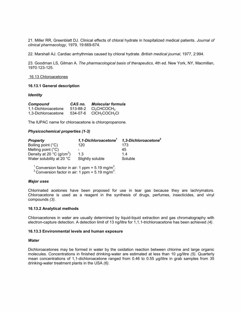
21. Miller RR, Greenblatt DJ. Clinical effects of chloral hydrate in hospitalized medical patients. Journal ofclinical pharmacology, 1979, 19:669-674.
22. Marshall AJ. Cardiac arrhythmias caused by chloral hydrate. British medical journal, 1977, 2:994.
23. Goodman LS, Gilman A. The pharmacological basis of therapeutics, 4th ed. New York, NY, Macmillan,1970:123-125.
16.13 Chloroacetones
16.13.1 General description
Identity
Compound CAS no. Molecular formula1,1-Dichloroacetone 513-88-2 Cl2CHCOCH31,3-Dichloroacetone 534-07-6 ClCH2COCH2Cl
The IUPAC name for chloroacetone is chloropropanone.
Physicochemical properties (1-3)
Property 1,1-Dichloroacetone1 1,3-Dichloroacetone2
Boiling point (°C) 120 173Melting point (°C) - 45Density at 20 °C (g/cm3) 1.3 1.4Water solubility at 20 °C Slightly soluble Soluble
1 Conversion factor in air: 1 ppm = 5.19 mg/m3.2 Conversion factor in air: 1 ppm = 5.19 mg/m3.
Major uses
Chlorinated acetones have been proposed for use in tear gas because they are lachrymators.Chloroacetone is used as a reagent in the synthesis of drugs, perfumes, insecticides, and vinylcompounds (3).
16.13.2 Analytical methods
Chloroacetones in water are usually determined by liquid-liquid extraction and gas chromatography withelectron-capture detection. A detection limit of 13 ng/litre for 1,1,1-trichloroacetone has been achieved (4).
16.13.3 Environmental levels and human exposure
Water
Dichloroacetones may be formed in water by the oxidation reaction between chlorine and large organicmolecules. Concentrations in finished drinking-water are estimated at less than 10 µg/litre (5). Quarterlymean concentrations of 1,1-dichloroacetone ranged from 0.46 to 0.55 µg/litre in grab samples from 35drinking-water treatment plants in the USA (6).
16.13.4 Effects on laboratory animals and in vitro test systems
Acute exposure
Oral LD50s of 250 mg/kg of body weight for 1,1-dichloroacetone and 25 mg/kg of body weight for 1,3-dichloroacetone have been reported in the mouse (7).
Short-term exposure
The hepatotoxicity of 1,1- and 1,3-dichloroacetone was investigated in mice. Single oral doses of eachcompound were administered to CD-1 mice (5-12 per dose). A dose of 0.25 ml/kg of body weight (325mg/kg of body weight) of 1,1-dichloroacetone caused significant increases in liver enzymes in serum, andhistological examination showed evidence of periportal necrosis. These effects were not observed atdoses of 130 mg/kg of body weight or lower. Liver glutathione levels were decreased at doses of 0.1 and0.25 ml/kg but not at 0.05 ml/kg (65 mg/kg of body weight). Based on measurements of serum enzymes,liver glutathione, and histopathological examination, 1,3-dichloroacetone did not cause liver toxicity atdoses of up to 20 mg/kg of body weight. NOAELs of 65 and 20 mg/kg of body weight for 1,1-dichloroacetone and 1,3-dichloroacetone, respectively, were identified in this study (7).
Mutagenicity and related end-points
A number of chlorinated acetones, including 1,1-, 1,3-, 1,1,1-, 1,1,3,3-, and pentachloroacetone, weredirect-acting mutagens in one or both of Salmonella typhimurium strains TA98 and TA100. Mutagenicactivity decreased with increased chlorine substitutions at the C-1 and C-3 positions, although 1,1,1-trichloroacetone was 25 times as potent as 1,1-dichloroacetone (8).
Carcinogenicity
The carcinogenic activity of 1,1-dichloroacetone and 1,1,1-trichloroacetone was studied in femaleSENCAR mice (60 per dose) that received a single oral (200 mg/kg) or topical (400 mg/kg) dose of 1,1-dichloroacetone or a single oral (50 mg/kg) or topical (400 mg/kg) dose of 1,1,1-trichloroacetone. Thevehicle was 0.2 ml of dimethyl sulfoxide for oral exposure and 0.2 ml of ethanol for topical exposure. Twoweeks after dosing, a tumour promotion schedule was begun with 1 µg of 12-O-tetradecanoyl-phorbol-13-acetate (TPA) three times per week for 20 weeks. However, 24 weeks after the start of the promotionschedule, there was no evidence of an increase in skin tumours attributable to either chemical (9).
The results of carcinogenicity studies with chlorinated acetones using the mouse skin assay have alsobeen reported. Groups of 40 SENCAR mice received topical doses of 1,1-dichloroacetone at 400, 600, or800 mg/kg; 1,3-dichloroacetone at 50, 75, or 100 mg/kg; or 1,1,3-trichloroacetone or 1,1,1-trichloroacetone at 50 mg/kg. Doses were applied six times over a 2-week period using 0.2 ml of ethanolas the vehicle. After 2 weeks, 1.0 µg of TPA in 0.2 ml of acetone was applied three times per week for 20weeks. After 24 weeks, the percentages of animals with tumours in the respective dose groups were: 5%in controls; 0, 5%, and 5% for 1,1-dichloroacetone; 48%, 45%, and 30% for 1,3-dichloroacetone; 10%,5%, and 0% for 1,1,1-trichloroacetone; and 10% for 1,1,3-trichloroacetone. The authors concluded that1,3-dichloroacetone is a tumour initiator in mouse skin (10).
16.13.5 Conclusions
The toxicological data on the chloroacetones are very limited, although studies with single doses of 1,1-dichloroacetone indicate that it affects the liver. There are insufficient data at present to permit theproposal of guideline values for any of the chloroacetones.

References
1. Weast RC. Handbook of chemistry and physics. Cleveland, OH, CRC Press, 1988.
2. Verschueren K. Handbook of environmental data on organic chemicals. New York, NY, Van NostrandReinhold, 1977.
3. Budavari S, O’Neill M, Smith A, eds. The Merck index. An encyclopedia of chemicals, drugs andbiologicals, 11th ed. Rahway, NJ, Merck, 1989.
4. Krasner SW et al. The occurrence of disinfection by-products in U.S. drinking water. Journal of theAmerican Water Works Association, 1989, 81(8):41-53.
5. Coleman WE et al. Identification of organic compounds in a mutagenic extract of a surface drinkingwater by a computerized gas chromatography/mass spectrometry system (GC/MS/COM). Environmentalscience and technology, 1980, 14:576-588.
6. Bull RJ, Kopfler FC. Formation and occurrence of disinfection by-products. In: Health effects ofdisinfectants and disinfection by-products. Denver, CO, American Water Works Association ResearchFoundation, 1991:55-103.
7. Laurie RD et al. Studies of the toxic interactions of disinfectant by-products. Environmental healthperspectives, 1986, 69:203-207.
8. Meier JR et al. Evaluation of chemicals used for drinking water disinfection for production ofchromosomal damage and sperm-head abnormalities in mice. Environmental mutagenesis, 1985, 7:201-211.
9. Bull RJ, Robinson M. Carcinogenic activity of haloacetonitrile and haloacetone derivatives in the mouseskin and lung. In: Jolley RL et al., eds. Water chlorination: chemistry, environmental impact and healtheffects, Vol. 5. Chelsea, MI, Lewis Publishers, 1985:221-227.
10. Robinson J, Laurie RD, Bull RJ. Carcinogenic activity associated with halogenated acetones andacroleins in the mouse skin assay. Cancer letter, 1989, 48:197-203.
16.14 Halogenated acetoniriles
16.14.1 General description
Identity
Compound CAS no. Molecular formulaDichloroacetonitrile 3018-12-0 CHCl2CNDibromoacetonitrile 3252-43-5 CHBr2CNBromochloroacetonitrile 83463-62-1 CHBrClCNTrichloroacetonitrile 545-06-2 CCl3CN
The IUPAC name for acetonitrile is ethanenitrile.
Physicochemical properties (1-3)
Property Dichloro-acetonitrile1
Dibromo-acetonitrile2
Bromochloro-acetonitrile3
Trichloro-acetonitrile4
Boiling point (°C) 112.3 67-69 125-130 84.6

Density at 20 °C (g/cm3) 1.37 2.30 1.68 1.44
1 Conversion factor in air: 1 ppm = 4.49 mg/m3.2 Conversion factor in air: 1 ppm = 8.14 mg/m3.3 Conversion factor in air: 1 ppm = 6.31 mg/m3.4 Conversion factor in air: 1 ppm = 5.91 mg/m3.
Major uses
Trichloroacetonitrile has been used as an insecticide (3).
Environmental fate
Halogenated acetonitriles are reported to undergo hydrolysis in water, yielding nonvolatile products (4).
16.14.2 Analytical methods
Draft EPA Method 551 can be used for the determination of haloacetonitriles, by capillary-column/electron-capture/gas chromatography. Extremely low detection limits are achievable. Monitoringdata indicate quantitation limits of 0.4 µg/litre for dichloro-, dibromo-, bromochloro-, andtrichloroacetonitrile.
16.14.3 Environmental levels and human exposure
Water
Halogenated acetonitriles are formed from organic precursors during the chlorination of drinking-water.Dihalogenated acetonitriles are present in chlorinated water at concentrations ranging from 0.3 to 40µg/litre (5,6); trichloroacetonitrile has been detected at a concentration of 0.1 µg/litre (6).
16.14.4 Kinetics and metabolism in laboratory animals and humans
Dichloroacetonitrile is well absorbed from the gastrointestinal tract. Most is excreted in urine, with smalleramounts being eliminated in expired air and faeces. Its metabolites are detected in highest concentrationsin liver, blood, muscle, and skin. Differences in the pattern of label distribution in tissues and of labelexcretion indicate that a substantial portion of dichloroacetonitrile is split into two one-carbon fragmentsthat undergo different metabolic reactions (7).
Halogenated acetonitriles may be formed in vivo following the ingestion of chlorinated water. Dichloro- anddibromoacetonitrile were detected in the stomach contents of rats following the oral administration ofsodium hypochlorite/potassium bromide, presumably formed by reaction with organic material in thestomach (8).
16.14.5 Effects on laboratory animals and in vitro test systems
Acute exposure
Acute oral LD50s for the halogenated acetonitriles in rodents range from 245 to 361 mg/kg of body weight(9-11).

Short-term exposure
Dichloroacetonitrile
In a 14-day toxicity study, doses of 0, 12, 23, 45, or 90 mg/kg of body weight per day were administered torats (10 per sex per dose). In males, a slight, nonsignificant depression in body weight gain was observedat the three highest doses, whereas decreased weight gain was noted only at the highest dose in females.No consistent treatment-related effects were observed in any of the haematological, serum chemistry, orurinary parameters measured. Absolute thymus weights in males given 23, 45, or 90 mg/kg of body weightper day were significantly decreased, but the thymus-to-body-weight ratio was decreased only at thehighest dose. Based on decreased body weight as the most sensitive end-point, the NOAEL in this studywas 45 mg/kg of body weight per day (11).
In a 90-day study, doses of 0, 8, 33, or 65 mg/kg of body weight per day were administered to groups ofrats (20 per sex per dose). No consistent compound-related effects were observed in any of thehaematological, urinary, or serum chemistry parameters measured. Body weight gain was significantlydepressed in females at 65 mg/kg of body weight per day and in males at 33 mg/kg of body weight perday. The NOAEL in this study was 8 mg/kg of body weight per day (11).
Dibromoacetonitrile
The 14-day toxicity of dibromoacetonitrile was investigated in adult rats (10 per sex per dose) thatreceived this compound by gavage at doses of 0, 23, 45, 90, or 180 mg/kg of body weight per day. Therewas increased mortality at 90 and 180 mg/kg of body weight per day (30% and 100%, respectively). Noconsistent compound-related effects were apparent in any of the serum chemistry, haematological, orurinary parameters measured. The authors concluded that decreased body weight was the most sensitiveindicator of toxicity. In males, dose-dependent decreases occurred at 45 and 90 mg/kg of body weight perday. No effect on body weight was noted in females. The authors concluded that the NOAEL was 45mg/kg of body weight per day, but the body weight decrease in male rats exposed at this dose suggeststhat the NOAEL should be 23 mg/kg of body weight per day (11).
In a 90-day study, dibromoacetonitrile doses of 0, 6, 23, or 45 mg/kg of body weight per day in corn oilwere administered by gavage to rats (20 per sex per dose). Body weight gain was significantly depressedonly in males at the highest dose tested. No compound-related deaths occurred, no consistent compound-related adverse effects were observed in the haematological, urinary, or serum chemistry parametersmeasured, and no remarkable findings were apparent at gross necropsy. The authors concluded thatdecreased body weight is the most sensitive end-point and identified a NOAEL of 23 mg/kg of body weightper day (11).
Reproductive toxicity, embryotoxicity, and teratogenicity
Dichloroacetonitrile
Dichloroacetonitrile was administered to pregnant Long-Evans rats by gavage at doses of 0, 5, 15, 25, or45 mg/kg of body weight per day on gestation days 6-18. At the two highest dose levels, fetal resorptionswere significantly increased and fetal weight and size were decreased. Malformations of thecardiovascular, digestive, and urogenital systems were observed in fetuses from dams at the two highestdose levels. No effects were described at lower doses. This study indicated that 15 mg/kg of body weightper day is a developmental and teratogenic NOAEL for dichloroacetonitrile (12).
Pregnant Long-Evans rats were exposed via gavage to dichloroacetonitrile at 55 mg/kg of body weight perday on days 7-21 of gestation. Dichloroacetonitrile decreased the percentage of females delivering littersand increased the percentage of fetal resorptions. Mean birth weights were reduced, and postnatalsurvival was significantly reduced. A LOAEL of 55 mg/kg of body weight per day was identified in this
study (13,14).
Groups of 10 male B6C3F1 mice were dosed with dichloroacetonitrile at 0, 12.5, 25, or 50 mg/kg of bodyweight by gavage for 5 consecutive days. No treatment-related effects were found on examination ofsperm for abnormal sperm-head morphology (9).
Dibromoacetonitrile
Pregnant Long-Evans rats were exposed via gavage to dibromoacetonitrile at 50 mg/kg of body weight perday on gestation days 7-21. Maternal weight gain decreased during gestation, and mean birth weightswere reduced. A LOAEL of 50 mg/kg of body weight per day was identified in this study (13,14).
Groups of 10 male B6C3F1 mice were dosed with dibromoacetonitrile at 0, 12.5, 25, or 50 mg/kg of bodyweight by gavage for 5 days. There were no treatment-related effects on sperm-head morphology. (9)
Bromochloroacetonitrile
In a study in which pregnant Long-Evans rats were exposed via gavage to bromochloroacetonitrile at 55mg/kg of body weight per day on days 7-21 of gestation, mean birth weights were reduced (13,14). Therewere no treatment-related effects on sperm-head morphology when groups of 10 male B6C3F1 mice weredosed with bromochloroacetonitrile at 0, 12.5, 25, or 50 mg/kg of body weight by gavage for 5 days (9).
Trichloroacetonitrile
Trichloroacetonitrile was administered to pregnant Long-Evans rats by gavage at doses of 0, 1, 7.5, 15,35, or 55 mg/kg of body weight per day on gestation days 6-18. The highest dose was lethal in four of 19dams, and there was 100% resorption in most survivors. Dose-related decreases in fetal weight and fetalviability were observed in animals exposed at all but the lowest dose. Numerous cardiovascular andurogenital malformations were seen in surviving fetuses. The frequency of soft-tissue malformations wasdose-dependent, ranging from 18% at 7.5 mg/kg of body weight per day to 35% at 35 mg/kg of bodyweight per day. The malformation frequency at 1 mg/kg of body weight per day (8.4%) was not statisticallydifferent from that in controls (3.8%), but the authors expressed concern that this level of malformationcould be of biological significance and suggested that a dose of 1 mg/kg of body weight per day might bethe NOAEL or close to the LOAEL for teratogenic effects. (15)
Pregnant Long-Evans rats were exposed via gavage to trichloroacetonitrile at 55 mg/kg of body weight perday on gestation days 7-21. Trichloroacetonitrile decreased the percentage of females delivering littersand increased the percentage of fetal resorptions, while mean birth weights were reduced, and postnatalsurvival was significantly reduced. A LOAEL of 55 mg/kg of body weight per day was identified in thisstudy (13,14).
Groups of 10 male B6C3F1 mice were dosed with trichloroacetonitrile at 0, 12.5, 25, or 50 mg/kg of bodyweight by gavage for 5 days. Examination of sperm for abnormal sperm-head morphology revealed notreatment-related effects (9).
Mutagenicity and related end-points
No significant increase in the frequency of micronuclei was observed for any of the four halogenatedacetonitriles in an in vivo assay in mice. All the halogenated acetonitriles induced sister chromatidexchanges in Chinese hamster ovary cells with metabolic activation and all except dichloroacetonitrilewithout it. Comparisons of potencies showed the order to be as follows: dibromoacetonitrile >bromochloroacetonitrile > trichloroacetonitrile > dichloroacetonitrile (16).
Results of the Salmonella/microsome assay, with and without metabolic activation by the S9 fraction of rat
liver homogenate, suggested that dichloro- and bromochloroacetonitrile were direct-acting mutagens andthat dibromo- and trichloroacetonitrile were nonmutagenic (16). Halogenated acetonitriles reportedlyproduced DNA strand breaks in cultured human lymphoblastic cells (17). The most potent initiator wastrichloroacetonitrile, followed by bromochloro-, dibromo-, and dichloroacetonitrile. It was reported thatdichloroacetonitrile formed a covalent adduct when incubated with calf thymus DNA (18).
Carcinogenicity
Groups of 40 A/J female mice were given a single oral dose of dichloro-, dibromo, bromochloro-, ortrichloroacetonitrile of 10 mg/kg (4.3 mg/kg of body weight per day) three times per week for 8 weeks. Theincidence of lung tumours (adenomas) was significantly increased in groups given trichloro- andbromochloroacetonitrile, while dichloro- and dibromoacetonitrile produced marginal and nonsignificantincreases in such tumours. The authors stated that the results should be interpreted with caution, as thereis a relatively large variation in background incidence of lung tumours in this strain of mice, and as thedose level tested was considerably below the maximum tolerated dose (19).
The ability of the four halogenated acetonitriles to act as tumour initiators was studied in mouse skin. Sixtopical doses of 200, 400, or 800 mg/kg were applied to the shaved backs of female SENCAR mice (40per dose) over a 2-week period to give total doses of 1200, 2400, and 4800 mg/kg of body weight. Twoweeks after the last dose, a tumour promotion schedule involving the application of 1 µg of 12-O-tetradecanoyl-phorbol-13-acetate (TPA) three times per week was begun and continued for 20 weeks.After 1 year, the incidence of squamous cell carcinomas was significantly increased relative to the controlgroup in mice treated with dibromo- and bromochloroacetonitrile. An increase in incidence withtrichloroacetonitrile at 2400 mg/kg was not reproducible. No significant increases in carcinomas wereobserved with dichloroacetonitrile (16). Dichloro-, dibromo-, and trichloroacetonitrile were inactive asinitiators in the rat liver?-glutamyl transpeptidase foci assay (20).
16.14.6 Guideline values
IARC has concluded that dichloro-, dibromo-, bromochloro-, and trichloroacetonitriles are not classifiableas to their carcinogenicity to humans (Group 3) (5).
Dichloroacetonitrile
A TDI of 15 µg/kg of body weight was calculated from the NOAEL of 15 mg/kg of body weight per day forfetal resorptions, decreases in fetal weight and size, and malformations of the cardiovascular, digestive,and urogenital systems in the offspring of rats exposed to this compound on gestation days 6-18 (12),incorporating an uncertainty factor of 1000 (100 for intra- and interspecies variation and 10 for the severityof the effects at doses above the NOAEL). This NOAEL is consistent with that observed for effects onbody weight in a 90-day study in rats. The provisional guideline value is 90 µg/litre, allocating 20% of theTDI to drinking-water. This guideline value is designated as provisional because of the limitations of thedatabase (i.e. lack of long-term toxicity and carcinogenicity bioassays).
Dibromoacetonitrile
A TDI of 23 µg/kg of body weight is calculated based on a NOAEL of 23 mg/kg of body weight per day foreffects on body weight in a 90-day study in rats (11), incorporating an uncertainty factor of 1000 (100 forintra- and interspecies variation and 10 for the short duration of the study). The provisional guideline valueis 100 µg/litre (rounded figure), allocating 20% of the TDI to drinking-water. This guideline value isdesignated as provisional because of the limitations of the database (i.e. lack of long-term toxicity andcarcinogenicity bioassays).

Bromochloroacetonitrile
Available data are insufficient to serve as a basis for derivation of a guideline value forbromochloroacetonitrile.
Trichloroacetonitrile
A TDI of 0.2 µg/kg of body weight was calculated from a NOAEL of 1 mg/kg of body weight per day forfetal resorption, decreased fetal weight and viability, and for numerous cardiovascular and urogenitalmalformations in a study in which rats were dosed on gestation days 6-18 (15), incorporating anuncertainty factor of 5000 (100 for intra- and interspecies variation, 10 for the severity of effects at dosesabove the NOAEL, and 5 for limitations of the database, i.e. lack of a 90-day study). The provisionalguideline value is 1 µg/litre (rounded figure), assuming the allocation of 20% of the TDI to drinking-water.This guideline value is designated as provisional because of the limitations of the database (i.e. lack oflong-term studies).
References
1. Weast RC. Handbook of chemistry and physics. Cleveland, OH, CRC Press, 1988.
2. Verschueren K. Handbook of environmental data on organic chemicals. New York, NY, Van NostrandReinhold, 1977.
3. Budavari S, O’Neill M, Smith A, eds. The Merck index. An encyclopedia of chemicals, drugs, andbiologicals, 11th ed. Rahway, NJ, Merck, 1989.
4. Bieber TI, Trehy ML. Dihaloacetonitriles in chlorinated natural waters. In: Jolley RL et al., eds. Waterchlorination: environmental impact and health effects, Vol. 4, Book 1. Chemistry and water treatment. AnnArbor, MI, Ann Arbor Science Publishers, 1983:85-96.
5. International Agency for Research on Cancer. Chlorinated drinking-water; chlorination by-products;some other halogenated compounds; cobalt and cobalt compounds. Lyon, 1991:45-359 (IARCMonographs on the Evaluation of Carcinogenic Risks to Humans, Volume 52).
6. Bruchet A et al. Characterization of total halogenated compounds during various water treatmentprocesses. In: Jolley RL et al., eds. Water chlorination: chemistry, environmental impact and healtheffects. Vol. 5. Chelsea, MI, Lewis Publishers, 1985:1165-1184.
7. Roby MR et al. Excretion and tissue disposition of dichloroacetonitrile in rats and mice. Environmentalhealth perspectives, 1986, 69:215-220.
8. Mink FL et al. In vivo formation of halogenated reaction products following peroral sodium hypochlorite.Bulletin of environmental contamination and toxicology, 1983, 30:394-399.
9. Meier JR et al. Evaluation of chemicals used for drinking water disinfection for production ofchromosomal damage and sperm-head abnormalities in mice. Environmental mutagenesis, 1985, 7:201-211.
10. Smyth HF et al. Range-finding toxicity data: list VI. American Industrial Health Association journal,1962, 23:95-107.
11. Hayes JR, Condie LW, Borzelleca JF. Toxicology of haloacetonitriles. Environmental healthperspectives, 1986, 69:183-202.

12. Smith MK et al. Developmental toxicity of dichloroacetonitrile, a by-product of drinking waterdisinfection. Fundamental and applied toxicology, 1989, 12:765-772.
13. Smith MK, Zenik H, George EL. Reproductive toxicology of disinfection by-products. Environmentalhealth perspectives, 1986, 69:177-182.
14. Smith MK et al. Developmental toxicity of halogenated acetonitriles: drinking water by-products ofchlorine disinfection. Toxicology, 1987, 46:83-93.
15. Smith MK et al. Teratogenic effects of trichloroacetonitrile in the Long-Evans rat. Teratology, 1988,38:113-120.
16. Bull RJ et al. Evaluation of mutagenic and carcinogenic properties of brominated and chlorinatedacetonitriles: by-products of chlorination. Fundamental and applied toxicology, 1985, 5:1065-1074.
17. Lin EL et al. Haloacetonitriles: metabolism, genotoxicity, and tumor-initiating activity. Environmentalhealth perspectives, 1986, 69:67-71.
18. Daniel FB et al. Genotoxic properties of haloacetonitriles: drinking water by-products of chlorinedisinfection. Fundamental and applied toxicology, 1986, 6:447-453.
19. Bull RJ, Robinson M. Carcinogenic activity of haloacetonitrile and haloacetone derivatives in themouse skin and lung. In: Jolley RL et al., eds. Water chlorination: chemistry, environmental impact andhealth effects, Vol. 5. Chelsea, MI, Lewis Publishers, 1985:221-227.
20. Herren-Freund SL, Pereira MA. Carcinogenicity of by-products of disinfection in mouse and rat liver.Environmental health perspectives, 1986, 69:59-65.
16.15 Cyanogen chloride
16.15.1 General description
Identity
CAS no.: 506-77-4Molecular formula: CNCl
Physicochemical properties (1-3)1
1 Conversion factor in air: 1 ppm = 2.5 mg/m3
Property ValueBoiling point 12.7 °CMelting point -6 °CDensity 1.186 g/cm3 at 20 °CWater solubility Very soluble
Major uses
Cyanogen chloride is used in tear gas, in fumigant gases, and as a reagent in the synthesis of othercompounds (4).
16.15.2 Analytical methods
EPA Method 524.2, in which purge-and-trap gas chromatography is combined with mass spectroscopy,can be used for the determination of cyanogen chloride. This method has a practical quantification limit of0.3 µg/litre.
16.15.3 Environmental levels and human exposure
Water
Cyanogen chloride may be formed as a by-product of chloramination or chlorination of water. It has beenfound in finished water supplies at concentrations below 10 µg/litre. The concentration in water whenchlorination was used for disinfection was reported to be 0.4 µg/litre. The level was higher (1.6 µg/litre) inchloraminated water (6).
16.15.4 Kinetics and metabolism in laboratory animals and humans
In an in vitro study with rat blood, cyanogen chloride was metabolized to cyanide ion by haemoglobin andglutathione (7).
16.15.5 Effects on laboratory animals and in vitro test systems
Acute exposure
Estimates of inhalation LC50s range from 100 mg/m3 in cats to 7536 mg/m3 in rabbits (8). In other lethalitytests, a concentration of 100 mg/m3 was fatal to cats within 18 min, 120 mg/m3 for 6 h was fatal to dogs, 5mg/m3 for 2 min was fatal to goats, and a subcutaneous dose of 20 mg/kg of body weight was fatal torabbits (9). An LD50 of 6 mg/kg of body weight was reported in rats following oral administration (10). Toxicsigns included irritation of the respiratory tract, haemorrhagic exudate of the bronchi and trachea, andpulmonary oedema.
16.15.6 Effects on humans
On inhalation, a concentration of 2.5 mg/m3 causes irritation. Cyanogen chloride was used as a war gas inthe First World War. A concentration of 120 mg/m3 was lethal (5).
16.15.7 Guideline value
Cyanogen chloride is rapidly metabolized to cyanide in the body. There are few data on the oral toxicity ofcyanogen chloride, and the proposed guideline is therefore based on cyanide. A guideline value of 70µg/litre for cyanide as total cyanogenic compounds is recommended (see Cyanide, p. 226).
References
1. Weast RC. Handbook of chemistry and physics. Cleveland, OH, CRC Press, 1988.
2. Verschueren K. Handbook of environmental data on organic chemicals. New York, NY, Van NostrandReinhold, 1977.
3. Budavari S, O’Neill M, Smith A, eds. The Merck index. An encyclopedia of chemicals, drugs, andbiologicals, 11th ed. Rahway, NJ, Merck, 1989.
4. Hawley GG. The condensed chemical dictionary, 10th ed. New York, NY, Van Nostrand Reinhold,1981:241.

5. National Academy of Sciences. Drinking water and health. Washington, DC, 1977.
6. Krasner SW et al. The occurrence of disinfection by-products in U.S. drinking water. Journal of theAmerican Water Works Association, 1989, 81(8):41-53.
7. Aldridge WN. The conversion of cyanogen chloride to cyanide in the presence of blood proteins andsulphydryl compounds. Biochemical journal, 1951, 48:271-276 (reviewed in ref. 5).
8. Tarken RL, Lewis RJ Sr, eds. Registry of toxic effects of chemical substances, Vol. 2, 1981-82 ed.Cincinnati, OH, National Institute for Occupational Safety and Health, 1983 (DHHS (NIOSH) PublicationNo. 83-107).
9. Flury F, Zernik F. Schädliche Gase. [Harmful gases.] Berlin, Springer, 1931 (reviewed in ref. 5).
10. Leitch JL, Bauer VE. Oral toxicity of cyanogen chloride in water to rats. Edgewood Arsenal, MD, USChemical Warfare Service, 1945 (Medical Division Report No. 19) (reviewed in ref. 5).
16.16 Chloropicrin
16.16.1 General description
Identity
CAS no.: 76-06-2Molecular formula: CCl3NO2
The IUPAC name for chloropicrin is trichloronitromethane.
Physicochemical properties (1-3)1
1 Conversion factor in air: 1 ppm = 6.68 mg/m3
Property ValueBoiling point 112 °CMelting point -64 °CDensity 1.65 g/cm3 at 20 °CVapour pressure 2.27 kPa at 20 °C
Major uses
Chloropicrin is used as a reagent in the synthesis of organic chemicals, in the manufacture of methylviolet, and as a fumigant for stored grain; it has also been used as a chemical warfare agent (3,5).
Environmental fate
Chloropicrin in water is reduced to chloroform when reducing agents are added to remove excess chlorine(6). In the presence of light, it is degraded to carbon dioxide, chloride ion, and nitrate ion (7).
16.16.2 Analytical methods
Draft EPA Method 551 can be used for the determination of chloropicrin, by capillary-column/electron-capture/gas chromatography. Extremely low detection limits can be achieved.
16.16.3 Environmental levels and human exposure
Water
Chloropicrin is formed in water by the reaction of chlorine with humic acids, amino acids, and nitrophenols.The presence of nitrates increases the amount formed (6). Chloropicrin has been detected in drinking-water; however, in the presence of reducing agents, it is converted into chloroform (6). In one study, themean chloropicrin concentration was 0.6 µg/litre; the highest concentration observed was 5.6 µg/litre in 36water supplies expected to have high concentrations of chlorination by-products (8).
16.16.4 Effects on laboratory animals and in vitro test systems
Acute exposure
An oral LD50 of 250 mg/kg of body weight was reported in rats (9). An LC50 of 66 mg/m3 in mice wasreported following a 4-h exposure to chloropicrin aerosol (10).
Short-term exposure
In a 6-week range-finding test, Osborne-Mendel rats (5 per sex per group) were given chloropicrin bygavage at doses of 0, 16, 25, 40, 63, or 100 mg/kg of body weight per day, 5 days per week. Groups ofB6C3F1 mice were treated in the same manner with doses of 10, 16, 25, 40, or 63 mg/kg of body weightper day. In rats, chloropicrin produced no mortality at 40 mg/kg of body weight per day or less, except forone female at 25 mg/kg of body weight per day. At 40 and 63 mg/kg of body weight per day, mean bodyweight was depressed by 11% and 38% in males and by 17% and 30% in females, respectively. In mice,there was no mortality at any dose tested. At 40 and 63 mg/kg of body weight per day, mean body weightwas depressed by 12% and 20% in males and by 3% and 6% in females, respectively. In both species, aNOAEL of 25 mg/kg of body weight per day was identified (11).
Long-term exposure
The chronic toxicity of chloropicrin was investigated in a 78-week study on Osborne-Mendel rats andB6C3F1 mice. Chloropicrin in corn oil was administered 5 days per week by gavage to animals (50 per sexper dose) at initial doses of 23 or 46 mg/kg of body weight per day for rats and 25 or 50 mg/kg of bodyweight per day for mice in a complex dosing regimen. Survival was decreased in both rats and mice. Forrats, survival to the end of the study was 6% for high-dose males, 8% for low-dose males, 20% for high-dose females, and 22% for low-dose females. In both vehicle and untreated control groups, at least 50%of the animals survived past week 89. The associations between chloropicrin dose and acceleratedmortality in mice were also significant when compared with the vehicle controls for both males andfemales (11).
Mutagenicity and related end-points
The mutagenicity of chloropicrin in five strains of Salmonella typhimurium and one strain of Escherichiacoli was studied. Chloropicrin was either negative or weakly positive in the absence of the S9 fraction, butpositive in one strain in its presence. Chloropicrin significantly increased the number of sister chromatidexchanges in cultured human lymphocytes in vitro in the absence of metabolic activation (13).
Carcinogenicity
Osborne-Mendel rats (50 per sex per dose) and B6C3F1 mice (50 per sex per dose) were givenchloropicrin by gavage in corn oil 5 days per week for 78 weeks. A complex dosing regimen wasemployed in which varying doses were administered for varying intervals; there were also periods duringwhich no chloropicrin was given. The overall time-weighted average doses for the 78-week period were 25

or 26 mg/kg of body weight per day for male rats, 20 or 22 mg/kg of body weight per day for female rats,66 mg/kg of body weight per day for male mice, and 33 mg/kg of body weight per day for female mice.Post-dosing observation periods were 32 weeks (rats) or 13 weeks (mice). In rats, the incidence ofneoplasms in exposed animals was not higher than that in controls. However, mortality in exposed ratswas high, and it is likely that most animals did not survive long enough to be at risk from tumours with longlatency period. A rapid decrease in survival after the first year of the study was also observed among thehigh-dose mice of both sexes. Although the mice did not exhibit any statistically significant incidence oftumours, two carcinomas and a papilloma of the squamous epithelium of the forestomach were reported,which were rare in historical controls. The authors concluded that the results of tests with rats did notpermit an evaluation of carcinogenicity because of the short survival time of dosed animals, and that theresults in mice did not demonstrate conclusive statistical evidence for carcinogenicity under the conditionsof the study (11).
16.16.5 Effects on humans
Inhalation of chloropicrin at 2 mg/m3 caused pulmonary effects following a 1-min exposure (9).
16.16.6 Conclusions
Because of the high mortality in the carcinogenesis bioassay and the limited number of end-pointsexamined in the 78-week study, the available data are considered inadequate to support theestablishment of a guideline value for chloropicrin.
References
1. Weast RC. Handbook of chemistry and physics. Cleveland, OH, CRC Press, 1988.
2. Verschueren K. Handbook of environmental data on organic chemicals. New York, NY, Van NostrandReinhold, 1977.
3. Budavari S, O’Neill M, Smith A, eds. The Merck index. An encyclopedia of chemicals, drugs, andbiologicals, 11th ed. Rahway, NJ, Merck, 1989.
4. Hawley GG. The condensed chemical dictionary, 10th ed. New York, NY, Van Nostrand Reinhold,1981:241.
5. Clayton GD, Clayton FE. Patty’s industrial hygiene and toxicology. Vol. II, 2nd ed. New York, NY,Interscience, 1963:2082-2083.
6. Sayato Y, Nakamuro K, Matsui S. [Studies on mechanism of volatile chlorinated organic compoundformation. III. Mechanism of formation of chloroform and chloropicrin by chlorination of humic acid.]Suishitsu odaku kenkyu, 1982, 5:127-134 (in Japanese).
7. Castro CE, Belser NO. Photohydrolysis of methyl bromide and chloropicrin. Journal of agricultural andfood chemistry, 1981, 29:1005-1008.
8. Bull RJ, Kopfler FC. Formation and occurrence of disinfectant by-products. In: Health effects ofdisinfectants and disinfection by-products. Denver, CO, American Water Works Association ResearchFoundation, 1991:55-103.
9. Tatken RL, Lewis RJ Sr, eds. Registry of toxic effects of chemical substances, Vol. 2, 1981-82 ed.Cincinnati, OH, National Institute for Occupational Safety and Health, 1983 (DHHS (NIOSH) PublicationNo. 83-107).
10. Kawai M. [Inhalation toxicity of phosgene and trichloronitromethane (chloropicrin)]. Sangyo igaku,1973, 15:406-407 (abstract, in Japanese).
11. National Cancer Institute. Carcinogenesis bioassay of chloropicrin. Washington, DC, US Departmentof Health, Education and Welfare, 1978 (Technical Report Series No. 65).
12. Moriya M et al. Further mutagenicity studies on pesticides in bacterial reversion assay systems.Mutation research, 1983, 116:185-216.
13. Garry VF, Nelson R, Markins M. Detection of genotoxicity of grain fumigants in human lymphocytes.Environmental mutagenesis, 1987, 9(Suppl. 8):38-39.

Part 3. Radiological aspects
17. Radiological aspects
17.1 Introduction
The guideline levels for radioactivity in drinking-water recommended in the first edition of Guidelines fordrinking-water quality in 1984 were based on the data available at that time on the risks of exposure toradiation sources. Since then, additional information has become available on the health consequences ofexposure to radiation, risk estimates have been reviewed, and the recommendations of the InternationalCommission on Radiological Protection (ICRP) have been revised. This new information (1) has beentaken into account in the preparation of the recommendations in this chapter.
The purpose of these recommendations for radioactive substances in drinking-water is to guide thecompetent authorities in determining whether the water is of an appropriate quality for humanconsumption.
17.1.1 Environmental radiation exposure
Environmental radiation originates from a number of naturally occurring and man-made sources. TheUnited Nations Scientific Committee on the Effects of Atomic Radiation (UNSCEAR) has estimated thatexposure to natural sources contributes more than 98% of the radiation dose to the population (excludingmedical exposure) (2). There is only a very small contribution from nuclear power production and nuclearweapons testing. The global average human exposure from natural sources is 2.4 mSv/year. There arelarge local variations in this exposure depending on a number of factors, such as height above sea level,the amount and type of radionuclides in the soil, and the amount taken into the body in air, food, andwater. The contribution of drinking-water to the total exposure is very small and is due largely to naturallyoccurring radionuclides in the uranium and thorium decay series (2).
Levels of natural radionuclides in drinking-water may be increased by a number of human activities.Radionuclides from the nuclear fuel cycle and from medical and other uses of radioactive materials mayenter drinking-water supplies; the contributions from these sources are normally limited by regulatorycontrol of the source or practice, and it is through this regulatory mechanism that remedial action shouldbe taken in the event that such sources cause concern by contaminating drinking-water.
17.1.2 Potential health consequences of radiation exposure
Exposure to ionizing radiation, whether natural or man-made, can cause two kinds of health effects.Effects for which the severity of the damage caused is proportional to the dose, and for which a thresholdexists below which the effect does not occur, are called “deterministic” effects. Under normal conditions,the dose received from natural radioactivity and routine exposures from regulated practices is well belowthe threshold levels, and therefore deterministic effects are not relevant to these recommendations.
Effects for which the probability of the occurrence is proportional to dose are known as “stochastic”effects, and it is assumed that there is no threshold below which they do not occur. The main stochasticeffect of concern is cancer.
Because different types of radiation have different biological effectiveness and different organs andtissues in the body have different sensitivities to radiation, the ICRP has introduced radiation and tissue-weighting factors to provide a measure of equal effect. The sum of the doubly weighted dose received byall the tissues and organs of the body gives a measure of the total harm and is referred to as the effectivedose. Moreover, radionuclides taken into the body may persist and, in some cases, the resulting exposuremay extend over many months or years. The committed effective dose is a measure of the total effectivedose incurred over a lifetime following the intake of a radionuchde. It is this measure of exposure that is
relevant to the present discussion; in what follows, the term “dose” refers to the committed effective dose,which is expressed in sieverts (Sv). The risk of adverse health consequences from radiation exposure is afunction of the total dose received from all sources. A revised estimate of the risk (i.e., the mathematicalexpectation) of a lifetime fatal cancer for the general population has been estimated by the ICRP to be 5 ×10⁻² per sievert (1). (This does not include a small additional health risk from non-fatal cancers orhereditary effects.)
17.1.3 Recommendations
• The recommended reference level of committed effective dose is 0.1 mSv from 1 year’s consumptionof drinking-water. This reference level of dose represents less than 5% of the average effective doseattributable annually to natural background radiation.
• Below this reference level of dose, the drinking-water is acceptable for human consumption andaction to reduce the radioactivity is not necessary.
• For practical purposes, the recommended guideline activity concentrations are 0. 1 Bq/litre for grossalpha and 1 Bq/litre for gross beta activity.
The recommendations apply to routine operational conditions of existing or new water supplies. They donot apply to a water supply contaminated during an emergency involving the release of radionuclides intothe environment. Guidelines covering emergencies are available elsewhere (3).
The recommendations do not differentiate between natural and man-made radionuclides.
17.2 Application of the reference level of dose
For practical purposes, the reference level of dose needs to be expressed as an activity concentration ofradionuclides in drinking-water.
The dose to a human from radioactivity in drinking-water is dependent not only on intake but also onmetabolic and dosimetric considerations. The guideline activity concentrations assume an intake of totalradioactive material from the consumption of 2 litres of water per day for 1 year and are calculated on thebasis of the metabolism of an adult. The influence of age on metabolism and variations in consumption ofdrinking-water do not require modification of the guideline activity concentrations, which are based on alifetime exposure and provide an appropriate margin of safety. Metabolic and dosimetric considerationshave been included in the development of dose conversion factors, expressed as sieverts per becquerel,which relate a dose expressed in sieverts to the quantity (in becquerels) of radioactive material ingested.
Examples of radionuclide concentrations (reference concentrations) corresponding to the reference levelof dose, 0.1 mSv/year, are given in Table 17.1. These concentrations have been calculated using thedose conversion factors of the United Kingdom National Radiological Protection Board (4) from theformula:
reference concentration (Bq/litre)
(Sv/Bq) factor conversion dose(Sv/litre) 10 .4 1
(Sv/Bq) factor conversion dose r)(litre/yea 730(Sv/year) 10 1
7-
-4
�
�
�
�
�
The previous guidelines recommended the use of an average gross alpha and gross beta activity
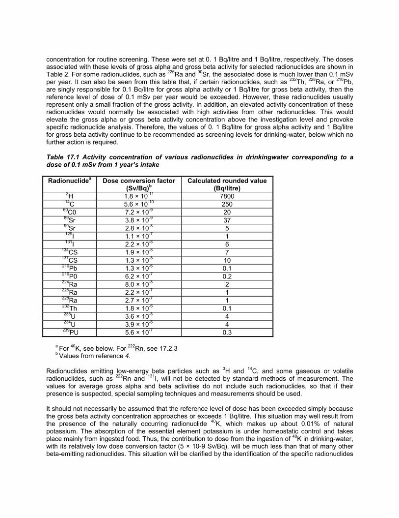
concentration for routine screening. These were set at 0. 1 Bq/litre and 1 Bq/litre, respectively. The dosesassociated with these levels of gross alpha and gross beta activity for selected radionuclides are shown inTable 2. For some radionuclides, such as 226Ra and 90Sr, the associated dose is much lower than 0.1 mSvper year. It can also be seen from this table that, if certain radionuclides, such as 232Th, 228Ra, or 210Pb,are singly responsible for 0.1 Bq/litre for gross alpha activity or 1 Bq/litre for gross beta activity, then thereference level of dose of 0.1 mSv per year would be exceeded. However, these radionuclides usuallyrepresent only a small fraction of the gross activity. In addition, an elevated activity concentration of theseradionuclides would normally be associated with high activities from other radionuclides. This wouldelevate the gross alpha or gross beta activity concentration above the investigation level and provokespecific radionuclide analysis. Therefore, the values of 0. 1 Bq/litre for gross alpha activity and 1 Bq/litrefor gross beta activity continue to be recommended as screening levels for drinking-water, below which nofurther action is required.
Table 17.1 Activity concentration of various radionuclides in drinkingwater corresponding to adose of 0.1 mSv from 1 year’s intake
Radionuclidea Dose conversion factor(Sv/Bq)b
Calculated rounded value(Bq/litre)
3H 1.8 × 10⁻¹¹ 780014C 5.6 × 10⁻¹⁰ 250
60C0 7.2 × 10⁻⁹ 2089Sr 3.8 × 10⁻⁹ 3790Sr 2.8 × 10⁻⁸ 5129I 1.1 × 10⁻⁷ 1131I 2.2 × 10⁻⁸ 6
134CS 1.9 × 10⁻⁸ 7137CS 1.3 × 10⁻⁸ 10210Pb 1.3 × 10⁻⁶ 0.1210P0 6.2 × 10⁻⁷ 0.2224Ra 8.0 × 10⁻⁸ 2226Ra 2.2 × 10⁻⁷ 1228Ra 2.7 × 10⁻⁷ 1232Th 1.8 × 10⁻⁶ 0.1238U 3.6 × 10⁻⁸ 4234U 3.9 × 10⁻⁸ 4
239PU 5.6 × 10⁻⁷ 0.3
a For 40K, see below. For 222Rn, see 17.2.3b Values from reference 4.
Radionuclides emitting low-energy beta particles such as 3H and 14C, and some gaseous or volatileradionuclides, such as 222Rn and 131I, will not be detected by standard methods of measurement. Thevalues for average gross alpha and beta activities do not include such radionuclides, so that if theirpresence is suspected, special sampling techniques and measurements should be used.
It should not necessarily be assumed that the reference level of dose has been exceeded simply becausethe gross beta activity concentration approaches or exceeds 1 Bq/litre. This situation may well result fromthe presence of the naturally occurring radionuclide 40K, which makes up about 0.01% of naturalpotassium. The absorption of the essential element potassium is under homeostatic control and takesplace mainly from ingested food. Thus, the contribution to dose from the ingestion of 40K in drinking-water,with its relatively low dose conversion factor (5 × 10⁻⁹ Sv/Bq), will be much less than that of many otherbeta-emitting radionuclides. This situation will be clarified by the identification of the specific radionuclides

in the sample.
Table 17.2 Examples of the doses arising from 1 year’s consumption of drinking-water containingany of the given alpha-emitting radionuclides at an activity concentration of 0.1 Bq/litre or of thegiven beta-emitting radionuclides at an activity concentration of 1 Bq/litrea
Radionuclide Dose (mSv)Alpha emitters (0.1 Bq/litre)210Po 0.045224Ra 0.006226Ra 0.016232Th 0.130234U 0.003238U 0.003239PU 0.04Beta emitters (1 Bq/litre)60Co 0.00589Sr 0.00390Sr 0.020129I 0.080131I 0.016134CS 0.014137CS 0.009210Pb 0.95228Ra 0.20
a Appropriate dose conversion factors taken from reference 4.
17.2.1 Analytical methods
The International Organization for Standardization (ISO) has published standard methods for determininggross alpha (5) and gross beta (6) activity concentrations in water. Although the detection limits depend onthe radionuclides present, the dissolved solids in the sample, and the counting conditions, therecommended levels for gross alpha and gross beta activity concentrations should be above the limits ofdetection. The ISO detection limit for gross alpha activity based on 239Pu is 0.04 Bq/litre, while that forgross beta activity based on 137CS is between 0.04 and 0.1 Bq/litre.
For analyses of specific radionuclides in drinking-water, there are general compendium sources in additionto specific methods in the technical literature (7-11).
17.2.2 Strategy for assessing drinking-water
If either the gross alpha activity concentration of 0.1 Bq/litre or the gross beta activity concentration of 1Bq/lltre is exceeded, then the specific radionuclides should be identified and their individual activityconcentrations measured. From these data, a dose estimate for each radionuclide should be made andthe sum of these doses determined. Where the following additive formula is satisfied, no further action isrequired:
� � 1 i
ii RC
C

where Ci is the measured activity concentration of radionuclide i and RCi is the reference activityconcentration of radionuclide i that, at an intake of 2 litres per day for 1 year, will result in a committedeffective dose of 0.1 mSv (see Table 17.1).
<<I>> p913.gif Fig. 17.1. Application of recommendations on radionuclides in drinking-waterbasedon annual reference level of dose of 0.1 mSv
If alpha-emitting radionuclides with high dose conversion factors are suspected, this additive formula mayalso be invoked when the gross alpha and gross beta activity screening values of 0. 1 Bq/litre and 1Bq/litre are approached. Where the sum exceeds unity for a single sample, the reference level of dose of0. 1 mSv would be exceeded only if the exposure to the same measured concentrations were to continuefor a full year. Hence, such a sample does not in itself imply that the water is unsuitable for consumptionand should be regarded only as a level at which further investigation, including additional sampling, isneeded.
The options available to the competent authority to reduce the dose should then be examined. Whereremedial measures are contemplated, any strategy considered should first be justified (in the sense that itachieves a positive net benefit) and then optimized in accordance with the recommendations of ICRP (1,12) in order to produce the maximum net benefit. The application of these recommendations issummarized in Fig. 1.
17.2.3 Radon
There are difficulties in applying the reference level of dose to derive activity concentrations of 222Rn indrinking-water (2). These difficulties arise from the ease with which radon is released from water duringhandling and the importance of the inhalation pathway. Stirring and transferring water from one containerto another will liberate dissolved radon. Water that has been left to stand will have reduced radon activity,and boiling will remove radon completely. As a result, it is important that the form of water consumed istaken into account in assessing the dose from ingestion. Moreover, the use of water supplies for otherdomestic uses will increase the levels of radon in the air, thus increasing the dose from inhalation. Thisdose depends markedly on the form of domestic usage and housing construction (13). The form of waterintake, the domestic use of water, and the construction of houses vary widely throughout the world. It istherefore not possible to derive an activity concentration for radon in drinking-water that is universallyapplicable.
The global average dose from inhalation of radon from all sources is about 1 mSv/year, which is roughlyhalf of the total natural radiation exposure. In comparison, the global dose from ingestion of radon indrinking-water is relatively low. In a local situation, however, the risks from inhalation and from ingestionmay be about equal. Because of this and because there may be other sources of radon gas entry to ahouse, ingestion cannot be considered in isolation from inhalation exposures.
All these factors should be assessed on a regional or national level by the appropriate authorities, in orderto determine whether a reference level of dose of 0.1 mSv is appropriate for that region, and to determinean activity concentration that may be used to assess the suitability of the water supply. These judgementsshould be based not only on the ingestion and inhalation exposures resulting from the supply of water, butalso on the inhalation doses from other radon sources in the home. In these circumstances, it wouldappear necessary to adopt an integrated approach and assess doses from all radon sources, especially todetermine the optimum action to be undertaken where some sort of intervention is deemed necessary.
References
1. 1990 Recommendations of the International Commission on Radiological Protection. Annals of theICRP, 1990, 21 (1-3).

2. United Nations Scientific Committee on the Effects of Atomic Radiation. Sources, effects and risks ofionizing radiation. New York, United Nations, 1988.
3. World Health Organization. Derived intervention levels for radionuclides in food Geneva, 1988.
4. National Radiological Protection Board. Committed equivalent organ doses and committed effectivedoses from intakes of radionuclides. A report of the National Radiological Protection Board of the UnitedKingdom. Chilton, Didcot, 1991 (NRPB-R245).
5. Association of Official Analytical Chemists. Official methods of analysis of the Association of OfficialAnalytical Chemists, 15th ed. Washington, DC, 1990.
6. Environmental Measurements Laboratory. EML procedures manual. New York, Department of Energy,1990 (HASL-300)
7. International Organization for Standardization. Water quality - measurement of gross alpha activity innon-saline water - thick source method. Geneva, 1990 (Draft International Standard 9696)
8. International Organization for Standardization. Water quality - measurement of gross beta activity innon-saline water. Geneva, 1990 (Draft International Standard 9697).
9. Suess MJ, ed. Examination of water for pollution control. 3 vols. Oxford, Pergamon Press, 1982.
10. United States Environmental Protection Agency. Eastern Environmental Radiation Facility.Radiochemistry procedures manual. Montgornery, AL, 1987 (EPA 520/5-84006).
11. American Public Health Association. Standard methods for the examination of water and wastewater,17th ed. Washington, DC, 1989.
12. Optimization and decision-making in radiological protection. Annals of the ICRP, 1989, 20(1).
13. National Council on Radiation Protection and Measurements. Control of radon in houses.Recommendations of the National Council on Radiation Protection and Measurements. Bethesda, MD,1989 (NCRP Report No. 103)
Annex 1. List of participants in preparatory meetings
Consultation on Revision of WHO Guidelines for Drinking-Water Quality (Rome, Italy, 17-19October 1988)
Members
L. Albanus, Head, Toxicology Laboratory, National Food Administration, Uppsala, Sweden
J. Alexander, Toxicological Department, National Institute of Public Health, Oslo, Norway
J.A. Cotruvo, Director, Criteria and Standards Division, United States Environmental Protection Agency,Washington, DC, USA
H. de Kruijf, Laboratory for Ecotoxicology, Environmental Chemistry and Drinking-Water, National Instituteof Public Health and Environmental Protection, Bilthoven, Netherlands
H.H. Dieter, Director and Professor, Institute for Water, Soil and Air Hygiene of the Federal Office ofHealth, Berlin
J.K. Fawell, Principal Toxicologist, Water Research Centre, Medmenham, England (Rapporteur)
E. Funari, Department of Environmental Hygiene, Istituto Superiore di Sanità, Rome, Italy
J.R. Hickman, Acting Director-General, Environmental Health Directorate, Health and Welfare Canada,Ottawa, Canada
Y. Magara, Director, Department of Sanitary Engineering, Institute of Public Health, Tokyo, Japan
R.F. Packham, Chief Scientist, Water Research Centre, Medmenham, England
M. Waring, Department of Health and Social Security, London, England
G.A. Zapponi, Environmental Impact Assessment Section, Istituto Superiore di Sanità, Rome, Italy
Observers
S. Blease, Administrator, Water Protection Division, Commission of European Communities, Brussels,Belgium
B. Julin, Regulatory Affairs Manager, International Group of National Associations of Manufacturers ofAgrochemical Products, Wilmington, DE, USA
A. Pelfrène, International Group of National Associations of Manufacturers of Agrochemical Products,Paris, France
N. Sarti, Division of Water and Soil, Ministry of Health, Rome, Italy
Secretariat
G. Burin, International Programme on Chemical Safety, Division of Environmental Health, World HealthOrganization, Geneva, Switzerland

R. Helmer, Prevention of Environmental Pollution, Division of Environmental Health, World HealthOrganization, Geneva, Switzerland
M. Mercier, Manager, International Programme on Chemical Safety, Division of Environmental Health,World Health Organization, Geneva, Switzerland
G. Ozolins, Manager, Prevention of Environmental Pollution, Division of Environmental Health, WorldHealth Organization, Geneva, Switzerland (Moderator)
S. Tarkowski, Director, Environment and Health, WHO Regional Office for Europe, Copenhagen, Denmark
Microbiology Consultation (London, England, 23 June 1989)
Members
U. Blumental, London School of Hygiene and Tropical Medicine, London, England
S. Cairncross, London School of Hygiene and Tropical Medicine, London, England
A.H. Havelaar, National Institute of Public Health and Environmental Protection, Bilthoven, Netherlands
R.F Packham, Marlow, England
W. Stelzer, Research Institute of Hygiene and Microbiology, Bad Elster, German Democratic Republic
H. Utkilen, Department of Sanitary Engineering and Environmental Protection, National Institute of PublicHealth, Oslo, Norway
R. Walter, Director, Institute for General and Community Hygiene, Dresden, German Democratic Republic
Secretariat
J.K. Fawell, Principal Toxicologist, Water Research Centre, Medmenham, England
R. Helmer, Prevention of Environmental Pollution, Division of Environmental Health, World HealthOrganization, Geneva, Switzerland
B. Lloyd, Environmental Health Unit, Robens Institute of Industrial and Environmental Health and Safety,Guildford, England
E.B. Pike, Water Research Centre, Medmenham, England
Coordination Consultation (Copenhagen, Denmark, 4-5 September 1989)
Members
J.K. Fawell, Principal Toxicologist, Water Research Centre, Medmenham, England (Co-Rapporteur)
E. Funari, Department of Environmental Hygiene, Istituto Superiore di Sanità, Rome, Italy
E.S. Jensen, Senior Technical Adviser on Water Supply and Sanitation Projects, Technical AdvisoryDivision, Danish International Development Agency, Copenhagen, Denmark

A. Minderhoud, Laboratory for Ecotoxicology, Environmental Chemistry and Drinking Water, NationalInstitute of Public Health and Environmental Protection, Bilthoven, Netherlands
B. Mintz, Chief, Health Effects Assessment Section, Criteria and Standards Division, Office of Drinking-Water, United States Environmental Protection Agency, Washington, DC, USA
P.A. Nielsen, Scientific Officer, Toxicologist, Institute of Toxicology, National Food Agency, Soborg,Denmark
E. Poulsen, Chief Adviser in Toxicology, Institute of Toxicology, National Food Agency, Soborg, Denmark
B. Schultz, Water Quality Institute, Horsholm, Denmark
Secretariat
G. Burin, International Programme on Chemical Safety, Division of Environmental Health, World HealthOrganization, Geneva, Switzerland (Co-Rapporteur)
O. Espinoza, Regional Officer for International Water Decade, Environment and Health, WHO RegionalOffice for Europe, Copenhagen, Denmark
R. Helmer, Prevention of Environmental Pollution, Division of Environmental Health, World HealthOrganization, Geneva, Switzerland
D. Kello, Project Officer for Toxicology and Food Safety, Environment and Health, WHO Regional Officefor Europe, Copenhagen, Denmark
S. Tarkowski, Director, Environment and Health, WHO Regional Office for Europe, Copenhagen, Denmark
Coordination Group Meeting (Geneva, Switzerland, 13-14 March 1990)
Members
J.K. Fawell, Principal Toxicologist, Water Research Centre, Medmenham, England
E. Funari, Department of Environmental Hygiene, Istituto Superiore di Sanità, Rome, Italy
J.R. Hickman, Acting Director-General, Environmental Health Directorate, Health and Welfare Canada,Ottawa, Canada
A. Minderhoud, Laboratory for Ecotoxicology, Environmental Chemistry and Drinking Water, NationalInstitute of Public Health and Environmental Protection, Bilthoven, Netherlands
B. Mintz, Chief, Health Effects Assessment Section, Criteria and Standards Division, Office of Drinking-Water, United States Environmental Protection Agency, Washington, DC, USA
B. Schultz, Water Quality Institute, Horsholm, Denmark
Secretariat
G. Burin, International Programme on Chemical Safety, Division of Environmental Health, World HealthOrganization, Geneva, Switzerland
O. Espinoza, Regional Officer for International Water Decade, Environment and Health, WHO RegionalOffice for Europe, Copenhagen, Denmark
R. Helmer, Prevention of Environmental Pollution, Division of Environmental Health, World HealthOrganization, Geneva, Switzerland
D. Kello, Project Officer for Toxicology and Food Safety, Environment and Health, WHO Regional Officefor Europe, Copenhagen, Denmark
G. Ozolins, Manager, Prevention of Environmental Pollution, Division of Environmental Health, WorldHealth Organization, Geneva, Switzerland
R. Plestina, International Programme on Chemical Safety, Division of Environmental Health, World HealthOrganization, Geneva, Switzerland
First Review Group Meeting on Pesticides (Busto Garolfo, Italy, 25-30 June 1990)
Members
H. Abouzaid, Chief, Water Quality Control Division, National Agency for Drinking-Water, Rabat-Chellah,Morocco
H. Atta-ur-Rahman, Director, H.E.J. Research Institute of Chemistry, Karachi, Pakistan
V. Benes, Chief, Toxicology and Reference Laboratory, Institute of Hygiene and Epidemiology, Prague,Czechoslovakia
J.F. Borzelleca, Pharmacology, Toxicology, Medical College of Virginia, Virginia CommonwealthUniversity, Richmond, VA, USA
L. Brener, Chief, Department of Mineral Analysis, Research Laboratory, Société Lyonnaise des Eaux-Dumez, Paris, France
D. Calamari, Institute of Agricultural Entomology, Faculty of Agriculture, University of Milan, Italy
J. Du, Office of Drinking-Water, United States Environmental Protection Agency, Washington, DC, USA
J. K. Fawell, Principal Toxicologist, Water Research Centre, Medmenham, England (Rapporteur)
J. Forslund, National Agency of Environmental Protection, Copenhagen, Denmark
E. Funari, Department of Environmental Hygiene, Istituto Superiore di Sanità, Rome, Italy
A. Jaron, Commission of the European Communities, Brussels, Belgium
M. Maroni, Director, International Centre for Pesticide Safety, Busto Garolfo, Italy
Y. Patel, Health Effects Assessment, Office of Drinking-Water, United States Environmental ProtectionAgency, Washington, DC, USA
E. Poulsen, Chief Adviser in Toxicology, Institute of Toxicology, National Food Agency, Soborg, Denmark(Chairman)
J. Rueff, Department of Genetics, Faculty of Medical Science, Lisbon, Portugal
B. Schultz, Water Quality Institute, Horsholm, Denmark
J.A. Sokal, Head, Department of Toxicity Evaluation, Institute of Occupational Medicine, Lodz, Poland
M. Takeda, Director of Environmental Chemistry, National Institute of Hygienic Science, Tokyo, Japan
E.M. den Tonkelaar, National Institute of Public Health and Environmental Protection, Bilthoven,Netherlands
G. Wood, Acting Head, Criteria Section, Monitoring and Criteria Division, Environmental HealthDirectorate, Health and Welfare, Ottawa, Canada
Observers
S. Behrendt, BASF AG, Limburgerhof, Federal Republic of Germany
S. Hahn, BASF AG, Limburgerhof, Federal Republic of Germany
H. Kieczka, BASF AG, Limburgerhof, Federal Republic of Germany
S. Kimura, Southern Fukuoka Prefecture, Water Spread Authority, Japan Water Works Association,Tokyo, Japan
F. Sarhan, CIBA-GEIGY Ltd, Basel, Switzerland
G.E. Veenstra, Shell International Petroleum, The Hague, Netherlands
Secretariat
G. Burin, International Programme on Chemical Safety, Division of Environmental Health, World HealthOrganization, Geneva, Switzerland
D. Kello, Project Officer for Toxicology and Food Safety, Environment and Health, WHO Regional Officefor Europe, Copenhagen, Denmark
R. Plestina, International Programme on Chemical Safety, Division of Environmental Health, World HealthOrganization, Geneva, Switzerland
First Review Group Meeting on Organics (Copenhagen, Denmark, 6-10 November 1990)
Members
C. Abernathy, Toxicologist, Health Effects Branch, Office of Drinking-Water, United States EnvironmentalProtection Agency, Washington, DC, USA
H.H. Dieter, Director and Professor, Institute for Water, Soil and Air Hygiene of the Federal Office ofHealth, Berlin, Germany
A.M. van Dijk-Looyaard, Drinking-Water Research Scientist, National Institute of Public Health andEnvironmental Protection, Bilthoven, Netherlands
J.K. Fawell, Principal Toxicologist, Water Research Centre, Medmenham, England (Rapporteur)

J. Forslund, National Agency of Environmental Protection, Copenhagen, Denmark
E. Funari, Department of Environmental Hygiene, Istituto Superiore di Sanità, Rome, Italy
K. Khanna, Pharmacologist, Health Effects Branch, Office of Drinking-Water, United States EnvironmentalProtection Agency, Washington, DC, USA
R. van Leeuwen, Toxicologist, National Institute of Public Health and Environmental Protection, Bilthoven,Netherlands
U. Lund, Head, Department of Chemistry, Water Quality Institute, Horsholm, Denmark
M.E. Meek, Head, Priority Substances Section, Environmental Health Centre, Health and WelfareCanada, Ottawa, Canada
T. Ookubo, Head, Water Quality Examination Laboratory, Hachinohe Regional Water Supply Cooperation,Hachinohe, Japan
E. Sandberg, Toxicologist, National Food Administration, Uppsala, Sweden
U. Schlosser, Research Institute for Hygiene and Microbiology, Bad Elster, Germany
E.A. Simpson, Commission of the European Communities, Brussels, Belgium
J.A. Sokal, Head, Department of Toxicity Evaluation, Institute of Occupational Medicine, Lodz, Poland(Chairman)
M. Takeda, Director of Environmental Chemistry, National Institute of Hygienic Science, Tokyo, Japan
Observer
A. Carlsen, Ministry of the Environment, National Agency of Environmental Protection, Miljöstyrelsen,Copenhagen, Denmark
Secretariat
P. Bérubé, Programme Assistant, International Water Decade, Environment and Health, WHO RegionalOffice for Europe, Copenhagen, Denmark
O. Espinoza, Regional Officer for International Water Decade, Environment and Health, WHO RegionalOffice for Europe, Copenhagen, Denmark
D. Kello, Project Officer for Toxicology and Food Safety, Environment and Health, WHO Regional Officefor Europe, Copenhagen, Denmark
D. Schutz, International Programme on Chemical Safety, Division of Environmental Health, World HealthOrganization, Geneva, Switzerland
S. Tarkowski, Director, Environment and Health, WHO Regional Office for Europe, Copenhagen, Denmark
J. Wilbourn, Unit of Carcinogen Identification and Evaluation, International Agency for Research onCancer, Lyon, France
First Review Group Meeting on Inorganics (Bilthoven, Netherlands, 18-22 March 1991)
Members
E.A. Bababumni, Department of Biochemistry, University of Ibadan, Ibadan, Nigeria
K.L. Bailey, Health Effects Assessment Section, Criteria and Standards Division, Office of Drinking-Water,United States Environmental Protection Agency, Washington, DC, USA
G.F. Craun, Chief Epidemiologist, United States Environmental Protection Agency, Washington, DC, USA
A.M. van Dijk-Looyaard, Drinking-Water Research Scientist, National Institute of Public Health andEnvironmental Protection, Bilthoven, Netherlands
J.K. Fawell, Principal Toxicologist, Water Research Centre, Medmenham, England (Rapporteur)
R. van Leeuwen, Toxicologist, National Institute of Public Health and Environmental Protection, Bilthoven,Netherlands (Chairman)
M.E. Meek, Head, Priority Substances Section, Environmental Health Centre, Health and WelfareCanada, Ottawa, Canada
E. Poulsen, Chief Adviser in Toxicology, Institute of Toxicology, National Food Agency, Soborg, Denmark
Y.A. Rakhmanin, Head of Laboratory, Ministry of Health of the USSR Academy of Medical Sciences, A.N.Sysin Institute of General and Communal Hygiene, Moscow, USSR
V.R. Rao, Assistant Director and Head, Department of Toxicology, The Haffkine Institute, Parel, Bombay,India
F.G.R. Reyes, Professor of Food Toxicology, Department of Food Science, State University of Campinas,Brazil
F. Sartor, Institute of Hygiene and Epidemiology, Ministry of Public Health and the Family, Brussels,Belgium
J.A. Sokal, Head, Department of Toxicity Evaluation, Institute of Occupational Medicine, Lodz, Poland
M. Takeda, Director of Environmental Chemistry, National Institute of Hygienic Science, Tokyo, Japan
Observers
J. Forslund, National Agency of Environmental Protection, Copenhagen, Denmark
I. Harimaya, Director of Water Quality Research, Kobe, Japan
M. Minowa, Director of Epidemiology, Institute of Public Health, Ministry of Health and Welfare, Tokyo,Japan
E.A. Simpson, Commission of the European Communities, Brussels, Belgium
J.F.M. Versteegh, National Institute of Public Health and Environmental Protection, Bilthoven, Netherlands
V. Vignier, Société Lyonnaise des Eaux Dumez, International Centre for Research on Water and theEnvironment (CIRSEE), Le Pecq, France
Secretariat
B. Chen, International Programme on Chemical Safety, Division of Environmental Health, World HealthOrganization, Geneva, Switzerland
H. Galal-Gorchev, International Programme on Chemical Safety, Division of Environmental Health, WorldHealth Organization, Geneva, Switzerland
Second Review Group Meeting on Organics (Copenhagen, Denmark, 8-12 April 1991)
Members
K. Bergman, Toxicologist, Medical Products Agency, Division of Pharmacology, Uppsala, Sweden
A. Carlsen, National Agency of Environmental Protection, Copenhagen, Denmark
H.H. Dieter, Director and Professor, Institute for Water, Soil and Air Hygiene of the Federal Office ofHealth, Berlin, Germany
P.M. Dudermel, Pasteur Institute, Lille, France
J.K. Fawell, Principal Toxicologist, Water Research Centre, Medmenham, England (Rapporteur)
J. Forslund, National Agency of Environmental Protection, Copenhagen, Denmark
R. Hasegawa, Section Chief, Division of Toxicology, National Institute of Hygienic Science, Tokyo, Japan
K. Hughes, Chemical Health Hazard Evaluator, Environmental Health Centre, Health and WelfareCanada, Ottawa, Canada
R. van Leeuwen, Toxicologist, National Institute of Public Health and Environmental Protection, Bilthoven,Netherlands
U. Lund, Head, Department of Chemistry, Water Quality Institute, Horsholm, Denmark
A. Patel, Toxicologist, Water Research Centre, Medmenham, England
Y. Richard, Chief, Department of Chemical Research, Société Degrémont, Rueil-Malmaison, France
E. Sandberg, Toxicologist, National Food Administration, Uppsala, Sweden
J.A. Sokal, Head, Department of Toxicity Evaluation, Institute of Occupational Medicine, Lodz, Poland(Chairman)
Secretariat
X. Bonnefoy, Acting Regional Officer for Health Planning/Ecology, WHO Regional Office for Europe,Copenhagen, Denmark
H. Galal-Gorchev, International Programme on Chemical Safety, Division of Environmental Health, WorldHealth Organization, Geneva, Switzerland
J. Gents, Secretary, International Water Decade, Environment and Health, WHO Regional Office forEurope, Copenhagen, Denmark
D. Kello, Project Officer for Toxicology and Food Safety, Environment and Health, WHO Regional Officefor Europe, Copenhagen, Denmark
S. Tarkowski, Director, Environment and Health, WHO Regional Office for Europe, Copenhagen, Denmark
Coordination Group Consultation (Geneva, Switzerland, 13-14 May 1991)
Members
J. K. Fawell, Principal Toxicologist, Water Research Centre, Medmenham, England
J.R. Hickman, Director-General, Environmental Health Directorate, Health and Welfare Canada, Ottawa,Canada (Moderator)
U. Lund, Head, Department of Chemistry, Water Quality Institute, Horsholm, Denmark
B. Mintz, Chief, Health Effects Assessment Section, Criteria and Standards Division, Office of Drinking-Water, United States Environmental Protection Agency, Washington, DC, USA
E.B. Pike, Water Research Centre, Medmenham, England
Secretariat
X. Bonnefoy, Acting Regional Officer for Health Planning/Ecology, WHO Regional Office for Europe,Copenhagen, Denmark (Co-Rapporteur)
H. Galal-Gorchev, International Programme on Chemical Safety, Division of Environmental Health, WorldHealth Organization, Geneva, Switzerland (Co-Rapporteur)
R. Helmer, Prevention of Environmental Pollution, Division of Environmental Health, World HealthOrganization, Geneva, Switzerland
J. Kenny, Prevention of Environmental Pollution, Division of Environmental Health, World HealthOrganization, Geneva, Switzerland
M. Mercier, Manager, International Programme on Chemical Safety, Division of Environmental Health,World Health Organization, Geneva, Switzerland
G. Ozolins, Manager, Prevention of Environmental Pollution, Division of Environmental Health, WorldHealth Organization, Geneva, Switzerland
P. Waight, Prevention of Environmental Pollution, Division of Environmental Health, World HealthOrganization, Geneva, Switzerland
Review Group on Disinfectants and Disinfectant By-products (Bethesda, MD, USA, 10-14 June1991)
Members
H. Abouzaid, Chief, Water Quality Control Division, National Agency for Drinking-Water, Rabat-Chellah,Morocco
W. Almeida, Department of Preventive Medicine, State University of Campinas, Campinas, Brazil
M. Ando, National Institute of Hygienic Science, Division of Environmental Chemistry, Tokyo, Japan
R. Bull, Pharmacology/Toxicology Graduate Program, College of Pharmacy, Washington State University,Pullman, WA, USA
G. Burin, United States Environmental Protection Agency, Washington, DC, USA (Vice-Chairman)
J.K. Fawell, Principal Toxicologist, Water Research Centre, Medmenham, England (Co-Rapporteur)
B. Havlik, Institute of Hygiene and Epidemiology, Prague, Czechoslovakia
N. Mahabhol, Ministry of Public Health, Bangkok, Thailand
M.E. Meek, Head, Priority Substances Section, Environmental Health Centre, Health and WelfareCanada, Ottawa, Canada (Co-Rapporteur)
B. Mintz, Chief, Health Effects Assessment Section, Criteria and Standards Division, Office of Drinking-Water, United States Environmental Protection Agency, Washington, DC, USA (Chairman)
R. Packham, Marlow, England
J.F.M. Versteegh, National Institute of Public Health and Environmental Protection, Bilthoven, Netherlands
Z. Zholdakova, Academy of Medical Sciences, A.N. Sysin Institute of General and Communal Hygiene,Moscow, USSR
Observers
J. Forslund, National Agency of Environmental Protection, Copenhagen, Denmark
E. Ohanian, Office of Science and Technology, United States Environmental Protection Agency,Washington, DC, USA
H. Sasaki, Water Quality Laboratory, Sapporro, Hokkaido, Japan
Secretariat
R. Cantilli, United States Environmental Protection Agency, Washington, DC, USA
N. Chiu, United States Environmental Protection Agency, Washington, DC, USA
J. Du, United States Environmental Protection Agency, Washington, DC, USA
H. Galal-Gorchev, International Programme on Chemical Safety, Division of Environmental Health, WorldHealth Organization, Geneva, Switzerland
J. Orme, Office of Science and Technology, United States Environmental Protection Agency, Washington,DC, USA
Review Meeting on Pathogenic Agents and Volume 3 on Surveillance of Community Supplies(Harare, Zimbabwe, 24-28 June 1991)
Members
H. Abouzaid, Chief, Water Quality Control Division, National Agency for Drinking-Water, Rabat-Chellah,Morocco
M.T. Boot, Programme Officer, IRC International Water and Sanitation Centre, The Hague, Netherlands
J.Z. Boutros, Consultant in Food and Water Control, Khartoum, Sudan (Rapporteur)
W. Fellows, Programme Officer, Water and Environmental Sanitation, UNICEF, Harare, Zimbabwe
F.J. Gumbo, Head of Water Laboratories, Operation, Maintenance and Water Laboratories Division,Ministry of Water (MAJI), Dar-es-Salaam, United Republic of Tanzania
A.H. Havelaar, National Institute of Public Health and Environmental Protection, Bilthoven, Netherlands
J. Hubley, Senior Lecturer in Health Education, Health Education Unit, Faculty of Health and Social Care,Leeds Polytechnic, Leeds, England
B. Jackson, Senior Engineering Advisor, British Development Division in East Africa, Nairobi, Kenya
E. Khaka, Ministry of Energy and Water Resources Development, Harare, Zimbabwe
S. Laver, Lecturer, Department of Community Medicine, University of Zimbabwe, Mount Pleasant, Harare,Zimbabwe
M.T. Martins, Associate Professor, Environmental Microbiology Laboratory, University of São Paulo, Brazil
P. Morgan, Advisor, Water and Sanitation, Ministry of Health, Blair Research Laboratory, Harare,Zimbabwe
S. Mtero, Principal Medical Research Officer, Ministry of Health, Blair Research Laboratory, Harare,Zimbabwe
S. Musingarabwi, Director, Environmental Health Services, Ministry of Health, Harare, Zimbabwe (Vice-Chairman)
F. Niang, Chief, Laboratory Service, Senegalese National Water Management Company, Dakar, Senegal
E.B. Pike, Water Research Centre, Medmenham, England
P.K. Ray, Director, Industrial Toxicology Research Centre, Lucknow, India
P. Taylor, Director, Training Centre for Water and Sanitation, Department of Civil Engineering, Universityof Zimbabwe, Harare, Zimbabwe (Chairman)
H. Utkilen, Scientist, National Institute of Public Health, Department of Environmental Medicine, Oslo,Norway
Observers
M. Ellis, Primary Health Consultant, The Robens Institute of Health and Safety, University of Surrey,Guildford, England

D. Tolson, Aid Secretary, British High Commission, Harare, Zimbabwe
Secretariat
J. Bartram, Manager, Overseas Development, The Robens Institute of Health and Safety, University ofSurrey, Guildford, England
H. Galal-Gorchev, International Programme on Chemical Safety, Division of Environmental Health, WorldHealth Organization, Geneva, Switzerland
R. Helmer, Prevention of Environmental Pollution, Division of Environmental Health, World HealthOrganization, Geneva, Switzerland
J. Kenny, Prevention of Environmental Pollution, Division of Environmental Health, World HealthOrganization, Geneva, Switzerland
V. Larby, The Robens Institute of Health and Safety, University of Surrey, Guildford, England
B. Lloyd, Head, Environmental Health, The Robens Institute of Health and Safety, University of Surrey,Guildford, England
K. Wedgwood, Research Officer, The Robens Institute of Health and Safety, University of Surrey,Guildford, England
F. Zawide, WHO Sanitary Engineer, Sub-region III, Harare, Zimbabwe
Second Review Group Meeting on Pesticides (Rennes, France, 2-6 September 1991)
Members
G. Burin, Toxicologist, United States Environmental Protection Agency, Washington, DC, USA (Co-Rapporteur)
A. Bruchet, Société Lyonnaise des Eaux Dumez, International Centre for Research on Water and theEnvironment (CIRSEE), Le Pecq, France
H.H. Dieter, Director and Professor, Institute for Water, Soil and Air Hygiene of the Federal Office ofHealth, Berlin, Germany
P.M. Dudermel, Pasteur Institute, Lille, France
J.K. Fawell, Principal Toxicologist, Water Research Centre, Medmenham, England (Co-Rapporteur)
J. Forslund, National Agency of Environmental Protection, Copenhagen, Denmark
E. Funari, Department of Environmental Hygiene, Istituto Superiore di Sanità, Rome, Italy
R. Halperin, Chief Engineer for Environmental Health, Ministry of Health, Jerusalem, Israel
K. Hughes, Chemical Health Hazard Evaluator, Priority Substances Section, Environmental SubstancesDivision, Environmental Health Directorate, Environmental Health Centre, Ottawa, Canada
S. Kojima, Director of Environmental Chemistry, National Institute of Hygienic Science, Tokyo, Japan

A.M. Mahfouz, Senior Toxicologist and Pesticides Team Leader, Office of Science and Technology,United States Environmental Protection Agency, Washington, DC, USA
A. Montiel, Water Quality Control Officer, Water Management Company of Paris, Paris, France(Chairman)
E. Poulsen, Chief Adviser in Toxicology, Institute of Toxicology, National Food Agency, Soborg, Denmark
R. Seux, National School of Public Health, Rennes, France
E. Simpson, Commission of the European Communities, Brussels, Belgium
Observers
M.J. Carroll, Area Registration Manager, Monsanto Services International, Brussels, Belgium
A. Hirata, Chief, Monitoring Section, Water Quality Management, Waterworks Bureau, Tokyo MetropolitanGovernment, Tokyo, Japan
H.P. Nigitz, Head, Regulatory Affairs, Agrolinz Agricultural Chemicals, Linz, Austria
E. Puri, Toxicologist, CIBA-GEIGY Ltd, Basel, Switzerland
G.A. Willis, Manager, Product Safety, ICI Agrochemicals, Fernhurst, Haslemere, Surrey, England
Secretariat
X. Bonnefoy, Regional Adviser, Health Planning/Ecology, WHO Regional Office for Europe, Copenhagen,Denmark
H. Galal-Gorchev, International Programme on Chemical Safety, Division of Environmental Health, WorldHealth Organization, Geneva, Switzerland
J. Gents, Programme Secretary, Environment and Health, WHO Regional Office for Europe, Copenhagen,Denmark
Second Review Group Meeting on Inorganics (Brussels, Belgium, 14-18 October 1991)
Members
Y. Aida, Senior Research Scientist, Division of Risk Assessment, National Institute of Hygienic Science,Kamiyoga, Setagayaku, Tokyo, Japan
J. Alexander, Deputy Director, Department of Environmental Medicine, National Institute of Public Health,Oslo, Norway
K.L. Bailey, Health Effects Assessment Section, Criteria and Standards Division, Office of Drinking-Water,United States Environmental Protection Agency, Washington, DC, USA
H.H. Dieter, Director and Professor, Toxicologist, Institute for Water, Soil and Air Hygiene of the FederalOffice of Health, Berlin, Germany
J.K. Fawell, Principal Toxicologist, Water Research Centre, Medmenham, England (Co-Rapporteur)
A. Lafontaine, Honorary Director, Institute of Hygiene and Epidemiology, Brussels, Belgium
M.E. Meek, Head, Priority Substances Section, Environmental Health Centre, Health and WelfareCanada, Ottawa, Canada
B. Naima, Director, Water Quality Laboratory, National Agency for Drinking-Water, Rabat - Chellah,Morocco
G.D. Nielsen, Department of Environmental Medicine, Odense University, Odense, Denmark
R.F. Packham, Marlow, England
Y.A. Rakhmanin, Head of Laboratory, Ministry of Health of the USSR Academy of Medical Sciences, A.N.Sysin Institute of General and Communal Hygiene, Moscow, USSR
Tharwat Saleh, Project Manager, WHO Project EFY/CWS/002, Cairo, Egypt
R. Sarin, Assistant Director, Scientist and Head, Basic Research Division, National EnvironmentalEngineering Research Institute (NEERI), Nehru Marg, Nagpur, India
F. Sartor, Institute of Hygiene and Epidemiology, Ministry of Public Health and the Family, Brussels,Belgium (Chairman)
J.F.M. Versteegh, National Institute of Public Health and Environmental Protection, Bilthoven, Netherlands
Observers
J. Forslund, National Agency of Environmental Protection, Copenhagen, Denmark
E.A. Simpson, Commission of the European Communities, Brussels, Belgium
V. Vignier, Société Lyonnaise des Eaux Dumez, International Centre for Research on Water and theEnvironment (CIRSEE), Le Pecq, France
Secretariat
X. Bonnefoy, Regional Adviser, Health Planning/Ecology, WHO Regional Office for Europe, Copenhagen,Denmark
H. Galal-Gorchev, International Programme on Chemical Safety, Division of Environmental Health, WorldHealth Organization, Geneva, Switzerland (Co-Rapporteur)
C. Martin, Prevention of Environmental Pollution, Division of Environmental Health, World HealthOrganization, Geneva, Switzerland
Radionuclides Meeting (Medmenham, England, 22-24 January 1992)
Members
O. Hydes, Drinking-Water Inspectorate, Department of the Environment, London, England
D.P. Meyerhof, Bureau of Radiation and Medical Devices, Department of National Health and Welfare,Ottawa, Canada

J.-C. Nénot, Director of Research, Institute for Nuclear Protection and Safety, Fontenay-aux-Roses,France
K.C. Pillai, Health Physics Division, Bhabha Atomic Research Centre, Bombay, India
A. Randell, Senior Officer, Food Quality and Standards Service, Food and Agriculture Organization of theUnited Nations, Rome, Italy
C. Robinson, National Radiological Protection Board, Chilton, Didcot, England (Co-Rapporteur)
L.B. Sztanyik, Director, "Frédéric Joliot-Curie" National Research Institute for Radiobiology andRadiohygiene, Budapest, Hungary (Chairman)
E. Wirth, Institute for Radiation Hygiene, Federal Office for Radiation Protection, Neuerberg, Germany
Secretariat
P.J. Waight, Prevention of Environmental Pollution, Division of Environmental Health, World HealthOrganization, Geneva, Switzerland (Co-Rapporteur)
Analytical and Treatment Methods (Medmenham, England, 27-29 January 1992)
Members
H. Abouzaid, Chief, Water Quality Control Division, National Agency for Drinking-Water, Rabat-Chellah,Morocco
S. Clark, Chief, Drinking Water Technology Branch, Office of Groundwater and Drinking Water, UnitedStates Environmental Protection Agency, Washington, DC, USA
A.M. van Dijk-Looyaard, Drinking-Water Research Scientist, National Institute of Public Health andEnvironmental Protection, Bilthoven, Netherlands
J. Forslund, National Agency of Environmental Protection, Copenhagen, Denmark
D. Green, Criteria Section, Environmental Health Centre, Department of National Health and Welfare,Ottawa, Canada (Co-Rapporteur)
I. Licsko, Research Centre for Water Resources Development (VITUKI), Budapest, Hungary
B. Lloyd, Head, Environmental Health, The Robens Institute of Health and Safety, University of Surrey,Guildford, England
D.P. Meyerhof, Bureau of Radiation and Medical Devices, Department of National Health and Welfare,Ottawa, Canada
A. Montiel, Water Quality Control Officer, Water Management Company of Paris, Paris, France (Co-Rapporteur)
R.F. Packham, Marlow, England (Chairman)
R. Sarin, Assistant Director, Scientist and Head, Basic Research Division, National EnvironmentalEngineering Research Institute (NEERI), Nehru Marg, Nagpur, India

Observers
T. Aizawa, Department of Sanitary Engineering, Institute of Public Health, Tokyo, Japan
R.A. Breach, Water Quality Manager, Severn Trent Water, Birmingham, England
O. Hydes, Drinking Water Inspectorate, Department of the Environment, London, England
M. Ichinohe, Bureau of Waterworks, Tokyo Metropolitan Government, Tokyo, Japan
E. Simpson, Commission of the European Communities, Brussels, Belgium
M. Tsuji, Ministry of Health and Welfare, Tokyo, Japan
Secretariat
X. Bonnefoy, Regional Adviser, Health Planning/Ecology, WHO Regional Office for Europe, Copenhagen,Denmark
B. Crathorne, Water Research Centre, Medmenham, England
J.K. Fawell, Principal Toxicologist, Water Research Centre, Medmenham, England
H. Galal-Gorchev, International Programme on Chemical Safety, Division of Environmental Health, WorldHealth Organization, Geneva, Switzerland
E.B. Pike, Water Research Centre, Medmenham, England
WHO Consolidation Meeting on Organics and Pesticides (Medmenham, England, 30-31 January1992)
Members
J.K. Fawell, Principal Toxicologist, Water Research Centre, Medmenham, England
R. van Leeuwen, Toxicologist, National Institute of Public Health and Environmental Protection, Bilthoven,Netherlands (Moderator)
U. Lund, Head, Department of Chemistry, Water Quality Institute, Horsholm, Denmark
M. Sheffer, Scientific Editor, Orleans, Canada
Secretariat
X. Bonnefoy, Regional Adviser, Health Planning/Ecology, WHO Regional Office for Europe, Copenhagen,Denmark (Co-Rapporteur)
H. Galal-Gorchev, International Programme on Chemical Safety, Division of Environmental Health, WorldHealth Organization, Geneva, Switzerland (Co-Rapporteur)
Final Drafts Preparation Meeting for Volumes 1 and 2 (Val David, Quebec, Canada, 19-22 May 1992)
Members

K. Bentley, Director, Environmental Health, Health Advancement Division, Australian Department ofHealth, Housing and Community Services, Woden, Australia
J.K. Fawell, Principal Toxicologist, Water Research Centre, Medmenham, England
J.R. Hickman, Director-General, Environmental Health Directorate, Department of National Health andWelfare, Ottawa, Canada (Chairman)
U. Lund, Head, Department of Chemistry, Water Quality Institute, Horsholm, Denmark
M.E. Meek, Head, Priority Substances Section, Environmental Health Centre, Department of NationalHealth and Welfare, Ottawa, Canada
B. Mintz, Chief, Health Effects Assessment Section, Criteria and Standards Division, Office of Drinking-Water, United States Environmental Protection Agency, Washington, DC, USA
R.F. Packham, Marlow, England
E.B. Pike, Water Research Centre, Medmenham, England
M. Sheffer, Scientific Editor, Orleans, Canada
P. Toft, Health Protection Branch, Environmental Health Directorate, Department of National Health andWelfare, Ottawa, Canada
G. Wood, Health Protection Branch, Environmental Health Directorate, Department of National Health andWelfare, Ottawa, Canada
Secretariat
X. Bonnefoy, Regional Adviser, Health Planning/Ecology, WHO Regional Office for Europe, Copenhagen,Denmark (Co-Rapporteur)
H. Galal-Gorchev, International Programme on Chemical Safety, World Health Organization, Geneva,Switzerland (Co-Rapporteur)
G. Ozolins, Manager, Prevention of Environmental Pollution, Division of Environmental Health, WorldHealth Organization, Geneva, Switzerland
Final Task Group Meeting (Geneva, Switzerland, 21-25 September 1992)
Members*
*Invited but unable to attend: Director-General of Health, Islamabad, Pakistan; F. Sartor, Institute ofHygiene and Epidemiology, Ministry of Public Health and the Family, Brussels, Belgium.
H. Abouzaid, Chief, Water Quality Control Division, National Agency for Drinking-Water, Rabat-Chellah,Morocco
M. Aguilar, Director of Basic Sanitation, Department of Environmental and Occupational Health and BasicSanitation, Mexico City, Mexico

J. Alexander, Deputy Director, Department of Environmental Medicine, National Institute of Public Health,Oslo, Norway
V. Angjeli, Chief of Communal Hygiene Division, Research Institute of Hygiene and Epidemiology, Tirana,Albania
L. Anukam, Federal Environmental Protection Agency (FEPA), Department of Planning and Evaluation,Federal Secretariat Complex (Phase II), Ikoyi, Lagos, Nigeria
W.S. Assoy, Director, Environmental Health Service, Department of Health, Manila, Philippines
Changjie Chen, Director, Institute of Environmental Health Monitoring, Chinese Academy of PreventiveMedicine, Beijing, China
M. Csanady, Department Leader, National Institute of Hygiene, Budapest, Hungary
H.H. Dieter, Director and Professor, Institute for Water, Soil and Air Hygiene of the Federal Office ofHealth, Berlin, Germany
F.K. El Jack, Head of Water Department, National Chemical Laboratories, Khartoum, Sudan
J. Forslund, National Agency of Environmental Protection, Copenhagen, Denmark (Vice-Chairman)
E. Funari, Department of Environmental Hygiene, Istituto Superiore di Sanità, Rome, Italy
E. Gonzalez, Chief, Department of Water Quality, Water Supply and Sewerage, San José, Costa Rica
F.J. Gumbo, Head of Water Laboratories, Operation, Maintenance and Water Laboratories Division,Ministry of Water (MAJI), Dar-es-Salaam, United Republic of Tanzania
B. Havlik, Head of Water Hygiene Branch, National Institute of Public Health, Prague, Czechoslovakia
H.M.S.S.D. Herath, Deputy Director General, Public Health Services, Ministry of Health, Colombo, SriLanka
L. Hiisvirta, Chief Engineer, Ministry of Social Affairs and Health, Helsinki, Finland
J. Kariuki, Senior Public Health Officer, Division of Environmental Health, Ministry of Health, Nairobi,Kenya
M. Kitenge, Director, Department of Local Production Control, Zaire Control Agency, Kinshasa, Zaire
F.X.R. van Leeuwen, Senior Toxicologist, National Institute of Public Health and Environmental Protection,Bilthoven, Netherlands
Y. Magara, Director, Department of Water Supply Engineering, Institute of Public Health, Tokyo, Japan
N.S. McDonald, Director, Water Branch, Department of Primary Industries and Energy, Canberra,Australia
B. Mintz, Chief, Exposure Assessment and Environmental Fate Section, Office of Science andTechnology, United States Environmental Protection Agency, Washington, DC, USA
F. Niang, Chief, Laboratory Service, Senegalese National Water Management Company, Dakar, Senegal

R.F. Packham, Marlow, England
Y.A. Rakhmanin, Academician of Russian Academy of Natural Sciences, A.N. Sysin Research Institute ofHuman Ecology and Environmental Health, Moscow, Russian Federation
F.G.R. Reyes, Professor of Food Toxicology, Department of Food Science, State University of Campinas,Brazil (Rapporteur)
T. Saleh, WHO Regional Support Office, Cairo, Egypt
E. Sandberg, Senior Toxicologist, National Food Administration, Uppsala, Sweden
Nantana Santatiwut, Director, Environmental Health Division, Department of Health, Ministry of PublicHealth, Bangkok, Thailand
R. Sarin, Scientist, National Environmental Engineering Research Institute (NEERI), Nehru Marg, Nagpur,India
C. Shaw, Senior Advisor Scientist, Public Health Services, Department of Health, Wellington, NewZealand
J.A. Sokal, Director, Institute of Occupational Medicine and Environmental Health, Sosnowiec, Poland
P. Toft, Health Protection Branch, Environmental Health Directorate, Department of National Health andWelfare, Ottawa, Canada (Chairman)
D. Tricard, Sanitary Engineer, Ministry of Health and Humanitarian Action, Department of Health, Paris,France
Observers
M.J. Crick, Radiation Safety Specialist, International Atomic Energy Agency, Vienna, Austria
A.M. van Dijk-Looyaard, Senior Scientist Drinking-Water Standards, KIWA N.V. Research andConsultancy, Nieuwegein, Netherlands
O. Hydes, Drinking Water Inspectorate, Department of the Environment, London, England
M. Rapinat, International Water Supply Association, Compagnie générale des Eaux, Paris, France
Y. Richard, Head Engineer, Société DEGREMONT-CIRSEE, Le Pecq, France
H. Rousseau, Division des Eaux de Consommation, Direction des Ecosystèmes urbains, Ministère del’Environnement, Ste Foy, Québec, Canada
J.E. Samdal, Norwegian Institute for Water Research (NIVA), Oslo, Norway
F. Sarhan, CIBA-GEIGY Ltd, Basel, Switzerland (representing the International Group of NationalAssociations of Manufacturers of Agrochemical Products)
E.A. Simpson, Commission of the European Communities, Brussels, Belgium
T. Yanagisawa, Director, Technical Management Section, Management and Planning Division, Bureau ofWaterworks, Tokyo, Japan
Secretariat
J. Bartram, Manager, Overseas Development, The Robens Institute of Health and Safety, University ofSurrey, Guildford, England
X. Bonnefoy, Environmental Health Planning/Ecology, WHO Regional Office for Europe, Copenhagen,Denmark
A. Enevoldsen, Environmental Health Planning/Ecology, WHO Regional Office for Europe, Copenhagen,Denmark
J.K. Fawell, Principal Toxicologist, Water Research Centre, Medmenham, England
B.H. Fenger, Water and Waste Scientist, WHO European Office for Environment and Health, Rome, Italy
H. Galal-Gorchev, International Programme on Chemical Safety, World Health Organization, Geneva,Switzerland
R. Helmer, Prevention of Environmental Pollution, Division of Environmental Health, World HealthOrganization, Geneva, Switzerland
J. Kenny, Prevention of Environmental Pollution, Division of Environmental Health, World HealthOrganization, Geneva, Switzerland
U. Lund, Water Quality Institute, Horsholm, Denmark
M. Mercier, Director, International Programme on Chemical Safety, World Health Organization, Geneva,Switzerland
H. Moller, Scientist, Unit of Carcinogen Identification and Evaluation, International Agency for Research onCancer, Lyon, France
G. Ozolins, Manager, Prevention of Environmental Pollution, Division of Environmental Health, WorldHealth Organization, Geneva, Switzerland
E.B. Pike, Water Research Centre, Medmenham, England
M. Sheffer, Scientific Editor, Orleans, Canada
S. Tarkowski, Director, Environment and Health, WHO Regional Office for Europe, Copenhagen, Denmark

Annex 2. Tables of guideline values
The following tables present a summary of guideline values for microorganisms and chemicals in drinking-water. Individual values should not be used directly from the tables. The guideline values must be usedand interpreted in conjunction with the information contained in the text.
Table A2.1. Bacteriological quality of drinking-watera
Organisms Guideline valueAll water intended for drinkingE. coli or thermotolerant coliform bacteriab,c Must not be detectable in any 100-ml sampleTreated water entering the distribution systemE. coli or thermotolerant coliform bacteriab Must not be detectable in any 100-ml sampleTotal coliform bacteria Must not be detectable in any 100-ml sampleTreated water in the distribution systemE. coli or thermotolerant coliform bacteriab Must not be detectable in any 100-ml sampleTotal coliform bacteria Must not be detectable in any 100-ml sample. In
the case of large supplies, where sufficientsamples are examined, must not be present in95% of samples taken throughout any 12-monthperiod
a Immediate investigative action must be taken if either E. coli or total coliform bacteria are detected.The minimal action in the case of total coliform bacteria is repeat sampling; if these bacteria aredetected in the repeat sample, the cause must be determined by immediate further investigation.
b Although E. coli is the more precise indicator of faecal pollution, the count of thermotolerant coliformbacteria is an acceptable alternative. If necessary, proper confirmatory tests must be carried out. Totalcoliform bacteria are not acceptable indicators of the sanitary quality of rural water supplies, particularlyin tropical areas where many bacteria of no sanitary significance occur in almost all untreated supplies.
c It is recognized that, in the great majority of rural water supplies in developing countries, faecalcontamination is widespread. Under these conditions, the national surveillance agency should setmedium-term targets for the progressive improvement of water supplies, as recommended in Volume 3of Guidelines for drinking-water quality.
Table A2.2. Chemicals of health significance in drinking-water
A. Inorganic constituents
Guideline value(mg/litre)
Remarks
antimony 0.005 (P)a
arsenic 0.01b (P) For excess skin cancer risk of 6 × 10⁻⁴
barium 0.7beryllium NADc
boron 0.3cadmium 0.003chromium 0.05 (P)copper 2 (P) ATOd
cyanide 0.07fluoride 1.5 Climatic conditions, volume of water consumed,
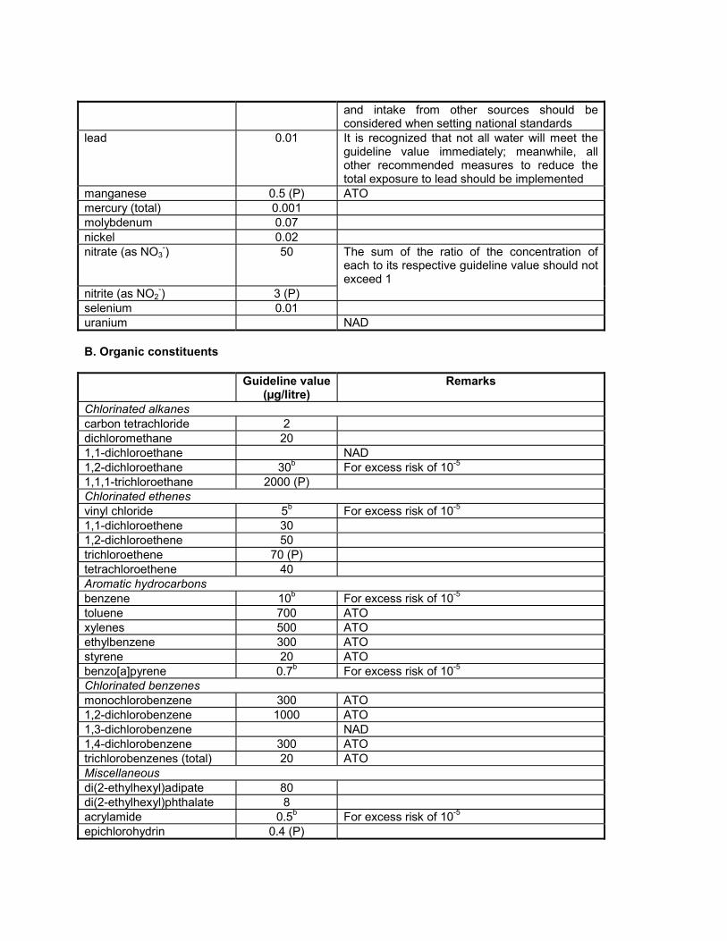
and intake from other sources should beconsidered when setting national standards
lead 0.01 It is recognized that not all water will meet theguideline value immediately; meanwhile, allother recommended measures to reduce thetotal exposure to lead should be implemented
manganese 0.5 (P) ATOmercury (total) 0.001molybdenum 0.07nickel 0.02nitrate (as NO3
-) 50 The sum of the ratio of the concentration ofeach to its respective guideline value should notexceed 1
nitrite (as NO2-) 3 (P)
selenium 0.01uranium NAD
B. Organic constituents
Guideline value(µg/litre)
Remarks
Chlorinated alkanescarbon tetrachloride 2dichloromethane 201,1-dichloroethane NAD1,2-dichloroethane 30b For excess risk of 10-5
1,1,1-trichloroethane 2000 (P)Chlorinated ethenesvinyl chloride 5b For excess risk of 10-5
1,1-dichloroethene 301,2-dichloroethene 50trichloroethene 70 (P)tetrachloroethene 40Aromatic hydrocarbonsbenzene 10b For excess risk of 10-5
toluene 700 ATOxylenes 500 ATOethylbenzene 300 ATOstyrene 20 ATObenzo[a]pyrene 0.7b For excess risk of 10-5
Chlorinated benzenesmonochlorobenzene 300 ATO1,2-dichlorobenzene 1000 ATO1,3-dichlorobenzene NAD1,4-dichlorobenzene 300 ATOtrichlorobenzenes (total) 20 ATOMiscellaneousdi(2-ethylhexyl)adipate 80di(2-ethylhexyl)phthalate 8acrylamide 0.5b For excess risk of 10-5
epichlorohydrin 0.4 (P)

hexachlorobutadiene 0.6edetic acid (EDTA) 200 (P)nitrilotriacetic acid 200dialkyltins NADtributyltin oxide 2
C. Pesticides
Guideline value(µg/litre)
Remarks
alachlor 20b For excess risk of 10-5
aldicarb 10aldrin/dieldrin 0.03atrazine 2bentazone 30carbofuran 5chlordane 0.2chlorotoluron 30DDT 21,2-dibromo-3-chloropropane
1b For excess risk of 10-5
2,4-D 301,2-dichloropropane 20 (P)1,3-dichloropropane NAD1,3-dichloropropene 20b For excess risk of 10-5
ethylene dibromide NADheptachlor and heptachlorepoxide
0.03
hexachlorobenzene 1b For excess risk of 10-5
isoproturon 9lindane 2MCPA 2methoxychlor 20metolachlor 10molinate 6pendimethalin 20pentachlorophenol 9 (P)permethrin 20propanil 20pyridate 100simazine 2trifluralin 20chlorophenoxy herbicides other than 2,4-D and MCPA
2,4-DB 90dichlorprop 100fenoprop 9MCPB NADmecoprop 102,4,5-T 9

D. Disinfectants and disinfectant by-products
Disinfectants Guideline value(mg/litre)
Remarks
monochloramine di- andtrichloramine
3 NAD
chlorine 5 ATO. For effective disinfection there should be aresidual concentration of free chlorine of �0.5mg/litre after at least 30 minutes contact time atpH <8.0
chlorine dioxide A guideline value has not been establishedbecause of the rapid breakdown of chlorinedioxide and because the chlorite guideline valueis adequately protective for potential toxicityfrom chlorine dioxide
iodine NADDisinfectants by-products
Guideline value(µg/litre)
Remarks
bromate 25b (P) For 7 × 10⁻⁵ excess riskchlorate NADchlorite 200 (P)chlorophenols
2-chlorophenol NAD2,4-dichlorophenol NAD2,4,6-trichlorophenol 200b For excess risk of 10-5, ATO
formaldehyde 900MX NADtrihalomethanes The sum of the ratio of the concentration of
each to its respective guideline value should notexceed 1
bromoform 100dibromochloromethane 100bromodichloromethane 60b For excess risk of 10-5
chloroform 200b For excess risk of 10-5
chlorinated acetic acidsmonochloroacetic acid NADdichloroacetic acid 50 (P)trichloroacetic acid 100 (P)
chloral hydrate(trichloroacetaldehyde)
10 (P)
chloroacetone NADhalogenated acetonitriles
dichloroacetonitrile 90 (P)dibromoacetonitrile 100 (P)bromochloroacetonitrile NADtrichloroacetonitrile 1 (P)
cyanogen chloride (as CN) 70chloropicrin NAD
a (P) - Provisional guideline value. This term is used for constituents for which there is some evidenceof a potential hazard but where the available information on health effects is limited; or where an

uncertainty factor greater than 1000 has been used in the derivation of the tolerable daily intake (TDI).Provisional guideline values are also recommended: (1) for substances for which the calculatedguideline value would be below the practical quantification level, or below the level that can beachieved through practical treatment methods; or (2) where disinfection is likely to result in theguideline value being exceeded.
b For substances that are considered to be carcinogenic, the guideline value is the concentration indrinking-water associated with an excess lifetime cancer risk of 10-5 (one additional cancer per 100 000of the population ingesting drinking-water containing the substance at the guideline value for 70 years).Concentrations associated with estimated excess lifetime cancer risks of 10-4 and 10-6 can becalculated by multiplying and dividing, respectively, the guideline value by 10.
In cases in which the concentration associated with an excess lifetime cancer risk of 10-5 is notfeasible as a result of inadequate analytical or treatment technology, a provisional guideline value isrecommended at a practicable level and the estimated associated excess lifetime cancer riskpresented.
It should be emphasized that the guideline values for carcinogenic substances have been computedfrom hypothetical mathematical models that cannot be verified experimentally and that the valuesshould be interpreted differently from TDI-based values because of the lack of precision of the models.At best, these values must be regarded as rough estimates of cancer risk. However, the models usedare conservative and probably err on the side of caution. Moderate short-term exposure to levelsexceeding the guideline value for carcinogens does not significantly affect the risk.
c NAD - No adequate data to permit recommendation of a health-based guideline value.
d ATO - Concentrations of the substance at or below the health-based guideline value may affect theappearance, taste, or odour of the water.
Table A2.3. Chemicals not of health significance at concentrations normally found in drinking-water
Chemical Remarksasbestos Usilver Utin U
U - It is unnecessary to recommend a health-based guideline value for these compounds because theyare not hazardous to human health at concentrations normally found in drinking-water.
Table A2.4. Radioactive constituents of drinking-water
Screening value(Bq/litre)
Remarks
gross alpha activity 0.1 If a screening value is exceeded, more detailedradionuclide analysis is necessary. Higher values donot necessarily imply that the water is unsuitable forhuman consumption
gross beta activity 1
Table A2.5. Substances and parameters in drinking-water that may give rise to complaints fromconsumers
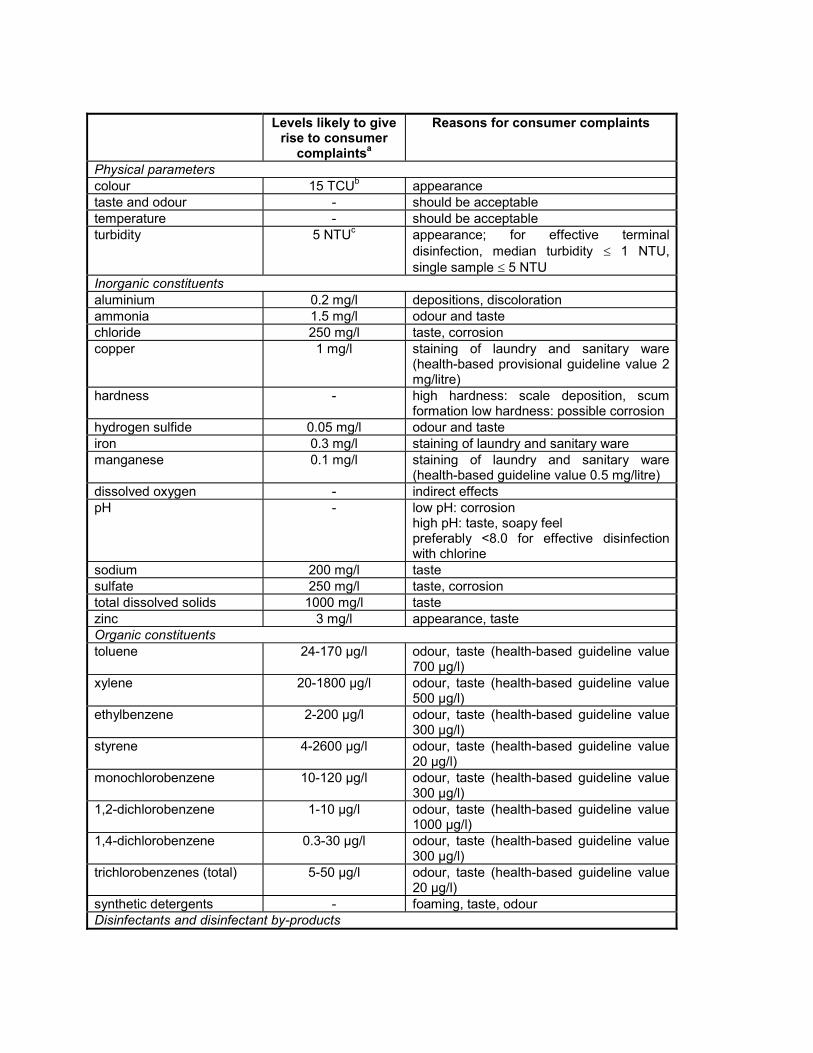
Levels likely to giverise to consumer
complaintsa
Reasons for consumer complaints
Physical parameterscolour 15 TCUb appearancetaste and odour - should be acceptabletemperature - should be acceptableturbidity 5 NTUc appearance; for effective terminal
disinfection, median turbidity � 1 NTU,single sample � 5 NTU
Inorganic constituentsaluminium 0.2 mg/l depositions, discolorationammonia 1.5 mg/l odour and tastechloride 250 mg/l taste, corrosioncopper 1 mg/l staining of laundry and sanitary ware
(health-based provisional guideline value 2mg/litre)
hardness - high hardness: scale deposition, scumformation low hardness: possible corrosion
hydrogen sulfide 0.05 mg/l odour and tasteiron 0.3 mg/l staining of laundry and sanitary waremanganese 0.1 mg/l staining of laundry and sanitary ware
(health-based guideline value 0.5 mg/litre)dissolved oxygen - indirect effectspH - low pH: corrosion
high pH: taste, soapy feelpreferably <8.0 for effective disinfectionwith chlorine
sodium 200 mg/l tastesulfate 250 mg/l taste, corrosiontotal dissolved solids 1000 mg/l tastezinc 3 mg/l appearance, tasteOrganic constituentstoluene 24-170 µg/l odour, taste (health-based guideline value
700 µg/l)xylene 20-1800 µg/l odour, taste (health-based guideline value
500 µg/l)ethylbenzene 2-200 µg/l odour, taste (health-based guideline value
300 µg/l)styrene 4-2600 µg/l odour, taste (health-based guideline value
20 µg/l)monochlorobenzene 10-120 µg/l odour, taste (health-based guideline value
300 µg/l)1,2-dichlorobenzene 1-10 µg/l odour, taste (health-based guideline value
1000 µg/l)1,4-dichlorobenzene 0.3-30 µg/l odour, taste (health-based guideline value
300 µg/l)trichlorobenzenes (total) 5-50 µg/l odour, taste (health-based guideline value
20 µg/l)synthetic detergents - foaming, taste, odourDisinfectants and disinfectant by-products
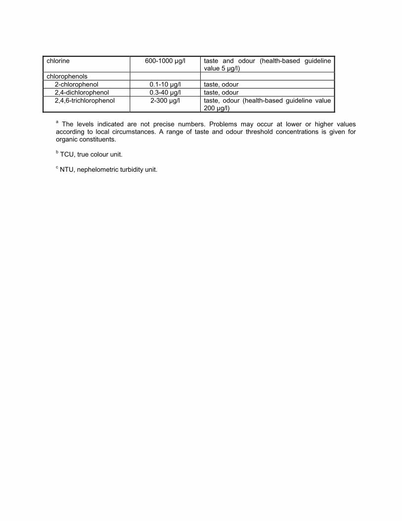
chlorine 600-1000 µg/l taste and odour (health-based guidelinevalue 5 µg/l)
chlorophenols2-chlorophenol 0.1-10 µg/l taste, odour2,4-dichlorophenol 0.3-40 µg/l taste, odour2,4,6-trichlorophenol 2-300 µg/l taste, odour (health-based guideline value
200 µg/l)
a The levels indicated are not precise numbers. Problems may occur at lower or higher valuesaccording to local circumstances. A range of taste and odour threshold concentrations is given fororganic constituents.
b TCU, true colour unit.
c NTU, nephelometric turbidity unit.

Back cover
The first edition of Guidelines for drinking-water quality, published in 1984-85, was widely used as a basisfor setting national standards to ensure the safety of the water supply. It established guideline values for a
large number of water constituents and contaminants, covering microbiological, biological, chemical,organoleptic, and radiological aspects. For this new edition, all the recommended guideline values havebeen reviewed and, where necessary, updated in the light of new scientific information. In addition, many
drinking-water contaminants not included in the first edition are evaluated.
Volume 2 explains how guideline values for drinking-water contaminants are to be used, defines thecriteria used to select the various chemical, physical, microbiological, and radiological contaminants
included in the report, describes the approaches used in deriving guideline values, and presents briefsummary statements either supporting the guideline values recommended or explaining why no health-
based guideline value is required at the present time.
It is emphasized that the guideline values recommended are not mandatory limits. Such limits should beset by national authorities, using a risk-benefit approach and taking into consideration local environmental,
social, economic and cultural conditions.
Price: Sw. fr. 260.-Price in developing countries: Sw. fr. 182.-
ISBN 92 4 154480 5


