14 A Comparative Study of Dysplasia and Cancer Risk in Intestinal Metaplasia (IM) and Non-Intestinal...
1
AGA Abstracts colon. CONCLUSION: Decreases in contractility and carbachol-induced depolarization in inflamed colon may be, in part, due to down-regulation of non-selective cation channels, in particular TRPC6 channels. Their down-regulation may underlie the more negative RMP and decreased excitability seen in inflamed colonic smooth muscle. 12 Loss of Gastric Interstitial Cells of Cajal and Impaired Electrical Pacemaking in Klotho Mice, an Animal Model of Aging Ferenc Izbeki, Andrea Lorincz, David T. Asuzu, Laura Popko, David L. Young, Michael R. Bardsley, Yujiro Hayashi, Makoto Kuro-o, Gianrico Farrugia, Tamas Ordog Background & Aims: There are several age-related changes in the upper gastrointestinal (GI) tract including increased prevalence of gastroesophageal reflux disease and inability to consume large portions of food, often resulting in decreased weight in the aged. Reduced food intake may lead to under-nutrition and promote aging-related chronic disorders. Gastric dysfunction arising from loss of interstitial cells of Cajal (ICC) and enteric neurons may contribute to these problems. We examined whether prematurely-aging, homozygous klotho mice severely deficient in Klotho, a pleiotropic protein with anti-aging properties, display gastric phenotypes. Methods: klotho mice and their wild-type, age-matched littermates (WT) were studied between 50 and 82 days of age (n=67). Serum hormones were measured by enzyme immunoassays. Gastric slow waves were recorded by intracellular technique. Gastric emptying of solids was measured by [ 13 C]-octanoic acid breath test. ICC were detected by Kit immunostaining and network volumes determined from confocal microscopic images reconstructed in 3D. ICC numbers were determined by flow cytometry. Enteric neurons were identified by immunostaining for HuC/D and counted by fluorescent microscopy. Results: As expected, klotho mice had reduced body weights (52±2% of WT; mean±SEM; P<0.001), non-fasting blood glucose (61±6%; P<0.001), serum insulin and IGF-I levels (31±2% and 53±5%, respectively P<0.02). Fecal output/3h was reduced to 54±9% (P= 0.001). These changes signify a profoundly reduced food intake. The gastric wall was stiff and had calcium deposits. Electrical slow waves were rhythmic but had reduced amplitudes ( klotho vs. WT: corpus: 6.7(4.9;9.1) vs. 9.0(7.6;10.3) mV; median(IQR); antrum: 8.5(6.2;12.0) vs. 12.4(10.6;14.4) mV; both P<0.001). Gastric emptying T 1/2 for solids was not changed. By flow cytometry, frequency of ICC and ICC progenitors was reduced to 48 and 38%, respectively. ICC network densities in the circular and longitudinal muscle layers and in the myenteric region were reduced to 10±6% (P=0.046), 6±3% (P=0.03) and 15±5% (P=0.004) of WT, respectively. In contrast, there was no difference in HuC/D + neuron counts in the greater curvature region of the entire gastric corpus and antrum. Conclusions: Premature aging associated with Klotho deficiency leads to loss of ICC and reduced electrical pacing of the gastric muscles. These changes may contribute to the low body weights of klotho mice. The stiffer stomach may attenuate the effects of impaired phasic activity on gastric emptying. klotho mice appear to be a useful model to study the mechanisms of GI aging. Supported by NIH DK58185 and the Rosztoczy Foundation. 13 Increasing Incidence of Barrett's Esophagus is Not Entirely Explained by Endoscopic Practice Helen G. Mulholland, Shivaram Bhat, Liam Murray, Damian T. McManus, Anna T. Gavin, Brian T. Johnston Introduction: Esophageal adenocarcinoma (EAC) incidence rates have increased dramatically in recent decades, particularly among white males in Western societies (1). There appears to be a concurrent rise in Barrett's Esophagus (BE) incidence, the pre-cursor condition for EAC, although it is debated whether this is a true rise, or a reflection of changes in endoscopy practices together with improvement in disease recognition (2). The aim of our investigation was to assess BE incidence over a 13 year period using a population-based register in Northern Ireland. Methods: The Northern Ireland Barrett's esophagus Register (NIBR) is a population-based register of all adults diagnosed with BE, defined as columnar epithelium of the esophagus, in Northern Ireland between 1993 and 2005. Data on all upper gastro- intestinal endoscopies and esophageal biopsies performed in Northern Ireland were obtained from healthcare providers. Annual BE incidence rates were calculated per 100,000 of the population, per 100 endoscopies and per 100 esophageal biopsies performed. Results: During the 13 year period, 197,635 patients underwent an endoscopy and 9,390 of these were diagnosed with BE, of whom 58% were male. Average annual BE incidence rates over this time period are presented in Table 1. A 2.5 fold increase in BE incidence in the population was observed. The observed rise in BE incidence was slightly more pronounced in males, rising from 37.3 to 97.5/100,000, compared with a 27.4 to 64.4/100,000 increase in females. Over the same time, there were 1.3 and 1.6 fold increases in endoscopy and biopsy rates in the population, respectively. Even with the increasing rates of endoscopy and biopsy, BE was still diagnosed more frequently per 100 endoscopies and per 100 biopsies. Discussion: These findings demonstrate that BE incidence rates in Northern Ireland have increased more rapidly than the rate of endoscopies or biopsies. This could indicate that a true rise in BE incidence has occurred, contributing to the increase in EAC seen in Western populations. This may have implications for efforts to prevent EAC. Table 1. Average annual BE incidence rates in Northern Ireland S-2 AGA Abstracts 14 A Comparative Study of Dysplasia and Cancer Risk in Intestinal Metaplasia (IM) and Non-Intestinal Metaplasia Epithelia: Clinical Implications for the Definition of Barrett's Esophagus Ajay Bansal, Mandeep Singh, Oksana Anand, Douglas H. McGregor, Philip G. Jones, Rachel Cherian, Vikas Singh, Srinivas Gaddam, Sachin B. Wani, Neil Gupta, Amit Rastogi, Prateek Sharma Background: Universal agreement on inclusion of intestinal metaplasia (IM) to diagnose Barrett's esophagus (BE) is lacking. Aim: To compare the prevalence of dysplasia/cancer in patients with columnar lined esophagus with (CLE-IM ) and without intestinal metaplasia (CLE-no IM). Methods: CLE-no IM patients (cases) were identified by querying the clinical pathology database using SNOMED codes for distal esophageal biopsies (January 1, 1999 to June 30, 2009) followed by manual review of all identified cases to include biopsies with glandular epithelium without IM. Cases were defined by presence of CLE on index endoscopy and lack of IM on all endoscopies. CLE-IM patients (controls) were identified from a well- established BE database. Subsequently, 2 models were generated to match cases and controls- Model 1 matched for age, gender, race and year of index EGD (±1 year) and Model 2 matched for CLE length in addition to Model 1. Since patients with CLE-no IM do not usually undergo surveillance, only prevalent dysplasia/cancer on index endoscopy was evalu- ated. Biopsies from CLE-no IM group were reviewed for IM and dysplasia by 2 expert GI pathologists (consensus diagnosis). Results: 253 cases and 763 controls were identified. In Model 1 (cases=134, controls 390), BE length was significantly lower in the CLE-no IM group vs. CLE-IM group (1.5±1.6 vs. 3.5±3.1, p<0.001). The proportion of patients with any dysplasia was 1.5% (95% CI 0.2%-5.3%) in the CLE-no IM group compared with 25.4% (95% CI18.3%-33.6%) in the CLE-IM group [RR 17.0 (4.1-70.8, p<0.001)]. Excluding patients with low-grade dysplasia (LGD), the proportion of patients with HGD/Cancer was 0% (95% CI 0%-2.7%) in the CLE-no IM group compared with 6% (95% CI 2.6%-11.4%) in the CLE-IM group [RR infinite, p<0.005). In Model 2 (cases=88, controls 150), the proportion of patients with any dysplasia was 1.1% (95% CI 0.0%-6.2%) in the CLE-no IM group compared with 18.2% (10.8%-27.8%) in the CLE-IM group [RR 16.0 (2.1-120.6), p<0.001)]. Excluding patients with LGD, the proportion of patients with HGD/Cancer was 0% (95% CI 0%-4.1%) in the CLE-no IM group compared with 2.3% (95% CI 0.3%-8%) in the CLE-IM group (estimated RR infinite, P=0.139). Mean body mass index, hiatal hernia and aspirin and NSAID use were not significantly different between cases and controls. Conclusion: In a large cohort of CLE-no IM patients, no cases of HGD/cancer were identified (only 3 patients with LGD); in contrast the prevalence of HGD/cancer was significantly higher in the CLE-IM group. Demonstration of intestinal metaplasia continues to be an essential element in the definition of Barrett's esophagus. 15 Insulin Resistance and Central Adiposity as a Risk Factors for Barrett's Esophagus Katarina B. Greer, Cheryl Thompson, Lacie Brenner, Beth Bednarchik, Gary W. Falk, William M. Grady, Joseph Willis, Gregory S. Cooper, Li Li, Amitabh Chak Background and aims: Obesity increases risk of developing esophageal adenocarcinoma (EAC) and its precursor Barrett's esophagus (BE), independent of gastroesophageal reflux (GERD). Hyperinsulinemia related to obesity leads to increased cellular proliferation and decreased apoptosis, which is postulated to be one mechanism for carcinogenesis. The aim of this study was to investigate central adiposity and hyperinsulinemia as risk factors for BE. Methods: BE patients (n=133) were recruited from consecutive patients presenting to a tertiary care institution and compared with two separate control groups; subjects with GERD (n=135) as well as colonoscopy controls (n=932). Baseline fasting insulin was measured in all subjects. HOMA-IR was calculated from baseline fasting insulin and glucose levels. Central adiposity was measured using waist to hip ratio (WHR). Univariate and multivariate regression analysis was performed on all variables of interest. For purposes of multivariate analysis the two control groups were combined. Results: Mean waist to hip ratio did not differ in BE cases compared to GERD controls (0.98 vs. 0.97, p=0.08), however, it was significantly higher in subjects with BE cases compared to colonoscopy controls (0.98 vs. 0.91, p<0.001). Mean fasting levels of insulin were lowest in colonoscopy controls and highest in BE cases (7.4 vs. 10.01μIU/mL, p<0.001). BE cases appeared to be more insulin resistant than both GERD and colonoscopy controls (mean HOMA-IR 2.7 ±2.6 vs. 1.8±3.2, respectively). In a multivariate logistic regression analysis adjusted for age, sex, gender, and WHR, both individuals in the highest quartile of serum insulin and individuals in the highest quartile of HOMA-IR showed increased odds of development of BE compared to those in the lowest quartile (ORinsulin = 2.80, 95% CI 1.43-5.49 and OR HOMA-IR 3.16, 95% CI 1.54-6.47). Conclusions: We provide evidence that central adiposity and insulin resistance are associated with increased risk of BE. Future prospective studies should explore whether weight loss as well as treatment of insulin resistance could disrupt or reverse the metaplasia- dysplasia progression in subjects with BE. 16 A Segregation Analysis of Barrett's Esophagus and Associated Adenocarcinomas Xiangqing Sun, Robert Elston, Jill Barnholtz-Sloan, Gary W. Falk, William M. Grady, Margaret F. Kinnard, Sumeet K. Mittal, Joseph Willis, Sanford Markowitz, Wendy E. Brock, Amitabh Chak INTRODUCTION: Familial aggregation of esophageal adenocarcinomas, esophagogastric junction adenocarcinomas, and their precursor Barrett's esophagus has been termed Familial Barrett's Esophagus (FBE). Numerous studies documenting increased familial risk for these diseases raise the hypothesis that there may be an inherited susceptibility to the development of BE and its associated cancers. METHODS: Using segregation analysis for a binary trait as implemented in S.A.G.E. 6.0.1, we analyzed data on 881 singly ascertained pedigrees in order to determine whether FBE is caused by a common environmental or genetic agent
-
Upload
ajay-bansal -
Category
Documents
-
view
212 -
download
0
Transcript of 14 A Comparative Study of Dysplasia and Cancer Risk in Intestinal Metaplasia (IM) and Non-Intestinal...
AG
AA
bst
ract
scolon. CONCLUSION: Decreases in contractility and carbachol-induced depolarization ininflamed colon may be, in part, due to down-regulation of non-selective cation channels,in particular TRPC6 channels. Their down-regulation may underlie the more negative RMPand decreased excitability seen in inflamed colonic smooth muscle.
12
Loss of Gastric Interstitial Cells of Cajal and Impaired Electrical Pacemakingin Klotho Mice, an Animal Model of AgingFerenc Izbeki, Andrea Lorincz, David T. Asuzu, Laura Popko, David L. Young, Michael R.Bardsley, Yujiro Hayashi, Makoto Kuro-o, Gianrico Farrugia, Tamas Ordog
Background & Aims: There are several age-related changes in the upper gastrointestinal(GI) tract including increased prevalence of gastroesophageal reflux disease and inability toconsume large portions of food, often resulting in decreased weight in the aged. Reducedfood intake may lead to under-nutrition and promote aging-related chronic disorders. Gastricdysfunction arising from loss of interstitial cells of Cajal (ICC) and enteric neurons maycontribute to these problems. We examined whether prematurely-aging, homozygous klothomice severely deficient in Klotho, a pleiotropic protein with anti-aging properties, displaygastric phenotypes. Methods: klotho mice and their wild-type, age-matched littermates (WT)were studied between 50 and 82 days of age (n=67). Serum hormones were measured byenzyme immunoassays. Gastric slow waves were recorded by intracellular technique. Gastricemptying of solids was measured by [13C]-octanoic acid breath test. ICC were detected byKit immunostaining and network volumes determined from confocal microscopic imagesreconstructed in 3D. ICC numbers were determined by flow cytometry. Enteric neuronswere identified by immunostaining for HuC/D and counted by fluorescent microscopy.Results: As expected, klotho mice had reduced body weights (52±2% of WT; mean±SEM;P<0.001), non-fasting blood glucose (61±6%; P<0.001), serum insulin and IGF-I levels(31±2% and 53±5%, respectively P<0.02). Fecal output/3h was reduced to 54±9% (P=0.001). These changes signify a profoundly reduced food intake. The gastric wall was stiffand had calcium deposits. Electrical slow waves were rhythmic but had reduced amplitudes(klotho vs. WT: corpus: 6.7(4.9;9.1) vs. 9.0(7.6;10.3) mV; median(IQR); antrum:8.5(6.2;12.0) vs. 12.4(10.6;14.4) mV; both P<0.001). Gastric emptying T1/2 for solids wasnot changed. By flow cytometry, frequency of ICC and ICC progenitors was reduced to 48and 38%, respectively. ICC network densities in the circular and longitudinal muscle layersand in the myenteric region were reduced to 10±6% (P=0.046), 6±3% (P=0.03) and 15±5%(P=0.004) of WT, respectively. In contrast, there was no difference in HuC/D+ neuroncounts in the greater curvature region of the entire gastric corpus and antrum. Conclusions:Premature aging associated with Klotho deficiency leads to loss of ICC and reduced electricalpacing of the gastric muscles. These changes may contribute to the low body weights ofklotho mice. The stiffer stomach may attenuate the effects of impaired phasic activity ongastric emptying. klotho mice appear to be a useful model to study the mechanisms of GIaging. Supported by NIH DK58185 and the Rosztoczy Foundation.
13
Increasing Incidence of Barrett's Esophagus is Not Entirely Explained byEndoscopic PracticeHelen G. Mulholland, Shivaram Bhat, Liam Murray, Damian T. McManus, Anna T. Gavin,Brian T. Johnston
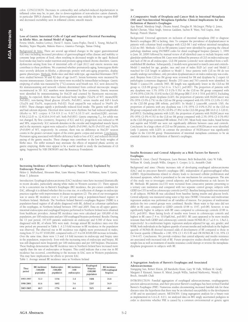
Introduction: Esophageal adenocarcinoma (EAC) incidence rates have increased dramaticallyin recent decades, particularly among white males in Western societies (1). There appearsto be a concurrent rise in Barrett's Esophagus (BE) incidence, the pre-cursor condition forEAC, although it is debated whether this is a true rise, or a reflection of changes in endoscopypractices together with improvement in disease recognition (2). The aim of our investigationwas to assess BE incidence over a 13 year period using a population-based register inNorthern Ireland. Methods: The Northern Ireland Barrett's esophagus Register (NIBR) is apopulation-based register of all adults diagnosed with BE, defined as columnar epitheliumof the esophagus, in Northern Ireland between 1993 and 2005. Data on all upper gastro-intestinal endoscopies and esophageal biopsies performed in Northern Ireland were obtainedfrom healthcare providers. Annual BE incidence rates were calculated per 100,000 of thepopulation, per 100 endoscopies and per 100 esophageal biopsies performed. Results: Duringthe 13 year period, 197,635 patients underwent an endoscopy and 9,390 of these werediagnosed with BE, of whom 58% were male. Average annual BE incidence rates over thistime period are presented in Table 1. A 2.5 fold increase in BE incidence in the populationwas observed. The observed rise in BE incidence was slightly more pronounced in males,rising from 37.3 to 97.5/100,000, compared with a 27.4 to 64.4/100,000 increase in females.Over the same time, there were 1.3 and 1.6 fold increases in endoscopy and biopsy ratesin the population, respectively. Even with the increasing rates of endoscopy and biopsy, BEwas still diagnosed more frequently per 100 endoscopies and per 100 biopsies. Discussion:These findings demonstrate that BE incidence rates in Northern Ireland have increased morerapidly than the rate of endoscopies or biopsies. This could indicate that a true rise in BEincidence has occurred, contributing to the increase in EAC seen in Western populations.This may have implications for efforts to prevent EAC.Table 1. Average annual BE incidence rates in Northern Ireland
S-2AGA Abstracts
14
A Comparative Study of Dysplasia and Cancer Risk in Intestinal Metaplasia(IM) and Non-Intestinal Metaplasia Epithelia: Clinical Implications for theDefinition of Barrett's EsophagusAjay Bansal, Mandeep Singh, Oksana Anand, Douglas H. McGregor, Philip G. Jones,Rachel Cherian, Vikas Singh, Srinivas Gaddam, Sachin B. Wani, Neil Gupta, AmitRastogi, Prateek Sharma
Background: Universal agreement on inclusion of intestinal metaplasia (IM) to diagnoseBarrett's esophagus (BE) is lacking. Aim: To compare the prevalence of dysplasia/cancer inpatients with columnar lined esophagus with (CLE-IM ) and without intestinal metaplasia(CLE-no IM). Methods: CLE-no IM patients (cases) were identified by querying the clinicalpathology database using SNOMED codes for distal esophageal biopsies (January 1, 1999to June 30, 2009) followed by manual review of all identified cases to include biopsies withglandular epithelium without IM. Cases were defined by presence of CLE on index endoscopyand lack of IM on all endoscopies. CLE-IM patients (controls) were identified from a well-established BE database. Subsequently, 2 models were generated to match cases and controls-Model 1 matched for age, gender, race and year of index EGD (±1 year) and Model 2matched for CLE length in addition to Model 1. Since patients with CLE-no IM do notusually undergo surveillance, only prevalent dysplasia/cancer on index endoscopy was evalu-ated. Biopsies from CLE-no IM group were reviewed for IM and dysplasia by 2 expert GIpathologists (consensus diagnosis). Results: 253 cases and 763 controls were identified. InModel 1 (cases=134, controls 390), BE length was significantly lower in the CLE-no IMgroup vs. CLE-IM group (1.5±1.6 vs. 3.5±3.1, p<0.001). The proportion of patients withany dysplasia was 1.5% (95% CI 0.2%-5.3%) in the CLE-no IM group compared with25.4% (95%CI18.3%-33.6%) in the CLE-IM group [RR 17.0 (4.1-70.8, p<0.001)]. Excludingpatients with low-grade dysplasia (LGD), the proportion of patients with HGD/Cancer was0% (95% CI 0%-2.7%) in the CLE-no IM group compared with 6% (95% CI 2.6%-11.4%)in the CLE-IM group [RR infinite, p<0.005). In Model 2 (cases=88, controls 150), theproportion of patients with any dysplasia was 1.1% (95% CI 0.0%-6.2%) in the CLE-noIM group compared with 18.2% (10.8%-27.8%) in the CLE-IM group [RR 16.0 (2.1-120.6),p<0.001)]. Excluding patients with LGD, the proportion of patients with HGD/Cancer was0% (95% CI 0%-4.1%) in the CLE-no IM group compared with 2.3% (95% CI 0.3%-8%)in the CLE-IM group (estimated RR infinite, P=0.139). Mean body mass index, hiatal herniaand aspirin and NSAID use were not significantly different between cases and controls.Conclusion: In a large cohort of CLE-no IM patients, no cases of HGD/cancer were identified(only 3 patients with LGD); in contrast the prevalence of HGD/cancer was significantlyhigher in the CLE-IM group. Demonstration of intestinal metaplasia continues to be anessential element in the definition of Barrett's esophagus.
15
Insulin Resistance and Central Adiposity as a Risk Factors for Barrett'sEsophagusKatarina B. Greer, Cheryl Thompson, Lacie Brenner, Beth Bednarchik, Gary W. Falk,William M. Grady, Joseph Willis, Gregory S. Cooper, Li Li, Amitabh Chak
Background and aims: Obesity increases risk of developing esophageal adenocarcinoma(EAC) and its precursor Barrett's esophagus (BE), independent of gastroesophageal reflux(GERD). Hyperinsulinemia related to obesity leads to increased cellular proliferation anddecreased apoptosis, which is postulated to be one mechanism for carcinogenesis. The aimof this study was to investigate central adiposity and hyperinsulinemia as risk factors forBE. Methods: BE patients (n=133) were recruited from consecutive patients presenting toa tertiary care institution and compared with two separate control groups; subjects withGERD (n=135) as well as colonoscopy controls (n=932). Baseline fasting insulinwas measuredin all subjects. HOMA-IR was calculated from baseline fasting insulin and glucose levels.Central adiposity was measured using waist to hip ratio (WHR). Univariate and multivariateregression analysis was performed on all variables of interest. For purposes of multivariateanalysis the two control groups were combined. Results: Mean waist to hip ratio did notdiffer in BE cases compared to GERD controls (0.98 vs. 0.97, p=0.08), however, it wassignificantly higher in subjects with BE cases compared to colonoscopy controls (0.98 vs.0.91, p<0.001). Mean fasting levels of insulin were lowest in colonoscopy controls andhighest in BE cases (7.4 vs. 10.01μIU/mL, p<0.001). BE cases appeared to be more insulinresistant than both GERD and colonoscopy controls (mean HOMA-IR 2.7 ±2.6 vs. 1.8±3.2,respectively). In a multivariate logistic regression analysis adjusted for age, sex, gender, andWHR, both individuals in the highest quartile of serum insulin and individuals in the highestquartile of HOMA-IR showed increased odds of development of BE compared to those inthe lowest quartile (ORinsulin = 2.80, 95% CI 1.43-5.49 and OR HOMA-IR 3.16, 95% CI1.54-6.47). Conclusions: We provide evidence that central adiposity and insulin resistanceare associated with increased risk of BE. Future prospective studies should explore whetherweight loss as well as treatment of insulin resistance could disrupt or reverse the metaplasia-dysplasia progression in subjects with BE.
16
A Segregation Analysis of Barrett's Esophagus and AssociatedAdenocarcinomasXiangqing Sun, Robert Elston, Jill Barnholtz-Sloan, Gary W. Falk, William M. Grady,Margaret F. Kinnard, Sumeet K. Mittal, Joseph Willis, Sanford Markowitz, Wendy E.Brock, Amitabh Chak
INTRODUCTION: Familial aggregation of esophageal adenocarcinomas, esophagogastricjunction adenocarcinomas, and their precursor Barrett's esophagus has been termed FamilialBarrett's Esophagus (FBE). Numerous studies documenting increased familial risk for thesediseases raise the hypothesis that there may be an inherited susceptibility to the developmentof BE and its associated cancers. METHODS: Using segregation analysis for a binary traitas implemented in S.A.G.E. 6.0.1, we analyzed data on 881 singly ascertained pedigrees inorder to determine whether FBE is caused by a common environmental or genetic agent