13 04-2015 bone biopsy and its decalcification stmu
29
Page 1 BONE BIOPSY AND DECALCIFICATION MUHAMMAD SHOAIB M.PHIL. Biochemistry
-
Upload
muhammad-shoaib -
Category
Health & Medicine
-
view
34 -
download
3
Transcript of 13 04-2015 bone biopsy and its decalcification stmu

Page 1
BONE BIOPSY AND
DECALCIFICATION
MUHAMMAD SHOAIBM.PHIL. Biochemistry

HENRY MOORE
NO ONE EVER KNOWS WHAT EACH DAY IS GOING TO BRING. THE IMPORTANT THING IS TO BE OPEN AND READY FOR IT.

SHIFA TAMEER E MILLAT
UNIVERSITY FOR
APPLIED SCIENCES STUDIES

Calcium ions found in the bones are responsible for its rigid posture and without it, bones may be flexible and most of time unable to carry their body weight. Diseases such as osteomalacia and rickets are as a result of lack of calcium in the bones of those areas.
De-calcification of bones as our topic is about the removal of these calcium from the bones. As such, we are going to look at the following about the topic:
• The definition of decalcification of bones• The purpose of decalcification of bones• The principle of bone decalcification• The method of decalcification of bonesHow to test for the completion of decalcification

INTRODUCTION
BIOPSYA biopsy is a procedure performed to remove tissue
or cells from the body for examination under a microscope.
BONE BIOPSY A bone biopsy is a procedure in which a small
sample of bone is taken from the body and looked at under a microscope for cancer, infection, or other bone disorders.
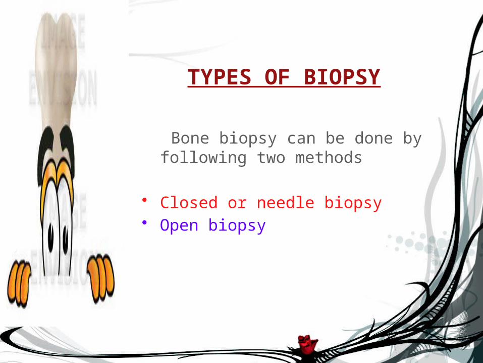
TYPES OF BIOPSY
Bone biopsy can be done by following two methods
• Closed or needle biopsy• Open biopsy

Closed or needle biopsy
• A drill biopsy is generally used to obtain a small specimen
• clean the skin over the bone where the biopsy sample will be taken and local anesthesia in skin is given.
• The radiologist or surgeon puts a long, thin needle through the skin into the bone
• Doctor may make a small cut in your skin before putting the needle in so the needle passes easily
• Take out a small amount of bone through the needle.
• After a closed or needle biopsy, a small bandage is placed over
the area and pressure is put on the area to stop any bleeding

Open biopsy• An open biopsy takes 30 to 60 minutes
• Before an open biopsy, general anesthesia is given to the patients
• Before making a skin cut the area need to be shaved and clean
• The surgeon makes a cut to see the bone and take out a small piece.
• After an open biopsy, the cut is cleaned and closed with stitches (sutures). A bandage is put on the area. The stitches are taken out about 14 days after the biopsy.

Purpose of bone biopsy
• Confirm the diagnosis of a bone disorder
• determine if a bone tumor is malignant (cancerous) or benign
• evaluate bone pain or tenderness .
• determine the cause of an unexplained infection or inflammation
• Find the cause of ongoing bone pain.
• Check bone problems seen on an X-ray.

Risks after bone biopsy
• bruising and discomfort at the biopsy site
• bone fracture
• prolonged bleeding from the biopsy site
• infection near the biopsy site or in the bone

Bone biopsy resultsBone biopsy results
Normal The biopsy sample shows normal bone tissue.
Abnormal
Bone tissue may show signs of infection, cancer, or another bone disorder (including Paget's disease, osteomyelitis, a bone cyst, or a benign bone growth called an osteoma). The bone tissue may also show osteoporosis or osteomalacia, which means the bones are weak.
Most cancer of the bone spreads to the bone from another part of the body, such as the breast, lungs, prostate, or other organs. But bone cancer can also start in the bone itself (such as osteosarcoma or multiple myeloma

BONEDECALCIFICATION
BONEDECALCIFICATION

INTRODUCTION
Decalcification Loss of calcium salts from a bone or tooth. OR The process of removing calcareous matter.
Bone decalcification Removal of calcium ions from the bone
through histological process thereby making the bone flexible and easy for pathological investigation.

PURPOSE OF BONE DECALCIFICATION
The purpose is to make the bone flexible and easy for pathological investigation. This is necessary in order to obtain soft sections of the bone using the microtome
Failure to decalcification results in torn, ragged sections and damage to the cutting edge of the microtome knife

PRINCIPLE OF DECALCIFICATION
Insoluble calcium salt are converted into soluble calcium salts by the action of decalcifying agent so that the tissue become soft.
Chelating agent binds to calcium ions present in the bone and decalcification is carried out.
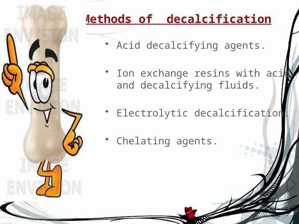
Methods of decalcification
• Acid decalcifying agents.
• Ion exchange resins with acid and decalcifying fluids.
• Electrolytic decalcification.
• Chelating agents.

Acid decalcifying agent
• The commonest method of decalcification is dissolving calcium salts in an acid solution.
• Some of the acid decalcifying agents are..
Nitric acid. Formic acid. Trichloroacetic acid

Nitric acid
MEHTODThin slices of fixed tissue are placed in a
freshly prepared 5-10% solution of nitric acid in distilled water.
Decalcification through this should not extends beyond 48 hours.
Formalin is added to nitric acid to protect the tissue against maceration and swelling.
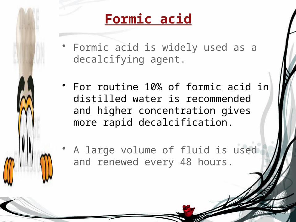
Formic acid
• Formic acid is widely used as a decalcifying agent.
• For routine 10% of formic acid in distilled water is recommended and higher concentration gives more rapid decalcification.
• A large volume of fluid is used and renewed every 48 hours.

Ion exchange resins with acid decalcifying fluids
The removal of calcium ions from the decalcifying fluid by the resins leads to quicker and more efficient decalcification.
Electrolytic DecalcificationIt is the speedier decalcification without damage
to cytological features and staining.
Drawback:
Heat produced in the process may cause the charring of specimen in the process

Chelating Agents
• EDTA is a chelating agent, it is a white crystalline powder soluble in distilled water to about 20%.
• As a decalcifying agent it combines with calcium ions to form soluble, non ionized compounds.
• The volume of solution for decalcifying should be 150 times that of the tissue.
• The solution should be renewed every
5 to 7 days during decalcification.

Advantages of EDTA
• Deposits of iron and other metals may also be removed by EDTA.
• Tissue is not hardened after decalcification.
• It can be good for Bone ,Teeth and any calcified tissue.
• This is also the preferred solution for
decalcifying bone material for transmission electron microscopy .

Test for completion of Decalcification
There are two method for determining the completion of progress of decalcification.
By X-ray examination.
By Ammonia method.

X-ray Examination
• The most reliable method for determining decalcification.
• But as this facility is not presented in all the laboratories

Ammonia Method
• In this method ammonia is added drop by drop in the decalcifying solution cloudiness indicate the presence of calcium.
• The specimen is then placed in a fresh solution of decalcifying fluid and test is repeated after a suitable interval of time.

SUMMARYSUMMARY
BONE BIOPSY
It is a procedure in which bone samples are removed (with a special biopsy needle or during surgery) to determine if cancer or other abnormal cells are present.
Done by two methods • closed method• Open method
BONE DECALCIFICATION Removal of calcium ions from the bone through histological
process thereby making the bone flexible and easy for pathological investigation.

QUOTE :
THE ONLY WAY TO DISCOVER THE LIMIT OF THE POSSIBLE IS TO GO BEYOND THEM INTO THE IMPOSSIBLE.