(12) United States Patent (10) Patent No.: US 6,815,540 B1 · ES C2S 2S OS 87 97 . U.S. Patent Nov....
64
USOO6815540B1 (12) United States Patent (10) Patent No.: US 6,815,540 B1 Pickthun et al. (45) Date of Patent: Nov. 9, 2004 (54) IMMUNOGLOBULIN SUPERFAMILY Dubel et al Journal of Immunological Methods DOMAINS AND FRAGMENTS WITH INCREASED SOLUBILITY (75) Inventors: Andreas Pickthun, Zürich (CH); Lars Nieba, Zürich (CH); Annemarie Honegger, Zürich (CH) (73) Assignee: University of Zurich, Zurich (CH) (*) Notice: Subject to any disclaimer, the term of this patent is extended or adjusted under 35 U.S.C. 154(b) by 0 days. (21) Appl. No.: 09/232,290 (22) Filed: Jan. 15, 1999 Related U.S. Application Data (63) Continuation of application No. PCT/EP97/03792, filed on Jul. 16, 1997. (30) Foreign Application Priority Data Jul. 16, 1996 (EP) ............................................ 96.111441 (51) Int. Cl." ................................................ C07H 21/04 (52) U.S. Cl. ................................ 536/23.53; 435/320.1; 435/325; 435/326; 530/387.3 (58) Field of Search ............................... 435/69.1, 69.7, 435/752.3, 69.6, 320.1, 325, 326; 536/23.4, 23.5, 23.53: 530/387.3 (56) References Cited U.S. PATENT DOCUMENTS 5,514,582 A * 5/1996 Capon et al. 6,485,943 B2 * 11/2002 Stevens et al. FOREIGN PATENT DOCUMENTS 2/1992 OTHER PUBLICATIONS WO PCT WO 92 O1787 Nieba, Honnegar, Krebber and Pluckthun, Protein Engineer ing, 10(4):435–444, Apr. 1997.* Knappik et al. Biotechniques 17(4):754-761, Oct. 1994.* Kostelny et al The Journal of Immunology, 148: 1547-1553, 1992.* 178:201-209, 1995.* Hopp, T.P. et al., “A Short polypeptide marker Sequence useful for recombinant protein identification and purifica tion", BIO/TECHNOLOGY 6, 1204-1210 (1988). Muyldermans, S. et al., “Sequence and structure of Vh domain from naturally occurring camel heavy chain immu noglobulins lacking light chains”, Protein Engineering 7(9), 1129-1135 (1994). Jenkins, T.M. et al., “Catalytic domain of human immuno defiency virus type 1 integrase: Identification of a Soluble mutant by Systemic replacement of hydrophobic residues', PNAS 92,6057–6061 (1995). Breitling, F. et al., “A Surface expression vector for antibody screning", Gene 104, 147-153 (1991). Dale, J.E., et al., “Improving protein Solubility through reationally designed amino acid replacements: Solubilization of the trimethoprim resistant type S1 dihydrofolate reduc tase", Protein Engineering 7(7), 933–939 (1994). * cited by examiner Primary Examiner-Larry R. Helms (74) Attorney, Agent, or Firm-Heller Ehrman White and McAuliffe LLP (57) ABSTRACT The present invention relates to the modification of immu noglobulin Superfamily (IgSF) domains, IgSF fragments and fusion proteins thereof, especially to the modification of antibody derivatives, So as to improve their Solubility, and hence the yield, and ease of handling. The inventors have found that this can be achieved by making the region which comprised the interface with domains adjoined to Said IgSF domain in a larger fragment or a full IgSF protein, and which becomes exposed in the IgSF domain, more hydrophilic by modification. The present invention describes DNA Sequences encoding modified IgSF domains or fragments and fusion proteins thereof, vectors and hosts containing these DNA sequences, IgSF domains or fragments or fusion proteins obtainable by expressing Said DNA sequences in Suitable expression Systems, and a method for modifying IgSF domains, So as to improve their Solubility, expressibil ity and ease of handling. 22 Claims, 28 Drawing Sheets
-
Upload
truongminh -
Category
Documents
-
view
214 -
download
0
Transcript of (12) United States Patent (10) Patent No.: US 6,815,540 B1 · ES C2S 2S OS 87 97 . U.S. Patent Nov....

USOO6815540B1
(12) United States Patent (10) Patent No.: US 6,815,540 B1 Pickthun et al. (45) Date of Patent: Nov. 9, 2004
(54) IMMUNOGLOBULIN SUPERFAMILY Dubel et al Journal of Immunological Methods DOMAINS AND FRAGMENTS WITH INCREASED SOLUBILITY
(75) Inventors: Andreas Pickthun, Zürich (CH); Lars Nieba, Zürich (CH); Annemarie Honegger, Zürich (CH)
(73) Assignee: University of Zurich, Zurich (CH)
(*) Notice: Subject to any disclaimer, the term of this patent is extended or adjusted under 35 U.S.C. 154(b) by 0 days.
(21) Appl. No.: 09/232,290 (22) Filed: Jan. 15, 1999
Related U.S. Application Data
(63) Continuation of application No. PCT/EP97/03792, filed on Jul. 16, 1997.
(30) Foreign Application Priority Data
Jul. 16, 1996 (EP) ............................................ 96.111441
(51) Int. Cl." ................................................ C07H 21/04 (52) U.S. Cl. ................................ 536/23.53; 435/320.1;
435/325; 435/326; 530/387.3 (58) Field of Search ............................... 435/69.1, 69.7,
435/752.3, 69.6, 320.1, 325, 326; 536/23.4, 23.5, 23.53: 530/387.3
(56) References Cited
U.S. PATENT DOCUMENTS
5,514,582 A * 5/1996 Capon et al. 6,485,943 B2 * 11/2002 Stevens et al.
FOREIGN PATENT DOCUMENTS
2/1992
OTHER PUBLICATIONS
WO PCT WO 92 O1787
Nieba, Honnegar, Krebber and Pluckthun, Protein Engineer ing, 10(4):435–444, Apr. 1997.* Knappik et al. Biotechniques 17(4):754-761, Oct. 1994.* Kostelny et al The Journal of Immunology, 148: 1547-1553, 1992.*
178:201-209, 1995.* Hopp, T.P. et al., “A Short polypeptide marker Sequence useful for recombinant protein identification and purifica tion", BIO/TECHNOLOGY 6, 1204-1210 (1988). Muyldermans, S. et al., “Sequence and structure of Vh domain from naturally occurring camel heavy chain immu noglobulins lacking light chains”, Protein Engineering 7(9), 1129-1135 (1994). Jenkins, T.M. et al., “Catalytic domain of human immuno defiency virus type 1 integrase: Identification of a Soluble mutant by Systemic replacement of hydrophobic residues', PNAS 92,6057–6061 (1995). Breitling, F. et al., “A Surface expression vector for antibody screning", Gene 104, 147-153 (1991). Dale, J.E., et al., “Improving protein Solubility through reationally designed amino acid replacements: Solubilization of the trimethoprim resistant type S1 dihydrofolate reduc tase", Protein Engineering 7(7), 933–939 (1994).
* cited by examiner
Primary Examiner-Larry R. Helms (74) Attorney, Agent, or Firm-Heller Ehrman White and McAuliffe LLP
(57) ABSTRACT
The present invention relates to the modification of immu noglobulin Superfamily (IgSF) domains, IgSF fragments and fusion proteins thereof, especially to the modification of antibody derivatives, So as to improve their Solubility, and hence the yield, and ease of handling. The inventors have found that this can be achieved by making the region which comprised the interface with domains adjoined to Said IgSF domain in a larger fragment or a full IgSF protein, and which becomes exposed in the IgSF domain, more hydrophilic by modification. The present invention describes DNA Sequences encoding modified IgSF domains or fragments and fusion proteins thereof, vectors and hosts containing these DNA sequences, IgSF domains or fragments or fusion proteins obtainable by expressing Said DNA sequences in Suitable expression Systems, and a method for modifying IgSF domains, So as to improve their Solubility, expressibil ity and ease of handling.
22 Claims, 28 Drawing Sheets
US 6,815,540 B1 Nov. 9, 2004 Sheet 1 of 28
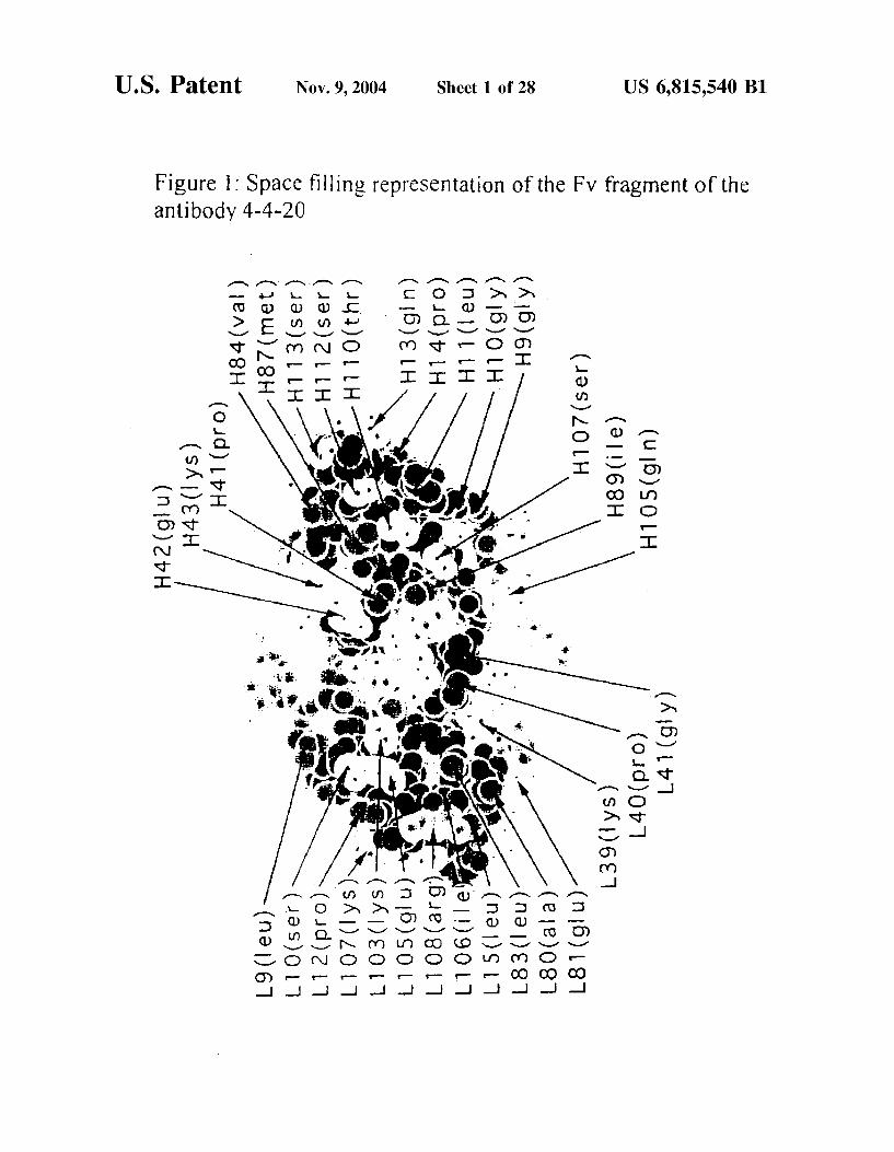
Figure 1: Space filling representation of the Fv fragment of the antibody 4-4-20
U.S. Patent
(u |6) SO IH (9 | !) 68H
(u º S)/. O || H
(K 16) 6H (K 16) O | H | (na 1) || || H
(u 16) $ | H(n 16) G O L T
?— (SK I) E O L T
( uu? !) O || || H`(sKI) ZO II ( 13 s) z 1 || H(o ud)? I T
§§( 13 s) O L T
] © Uu) / 8H(na I) 6T

U.S. Patent Nov. 9, 2004 Sheet 2 of 28 US 6,815,540 B1
Figure 2a: Variable/constant domain interface residues for VL
ty9
28
O
82
eZ2
ozz
bA.
92
72
22
O2
C O d vs. 8 S. O
e3ueue O 96

U.S. Patent Nov. 9, 2004 Sheet 3 of 28 US 6,815,540 B1
Figure 2a: Variable/constant domain interface residues for VL (cont.)

US 6,815,540 B1 U.S. Patent Nov. 9, 2004 Sheet 4 of 28
Figure 2a: Variable/constant domain interface residues for V. (cont.)
96
e G6
ty6
26
O6
98
99
-s z" -2S

U.S. Patent Nov. 9, 2004 Sheet 5 of 28 US 6,815,540 B1
Figure 2a: Variable/constant domain interface residues for VL (cont.)
N
o
Copepo (ped
i vio Vivlovo-colou–
...'... >ivivolo voivo Ooooo to -- o'-la---
------allel a co-o-o-o-o-o-o-o- to univ -in viou? vivuo-|u cooooooooooooo --|--|--|--|-- - HH--- > --> 22 >;-Ji>> -->>>> >>io do oc dio> DDDD --->|--|z--- . . ; oloo old-oluloloo
O
n
w
y
O N H
O C
o
N.
O
f
w
(Y)
N
-
<i>> CCD
o'clocs/laoloz
cococooloolooloolo


U.S. Patent Nov. 9, 2004 Sheet 7 of 28 US 6,815,540 B1
Figure 2a: Variable/constant domain interface residues for VL (cont.)
ul-ulu
| local
CD (DD:
-- ce---jor ZZ z'un-z-
-a al-ajo
------
D - -
slaisalsalsala alszisz Zsa
; : Sai O Sai Yi-CO' Sa
pupipio pico. D up
aloes on a

U.S. Patent Nov. 9, 2004 Sheet 8 of 28 US 6,815,540 B1
Figure 2a: Variable/constant domain interface residues for VL (cont.)
: .
hi-h -----
i
odoo
hola--
un; H-H -- wh
>u. L - ul-Is und alololo :
coup coup

U.S. Patent Nov. 9, 2004 Sheet 9 of 28 US 6,815,540 B1
Figure 2a: Variable/constant domain interface residues for VL (cont.)
Yori Y: YYYYYY: YY :
:
le H-i- & D COCO
i
olooloo oleioko cellull. CD: Di
|
o
s !----> . . . . . --J
wo
it was a ot z
i olor oz co
: U:O: Ulu OOOOO!oiu : :
H/> - wh
lolo
s:-
YY
| --
CD
-- ls
. . . : : . . . i - : :
ciolo v.o.o.o.o. oo'-too-cul OO
t
lulu. >-
Eco
YY
-- He
O
i
CDC
| viou? notic o
Cooloo
-
sts
<o
DC)
Viczi-zo
selotallels oloiz.o clotovole UOC-Q: Qiu

U.S. Patent Nov. 9, 2004 Sheet 10 of 28 US 6,815,540 B1
Figure 2a: Variable/constant domain interface residues for VL (cont.)

U.S. Patent Nov. 9, 2004 Sheet 11 of 28 US 6,815,540 B1
Figure 2b: Variable/constant domain interface residues for VH
8 d O C d t N 8
e3Uee, O %

U.S. Patent Nov. 9, 2004 Sheet 12 of 28 US 6,815,540 B1
Figure 2b: Variable/constant domain interface residues for VH (cont.)
6A.
44.
SA
EA
A.
69
A9
S9
99
9
s 6S
AS
SS
ES
C2S
2S
OS
87
97

U.S. Patent Nov. 9, 2004 Sheet 13 of 28 US 6,815,540 B1
Figure 2b. Variable/constant domain interface residues for VH (cont.)
O
OO .
OOL
POO .
GOOL
OO
96
96.
76


U.S. Patent Nov. 9, 2004 Sheet 16 of 28 US 6,815,540 B1
Figure 2b: Variable/constant domain interface residues for VH (cont.) Y (Y Y a Y a S4 Y Y Y > Y a Y \, \ Y Y Y a Na Y a Y Y Y Y Y
- - 1. > D - l D D - - > ll D U D - U l U Ll D - l D D -
vo (?o na V V < \ un on V) <( / na V) u v V u a
Ll d X X >- X d X X X CD X - - X X X X >-
2. d CY 1. 2 - X
- Q
() D X CO O CO CO cy) d CO ? X CD
- > - - - - - - - - - - - - - - - - - - - - - - - / >
or >- >- or ( p > Z - or D H >- H - Ll - St. LJ S >- - Lu u CD < Y
co ( p (p CC u? to D C C C CD CC (D CC CD (p up up to co ( p <C (p up < <C & D
> > - >> - J - > > -u -i > - > - - - > - > - > --> -->
& s 3 s 3 & 33 33 3 s > 3 > 3 > 3 > 3 > 3 > 3 3 s >-
Liu Lu Liu Lu u u u u LLJ u u Lu O U U U U Yu Y Lu Liu Lu Li Li Li Li.
- - - - - - - - - - - - - - - - - - - - - - - - - - -

U.S. Patent Nov. 9, 2004 Sheet 17 of 28 US 6,815,540 B1
Figure 2b: Variable/constant domain interface residues for VH (cont.)
O -(
H H - M. Cz I O W, or o O Y H or - cz - YHY - occe H de cc Hoc h or
- Y Z ZZ Z H- - Z H- a V or z <( Z v z v. Zozo w Z v ZZ Z ozz
2. 2 - - - 2 - - - - - - - > 2 - 22 - 2 - > - - - 2 > 2 - 2 2 -
O' o O O'o at u o' Y 2 o' u o' u osa o o' u do o O'o do O'o'c - > - - - > - - - - - 2 - 2 - V -> - 2 - - 2 - - - - >, >
X - X Ul Ll >- X
ul Li- C - - > - - - D D - (C - LL CC <C (C <( - - (C - J -->- > <C -u
- H W. O'H > v) O'H - - O' O'H H Z - - Cy H H - - /) H
f f 2 2 V w cy 1. 2. 2 u Z 2 v 2 (M) 2 Z 2 2 M Z (M) 2 v ) 2 ( 2.
Y Y 2 Na u)
4. C 2 - H. - H-.
u) u)
Y Z Y 2 Y
C. 1.
C) ) 1 O Z O 2 ul C) O O C C) :
-- al - u -- - --- u ru a - u m - L1 ll l - - 1 D --- - - Dm
- u? H - - Y (/) - H - H. H. H. H. C.
Ll - < Ul li - D U CC U < U D L L H L L. ti U t

U.S. Patent Nov. 9, 2004 Sheet 18 of 28 US 6,815,540 B1
Figure 2b: Variable/constant domain interface residues for VH (cont.)
O
C C C /), D C C CC
O

U.S. Patent Nov. 9, 2004 Sheet 19 of 28 US 6,815,540 B1
Figure 2b: Variable/constant domain interface residues for VH (cont.)
6
s ss ss ss ss is ss ss ss ss ss is s > s > s > s ss
o <C (9 < o o H. <C O - o KC O > d < Zoo o cho < o do o ?o a o
- > Llul - Llull 2 >- > u > ul LL - > v > u-ul Q. Zululu, o 2 u. --
H- <C d > > Y >- > Z & d Du Vo a s > Ll D >- Ll e >- > - ul
> 1 a

U.S. Patent Nov. 9, 2004 Sheet 20 0f 28 US 6,815,540 B1
Figure 3: Western blots showing the insoluble (i) and Soluble (s) fractions of cell extracts
CO of OO up is 2 22
- - if O in CO of s 5
2 st E
to - to - wr -
As
- O ve
a oi o s s
- - - - O
CO a c co to O) 3
N - l N - L CO a CN CO to o
2 2
to - to - 1. w up se st a
N. al
l t
ves
s s
s

U.S. Patent Nov. 9, 2004 Sheet 21 of 28 US 6,815,540 B1
Figure 4: Scatchard plots of fluorescence titration of fluorescein with antibody: a) Titration of wt ScFv; b) Titration of Flu4(V84D)
a 1.4x 10'
1.2x1 o'
2 Ox O
o 7
is 0.8x10 0. N 0.6 x 1 o'
0.4xt o'
O.2x1 o'
O
r
b 1.6 x 10'-
1.4x 1 o'
ar 1.2x 1 o' CD
9) 1.ox 1 o'
i O O.8x 1 o'
0.6 x 1 O
0.4x 10'
0.2x 1 o'

U.S. Patent Nov. 9, 2004 Sheet 22 of 28 US 6,815,540 B1
Figure 5: Overlay plot of urea denaturation. (x) wt Scriv, (o) Flu4
1 OO
4.68 OOO 2 O
urea) (M)
Figure 6: Thermal denaturation time courses at 40°C and 44°C for wt and Flu4 schv fragments
250
2OO
150
100
50
onal Oscaloa math
60 80 OO 120 40 160
Ellisi 4. O
time (min)

U.S. Patent Nov. 9, 2004 Sheet 23 of 28 US 6,815,540 B1
Table 1: Sequence variability of residues contributing to the v/c interface
O
Co o
v o
o N
u) rr
O
n s
C
O
e
l CO C N O
9 to 9 un c > r3 O ri is 2 S E. C. . . . . 2 at >. Sg - it f : 3 (5 (53. 3 S S S 3 g is

U.S. Patent Nov. 9, 2004 Sheet 24 of 28 US 6,815,540 B1
Table 1: Sequence variability of residues contributing to the v/c interface (cont.)
O
N o
y d
O
O o
O
O
o
Cd

U.S. Patent Nov. 9, 2004 Sheet 25 of 28 US 6,815,540 B1
Table 1: Sequence variability of residues contributing to the v/c interface (cont.)
O
w
s
s a
cvs O O
(v.
s y o O E
t o C
S.

U.S. Patent Nov. 9, 2004 Sheet 26 of 28 US 6,815,540 B1
Table I: Sequence variability of residues contributing to the v/c interface (cont.)
to mo C
o r C Cd
or cy e O 9 C. N. O Co
2 g 22 5 is a se S 9 so a 25, 8. ig SSA ; ; 5 is SSS gigse 2 -ra u a O SS R v 5 . C -
.9 d. c. w d is s V
3 5. R R 5 as

U.S. Patent Nov. 9, 2004 Sheet 27 of 28 US 6,815,540 B1
Table l: Sequence variability of residues contributing to the v/c interface (cont.)

U.S. Patent Nov. 9, 2004 Sheet 28 of 28 US 6,815,540 B1
Table 2: Mutations introduced in the scFv fragment of the antibody 4-4-20
15E (V) L11N (VH) 11 D (VH) V84D (VH) Fit O
Flu 2
Fu 3 p
F 4 O
Flu 5 O
Flu 6 d O
Flu 7
Flu 8 d g
Fit 9 9 () O
Flu 4 short
Table 3: KD values of the different scFv mutants determined in fluorescence titration
Flu W Fu 3 Flu 4 Flu 6 Flu wt.
KD (nM) 80 - 7 60 12 70 it 10 75 13 90

US 6,815,540 B1 1
IMMUNOGLOBULIN SUPERFAMILY DOMAINS AND FRAGMENTS WITH
INCREASED SOLUBILITY
CROSS-REFERENCE TO RELATED APPLICATIONS
This application is a Continuation of International Appli cation PCT/EP97/03792, filed Jul 16, 1997, which claims priority from European patent application EP 96111441.0, filed Jul. 16, 1996.
BACKGROUND OF THE INVENTION
The present invention relates to the modification of immu noglobuling Superfamily (IgSF) domains and derivatives thereof So as to increase their Solubility, and hence the yield, and ease of handling.
Small antibody fragments show exciting promise for use as therapeutic agents, diagnostic reagents, and for biochemi cal research. Thus, they are needed in large amounts, and the expression of antibody fragments, e.g. Fv, Single-chain-FV (scFv), or Fab in the periplasm of E. coli (Skerra & Pluckthun 1988; Better et al., 1988) is now used routinely in many laboratories. Expression yields vary widely, however, especially in the case of ScFvs. While some fragments yield up to Several mg of functional, Soluble protein per litre and OD of culture broth in shake flask 25 culture (Carter et al., 1992, Pluckthun et al. 1996), other fragments may almost exclusively lead to insoluble material, often found in So-called inclusion bodies. Functional protein may be obtained from the latter in modest yields by a laborious and time-consuming refolding process. The factors influencing antibody expression levels are Still only poorly understood.
Folding efficiency and Stability of the antibody fragments, protease lability and toxicity of the expressed proteins to the host cells often Severely limit actual production levels, and Several attempts have been tried to increase expression yields. For example, Knappik & Pluckthun (1995) have identified key residues in the antibody framework which influence expression yields dramatically. Similarly, Ullrich et al. (1995) found that point mutations in the CDRs can increase the yields in periplasmic antibody fragment expres Sion. Nevertheless, these Strategies are only applicable to a few antibodies.
The observations by Knappik & Pluckthun (1995) indi cate that optimizing those parts of the antibody fragment which are not directly involved in antigen recognition can Significantly improve folding properties and production yields of recombinant Fv and scEv constructs. The causes for the improved expression behavior lie in the decreased aggregation behavior of these molecules. For other molecules, fragment Stability and protease resistance may also be affected. The understanding of how Specific Sequence modifications change these properties is still very limited and currently under active investigation.
Difficulties in expressing and manipulating protein domains may arise because amino acids which are normally buried within the protein Structure become exposed when only a portion of the whole molecule is expressed. Aggre gation may occur through interaction of newly Solvent exposed hydrophobic residues originally forming the con tact regions between adjacent domains. Leistler and Perham (1994) could show that a certain domain of glutathione reductase may be expressed Separately from its neighboring domains, but the protein showed non-specific association in Vitro forming multimeric protein Species. The introduction
15
25
35
40
45
50
55
60
65
2 of hydrophilic residues instead of exposed hydrophobic amino acids could decrease this aggregation tendency and thus stabilize this isolated domain. Both wild type and modified domains were exclusively found in inclusion bod ies and had to be refolded. Although in vitro experiments contributed a lot to define various intermolecular interactions, which drive folding processes, they are only of limited value in predicting the folding behaviour of different polypeptide chains in vivo (Gething & Sambrook, 1992). Thus, Leistler and Perham do not teach or Suggest how to increase expression yields of Soluble protein domains.
In the case of antibodies, two chains comprising Several domains dimerize, each domain consisting of a b-barrel whose two b-sheets are held together by a disulphide bond, forming the So-called immunoglobulin fold. Two domains, one variable domain (VL) and one constant domain (CL) are adjacent along the longitudinal axis in the light chain (VL-CL), and four domains, one variable domain (VH) and three constant domain (CH1 to CH3) are adjacent along the longitudinal axis in the heavy chain (VH-CH1-CH2-CH3). In the dimer formed by chains a and b, two Such domains associate laterally: VLa with VHa, CLa with CH1a, VLb with VHb, CLb with CH1b CH2a with CH2b and CH3a with CH3b. In WO92/01787 (Johnson et al., 1992), it is taught that isolated Single domains, e.g. VH, can be modified in the former VL/VH interface region by exchanging hydrophobic residues by hydrophilic ones without changing the Specific ity of the parent domain. The rationale for WO92/01787 was the assumption that exposed hydrophobic residues might lead to non-specific binding, interaction with Surfaces and decreased Stability. Data for increase in binding Speci ficity was given, but increase in expression level was not shown. Furthermore, WO92/01787 would not be applicable to any antibody fragment containing the complete antigen binding site, as it must contain VL and VH. In the case of T cell receptors, two chains (a and b) dimerize, each consisting of a variable (V) and a constant (C) domain with the immunoglobulin fold, and one transmembrane domain. In each chain, the variable and constant domains are adjacent along the longitudinal axis in the chains (Va.-Ca.; Vb-Cb) and asSociate laterally with the corresponding domains of the second chain (Va.-Vb, Ca-Cb).
Various other molecules of the immunoglobulin Superfamily, such as CD2, CD4, CD16, CD22, comprise only one chain, wherein two or more domains (variable and/or constant) with the immunoglobulin fold are adjacent along the longitudinal axis in the chains.
SUMMARY OF THE INVENTION
The present inventors have found that expression prob lems are largely associated with a part of the molecule that has hitherto not been regarded relevant for expression Stud ies and which comprises the interface between adjacent domains within an immunoglobulin chain. This Surprising finding forms the basis of the present invention, which provides a general Solution to the problems associated with production of domains or fragments of the immunoglobulin Superfamily (IgSF), especially antibody fragments, which exhibit poor solubility or reduced levels of expression.
In addition to lateral interactions between domains of different chains described above, there are well documented contacts between adjacent domains within individual chains along the longitudinal axis. For example, in the case of an antibody (Lesk & Chothia, 1988), the “bottom” of VL makes contact with the “top” of CL, and, in a Similar manner there are contacts between VH and CH1. The contacts at these

US 6,815,540 B1 3
inter-domain interfaces are probably essential for the com pact arrangement of the Fab fragment, and, as is typical for Such contacts, are at least partially hydrophobic in nature (Lesk & Chothia, 1988).
The basis of the present invention is the Surprising finding that the solubility (and hence the yield) of antibody frag ments comprising at least one domain can be dramatically increased by decreasing the hydrophobicity of former inter faces at the “end” of said domain, where it would normally adjoin a Second domain within a chain in a larger antibody fragment or full antibody. This is Surprising and could not have been predicted from the prior art (WO 92/01787), because the size of the longitudinal interface, for example, in a scFv fragment, is much smaller than that between VH and VL, and therefore, the amino acids which make up the interfaces between VH and CH1 or between VL and CL in a Fab fragment represent a much Smaller proportion of the total Surface area of the ScFv molecule, and would accord ingly be expected to play less of a role in determining the physical properties of the molecule.
The present invention has the additional advantage that because the alterations effected in the molecules that lead to Said decreased hydrophobicity of former interfaces are located at the most distant part of the domain from the CDRS, applying the invention is unlikely to have a delete rious effect on the binding properties of the molecule. This is not the case in WO92/01787, where at least one modi fication is close to the CDRs and may therefore be expected to have an impact on antigen binding. Furthermore, WO 92/01787 cannot be applied to VL/VH heterodimers, as explained above.
BRIEF DESCRIPTION OF THE DRAWINGS
FIG. 1 provides a space filling representation of the Fv. fragment of the antibody 4-4-20.
FIG. 2 presents the variable/constant domain interface residues for VL(2a) and VH (2b). For 30 non-redundant Fab fragments taken from the Brookhaven Databank, the solvent accessible Surface of the amino acid Side chains was calcu lated in the context of an Fv and of an Fab fragment. The plot shows the relative reduction in accessible Surface upon contact with the constant domains (overlay plot for all 30 Fv fragments). In the Sequence alignment, residues contributing to the v/c interface are highlighted. The symbols indicate the relative reduction of Solvent accessible Surface upon remov ing the constant domains (symbols: no symbol <1%; 1<20%; nk40%; S-60%; t-80%, and u 80%). Circles indicate those positions which are further analyzed (see Table 1).
FIG. 3 presents Western blots showing the insoluble (i) and Soluble (S) fractions of cell extracts, prepared as described in Material and Methods, expressing the scFv fragments of the antibody 4-4-20. The amino acids substi tuted in the various mutants are given in Table 2.
FIG. 4 presents a Scatchard plot of the fluorescence titration of fluorescein (20 nM) with antibody (4 to 800 nM), measured at 510 nm. The value r was obtained from (F-Fo)/ (FY-Fo), where F is the measured fluorescein fluorescence at a given antibody concentration, Fo is the fluorescence in the absence of antibody and FY when antibody is present in large excess. Note that r gives the Saturation of fluorescein by antibody. (a) Titration of wt scFv, (b) titration of Flu4 (V84D).
FIG. 5 presents an overlay plot of the urea denaturation curves ((X) wt scFv, (o) Flu4).
FIG. 6 presents the thermal denaturation time courses at 40 and 44 C. for wt and Flu4 scEv fragment ((a) wt scFv at 40°C., (b) Flu.4 at 40°C., (c) Flua at 44°C., (d) wt scFv at 44° C.).
15
25
35
40
45
50
55
60
65
4 Table 1 describes the sequence variability of residues
contributing to the V/c interface. Residue Statistics are based on the variable domain Sequences in the Kabat database (March 1996). Sequences which were <90% complete were excluded from the analysis. Number of Sequences analyzed: human VL kappa: 404 of 881, murine VL kappa: 1061 of 2239, human VL lambda: 223 of 409, murine VLlambda: 71 of 206, human VH: 663 of 1756, murine VH: 1294 of 3849. Position refers to the Sequence position according to Kabat et al. 1991, 9%exp. (Fab) to the relative side chain accessi bility in an Fab fragment as calculated by the program NACCESS (NACCESS v2.0 by Simon Hubbard, 76exp. (ind.) to the relative side chain accessibility in the isolated VL or VH domain, %buried to the relative difference in side chain accessibility between Fv and Fab fragment. Consensus refers to the Sequence consensus, and Distribution to the distribution of residue types.
Table 2 describes mutations introduced in the sclv frag ment of the antibody 4-4-20. Each line represents a different protein carrying the mutations indicated. The residues are numbered according to Kabat et al. (1991).
Table 3 describes KD values of the different ScFv mutants determined in fluorescence titration. The KD values are given in nM, the error was calculated from the Scatchard analysis (FIG. 4). # determined by Miklasz et al. (1995).
DESCRIPTION OF THE PREFERRED EMBODIMENTS
The present invention relates to a modified immunoglo bulin superfamily (IgSF) domain or fragment which differs from a parent IgSF domain or fragment in that the region which comprised or would comprise the interface with a Second domain adjoined to Said parent IgSF domain or fragment within the protein chain of a larger IgSF fragment or a full IgSF protein, and which is exposed in Said parent IgSF domain or fragment in the absence of Said Second domain, is made more hydrophilic by modification.
In the context of the present invention, the term immu noglobulin Superfamily (IgSF) domain refers to those parts of members of the immunoglobulin Superfamily which are characterized by the immunoglobulin fold, Said Superfamily comprising the immunoglobulins or antibodies, and various other proteins Such as T-cell receptorS or integrins. The term IgSF fragment refers to any portion of a member of the immunoglobulin Superfamily, Said portion comprising at least one IgSF domain. The term adjoining domain refers to a domain which is contiguous with a first domain. The term interface refers to a region of Said first domain where interaction with the adjoining domain takes place. The terms hydrophobic and hydrophilic refer to a physical property of amino acids, which can be estimated quantitatively: tabu lated values of hydrophobicity for the twenty naturally occurring amino acids are available (Nozaki & Tanford, 1971; Casari & Sippl, 1992; Rose & Wolfenden, 1993). The residues to be modified can be identified in a number
of ways. For example, in one way, the Solvent accessibilities (Lee & Richards, 1971) of hydrophobic interface residues in Said parent IgSF fragment compared to Said larger IgSF fragment or full IgSF protein are calculated, with high accessibilities indicating highly exposed residues. In a Sec ond way, the number of van der Waals contacts of hydro phobic interface residues in Said larger IgSF fragment or full IgSF protein is calculated. A large number for a residue of Said parent domain indicates that it will be highly Solvent exposed in the absence of an adjoining domain. There are other ways of calculating or determining residues to be

US 6,815,540 B1 S
modified according to the present invention, and one of ordinary skill in the art will be able to identify and practice these ways.
By analyzing computer models of Said parent IgSF fragment, interactions of Said highly exposed residues within the fragment can be identified. Such interactions could Stabilize the parent IgSF fragment. Residues, which interact closely with other hydrophobic residues and which can be identified by anyone of ordinary skill in the art, should not preferentially be mutated.
The modification referred to above may be effected in a number of ways which are well known to one skilled in the art. In a preferred embodiment, the modification is a Sub Stitution of one or more amino acids at the exposed interface, identified as described above, with amino acids which are more hydrophilic Alternatively, one or more amino acids can be inserted in Said interface, or one or more amino acids can be deleted from Said interface, So as to increase its overall hydrophilicity. Furthermore, any combination of Substitution, insertion and deletion can be effected to reduce the hydrophobicity of said interface. Also comprised by the present invention is the possibility that the Substitution or insertion comprises amino acids with a relatively high hydrophobicity value, or that the deletion comprises amino acids with relatively low hydrophobicity value, as long as the Overall hydrophilicityValue is increased in Said interface region. Modifications Such as Substitution, insertion and deletion can be effected using Standard methods which are well known to practitioners skilled in the art. By way of example, the Skilled artisan can use either Site-directed or PCR-based mutagenesis (Ho et al., 1989; Kunkel et al., 1991; Trower, 1994; Viville, 1994), or total gene synthesis (Prodromou & Pearl, 1992) to effect the necessary modifications). In a further embodiment, the mutations may be obtained by random mutagenesis and Screening of ran dom mutants, using a Suitable expression and Screening System (see, for example, Stemmer, 1994, Crameri et al., 1996).
In a preferred embodiment, the amino acid(s) which replace(s) the more hydrophobic amino acids include ASn, Asp, Arg, Gln, Glu, Gly, His, LyS, Ser, and Thr. These are among the more hydrophilic of the 20 naturally-occurring amino acids, and have proven to be particularly effective in the application of the present invention. Said amino acids, alone or in combination, or in combination with other amino acids, can also be used to form the above mentioned insertion which makes the interface region more hydro philic.
The parent IgSF domain or fragment referred to above can be one of several different types. In a preferred embodiment, Said parent domain or fragment is derived from an antibody. In one embodiment, Said parent antibody fragment com prises an Fv fragment. In this context, the term Fv fragment refers to a complex comprising the VL (variable light) and VH (variable heavy) portions of the antibody molecule. In a further embodiment, the parent antibody fragment may be a single-chain Fv fragment (scFv, Bird et al., 1988; Huston et al., 1988), in which the VL and VH chains are joined, in either a VL-VH, or VH-VL orientation, by a peptide linker. In yet a further embodiment, the parent antibody fragment may be an Fv fragment Stabilized by an inter-domain dis ulphide bond. This is a structure which can be made by engineering into each chain a Single cysteine residue, wherein Said cysteine residues from two chains become linked through oxidation to form a disulphide (Glockshuber et al., 1990; Brinkmann et al., 1993).
In a most preferred embodiment, the interface region of the variable domains mentioned above comprises residues 9,
15
25
35
40
45
50
55
60
65
6 10, 12, 15, 39, 40, 41,80, 81, 83, 103,105,106, 106A, 107, 108 for VL, and residues 9, 10, 11, 13, 14, 41, 42, 43, 84, 87, 69, 105, 108, 110, 112, 113 for VH according to the Kabat numbering system (Kabat et al., 1991). Said numbering System was established for the Sequences of whole antibodies, but can be adapted correspondingly to describe the Sequences of isolated antibody domains or antibody fragments, even in the case of ScFv fragments, where VL and VH are connected via a peptide linker, and where the protein sequence from N- to C-terminus has to be numbered differ ently. This means that the Kabat numbering System is used in the present invention as a Sequence description relative to the existing data on antibody Sequences, not as an absolute description of actual positions within the antibody fragment Sequences of interest.
In a further embodiment, Said parent antibody fragment comprises a Fab fragment. In this context, the term Fab refers to a complex comprising the VL-CL (variable and constant light) and VH-CH1 (variable and first constant heavy) portions of the antibody molecule, and the term interface region refers to a region in the first constant domain of the heavy chain (CH1) which is, or would be adjoined to, the CH2 domain in a larger antibody fragment or full antibody.
In a still further embodiment, said parent IgSF fragment is a fusion protein of any of Said domains or fragments and another protein domain, derived from an antibody or any other protein or peptide. The advent of bacterial expression of antibody fragments has opened the way to the construc tion of proteins comprising fusions between antibody frag ments and other molecules. A further embodiment of the present invention relates to such fusion proteins by provid ing for a DNA sequence which encodes both the modified IgSF domain or fragment, as described above, as well as an additional moiety. Particularly preferred are moieties which have a useful therapeutic function. For example, the addi tional moiety may be a toxin molecule which is able to kill cells (Vitetta et al., 1993). There are numerous examples of Such toxins, well known those skilled in the art, Such as the bacterial toxins Pseudomonas eXotoxin A, and diphtheria toxin, as well as the plant toxins ricin, abrin, modeccin, Saporin, and gelonin. By fusing Such a toxin to an antibody fragment, the toxin can be targeted to, for example, diseased cells, and thereby have a beneficial therapeutic effect. Alternatively, the additional moiety may be a cytokine, Such as IL-2 (Rosenberg & Lotze, 1986), which has a particular effect (in this case a T-cell proliferative effect) on a family of cells. In a further preferred embodiment, the additional moiety is at least part of a Surface protein which may direct the fusion protein to the Surface of an organism, for example, a cell or a phage, and thereby displays the IgSF partner. Preferably, the additional moiety is at least part of a coat protein of filamentous bacteriophages, most preferably of the geneIII protein. In a further embodiment, the additional moiety may confer on its IgSF partner a means of detection and/or purification. For example, the fusion protein could comprise the modified IgSF domain or fragment and an enzyme commonly used for detection purposes, Such as alkaline phosphatase (Blake et al., 1984). There are numer ous other moieties which can be used as detection or purification tags, which are well known to the practitioner skilled in the art. Particularly preferred are peptides com prising at least five histidine residues (Hochuliet al., 1988), which are able to bind to metal ions, and can therefore be used for the purification of the protein to which they are fused (Lindner et al., 1992). Also provided for by the invention are additional moieties Such as the commonly used

US 6,815,540 B1 7
c-myc and FLAG tags (Hopp et al., 1988; Knappik & Pluckthun, 1994).
By engineering one or more fused additional domains, IgSF domains or fragments can be assembled into larger molecules which also fall under the Scope of the present invention. To the extent that the physical properties of the IgSF domain or fragment determine the characteristics of the assembly, the present invention provides a means of increas ing the Solubility of Such larger molecules. For example, mini-antibodies (Pack, 1994) are dimers comprising two antibody fragments, each fused to a Self-associating dimer ization domain. Dimerization domains which are particu larly preferred include those derived from a leucine Zipper (Pack & Pluckthun, 1992) or helix-turn-helix motif (Packet al., 1993).
All of the above embodiments of the present invention can be effected using Standard techniques of molecular biology known to anyone skilled in the art.
The compositions described above may have utility in any one of a number of Settings. Particularly preferred are diagnostic and therapeutic compositions.
The present invention also provides methods for making the compositions and compounds comprised therein described above. Particularly preferred is a method com prising the following Steps: i) analyzing the interface region of an IgSF domain for
hydrophobic residues which are Solvent-exposed using either a Solvent-accessibility approach (Lee & Richards, 1971), analysis of van der Waals interactions in the interface region, or similar methods which are well known to one skilled in the art,
ii) identifying one or more of the hydrophobic residues to be Substituted by more hydrophilic residues, or one or more positions where hydrophilic residues or amino acid Stretches enhancing the overall hydrophilicity of the inter face region can be inserted into Said interface region, or one or more positions where hydrophobic residues or amino acid stretches enhancing the overall hydrophobic ity of the interface region can be deleted from Said interface region, or any combination of Said Substitutions, Said insertions, and Said deletions to give one or more mutants of Said parent IgSF domain,
iii) preparing DNA encoding mutants of Said IgSF domain, characterized by the changes identified inii), by using e.g. conventional mutagenesis or gene Synthesis methods, Said DNA being prepared either Separately or as a mixture,
iv) introducing said DNA or DNA mixture in a vector system Suitable for expression of Said mutants,
V) introducing said vector System into Suitable host cells and expressing Said mutant or mixture of mutants,
Vi) identifying and characterizing mutants which are obtained in higher yield in soluble form, and
Vii) if necessary, repeating steps iii) to vi) to increase the hydrophilicity of said identified mutant or mutants further. The host referred to above may be any of a number
commonly used in the production of heterologous proteins, including but not limited to bacteria, Such as E. coli (Geet al, 1995), or Bacillus Subtilis (Wu et al., 1993), fungi, such as yeasts (Horwitz et al., 1988; Ridder et al., 1995) or filamentous fungus (Nyyssonen et al., 1993), plant cells (Hiatt, 1990, Hiatt & Ma, 1993; Whitelam et al., 1994), insect cells (Potter et al., 1993; Ward et al., 1995), or mammalian cells (Trill et al., 1995). The invention also relates to a method for the production
of an IgSF domain or fragment of the invention comprising culturing a host cell of the invention and isolating Said domain or fragment.
1O
15
25
35
40
45
50
55
60
65
8 The invention is now demonstrated by the following
examples, which are presented for illustration only and are not intended to limit the Scope of the invention.
EXAMPLES
i) Abbreviations Abbreviations are defined as follows: CDR: complemen
tarity determining region; dsFv: disulfide-linked FV frag ment; IMAC: immobilized metal ion affinity chromatogra phy; IPTG: isopropyl-b-D-thiogalactopyranoside, i?s: ratio insoluble/soluble; H(X): heavy chain residue number X; L(X): light chain residue number X; NTA: nitrilo-triacetic acid; OD550: optical density at 550 nm; PDB: protein database; scFv: single-chain Fv fragment; SDS-PAGE: Sodium dodecyl Sulfate polyacrylamide gel electrophoresis, V/c: Variable/constant, wit: wild type.
ii) Material and Methods (a) Calculation of Solvent Accessibility
Solvent accessible Surface areas for 30 non-redundant Fab fragments and the Fv fragments derived from these by deleting the constant domain coordinates from the PDB file were calculated using the latest version, as of March 1996, of the program NACCESS (http://www.biochem.ucl.ac. uk/~roman/naccess/naccess) based on the algorhithm described by Lee & Richards (1971). (b) sclv Gene Synthesis The single-chain Fv fragment (scFv) in the orientation
VL-linker-VH of the antibody 4-4-20 (Bedzyk et al., 1990) was obtained by gene synthesis (Prodromou and Pearl, 1992). The VL domain carries a three-amino acid long FLAG tag (Knappik and Pluckthun, 1994). We have used two different linkers with a length of 15 (Gly4Ser)3 and 30 amino acids (Gly4Ser)6, respectively. The gene So obtained was cloned into a derivative of the vector pIG6 (Ge et al., 1995). The mutant antibody fragments were constructed by Site-directed mutagenesis (Kunkel et al., 1987) using Single Stranded DNA and up to three oligonucleotides per reaction. (c) Expression
Growth curves were obtained as follows: 20 ml of 2XYT medium containing 100 g/ml amplicillin and 25 g/ml Strep tomycin were inoculated with 250 1 of an overnight culture of E. coli JM83 harboring the plasmid encoding the respec tive antibody fragment and incubated at 24.5 C. until an OD550 of 0.5 was reached. IPTG (Biomol Feinchemikalien GmbH) was added to a final concentration of 1 mM and incubation was continued for 3 hours. The OD was mea Sured every hour, as was the b-lactamase activity in the culture Supernantant to quantify the degree of cell leakiness. Three hours after induction an aliquot of the culture was removed and the cells were lysed exactly as described by Knappik and Pluckthun (1995). The b-lactamase activity was measured in the Supernatant, in the insoluble and in the soluble fraction. The fractions were assayed for antibody fragments by reducing SDS-PAGE, with the samples nor malized to OD and b-lactamase activity to account for possible plasmid loSS as well as for cell leakineSS. The gels were blotted and immunostained using the FLAG antibody M1 (Prickett et al., 1989) as the first antibody, an Fc-specific anti-mouse antiserum conjugated to horseradish peroxidase (Pierce) as the Second antibody, using a chemoluminescent detection assay described elsewhere (Ge et al., 1995). (d) Purification
Mutant scFv fragments were purified by a two-column procedure. After French press lysis of the cells, the raw E. coli extract was first purified by IMAC (Ni-NTA Superflow,

US 6,815,540 B1 9
Qiagen) (20 mM HEPES, 500 mM NaCl, pH 6.9; step gradient of imidazole 10, 50 and 200 mM) (Lindner et al., 1992) and, after dialyzing the IMAC eluate against 20 mM MES pH 6.0, finally purified by cation exchange chroma tography (S-Sepharose fast flow column, Pharmacia) (20 mM MES, pH 6.0; salt gradient 0-500 mM NaCl). Purity was controlled by Coomassie stained SDS-PAGE. The func tionality of the scFv was tested by competition ELISA.
Because of its very poor Solubility in the periplasmic System, the wt 4-4-20 was expressed as cytoplasmic inclu sion bodies in the T7-based system (Studier & Moffatt, 1986; Ge et al., 1995). The refolding procedure was carried out as described elsewhere (Ge et al., 1995). For purification, the refolding solution (21) was loaded over 10 h without prior dialysis onto a fluorescein affinity column, followed by a washing step with 20 mM HEPES, 150 mM NaCl, pH 7.5. Two column volumes of 1 mM fluorescein (sodium salt, Sigma Chemicals Co.) pH 7.5 were used to elute all functional scFv fragment. Extensive dialysis (7 days with 12 buffer changes) was necessary to remove all fluorescein. All purified ScFv fragments were tested in gel filtration (Superose-12 column, Pharmacia SMART-System, 20 mM HEPES, 150 mM NaCl, pH 7.5). (e) KD Determination by Fluorescence Titration
The concentrations of the proteins were determined pho tometrically using an extinction coefficient calculated according to Gill and von Hippel (1989). Fluorescence titration experiments were carried out by taking advantage of the intensive fluorescence of fluorescein. Two ml of 20 mM HEPES, 150 mM NaCl, pH 7.5 containing 10 or 20 nM fluorescein were placed in a cuvette with integrated Stirrer. The excitation wavelength was 485 nm, emission spectra were recorded from 490 to 530 nm. Purified scFv (in 20 mM HEPES, 150 mM NaCl, pH 7.5) was added in 5 to 100 1 aliquots, and after a 3 min equilibration time a spectrum was recorded. All spectra were recorded at 20° C. The maximum of emission at 510 nM was used for determining the degree of complexation of ScFv to fluorescein, Seen as quenching as a function of the concentration of the antibody fragment. The KD value was determined by Scatchard analysis. (f) Equilibrium denaturation measurement
Equilibrium denaturation curves were obtained by dena turation of 0.2 M protein in HEPES buffered saline (HBS) buffer (20 mM HEPES, 150 mM NaCl, 1 mM EDTA, pH 7.5) and increasing amounts of urea (1.0–7.5 M; 20 mM HEPES, 150 mM NaCl, pH 7.4; 0.25 M steps) in a total volume of 1.7 ml. After incubating the samples at 10 C. for 12 hours and an additional 3 hours at 20° C. prior to measurements, the fluorescence spectra were recorded at 20 C. from 320-360 nm with an excitation wavelength of 280 nm. The emission wavelength of the fluorescence peak shifted from 341 to 347 nm during denaturation and was used for determining the fraction of unfolded molecules. Curves were fitted according to Pace (1990). (g) Thermal Denaturation
For measuring the thermal denaturation rates, purified ScFv was dissolved in 2 ml HBS buffer to a final concen tration of 0.5M. The aggregation was followed for 2.5 h at 40° C. and at 44° C. by light scattering at 400 nm.
iii) Results (a) Comparison of Known Antibody Sequences
Compared to other domain/domain interfaces in proteins, the interface between immunoglobulin variable and constant domains is not very tightly packed. A comparison of 30 non-redundant Fab structures in the PDB database showed that between the light chain variable and constant domain an area of 410 +90 A2 per domain is buried, while the heavy
15
25
35
40
45
50
55
60
65
10 chain variable and constant domains interact over an area of 710-180 A2. Some, but not all of the interface residues are hydrophobic, predominantly aliphatic. Generally, Sequence conservation of the residues contributing to the V/c domain interface is not particularly high. Still, the V/c domain interface Shows up as a marked hydrophobic patch on the surface of an Fv fragment (FIG. 1).
Solvent accessible Surface areas for 30 non-redundant Fab fragments and their corresponding Fv fragments (derived from the Fab fragment by deleting the constant domain coordinates from the PDB file) were calculated using the program NACCESS (Lee & Richards, 1971). Residues participating in the V/c domain interface were identified by comparing the Solvent-accessible Surface area of each amino acid Side chain in the context of an Fv fragment to its accessible surface in the context of an Fab fragment. FIG.2 shows a plot of the relative change in Side chain accessibility upon deletion of the constant domains as a function of Sequence position. Residues which show a significant reduc tion of Side chain accessibility are also highlighted in the Sequence alignment. To assess Sequence variability in the positions identified in FIG. 2, the variable domain Sequences collected in the Kabat database (status March 1996) were analyzed (Table 1). Of the 15 interface residues identified in the VL domain of the antibody 4-4-20 (FIG. 1 and Table 1), L9(leu), L12(pro), L15(leu), L40(pro), L83(leu), and L106 (ile) are hydrophobic and therefore candidates for replace ment. Of the 16 interface residues in the VH domain, Hll(leu), H14(pro), H41(pro), H84(val), H87(met) and H89 (ile) were identified as possible candidates for Substitution by hydrophilic residues in the scFv fragment of the antibody 4-4-20 (FIG. 1 and Table 1). Not all of these hydrophobic residues are equally good
candidates for replacements, however. While residues which are hydrophobic in one particular Sequence but hydrophilic in many other Sequences may appear most attractive, the conserved hydrophobic residues listed in Table 1 have also been investigated, since the evolutionary pressure which kept these conserved residues acted on the Fab fragment within the whole antibody, but not the isolated Fv portion. In this study, we did not replace the proline residues since proL40 and pro H41 form the hairpin turns at the bottom of the framework II region, while the conserved VLcis-proline L8 and proline residues H9 and H14 determine the shape of framework I of the immunoglobulin variable domains.
Excluding prolines, this leaves residues L9 (leu in 4-4-20, Ser in most kappa chains), L15 (leu, usually hydrophobic), L83 (leu, usually val or phe) and L106 (Ile, as in 86% of all kappa chains) in the VL domain and Hll (leu as in 60% of all heavy chains), H84 (val, in other VH domains frequently ala or ser), H87 (net, usually ser) and H89 (ile, most frequently val) in VH as possible candidates for replacement in the 4-4-20 scFv fragment. (b) Mutations in the 4-4-20 scFv
For the 4-4-20 scFv fragment some of the crucial residues identified in the Sequence analysis described above are already hydrophilic, but nevertheless 9 residues are of hydrophobic nature (including pro12 in the light chain) (Table 1). We chose three residues for closer analysis by mutations.
Leu15 in VL is a hydrophobic amino acid in 98% of all kappa chains (Table 1). Leu11 is conserved in VH (Table 1) and is involved in V/c interdomain contacts (Lesk & Chothia, 1988). In contrast, valine occurs very infrequently at posi tion H84; mainly found at this position are threonine or serine and alanine (Table 1). As can be seen in FIG. 1, val84 is contributing to a large hydrophobic patch at the newly

US 6,815,540 B1 11
exposed surface of VH. All three positions were mutated into acidic residues, and L11 was also changed to asparagine (Table 2).
The scFv fragment was tested and expressed with two different linkers, the 15-mer linker (Gly4Ser)3 (Huston et al., 1995) and the same motif extended to 30 amino acids (Gly4Ser)6. All mutations were tested in both constructs. The in vivo results of the different mutations on solubility were identical, and therefore only the results of the 30-mer linker are described in more detail. The periplasmic expres Sion experiments were carried out at 24.5 C., and all constructs were tested for soluble and insoluble protein by immunoblotting. The ratio of insoluble to soluble (i/s) protein was determined for every mutant. In FIG. 3 A-D, insoluble (lane 1) and soluble (lane 2) fractions of the wt scFv are shown. Nearly no soluble material occurs in periplasmic expression, which is consistent with previous reports of Bedzyk et al. (1990) and Denzin et al. (1991), who described earlier that the periplasmic expression of the wt ScFv leads mainly to periplasmic inclusion bodies.
The single point mutation L15E in VL (Flul) shows no effect on the ratio i?s when compared with the wt (FIG. 3A, lane 3, 4). Mutating leu at position 11 in the heavy chain to asparagine (Flu2) also shows nearly no effect compared to the wt, whereas the subtitution with aspartic acid (Flu3) changes the i/S ratio to more Soluble protein, but still this effect is not very dramatic. In contrast, the point mutation at position 84 (Flu4, FIG. 3B, lane 3, 4 and FIG. 3D, lane 3, 4) had a dramatic influence on the solubility of the scFv fragment of the antibody 4-4-20. The ratio i?s is changed to about 1:1, resulting in a 25-fold increase of Soluble protein compared to the wt.
The combination of V84D with LIN or L11D (Flu5, Fluó) also changes the ratio i?s compared to the wt, but this ratio compared to V84D alone is not improved further (FIG.3B). Interestingly, the combination of Flu5 with the light chain mutation at position 15 (Flu9) leads to less soluble material (FIG. 3C lane 7,8) than Flu5 itself (FIG.3B, lane 5, 6). The negative influence of the L15E mutations can also be seen in Flu8 (FIG.3C, lane 5, 6) compared with Flu3 (FIG. 3A, lane 7, 8). In FIG. 3D the comparison of the wt (lane 1, 2 and 5, 6) and Flu4 (lane 3, 4 and 7, 8) is shown in both the 15-mer and the 30-mer construct. The negative effect of L15E can be rationalized by
looking at a model of the 4-4-20 scFv fragment. L15 is forming a hydrophobic pocket together with residues A80, L83, and L106. Apparently, L15 stabilizes the scFv fragment by hydrophobic interactions with its closest neighbourS. Thus the exchange L15E for making the ScFV fragment more hydrophilic and more Soluble is made at the expense of the fragment Stability. The analysis of hydrophobic interactions within a fragment should thereby by used to choose the Solvent-exposed residues to be mutated in the case of any other antibody fragment. Combinations of various Serine mutations in VH led to further improvements in the i/s ratio. The mutants FH15 (V84S, M87S, I89S) and FH20 (L11S, v84s, M87S, I89S) both showed more than 70% of soluble protein in immunoblots (data not shown). The negative effect of L15E (c) Functional Expression and Purification
The oligomerization of ScFv fragments as a function of linker length has been investigated previously. A continuous decrease in the amount of dimer and multimer formation as a function of linker length has been reported (Desplancq et al., 1994; Whitlow et al., 1994). While the (Gly4Ser)3 linker has been shown to lead to monomeric ScFVS in many cases in the VH-VL direction, this is often not the case in the
15
25
35
40
45
50
55
60
65
12 VL-VH direction. This is caused by an asymmetry in the VL/VH arrangement, leading to a longer distance between the end of VH and the N-terminus of VL than between C-terminus of VL and N-terminus of VH (Huston et al., 1995). Consequently, a linker of identical length may lead to different properties of the resulting molecules.
Since we have chosen to use the minimal pertubation FLAG (Knappik & Pluckthun, 1994) at the N-terminus of VL in our constructs and thus the VL-linker-VH orientation, we have investigated the use of longer linkers. In the periplasmic expression in E. coli no difference between the 15-mer and the 30-mer linker in the corresponding mutants is visible (FIG. 3D), but when we attempted to purify the two Flu4 ScFVS with long and short linker, a big discrepancy between the two constructs was found. The purification of the Flu4 mutant (V84D) with the 15-mer linker leads to very Small amounts of partially purified protein (about 0.015 mg per liter and OD; estimated from SDS-PAGE after IMAC purification), whereas the 30-mer linker construct gives about 0.3 mg per liter and OD of highly pure functional protein. All mutants with 30-mer linker were tested in gel filtration and found to be monomeric (data not shown).
For further in vitro characterization five mutants were purified with the 30-mer linker, V84D (Flu4), V84D/L11D (Fluo), L11D (Flu3), and the serine mutants FH15 and FH20 (see iii.(b)). A two-step chromatography, first using IMAC and then cation-exchange chromatography, led to homoge neous protein. The i/s ratio of the antibody fragments (FIG. 3) was also reflected in the purification yield of functional protein. The highly soluble mutant Flu.4 (V84D) (FIG. 3B lane 3, 4) yielded about 0.3 mg purified and functional protein per liter and OD, Fluó (L11D/V84D) (FIG. 3B lane 7, 8) yielded about 0.25 mg per liter and OD and Flu3 (less soluble material on the blot in FIG. 3A lane 7, 8) yielded 0.05 mg per liter and OD. The serine mutants FH15 and FH20 yielded 0.3 mg and 0.4 mg per liter and OD, respec tively. The wt scFv of the antibody 4-4-20 did not give any Soluble protein at all in periplasmic expression with either linker, and it was therefore expressed as cytoplasmic inclu sion bodies, followed by refolding in vitro and fluorescein affinity chromatography. The refolded wit scFv was shown by gel filtration to be monomeric with the 30-mer linker (data not shown). (d) Biophysical Properties of the Mutant scFvs
Since we changed amino acids which are conserved, it cannot be excluded that changes at these positions may be transmitted through the Structure and have an effect on the binding constant, even though they are very far from the binding site (Chatellier et al., 1996). To eliminate this possibility, we determined the binding constant of the mutants Flu3, Flua, Flu.6 and the wt ScFv. Fluorescence titration was used to determine KD in Solution by using the quenching of the intrinsic fluorescence of fluorescein when it binds to the antibody. The fluorescence quenching at 510 nm was measured as a function of added ScFv. The KD values (Table 3 and FIG. 4) obtained for all three mutant scFVs and the wt scFv are very similar and correspond very well to the recently corrected KD of the monoclonal anti body 4-4-20 (Miklasz et al., 1995). To determine whether the mutations had an influence on
the thermodynamic stability of the protein we determined the equilibrium unfolding curves by urea denaturation. V84D mutant and the wt scFv were used for this analysis, and in FIG. 5 an overlay plot is shown. The midpoint of both curves is at 4.1 M urea. Both curves were fitted by an algorithm for a two-state model described by Pace (1990), but the apparent small difference between the V84D mutant

US 6,815,540 B1 13
and the wt Scev is not of Statistical significance. Aggregation of folding intermediates could be another explanation for the different in Vivo results between the mutant sclvs and the wt ScFv (FIG. 3). In the periplasm of E. coli, the protein concentrations are assumed to be rather high (van Wielink & Duine, 1990) and the aggregation effects could thus be pronounced. In order to estimate the aggregation behavior in Vitro, We have measured the thermal aggregation rates at different temperatures. In FIG. 6 it is clearly seen that the wt Scev is significantly aggregating already at 44°C., whereas the mutant V84D tends to aggregate more slowly. The wt Scev is thus clearly more aggregation prone than the mutant ScFv. This is very similar to the observations made with different mutations on the antibody McPC603 (Knappik and Pluckthun, 1995), where no correlation was found between equilibrium denaturation curves and expression behavior, but a good correlation was found with the thermal aggre gation rates.
OTHER PUBLICATIONS
Better, M., Chang, P., Robinson, R. & Horwitz, A. H. (1988). E. coli Secretion of an active chimeric antibody fragment. Science 240, 1041-1043.
Bird, R. E., Hardman, K. D., Jacobson, J. W., Johnson, S., Kaufman, B. M., Lee S. M., Lee T., Pope S. H., Riordan G. S., & Whitlow M. (1988). Single-chain antigen binding proteins published erratum appears in (1989). Science 244, 4091). Science 242, 423–6.
Bedzyk, W. D., Weidner, K. M., Denzin, L. K., Johnson, L. S., Hardman, K. D., Pantoliano, M. W., Asel, E. D. & Voss, E. W., Jr. (1990). Immunological and structural characterization of a high affinity anti-fluorescein single chain antibody. J. Biol. Chem. 265, 18615-18620.
Blake, M. S., Johnston, K. H., Russel-Jones, G. J. & Gotschlich, E. C. (1984). A rapid, sensitive method for detection of alkaline phosphatase-conjugated anti antibody on Western blots. Anal. Biochem. 136, 175-179.
Brinkmann, U., Reiter, Y., Jung, S., Lee, B. & Pastan, I. (1993). A recombinant immunotoxin containing a disulfide-stabilized Fv fragment. Proc. Natl. Acad. Sci. U.S.A. 90, 7538.07542.
Carter, P., Kelley, R. F., Rodrigues, M. L., Snedecor, B., Covarrubias, M., Velligan, M. D., Wong, W. L. T., Rowland, A. M., Kotts, C. E., Carver, M. E., Yang, M., Bourell, J. H., Shepard, H. M. & Henner, D. (1992). High level Escherichia coli expression and production of a bivalent humanized antibody fragment. Bio/Technology 10, 163-167.
Casari, G. & Sippl, M. J. (1992). Structure-derived hydro phobic potential. Hydrophobic potential derived from X-ray structures of globular proteins is able to identify native folds. J. Mol. Biol., 224, 725–32.
Chatellier, J., van Regenmortel, M. H. V., Vernet, T. & Altschuh, D. (1996). Functional mapping of conserved residues located at the VL and VH domain interface of an Fab. J. Mol. Biol., in press.
Denzin, L. K., Whitlow, M & Voss, E. W., Jr. (1991). Single-chain site-specific mutation of fluorescein-amino acid contact residues in high affinity monoclonal antibody 4-4-20. J. Biol. Chem. 266, 14095-14102.
Desplancq, D., King, D. J., Lawson, A. D. & Mountain, A. (1994). Multimerization behaviour of single chain Fv variants for the tumour-binding antibody B72.3. Protein Eng. 7, 1027-1033.
Ge, L., Knappik, A., Pack, P, Freund, C. & Pluckthun, A. (1995). Expressing antibodies in Escherichia coli. Anti body Engineering. A Practical Approach (Ed. C.A.K. Borrebaeck). IRL Press, Oxford, pp. 229-266.
15
25
35
40
45
50
55
60
65
14 Gething, M.J. & Sambrook, J. (1992). Protein folding in the
cell. Nature 355, 33-45. Gill, S. C. von Hippel, P. H. (1989). Calculation of protein
extinction coefficients from amino acid sequence data. Anal. Biochem. 182, 319-326.
Glockshuber, R., Malia, M., Pfitzinger, I. & Pluckthun, A. (1992). A comparison of strategies to stabilize immuno globulin FV-fragments. Biochemistry 29, 1362–1366.
Hiatt, A. (1990). Antibodies produced in plants. Nature 344, 469-470.
Hiatt, A. & Ma, J. K. (1993). Characterization and applica tions of antibodies produced in plants. Int. Rev. Immunol. 10, 139-152.
S. N., nt, H. D., Horton, R. M., Pullen, J. K. & Pease, L. R. (1989). Site-directed mutagenesis by overlap extension using the polymerase chain reaction. Gene 77, 51-9.
Hochuli, E., Bannwarth, W., Ddbeli, H., Gentz, R. & Stuber, D. (1988). Genetic approach to facilitate purification of recombinant proteins with a novel metal chelate adsor bent. Bio/Technology 6, 1321–1325.
Hopp, T. P., Prickett, K. S.Price V. L., Libby, R. T., March, C. J., Cerretti, D. P., Urdal, D. L. & Conlon, P.J. (1988). A short polypeptide marker Sequence useful for recom binant protein identification and purification. Bio/ Technology 6, 1204-1210.
Horwitz, A. H., Chang, C. P., Better, M., Hellstrom, K. E. & Robinson, R. R. (1988). Secretion of functional antibody and Fab fragment from yeast cells. Proc. Natl. Acad. Sci. U.S.A. 85, 8678–8682.
Huston, J., George, A. J. T., Tai, M., McCartney, J. E., Jin, D., Segal, D.M., Keck, P. & Oppermann, H. (1995). Single-chain Fv design and production by preparative folding. Antibody Engineering. A Practical Approach (Ed. C.A.K. Borrebaeck). IRL Press, Oxford, pp. 185–228.
Huston, J. S., Levinson, D., Mudgett-Hunter, M., Tai, M. S., Novotny, J., Margolies, M. N., Ridge, R.J., Bruccoleri, R. E., Haber, E. & Crea, R. (1988). Protein engineering of antibody binding sites recovery of specific activity in an anti-digoxin Single-chain Fv analogue produced in Escherichia coli. Proc. Natl. Acad. Sci. U. S. A. 85, 5879-83.
Johnson, K. S., Jackson, R. H. & Chiswell, D. J. (1992). Binding Domains. PCT Application WO92/01787.
Kabat, E. A., Wu, T. T., Perry, H. M., Gottesmann, K. S. & Foeller, C. (1991). Sequences of proteins of immunologi cal interest. U.S. Dept. of Health and Human Services, Public Health Service, National Institutes of Health. NIH Publication 91-3242.
Knappik, A. & Plickthun, A. (1994). An improved affinity tag based on the FLAG peptide for detection and purifi cation of recombinant antibody fragments. BioTech niques 17, 754–761.
Knappik, A. & Plickthun, A. (1995). Engineered turns of a recombinant antibody improve its in vivo folding. Protein Eng. 8, 81-89.
Kunkel, T. A., Bebenek, K. & McClary, J. (1991). Efficient Site-directed mutagenesis using uracil-containing DNA. Methods in Enzymol. 204, 125-39.
Kunkel, T. A., Roberts, J. D. & Zakour, R. A. (1987). Rapid and efficient site-specific mutagenesis without phenotypic selection. Methods in Enzymol. 154: 367-382.
Lee, B. & Richards, F. M. (1971). The interpretation of protein structures: estimation of static accessibility. J. Mol. Biol. 55, 379-400.
Lesk, A. M. & Chothia, C. (1988). Elbow motion in the immunoglobulins involves a molecular ball-and-socket joint. Nature (London) 335,188–190.

US 6,815,540 B1 15
Leistler, B. & Perham, R. N. (1994). Solubilizing buried domain proteins: A Self-assembling interface domain from glutathione reductase. Biochemistry 33, 2773-2781.
Lindner, P., Guth, B., Wilfing, C., Krebber, C., Steipe, B., M tiller, F. & Pluckthun, A. (1992). Purification of native proteins from the cytoplasm and periplasm of Escherichia coli using IMAC and histidine tails: a comparison of proteins and protocols. Methods: A Companion to Meth ods Enzymol. 4, 41-56.
Miklasz, S. D., Gulliver, G. A. & Voss, E. W., Jr. (1995). High-affinity rat anti-fluorescein monoclonal antibody with unique fine Specificity properties including differen tial recognition of dynamic ligand analogues. J. Mol. Recognition 8, 258-269.
Munro, S. & Pelham, H. R. B. (1986). An Hsp70-like protein in the ER: identity with the 78 kid glucose-regulated protein and immunoglobulin heavy chain binding protein. Cell, 46, 291-300.
Nozaki, Y. & Tanford, C. (1971). The solubility of amino acids and two glycine peptides in aqueous ethanol and dioxaneolutions. Establishment of a hydrophobicity Scale. J. Biol. Chem. 246, 22.11-7.
Nyyssonen, E., Penttila, M., Harkki, A., Saloheimo, A., Knowles, J. K. & Keranen, S. (1993). Efficient production of antibody fragments by the filamentous fungus Tricho derma reesei. Bio/Technology 11, 591-595.
Pace, C.N. (1990). Measuring and increasing protein stabil ity. Trends Biotechnol. 8,93–98. Pack, P. & Pluckthun, A. (1992). Miniantibodies: use of amphipathic helices to produce functional, flexibly linked dimeric Fv fragments with high avidity in Escherichia coli. Biochemistry 31, 1579-1584.
Pack, P., Kujau, M., Schroeckh, V., Knupfer, U., Wenderoth, R., Riesenberg D. & Pluckthun, A. (1993). Improved bivalent miniantibodies, with identical avidity as whole antibodies, produced by high cell density fermentation of in Escherichia coli. Bio/Technology 11, 1271-1277.
Pack, P. (1994). Mini-Antikörper. Bivalente, tetravalente und bispezifische Immunglobuline aus E. coli. Ph.D. thesis, Ludwig-Maximilians-Universitat Minchen.
Pluckthun, A. (1992). Mono- and bivalent antibody frag ments produced in Escherichia coli: engineering, folding and antigen binding. Immun. Rev. 130,151-188.
Pluckthun, A., Krebber, A., Krebber, C., Horn, U., Knupfer, U., Wenderoth, R., Nieba, L., Proba, K. & Riesenberg, D. (1996). Producing antibodies in Escherichia coli: From PCR to fermentation. A practical apprach. Antibody Engi neering (Ed. J. McCafferty). IRL Press, Oxford, pp. 2O3-252.
Potter, K. N., Li, Y. & Capra, J. D. (1993). Antibody production in the baculovirus expression System. Int. Rev. Immunol. 10, 103-112.
SEQUENCE LISTING
<160> NUMBER OF SEQ ID NOS: 60
SEQ ID NO 1 LENGTH 113 TYPE PRT ORGANISM: Murine
<400 SEQUENCE: 1
5
15
25
35
40
45
50
16 Prickett, K. S., Amberg, D.C. & Hopp, T. P. (1989). A
calcium-dependent antibody for identification and purifi cation of recombinant proteins. BioTechniques 7, 580-589.
Prodromou, C. & Pearl, L.H. (1992). Recursive PCR: a novel technique for total gene Synthesis. Protein Eng. 5, 827-829.
Ridder, R., Schmitz, R., Legay, F. & Gram, H. (1995). Generation of rabbit monoclonal antibody fragments from a combinatorial phage display library and their production in the yeast Pichia pastoris. Bio/Technology 13, 255-260.
Rose, G. D. & Wolfenden, R. (1993). Hydrogen bonding, hydrophobicity, packing, and protein folding. Annu Rev. Biophys. Biomol. Struct. 22,381-415.
Rosenberg, S. A. & Lotze, M. T. (1986). Cancer immuno therapy using interleukin-2 and interleukin-2 activated lymphocytes. Ann. Rev. Immunol. 4, 681-709.
Skerra, A. & Pluckthun(1988). Assembly of a functional immunoglobulin Fv fragment in Escherichia coli. Sclence 240, 1038-1041.
Studier, F. W. & Moffatt, B.A. (1986). Use of bacteriophage T7 RNA polymerase to direct selective high-level expres sion of cloned genes. J. Mol. Biol. 189, 113-130.
Trill, J. J., Shatzman, A. R. & Ganguly, S. (1995). Produc tion of monoclonal antibodies in COS and CHO cells. Curr. 9pin. Biotechnol. 6, 553–560.
Trower. M. K. (1994). Site-directed mutagenesis using a uracil-containing phagemidtemplate. Methods Mol. Biol. 31, 67-77.
Ullrich, H. D., Patten, P. A., Yang, P. L., Romesberg, F. E. & Schultz, P. G. (1995). Expression studies of catalytic antibodies. Proc. Natl. Acad. Sci. USA92, 11907–11911.
Van Wielink, J. E. & Duine, J. A. (1990). How big is the periplasmic space. Trends Biochem. Sci. 15, 136-137. Vitetta, E. S., Thorpe, P. E. & Uhr, J. (1993). Immuno toxins: magic bullets or misguided missiles. Immunol. Today 14, 253–259.
Viville, S. (1994). Site-directed mutagenesis using a double stranded DNA template. Methods Mol. Biol. 31, 57–65.
Ward, V. K., Kreissig, S. B., Hammock, B. D. & Choudary, P. V. (1995). Generation of an expression library in the baculovirus expression vector system. J. Virol. Methods 53, 263-272.
Whitelam, G. C., Cockburn, W. & Owen, M. R. (1994). Antibody production in transgenic plants. Biochem. Soc. Trans. 22, 940–944.
Whitlow, M., Filupa, D., Rollence, M. L., Feng, S. & Wood, J. F. (1994). Multivalent Fvs: characterization of single chain Fv oligomers and preparation of a bispecific Fv. Protein Eng. 7, 1017-1026.
Wu, X. C., Ng, S. C., Near, R. I. & Wong, S. L. (1993). Efficient production of a functional Single-chain anti digoxin antibody via an engineered Bacillus Subtilis expression-Secretion System. Bio/Technology 11, 71-76.

Asp
Glin
Gly
Arg 65
Pro
Glu
Arg
<400
Glin 1
Glu
Asp
Gly 65
Asp
Thr
<400
Asp 1
Glu
Teu
Ser 65
Glu
Thr
Ile
Arg
Wall 5 O
Phe
Wall
Asp
Wall
Ala
Ser 35
Telu
Ser
Glu
Pro
Met Thr
Thir Ile 2O
Phe Met
Ile Tyr
Gly Ser
Ala Asp 85
Pro Pro 100
SEQ ID NO 2 LENGTH 108 TYPE ORGANISM: Murine
PRT
SEQUENCE: 2
Ile Wall Leu. Thr
Lys
Trp
Thr 5 O
Ser
Ala
Phe
Wall
Tyr 35
Ser
Gly
Ala
Gly
5
Thr Met 2O
Glin Glin
Asn. Teu
Thir Ser
Thr Tyr 85
Val Gly 100
SEQ ID NO 3 LENGTH 108 TYPE ORGANISM: Murine
PRT
SEQUENCE: 3
Ile Glin Met Thr
Thr
Ala
Ala 5 O
Gly
Asp
Phe
Wall
Trp 35
Ala
Ser
Phe
Gly
5
Thir Ile 2O
Tyr Glin
Thr Asn
Gly Thr
Gly Ser 85
Gly Gly 100
SEQ ID NO 4 LENGTH 113 TYPE ORGANISM: Murine
PRT
Glin
Ser
His
Ile
Gly 70
Asp
Thr
Glin
Thr
Lys
Ala
Tyr 70
Tyr
Thr
Glin
Thr
Glin
Teu
Glin 70
Tyr
Thr
17
Ser
Cys
Trp
Ala 55
Ser
Ala
Phe
Ser
Cys
Pro
Ser 55
Ser
Cys
Lys
Ser
Cys
Lys
Ala 55
Tyr
Tyr
Pro
Tyr 40
Ser
Arg
Ala
Gly
Pro
Ser
Gly 40
Gly
Telu
Glin
Telu
Pro
Arg
Glin 40
Asp
Ser
Telu
Ala
Ala 25
Glin
Asn
Thr
Thr
Ala 105
Ala
Ala 25
Ser
Wall
Thr
Glin
Asp 105
Ala
Ala 25
Gly
Gly
Telu
Glin
Glu 105
Ser 10
Ser
Glin
Telu
Asp
Tyr 90
Gly
Ile 10
Ser
Ser
Pro
Ile
Trp 90
Telu
Ser 10
Ser
Lys
Wall
His 90
Ile
Teu
Glu
Glu
Phe 75
Thr
Met
Ser
Pro
Wall
Ser 75
Ser
Teu
Glu
Ser
Pro
Ile 75
Phe
Lys
US 6,815,540 B1
-continued
Wall
Ser
Pro
Ser 60
Thr
Cys
Lys
Ser
Ser
Arg
Arg 60
Arg
Ser
Thr
Ser
Asn
Pro
Ser 60
Asn
Trp
Arg
Wall
Wall
Gly 45
Gly
Teu
Glin
Teu
Ala
Wall
Teu 45
Phe
Met
Wall
Ile
Glin 45
Arg
Ser
Gly
Ser
Asp 3O
Glin
Wall
Thr
Glin
Glu 110
Ser
Tyr
Telu
Ser
Glu
Pro
Ser
Tyr
Telu
Phe
Telu
Thr
Telu 15
Ser
Pro
Pro
Ile
Asn 95
Met
Pro 15
Ile
Gly
Ala
Pro 95
Wall 15
Ser
Telu
Ser
Glin
Pro 95
Gly
Pro
Ala
Asp 8O
Asn
Arg
Gly
Met
Ser
Glu 8O
Ile
Gly
Asn
Wall
Gly
Ser 8O
18

US 6,815,540 B1 19
-continued
<400 SEQUENCE: 4
Asp Val Val Met Thr Glin Thr Pro Leu Ser Leu Pro Val Ser Leu Gly 1 5 10 15
Asp Glin Ala Ser Ile Ser Cys Arg Ser Ser Glin Ser Lieu Val His Ser 2O 25 3O
Asn Gly Asn. Thir Tyr Lieu. His Trp Tyr Lieu Gln Lys Pro Gly Glin Ser 35 40 45
Pro Llys Leu Leu Ile Tyr Lys Val Ser Asn Arg Phe Ser Gly Val Pro 5 O 55 60
Asp Arg Phe Ser Gly Ser Gly Ser Gly Thr Asp Phe Tyr Lieu Lys Ile 65 70 75 8O
Ser Arg Val Glu Ala Glu Asp Leu Gly Val Tyr Phe Cys Ser Glin Ser 85 90 95
Thr His Val Pro Leu Thr Phe Gly Ala Gly Thr Lys Leu Glu Leu Lys 100 105 110
Arg
<210 SEQ ID NO 5 &2 11s LENGTH 106 &212> TYPE PRT
<213> ORGANISM: Homo sapiens
<400 SEQUENCE: 5
Asp Ile Gln Met Thr Glin Ser Pro Ser Thr Leu Ser Ala Ser Val Gly 1 5 10 15
Asp Arg Val Thr Ile Thr Cys Arg Ala Ser Glin Ser Ile Ser Arg Trp 2O 25 3O
Leu Ala Trp Tyr Glin Gln Lys Pro Gly Lys Val Pro Lys Lieu Lieu. Ile 35 40 45
Tyr Lys Ala Ser Ser Leu Glu Ser Gly Val Pro Ser Arg Phe Ser Gly 5 O 55 60
Ser Gly Ser Gly Thr Glu Phe Thr Leu Thir Ile Ser Ser Leu Gln Pro 65 70 75 8O
Asp Asp Phe Ala Thr Tyr Tyr Cys Glin Glin Tyr Asn Ser Tyr Ser Phe 85 90 95
Gly Pro Gly Thr Lys Val Asp Ile Lys Arg 100 105
<210> SEQ ID NO 6 &2 11s LENGTH 108 &212> TYPE PRT <213> ORGANISM: Murine
<400 SEQUENCE: 6
Asp Ile Gln Met Thr Glin Ser Pro Ala Ser Leu Ser Ala Ser Val Gly 1 5 10 15
Glu Thr Val Thr Ile Thr Cys Thr Ala Ser Gly Asn Ile His Asn Tyr 2O 25 3O
Leu Ala Trp Tyr Glin Gln Lys Glin Gly Lys Ser Pro Glin Leu Lieu Val 35 40 45
Tyr Tyr Thr Thr Thr Leu Ala Asp Gly Val Pro Ser Arg Phe Ser Gly 5 O 55 60
Ser Gly Ser Gly Thr Glin Tyr Ser Leu Lys Ile Asn Ser Leu Gln Pro 65 70 75 8O
Glu Asp Phe Gly Ser Tyr Tyr Cys Gln His Phe Trp Ser Thr Pro Arg
20

US 6,815,540 B1 21
-continued
85 90 95
Thr Phe Gly Gly Gly Thr Lys Lieu Glu Ile Lys Arg 100 105
<210 SEQ ID NO 7 &2 11s LENGTH 109 &212> TYPE PRT <213> ORGANISM: Murine
<400 SEQUENCE: 7
Glu Asin Val Leu Thr Glin Ser Pro Ala Ile Met Ser Ala Ser Pro Gly 1 5 10 15
Glu Lys Val Thr Met Ala Cys Arg Ala Ser Ser Ser Val Ser Ser Thr 2O 25 3O
Tyr Lieu. His Trp Tyr Glin Gln Lys Ser Gly Ala Ser Pro Llys Lieu Lieu 35 40 45
Ile Tyr Ser Thr Ser Asn Leu Ala Ser Gly Val Pro Ala Arg Phe Ser 5 O 55 60
Gly Ser Gly Ser Gly Thr Ser Tyr Ser Leu Tyr Ile Ser Ser Val Glu 65 70 75 8O
Ala Glu Asp Ala Ala Thr Tyr Tyr Cys Glin Gln Tyr Ser Gly Tyr Pro 85 90 95
Lieu. Thir Phe Gly Ala Gly. Thir Lys Lieu Glu Lieu Lys Arg 100 105
<210> SEQ ID NO 8 <211& LENGTH: 114 &212> TYPE PRT <213> ORGANISM: Murine
<400 SEQUENCE: 8
Asp Ile Val Met Thr Glin Ser Pro Ser Ser Leu Thr Val Thr Ala Gly 1 5 10 15
Glu Lys Val Thr Met Ser Cys Lys Ser Ser Glin Ser Leu Phe Asin Ser 2O 25 3O
Gly Lys Arg Lys Asn. Phe Lieu. Thir Trp Tyr His Glin Lys Pro Gly Glin 35 40 45
Pro Pro Llys Leu Leu Ile Tyr Trp Ala Ser Thr Arg Glu Ser Gly Val 5 O 55 60
Pro Asp Arg Phe Ser Gly Ser Gly Ser Gly Thr Asp Phe Thr Leu Thr 65 70 75 8O
Ile Thr Ser Val Glin Ala Glu Asp Leu Ala Ile Tyr Tyr Cys Glin Asn 85 90 95
Asp Tyr Ser His Pro Leu Thr Phe Gly Ala Gly Thr Lys Lieu Glu Lieu 100 105 110
Lys Arg
<210 SEQ ID NO 9 &2 11s LENGTH 108 &212> TYPE PRT
<213> ORGANISM: Artificial Sequence &220s FEATURE <223> OTHER INFORMATION: Description of Artificial Sequence: Humanized
Ille
<400 SEQUENCE: 9
Asp Ile Gln Met Thr Glin Ser Pro Ser Ser Leu Ser Ala Ser Val Gly 1 5 10 15
Asp Arg Val Thir Ile Thr Cys Arg Ala Ser Glin Asp Wall Asn. Thir Ala
22

Wall
Ser 65
Glu
Thr
<400
Asp 1
Glin
Gly
Arg 65
Asp
Glin
<400
Ala
Ser 5 O
Arg
Asp
Phe
Trp 35
Ala
Ser
Phe
Gly
Tyr
Ser
Gly
Ala
Glin 100
PRT
SEQUENCE:
Ile Wall Leu
Arg
Asn
Telu 5 O
Phe
Pro
Asp
Ala
Ser 35
Telu
Ser
Wall
Pro
Thr
Phe
Ile
Gly
Glu
Telu 100
PRT
SEQUENCE:
Glin Ala Wal Wall 1
Thr
Asn
Teu
Ser 65
Glin
His
Wall
Tyr
Ile 5 O
Gly
Thr
Trp
Thr
Ala 35
Glu
Ser
Glu
Wall
Telu
Asn
Glu
Telu
Asp
Wall 100
Glin
Phe
Thr
Thr 85
Gly
SEQ ID NO 10 LENGTH TYPE ORGANISM: Murine
112
10
Thr 5
Ile
Teu
Tyr
Ser
Ala 85
Thr
SEQ ID NO 11 LENGTH TYPE ORGANISM: Murine
111
11
Thr 5
Thr
Trp
Thr
Ile
Glu 85
Phe
<210> SEQ ID NO 12 &2 11s LENGTH &212> TYPE PRT
114
Glin
Teu
Asp 70
Tyr
Thr
Glin
Ser
His
Phe
Glin
Cys
Tyr
Asn
Gly 70
Ala
Gly
23
Lys
Glu 55
Phe
Tyr
Lys
Ser
Cys
Trp
Ser 55
Ser
Wall
Gly
Glu
Glin
Asn 55
Asp
Ile
Gly
Pro 40
Ser
Thr
Cys
Wall
Pro
Tyr 40
Ser
Ala
Ala
Ser
Ser
Glu 40
Lys
Tyr
Gly
25
Gly
Gly
Telu
Glin
Glu 105
Gly
Ala 25
Glin
Asn
Thr
Thr
Gly 105
Ala
Ser 25
Lys
Ala
Ala
Phe
Thr 105
Lys
Wall
Thr
Glin 90
Ile
Ser 10
Ser
Glin
Telu
Asp
Tyr 90
Thr
Telu 10
Thr
Pro
Pro
Ala
Cys 90
Lys
Ala
Pro
Ile 75
His
Lys
Teu
Glin
Lys
Ile
Phe 75
Tyr
Lys
Thr
Gly
Asp
Gly
Teu 75
Ala
Teu
US 6,815,540 B1
-continued
Pro
Ser 60
Ser
Arg
Ala
Ser
Pro
Ser 60
Thr
Teu
Thr
Ala
His
Wall 60
Thr
Teu
Thr
Lys 45
Arg
Ser
Thr
Wall
Wall
Gly 45
Gly
Teu
Glin
Glu
Ser
Wall
Teu 45
Pro
Ile
Trp
Wall
Telu
Phe
Telu
Thr
Ser
Asp
Glin
Ile
Thr
Glin
Ile 110
Pro
Thr
Phe
Ala
Thr
Telu 110
Telu
Ser
Glin
Pro 95
Telu 15
Asp
Pro
Pro
Ile
Ser 95
Lys
Gly 15
Thr
Thr
Gly
Ser 95
Gly
Ile
Gly
Pro
Pro
Gly
Asp
Pro
Asp
Asn
Asn
Arg
Glu
Ser
Gly
Phe
Ala
Asn
24

<213> ORGANISM: Murine
<400 SEQUENCE:
Asp 1
Glu
Gly
Pro
Pro 65
Ile
Asp
Lys
Ile Wal Met
Lys Val Thr 2O
Lys Gln Lys 35
Pro Llys Val 5 O
Asp Arg Phe
Ser Ser Wall
Tyr Ser Asn 100
Arg
<210> SEQ ID NO &2 11s LENGTH 113 &212> TYPE PRT
<400 SEQUENCE:
Asp 1
Asp
Asn
Pro
Asp 65
Ser
Thr
Arg
Wal Wal Met
Glin Ala Ser 2O
Gly Asn Thr 35
Lys Lieu Lieu 5 O
Thr Phe Ser
Arg Val Glu
His Val Pro 100
<210> SEQ ID NO &2 11s LENGTH 109 &212> TYPE PRT
<400 SEQUENCE:
12
Thr 5
Met
Asn
Teu
Thr
Glin 85
Pro
13
13
Thr 5
Ile
Tyr
Ile
Gly
Ala 85
Pro
14
14
Ala Wal Wall Thr Glin 1 5
Val Thr Leu Thr Cys
Tyr Ala Asn Trp Tyr 35
Ile Gly Gly Thr Asn 5 O
Gly Ser Lieu. Ile Gly
Glin
Ser
Tyr
Ile
Gly 70
Ala
Teu
ORGANISM: Murine
Glin
Ser
Teu
Tyr
Ser 70
Glu
Thr
ORGANISM: Murine
Glu
Arg
Glin
Asn
Asp
25
Ser
Cys
Teu
Tyr 55
Ser
Glu
Thr
Thr
Cys
Asn
Lys 55
Gly
Phe
Ser
Ser
Glu
Arg 55
Lys
Pro
Thr
Thr 40
Trp
Gly
Phe
Pro
Trp 40
Wall
Ser
Telu
Gly
Ala
Ser
Lys 40
Ala
Ala
Ser
Ser 25
Trp
Ala
Ser
Telu
Gly 105
Telu
Ser 25
Tyr
Ser
Gly
Gly
Gly 105
Telu
Thr 25
Pro
Pro
Ala
Ser 10
Ser
Tyr
Ser
Gly
Ala 90
Gly
Ser 10
Ser
Telu
Asn
Thr
Ile 90
Gly
Thr 10
Gly
Asp
Gly
Telu
Teu
Glin
Glin
Thr
Thr 75
Wall
Gly
Teu
Glin
Glin
Arg
Asp 75
Thr
Thr
Ala
His
Wall
Thr
US 6,815,540 B1
-continued
Thr
Ser
Glin
Arg 60
Asp
Thr
Pro
Ser
Phe 60
Phe
Phe
Ser
Wall
Teu
Pro 60
Ile
Wall
Teu
Lys 45
Glu
Phe
Wall
Teu
Ala 45
Ser
Thr
Teu
Pro
Thr
Phe 45
Ala
Thr
Thr
Phe 3O
Pro
Ser
Thr
Telu 110
Ser
Wall
Gly
Gly
Telu
Ser
Glu 110
Gly
Thr 3O
Thr
Arg
Gly
Ala 15
Asn
Gly
Gly
Telu
Glin 95
Glu
Telu 15
His
Glin
Wall
Glin 95
Ile
Glu 15
Ser
Gly
Phe
Ala
Gly
Ser
Glin
Wall
Thr
Asn
Telu
Gly
Ser
Ser
Pro
Ile
Thr
Thr
Asn
Telu
Ser
Glin
26

65
27
70 75
US 6,815,540 B1
-continued
Thr Glu Asp Glu Ala Arg Tyr Phe Cys Ala Leu Trp Tyr Ser Asn Lieu 85 90
Trp Val Phe Gly Gly Gly. Thir Lys Leu Thr Val Leu Gly
<400
100
SEQ ID NO 15 LENGTH 113 TYPE PRT ORGANISM: Murine
SEQUENCE: 15
Asp Val Leu Met Thr Glin Thr Pro 1 5
Asp Glin Ala Ser Ile Ser Cys Arg
Asn Gly Asn. Thir Tyr Lieu Glu Trp 35 40
Pro Llys Lieu Lieu. Ile Tyr Ser Ile 5 O 55
Asp Arg Phe Ser Gly Ser Gly Ser 65 70
Ser Arg Val Glin Ala Glu Asp Lieu 85
Ser His Val Pro Tyr Thr Phe Gly
Thr
<400
100
SEQ ID NO 16 LENGTH 108 TYPE PRT ORGANISM: Murine
SEQUENCE: 16
Asp Ile Gln Met Thr Glin Thir Thr 1 5
Asp Arg Val Thir Ile Ser Cys Arg
Lieu. Asn Trp Tyr Glin Gln Lys Pro 35 40
Tyr Tyr Thr Ser Arg Leu. His Ser 5 O 55
Ser Gly Ser Gly Thr Asp Tyr Ser 65 70
Glu Asp Met Ala Thr Tyr Ile Cys 85
Thr Phe Gly Ser Gly Thr Lys Leu
<400
100
SEQ ID NO 17 LENGTH 111 TYPE PRT
ORGANISM: Homo sapiens
SEQUENCE: 17
105
Ile
Ser 25
Tyr
Ser
Gly
Gly
Gly 105
Ser
Ala 25
Asp
Gly
Telu
Glin
Glu 105
Ser 10
Ser
Telu
Ser
Thr
Wall 90
Gly
Ser 10
Ser
Gly
Wall
Thr
Glin 90
Ile
Ile
Glin
Glin
Arg
Asp 75
Thr
Teu
Glin
Thr
Pro
Ile 75
Gly
Lys
Pro
Ser
Phe 60
Phe
Ser
Asp
Wall
Ser 60
Ser
Asn
Arg
Wall
Ile
Pro 45
Ser
Thr
Teu
Ala
Ile
Lys 45
Arg
Asn
Thr
Ser
Wall
Gly
Gly
Telu
Phe
Glu 110
Ser
Tyr
Telu
Phe
Telu
Telu
95
Telu 15
His
Glin
Wall
Glin 95
Ile
Telu 15
Asn
Telu
Ser
Asn
Pro 95
Gly
Gly
Ser
Pro
Ile
Gly
Lys
Gly
Tyr
Ile
Gly
Glin
Phe
Pro Ser Ala Leu Thr Glin Pro Pro Ser Ala Ser Gly Ser Leu Gly Glin 1 5 10 15
28

Ser Wall
Asn Tyr
Lieu. Ile 5 O
Ser Gly 65
Glin Ala
Asp Asn
Thr
Wall 35
Tyr
Ser
Glu
Phe
Ile
Ser
Glu
Lys
Asp
Wall 100
Ser
Trp
Wall
Ser
Glu 85
Phe
<210> SEQ ID NO 18 &2 11s LENGTH &212> TYPE <213> ORGANISM: Anous minutus &220s FEATURE
<223> OTHER INFORMATION: Noddy Tern
PRT
<400 SEQUENCE:
Asp Ile Val Met 1
Asp Arg
Wal Wall
5 O
Ser Gly 65
Glu Asp
Thir Phe
Wall
Trp 35
Ala
Ser
Telu
Gly
Thr
Tyr
Ser
Gly
Ala
Gly 100
108
18
Thr 5
Ile
Glin
Thr
Thr
Teu 85
Gly
<210 SEQ ID NO 19 &2 11s LENGTH &212> TYPE <213> ORGANISM: Murine
PRT
<400 SEQUENCE:
Glu Leu Wal Met 1
Asp Glin
Asn Gly
Pro Lys 5 O
Asp Arg 65
Ser Arg
Thir His
Arg
Ala
Asn 35
Telu
Phe
Wall
Wall
Ser
Thr
Telu
Ser
Glu
Pro 100
113
19
Thr 5
Ile
Tyr
Ile
Gly
Ala 85
Tyr
Cys
Tyr
Asn
Gly 70
Ala
Gly
Glin
Thr
Glin
Asp 70
Tyr
Thr
Glin
Ser
Teu
Tyr
Ser 70
Glu
Thr
29
Thr
Glin
Lys 55
Asn
Thr
Ser
Cys
Lys
His 55
Tyr
Tyr
Lys
Ser
Cys
His
Arg 55
Gly
Phe
Gly
Glin 40
Arg
Thr
Tyr
Gly
Pro
Lys
Pro 40
Ile
Thr
Cys
Telu
Pro
Trp 40
Wall
Ser
Telu
Gly
Thr 25
His
Pro
Ala
Tyr
Thr 105
Lys
Ala 25
Gly
Gly
Telu
Glin
Glu 105
Telu
Pro 25
Tyr
Ser
Gly
Gly
Gly 105
Ser
Ala
Ser
Ser
Cys 90
Lys
Phe 10
Ser
Glin
Wall
Thr
Glin 90
Ile
Ser 10
Ser
Telu
Asn
Thr
Wall 90
Gly
Ser
Gly
Gly
Teu 75
Ser
Wall
Met
Glin
Ser
Pro
Ile 75
His
Lys
Teu
Glin
Glin
Arg
Ala 75
Thr
US 6,815,540 B1
-continued
Asp Val Gly Gly
Lys
Wall 60
Thr
Ser
Thr
Ser
Asp
Pro
Asp 60
Ser
Arg
Pro
Ser
Phe 60
Phe
Phe
Ala 45
Pro
Wall
Tyr
Wall
Thr
Wall
Lys 45
Arg
Ser
Ser
Wall
Teu
Pro 45
Ser
Thr
Cys
Teu
Pro
Asp
Ser
Glu
Telu 110
Ser
Ser
Telu
Phe
Wall
Pro
Ser
Wall
Gly
Gly
Telu
Ser
Glu 110
Lys
Arg
Gly
Gly 95
Gly
Wall 15
Thr
Telu
Ala
Glin
Pro 95
Telu 15
His
Glin
Wall
Glin 95
Telu
Tyr
Wall
Phe
Telu
Ser
Gly
Ala
Ile
Gly
Ala
Trp
Gly
Ser
Ser
Pro
Ile
Gly
30

31
<210> SEQ ID NO 20 &2 11s LENGTH 113 &212> TYPE PRT <213> ORGANISM: Murine
<400 SEQUENCE: 20
Asp Val Val Met Thr Glin Ile Pro 1 5
Asp Glin Ala Ser Ile Ser Cys Arg 2O
Asn Gly Asn Thr Tyr Lieu. His Trp 35 40
Pro Llys Lieu Lleu Met Tyr Lys Val 5 O 55
Asp Arg Phe Ser Gly Ser Gly Ser 65 70
Ser Arg Val Glu Ala Glu Asp Lieu 85
Ser His Val Pro Pro Thr Phe Gly 100
Arg
<210> SEQ ID NO 21 &2 11s LENGTH 108 &212> TYPE PRT <213> ORGANISM: Murine
<400 SEQUENCE: 21
Asp Ile Gln Met Thr Glin Thir Thr 1 5
Asp Arg Val Thir Ile Ser Cys Arg 2O
Lieu. Asn Trp Tyr Glin Gln Lys Pro 35 40
Tyr Tyr Thr Ser Arg Leu. His Ser 5 O 55
Ser Gly Ser Gly Thr Asp Tyr Ser 65 70
Glu Asp Ile Ala Thr Tyr Phe Cys 85
Thr Phe Gly Gly Gly Thr Lys Leu 100
<210> SEQ ID NO 22 <211& LENGTH 111 &212> TYPE PRT
<213> ORGANISM: Homo sapiens
<400 SEQUENCE: 22
Glin Ser Wall Leu. Thr Glin Pro Pro 1 5
Arg Val Thr Ile Ser Cys Ser Gly 2O
Thr Val Asn Trp Tyr Glin Gln Leu 35 40
Ile Tyr Arg Asp Ala Met Arg Pro
Telu
Ser 25
Tyr
Ser
Gly
Gly
Gly 105
Ser
Ala 25
Asp
Gly
Telu
Glin
Glu 105
Ser
Thr 25
Pro
Ser
Ser 10
Ser
Telu
Asn
Thr
Ile 90
Gly
Ser 10
Ser
Gly
Wall
Thr
Glin 90
Ile
Ala 10
Ser
Gly
Gly
Teu
Glin
Glin
Arg
Asp 75
Thr
Teu
Glin
Thr
Pro
Ile 75
Gly
Ser
Ser
Met
Wall
US 6,815,540 B1
-continued
Pro
Ser
Phe 60
Phe
Phe
Ser
Asp
Wall
Ser 60
Ser
Ser
Arg
Gly
Asn
Ala
Pro
Wall
Teu
Pro 45
Teu
Ala
Ile
Lys 45
Arg
Asn
Thr
Thr
Ile
Pro 45
Asp
Asn
Ile
Gly
Gly
Telu
Ser
Glu 110
Ser
Ser
Telu
Phe
Telu
Telu
Pro
Gly 3O
Arg
Telu 15
His
Glin
Wall
Glin 95
Ile
Telu 15
Asn
Telu
Ser
Glu
Pro 95
Gly 15
Ser
Telu
Phe
Gly
Ser
Ser
Pro
Ile
Ser
Gly
Ile
Gly
His
Arg
Glin
Ser
Telu
Ser
32

Gly 65
Ser
Asn
<400
Glu 1
Glin
Asn
Glu
Gly 65
Asp
Phe
<400
Asp 1
Glu
Asp
Gly 65
Asp
Gly
<400
5 O
Ser
Glu
Ala
33
55
Lys Ser Gly Ala Ser Ala Ser Lieu Ala 70
Asp Glu Thir Asp Tyr Tyr Cys 85
Tyr Val Phe Gly Thr Gly. Thr 100
PRT
SEQUENCE:
Ile Wall Leu
Lys
Trp
Ile 5 O
Ser
Ala
Gly
Wall
Tyr 35
Ser
Gly
Ala
Ala
Thr 2O
Glin
Thr
Ile
Gly 100
PRT
SEQUENCE:
Ile Wall Leu
Lys
Trp
Thr 5 O
Ser
Ala
Gly
Wall
Tyr 35
Ser
Gly
Ala
Gly
Thr 2O
Glin
Thr
Glu
Thr 100
PRT
SEQUENCE:
SEQ ID NO 23 LENGTH TYPE ORGANISM: Murine
107
23
Thr 5
Ile
Glin
Teu
Ser
Tyr 85
Thr
SEQ ID NO 24 LENGTH TYPE ORGANISM: Murine
106
24
Thr 5
Met
Glin
Teu
Ser
Tyr 85
Lys
SEQ ID NO 25 LENGTH TYPE ORGANISM: Murine
113
25
Glin
Thr
Lys
Ala
Tyr 70
Tyr
Lys
Glin
Thr
Lys
Ala
Tyr 70
Tyr
Teu
Ser
Cys
Ser
Ser 55
Ser
Cys
Teu
Ser
Cys
Ser
Ser 55
Ser
Cys
Glu
Pro
Ser
Gly 40
Gly
Telu
Glin
Glu
Pro
Ser
Gly 40
Gly
Telu
Glin
Ile
105
Ala
Ala 25
Thr
Wall
Thr
Glin
Telu 105
Ala
Ala 25
Thr
Wall
Thr
Glin
Lys 105
Ala 90
Lys
Ile 10
Ser
Ser
Pro
Ile
Trp 90
Ile 10
Ser
Ser
Pro
Ile
Trp 90
Arg
75
Ala
Wall
Thr
Ser
Pro
Ala
Asn 75
Thr
Arg
Met
Ser
Pro
Wall
Ser 75
Gly
US 6,815,540 B1
-continued
60
Ile Gly Gly
Trp Asp Wall
Thr Wall Leu
Ala
Ser
Arg 60
Thr
Ser
Ser
Arg 60
Ser
Thr
Ala
Wall
Pro 45
Phe
Met
Pro
Ala
Wall
Arg 45
Phe
Met
Asn
110
Ser
Ser
Trp
Ser
Glu
Telu
Ser
Asn
Trp
Ser
Glu
Pro
Telu
Ser 95
Gly
Telu 15
Ser
Ile
Gly
Ala
Ile 95
Pro 15
Ile
Gly
Thr
Thr 95
Glin 8O
Telu
Gly
Ser
Glu 8O
Thr
Gly
Met
Ser
Glu 8O
Phe
Asp Val Leu Met Thr Glin Thr Pro Leu Ser Leu Pro Val Ser Leu Gly 1 5 10 15
34

Asp
Asp
Pro
Asp 65
Ser
Ser
Arg
<400
Asp 1
Glu
Gly
Pro
Pro 65
Ile
Asp
Lys
<400
Asp 1
Asn
Teu
Ser 65
Glu
Thr
Glin
Gly
Lys 5 O
Arg
Arg
His
Ala
Asp 35
Telu
Phe
Wall
Wall
Ser
Thr
Telu
Ser
Glu
Pro 100
PRT
SEQUENCE:
Ile Wal Met
Arg
Asn
Pro 5 O
Asp
Ser
His
Arg
Wall
Glin 35
Arg
Ser
Ser
Thr
Lys
Telu
Phe
Wall
Tyr 100
PRT
SEQUENCE:
Ile Wall Leu
Ser
His
Tyr 5 O
Gly
Asp
Phe
Wall
Trp 35
Ala
Ser
Phe
Gly
Ser
Tyr
Ser
Gly
Gly
Gly
Ile
Tyr
Ile
Gly
Ala 85
Pro
SEQ ID NO 26 LENGTH TYPE ORGANISM: Murine
114
26
Thr 5
Met
Asn
Teu
Thr
Glin 85
Pro
SEQ ID NO 27 LENGTH TYPE ORGANISM: Murine
108
27
Thr 5
Teu
Glin
Glin
Thr
Met 85
Gly
Ser
Teu
Tyr
Ser 70
Glu
Thr
Glin
Ser
Phe
Ile
Gly 70
Ala
Teu
Glin
Ser
Glin
Ser
Asp 70
Tyr
Thr
35
Cys
Glu
Lys 55
Gly
Phe
Ser
Cys
Teu
Tyr 55
Ser
Glu
Thr
Ser
Cys
Lys
Ile 55
Phe
Phe
Lys
Trp 40
Wall
Ser
Telu
Gly
Pro
Lys
Ala 40
Gly
Gly
Phe
Pro
Ser 40
Ser
Thr
Cys
Telu
Ser 25
Tyr
Ser
Gly
Gly
Gly 105
Ser
Ser 25
Trp
Ala
Ser
Telu
Gly 105
Ala
Ala 25
His
Gly
Telu
Glin
Glu
Asn
Telu
Asn
Thr
Wall 90
Gly
Ser 10
Ser
Tyr
Ser
Gly
Ala 90
Ala
Thr 10
Ser
Glu
Ile
Ser
Glin 90
Ile
Glin
Glin
Arg
Asp 75
Thr
Teu
Glin
Glin
Thr
Thr 75
Wall
Gly
Teu
Glin
Ser
Pro
Ile 75
Ser
Lys
US 6,815,540 B1
-continued
Thr
Lys
Phe 60
Phe
Ser
Ser
Glin
Arg 60
Asp
Thr
Ser
Ser
Pro
Ser 60
Asn
Asn
Arg
Ile
Pro 45
Ser
Thr
Teu
Wall
Teu
Lys 45
Glu
Phe
Wall
Ile
Arg 45
Arg
Ser
Ser
Telu
Gly
Gly
Telu
Phe
Glu 110
Ser
Telu 3O
Pro
Ser
Thr
Telu 110
Thr
Gly
Telu
Phe
Wall
Trp
Telu
Glin
Wall
Glin 95
Ile
Ala 15
Asn
Gly
Glu
Telu
Glin 95
Glu
Pro 15
Asn
Telu
Ser
Glu
Pro 95
Ser
Ser
Pro
Ile
Gly
Gly
Ser
Glin
Wall
Thr
Asn
Ile
Gly
Asn
Ile
Gly
Thr 8O
36

<400
37
100
SEQ ID NO 28 LENGTH 113 TYPE PRT ORGANISM: Murine
SEQUENCE: 28
Asp Val Val Met Thr Glin Thr Pro 1 5
Asp Glin Ala Ser Ile Ser Cys Arg
Glin Gly Asn. Thir Tyr Lieu Arg Trp 35 40
Pro Llys Wall Lys Ile Tyr Lys Val 5 O 55
Asp Arg Phe Ser Gly Ser Gly Ser 65 70
Ser Arg Val Glu Ala Glu Asp Lieu 85
Thr His Val Pro Trp Thr Phe Gly
Arg
<400
Asp I 1
100
SEQ ID NO 29 LENGTH 107 TYPE PRT ORGANISM: Murine
SEQUENCE: 29
le Glin Met Thr Glin Ile Pro 5
Asp Arg Val Ser Ile Ser Cys Arg
Lieu. Asn Trp Tyr Glin Gln Lys Pro 35 40
Tyr Phe Thr Ser Arg Ser Glin Ser 5 O 55
Ser Gly Ser Gly Thr Asp Tyr Ser 65 70
Glu Asp Ile Ala Thr Tyr Phe Cys 85
Thr Phe Gly Gly Thr Lys Leu Glu
<400
100
SEQ ID NO 30 LENGTH 105 TYPE PRT
ORGANISM: Homo sapiens
SEQUENCE: 30
Glu Lieu. Thr Glin Pro Pro Ser Wall 1 5
105
Teu Ser 10
Ser Ser 25
Tyr Leu
Ser Asn
Gly Thr
Gly Met 90
Gly Gly 105
Ser Ser 10
Ala Ser 25
Asp Gly
Gly Val
Leu. Thr
Glin Glin 90
Ile Lys 105
Teu
Glin
Glin
Arg
Asp 75
Tyr
Thr
Teu
Glin
Thr
Pro
Ile 75
Gly
Arg
US 6,815,540 B1
-continued
Pro
Ser
Phe 60
Phe
Phe
Ser
Asp
Ile
Ser 60
Ser
Asn
Wall
Teu
Pro 45
Ser
Thr
Teu
Ala
Ile
Lys 45
Arg
Asn
Ala
Ser
Wall
Gly
Gly
Telu
Ser
Glu 110
Ser
Asn
Telu
Phe
Telu
Telu
Ser Val Ser Pro Gly Glin 10
Arg Ile Thr Cys Ser Ala Asn Ala Lieu Pro Asn Glin Tyr 25
Tyr Glin Gln Lys Pro Gly Arg Ala Pro Val Met Val Ile 35 40 45
Ala 3O
Tyr
Telu 15
His
Glin
Wall
Glin 95
Ile
Telu 15
Asn
Telu
Ser
Glu
Pro 95
Thr 15
Tyr
Gly
Ser
Ser
Pro
Ile
Ser
Lys
Gly
Phe
Ile
Gly
Glin 8O
Ala
Trp
Asp
38

Thr Glin Arg Pro Ser Gly 5 O
Gly. Thir Thr Val Thr Leu 65 70
Ala Asp Tyr Tyr Cys Glin 85
Gly Gly. Thir Lys Leu Thr
<400
Glin 1
Ser
Asn
Gly
Ser 65
Glin
Arg
Thr
<400
100
PRT
SEQUENCE:
Wall Lys Lieu
Telu
Arg 5 O
Arg
Telu
Glu
Thr
Ser
Cys 35
Ile
Ser
Ile
Asn
Wall 115
Telu
Trp
Cys
Thr
Ser
His 100
Thr
PRT
SEQUENCE:
Asp Val Glin Lieu 1
Ser
Ser
Glu
Ser 65
Phe
Phe
Wall
Glin
Asp
Trp 5 O
Telu
Phe
Cys
Ser
Ser
Tyr 35
Met
Arg
Telu
Ala
Wall 115
Glin 2O
Ala
Gly
Ser
Glin
Arg 100
Ser
SEQ ID NO 31 LENGTH TYPE ORGANISM: Murine
119
31
Glin Glu 5
Thr Cys
His Trp
Tyr Glu
Ile Ser 70
Wall Thr 85
Met Tyr
Wal Ser
SEQ ID NO 32 LENGTH TYPE ORGANISM: Murine
117
32
Glin Glu 5
Ser Leu
Trp Asn
Tyr Met
70
Leu Lys 85
Gly Trp
Glu
<210 SEQ ID NO 33 &2 11s LENGTH 115
39
Ile Pro Glin 55
Thir Ile Ser
Ala Trp Asp
Val Lieu Gly 105
Ser Gly Pro
Ile Wal Ser 25
Ile Arg Pro 40
Glu Ser Ile 55
Arg Asp Thr
Asn Glu Asp
Glu Thr Tyr 105
Ser
Ser Gly Pro
Thr Cys Thr 25
Trp Ile Arg 40
Ser Tyr Ser 55
Ser Ile Thr
Ser Wall Thr
Pro Leu Ala 105
Arg
Gly
Asn 90
Ala 10
Gly
Gly
Ser
Thr 90
Phe
Gly 10
Wall
Glin
Gly
Arg
Thr 90
Phe
Wall 75
Ser
Wall
Phe
Lys
Teu 75
Ala
Asp
Teu
Thr
Phe
Ser
Asp 75
Glu
Trp
US 6,815,540 B1
-continued
Ser 60
Glin
Ala
Ile
Ser
Gly
Ser 60
Asn
Met
Wall
Wall
Gly
Pro
Thr 60
Thr
Asp
Gly
Ser Ser Thr
Ala Glu Asp
Ser Ile Phe
Ile
Teu 45
Pro
Trp
Gly 45
Arg
Ser
Thr
Glin
Pro
Thr 3O
Glu
Ser
Phe
Gly 110
Pro
Ser 3O
Asn
Ala
Gly 110
95
Ser 15
Arg
Trp
Ile
Phe
Cys 95
Glin
Ser 15
Ile
Asn
Asn
Thr 95
Thr
Ser
Glu 8O
Gly
Glin
Thr
Met
Ile 8O
Ser
Gly
Glin
Thr
Telu
Pro
Glin
Glin
40
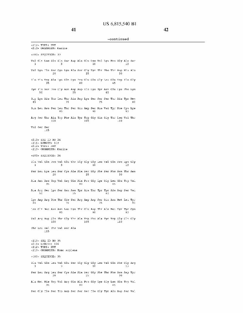
US 6,815,540 B1 41
-continued
&212> TYPE PRT <213> ORGANISM: Murine
<400 SEQUENCE: 33
Val Glin Leu Glin Glin Ser Asp Ala Glu Lys Wall Lys Pro Gly Ala Ser 1 5 10 15
Val Lys Ile Ser Cys Lys Ala Ser Gly Tyr Thr Phe Thr Asp His Ala 2O 25 3O
Ile His Trp Ala Lys Glin Lys Pro Glu Glin Gly Lieu Glu Trp Ile Gly 35 40 45
Tyr Ile Ser Pro Gly Asn Asp Asp Ile Lys Tyr Asn. Glu Lys Phe Lys 5 O 55 60
Gly Lys Ala Thr Leu Thr Ala Asp Llys Ser Ser Ser Thr Ala Tyr Met 65 70 75 8O
Glin Lieu. Asn. Ser Lieu. Thir Ser Glu Asp Ser Ala Val Tyr Phe Cys Lys 85 90 95
Arg Ser Thr Ala Trp Phe Ala Tyr Trp Gly Glin Gly Thr Leu Val Thr 100 105 110
Wal Ser Ser 115
<210> SEQ ID NO 34 &2 11s LENGTH 119 &212> TYPE PRT <213> ORGANISM: Murine
<400> SEQUENCE: 34
Glu Val Glin Pro Val Glu Thr Gly Gly Gly Leu Val Glin Pro Lys Gly 1 5 10 15
Ser Lieu Lys Lieu Ser Cys Ala Ala Ser Gly Phe Ser Phe Asn. Thir Asn 2O 25 3O
Ala Asn. Asn Trp Val Arg Glin Ala Pro Gly Lys Gly Lieu Glu Trp Val 35 40 45
Ala Arg Ser Lys Ser Asn. Asn Tyr Ala Thr Tyr Tyr Ala Asp Ser Val 5 O 55 60
Lys Asp Arg Phe Thr Ile Ser Arg Asp Asp Ser Glin Asn Met Leu Tyr 65 70 75 8O
Leu Gln Met Asn. Asn Lieu Lys Thr Glu Asp Thr Ala Met Tyr Tyr Cys 85 90 95
Val Arg Asp Gln Thr Gly Thr Ala Trp Phe Ala Tyr Trp Gly Glin Gly 100 105 110
Thir Leu Wall Thr Wal Ser Ala 115
<210 SEQ ID NO 35 <211& LENGTH 126 &212> TYPE PRT
<213> ORGANISM: Homo sapiens
<400 SEQUENCE: 35
Glu Val Glin Leu Val Glu Ser Gly Gly Gly Lieu Val Glin Phe Gly Arg 1 5 10 15
Ser Lieu Arg Lieu Ser Cys Ala Ala Ser Gly Phe Thr Phe Asn Asp Tyr 2O 25 3O
Ala Met His Trp Val Arg Glin Ala Pro Gly Lys Gly Lieu Glu Trp Val 35 40 45
Ser Gly Ile Ser Trp Asp Ser Ser Ser Ile Gly Tyr Ala Asp Ser Val
42

5 O
Lys Gly Arg Phe Thr Ile 65 70
Leu Gln Met Asn. Ser Lieu 85
Wall Lys Gly Arg Asp Tyr 100
Phe Asp Ile Trp Gly Glin 115
<210 SEQ ID NO 36 &2 11s LENGTH &212> TYPE <213> ORGANISM: Murine
PRT
<400 SEQUENCE:
Glin Wall Glin Leu 1
Ser Lieu
Gly Val
Gly Met 5 O
Ser Arg 65
Lys Met
Arg Glu
Thir Wall
Ser
Asn 35
Ile
Telu
Asn
Arg
Ser 115
Ile
Trp
Trp
Ser
Ser
Asp 100
Ser
116
36
Lys 5
Thr
Wall
Gly
Ile
Teu 85
Tyr
<210 SEQ ID NO 37 &2 11s LENGTH &212> TYPE <213> ORGANISM: Murine
PRT
<400 SEQUENCE:
Asp Val Glin Lieu 1
Ser Wall
Asn. Ile
Gly Asn 5 O
Lys Gly 65
Met His
Ala Arg
Thir Leu
Lys
Asn 35
Ile
Lys
Telu
Wall 115
Ile
Trp
Asp
Ala
Thr
Arg 100
Thr
119
37
Glin 5
Ser
Ile
Pro
Thr
Ser 85
Asp
Wall
Glu
Cys
Arg
Ser 70
His
Glin
Cys
Wall
Tyr
Teu 70
Teu
Gly
Ser
43
55
Ser
Arg
Tyr
Gly
Ser
Thr
Glin
Gly 55
Lys
Thr
Teu
Ser
Lys
Glin
Tyr 55
Thr
Glin
Asn
Ala
Thr 120
Gly
Wall
Pro 40
Asn
Asp
Gly
Ala
Arg 40
Gly
Wall
Ser
Tyr
Asp
Glu
Ser 105
Met
Pro
Ser 25
Pro
Thr
Asn
Asp
Tyr 105
Pro
Ser 25
Asn
Gly
Asp
Glu
Gly 105
Asn
Asp 90
Gly
Wall
Gly 10
Gly
Gly
Ser
Thr 90
Trp
Glu 10
Gly
Gly
Thr
Lys
Asp 90
Phe
Ala 75
Met
Gly
Thr
Teu
Phe
Lys
Lys 75
Ala
Gly
Teu
Phe
Lys
Asn
Ser 75
Ser
Thr
US 6,815,540 B1
-continued
60
Lys
Ala
Tyr
Wall
Wall
Ser
Gly
Asn 60
Ser
Arg
Glin
Glu
Ser
Ser
Phe 60
Ser
Ala
Asn
Teu
Phe
Ser 125
Ala
Teu
Teu 45
Ser
Glin
Gly
Teu
Teu 45
Asn
Ser
Wall
Trp
Ser
Thr 110
Ser
Pro
Thr 3O
Glu
Ala
Wall
Thr 110
Pro
Pro 3O
Glu
Pro
Thr
Gly 110
Telu
Tyr 95
Wall
Ser 15
Gly
Trp
Telu
Phe
Cys 95
Thr
Gly 15
Gly
Trp
Telu
Tyr 95
Glin
Ala
Glin
Tyr
Telu
Telu 8O
Ala
Telu
Ala
His
Ile
Phe
Tyr
Gly
44

<400
45
SEQ ID NO 38 LENGTH 120 TYPE PRT ORGANISM: Murine
SEQUENCE: 38
Glu Val Lieu Lieu Val Glu Ser Gly Gly Asp 1 5 10
Ser Lieu Lys Lieu Ser Cys Ala Ala Ser Gly 2O 25
Gly Met Ser Trp Val Arg His Thr Pro Asp 35 40
Ala Thr Ile Ser Asn Gly Gly Gly Tyr Thr 5 O 55
Lys Gly Arg Phe Thr Ile Ser Arg Asp Asn 65 70
Leu Glu Met Thr Ser Lieu Lys Ser Glu Asp 85 90
Ala Arg Arg Glu Arg Tyr Asp Glu Lys Gly 100 105
Gly Thr Leu Val Thr Val Ser Ala
<400
115 120
SEQ ID NO 39 LENGTH 120 TYPE PRT
ORGANISM: Artificial Sequence FEATURE:
OTHER INFORMATION: Description of murine
SEQUENCE: 39
Glu Val Glin Leu Val Glu Ser Gly Gly Gly 1 5 10
Phe Leu Arg Lieu Ser Cys Ala Ala Ser Gly 2O 25
Tyr Ile His Trp Val Arg Glin Ala Pro Gly 35 40
Ala Arg Ile Tyr Pro Thr Asn Gly Tyr Thr 5 O 55
Lys Gly Arg Phe Thr Ile Ser Ala Asp Thr 65 70
Leu Gln Met Asn. Ser Lieu Arg Ala Glu Asp 85 90
Ser Arg Trp Gly Gly Asp Gly Phe Tyr Ala 100 105
Gly Thr Leu Val Thr Val Ser Ser
<400
115 120
SEQ ID NO 40 LENGTH 115 TYPE PRT ORGANISM: Murine
SEQUENCE: 40
Teu
Phe
Ala 75
Ala
Phe
US 6,815,540 B1
-continued
Wall
Thr
Arg
Tyr 60
Gly
Ala
Phe
Teu 45
Glin
Asn
Teu
Pro
Ser
Glu
Asp
Thr
Trp 110
Gly 15
Ser
Trp
Ser
Telu
Tyr 95
Gly
Artificial Sequence:
Teu
Phe
Lys
Arg
Ser 75
Thr
Met
Wall
Asn
Gly
Tyr 60
Ala
Asp
Glin
Ile
Teu 45
Ala
Asn
Wall
Wall
Pro
Lys
Glu
Asp
Thr
Trp 110
Glin 15
Asp
Trp
Ser
Telu
Tyr 95
Gly
Gly
Phe
Wall
Wall
Phe
Arg
Humanized
Glin
Thr
Wall
Wall
Tyr
Glin
Glin Val Glin Leu Gln Glu Ser Gly Pro Gly Ile Leu Gln Pro Ser Glin 1 5 10 15
Ser Leu Ser Leu Thr Cys Ser Phe Ser Gly Phe Ser Leu Ser Thr Tyr 2O 25
46

Gly Met Gly Val Ser Trp 35
Trp Leu Ala His Ile Phe 5 O
Leu Lys Ser Arg Lieu Lys 65 70
Phe Leu Lys Ile Thr Ser 85
Cys Val Glin Glu Gly Tyr
Wall S
<400
100
er Ser 115
SEQ ID NO 41 LENGTH 122 TYPE PRT ORGANISM: Murine
SEQUENCE: 41
Glin Val Glin Leu Lys Glu 1 5
Thr Leu Ser Ile Thr Cys
Gly Val His Trp Val Arg 35
Gly Val Ile Trp Ala Gly 5 O
Ser Arg Val Ser Ile Ser 65 70
Lys Met Lys Ser Leu Glin 85
Arg Asp Phe Tyr Asp Tyr 100
Gly Glin Gly Thr Ser Val
<400
115
SEQ ID NO 42 LENGTH 120 TYPE PRT ORGANISM: Murine
SEQUENCE: 42
Glu Wall Glin Leu Wall Glu 1 5
Ser Lieu Lys Lieu Ser Cys
Gly Met Ser Trp Val Arg 35
Ala Thr Ile Ser Asn Gly 5 O
Lys Gly Arg Phe Thr Ile 65 70
Leu Gln Met Ser Ser Leu 85
Ala Arg Arg Glu Arg Tyr 100
47
Ile
Trp 55
Ile
Wall
Ile
Ser
Thr
Glin
Gly 55
Lys
Thr
Asp
Thr
Ser
Ala
Glin
Gly 55
Ser
Lys
Arg 40
Asp
Ser
Tyr
Gly
Wall
Pro 40
Asn
Wall
Wall 120
Gly
Ala
Thr 40
Gly
Ser
Glu
Glin
Gly
Thr
Trp 105
Pro
Ser 25
Pro
Thr
Asn
Asp
Phe 105
Ser
Gly
Ser 25
Pro
Tyr
Asp
Glu
Asn 105
Pro
Asp
Asp
Ala 90
Gly
Gly 10
Gly
Glin
Asn
Ser
Thr 90
Tyr
Ser
Asp 10
Gly
Asp
Thr
Asn
Asp 90
Gly
Ser
Thr 75
Asp
Glin
Teu
Phe
Lys 75
Ala
Teu
Phe
Ala 75
Ser
Phe
US 6,815,540 B1
-continued
Gly
Arg 60
Ser
Thr
Gly
Wall
Teu
Gly
Asn 60
Ser
Met
Ala
Wall
Ser
Arg
Tyr 60
Ala
Ala
Lys 45
Tyr
Asn
Ala
Thr
Ala
Teu
Teu 45
Ser
Glin
Met
Phe
Teu 45
Pro
Asn
Met
Gly
Asn
Asn
Thr
Ser 110
Pro
Ile 3O
Glu
Ala
Wall
Asp 110
Pro
Ser 3O
Glu
Asp
Thr
Trp 110
Telu
Pro
Glin
Tyr 95
Wall
Ser 15
Ser
Trp
Telu
Phe
Cys 95
Tyr
Gly 15
Ser
Trp
Ser
Telu
Tyr 95
Gly
Glu
Ser
Wall 8O
Thr
Glin
Asn
Telu
Met
Telu
Ala
Trp
Gly
Wall
Wall
Tyr
Glin
48

49
Gly Thr Leu Val Thr Val Ser Ala
<400
Wall 1
Wall
Met
Gly 65
Glu
Gly
Ser
<400
Glu 1
Ser
Thr
Ala
Lys 65
Teu
Ala
Ser
<400
115
PRT
SEQUENCE:
Gln Leu Glin
Arg
Asn
Ile 5 O
Lys
Telu
Ser
Wall
Met
Trp 35
Ser
Ala
Arg
Ser
Thr 115
Ser
Wall
Pro
Thr
Ser
Gly 100
Wall
PRT
SEQUENCE:
Wall Thr Lieu
Telu
Met
Thr 5 O
Gly
Glin
Ser
Ala
Lys
Ser 35
Thr
Telu
His
Lys
Trp
Telu
Phe
Asn
Arg 100
PRT
SEQUENCE:
SEQ ID NO 43 LENGTH TYPE ORGANISM: Murine
118
43
Glin 5
Cys
Arg
Tyr
Teu
Teu 85
Asn
Ser
SEQ ID NO 44 LENGTH TYPE ORGANISM: Murine
114
44
Wall 5
Ser
Wall
Ser
Thr
Ser 85
Phe
SEQ ID NO 45 LENGTH TYPE ORGANISM: Murine
118
45
Ser
Glin
Ser
Thr 70
Thr
Ser
Glu
Cys
Arg
Gly
Ile 70
Teu
Wall
Gly
Ser
Ser
Gly 55
Wall
Ser
Trp
Ser
Ala
Glin
Gly 55
Ser
His
120
Pro
Ser
His 40
Wall
Asp
Glu
Ala
Gly
Ala
Thr 40
Gly
Ser
Trp
Glu
Gly 25
Gly
Thr
Lys
Asp
Met 105
Gly
Ser 25
Pro
Phe
Asp
Glu
Gly 105
Telu 10
Tyr
Lys
Gly
Ser
Ser 90
Asp
Asp 10
Gly
Glu
Thr
Asn
Asp 90
His
Wall
Ile
Ser
Tyr
Ser 75
Ala
Tyr
Ser
Phe
Lys
Phe
Ala 75
Thr
Gly
US 6,815,540 B1
-continued
Phe
Teu
Asn 60
Ser
Wall
Trp
Wall
Thr
Arg
Tyr 60
Glin
Ala
Thr
Pro
Thr
Asp 45
Glin
Thr
Gly
Teu
Teu 45
Ser
Asn
Teu
Teu
Gly
Asp 3O
His 110
Pro
Ser 3O
Glu
Ala
Asn
Wall 110
Ala 15
Phe
Ile
Phe
Cys 95
Gly
Gly 15
Gly
Trp
Ser
Telu
Phe 95
Thr
Ser
Met 8O
Ala
Ala
Gly
Glu
Wall
Wall
Tyr
Wall
Glin Val Glin Leu Gln Glin Ser Gly Pro Glu Leu Val Arg Pro Gly Ser 1 5 10 15
Ser Val Lys Ile Ser Cys Lys Gly Ser Gly Tyr Thr Phe Thr Thr Tyr
SO
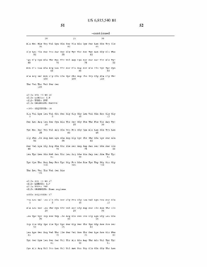
US 6,815,540 B1 S1
-continued
2O 25 3O
Ala Met His Trp Wall Lys Glin Ser His Ala Lys Ser Leu Glu Trp Ile 35 40 45
Gly Leu Ile Ser Pro Ser Ser Gly Tyr Thr Ser Tyr Asn Gly Glu Phe 5 O 55 60
Lys Gly Lys Ala Thr Met Thr Val Asp Llys Ser Ser Ser Thr Ala Tyr 65 70 75 8O
Met Glu Lieu Ala Arg Lieu. Thir Ser Glu Asp Ser Ala Ile Tyr Tyr Cys 85 90 95
Ala Arg Val Met Gly Glu Gln Tyr Phe Asp Phe Trp Gly Ala Gly Thr 100 105 110
Thr Wall Thr Wal Ser Ser 115
<210> SEQ ID NO 46 &2 11s LENGTH 119 &212> TYPE PRT <213> ORGANISM: Murine
<400 SEQUENCE: 46
Glu Wall Lys Lieu Val Glu Ser Gly Gly Gly Lieu Val Glin Pro Gly Gly 1 5 10 15
Ser Leu Arg Leu Ser Cys Ala Thr Ser Gly Phe Thr Phe Thr Asp Tyr 2O 25 3O
Tyr Met Ser Trp Val Arg Gln Pro Pro Gly Lys Ala Leu Glu Trp Leu 35 40 45
Gly Phe Ile Arg Asn Lys Ala Asp Gly Tyr Thr Thr Glu Tyr Ser Ala 5 O 55 60
Ser Val Lys Gly Arg Phe Thir Ile Ser Arg Asp Asn. Ser Glin Ser Ile 65 70 75 8O
Leu Tyr Lieu Gln Met Asn. Thir Lieu Arg Ala Glu Asp Ser Ala Thr Tyr 85 90 95
Tyr Cys Thr Arg Asp Pro Tyr Gly Pro Ala Ala Tyr Trp Gly Glin Gly 100 105 110
Thir Leu Wall Thr Wal Ser Ala 115
<210> SEQ ID NO 47 &2 11s LENGTH 117 &212> TYPE PRT <213> ORGANISM: Homo sapiens
<400 SEQUENCE: 47
Pro Leu Val Leu Gln Glu Ser Gly Pro Gly Leu Val Lys Pro Ser Glu 1 5 10 15
Ala Leu Ser Leu Thr Cys Thr Val Ser Gly Asp Ser Ile Asn Thr Ile 2O 25 3O
Leu Tyr Tyr Trp Ser Trp Ile Arg Gln Pro Pro Gly Lys Gly Leu Glu 35 40 45
Trp Ile Gly Tyr Ile Tyr Tyr Ser Gly Ser Thr Tyr Gly Asn Pro Ser 5 O 55 60
Leu Lys Ser Arg Val Thr Ile Ser Val Asn. Thir Ser Lys Asn Glin Phe 65 70 75 8O
Tyr Ser Lys Leu Ser Ser Val Thr Ala Ala Asp Thr Ala Val Tyr Tyr 85 90 95
Cys Ala Arg Val Pro Leu Val Val Asn Pro Trp Gly Glin Gly Thr Leu
52

S3
100
Wall Thr Wal Ser Ser 115
&212> TYPE
<400
Glin 1
Thr
Gly
Gly
Lys 65
Teu
Ala
Gly
Ile Glin
Wall Lys
Met Asn 35
Trp Ile 5 O
Gly Arg
Glin Ile
Arg Gly
Th Thr 115
&212> TYPE
<400
Arg 1
Ser
Trp
Gly
Lys 65
Met
Thr
Thr
Wall Glin
Wall Glin
Ile Glu 35
Glu Ile 5 O
Gly Lys
Gln Leu
Arg Gly
Wal Ser 115
&212> TYPE
<400
SEQ ID NO 48 LENGTH 120
PRT ORGANISM: Anous Minutus FEATURE:
OTHER INFORMATION: Noddy Tern
SEQUENCE: 48
Leu Val Glin Ser Gly 5
Ile Ser Cys Lys Ala 2O
Trp Val Lys Glin Ala 40
Asn Thr Asn Thr Gly 55
Phe Ala Phe Ser Leu 70
Asn Asn Lieu Lys Asn 85
Glu Asp Asn. Phe Gly 100
Wall Thr Wal Ser Ser 120
SEQ ID NO 49 LENGTH 116
PRT ORGANISM: Murine
SEQUENCE: 49
Lieu Lleu Glu Ser Gly 5
Ile Ser Cys Lys Ala 2O
Trp Val Lys Glu Arg 40
Leu Pro Gly Ser Gly 55
Ala Thr Phe Thr Ala 70
Ser Ser Lieu. Thir Ser 85
Tyr Ser Ser Met Asp 100
Ala
SEQ ID NO 50 LENGTH 119
PRT ORGANISM: Murine
SEQUENCE: 50
105
Pro
Ser 25
Pro
Glu
Glu
Glu
Ser 105
Ala
Thr 25
Pro
Arg
Asp
Glu
Tyr 105
Glu 10
Gly
Gly
Pro
Thr
Asp 90
Telu
Glu 10
Gly
Gly
Thr
Thr
Asp 90
Trp
Teu
Tyr
Lys
Thr
Ser 75
Thr
Ser
Teu
Tyr
His
Asn
Ser 75
Ser
Gly
US 6,815,540 B1
-continued
Thr
Gly
Tyr 60
Ala
Ala
Asp
Met
Thr
Gly
Tyr 60
Ser
Ala
Glin
Phe
Teu 45
Gly
Ser
Thr
Phe
Teu 45
Arg
Asn
Wall
Gly
110
Pro
Thr
Glu
Thr
Phe
Trp 110
Pro
Ser
Glu
Glu
Thr
Thr 110
Gly 15
Asn
Trp
Glu
Ala
Phe 95
Gly
Gly 15
Glu
Trp
Lys
Ala
Tyr 95
Ser
Glu
Met
Phe
Glin
Ala
Ile
Phe
Tyr
Wall

US 6,815,540 B1 SS
-continued
Glin Ile Glin Leu Val Glin Ser Gly Pro Glu Lieu Lys Lys Pro Gly Glu 1 5 10 15
Thr Val Lys Ile Ser Cys Lys Ala Ser Gly Tyr Ala Phe Thr Asn Tyr 2O 25 3O
Gly Val Asn Trp Val Lys Glu Ala Pro Gly Lys Glu Lieu Lys Trp Met 35 40 45
Gly Trp Ile Asin Ile Tyr Thr Gly Glu Pro Thr Tyr Val Asp Asp Phe 5 O 55 60
Lys Gly Arg Phe Ala Phe Ser Leu Glu Thir Ser Ala Ser Thr Ala Tyr 65 70 75 8O
Leu Glu Ile Asn. Asn Lieu Lys Asn. Glu Asp Thr Ala Thr Tyr Phe Cys 85 90 95
Thr Arg Gly Asp Tyr Val Asn Trp Tyr Phe Asp Val Trp Gly Ala Gly 100 105 110
Thir Thr Wall Thr Wal Ser Ser 115
<210 SEQ ID NO 51 <211& LENGTH 124 &212> TYPE PRT <213> ORGANISM: Murine
<400 SEQUENCE: 51
Glin Val Glin Leu Glin Glin Ser Gly Ala Glu Lieu Val Arg Ala Gly Ser 1 5 10 15
Ser Val Lys Met Ser Cys Lys Ala Ser Gly Tyr Thr Phe Thr Ser Tyr 2O 25 3O
Gly Val Asn Trp Val Lys Glin Arg Pro Gly Glin Gly Lieu Glu Trp Ile 35 40 45
Gly Tyr Ile Asin Pro Gly Lys Gly Tyr Lieu Ser Tyr Asn. Glu Lys Phe 5 O 55 60
Lys Gly Lys Thr Thr Leu Thr Val Asp Arg Ser Ser Ser Thr Ala Tyr 65 70 75 8O
Met Glin Leu Arg Ser Leu Thir Ser Glu Asp Ala Ala Val Tyr Phe Cys 85 90 95
Ala Arg Ser Phe Tyr Gly Gly Ser Asp Leu Ala Val Tyr Tyr Phe Asp 100 105 110
Ser Trp Gly Glin Gly. Thir Thr Leu Thr Val Ser Ser 115 120
<210> SEQ ID NO 52 <211& LENGTH 126 &212> TYPE PRT <213> ORGANISM: Homo sapiens
<400 SEQUENCE: 52
Glu Val Glin Leu Val Glin Ser Gly Gly Gly Val Val Glin Pro Gly Arg 1 5 10 15
Ser Leu Arg Leu Ser Cys Ser Ser Ser Gly Phe Ile Phe Ser Ser Tyr 2O 25 3O
Ala Met Tyr Trp Val Arg Glin Ala Pro Gly Lys Gly Leu Glu Trp Val 35 40 45
Ala Ile Ile Trp Asp Asp Gly Ser Asp Gln His Tyr Ala Asp Ser Val 5 O 55 60
Lys Gly Arg Phe Thr Ile Ser Arg Asn Asp Ser Lys Asn. Ser Lieu Phe 65 70 75 8O

57 US 6,815,540 B1
-continued
Leu Gln Met Asp Ser Lieu Arg Pro Glu Asp Thr Gly Val Tyr 85
Ala Arg Asp Gly Gly. His Gly Phe Cys 100 105
90
Ser Ser Ala Ser Cys 110
Pro Asp Tyr Trp Gly Glin Gly Thr Pro Val Thr Val Ser Ser
<400
Glu 1
Ser
Trp
Gly
Lys 65
Teu
Ala
Teu
<400
Wall 1
Wall
Ile
Glu
Gly 65
Glin
His
Wall
115
SEQ ID NO 53 LENGTH 118 TYPE ORGANISM: Murine
PRT
SEQUENCE: 53
Wall Lys Lieu Lieu
Telu
Met
Glu 5 O
Asp
Glin
Arg
Wall
Lys
Ser 35
Ile
Lys
Met
Telu
Thr 115
5
Teu Ser 2O
Trp Val
His Pro
Phe Ile
Ser Lys 85
His Tyr 100
Wal Ser
SEQ ID NO 54 LENGTH 115 TYPE ORGANISM: Murine
PRT
SEQUENCE: 54
Gln Leu Glin Glin
Lys
Glu
Ile 5 O
Lys
Telu
Gly
Ser
Ile
Trp 35
Telu
Ala
Asn
Asn
Ser 115
Ser Cys 2O
Wall Lys
Pro Gly
Thr Phe
Ser Teu 85
Tyr Asp 100
SEQ ID NO 55 LENGTH 119 TYPE ORGANISM: Murine
PRT
Glu
Cys
Arg
Asp
Ile 70
Wall
Tyr
Ala
Ser
Lys
Glin
Ser
Thr 70
Thr
Phe
Ser
Ala
Glin
Ser 55
Ser
Arg
Gly
Gly
Ala
Gly 55
Ala
Ser
120
Gly
Ala
Ala 40
Gly
Ser
Tyr
Ala
Ser
Pro 40
Ser
Glu
Gly
Gly
Ser 25
Pro
Thr
Asp
Glu
Asn 105
Glu
Gly 25
Gly
Thr
Thr
Asp
Trp 105
Gly 10
Gly
Gly
Ile
Asn
Asp 90
Ala
Telu 10
Tyr
His
Asn
Ser
Ser 90
Gly
Teu
Phe
Lys
Asn
Ala 75
Thr
Tyr
Met
Thr
Gly
Ser 75
Ala
Glin
Wall
Asp
Gly
Tyr 60
Ala
Trp
Phe
Teu
His 60
Ser
Wall
Gly
125
Glin
Phe
Teu 45
Thr
Asn
Teu
Gly
Pro
Ser
Glu 45
Glu
Thr
Thr
Pro
Ser 3O
Glu
Pro
Thr
Glin 110
Gly
Asp 3O
Trp
Arg
Ala
Thr 110
Phe 95
Phe
Gly 15
Trp
Ser
Telu
Tyr 95
Gly
Ala 15
Ile
Phe
Cys 95
Telu
Gly
Gly
Ile
Telu
Tyr
Thr
Ser
Trp
Gly
Met 8O
Telu
Thr
58

<400 SEQUENCE: 55
Glu 1
Ser
Ala
Ala
Lys 65
Teu
Thr
Thr
<400
Glu 1
Ser
Ala
Ser 65
Teu
Gly
<400
Wall Glin Leu Wall Glu
Telu
Met
Gly 5 O
Gly
Glin
Arg
Thr
Lys
Ser 35
Ile
Arg
Met
Tyr
Telu 115
5
Leu Ser Cys 2O
Trp Val Arg
Ser Ser Gly
Phe Ile Ile 70
Ser Ser Teu 85
Ser Ser Asp 100
Thr Wal Ser
SEQ ID NO 56 LENGTH 122 TYPE ORGANISM: Murine
PRT
SEQUENCE: 56
Wall Glin Leu Wall Glu
Telu
Met
Ala 5 O
Wall
Ala
Arg
Glu 35
Ser
Telu
Ala
Gly 115
5
Leu Ser Cys 2O
Trp Val Arg
Arg Asn Lys
Gly Arg Phe 70
Gln Met Asn 85
Arg Asn Tyr 100
Thir Thr Wall
SEQ ID NO 57 LENGTH 113 TYPE ORGANISM: Murine
PRT
SEQUENCE: 57
Asp Val Glin Leu Glin Glu 1
Thr
Tyr
Gly
Telu
Trp
Tyr 5 O
Ser
Ser 35
Wall
5
Leu Thir Cys 2O
Trp Ile Arg
Ser Tyr Ser
59
Ser
Ala
Glin
Gly 55
Ser
Pro
Ser
Ser
Ala
Glin
Gly 55
Ile
Ala
Tyr
Thr
Ser
Ser
Lys
Gly 55
Gly
Ala
Thr 40
Ser
Ser
Phe
Gly
Thr
Pro 40
Asn
Wall
Telu
Gly
Wall 120
Gly
Wall
Phe 40
Ser
Gly
Ser 25
Pro
Tyr
Asn
Glu
Tyr 105
Gly
Ser 25
Pro
Lys
Ser
Ser 105
Ser
Pro
Thr 25
Pro
Thr
Asp 10
Gly
Glu
Thr
Asn
Asp 90
Phe
Gly 10
Gly
Gly
Arg
Ala 90
Thr
Ser
Ser 10
Gly
Gly
Tyr
Teu
Phe
Phe
Ala 75
Thr
Asp
Teu
Phe
Lys
Thr
Asp 75
Glu
Trp
Teu
Asp
Asn
US 6,815,540 B1
-continued
Wall
Thr
Arg
Tyr 60
Arg
Ala
Wall
Thr
Arg
Thr 60
Thr
Asp
Wall
Ser
Arg
Asn 60
Phe
Teu 45
Pro
Asn
Ile
Trp
Glin
Phe
Teu 45
Glu
Ser
Thr
Phe
Ile
Teu 45
Pro
Pro
Ser 3O
Glu
Asp
Thr
Gly 110
Pro
Ser 3O
Glu
Glin
Ala
Asp 110
Pro
Thr 3O
Glu
Ser
Gly 15
Arg
Trp
Thr
Telu
Tyr 95
Glin
Gly 15
Asp
Trp
Ser
Ser
Ile 95
Wall
Ser 15
Ser
Telu
Gly
Wall
Wall
Ser 8O
Gly
Gly
Phe
Ile
Ala
Ile 8O
Trp
Glin
Asp
Met
60

Ser
65
Asp
Asn
Ala
<400
Glu 1
Pro
Trp
Ala
Ser
65
Wall
Ser
<400
Glu
1
Ser
61
Arg Ile Ser Ile Thr Arg Asp Thr 70
Leu Asin Ser Val Thr Thr Glu Asp 85
Trp Asp Gly Asp Tyr Trp Gly Glin 100
PRT
SEQUENCE:
Wall Glin Leu
Met
Met
Glin 5 O
Wall
Wall
Lys
Asn
35
Ile
Telu
Thr
Thr 115
Telu
2O
Trp
Gly
Glin
Gly 100
Wall
PRT
SEQUENCE:
Wall Glin Leu
Wall Lys
Gly Ile Asn 35
Gly Tyr Asn
Lys 65
Met
5 O
Gly
Glin
Lys
Telu
Met
Trp
Asn
Thr
Arg
SEQ ID NO 58 LENGTH
TYPE
ORGANISM: Murine
118
58
Asp 5
Ser
Wall
Asn
Met
85
Ser
Ser
SEQ ID NO 59 LENGTH
TYPE
ORGANISM: Murine
121
59
Glin
5
Ser
Wall
Pro
Thr
Ser
85
Glu
Cys
Arg
Lys
Phe 70
Asn
Tyr
Ser
Glin
Cys
Lys
Gly
Teu
70
Teu
Thr
Wall
Glin
Pro
55
Thr
Asn
Tyr
Ser
Lys
Glin
Asn
55
Thr
Thr
Gly
Ala
Ser
40
Tyr
Ile
Telu
Gly
Gly
Ala
40
Gly
Wall
Ser
105
Gly
Ser
25
Pro
Asn
Ser
Met
105
Wall
Ser
25
Pro
Tyr
Asp
Glu
Ser
Thr 90
Gly
Gly 10
Gly
Glu
Arg
Wall 90
Asp
Glu
10
Gly
Gly
Ile
Asp 90
Lys 75
Ala
Thr
Teu
Phe
Lys
Glu
Asp 75
Glu
Teu
Tyr
Glin
Ala
Ser
75
Ser
US 6,815,540 B1
-continued
Asn Glin Tyr
Thr Tyr Tyr
Leu Wall Thr
Wall
Thr
Gly
Thr 60
Asp
Asp
Trp
Wall
Thr
Gly
60
Ser
Ala
Glin
Phe
Teu
45
Ser
Met
Gly
Arg
Phe
Teu
45
Asn
Ser
Wall
110
Pro
Ser
3O
Glu
Gly
Glin 110
Ala
Thr
Glu
Glu
Thr
Tyr
Cys 95
Wall
Gly 15
Asp
Trp
Ser
Ser
Ile 95
Gly
Gly 15
Ser
Trp
Ala
Phe 95
Telu
8O
Ala
Ser
Arg
Wall
Asp
Ser
8O
Thr
Ser
Asn
Ile
Phe
62

US 6,815,540 B1 63
-continued
64
Ala Arg Ser Glu Tyr Tyr Gly Gly Ser Tyr Lys Phe Asp Tyr Trp Gly 100 105 110
Gln Gly. Thir Thr Leu Thr Val Ser Ser 115 120
<210 SEQ ID NO 60
<211& LENGTH 121
&212> TYPE PRT
<213> ORGANISM: Homo sapiens
<400 SEQUENCE: 60
Ala Val Glin Leu Val Glin Ala Gly Gly Gly Val Val Glin Pro Gly Arg
Ser Leu Arg Leu Ser Cys Ile Ala Ser Gly Phe Thr Phe Ser Asn Tyr 2O 25 3O
Gly Met His Trp Val Arg Glin Ala Pro Gly Lys Gly Leu Glu Trp Val 35 40 45
Ala Val Ile Trp Tyr Asn Gly Ser Arg Thr Tyr Tyr Gly Asp Ser Val 5 O 55 60
Lys Gly Arg Phe Thr Ile Ser Arg Asp Asn. Ser Lys Arg Thr Lieu. Tyr 65 70 75 8O
Met Glin Met Asn Ser Leu Arg Thr Glu Asp Thr Ala Val Tyr Tyr Cys 85 90 95
Ala Arg Asp Pro Asp Ile Leu Thir Ala Phe Ser Phe Asp Tyr Trp Gly 100 105 110
Gln Gly Val Leu Val Thr Val Ser Ser 115 120


; 2
i.
3.
5
S
3
i 3. s
O
s
s
67 O
O
. O
O
S
S
s
&
5 2. H
ii2.3.i.i E &
E
US 6,815,540 B1
& g
&sS. S.2 ww2s s S ca
S g
2 2 O
s 2
S
2. --
22222222 5; 2
5.
g5;E E
s i
É
s5
;
s
5
2
2.
i.
s
5
o
2.
É
gg :
s
o
&
O
2
2
2
i.
5
s
s
2
S.
2
2.
i 3. s
st
st
2
2
2
s
s
2
3
3.
S
s 5 2.

US 6,815,540 B1 69
TABLE 2
Mutations introduced in the scFv fragment of the antibody 4-4-20
L15E (V) L11N (V) L11D (V) V84D (V) Flu 1 O Flu. 2 O Fu 3 O Fu 4 O Fu S O O Flu 6 O O Fu 7 O O Fu 8 O O Full 9 O O O Flu 4 short O
TABLE 3
K values of the different scFv mutants determined in fluorescence titration
Flu wt Fu 3 Flu 4 Flu 6 Flu wt #
KD (nM) 80 - 7 60 - 12 70 - 10 7S 13 90
What is claimed is: 1. A nucleic acid Sequence encoding a modified Single
chain Fv molecule, wherein Said Single chain Fv molecule comprises a heavy
chain variable domain and a light chain variable domain that interact to form an antigen binding site,
wherein said heavy chain variable domain comprises an intra-heavy chain inter-domain interface region and Said light chain variable domain comprises an intra light chain inter-domain interface region
and wherein at least one amino acid in an intrachain inter-domain interface region is modified So as to decrease the hydrophobicity in Said intra-chain inter domain interface region.
2. A nucleic acid Sequence encoding a modified polypep tide comprising a heavy chain variable domain, wherein Said heavy chain variable domain comprises an intra-heavy chain inter-domain interface region,
and wherein at least one aznino acid in Said intra-heavy chain inter-domain interface region is modified So as to decrease the hydrophobicity in Said region.
3. The nucleic acid Sequence according to claim 2, wherein Said intra-heavy chain inter-domain interface com prises residues 9, 10, 11, 13, 14, 41, 42, 43, 84, 87, 89, 105, 108, 110, 112, 113 of said heavy chain variable domain.
4. A nucleic acid Sequence encoding a modified polypep tide comprising a light chain variable domain, wherein Said light chain variable domain comprises an intra-light chain inter-domain interface region,
and wherein at least one amino acid in Said intra-light chain inter-domain interface region is modified So as to decrease the hydrophobicity in Said region.
5. The nucleic acid Sequence according to claim 4, wherein Said inter-domain interface comprises residues 9,
1O
15
25
35
40
45
50
55
70 10, 12, 15, 39, 40, 41,80, 81, 83, 103,105,106, 106A, 107, 108 of said light chain variable domain.
6. The nucleic acid Sequence according to claim 1 in which Said modification comprises insertion of one or more hydrophilic amino acids or deletion of one or more hydro phobic amino acids.
7. The nucleic acid Sequence according to claim 1 which Said modification comprises any two or more of:
a) a Substitution of one or more amino acids with amino acids which are more hydrophilic than the one or more amino acids being Substituted for;
b) an insertion of one or more hydrophilic amino acids; and
c) a deletion of one or more hydrophobic amino acids. 8. The nucleic acid Sequence according to claim 6 or 7 in
which said Substituted or inserted amino acid is Selected from the group consisting of ASn, Asp, Arg, Gln, Glu, Gly, His, Lys, Ser, and Thr.
9. The nucleic acid Sequence according to claim 2, 3 or 5, further comprising a contiguous Sequence which encodes one or more additional moieties.
10. The nucleic acid Sequence according to claim 9 in which at least one of Said additional moieties is a toxin, a cytokine, or a reporter enzyme.
11. The nucleic acid Sequence according to claim 10 in which at least one of Said additional moieties is at least part of a Surface protein of an organism.
12. The nucleic acid Sequence according to claim 11 in which Said organism is a filamentous bacteriophage.
13. The nucleic acid sequence according to claim 12 which Said Surface protein is the geneII protein.
14. The nucleic acid Sequence according to claim 9 in which at least one of Said additional moieties is capable of binding a metal ion.
15. The nucleic acid Sequence according to claim 14 in which at least one of Said additional moieties comprises at least five hisitidines.
16. The nucleic acid Sequence according to claim 9 in which Said additional moiety is a labeling tag peptide.
17. The nucleic acid Sequence according to claim 16 in which Said labeling tag peptide is c-myc or FLAG.
18. The nucleic acid Sequence according to claim 9 in which Said additional moiety comprises a peptic comprising an association domain.
19. The nucleicacid Sequence according to claim 18 in which Said asSociation domain comprises a leucine Zipper or a helix-turn-helix motif.
20. A vector comprising a nucleic acid Sequence accord ing to claim 1, 2 or 3.
21. A host cell comprising a vector according to claim 20. 22. The nucleic acid Sequence according to claim 1,
wherein at least one amino acid in Said intra-heavy chain inter-domain interface and at least one amino acid in Said intra-light chain inter-domain interface is modified.
k k k k k