12 cerebellar masses on computed tomography
12
12 Cerebellar Masses on Computed Tomography
-
Upload
muhammad-bin-zulfiqar -
Category
Education
-
view
340 -
download
0
Transcript of 12 cerebellar masses on computed tomography

12 Cerebellar Masses on Computed Tomography

CLINICAL IMAGAGINGAN ATLAS OF DIFFERENTIAL DAIGNOSIS
EISENBERG
DR. Muhammad Bin Zulfiqar PGR-FCPS III SIMS/SHL
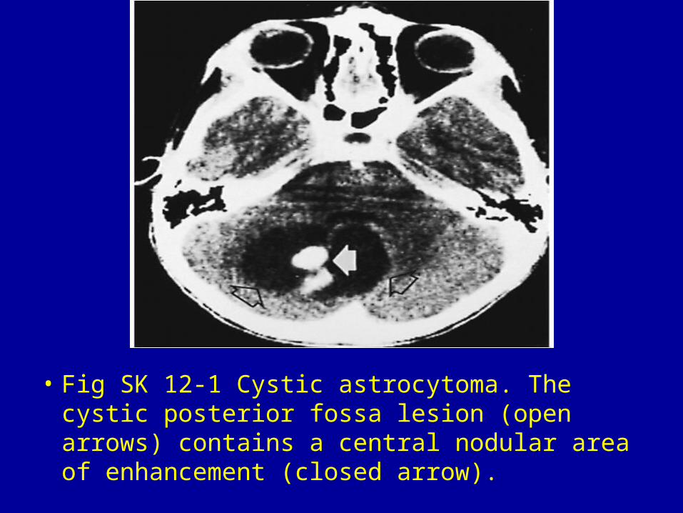
• Fig SK 12-1 Cystic astrocytoma. The cystic posterior fossa lesion (open arrows) contains a central nodular area of enhancement (closed arrow).
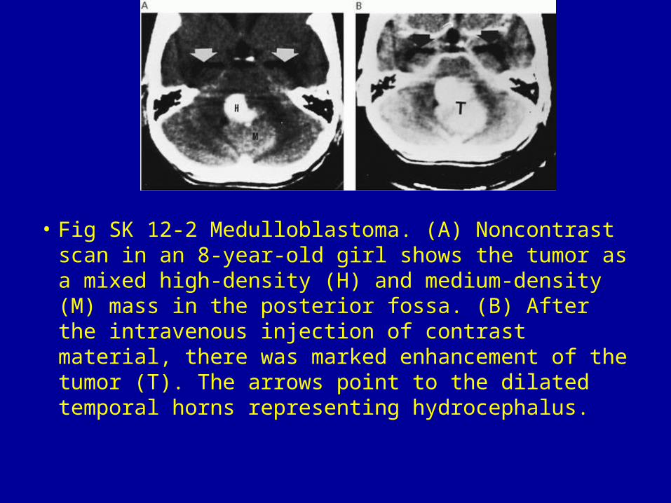
• Fig SK 12-2 Medulloblastoma. (A) Noncontrast scan in an 8-year-old girl shows the tumor as a mixed high-density (H) and medium-density (M) mass in the posterior fossa. (B) After the intravenous injection of contrast material, there was marked enhancement of the tumor (T). The arrows point to the dilated temporal horns representing hydrocephalus.
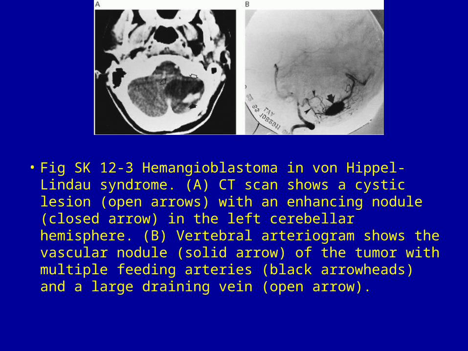
• Fig SK 12-3 Hemangioblastoma in von Hippel-Lindau syndrome. (A) CT scan shows a cystic lesion (open arrows) with an enhancing nodule (closed arrow) in the left cerebellar hemisphere. (B) Vertebral arteriogram shows the vascular nodule (solid arrow) of the tumor with multiple feeding arteries (black arrowheads) and a large draining vein (open arrow).

• Fig SK 12-4 Cerebellar sarcoma. (A) Noncontrast scan shows dense tumor (straight arrows) in the left cerebellar hemisphere. Note the cystic region (open curved arrow) within it. (B) After intravenous contrast infusion, the tumor is notably enhanced (straight arrows). The fourth ventricle is displaced severely from left to right (open curved arrow), causing noncommunicating hydrocephalus.1
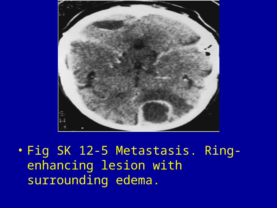
• Fig SK 12-5 Metastasis. Ring-enhancing lesion with surrounding edema.
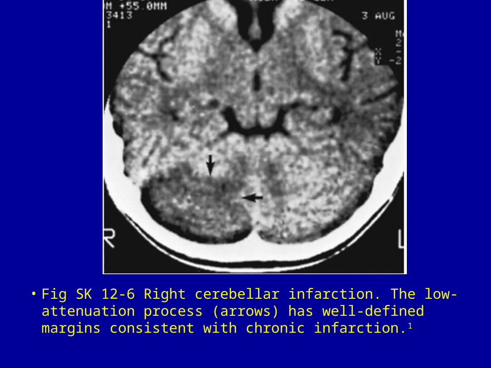
• Fig SK 12-6 Right cerebellar infarction. The low-attenuation process (arrows) has well-defined margins consistent with chronic infarction.1
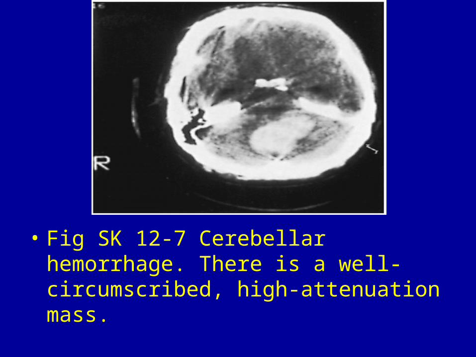
• Fig SK 12-7 Cerebellar hemorrhage. There is a well-circumscribed, high-attenuation mass.

• Fig SK 12-8 Arteriovenous malformation. Irregular mass of increased attenuation in the vermis (arrow). Note the dilated vein (arrowheads) draining the lesion.