![8 Referências Bibliográficas€¦ · 8 Referências Bibliográficas [1] Site da TRANSPETRO – consultado em 21/11/2006. [2] KIEFNER, J. F., et al., "Failure Stress Levels of Flaws](https://static.fdocuments.us/doc/165x107/5ad916bc7f8b9ae1768b604a/8-referncias-8-referncias-bibliogrficas-1-site-da-transpetro-consultado.jpg)
11 Referências - maxwell.vrac.puc-rio.br
29
77 11 Referências Abbott, FV. Peripheral and central antinociceptive actions of ethylketocyclazocine in the formalin test, European Journal of Pharmacology. V. 152, pp. 93-100, 1988. Abbott, FV., Franklin, KB., Ludwick, RJ., Melzack, R. Apparent lack of tolerance in the formalin test suggests different mechanisms for morphine analgesia in different types of pain. Pharmacol Biochem Behav. V.15, pp. 637-40, 1981. Abbott, FV., Melzack, R. Dissociation of the mechanisms of stimulation-produced analgesia in tests of tonic and phasic pain, Advances in Pain Research and Therapy. V. 5, pp. 401-409, 1983. Abbott, FV., Melzack, R., Leber, BF. Morphine analgesia and tolerance in the tail-flick and formalin tests: dose-response relationships, Pharmacology Biochemistry Behavior. V 17, pp. 1213-1219, 1982a. Abbott, FV., Melzack, R., Samuel, C. Morphine analgesia in tail-flick and formalin pain tests is mediated by different neural systems, Experimental Neurology. V. 75, pp. 644-651, 1982. Abbott, FV., Melzack, R., Samuel, C. Morphine analgesia in tail-flick and formalin pain tests is mediated by different neural systems, Experimental Neurology. V. 75, pp. 644-651, 1982b. Adams, JC. Ascending projections to the inferior colliculus. Journal of Comparative Neurology. V.183, pp. 519-538, 1979. Adler, CM., Craske, MG., Barlow, DH. Relaxation-induced panic (RIP): When resting isn't peaceful. Integrative Psychiatry V. 5, pp. 94-100, 1987. Allen, GV., Hopkins, DA. Topography and synaptology of mammillary body projections to the mesencephalon and pons in the rat, Journal of. Comparative. Neurology, V. 301, pp. 214-231, 1990. Almeida-Filho, N., Mari, JJ., Coutinho, E., França, JF., Fernandes, J., Andreoli, SB., Busnello, ED. Brazilian multicentric study of psychiatric morbidity: methodological features and prevalence estimates. Brazilian Journal of Psychiatry V. 171, pp. 524-9, 1997. Alreja, M., Mutalik, P., Nayar, U., Manchanda, SK. The formalin test: a tonic pain model in the primate, Pain. V. 20, pp. 97–105, 1984.
Transcript of 11 Referências - maxwell.vrac.puc-rio.br

77
11
Referências
Abbott, FV. Peripheral and central antinociceptive actions of ethylketocyclazocine
in the formalin test, European Journal of Pharmacology. V. 152, pp. 93-100,
1988.
Abbott, FV., Franklin, KB., Ludwick, RJ., Melzack, R. Apparent lack of tolerance
in the formalin test suggests different mechanisms for morphine analgesia in
different types of pain. Pharmacol Biochem Behav. V.15, pp. 637-40, 1981.
Abbott, FV., Melzack, R. Dissociation of the mechanisms of stimulation-produced
analgesia in tests of tonic and phasic pain, Advances in Pain Research and
Therapy. V. 5, pp. 401-409, 1983.
Abbott, FV., Melzack, R., Leber, BF. Morphine analgesia and tolerance in the
tail-flick and formalin tests: dose-response relationships, Pharmacology
Biochemistry Behavior. V 17, pp. 1213-1219, 1982a.
Abbott, FV., Melzack, R., Samuel, C. Morphine analgesia in tail-flick and
formalin pain tests is mediated by different neural systems, Experimental
Neurology. V. 75, pp. 644-651, 1982.
Abbott, FV., Melzack, R., Samuel, C. Morphine analgesia in tail-flick and
formalin pain tests is mediated by different neural systems, Experimental
Neurology. V. 75, pp. 644-651, 1982b.
Adams, JC. Ascending projections to the inferior colliculus. Journal of
Comparative Neurology. V.183, pp. 519-538, 1979.
Adler, CM., Craske, MG., Barlow, DH. Relaxation-induced panic (RIP): When
resting isn't peaceful. Integrative Psychiatry V. 5, pp. 94-100, 1987.
Allen, GV., Hopkins, DA. Topography and synaptology of mammillary body
projections to the mesencephalon and pons in the rat, Journal of. Comparative.
Neurology, V. 301, pp. 214-231, 1990.
Almeida-Filho, N., Mari, JJ., Coutinho, E., França, JF., Fernandes, J., Andreoli,
SB., Busnello, ED. Brazilian multicentric study of psychiatric morbidity:
methodological features and prevalence estimates. Brazilian Journal of
Psychiatry V. 171, pp. 524-9, 1997.
Alreja, M., Mutalik, P., Nayar, U., Manchanda, SK. The formalin test: a tonic pain
model in the primate, Pain. V. 20, pp. 97–105, 1984.
DBD
PUC-Rio - Certificação Digital Nº 0812188/CA

78
Amir, S., Brown, ZW., Amit, Z. The role of endorphins in stress: Evidence and
speculations. Neuroscience & Biobehavioral Reviews, V. 4, pp. 77-86, 1980.
Amodei, N., Paxinos, G. Unilateral knife cuts produce ipsilateral supression of
responsiveness to pain in the formalin test, Brain Research. V. 193, pp. 85-94,
1980.
Anagnostaras, SG., Maren, S., Fanselow, MS. Scopolamine selectively disrupts
the acquisition of contextual fear conditioning in rats. Neurobiology of Learning
and Memory. V. 64, pp. 191-4, 1995.
Ansuategui, M., Naharro, L., Feria, M. Noradrenergic and opioidergic influences
on the antinociceptive effect of clomipramine in the formalin test in rats,
Psychopharmacology. V. 98, pp. 93-96, 1989.
Arikian, SR., Gorman, JM. A review of the diagnosis pharmacologic treatment
and economic aspects of anxiety disorders. Journal of Clinical Psychiatry V. 3,
pp. 110-117, 2001.
Atkinson, AP., Adolphs, R. The neuropsychology of face perception: Beyond
simple dissociations and functional selectivity. Philosophical Transactions of the
Royal Society, Series B, Biological Sciences, V. 366,pp. 1726-1738, 2011.
Aurbach, S., Fornal, C., Jacobs, BL. Response of serotonin-containing neurons in
nucleus raphe magnus to morphine, noxious stimuli and periaqueductal gray
stimulation in freely moving cats, Experimental Neurology. V. 88, pp. 609-628,
1985.
Averill, JR. Studies on Anger and Aggression: Implications for Theories of
Emotion, American Psychologist. V.38, pp. 1145-1160, 1983.
Ballantine, HT., Cassidy, WL., Flanagan, NB., Marino, R Jr. Stereotaxic anterior
cingulotomy for neuropsychiatric illness and intractable pain. Journal of
Neurosurgery. V. 26, pp. 488-495, 1967.
Barlow, DH. Anxiety and its disorders: the nature and treatment of anxiety
and panic. New York: Guilford, 1988.
Bartels, A., Zeki, S. The neural basis of romantic love. NeuroReport. V. 11, pp.
3829–3834, 2000.
Basoglu, M., Marks, IM., e Sengun, SA. Prospective study of panic and anxiety in
agoraphobia with panic disorder. British Journal of Psychiatry V. 160, pp. 57-
64, 1992.
DBD
PUC-Rio - Certificação Digital Nº 0812188/CA

79
Bear, MF., Cooper, LN., Ebner, FF. A physiological basis for a theory of synaptic
modification, Science. V. 237, pp. 42118, 1987.
Beecher, HK. Pain in Men Wounded in Battle. Annals of Surgery. V. 123, pp.
96-105, 1946.
Beecher, HK. The powerful placebo. JAMA. V. 159, pp.1602 -1606, 1955.
Bingel, U., Tracey, I. Imaging CNS modulation of pain in humans. Physiology.
V. 23, pp. 371-380, 2008.
Bishop, SJ. Neural mechanisms underlying selective attention to threat, Annals of
the New York Academy of Sciences. V. 1129, pp. 141-152, 2008.
Bissière S, Plachta N, Hoyer D, McAllister KH, Olpe HR, Grace AA, Cryan JF.
The rostral anterior cingulate cortex modulates the efficiency of amygdala-
dependent fear learning, Biological Psychiatry.V. 63, pp.821-31, 2008.
Bissiere, S., Plachta, N., Hoyer, D., McAllister, KH., Olpe, HR., Grace, AA.,
Cryan, JF. The rostral anterior cingulate cortex modulates the efficiency of
amygdala-dependent fear learning. Biological. Psychiatry. V. 63, pp. 821–831,
2008.
Blanchard RJ, Blanchard DC (1971) Defensive reactions in the albino rat.
Learning & Motivation. V. 2, pp. 351–362, 1971.
Blanchard, DC., Griebel, G., Blanchard, RJ. Mouse defensive behaviors:
pharmacological and behavioral assays for anxiety and panic. Neuroscience
Biobehavioral Review. V.25, pp. 205-18, 2001.
Blanchard, DC., Griebel, G., e Blanchard, RJ. (2001). Mouse defense behaviors:
pharmacological and behavioral assays for anxiety and panic. Neuroscience and
Biobehavioral Reviews, V. 25, pp. 205-218, 2001.
Blanchard, RJ., Blanchard, DC. Antipredator defensive behaviors in a visible
burrow system. Journal of Comparative Psychology, V. 103, pp. 70-82, 1989.
Blanchard, RJ., Blanchard, DC., Attack and defense in rodents as
ethoexperimental models for the study of emotion. Progress in
Neuropsychopharmacol & Biolological Psychiatry. V.13, pp. 3-14, 1989.
Blanchard, RJ., Flannelly, KJ., e Blanchard, DC. Defensive behaviors of
laboratory and wild Rattus norvegicus. Journal of Comparative Psychology, V.
100, pp. 101-107, 1986.
Boecker, H., Sprenger, T., Spilker, ME., Henriksen, G., Koppenhoefer, M.,
Wagner, KJ., Valet, M., Berthele, A., Tolle, TR. The runner's high: opioidergic
mechanisms in the human brain. Cereb Cortex. V.18, pp. 2523-2531, 2008.
DBD
PUC-Rio - Certificação Digital Nº 0812188/CA

80
Bolles, RC., Collier, AC. The effect of predictive cues on freezing rats. Animal
learning & Behavior, V. 4, pp. 6-8, 1976.
Bolles, RC., Fanselow, MS. A perceptual defensive recuperative model of fear
and pain. Behavioral & Brain Sciences, V. 3, pp. 291-323, 1980.
Bolles, RC., Fanselow, MS. Endorphins and behavior. Annual Review of
Psychology, V. 33, pp. 87-101, 1982.
Borelli, KG., Ferreira-Netto, C., Coimbra, NC., Brandão, ML. Fos-like
immunoreactivity in the brain associated with freezing or escape induced by
inhibition of either glutamic acid decarboxylase or GABAA receptors in the
dorsal periaqueductal gray. Brain Research. V. 1051, pp. 100-11, 2005.
Bouton, ME., Mineka, S., e Barlow, DH. A modern learning theory perspective on
the etiology of panic disorder. Psychological Review V. 108, pp. 4-32, 2001.
Brandão, M.L., Zanoveli, JM., Ruiz-Martinez, RC., Oliveira LC., e Landeira-
Fernandez, J. Different patterns of freezing behavior organized in the
periaqueductal gray of rats: Association with different types of
anxiety. Behavioral Brain Research, V. 188, pp. 1-13, 2008.
Brandão, ML., Aguiar, JC., Graeff, FG. GABA mediation of the anti-aversive
action of minor tranquilizers. Pharmacology, Biochemistry and Behavior, V.
16, pp. 397-402, 1982.
Brandão, ML., Anseloni, VZ., Pandóssio, JE., De Araújo, JE., Castilho, VM.
Neurochemical mechanisms of the defensive behavior in the dorsal midbrain.
Neuroscience Biobehavioral Review. V. 23, pp. 863-75, 1999.
Brandão, ML., Coimbra, NC., Borges, PC. Effects of morphine and midazolam on
reactivity to peripheral noxious and central aversive stimuli. Neuroscience
Biobehavioral Review. V. 14, pp. 495-9, 1990.
Brandão, ML., Di Scala, G., Bouchet, MJ., Schmitt, P. Escape behavior produced
by the blockade of glutamic acid decarboxylase (GAD) in mesencephalic central
gray or medial hypothalamus. Pharmacology Biochemistry and Behavior. V.
24, pp. 497-501,1986.
Brandão, ML.,Vasquez, EC., Cabral, AM., Schmitt, P. Chlordiazepoxide and
morphine reduce pressor response to brain stimulation in awake rats.
Pharmacology Biochemical Behavior. V. 6, pp. 1069-71, 1985.
Brown, RE. Mammalian social odors: A critical review ,in: J.S. Rosenblatt,
R.A. Hinde, C. Beer, M. Claire-Busnel (Eds.), Advances in the Study of Behavior,
Academic Press, New York. V.10, pp. 103–162, 1979.
DBD
PUC-Rio - Certificação Digital Nº 0812188/CA

81
Bruehl, S., Burns, JW., Chung, OY., Ward, P., Johnson, B. Anger and pain
sensitivity in chronic low back pain patients and pain-free controls: The role of
endogenous opioids, Pain. V. 99, pp. 223-233, 2002.
Buchanan, A., Reed, A., Wessely, S. Acting on delusions. II: The
phenomenological correlates of acting on delusions, British Journal of
Psychiatry, V. 163, pp. 77-81, 1993.
Buchanan, SL., Thompson, RH., Maxwell, BL., Powell, DA. Efferent connections
of the medial prefrontal cortex in the rabbit. Experimental Brain Research. V.
100, pp. 469-483, 1994.
Buchel, C., Bornhovd, K., Quante, M., Glauche, V., Bromm, B., Weiller, C.
Dissociable neural responses related to pain intensity, stimulus intensity, and
stimulus awareness within the anterior cingulate cortex: a parametric singletrial
laser functional magnetic resonance imaging study, Journal of Neuroscience. V.
22, pp. 970-976, 2002.
Buchel, C., Dolan, RJ. Classical fear conditioning in functional neuroimaging,
Curr Opin Neurobiol. V. 10, pp. 219-223, 2000.
Bush, G., Luu, P., Posner, MI. Cognitive and emotional influences in anterior
cingulate cortex, Trends in Cognitive Science. V. 4, pp. 215-22, 2000.
Bush, G., Vogt, BA., Holmes, J., Dale, AM., Greve, D., Jenike, MA., Rosen, BR.
Dorsal anterior cingulate cortex: a role in reward-based decision making. Proc
Natl Acad Sci. V. 99, pp. 523-528, 2002.
Calcagnetti, DJ., Helmstetter, FJ., Fanselow, MS. Quaternary naltrexone reveals
the central mediation of conditional opioid analgesia, Pharmacology
Biochemistry Behavior. V27, pp. 529-531.
Cannistraro, PA., Rauch, SL. Neural circuitry of anxiety: Evidence from structural
and functional neuroimaging studies, Psychopharmacology. Bulletin. V. 37, pp.
:8-25, 2003.
Carrive, P., Lee, J., Su, A. Lidocaine blockade of amygdala output in fear-
conditioned rats reduces Fos expression in the ventrolateral periaqueductal gray.
Neuroscience.V.95, pp. 1071-80, 2000.
Casey, KL. Forebrain mechanism of nociception and pain: Analysis through
imaging, Proc. Natl. Sci. V. 96, pp. 7668-7674, 1999.
Cassano, GB., Perugi, G., & McNair, DM. Panic disorder: review of the empirical
and rational basis of pharmacological treatment. Pharmacopsychiatry, V. 21, pp.
157-65, 1988.
DBD
PUC-Rio - Certificação Digital Nº 0812188/CA

82
Cassell, M.D., Wright, D.J. Topography of projections from the medial prefrontal
cortex to the amygdala in the rat. Brain Research. Bulletin, 1986.
Castilho, VM., Brandão, ML. Conditioned antinociception and freezing using
electrical stimulation of the dorsal periaqueductal gray or inferior colliculus as
unconditioned stimulus are differentially regulated by 5-HT2A receptors in rats.
Psychopharmacology (Berl). V.155, pp. 154-162, 2001.
Chapman, WP., Cohen, ME., Cobb, S. Measurements related to pain in neuro-
circulatory asthenia, anxiety neuroses, or effort syndromes: Levels of heat
stimulus preceived as painful and producing wince and withdrawal reactions,
Journal of Clinical Investigation. V. 25, pp. 890, 1946.
Cheng, DT., Knight, DC., Smith, CN., Helmstetter, FJ. Human amygdala activity
during the expression of fear responses. Behavioral Neuroscience. V. 120,
pp.1187-95, 2006.
Clavelou, P., Pajot, J., Dallel, R., Raboisson, P. Aplication of the formalin test to
the study of orofacial pain in the rat, Neuroscience Letters. V. 103, pp. 349-353,
1989.
Coderre, TJ., Abbott, FV., Melzack, R. Effects of peripheral antisympathetic
treatements in the tail- flick, formalin and autotomy tests, Pain. V. 18, pp. 13-23,
1984b.
Coderre, TJ., Abbott., Melzack, R. Behavioral evidence in rats for peptidergic-
noradrenergic interaction in cutaneous sensory and vascular function,
Neuroscience Letters. V. 47, pp. 113-118, 1984a.
Conti, LH., Maciver, CR., Ferkany, JW., Abreu, ME. Footshock-induced freezing
behavior in rats as a model for assessing anxiolytics. Psychopharmacology
(Berl). V. 102, pp. 492-7. 1990
Cowan, A., Porreca, F. Wheeler, H. Use of the formalin test in evaluating
analgesics, NIDA Res Monog, V. 95, pp. 116-122, 1989.
Craig, AD. Pain mechanisms: labeled lines versus convergence in central
processing, Annual Review of Neuroscience, V 26, pp. 1-30, 2003.
Cruz, APM., Landeira-Fernandez, J. A ciência do medo e da dor. Ciência Hoje. V.
29, pp. 16-23, 2001.
Cruz, APM., Landeira-Fernandez, J. Por uma psicologia baseada em um
cérebro em transformação, Landeira-Fernandez, J. e Silva, M.T.A
(Orgs.).Intersecções entre Neurosciência e Psicologia. Editora MedBook, Rio de
Janeiro, 2007.
DBD
PUC-Rio - Certificação Digital Nº 0812188/CA

83
Darwin, CR: The expression of the emotions in man and animals. London:
John Murray, 1st edition, 1872.
Davis KD, Taylor KS, Hutchison WD, Dostrovsky JO, McAndrews MP, Richter EO,
Lozano AM. Human anterior cingulate cortex neurons encode cognitive and emotional
demands. Journal of Neuroscience. V. 25, pp. 8402-6, 2005.
Davis, M. Neural system involved in fear potentiated startle. In M. Davis , BL,
Jacobs & RI Schonfeld (Eds), Annals of The New York Academy of Sciences. V.
563, pp. 165-183, 1989.
Davis, M. The role of the amygdala in fear and anxiety. Annu. Rev. Neurosci. V.
5, pp.:353 –375, 1992.
De Carvalho, MR., Dias, GP., Cosci, F., de-Melo-Neto, VL., Bevilaqua, MC.,
Gardino, PF., Nardi, AE, Current findings of fMRI in panic disorder:
contributions for the fear neurocircuitry and CBT effects Expert Rev.
Neurother. V. 10, pp. 291-303, 2010.
De Oca BM., DeCola, JP., Maren, S., Fanselow, MS. Distinct regions of the
periaqueductal gray are involved in the acquisition and expression of defensive
responses, Journal of. Neuroscience.V.18, pp. 3426–3432, 1998.
Deakin, JFW., Graeff, FG. 5-HT and mechanisms of defence. Journal of
Psychopharmacology, V. 5, pp. 305-315, 1991.
Del-Ben, CM., Graeff, FG. Panic disorder: is the PAG involved? Neural
Plasticity. Publicação online, 2009.
Delgado, JMR., Roberts. WW., Miller, NE. Learning motivated by electrical
stimulation of the brain. American. Journal of Physiology, V. 179, pp. 587,
1954.
Dennis, SG., Melzack, R. Effects of cholinergic and dopaminergic agents on pain
and morphine analgesia measured by three pain tests, Experimental Neurology.
V. 81, pp. 167-176, 1983.
Dennis, SG., Melzack, R. Pain modulation by 5-hydroxytrypt-aminergic agents
and morphine as measured by three pain tests, Experimental Neurology. V. 69,
pp. 260-270, 1980.
Dennis, SG., Melzack, R., Gutman, S., Boutcher, F. Pain modulation by
adrenergic agents and morphine as measured by three pain tests, Life Science, V.
26, pp. 1247-1259, 1980.
Derbyshire, SWG. Exploring the pain “neuromatrix.” Current Review of Pain.
V. 6, pp. 467-477, 2000.
DBD
PUC-Rio - Certificação Digital Nº 0812188/CA

84
Devinsky, O., Morrell, MJ., Vogt, BA. Contributions of anterior cingulate to
behavior, Brain, V. 118, pp. 279-306, 1995.
Devinsky, O., Vickrey, BG., Cramer, J., Perrine, K., Hermann, B., Meador, K.
Development of the Quality of Life in Epilepsy Inventory, Epilepsia. V. 36, pp.
1089-1104, 1995.
Dias, GP., Bevilaqua, MC., Silveira, AC., Landeira-Fernandez, J., Gardino, PF.
Behavioral profile and dorsal hippocampal cells in carioca high-conditioned
freezing rats. Behavioral Brain Research, V. 205, pp. 342-8, 2009.
Ding, HK., Teixeira, CM., Frankland, PW. Inactivation of the anterior cingulate cortex
blocks expression of remote, but not recent, conditioned taste aversion memory.
Learning Memory. V.25, pp. 290-3, 2008.
DSM IVTR
: Manual diagnóstico e estatístico de transtornos mentais. Porto
Alegre: Artes Médicas, 1995.
Dubuisson, D., Dennis, SG. The formalin test: a quantative study of the analgesic
effects of morphine, meperidine and brain-stem stimulation in rats and cats, Pain.
V. 4, pp. 161-174, 1977.
DuPont, RL., Rice, DP., Miller, LS., Shiraki, SS., Rowland, CR., Horwood, HJ.
Economic costs of anxiety disorders. Anxiety V. 2, pp. 167-172, 1996.
Eippert, F., Bingel, U., Schoell, ED., Yacubian, J., Klinger, R., Lorenz, J.
Activation of the opioidergic descending pain control system underlies placebo
analgesia. Neuron. V. 63, pp. 533-543, 2009a
Eisenberg, NI., Fabes, RA., Spinrad, TL. Prosocial development. W. Damon
(ed): Handbook of child psychology, volume 3: Social, emotional, and personality
development. 5th edition. New York: Wiley, 2006.
Eisenberger, NI., Lieberman, MD., Williams, KD. Does rejection hurt? An fMRI
study of social exclusion. Science. V. 302, pp. 290–292, 2003.
Ekman, P. Facial expression of emotion. American Psychologist V. 48, pp. 384-
92, 1993.
Fanselow MS. Odors released by stressed rats produce opioid analgesia in
unstressed rats. Behavioral Neuroscience.V. 99, pp. 589-92, 1985.
Fanselow, M. S., & Bolles, R. C. (1979). Naloxone and shock-elicited freezing in
the rat. Journal of Comparative and Physiological Psychology, 93, 736-744.
Fanselow, MS. Associative vs topographical accounts of the immediate shock-
freezing deficit in rats: implications for the response selection rules governing
DBD
PUC-Rio - Certificação Digital Nº 0812188/CA

85
species-specific defensive reactions. Learning and Motivation, V. 17, pp. 16-39,
1986.
Fanselow, MS. Conditional and Unconditional components of post-shock
freezing, Pavlovian Journal of Biological Science, V. 15, pp. 177-82, 1980.
Fanselow, MS. Shock-induced analgesia on the formalin test: effects of shock
severity naloxone, hypophysectomy, and associative variables. Behavioral
Neuroscience. V. 98, pp. 79-95, 1984.
Fanselow, MS., Bolles, RC. Naloxone and shock-elicited freezing in the rat.
Journal of Comparative and Physiological Psychology. V. 93, pp.736-744,
1979b.
Fanselow, MS., Bolles, RC. Triggering of the endorphin analgesic reaction by a
cue previously associated with shock: Reversal by naloxone. Bulletin of the
Psychonomic Society. V. 14, pp. 88-90, 1979a
Fanselow, MS., Calcagnetti, DJ., Helmstetter, FJ. Delta opioid antagonist, 16-Me
cyprenorphine, selectively attenuates conditional fear and DPDPE-induced
analgesia on the formalin test, Pharmacology Biochemistry Behavior. V. 32, pp.
469-473, 1989a.
Fanselow, MS., Lester, LS. A functional behavioristic approach to aversively
motivated behavior: Predatory imminence as a determinant ofthetopography
of defensive behavior. In R. C. Bolles & M. D. Beecher (Eds.), Evolution and
learning. Hillsdale, NJ: Erlbaum, 1986.
Fanselow, MS., Sigmundi, RA. Species-specific danger signals, endogenous
opioid analgesia, and defensive behavior. Journal of Experimental Psychology
and Animal Behavioral Process. V. 12, pp. 301-9, 1986.
Fanselow, MS., Sigmundi, RA. The enhancement and reduction of defensive
fighting by naloxone pretreatment. Physiological Psychology, V 10, 313-316,
1982.
Fasmer OB., Berger, OG., Hole, K. Development of tolerance to the
antinociceptive effect of metergoline, Psychopharmacology. V. 93, pp. 16-18,
1987a.
Fasmer OB., Post, C., Hole, K. Changes in nociception after acute and chronic
administration of zimelidine: different effects in the formalin test and the
substance P behavioural assay, Neuropharmacology. V. 26, pp. 309-312, 1987c.
Fasmer, OB., Berge, OG., Hole, K. Changes in nociception after lesions of
descending serotonergic pathways induced with 5,6-dihydroxytryptamine.
Different effects in the formalin and tail-flick tests, Neuropharmacology. V. 24,
pp. 729-734, 1985.
DBD
PUC-Rio - Certificação Digital Nº 0812188/CA
86
Fasmer, OB., Berge, OG., Jorgensen, HA., Hole, K. Antinociceptive effects of
(+/-) and (-) nefopam in mice, J. Pharm. Pharmacol. V.39, pp. 508-511, 1987b.
Fasmer, OB., Berge, OG., Post, C., Hole, K. Efects of the putative: 5-HT1A:
receptor agonist 8-OH-2-(di-n-propylamino)-tetralin on nociceptive sensitivity in
mice, Pharmacology Biochemistry and behavior. V 25, pp. 883-888, 1986a.
Fasmer, OB., Berge, OG., Tveiten, L., Hole, K. Changes in nociception after 6-
hydroxydopamine lesion of descending catecholaminergic pathways in mice,
Pharmacology Bichemistry and Behavior. V. 24, pp. 1441-1444, 1986b.
Fasmer, OB., Hunskaar, S., Hole, K. Antinociceptive effects of serotonergic
reuptake inhibition in mice, Neuropharmacology. V. 28, pp. 1363-1366, 1989.
Fendt, M., Fanselow, MS. The neuroanatomical and neurochemical basis of
conditioned fear, Neuroscience. Biobehavioral. Review.V. 23, pp. 743-
760,1999.
Fendt, M., Fanselow, MS. The neuroanatomical and neurochemical basis of
conditioned fear, Neuroscience. Biobehavioral. Review. V.23, pp. 743-760,
1999.
Ferreira-Netto, C., Borelli, KG., Brandão, ML. Neural segregation of Fos-protein
distribution in the brain following freezing and escape behaviors induced by
injections of either glutamate or NMDA into the dorsal periaqueductal gray of
rats. Brain Research.V. 1031, pp. 151-63, 2005.
Fields, HL., Basbaum, AI., ,Heinricher, M. Central nervous system mechanisms
of pain modulation SB. McMahon, M. Koltzenburg (Eds.), Wall and Melzack’s
textbook of pain (5th edition), Elsevier, London, pp. 125-142, 2005.
Foltz, EL., White, LE. Journal of. Pain “relief” by frontal cingulumotomy,
Journal. of Neurosurgery, V. 19, pp. 89-100, 1962.
Frankland, PW., Cestari, V., Filipkowski, R., McDonald, RJ., Silva, AJ. The
dorsal hippocampus is essential for context discrimination but not for contextual
conditioning, Behavioral Neuroscience.V. 112, pp. 863–874, 1998.
Freud, S. (1976). Sobre os critérios para destacar da neurastenia uma síndrome
particular intitulada neurose de angústia Neurastenia e neurose de Angustia.
(Trabalho original publicado em 1895) S. Freud, Edição standard brasileira das
obras psicológicas completas de Sigmund Freud, Rio de Janeiro: Imago V. 3,
pp. 107-136, 1993.
Frew, AK., Drummond, PD. Stress-evoked opioid release inhibits pain in major
depressive disorder. Pain. V.139, pp. 284-292, 2008.
DBD
PUC-Rio - Certificação Digital Nº 0812188/CA

87
Fujii, T., Sakurada, S., Tanno, K., Yabuta, M., Sasaki, T., Yamaguchi, K., Andob,
R., Kisara, K. The effect of nonanoylvanillylamide on the chemically-induced
nociception, Nippon Yakuigaku Zasshi. V. 90, pp. 267-272, 1987.
Gabbott, PL., Warner, TA., Jays, PR., Salway, P., Busby, SJ. Prefrontal cortex in
the rat: Projections to subcortical autonomic, motor and limbic centres. Journal
of Computacional Neurology. V. 492, pp. 145-177, 2005.
Gabriel, M., Kubota, Y., Sparenborg, S., Straube, K., Vogt, BA. Effects of
cingulate cortical lesions on avoidance learning and training-induced unit activity
in rabbits, Experimental Brain Research. V. 86, pp. 585-600, 1991.
Galvão, BO., Larrubia, BC., Hommes, W.J., Cardenas, F.P., Cruz, A.P.M., e
Landeira-Fernandez, J. Effects of contextual fear conditioning and
pentylenetetrazol on panic-like reactions induced by dorsal periaqueductal gray
stimulation with N-methylD-aspartate. Psychology & Neuroscience, V. 3, pp. 67-
72, 2010.
Gamble, GD., Milne, RJ. Hypercapnia depresses nociception: endogenous
opioids implicated, Brain Research. V. 479, pp. 306-312, 1989.
George, MS., Ketter, TA., Parekh, PI., Horwitz, B., Hercovitch, P., RM. Post
Brain activity during transient sadness and happiness in healthy women,
American. Journal. of Psychiatry. V. 152, pp. 341–351, 1995.
Gomes, V.C., & Landeira-Fernandez, J. Amygdaloid lesions produced similar
contextual fear conditioning disruption in the Carioca high- and low-conditioned
freezing rats. Brain Research. V. 1233, 137-45, 2008.
Gorman, JM., Liebowitz, MR., Fyer, AJ., Goetz, D., Campeas, RB., Fyer, MR.
An open trial of fluoxetine in the treatment of panic attacks. Journal of Clinical
Psychopharmacology, V. 7, pp. 329-332, 1987.
Graeff FG, Viana MB, Mora PO. Dual role of 5-HT in defense and anxiety.
Neuroscience Biobehavioral Review. V. 21, pp. 791-9, 1997.
Graeff, FG. On serotonin and experimental anxiety. Psychopharmacology
(Berl).V.163, pp. 467-76, 2002.
Graeff, FG.; Garcia-Leal, C.; Del-Ben, CM.; Guimaraes, FS. Does the panic
attack activate the hypothalamic-pituitary-adrenal axis? Anais da Academia
Brasileira de Ciências. V. 77, pp. 1-15, 2005.
Graeff, FG. New perspective on the pathophysiology of panic: merging serotonin
and opioids in the periaqueductal gray. Brazilian Journal of Medical and
Biological Research. V. 45, pp. 366-375, 2012.
DBD
PUC-Rio - Certificação Digital Nº 0812188/CA

88
Gray, J.A. and McNaughton, N. The Neuropsychology of Anxiety: an enquiry
into the functions of the septo-hippocampal system. Oxford: University Press,
2nd edition, 2000.
H.E. Webb, R.G. Lascelles Treatment of facial and head pain associated with
depression, Lancet. V 1, pp. 355–356, 1962.
Hadjipavlou, G., Dunckley, P., Behrens, TE., Tracey, I. Determining anatomical
connectivities between cortical and brainstem pain processing regions in humans:
a diffusion tensor imaging study in healthy controls, Pain. V. 123, pp. 169-178,
2006.
Hardy, SGP., Leichnetz, GR. Frontal cortex projections to the periaqueductal gray
in the rat: a retrograde and orthograde horseradish peroxidase study. Neuroscience
Letters. V. 23, pp. 13-17, 1981.
Harte, SE., Spuz, CA., Borszcz, GS. Functional interaction between medial
thalamus and rostral anterior cingulate cortex in the suppression of pain affect.
Neuroscience. V. 172, pp. 460-73, 2011.
Hassenbusch, SJ., Pillay, PK., Magdinec, M., Currie, K., Bay, JW., Covington,
ED., Tomaszewki, MZ. Constant infusion of morphine for intractable cancer pain
using an implanted pump. Neurosurgery, V.73, pp. 405-409, 1990.
Hemphill, RE., Hall, KRL., Crookes, TG. A preliminary report on fatigue and
pain tolerance in depressive and psychoneurotic patients, Journal of Mental
Science. V. 98, pp.433–440, 1952.
Hunskaar, S., Berge, OG., Broch, OJ., Hole, K. Lesions of the ascending
serotonergic pathways and antinociceptive effects after systemic administration of
p-chloroamphetamine in mice, Pharmacology Biochemistry and Behavior. V.
24, pp. 709-714, 1986b.
Hunskaar, S., Berge, OG., Hole, K. Orphenadrine citrate in mice, Acta
Pharmacol. Toxicol. V. 59, pp. 53-59, 1986c.
Hunskaar, S., Berge., OG., Hole, K. Antinociceptive effects of orphenadrine
citrate in mice, Journal of Pharmacology. V. 111, pp. 221-226, 1985a
Hunskaar, S., Fasmer, OB., Broch, OJ., Hole, K. Involvement of central
serotonergic pathways in nefopam-induced antinociception, European Journal of
Pharmacology. V. 138, pp. 77-82, 1987.
Hunskaar, S., Fasmer, OB., Hole, K. Formalin test in mice a useful technique for
evaluating mild analgesics, Journal of Neruscience Methods. V. 14, pp. 69-76,
1985b.
DBD
PUC-Rio - Certificação Digital Nº 0812188/CA

89
Hunskaar, S., Rosland, JH., Hole, K. Mechanism of orphenadrine-induced
antinociception in mice: a role for serotonergic pathways, European Journal of
Pharmacology. V. 160, pp. 83-91, 1989.
Hutchison, WD., Davis, KD., Lozano, AM., Tasker, RR., Dostrovsky, JO.Pain-
related neurons in the human cingulate cortex. Nature Neuroscience. V. 2, pp.
403-405, 1999.
Janssen, SA. Negative affect and sensitization to pain, Scandinavian Journal of
Psychology.V. 43, pp.131–37, 2002.
Jeon, D., Kim, S., Chetana, M., Jo, D., Ruley, HE., Lin, SY., Rabah, D., Kinet,
JP., Shin, HS. Observational fear learning involves affective pain system and
Cav1.2 Ca2+ channels in ACC, Nature Neuroscience. V. 13, pp. 482-8, 2010.
Johansen, JP., Fields, HL., Manning, BH. The affective component of pain in
rodents: direct evidence for a contribution of the anterior cingulate cortex,
Proceedings of the National Academy of Sciences of the United States of
America.V. 98, pp. 8077-8082, 2001.
Kavaliers, M. Evolutionary and comparative aspects of nociception. Brain
Research. Bulletin. V. 21, pp. 923-931, 1988.
Kavaliers, M. Nociception & Pain in Animals. Kluwer Academic Publishers,
1999.
Kemp, AH., e Felmigham, KL. The psychology and neuroscience of depression
and anxiety: towards an integrative model of emotion disorders. Psychology &
Neuroscience, V. 1, pp. 177-181, 2008.
Kimble, DP., and Gostnell, D. Role of cingulate cortex in shock avoidance
behavior of rats, Journal of Comparative.& Physiological. Psychology. V. 65,
pp. 290-294, 1968.
Klein, DF., e Fink, M. Psychiatric reaction patterns to imipramine. American
Journal of Psychiatry V. 119, pp. 432-438, 1962.
Klein, DF., e Klein, HM. The utility of the panic disorder concept. European
Archives of Psychiatry and Neurological Sciences, V. 238, pp. 268-279, 1989.
Knight, DC., Smith, CN., Stein, EA., Helmstetter, FJ. Functional MRI of human
pavlovian fear conditioning: Patterns of activation as a function of learning,
Neuroreport. V. 10, pp. 3665-3670, 1999.
Krettek, JE., Prince, JL. Projections from the amygdaloid complex to the cerebral
cortex and thalamus in the cat and rat. Journal of. Cornputacional.
Neurolology. V. 172, pp. 687-722, 1977.
DBD
PUC-Rio - Certificação Digital Nº 0812188/CA

90
Krout, KE., Loewy, AD. Parabrachial nucleus projections to midline and
intralaminar thalamic nuclei of the rat Journal of Comparative Neurology. V.
428, pp. 475-494, 2000.
Kung, JC., Su, NM., Fan, RJ., Chai, SC., Shyu, BC. Contribution of the anterior
cingulate cortex to laser-pain conditioning in rats, Brain Research. V. 970, pp.
58-72, 2003.
Landeira-Fernandez, J. Context and Pavlovian conditioning. Brazilian Journal of
Medical Biology Research, V.29, pp. 149-73, 1996.
Landeira-Fernandez, J., Cruz, APM., e Brandão, ML. Padrões de respostas
defensivas de congelamento associados a diferentes transtornos de ansiedade.
Psicologia USP, V. 17, pp. 175-192, 2006.
Landeira-Fernandez, J., Fanselow, M.S., DeCola, JP., Kim, JJ. Effects of
Handling and Context Preexposure on the Immediate Shock Deficit. Animal
Learning and Behavior. V. 23, pp. 335-338, 1995.
LeDoux, JE., Farb, CF., Ruggiero, DA. Topographic organization of neurons in
the acoustic thalamus that project to the amygdala. Journal of. Neuroscience. V.
10, pp. 1043-1054, 1990
Lester, LS., Fanselow, MS. Exposure to a cat produces opioid analgesia in rats.
Behavioral Neuroscience, V. 99, pp. 756-759, 1985.
Levine, JD., Gordon, NC., Fields, HL. The mechanism of placebo analgesia.
Lancet, pp. 654-657, 1978a.
Lewis, T., Kellgren, JH. Observations relating to referred pain, visceromotor
reflexes and other associated phenomena, Clinical Science.V. 4, pp. 47-71, 1939.
Lichtman, AH., Fanselow, MS. Cats produce analgesia in rats on the tail-flick
test: naltrexone sensitivity is determined by the nociceptive test stimulus, Brain
Research. V. 533, pp. 91-94, 1990.
Liebowitz, MR., Fyer, AJ., Gorman, JM., Dillon, D., Appleby. IL., Levy, G.
Lactate provocation of panic attacks. I. Clinical and behavioral findings. Archives
of General Psychiatry pp. 764-770, 1984.
Liebowitz, MR., Fyer, AJ., Gorman, JM., Levitt, M., Dillon, D., Levy, G. Lactate
provocation of panic attacks. II. Biochemical and physiological findings.
Archives of General Psychiatry V. 42, pp. 709-719, 1985.
Lin, Y., Morrow, TJ., Kiritsy-Roy, JA., Terry, LC., Casey, KL. Cocaine: evidence
for supraspinal, dopamine-mediated, non-opiate analgesia, Brain Research. V.
479, pp. 306-312, 1989.
DBD
PUC-Rio - Certificação Digital Nº 0812188/CA

91
Linton, S.J. Understanding Pain for Better Clinical Practice. A Psychological
Perspective. Pain Research and Clinical Management, Edinburgh: Elsevier,
pp.180,2005.
Linton, SJ. Psychologic risk factors for neck and back pain. Nachemsom A,
Jonsson E, editors. Neck and back pain: The scientific evidence of causes,
diagnosis, and treatment. Philadelphia: Lippincott Williams & Wilkins, p 57–
78, 2000.
MacDonald, G., e Leary, MR. Why does social exclusion hurt? The relationship
between social and physical pain. Psychological Bulletin, V. 131, pp. 202-223,
2005.
Macedo, CE., Martinez, RC., Brandão, ML., Conditioned and unconditioned fear
organized in the inferior colliculus are differentially sensitive to injections of
muscimol into the basolateral nucleus of the amygdala. Behavioral
Neuroscience. V. 120, pp. 625-31, 2006.
MacLean PD, ed. The triune brain in evolution: Role in paleocerebral
functions. New York: Plenum, 1990.
Magierek, V., Ramos, PL., da Silveira-Filho, NG., Nogueira, RL., Landeira-
Fernandez J. Context fear conditioning inhibits panic-like behavior elicited by
electrical stimulation of dorsal periaqueductal gray, Neuroreport, V. 14, pp.
1641-4, 2003.
Malin, EL., Ibrahim, DY., Tu, JW., McGaugh, JL. Involvement of the rostral
anterior cingulate cortex in consolidation of inhibitory avoidance memory:
Interaction with the basolateral amygdala. Neurobiology of Learning and
Memory. V. 87, pp. 295-302, 2007.
Martin del Campo, AF., Dowson, J., Herbert, J., Paykel, E. Effects of naloxone on
diurnal rhythms in mood and endocrine function, a dose/response study in man
Psychopharmacology. V. 114, pp. 583-590, 1994.
McGaugh, JL. The amygdala modulates the consolidation of memories of
emotionally arousing experiences, Annual Review Neuroscience. V. 27, pp. 1-
28, 2004.
Mclaughlin, CR., Lichtman, AH., Fanselow, MS., Cramer, CP. Tonic nociception
in neonatal rats, Pharmacology Biochemistry Behavior. V. 36, pp. 859-862,
1990.
Melzack, R., Casey, K. The skin senses, Sensory, motivational, and central
control determinants of pain, Kenshalo, pp 423–443, 1968.
Melzack, R., Melinkoff, DF. Analgesia produced by brain stimulation: Evidence
of a prolonged onset period, Experimental Neurology. V. 43, pp. 369-374, 1974.
DBD
PUC-Rio - Certificação Digital Nº 0812188/CA

92
Mikulis, DJ., Jurkiewicz, MT., McIlroy, WE., Staines, WR., Rickards, L., Kalsi-
Ryan, S., Crawley, AP., Fehlings, MG., Verrier, MC. Adaptation in the motor
cortex following cervical spinal cord injury, Neurology. V. 58, pp.794-801, 2002.
Miranda MI, McGaugh JL. Enhancement of inhibitory avoidance and conditioned
taste aversion memory with insular cortex infusions of 8-Br-cAMP: involvement
of the basolateral amygdala, Learning and Memory. V. 2004; V. 11, pp. 312-
317, 2004.
Mobbs, D., Petrovic, P., Marchant, JL., Hassabis, D., Weiskopf, N., Seymour, B., Dolan,
RJ., Frith, CD. When fear is near: threat imminence elicits prefrontal-
periaqueductal gray shifts in humans. Science. V. 317, pp. 1079-83, 2007.
Nashold, Jr., BS., Wilson, WP., e Slaughter, DG. Sensations evoked by
stimulation in the midbrain of man. Journal of Neurosurgery, V. 30, pp. 14-24,
1969.
Neafsey, EJ., Terreberry, RR., Hurley, KM., Ruit, KG., Frysztak, RJ. Anterior
cingulate cortex in rodents: Connections, visceral control functions, and
implications for emotion, In "Neurobiology of Cingulate Cortex and Limbic
Thalamus" (B. A. Vogt and M. Gabriel, Eds.), pp. 207-223. Birkhäuser, Boston,
1993.
O`Keefe, J. Spinal cord mechanisms subserving pain perception. Unpublished
M.Sc. thesis, McGill University, 1984.
Ohkubo, T., Shibata, M., Takahashi, H., Inoki, R. Roles of substance P and
somatostatin on transmission of nociceptive information induced by formalin in
spiral Cord, Journal of Pharmacology Experimental Therapy, V. 252, pp.
1261-1268.
Oliveira, LC., Nobre, MJ., Brandão, ML., Landeira-Fernandez, J. Role of
amygdala in conditioned and unconditioned fear generated in the periaqueductal
gray. Neuroreport. V.15, pp. 2281-5, 2004.
Packard, MG., Cahill, L., McGaugh, JL. Amygdala modulation of hippocampal-
dependent and caudate nucleus-dependent memory processes, Proc. Natl. Acad.
Sci.. V. 91, pp. 8477-8481, 1994.
Peretz, E. The effects of lesions of the anterior cingulate cortex on the behavior of
the rat, Journal of Comparative & Physiological Psycho1ogy. V. 53, pp. 540-
548, 1960.
Pertovaara, A., Kauppila, T., Tukeva, T. The effect of medetomidine, an alpha 2
adreno captor agonist, in various pain tests, European Journal of
Pharmacology. V. 179, pp. 323-328, 1990.
DBD
PUC-Rio - Certificação Digital Nº 0812188/CA

93
Petrovic, P., Kalso, E., Peterson, KM., Ingvar, M. Placebo and opioid analgesia
imaging a shared neuronal network. Science. V. 295, pp. 1737–40, 2002.
Peyron, C., Faraco, J., Rogers, W., Ripley, B., Overeem, S., Charnay, Y.,
Nevsimalova, S., Aldrich, M., Reynolds, D., Albin, R., Li, R., Hungs, M.,
Pedrazzoli, M., Padigaru, M., Kucherlapati, M., Fan, J., Maki, R., Lammers, GJ.,
Bouras, C., Kucherlapati, R. A mutation in a case of early onset narcolepsy and a
generalized absence of hypocretin peptides in human narcoleptic brains, Nature
Medicine. V. 6, pp. 991-997, 2000.
Peyron, R., Faillenot, I., Mertens, P., Laurent, B., Garcia-Larrea, L. Motor cortex
stimulation in neuropathic pain. Long term effects and their correlation with
protracted brain activations. A PET study, NeuroImage. V. 34, pp. 310–321,
2007.
Peyron, R., Laurent, B., García-Larrea, L. Functional imaging of brain responses
to pain. A review and meta-analysis. Neurophysiologie Clinique. V. 30, pp. 263-
88, 2000.
Pickar, D., Cohen, MR., Naber, D., Cohen, RM. Clinical studies of the
endogenous opioid system. Biological. Psychiatry. V. 17, pp. 1243-1276, 1982.
Pitkanen, A. Connectivity of the rat amygdaloid complex. In: The Amygdala: A
Functional Analysis, edited by Aggleton JP: Oxford: Oxford University Press, pp.
31-116, 2000.
Ploner, M., Gross, J., Timmermann, L., Schnitzler, A. Cortical representation of
first and second pain sensation in humans, Proc Natl Acad Sci. V. 99, pp. 12444-
12448, 2002.
Ponder, CA., Kliethermes, CL., Drew, MR., Muller, J., Das, K., Risbrough, VB.,
Crabbe, JC., Gilliam, TC., Palmer, AA.. Selection for contextual fear conditioning
affects anxiety-like behaviors and gene expression. Genes, Brain and Behavior,
2007.
Porro, CA. Does anticipation of pain affect cortical nociceptive systems? Journal
of. Neuroscience. V. 22, pp. 3206-3214, 2002.
Porro, CA. Functional imaging and pain: behavior, perception, and modulation,
The Neuroscientist. V. 9, pp. 354 -369, 2003.
Porro, CA., Facchinetti, F., Pozzo, P., Benassi, C., Biral, GP., Genazzani, AR.
Tonic pain time-dependently affects beta-endorphin-like immunoreactivity in the
ventral periaqueductal Gray matter of the rat brain, Neuroscience Letters. V.86,
pp. 89-93, 1988.
Porro, CA., Tassinari, G., Facchinetti, F., Panerai, AE., Carli, G. Central beta-
endorphin system involvement in the reaction to acute tonic pain, Experimental
Brain Research. V. 83, pp. 549-554, 1991c.
DBD
PUC-Rio - Certificação Digital Nº 0812188/CA

94
Powell JE, Taylor D. Anger, depression, and anxiety following heroin withdrawal.
Int J Addict. V. 27, pp.25-35, 1992.
Price, DD. Psychological and neural mechanisms of the affective dimension of
pain. Science. V.288, pp. 1769-1772, 2000.
Raboisson, P., Bourdiol, P., Dallel, R., Clavelou, P., Woda, A. Responses of
trigeminal submucleus oralis nociceptive neurons to subcutaneous formalin in the
rat, Neuroscience Letters. V. 125, pp. 179-182, 1991.
Rainville, P., Duncan, GH., Price, DD., Carrier, B., Bushnell, MC. Pain affect
encoded in human anterior cingulate but not somatosensory cortex. Science. V.
277, pp. 968-71, 1997.
Rainville, P., Duncan, GH., Price, DD., Carrier, B.,Bushnell, MC. Pain affect
encoded in human anterior cingulate but not somatosensory cortex, Science. V.
277, pp. 968-970, 1997.
Ramzy, I., Wallerstein, RS. Pain, fear, and anxiety, The Psychoanalytic Study of
the Child. V. 13, pp. 147-90, 1958.
Reiss, S., McNally, RJ. (1985). Expectancy model of fear. S. Reiss & R. R.
Bootzin (Eds.), Theoretical issues in behavior therapy. New York: Academic, pp.
107-121, 1985.
Restivo, L., Vetere, G., Bontempi, B., Ammassari-Teule, M. The formation of
recent and remote memory is associated with time-dependent formation of
dendritic spines in the hippocampus and anterior cingulate cortex. Journal of
Neuroscience. V.29, pp. 8206-14, 2009.
Rizvi, TA., Ennis, M., Behbehani, MM., Shipley,MT., Connections between the
central nucleus of the amygdala and the midbrain periaqueductal gray: topography
and reciprocity. Journal of Comparative Neurology V. 303, pp. 121-31, 1991.
Roesler, R., Roozendaal, B., McGaugh, JL. Basolateral amygdala lesions block
the memory-enhancing effect of 8-Br-cAMP infused into the entorhinal cortex of
rats after training, The European Journal of Neuroscience. V. 15, pp. 905-910,
2002.
Rolls, ET., O'Doherty, J., Kringelbach, ML., Francis, S., Bowtell, R., McGlone, F.
Representations of pleasant and painful touch in the human orbitofrontal and
cingulate cortices, Cerebral Cortex, V. 13, pp. 308-17, 2003a
Roozendaal, B., de Quervain, DJ., Ferry, B., Setlow, B., McGaugh, JL.
Basolateral amygdala-nucleus accumbens interactions in mediating glucocorticoid
DBD
PUC-Rio - Certificação Digital Nº 0812188/CA

95
enhancement of memory consolidation, The Journal of Neuroscience. V. 21, pp.
2518-2525, 2001.
Roozendaal, B., McGaugh, JL. Basolateral amygdala lesions block the memory-
enhancing effect of glucocorticoid administration in the dorsal hippocampus of
rats. The European Journal of Neuroscience.V.9, pp.76–83, 1997.
Roozendaal, B., McReynolds, JR., McGaugh, JL. The basolateral amygdala
interacts with the medial prefrontal cortex in regulating glucocorticoid effects on
working memory impairment, The Journal of Neuroscience. V. 24, pp. 1385–
1392, 2004.
Rosland, JH. The formalin test in mice: the influence of ambient temperature,
Pain. V. 45, pp. 211-216, 1991.
Royce, GJ. Cortical neurons with collateral projections to both the caudate
nucleus and the centromedian-pararascicular thalamic complex: a fluorescent
retrograde double labeling study in the cat, Experimental Brain Research, V.50,
pp. 157, 1983.
Ruiz-Martinez, RC., de Oliveira, AR., e Brandão, ML. Conditioned and
unconditioned fear organized in the periaqueductal gray are differentially sensitive
to injections of muscimol into amygdaloid nuclei. Neurobiology Learning and
Memory. V. 85, pp. 58-65, 2006.
S.G. Hardy, H.J. Haigler Prefrontal influences upon the midbrain: a possible route
for pain modulation, Brain Research. V. 339, pp. 285-293, 1985.
Sanders, MJ., Kieffer, BL., Fanselow, MS. Deletion of the mu opioid receptor
results in impaired acquisition of Pavlovian context fear, Neurobiology Learning
& Memory. V. 84, pp. 33-41, 2005.
Santos JM., Martinez, RC., Brandão, ML. Effects of acute and subchronic
treatments with fluoxetine and desipramine on the memory of fear in moderate
and high-intensity contextual conditioning. European Journal of Pharmacology,
V. 542, pp. 121–128, 2006.
Santos, JM., Gárgaro, AC., Oliveira, AR., Masson, S., Brandão, ML.
Pharmacological dissociation of moderate and high contextual fear as assessed by
freezing behavior and fear-potentiated startle. European
Neuropsychopharmacology. V.15, pp. 239-46, 2005.
Sarter, M., Markowitsch, HJ. Convergence of basolateral amygdaloid and
mediodorsal thalamic projections in different areas of the frontal cortex in the rat,
Brain Research Bulletin. V.10, pp. 607-622, 1983.
DBD
PUC-Rio - Certificação Digital Nº 0812188/CA

96
Schachter, J. Pain, fear, and anger in hypertensives and normotensives,
Psychosomatic Medicine. V.19, pp. 17-29, 1957.
Schenberg, LC., Bittencourt, AS., Sudré, EC., Vargas, LC. Modeling panic attack.
Neuroscience Biobehavioral Review. V. 25, pp. 647-59, 2001.
Schmitt, P., Carrive, P., Di Scala, G., Jenck, F., Brandao, M., Bagri, A., Moreau,
JL., Sandner, G. A neuropharmacological study of the periventricular neural
substrate involved in flight. Behavioral Brain Research. V. 22, pp. 181-90,
1986.
Selye, H. Further studies concerning the participation of the adrenal cortex in the
pathogenesis of arthritis, British Medical Journal. V. 2, pp. 1129-1135, 1949.
Sewards, TV., Sewards, MA. Fear and power-dominance drive motivation: neural
representations and pathways mediating sensory and mnemonic inputs, and
outputs to premotor structures, Neuroscience Biobehavioral Review. V. 26, pp.
553-79, 2002.
Sheehan, D. Treatment of endogenous anxiety with phobic, hysterical, and
hypochondriacal symptoms. Archives of General Psychiatry, V. 37, pp. 51-59,
1980.
Shibata, M., Ohkubo, T., Takahashi, H., Inoki, R. Modified formalin test:
characteristic biphasic pain response, Pain. V. 38, pp. 347-352, 1989b.
Shima, K., Nakahama, H., Yamamoto, M., Aya, K., Inase, M. Effects of morphine
on two types of nucleus raphe dorsalis neurons in awake cats, Pain. V. 29, pp.
375-386, 1987.
Sigmund, .TRA., BOUTON, ME., BOLLES, RC. Conditioned freezing in the rat
as a function of shock intensity and CS modality. Bulletin of the Psychonomic
Society V.15, pp..254-256, 1980.
Sikes, RW., Vogt, BA. Nociceptive neurons in area 24 of rabbit cingulate cortex,
Journal of. Neurophysiology. V. 68, pp. 1720-1732, 1992.
Simpson, JR. Emotion-induced changes in human medial prefrontal cortex: II.
During anticipatory anxiety, Proc. Natl. Acad. Sci.. V.98, pp. 688-693, 2001.
Singewald, N., Salchner, P., Sharp, T. Induction of c-Fos expression in specific
areas of the fear circuitry in rat forebrain by anxiogenic drugs, Biological
Psychiatry. V.53, pp. 275- 283, 2003.
Sprenger, T., Valet, M., Boecker, H., Henriksen, G., Spilker, ME., Willoch, F.,
Wagner, KJ., Wester, HJ., Tölle, TR. Opioidergic activation in the medial pain
system after heat pain. Pain. V. 122, pp. 63-7, 2006.
DBD
PUC-Rio - Certificação Digital Nº 0812188/CA

97
Sripanidkulchai, K., Sripanidkulchai, B., Wyss, JM. The cortical projection of the
basolateral amygdaloid nucleus in the rat: a retrograde fluorescent dye study. The
Journal of Comparative Neurology. V.229, pp. 419–431, 1984.
Staud, R. Fibromyalgia pain: do we know the source? Curr Opin Rheumatol.
V.16, pp. 157-63, 2004.
Sugimoto, M., Kuraishi, Y., Satoh, M., Takagi, H. Involvement of medullary
opioid-peptidergic and spinal noradrenergic systems in the regulation of formalin-
induced persistent pain, Neuropharmacology. V. 25, pp. 481-485, 1986.
Takahashi, H., Onkubo, T., Shibata, M., Naruse, S. A modified formalin test for
measuring analgesia in mice, Jpn J. Oral Biol. V. 26 ,pp. 543-548, 1984a.
Tang, J., Ko, S., Ding, HK., Qiu, CS., Calejesan, AA., Zhuo, M. Pavlovian fear
memory induced by activation in the anterior cingulate cortex, Molecular Pain.
V. 1, pp. 6, 2005.
Tang, J., Ko, S., Ding, HK., Qiu, CS., Calejesan, AA., Zhuo, M. Pavlovian fear
memory induced by activation in the anterior cingulate cortex. Molecular pain. ,
pp.. 1: 6, 2005.
Targum, SD., e Marshall, LE. Fenfluramine provocation of anxiety in patients
with panic disorder. Psychiatry Research, V. 28, pp. 295-306, 1989.
Tarpley, JW., Shlifer, IG., Halladay, LR., Blair, HT. Conditioned turning
behavior: a Pavlovian fear response expressed during the post-encounter period
following aversive stimulation. Neuroscience, V. 169, pp. 1689-704, 2010.
Taylor, S., Thordarson, DS., Maxfield, L., Fedoroff, IC., Lovell, K., Ogrodniczuk,
J. Comparative efficacy, speed, and adverse effects of three PTSD treatments:
exposure therapy, EMDR, and relaxation training, Journal of Consulting and
Clinical Psychology. V.71, pp. 330-338, 2003.
Tracey, I., Mantyh, PW. 'The cerebral signature for pain perception and its
modulation,' Neuron. V. 55, pp. 377-391, 2007.
Valet, M., Sprenger, T., Boecker, H., Willoch, F., Rummeny, E., Conrad, B.
Distraction modulates connectivity of the cingulo-frontal cortex and the midbrain
during pain-an fMRI analysis, Pain. V. 109, pp. 399-408, 2004.
Vianna, DM., Borelli, KG., Ferreira-Netto, C., Macedo, CE., Brandão, ML. Fos-
like immunoreactive neurons following electrical stimulation of the dorsal
periaqueductal gray at freezing and escape thresholds. Brain Research Bulletin.
V. 62, pp. 179-89, 2003.
Vianna, DM., Graeff, FG., Brandão, ML., Landeira-Fernandez, J. Defensive
freezing evoked by electrical stimulation of the periaqueductal gray: comparison
DBD
PUC-Rio - Certificação Digital Nº 0812188/CA

98
between dorsolateral and ventrolateral regions. Neuroreport.V. 12, pp. 4109-12,
2001.
Vianna, DM., Graeff, FG., Landeira-Fernandez, J., Brandão, ML. Lesion of the
ventral periaqueductal gray reduces conditioned fear but does not change freezing
induced by stimulation of the dorsal periaqueductal gray. Learning and
Memory.V. 8, pp. 164-9, 2001.
Vianna, DM., Landeira-Fernandez, J., Brandão, ML. Dorsolateral and ventral
regions of the periaqueductal gray matter are involved in distinct types of fear.
Neuroscience Biobehavioral Review. V. 25, pp. 711-9, 2001.
Vogt, BA., Derbyshire, S., Jones, AKP. Pain processing in four regions of human
cingulate cortex localized with coregistered PET and MRI, European. Journal
of. Neuroscience. V. 8, pp. 1461–1473, 1996.
Vogt, BA., Sikes, RW. Cingulate Nocieceptive circuitry and roles in pain
processing: The cingulate premot pain model. Vogt BA Cingulate
Neurobiology and disease Oxford University Press, Oxford, 2009.
Vogt, BA., Sikes, RW., Vogt, LJ. Anterior cingulate cortex and the medial
pain system. In: Vogt BA, Gabriel M (eds) Neurobiology of cingulate cortex and
limbic thalamus. Birkhauser, Boston, pp. 313-344, 1993.
Wagner, KJ., Sprenger, T., Kochs, EF., Tolle, TR., Valet, M., Willoch, F. Imaging
human cerebral pain modulation by dose-dependent opioid analgesia: a positron
emission tomography activation study using remifentanil. Anesthesiology. V.
106, pp. 548-556, 2007.
Wall, PD. On the relation of injury to pain, Pain. V. 6, pp. 253-264, 1979.
Whalen, PJ., Rauch, SL., Etcoff, NL., McInerney, SC., Lee, MB., Jenike, MA.
Masked presentations of emotional facial expressions modulate amygdala activity
without explicit knowledge, Journal of Neuroscience. V. 18, pp. 411-418, 1998.
Wheeler-Aceto, H., Porreca, F., Cowan, A. The rat paw formalin test: comparison
of noxious agents, Pain. V 40, pp. 229-238, 1990.
Wildmann, J., Kruger, A., Schmole, M., Niemann, J., Matthaei, H. Increase of
circulating beta-endorphin-like immunoreactivity correlates with the change in
feeling of pleasantness after running. Life Science. V. 38, pp. 997-1003, 1986.
Winter, CA. Anti-inflamatory testing methods: comparative evaluation of
indomethacin and other agents. S. Garattini and M.N.G Dukes, Non-Steroidal
Anti-Inflammatory Drugs, V. 82, pp. 190-202, 1965.
Wyss, JM., Sripanidkulchai, K. The topography of the mesencephalic and pontine
projections from the cingulate cortex of the rat, Brain Research. V. 293, pp. :l-
15, 1984.
DBD
PUC-Rio - Certificação Digital Nº 0812188/CA
99
Zhou, Y., Lin, FC., Du, YS., Qin, LD., Zhao, ZM., Xu, JR. Gray matter
abnormalities in internet addiction: a voxel-based morphometry study, European
Journal of Radiology. V. 79, pp. 92–95, 2011.
Zhuo, M. Cortical excitation and chronic pain, Trends in Neuroscience. V. 31,
pp. 199-207, 2008.
Zhuo, M. Cortical excitation and chronic pain. Trends in Neuroscience. V. 31,
pp.:199-207, 2008.
Zorrilla, EP., DeRubeis, RJ., Redei, E. High self-esteem, hardiness and affective
stability are associated with higher basal pituitary-adrenal hormone levels
Psychoneuroendocrinology. V. 20, pp. 591-601, 1995.
Zubieta, JK., Bueller, JA., Jackson, LR., Scott, DJ., Xu, Y., Koeppe, RA., Stohler,
CS. Placebo effects mediated by endogenous opioid neurotransmission and μ-
opioid receptors. Journal of Neuroscience. V. 25, pp. 7754-7762, 2005a.
Zubieta, JK., Ketter, TA., Bueller, JA., Xu, Y., Kilbourn, MR., Young, EA.
Regulation of human affective responses by anterior cingulate and limbic mu-
opioid neurotransmission. Archives of Gen Psychiatry 60: 1145–1153, 2003.
Zubieta, JK., Smith, YR., Bueller, JA., Xu, Y., Kilbourn, MR., Jewett, DM.,
Meyer, CR., Koeppe, RA., Stohler, CS. Regional mu opioid receptor regulation of
sensory and affective dimensions of pain. Science. V.293, pp.:311-5, 2001.
DBD
PUC-Rio - Certificação Digital Nº 0812188/CA

100
12
Anexo
DBD
PUC-Rio - Certificação Digital Nº 0812188/CA

101
Brandão, & Landeira-Fernandez, 2001). Freezing and
escape responses triggered by dPAG stimulation are
mediated by the same neurochemical processes and
represent a model of panic attack, whereas dPAG post-
stimulation freezing at the aversive escape threshold
is mediated by a distinct neuronal substrate and
appears to be a model of panic disorder (for review,
see Brandão, Zanoveli, Ruiz-Martinez, Oliveira, &
Landeira-Fernandez, 2008).
Considerable evidence also indicates that the
freezing response to contextual cues previously
associated with electrical footshock is one of the
most reliable animal models of anticipatory anxiety
(Brandão et al., 2008). In a typical experiment,
a rat is exposed to a novel chamber, and a brief
unsignaled footshock is presented several minutes
later. Some time later (i.e., the next day), the animal
freezes when returned to the same chamber in the
absence of footshock (Landeira-Fernandez, 1996).
This defensive freezing response differs from the
one triggered by dPAG stimulation because no
piloerection or exophthalmus is observed. Moreover,
freezing in response to contextual cues previously
paired with footshock involves an initial active motor
component with the purpose of withdrawing to a safe
and hidden location next to an object (thigmotaxis),
such as a corner or a wall.
Previous studies indicated that rats exposed to
contextual cues previously associated with electrical
footshock exhibited a robust defensive freezing
response and exhibited a higher dPAG electrical
stimulation threshold to induce escape responses
compared with control animals that were not exposed
to contextual fear conditioning (Magierek, Ramos,
da Silveira-Filho, Nogueira, & Landeira-Fernandez,
2003). This pattern of results has been replicated
recently (Galvão, Larrubia, Hommes, Cardenas, &
Cruz, 2010) and indicates that anticipatory anxiety
might play an inhibitory role on the occurrence of
panic attack-like behavior. However, still unclear
is the extent to which anxiety might influence the
development of panic disorder.
Two new lines of Wistar rats, termed Carioca
High- and Low-Freezing (CHF, CLF), were selectively
bred for high and low levels of freezing in response to
contextual cues previously associated with footshock
(Gomes & Landeira-Fernandez, 2008 ). After three
generations of breeding, CHF rats were considered to
naturally have a greater propensity for exhibiting higher
freezing responses compared with CLF animals. A
recent study indicated isomorphism between CHF rats
and anticipatory anxiety (Dias, Bevilaqua, Silveira,
Landeira-Fernandez, & Gardino, 2009). Therefore,
these lines of animals may be an important tool for
investigating the relationship between anxiety with panic
attack and panic disorder. Accordingly, the purpose of
the present study was to investigate whether CHF and
CLF animals exhibit different patterns of panic attack-
and panic disorder-like behaviors induced by electrical
stimulation of the dPAG.
Method Subjects
Experimental animals selectively bred for high
(CHF) and low (CLF) contextual fear conditioning
were obtained according to a procedure described in a
previous study (Gomes & Landeira-Fernandez, 2008).
Briefly, albino Wistar rats were placed in an observation
chamber. Three minutes later, three unsignaled electrical
footshocks (0.7 mA, 1 s duration) were delivered 20 s
apart. Three minutes after the last shock, the animal was
returned to its home cage. Approximately 24 h after the
training session, the animal was returned to the same
observation chamber for an 8-min test session in the
absence of any stimulation. The present study employed
male CHF and CLF rats from the ninth generation
with the highest (CHF) and lowest (CLF) conditioned
freezing scores. All animals were employed as breeders
in our ongoing selective breeding program before the
beginning of the experiment.
Animals were housed in groups of five to seven,
according to their respective lines, in polycarbonate cages
measuring 18 × 31 × 38 cm3, with food and water available
ad libitum. The room temperature was controlled (24
± 1°C), with a 12 h/12 h light/dark cycle (07:00–19:00
h). The experiment was conducted during the light
phase of the cycle. Animals were 6 months old at the
beginning of the experiment. The experimental procedures
were performed in accordance with the guidelines for
experimental animal research established by the Brazilian
Society of Neuroscience and Behavior, which are based on
the United States National Institutes of Health Guide for
the Care and Use of Laboratory Animals (revised 1996).
Surgery
All animals were implanted with a stainless steel
unilateral guide-cannula aimed at the dPAG. Under
tribromoethanol anesthesia (250 mg/kg, i.p.), each
animal was fixed in a Kopf stereotaxic frame and injected
locally with lidocaine (20 mg/ml). The upper incisor bar
was set 3.3 mm below the interaural line such that the
skull was horizontal between bregma and lambda. The
following coordinates were used for implantation of the
guide-cannula, with lambda serving as the reference for
each plane according to the Paxinos and Watson (1986)
rat brain atlas: anterior/posterior, +2.3 mm; medial/
lateral, -1.7 mm; dorsal/ventral, -4.5 mm. The guide-
cannula was attached to the skull of the animal with
acrylic resin and three stainless steel screws. A stylet
of the same length as the guide-cannula was introduced
inside the guide-cannula to prevent obstruction.
DBD
PUC-Rio - Certificação Digital Nº 0812188/CA

102
Material
The experiment was conducted in the same
observation chamber (25 × 20 × 20 cm3) where CHF
and CLF animals were phenotyped for contextual fear
conditioning. The observation chamber was placed
inside a sound-attenuating box. A red light bulb (25
W) was placed inside the box, and a video camera
mounted on the back of the observation chamber was
used to observe the animal’s behavior on a monitor
placed outside the experimental room. A ventilation fan
attached to the box supplied 78 dB background noise.
Procedure
Seven days after surgery, each animal was
placed inside the observation chamber. Five minutes
later, freezing and escape aversive thresholds were
determined using electrical stimuli (alternating current,
60 Hz, 20 s) presented through a removable electrode
connected to the guide-cannula aimed at the dPAG.
Brain stimulation was presented at 20-s intervals,
with the current intensity beginning at 5 µA and
increasing by 5 µA steps. The freezing threshold was
operationally defined as the lowest current intensity that
produced an absence of movement, with the exception
of respiration, accompanied by at least two of the
following autonomic reactions: urination, defecation,
piloerection, or exophthalmia. The current intensity
producing running or jumping was considered to be
the escape threshold. The dPAG electrical stimulation
was stopped when the threshold for eliciting an escape
was reached. To investigate freezing behavior that
occurred after cessation of dPAG stimulation applied
at the escape threshold, the animals remained in the
observation chamber for an additional 8 min without
any stimulation. During this period, freezing was scored
using a time-sample procedure. Every 2 s, the animal’s
freezing behavior was scored by a well-trained observer.
After the experiment, animals were sacrificed under
deep anesthesia with chloral hydrate. The brain was
perfused through the heart with saline solution (0.9%)
followed by 10% formalin solution, removed, and
postfixed in 10% formalin. Frozen 55-µm sections were
cut using a microtome to localize the positions of the
electrode tips according to the atlas of Paxinos and Watson
(1986). Only data from rats with electrode tips located
inside the dPAG were included in the statistical analysis.
Results
Data are expressed as mean ± SEM. Two-way
repeated-measures analysis of variance (ANOVA) was
used to evaluate differences in aversive threshold between
CHF and CLF animals. The breeding line (CHF and
CLF) was considered the between-subjects factor, and
aversive thresholds (freezing and escape) were considered
the within-subjects factors. Two-way repeated-measures
ANOVA was also used for dPAG-evoked post-stimulation
freezing analysis, with breeding line (CHF and CLF) the
between-subjects factor and time (min) the within-subjects
factor. Significant effects in the ANOVA were followed by
the Newman-Keuls post hoc test. Values of p < 0.05 were
considered statistically significant.
Histological examination of the brain slices
indicated that all electrode tips were located inside the
dPAG. The final group samples were the following:
CHF, n = 7; CLF, n = 7. The representative sites of the
dPAG stimulation are shown in Figure 1.
Figure 1. Composite of stimulation electrode tips within the dPAG. According to the Paxinos and Watson (1986) atlas, the numbers on the right-hand side of each plate indicate the distance in millimeters from bregma. CHF animals are represented by circles, and CLF animals are represented by squares.
DBD
PUC-Rio - Certificação Digital Nº 0812188/CA
103
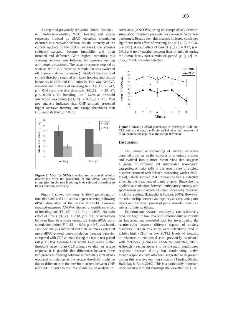
As reported previously (Oliveira, Nobre, Brandão,
& Landeira-Fernandez, 2004), freezing and escape
responses induced by dPAG electrical stimulation
occurred in a stepwise fashion. As the intensity of the
current applied to the dPAG increased, the animals
suddenly stopped, became immobile, and often
urinated and defecated. With higher intensities, this
freezing behavior was followed by vigorous running
and jumping reactions. The escape response stopped as
soon as the dPAG electrical stimulation was switched
off. Figure 2 shows the mean (± SEM) of the electrical
current threshold required to trigger freezing and escape
behaviors in CHF and CLF animals. Two-way ANOVA
revealed main effects of breeding line (F[1,12] = 5.42,
p < 0.05) and aversive threshold (F[1,12] = 234.37,
p < 0.0001). No breeding line ´ aversive threshold
interaction was found (F[1,12] = 0.37, p > 0.5). Post
hoc analysis indicated that CHF animals presented
higher aversive freezing and escape thresholds than
CFL animals (both p < 0.05).
Figure 2. Mean (± SEM) freezing and escape thresholds determined with the procedure of the dPAG electrical stimulation in the two breeding lines selected according to their emotional reactivity.
Figure 3 shows the mean (± SEM) percentage of
time that CHF and CLF animals spent freezing following
dPAG stimulation at the escape threshold. Two-way
repeated-measures ANOVA showed a significant effect
of breeding line (F[1,12] = 12.10, p < 0.005). No main
effect of time (F[1,12] = 2.18, p > 0.1) or interaction
between lines of animals during the 8-min dPAG post-
stimulation period (F [1,12] = 0.34, p > 0.5) was found.
Post hoc analysis indicated that CHF animals expressed
more dPAG-evoked post-stimulation freezing behavior
compared with CLF animals during the 8-min test period
(all p < 0.05). Because CHF animals required a higher
threshold current than CLF animals to elicit an escape
response it is possible that differences between these
two groups in freezing behavior immediately after dPAG
electrical stimulation at the escape threshold might be
due to differences in the threshold current between CHF
and CLF. In order to test this possibility, an analysis of
covariance (ANCOVA) using the escape dPAG electrical
stimulation threshold parameter as covariant factor was
performed. Results from this analysis indicated confirmed
significant main effect of breeding line (F [1,12] = 9.34,
p = 0.01). A main effect of time (F [1,12] = 8.47, p =
0.01) and no interaction between lines of animals during
the 8-min dPAG post-stimulation period (F [1,12] =
0.55, p > 0.4) was also detected.
Figure 3. Mean (± SEM) percentage of freezing in CHF and CLF animals during the 8-min period after the cessation of dPAG stimulation applied at the escape threshold.
Discussion
The current understanding of anxiety disorders
departed from an earlier concept of a unitary process
and evolved into a more recent view that suggests
a group of different but interrelated nosological
categories. A major shift in this recent view of anxiety
disorder occurred with Klein’s pioneering work (1962;
1964), which showed that imipramine had a selective
effect in the treatment of panic attacks. Since then, a
qualitative distinction between anticipatory anxiety and
spontaneous panic attack has been repeatedly observed
in clinical settings (Battaglia & Ogliari, 2005). However,
the relationship between anticipatory anxiety with panic
attack and the development of panic disorder remains a
subject of intense debate.
Experimental research employing rats selectively
bred for high or low levels of emotionality represents
an important and powerful tool for investigating the
relationships between different aspects of anxiety
disorders. Rats in this study were selectively bred to
exhibit high (CHF) or low (CFL) levels of freezing
in response to contextual cues previously associated
with footshock (Gomes & Landeira-Fernandez, 2008).
Although freezing appears to be the main conditioned
response observed during fear conditioning, active
escape responses have also been suggested to be present
during this aversive learning situation (Tarpley, Shlifer,
Halladay, & Blair, 2010). This is a particularly important
issue because it might challenge the view that the CHF
DBD
PUC-Rio - Certificação Digital Nº 0812188/CA

104
1A
phenotype is associated with more anxiety-like behavior
than the CLF phenotype. Thus, CLF rats may freeze less
not because they are less “afraid,” but because they are
more “frightened” and thus more prone to exhibit active
escape responses than defensive freezing behavior.
Much evidence appears to exclude this possibility.
For example, conditioned freezing is a direct function
of footstock intensity (Morris & Bouton, 2008) and has
been pharmacologically validated as an animal model
of anticipatory anxiety. Accordingly, benzodiazepine
receptor agonists such as diazepam and midazolam
reduced the amount of conditioned freezing, whereas
the benzodiazepine inverse agonist dimethoxy-β-
carboline produced freezing behavior similar to that
elicited by context fear conditioning (Fanselow, 1991).
Consistent with this, anxiolytic-like substances such as
the dPAG, which in turn might inhibit defensive reactions
triggered by this structure. Therefore, activation of the
neural circuitry involved in anxiety might indeed inhibit
the occurrence of panic attack-like behavior associated
with neurons located within the dPAG.
The present results also indicated that CHF animals
displayed more freezing behavior immediately after
dPAG electrical stimulation at the escape threshold
compared with CLF animals. This difference might
be attributable to the fact that CHF animals required
a higher threshold current than CLF animals to elicit
an escape response. An ANCOVA contested this
hypothesis indicating that although CHF animals were
more resistant to the expression of escape behavior in
response to dPAG stimulation, they were more prone
to freezing after the occurrence of the dPAG aversive
5-HT receptor agonists, selective serotonin reuptake stimulation compared with CLF animals.
inhibitors, and monoamine oxidase inhibitors with
verified clinical efficacy in the treatment of anxiety
symptoms, attenuated conditioned behavior in rats,
indicating considerable construct and face validity
of this paradigm to human anxiety (Conti, Maciver,
Ferkany, & Abreu, 1990; Maki et al., 2000). Moreover,
mice selectively bred for high and low levels of freezing
in response to contextual cues previously associated with
footshock also presented, respectively, higher and lower
levels of anticipatory anxiety in the fear-potentiated
startle test (Ponder et al., 2007). Finally, previous results
from our laboratory with different models of anxiety,
such as the elevated plus maze and social interaction
test, indicated that CHF animals exhibited significantly
more anxiety-like behavior than control rats (Dias et al.,
2008) Therefore, the CHF line appears to represent a
robust animal model of anticipatory anxiety.
The results of the present study indicated that
CHF animals had a higher dPAG electrical stimulation
aversive threshold for producing freezing and escape
reactions than CLF animals. This result is consistent with
several other studies, which indicated that contextual fear
conditioning can inhibit defensive responses to aversive
proximal or painful stimuli such as the tail-flick response
to radiant heat, complex and elaborated nociceptive
responses elicited in the formalin test, vigorous running
and jumping triggered by footshock (Fanselow, 1982)
and shock-induced defensive fight reactions (Bolles and
Collier, 1976). Moreover, contextual fear conditioning
can also inhibit vigorous escape responses induced by
N-methyl-D-aspartate (Galvão et al., 2010) or electrical
stimulation (Magierek et al.,2003) of the dPAG. Much
evidence indicates that the amygdaloid complex and its
projections to the ventral portion of the PAG are critically
involved in the regulation of contextual fear conditioning.
Malfunctioning of this system might be associated
with pathological forms of anticipatory anxiety (e.g.,
generalized anxiety disorder). Descending inhibitory
projections from the amygdaloid complex might reach
The dPAG post-stimulation freezing is not fear-
conditioning in response to contextual cues associated
with the dPAG electrical stimulation. Previous studies
employed a context shift procedure and indicated that
freezing after dPAG stimulation persisted when animals
were placed in a different context immediately after
the dPAG stimulation (Vianna et al., 2001). Moreover,
several studies indicated that freezing observed after
dPAG stimulation has a different neural mechanism
from freezing and escape responses elicited by dPAG
electrical stimulation. For example, electrolytic lesions
or muscimol-induced inactivation of the amygdaloid
complex reduced dPAG post-stimulation freezing but
did not affect freezing or escape responses induced
by dPAG electrical stimulation (Oliveira et al. 2004;
Ruiz-Martinez, de Oliveira, & Brandão, 2006). Indeed,
dPAG post-stimulation freezing appears to be mediated
by ascending projections, possibly relayed through the
thalamus to forebrain structures related to the sensory
processing of aversive stimuli. These findings suggest
the possibility that dPAG post-stimulation freezing might
represent an animal model of panic disorder. Therefore,
the fact that CHF animals expressed more dPAG post-
stimulation freezing than CLF animals might indicate
that anticipatory anxiety could enhance the development
of panic disorder triggered by panic attacks.
The complex nature of the symptoms involved in
panic disorders suggests that several brain regions may be
implicated in this pathology. For example, brain imaging
techniques indicated that anxiety reactions among
patients who suffer from panic disorder recruits several
forebrain areas such as the anterior cingulate cortex,
hippocampus, basal ganglia, and insula (de Carvalho et
al., 2010). Another important brain region that appears
to be implicated in the modulation of anxiety in panic
disorder is the amygdaloid complex. Indeed, ascending
projections from the dPAG to the amygdaloid complex
might represent one of the main routes responsible for
the pathogenesis of panic disorder. Therefore, anxiety
DBD
PUC-Rio - Certificação Digital Nº 0812188/CA

105
reactions that are present in panic disorder might also
recruit some forebrain structures related to anticipatory
anxiety. Further studies are needed to elucidate whether
activation of neural circuitries associated with the
amygdaloid complex might play an inhibitory role in the
occurrence of panic attack and excitatory modulation of
structures associated with panic disorders.
Acknowledgment
This research was supported by Brazilian National
Research Council CNPq, 522720/95-10 awarded to J.
Landeira-Fernandez.
References
American Psychiatric Association. (1994) Diagnostic and statistical
manual of mental disorders, (4th ed). Washington: American Psychiatric Association.
Battaglia, M., & Ogliari, A. (2005). Anxiety and panic: from human studies to animal research and back. Neuroscience Biobehavioral
Reviews, 29, 169-79. Bolles, RC., & Collier, A. C. (1976). The effect of predictive cues of
freezing in rats. Animal Learning Behavior, 4, 6-8. Brandão, M.L., de Aguiar, J.C., & Graeff, F.G. (1982) GABA
mediation of the anti-aversive action of minor tranquilizers. Pharmacology Biochemistry Behavior, 16, 397-402.
Brandão, M.L., Zanoveli, J.M., Ruiz-Martinez, R.C., Oliveira L.C., & Landeira-Fernandez J. (2008). Different patterns of freezing
behavior organized in the periaqueductal gray of rats: association with different types of anxiety. Behavioral Brain Reviews, 188, 1-13.
Conti, L.H., Maciver, C.R., Ferkany, J.W., & Abreu, M.E. (1990). Footshock-induced freezing behavior in rats as a model for
assessing anxiolytics. Psychopharmacology (Berl), 102, 492-497. de Carvalho, M.R., Dias, G.P., Cosci, F., de-Melo-Neto, V.L., Bevilaqua,
M.C., Gardino, P.F, & Nardi, A.E. (2010) Current findings of fMRI in panic disorder: contributions for the fear neurocircuitry and CBT
effects. Expert Reviews of Neurotherapeutics, 10, 291-303. Dias, G.P., Bevilaqua, M.C., Silveira, A.C., Landeira-Fernandez, J.,
& Gardino P.F. (2009). Behavioral profile and dorsal hippocampal cells in carioca high-conditioned freezing rats. Behavioral Brain
Research, 205, 342-8. Fanselow, M.S. (1982). The post-shock activity burst. Animal
Learning and Behavior, 10, 448-54. Fanselow, M.S. (1991). Analgesia as a response to aversive Pavlovian
conditional stimuli: Cognitive and emotional mediators. In Denny
M.R. Fear, avoidance, and phobias: A fundamental analysis, (pp. 61-86) Hillsdale, NJ: Lawrence Erlbaum.
Galvão, B., Larrubia, B., Hommes, W.J., Cardenas, F.P., Cruz,
A.P.M., & Landeira-Fernandez, J. (2010). Effects of contextual fear conditioning and pentylenetetrazol on panic-like reactions
induced by dorsal periaqueductal gray stimulation with N-methyl- D-aspartate. Psychology Neuroscience, 3, 67-72.
Gomes, V.C., & Landeira-Fernandez, J. (2008). Amygdaloid lesions produced similar contextual fear conditioning disruption in the
Carioca high- and low-conditioned freezing rats. Brain Research, 1233, 137-45.
Klein, D.F. (1964). Delineation of two drug-responsive anxiety
syndromes. Psychopharmacologia, 5, 397-408. Klein, D.F., & Fink, M. (1962). Psychiatric reaction patterns to
imipramine. American Journal of Psychiatry, 119, 432-8. Landeira-Fernandez, J. (1996). Context and Pavlovian conditioning.
Brazilian Journal of Medical Biology Research, 29, 149-73.
Magierek, V., Ramos, P.L., da Silveira-Filho, N.G., Nogueira, R.L., & Landeira-Fernandez, J. (2003). Context fear conditioning inhibits
panic-like behavior elicited by electrical stimulation of dorsal periaqueductal gray. Neuroreport, 14, 1641-4.
Maki, Y., Inoue, T., Izumi, T., Muraki, I., Ito, K., Kitaichi, Y., Li X., & Koyama T. (2000). Monoamine oxidase inhibitors reduce
conditioned fear stress-induced freezing behavior in rats. European Journal of Pharmacology, 406, 411-8.
Morris, R.W., & Bouton, M.E. (2006). Effect of unconditioned stimulus magnitude on the emergence of conditioned responding. Journal of
Experimental Psychology: Animal Behavior Processes, 32, 371-85. Oliveira, L.C., Nobre, M.J., Brandão, M.L., & Landeira-Fernandez,
J. (2004) Role of amygdala in conditioned and unconditioned fear generated in the periaqueductal gray. Neuroreport, 15, 2281-5.
Paxinos G., & Watson C. (1986). The rat brain in stereotaxic coordinates, (2nd ed). New York: Academic Press.
Ponder ,C.A., Kliethermes, C.L., Drew, M.R., Muller, J., Das, K., Risbrough, V.B., Crabbe, J.C., Gilliam, T.C., & Palmer, A.A. (2007).
Selection for contextual fear conditioning affects anxiety-like behaviors and gene expression. Genes Brain and Behavior, 6, 736-49.
Ruiz-Martinez, R.C., de Oliveira, A.R., & Brandão, M.L. (2006). Conditioned and unconditioned fear organized in the periaqueductal
gray are differentially sensitive to injections of muscimol into amygdaloid nuclei. Neurobiology Learning and Memory, 85, 58-65.
Tarpley, J.W., Shlifer, I.G., Halladay, L.R., & Blair, HT. (2010). Conditioned turning behavior: a Pavlovian fear response expressed
during the post-encounter period following aversive stimulation. Neuroscience, 169,1689-704.
Vianna, D.M., Graeff, F.G., Brandão, M.L., & Landeira-Fernandez
J. (2001) Defensive freezing evoked by electrical stimulation of
the periaqueductal gray: comparison between dorsolateral and ventrolateral regions. Neuroreport 12, 4109-12.
DBD
PUC-Rio - Certificação Digital Nº 0812188/CA


![11 Referências bibliográficas e webográficas · 190 11 Referências bibliográficas e webográficas [1] Burger, Kálmán, Biocoordination Chemistry: Coordination Equilibria in](https://static.fdocuments.us/doc/165x107/5eb6df383b2e2668687a9f05/11-referncias-bibliogrficas-e-webogr-190-11-referncias-bibliogrficas-e.jpg)