10 October 2011 Chapter 6 Section D: Anatomy Chapter 7: Sensory Physiology Lab this week: Part 1:...
14
10 October 2011 Chapter 6 Section D: Anatomy Chapter 7: Sensory Physiology Lab this week: Part 1: Visual System A (lecture/demo) Part 2: Sensory physiology: localizing a stimulus applied to the skin Lab next week: Part 1: Visual System B (lecture/demo) Part 2: Sensory physiology: 2 more experiments Part 3: Auditory System (lecture/demo) Progress report on Take-home portion of Test 1
-
Upload
jack-fisher -
Category
Documents
-
view
215 -
download
0
Transcript of 10 October 2011 Chapter 6 Section D: Anatomy Chapter 7: Sensory Physiology Lab this week: Part 1:...
10 October 2011Chapter 6 Section D: Anatomy Chapter 7: Sensory Physiology
Lab this week: Part 1: Visual System A (lecture/demo)Part 2: Sensory physiology: localizing a stimulus applied to the skin
Lab next week: Part 1: Visual System B (lecture/demo)Part 2: Sensory physiology: 2 more experimentsPart 3: Auditory System (lecture/demo)
Progress report on Take-home portion of Test 1
1QQ # 15 for 8:30 class
1. Why would you insist that an epidural injection of lidocaine not be given in the region of cervical vertebra?
2. Describe the challenges that face neuroscientists who wish to replace dying or damaged neurons in the brain with cells donated from elsewhere.
3. You suspect that the neurons in Nucleus X are communicating with their target cells by releasing NE as their neurotransmitter. Describe an experiment that would allow you to test your hypothesis.
4. Use your knowledge of cortical neuroanatomy to explain why stroke victims very often have deficits involving the face and hands.
1QQ # 15 for 9:30 class
1. Describe the challenges that face neuroscientists who wish to replace dying or damaged neurons in the brain with cells donated from elsewhere.
2. You suspect that the neurons in Nucleus Z are communicating with their target cells by releasing ACh as their neurotransmitter. Describe an experiment that would allow you to test your hypothesis.
3. Use your knowledge of cortical neuroanatomy to explain why stroke victims very often have deficits involving the face and hands.
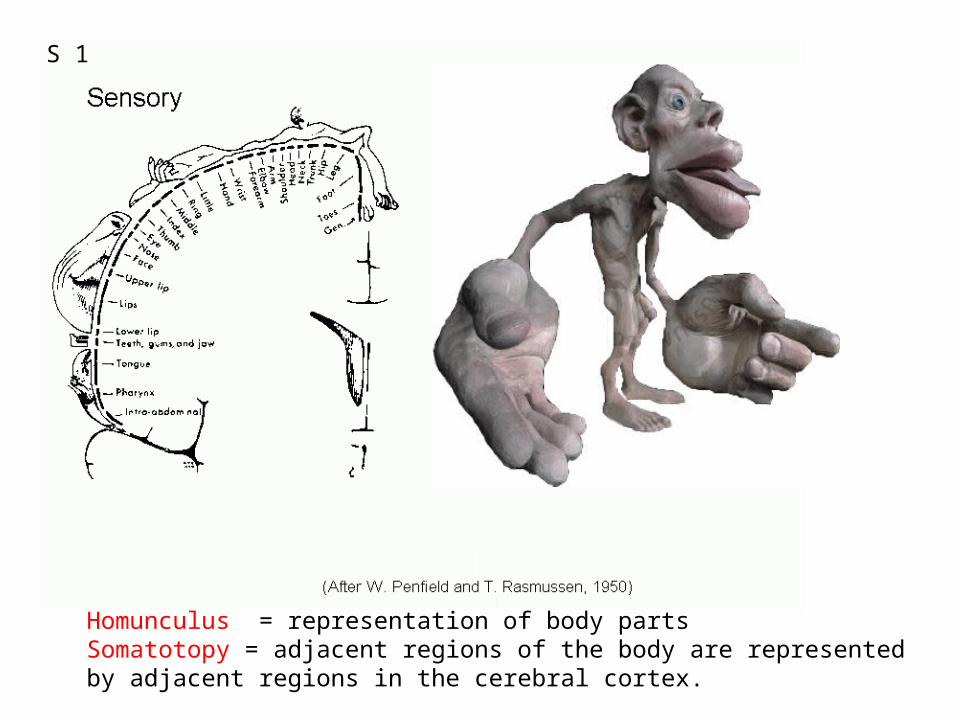
Homunculus = representation of body partsSomatotopy = adjacent regions of the body are representedby adjacent regions in the cerebral cortex.
S 1
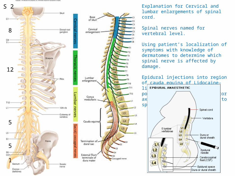
Explanation for Cervical and lumbar enlargements of spinal cord.
Spinal nerves named for vertebral level.
Using patient’s localization of symptoms with knowledge of dermatomes to determine which spinal nerve is affected by damage.
Epidural injections into region of cauda equina of Lidocaine-like agents to block action potentials in sensory and motor axons without risk of damage to spinal cord.
8
12
5
5
1
S 2
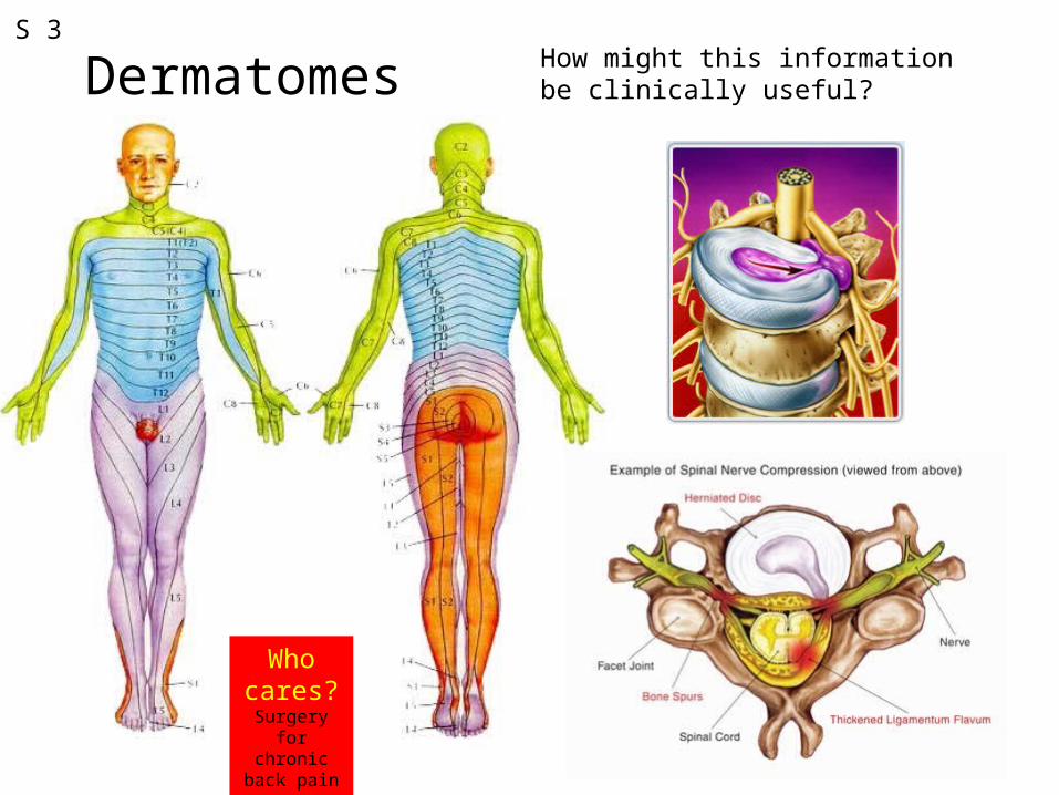
DermatomesS 3
How might this information be clinically useful?
Who cares?Surgery for
chronic back pain
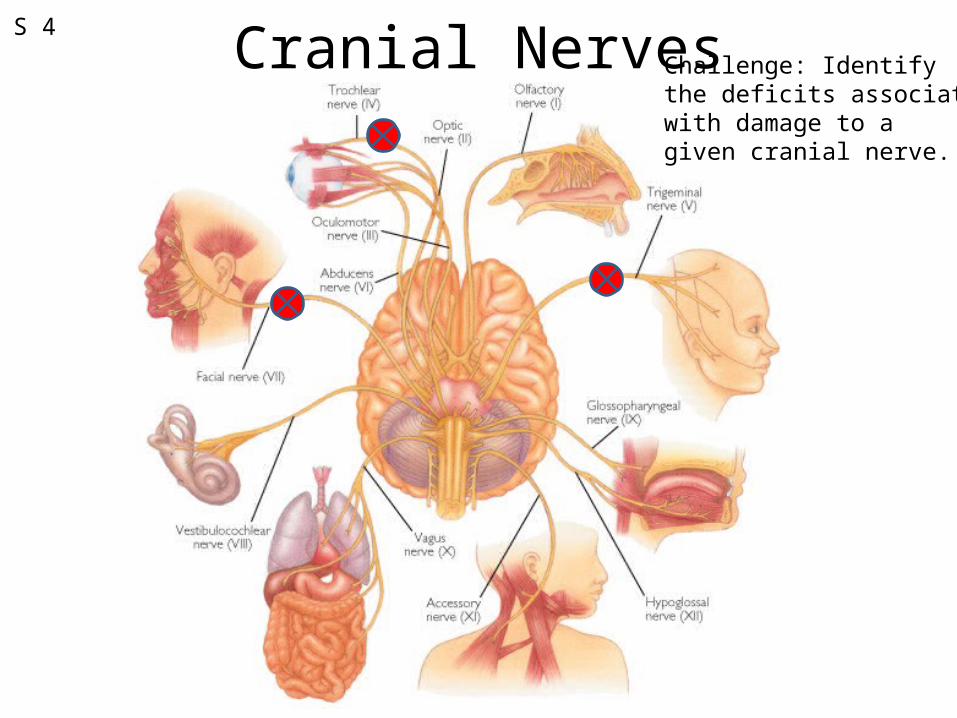
Cranial NervesS 4
Challenge: Identifythe deficits associatedwith damage to agiven cranial nerve.
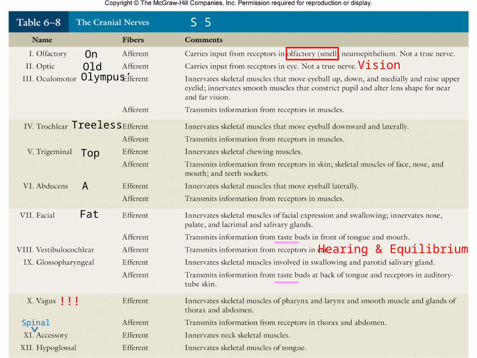
Vision
Hearing & Equilibrium
!!!
S 5
Spinal
OnOld
Fat
Olympus’
Treeless
A
Top
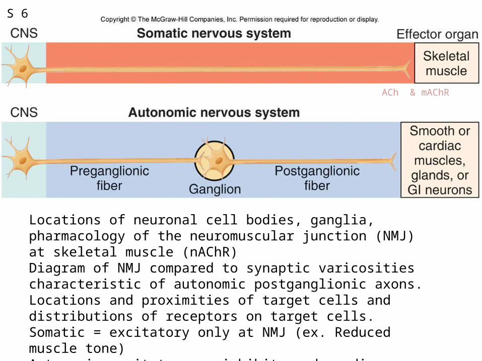
Figure 6.43
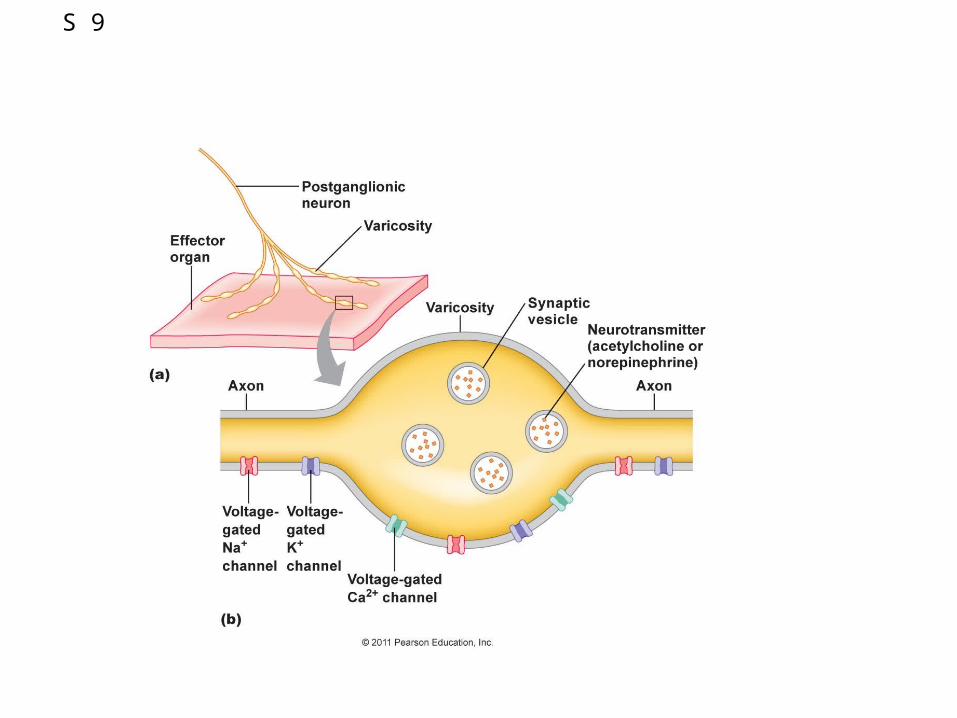
Locations of neuronal cell bodies, ganglia, pharmacology of the neuromuscular junction (NMJ) at skeletal muscle (nAChR)Diagram of NMJ compared to synaptic varicosities characteristic of autonomic postganglionic axons. Locations and proximities of target cells and distributions of receptors on target cells.Somatic = excitatory only at NMJ (ex. Reduced muscle tone)Autonomic= exitatory or inhibitory depending on NTs and their receptors.
S 6
ACh & mAChR
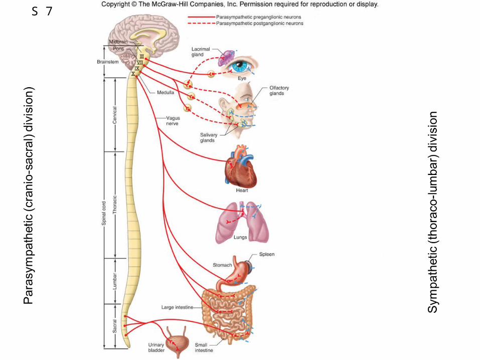
Figure 6.44S 7
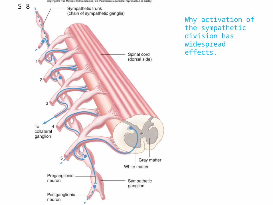
Why activation of the sympathetic division has widespread effects.
S 8
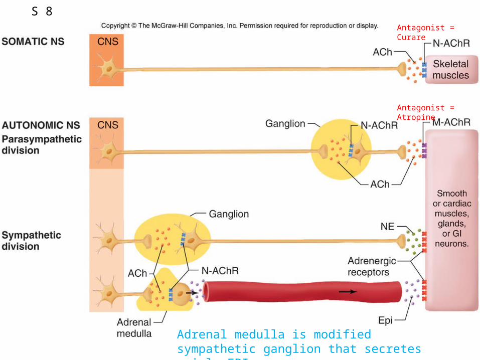
Figure 6.46
Adrenal medulla is modified sympathetic ganglion that secretes mainly EPI
Antagonist = Curare
Antagonist = Atropine
S 8
S 9
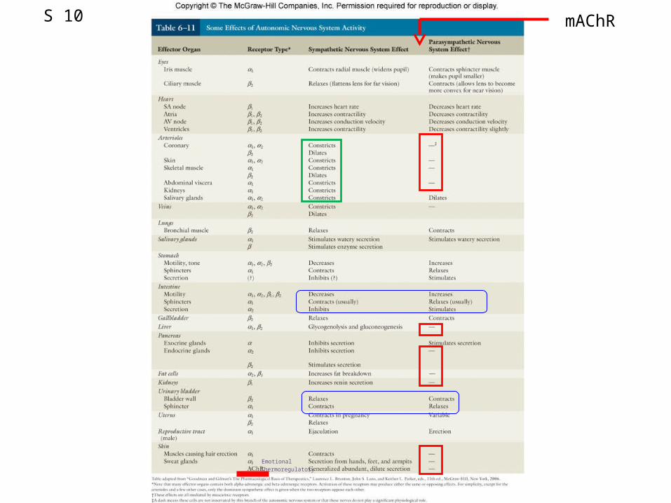
Table 6.11S 10
EmotionalThermoregulatory
mAChR