
10-01-01 Developing a Core Platform for the Tissue Engineering of Vital Organs
16
Developing a Core Platform for the Tissue Engineering of Vital Organs Erin R. Ochoa and Joseph P. Vacanti T he field of tissue engineering was last reviewed in this journal in 1993 by Mooney and Vacanti’ (“Tissue Engineering Using Cells and Synthetic Polymers”), with particular emphasis on the in vivo culture of liver, cartilage, bone, uroepithelial, and intestinal tissues. At that time, our experiments were designed to show the potential applicability of a tissue-engineering approach to a wide range of clin- ical surgical problems and to weigh the relative ad- vantages of diKerent approaches to tissue engincer- ing. These early experiments assessed whether to culture autologous versus allogenic donor tissues, whether to use such naturally occurring biomaterials as collagen or to develop synthetic materials to use as support structures for tissue culture, and whether to culture transplantable tissues in vitro before trans- plantation or to culture new tissue in viva with the aid of signaling factors and neovasculature mar- shaled from the host.‘J In a short time, the field of tissue engineering has undergone explosive development, and now is yield- ing practical clinical results. Versatile, cost-effective, engineered skin replacements are commercialI). available, and several cartilage replacement devices are in the clinical trial stage.“.” As the field continues to mature and new microfabrication technologies emerge, the possibility of whole-organ fabrication becomes more credible. Our team, now part of the Center for the Inte- gration of Medicine and Innovative Technology lab- oratories at Massachusetts General Hospital (Bos- ton, MA), has evolved into a highly integrated multidisciplinary network of complementary projects. WC have active collaborations with rcscarch groups throughout Massachusetts General Hospital, Massa- chusetts Institute ofTechnoloa, Draper Laboratory, Brigham and Women’s Hospital, Children’s Hospi- tal, the HalTlard Center for Genomic Research, Har- vard School of Dental Medicine, Forsythe Dental Institute, Shrine& Burn Institute, and the Univer- si$ of Michigan. Subgroups of our team integrate the resources of these institutions into focused tis- sue-specific projects, including liver, kidney, lung, intestine, heart muscle, a biological tooth, auricular cartilage, manclible, esophagus, heart valves, and tu- mor control. Although these projects use a variety of tissue-engineering strategies and are at various stages of development, the bulk of our work has focused on the development of a core strategy fol overcoming a specilic set of problems in engineering lital organs and dense vascularized tissue. To bring our core problem set into focus, first consider that to date, successes in tissue engineering have been achieved through relatively simple strat- cgies. For example, extremely cost-effective type I collagen and sulfatecl glycosaminoglycan grafts have been used to induce dermal regeneration by inhibit- ing wound contraction in viva.‘; Human-shaped ears have been grown from auricular cartilage seeded onto a biodegradable synthetic scaffold cultured in subcutaneous pockets of dorsa of athymic mice.’ A wicle range ofother tissue types has been engineered using a great variety of relatively simple technologies relying on angiogenesis .x Although useful for many specific clinical applications, these techniques and even the materials used in successful devices have proven inadequate for engineering large tissue masses or vital organs. Our experience supports two hypotheses. First, many types of donor cells will develop into morpho- logically and functionally normal new tissue if given adequate structural cues in the form of a support matrix and enough nutrition and diffusion to sustain culture. Second, although a variety of relatively sim- ple procedures will sustain the culture of thin layers of tissue types with low oxygen and nutrition require- 184 Trattsplanlation Review, Vol 15, No 4 (Octoltur), 2001: J~JI 184-199
-
Upload
michael-ochoa -
Category
Documents
-
view
26 -
download
1
Transcript of 10-01-01 Developing a Core Platform for the Tissue Engineering of Vital Organs

Developing a Core Platform for the Tissue Engineering of Vital Organs
Erin R. Ochoa and Joseph P. Vacanti
T he field of tissue engineering was last reviewed in this journal in 1993 by Mooney and Vacanti’
(“Tissue Engineering Using Cells and Synthetic Polymers”), with particular emphasis on the in vivo culture of liver, cartilage, bone, uroepithelial, and
intestinal tissues. At that time, our experiments were designed to show the potential applicability of a tissue-engineering approach to a wide range of clin- ical surgical problems and to weigh the relative ad-
vantages of diKerent approaches to tissue engincer- ing. These early experiments assessed whether to culture autologous versus allogenic donor tissues,
whether to use such naturally occurring biomaterials as collagen or to develop synthetic materials to use as support structures for tissue culture, and whether to
culture transplantable tissues in vitro before trans- plantation or to culture new tissue in viva with the aid of signaling factors and neovasculature mar- shaled from the host.‘J
In a short time, the field of tissue engineering has undergone explosive development, and now is yield-
ing practical clinical results. Versatile, cost-effective, engineered skin replacements are commercialI). available, and several cartilage replacement devices
are in the clinical trial stage.“.” As the field continues to mature and new microfabrication technologies emerge, the possibility of whole-organ fabrication
becomes more credible. Our team, now part of the Center for the Inte-
gration of Medicine and Innovative Technology lab-
oratories at Massachusetts General Hospital (Bos- ton, MA), has evolved into a highly integrated multidisciplinary network of complementary projects.
WC have active collaborations with rcscarch groups
throughout Massachusetts General Hospital, Massa- chusetts Institute ofTechnoloa, Draper Laboratory, Brigham and Women’s Hospital, Children’s Hospi- tal, the HalTlard Center for Genomic Research, Har-
vard School of Dental Medicine, Forsythe Dental Institute, Shrine& Burn Institute, and the Univer- si$ of Michigan. Subgroups of our team integrate the resources of these institutions into focused tis- sue-specific projects, including liver, kidney, lung,
intestine, heart muscle, a biological tooth, auricular cartilage, manclible, esophagus, heart valves, and tu- mor control. Although these projects use a variety of
tissue-engineering strategies and are at various stages of development, the bulk of our work has focused on the development of a core strategy fol
overcoming a specilic set of problems in engineering lital organs and dense vascularized tissue.
To bring our core problem set into focus, first
consider that to date, successes in tissue engineering have been achieved through relatively simple strat- cgies. For example, extremely cost-effective type I
collagen and sulfatecl glycosaminoglycan grafts have been used to induce dermal regeneration by inhibit- ing wound contraction in viva.‘; Human-shaped ears
have been grown from auricular cartilage seeded onto a biodegradable synthetic scaffold cultured in subcutaneous pockets of dorsa of athymic mice.’ A
wicle range ofother tissue types has been engineered using a great variety of relatively simple technologies relying on angiogenesis .x Although useful for many
specific clinical applications, these techniques and even the materials used in successful devices have proven inadequate for engineering large tissue masses or vital organs.
Our experience supports two hypotheses. First, many types of donor cells will develop into morpho- logically and functionally normal new tissue if given
adequate structural cues in the form of a support matrix and enough nutrition and diffusion to sustain
culture. Second, although a variety of relatively sim- ple procedures will sustain the culture of thin layers of tissue types with low oxygen and nutrition require-
184 Trattsplanlation Review, Vol 15, No 4 (Octoltur), 2001: J~JI 184-199

Core Plal/orm for Time EngineeGg 185
merits (ie, skin and cartilage),” culturing dense tis- sues and solid organs will require the development of new techniques and materials that tightly control a host of parameters governing nutrition and diffu- sion.“’ Thus, microengineering a synthetic microvas- cularized scaffold to meet the culture-flow dynamic requirements of dense tissues and solid organs has become the principle goal of our laboratory. If suc- cessful, it could provide a core platform that would be applicable to a wide range of tissue-engineering scenarios and could bc scaled up to provide a general solution to dire clinical tissue shortages.”
To better understand our strategy for addressing this central challenge facing tissue engineering, we take a vertical slice though our network of liver project collaborations. This exposes a series of closely related experiments that are simultaneously attack- ing specific complementary aspects of our core prob- lems and also providing data to feed back into other parts of the liver project, as well as into parallel developments in our other tissue-specific projects. To understand our liver project we must touch upon: (1) the theoretical background that shapes our ap- proach; (2) the development of computational mod- els of culture flow through branching vascular net- works; (3) the evolution of our synthetic vascular network architectural designs; (4) the adaptation of a 2-dimensional microfabrication technology bor- rowed from the integrated circuit industry to the production of 3-dimensional biodegradable synthetic vasculature scaffolds; (5) the development of flow systems to seed vascularized scaffolds with donor cells, maintain specific in vitro culture Row condi- tions, and conduct pressure and flow studies of syn- thetic vascular networks; (6) isolating, seeding, cul- turing, and implanting optimal donor cell types; and (7) the development of a host of tools for analyzing our engineered liver tissues. We conclude with a brief survey of some of our other parallel and tan- gential projects.
Tissue Biology and Tissue-Engineering Strategy Parameters
Some general observations about the biological com- position of all living tissue are needed to establish the basic parameters of our core tissue-engineering ap- proach. We must distinguish the variety of cell types that compose a given tissue, the extracellular matrix (ECM) that forms the immediate environment of
each cell, and the host of signaling molecules traded back and forth between individual cells, the ECM, and neighboring or even far distant cells. The dy- namic chemical and mechanical interactions of these 3 tissue components determine the course of tissue development and regulate normal function.‘” To en- gineer new tissue we must begin by making strategic decisions about how to treat each of these compo- nents.
In engineering the microvasculature needed to grow dense functional liver tissue, we begin with syngeneic samples of isolated adult male Lewis rat hepatocytes and endothelium. The most straightfor- ward strategy to avoid later tissue rejection by the implant host would be to culture autologous tissue. However, such an approach would not provide the most cost-effective o&the-shelf solutions needed to meet the huge clinical demand for donor tissue in the long run. The solution eventually may be pro- vided by the emerging field of stem-cell research. Many stem-cell sources from adult and fetal animals show pluripotent developmental potential and a high degree of immunocompatibility.‘“*‘+~‘j However, much work remains to be done in this area, and legal and ethical constraints on stem-cell research have yet to be resolved.“j.”
The ECM is an important nest consideration. The most advanced tissue-engineering products, skin grafts, generally use a Form of collagen process- ing, such as foam, gel, fiber mesh, or sponge, and are often implanted with growth-stimulating proteins. These devices closely mimic the chemical properties of the native ECM, but present drawbacks as a uni- versalizable tissue-engineering materials solution.ls Because collagen is generally isolated from human or animal tissue, it has large batch-to-batch varia- tions. More important for our purposes, collagen offers limited versatility in designing microstruc- tures and tightly controlling such properties as me- chanical strength and degradation time. To focus on the structural requirements necessary to promote microvascularization, our team pioneered the use of synthetic bioabsorbable polymers as our core support matrix material.
Finally, any tissue-engineering strategy must make decisions about how to facilitate or direct the complex molecular activities that guide tissue devel- opment.‘” The neighboring field of molecular biology is rapidly changing the life sciences. The tools it offers tissue engineering include the possibility of altering the genetic composition of donor tissue to

186 Ociwa and Vacanti
eliminate genetic defects or promote compatibility, altering genetic expression patterns of donor tissue more tightly to control tissue development, and iden- ti@ing and isolating specific signaling factors that may be incorporated into engineered devices. The general hypothesis that dictates our attitude toward these possibilities is that if adequate structural cues and culture conditions are provided, the donor tissue and host can provide the molecular signals necessary to guide new tissue development. Our goal is to facilitate the natural molecular communication be- tween cells, rather than to control it. Although we have experimented with enriching support scaffolds with well-documented growth factors, our principal use of the tools of molecular biology in our liver project is to compare the in vitro developmental process with in vivo organogenesis.
Taken together, our basic decisions about cell sourcing, support materials, and molecular biology determine what we may call our “mechanical-struc- tural” core tissue-engineering strategy.
Modeling Nutrition and Diffusion Dynamics
Given the task of microfabricating the vasculature needed to culture large tissue masses and vital or- gans, tissue engineering must transform our existing knowledge of in vivo vascular architecture into syn- thetic vascular systems. The new radiological tech- nologies of micro-magnetic resonance and micro- computed tomography may offer a technique to understand the topography of vascular systems.21*22+23 Using these and similar techniques, tissue engineers are developing computational models that can mimic the essential parameters of any specific in vivo target type. After these features have been quanti- fied, these models can be translated into synthetic systems that preserve the essential feature of in vivo vasculature (Fig 1).
On our own team, Kaazempur-Mofrad developed independent complementary mathematical models that simulate the branching architecture of fractal
Figure 1. (A) Topology parameter schematic. (B) Two- sample fractal networks. (A, courtesy of Mohammad R Kaazempur, PhD, B reproduced from Kaazempur-Mofrad et al *O; with permission)
Core PlatjiiJi Tissue Engineetfq 187
A Du Y nbuu om of WOI mohxcd Fl ow Rare
IS,ru lburmo orl-ubc C~C~O~CIIL
hutbuuoo ofRBC Flux
0 I OJO on0
030 0110 010 000 010 0.10 100
OLD
cr,
I
oca OLr 013 ON OLS OCI OCO
009 oar oaa
F #IL
I
oao
on0
030 0 030 oco 000 010 0.10 100 JO
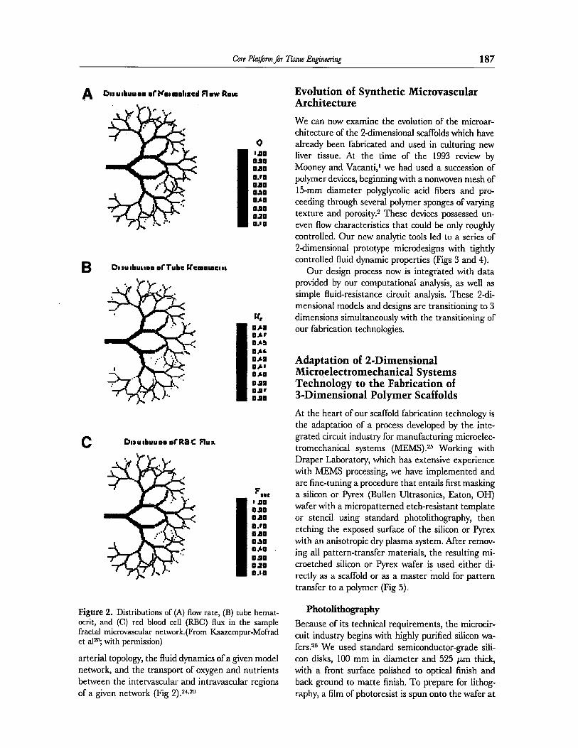
Figure 2. Distributions of (A) flow rate, (B) tube hemat- ocrit, and (C) red blood cell (RBC) flux in the sample fractal microvascular network.(From Kaazempur-Mofrad et aP; with permission)
arterial topology, the fluid dynamics of a given model network, and the transport of oxygen and nutrients between the intervascular and intravascular regions of a given network (Fig 2).2+*20
Evolution of Synthetic Microvascular Architecture
We can now examine the evolution of the microar- chitecture of the 2-dimensiona.l scaffolds which have already been fabricated and used in culturing new liver tissue. At the time of the 1993 review by Mooney and Vacanti,’ we had used a succession of polymer devices, beginning with a nonwoven mesh of 15-mm diameter polyglycolic acid fibers and pro- ceeding through several polymer sponges of varying texture and porosity.* These devices possessed un- even flow characteristics that could be only roughly controlled. Our new analytic tools led to a series of P-dimensional prototype microdesigns with tightly controlled fluid dynamic properties (Figs 3 and 4).
Our design process now is integrated with data provided by our computational analysis, as well as simple fluid-resistance circuit analysis. These 2di- mensional models and designs are transitioning to 3 dimensions simultaneously with the transitioning of our fabrication technologies.
Adaptation of 2-Dimensional Micrqelectromechanical Systems Technology to the Fabrication of 3-Dimensional Polymer Staff olds
At the heart of our scaffold fabrication technology is the adaptation of a process developed by the inte- grated circuit industry for manufacturing microelec- tromechanical systems (MEMS).25 Working with Draper Laboratory, which has extensive experience with MEMS processing, we have implemented and are fine-tuning a procedure that entails first masking a silicon or Pyrex (Bullen Ultrasonics, Eaton, OH) wafer with a micropatterned etch-resistant template or stencil using standard photolithography, then etching the exposed surface of the silicon or Pyrex with an anisotropic dry plasma system. After remov- ing all pattern-transfer materials, the resulting mi- croetched silicon or F’yrex wafer is used either di- rectly as a scaffold or as a master mold for pattern transfer to a polymer (Fig 5).
Photolithography
Because of its technical requirements, the microcir- cuit industry begins with highly purified silicon wa- fers.2G We used standard semiconductor-grade sili- con disks, 100 mm in diameter and 525 pm thick, with a front surface polished to optical finish and back ground to matte finish. To prepare for lithog- raphy, a film of photoresist is spun onto the wafer at

188 oclloa and Vacanti
Figure 3. Polymer templates used for cell substrates. (A) Scanning electron micrograph of poly(lactic-co-glycolic acid) (85:15). The scaffold was prepared by the particulate leaching method, resulting in an interconnected porous network. (B) Fiber-based device consisting of a nonwoven mesh of polyglycolic acid (PGA) fibers (- 15 mm in diameter).

Core Platjii~for Tissue Engimring 189
Figure 3. (Cont’d) (C) Poly-4-hydroxybutyrate (P4HB) scaffold. Abbreviations: AccV, ; Det SE, ; WD, ; Exp, ; STl, ; Co& (A, courtesy of Michael Y. Shin, PhD; B, C, courtesy of Irina Pomerantseva, MD).
4,000 rpm to a thickness sufficient to serve as a mask for the subsequent chemical etching.z7
As an alternative, we experimented with inexpen- sive Pyrex, which is commonly used for cell-culture and biotech applications but is not sufficiently pure for semiconductor fabrication.28 We used loo-mm diam- eter, 775-mm thick Pyrex wafers, with the front and
back polished to optical finish. It is known that a layer of photoresist similar to that used on silicon wafers is not sufXcient to withstand the standard etch chemis- tries that we use. So, we tried depositing an interme- diate layer of etch-resistant polysilicon to the Pyrex before applying the photoresist. However, the high temperatures needed to adhere the polysilicon (550°C
Figure 4. Succession of prototype designs. (Courtesy of Jeffery T. Borenstein, PhD)

190 Ckhoa and Vacanti
Pour Biodegradable Polymer Mold
Separate Polymer
Build Copy
Figure 5. Schematic of 2aimensional polymer scaffold produced by h4Eh4S technology. (Courtesy of Jeffery T. Boren- stein, PhD)
to 650°C) caused the glass to flow and buckle, leading to severe downstream processing problems. We now use a modified process to mask the Pyrex before etch- ing. If our promising trials are confirmed, we may have arrived at the optimal starting material.
In addition to preparing a wafer, we must repli- cate our desired pattern on a glass photomask plate by electron-beam processing. This is done for us by a commercial lithography service (Align-Rite, Bur- bank, CA). Light is passed through this photomask, leaving a pattern on the layer of photoresist. These patterned areas are chemically removed, leaving an exposed surface of silicon or Pyrex outlined by a thick etch-resistant template or stencil.
Biocompatible and Bioabsorbable Scaffolds
Over the years, we experimented with a number of polymer materials, including polyglycolic acid, poly-L, lactic acid and poly(lacticcoglycolide), and poly dimethyl silazone.29 These and similar materials allow tissue engineers to create a variety of biocom- patible scaffolds with tightly controlled bioabsorbable properties.
Our experiments with a variety of permanent and bioabsorbable polymers have shown the versatility of these materials and revealed several obstacles that polymer-based tissue-engineering strategies must address. Polymers present a smoother surface than such natural ECM analogues as collagen. By exper- imenting with polymer surfaces coated with various binding proteins, such as collagen or vitrogen, we are beginning to understand the complex mechanical and chemical processes that occur as individual do- nor cells attach to a scaffold and initiate new tissue growth.s@siJs In addition, techniques must be per- fected for modifying polymer surface chemistry to promote the bonding of polymer sheets with sufh- cient strength to withstand culture-flow pressures.
Dynamic Seeding, Culture, and Flow Studies
Our early tissue-engineering strategies used a vari- ety of static cell seeding and culture techniques. We gradually developed a bioreactor system for much

Core Platjiifor Tissue Engheering 191
more effective dynamic cell seeding to provide con- tinuous culture-flow conditions to the expanding new tissue and conduct fluid dynamic studies of our 2di- mensional sealed vascularized systems (Fig 6).s3r3*
In our current bioreactor configuration, cell seed- ing is accomplished by pumping a medium of the desired cell type through the sealed scaffold network, at a low pulsate flow rate and culture viscosity coor- dinated to optimize the adhesion of cells to walls of the scaffold without permitting the channels to be- come occluded. By switching to an oxygen- and nu- trient-rich culture medium, we can maintain flow conditions while seeded donor cells develop into a tubular layer of new tissue lining the scaffold. The result is a network of living vessels that mirrors our architectural and flow designs.
Our final hurdle will be to modify our designs and fine-tune our fabrication process so that our compu- tational fluid dynamic and experimental values both match the normal physiological hematocrit and blood pressure values of in vivo liver. The success of a tissue-engineering microvascular strategy is measured by how closely it can bring these 3 sets of values into agreement for the set of parameters de- fined by the specific needs of a particular in vivo tissue or organ type.
Isolating, Seeding, Culturing, and Implanting Optimal Donor Tissue
Although there are many cell types in native liver, 3 types are of particular interest to tissue engineering because of their architectural features: vascular en- dothelium, parenchymal hepatocytes, and biliary ep- ithelium. The vasculature of the liver has 3 parts. Two independent blood supply systems enter through the hepatic artery and portal vein and meet at the innumerable vascular microspaces called liver sinusoids. Red blood cells arrive through these 2 vascular networks and deposit oxygen and nutrition to surrounding tissue through fenestrations or pores in the sinusoid walls, then are collected into ramifi- cation of the hepatic vein. This venous network con- stitutes the third part of the liver vasculature and provides an exit to depleted red blood cells through the inferior vena cava. Together, these 3 parts form 1 vast continuous vascular space composed of endo- thelial cells. Our synthetic networks lined with en- dothelial cells will be our engineering analogue for this vascular system.
Surrounding the liver sinusoids and drawing oxy- gen and nutrients through the sinusoidal fenestra is a large mass of parenchymal tissue organized into cribriform, anastomosing sheets or “plates” of which
Air Trap
Pressure
Medium
Figure-b. Schematic explanation of microcirculation bioreactor system. (Courtesy of Kohei Ogawa, MD)
192 Ochoa and Vacanti
the hepatocyte is the main functional cell type. Growing a dense mass of functioning liver parenchy- mal tissue from donor hepatocytes is the primary strategic goal of our liver-engineering project and the end to which we need our endothelial networks.
Finally, there is another ductal or vascular-type space completely surrounded by parenchymal tissue, separated from this tissue by the space of Disse. Composed of specialized biliary epithelial cells, this system of canaliculi forms the beginning of the bile duct system. Bile is excreted by the parenchymal hepatocytes, passes through the space of Disse and into the biliary canaliculi, and is moved out of the liver by mechanical forces in a direction opposite to the flow of blood. Engineering this space composed of biliary epithelial cells is the secondary goal of any liver-engineering project. The engineering of new tissue from each of these 3 cell types (vascular endo- thelial cells, parenchymal hepatocytes, and biliary
epithelial cells) needs to be understood separately before putting them together.
Hepatocytes, Small Hepatocyte Cells, and Stem Cells
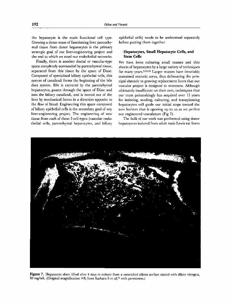
We have been culturing small masses and thin sheets of hepatocytes by a large variety of techniques for many years.sJj.:j” Larger masses have invariably contained necrotic cores, thus delineating the prin- cipal obstacle to growing replacement livers that our vascular project is designed to overcome. Although ultimately insufficient on their own, techniques that our team painstakingly has acquired over 15 years for isolating, seeding, culturing, and transplanting hepatocytes will guide our initial steps toward the new horizon that is opening up to us as we perfect our engineered vasculature (Fig 7).
The bulk of our work was performed using donor hepatoyctes isolated from adult male Lewis rat livers
Figure 7. Hepatocyte sheet lifted after 4 days in culture from a nonetched silicon surface coated with dilute vitrogen, 30 mg/mL. (Original magnilication X8; from Kaihara S et al;ts with permission.)

Core Plal/mJw Tissue Engineming 193
in a modified P-step colleganase procedure37n3* that results in a purified single-cell suspension containing hepatocyte growth medium. We developed the tech- nique of centrifuging this suspension and collecting the supernatant. This procedure results in a viable single-cell suspension of a class of hepatocytes that we call small hepatocyte cells (SHCS),~” along with a few nonparenchymal cells (NFCs). The high prolif- erative potential of SHCs led us to postulate that they are committed liver progenitor cells. Regard- less, we have been more successful culturing these cells than either mature hepatocytes or so-called fetal stem cells, and SHCs ultimately may be suffi- cient to engineer new liver when combined with our synthetic vasculature.
Single-cell suspensions of various mixtures of large hepatocyte cells, SHCs, and NPCs have been seeded onto a variety of collagen, polymer, silicon, and Pyrex materials (both uncoated and coated with various binding proteins) in a variety of architectural configurations using multiple static and dynamic cell-seeding processes. Our optimal method of dy- namic cell seeding takes approximately 90 minutes to line the support surface uniformly.M By repeatedly culturing various cell/matrix combinations in a bio- reactor for a range of times before harvesting, we determined that our optimal suspensions attach to a variety of materials, proliferate, and grow to conflu- ence within 4 days of seeding. Culture has been continued for periods up to 21 days. The number of live hepatocytes invariably decreases over time; those that survive lose their liver-specific function, and all dense masses contain necrotic cores.
A variety of hepatocyte configurations also have been transplanted onto the omentum of host rats. Our optimal method involves injecting retrosine into the host peritoneal cavity at a dose of 3 mg/ 100 g at 5 weeks before and again at 3 weeks before implantation. This inhibits the regeneration of nor- mal liver by producing a block in the hepatocyte cell cycle with an accumulation of cells in late S or G2 phase. +i Optimal implantation entails creating a portacaval shunt, implanting a hepatocyte mass that has been cultured for 4 to 14 days onto the micro- vasculature of the rat omentum, and performing a 60% hepatectomy on the host. Such implants have been grown for periods up to 3 months before har- vesting and testing.42sss3G
Although our SHC suspensions have shown the potential to proliferate, function, and begin to form morphologically correct new tissue when cultured in vitro in our bioreactor, as well as when implanted
into a host, the number of viable cells that we have been able to culture and transplant has been insuf- ficient to reach the critical mass needed in vivo to regenerate a stable functioning adult liver. We an- ticipate that the implementation of novel strategies to combine the culture of SHCs with our new endo- thelial vascular systems will enable us to culture much larger and further differentiated parenchymal masses before implantation.
Vascular Endothelial and Biliary
Epithelial Cells
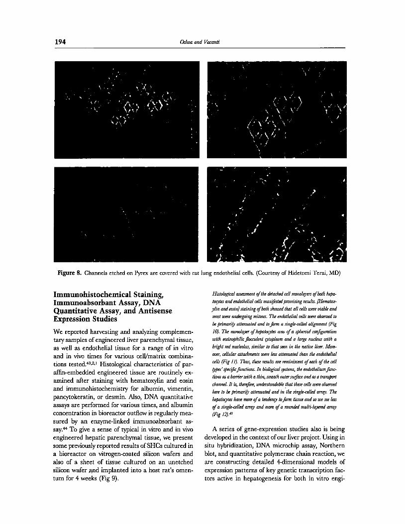
The engineering challenge that we now face is to combine the long-term culture of SHCs with our vascular endothelial systems. Independently, tissue engineers can create small masses, or sheets of hepa- tocytes that can survive being removed from the support scaffold and being folded and manipulated into various configurations, and we can create 2di- mensional vascular endothelial networks embedded in biodegradable polymer (Fig 8). When we can re- move an intact endothelial vasculature from the polymer, we may be able to layer vascular networks and hepatocyte sheets and continue culture with oxygen and nutrition provided through the new en- gineered vasculature. Alternatively, we may be able to seed SHCs or another donor hepatocyte type di- rectly onto a vascular system, growing dense new parenchymal tissue in 1 continuous process on pre- fabricated vascular systems.
These possibilities leave open the question of how we can meet our secondary goal of engineering a bile-duct system lined with biliary epithelial cells. Some of our more successful SHC cultures show cuboidal cells resembling biliary epithelial cells and duct-like formations. This produces the tantalizing suggestion that a biliary system may spontaneously differentiate and regenerate from our SHC culture when we can sustain the process for longer periods. Again, such a system may have to be incorporated into an engineering strategy. At this juncture, we are thrown back on the primary hypothesis of our me- chanical-structural approach, that our challenge is to engineer the basic structural cues and culture envi- ronment, and at some point, our new tissue will take
over and complete the process of regenerating whole functioning liver. The near future will tell whether there are significant engineering challenges still to be faced.
194 Ocha and Vacanti
Figure 8. Channels etched on Pyrex are covered with rat lung endothelial cells. (Courtesy of Hidetomi Terai, hID)
Immunohistochemical Staining, Immunoabsorbant Assay, DNA Qvantitative Assay, and Antisense Expression Studies
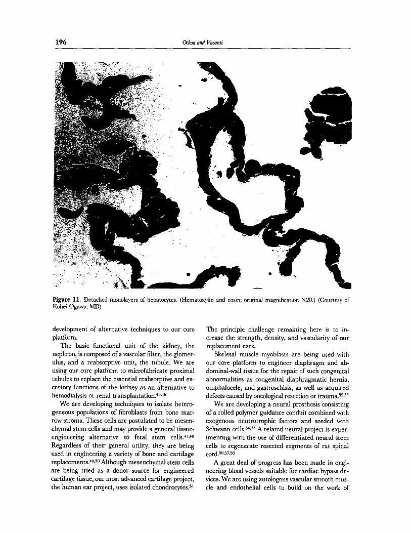
We reported harvesting and analyzing complemen- tary samples of engineered liver parenchymal tissue, as well as endothelial tissue for a range of in vitro and in vivo times for various cell/matrix combina- tions tested.43***t Histological characteristics of par- affin-embedded engineered tissue are routinely ex- amined after staining with hematoxylin and eosin and immunohistochemistry for albumin, vimentin, pancytokeratin, or desmin. Also, DNA quantitative assays are performed for various times, and albumin concentration in bioreactor outflow is regularly mea- sured by an enzyme-linked immunoabsorbant as- say.& To give a sense of typical in vitro and in vivo engineered hepatic parenchymal tissue, we present some previously reported results of SHCs cultured in a bioreactor on vitrogen-coated silicon wafers and also of a sheet of tissue cultured on an unetched silicon wafer and implanted into a host rat’s omen- turn for 4 weeks (Fig 9).
Histological auessmenl of fhe detached cell monolayers ofboth hepa-
kxytes and endorlrelial cells man$s~edpromising results. [Nemalox-
ylin and ean’n]staining rfbdi showed Ihat all cells were viable and
most were undergoing mikxes. The endolhelial cells were observed lo
be primarib aknuated and /o&n a single~elled alignmenl (Fig
10). The monolayer oJhepak@es was oja spheroid configuration
wilh eosinophilicJhuculenl @oplasm and a large nucleus will1 a
brig111 red nucleolus, similar to !hat seen in lhe nalive liver. More-
over, cellular altachmenls were less alterwaled than the endo&lial
cells (Fig I I). Thus, these reruhs are reminiscem of each of /he cell
ppeJspc$cJinctiorrr. In biological Tstemt, !he endohelium finc-
lions as a barrier with a thin, smooth oumrsu@ce and as a transport
channel. II is, krefore, understandable that lhese cells were observed
here IO be prima+ attenualed and in ‘lhe single-celled array. The
lepalo$es have more of a &ndery lofbnn lis.rue and so we see less
of a single-celled arq and more of a mun&d mul&+red array
(Fig 1.3.”
A series of gene-expression studies also is being developed in the context of our liver project. Using in situ hybridization, DNA microchip assay, Northern blot, and quantitative polymerase chain reaction, we are constructing detailed 4-dimensional models of expression patterns of key genetic transcription fac- tors active in hepatogenesis for both in vitro engi-

Core Pla@nn fw Tti Engineering 195
Figure 9. Hematoxylin and eosin staining for hepatocyte sheets implanted into the rat omentum after 4 weeks. (Courtesy of Kohei Ogawa, MJX
neered rat hepatogenesis and normal in vivo fetal rat hepatogenesis. These are the first large-scale molec- ular studies of tissue engineering. The data will be used to test the fundamental hypothesis underlying all tissue-engineering projects, that in vitro organo- genesis reiterates in vivo organogenesis at the mo- lecular level. The results will either strengthen our mechanical-structural assumptions or help deter- mine the minimal amount of molecular intervention
needed to create functioning replacement livers, as well as other tissues.
Parallel and Tangential Projects
Having traversed a vertical slice through our inte- grated multidisciplinary liver project, we can easily comprehend parallel developments in some of our other organ and dense-tissue projects, as well as the
Figure 10. Detached mono- layers of endothelial cells. (Hematoxylin and eosin; original magnification X20.) (Courtesy of Kohei Ogawa, MJJ
196 Ochoa and Vacanti
Figure 11. Detached monolayers of hepatocytes. (Hematoxylin and eosin; original magnification x20.) (Courtesy of Kohei Ogawa, MD)
development of alternative techniques to our core platform.
The basic functional unit of the kidney, the nephron, is composed of a vascular filter, the glomer- ulus, and a reabsorptive unit, the tubule. We are using our core platform to microfabricate proximal tubules to replace the essential reabsorptive and ex- cretory functions of the kidney as an alternative to hemodialysis or renal transplantation.4s~ti
We are developing techniques to isolate hetero- geneous populations of fibroblasts from bone mar- row stroma. These cells are postulated to be mesen- chymal stem cells and may provide a general tissue- engineering alternative to fetal stem cells.+7~4a Regardless of their general utility, they are being used in engineering a variety of bone and cartilage replacements.4a~5eAlthough mesenchymal stem cells are being tried as a donor source for engineered cartilage tissue, our most advanced cartilage project, the human ear project, uses isolated chondrocytes.5’
The principle challenge remaining here is to in- crease the strength, density, and vascularity of our replacement ears.
Skeletal muscle myoblasts are being used with our core platform to engineer diaphragm and ab- dominal-wall tissue for the repair of such congenital abnormalities as congenital diaphragmatic hernia, omphalocele, and gastroschisis, as well as acquired defects caused by oncological resection or trauma.5**53
We are developing a neural prosthesis consisting of a rolled polymer guidance conduit combined with exogenous neutrotrophic factors and seeded with Schwann cells.s@5 A related neural project is exper- imenting with the use of differentiated neural stem cells to regenerate resected segments of rat spinal cord.W57,58
A great deaf of progress has been made in engi- neering blood vessels suitable for cardiac bypass de- vices. We are using autologous vascular smooth mus- cle and endothelial cells to build on the work of

Core Pla$mn fm Tisnre Engheenhg 197
Figure 12. Endothelial cells grown on Vitrogen-coated (30 mg/tnL) Pyrex wafers after 4 days in culture. (Courtesy of Jeffery T. Borenstein, PhD)
Weinberg and Bell,59 as well as that of L’heaureaux et alGo and Niklason et al,“’ to increase the mechan- ical strength of such devices. Other cardiac work includes the fabrication of muscle patches to correct such congenital defects as hypoplastic heart syn- drome.
In collaboration with our former team member, David Mooney, now at the University of Michigan, we can routinely create organoid units, 0.1 X 0.25 mm, of mesenchymal cell cores with epithelial cell coverage. These organoids are the basis of several strategies to separately engineer replacement small intestine, large intestine, and duodenum.G2
Conclusion
The maturing field of tissue engineering is yielding practical results. Engineered dermal replacements
are commercially available, and several cartilage re- placement devices are entering the clinical trial stage. To fullill its promise of providing a large-scale cost-effective solution to the growing clinical demand for a variety of human tissues for surgical replace- ment and reconstruction, tissue engineering must now master techniques for creating dense vascular- ized tissue and solid vital organs. To meet this chal- lenge, our Advanced Technology Team at the Cen- ter for the Integration of Medicine and Innovative Technology laboratories developed a minimally inva- sive mechanical-structural platform for engineering living endothelial vascular systems in which dynamic features can be customized to meet the oxygen and nutrition requirements of any target tissue type. Combined with our expertise in engineering func- tioning hepatic parenchymal tissue, we soon may have the capacity to engineer stable mature human liver for the treatment of end-stage liver disease.
198 Ochoa and Vacanli
References
I. Mooney DJ, VacantiJP: Tissue engineering using cells and
synthetic polymers. Transplant Rev 1993, 7:153 2. Mooney DJ, Parks S, Kaukmann PM, et al: Biodegradable
sponges for hepatocyte transplantation. J Biomed Mater Res
1995,29:959 3. VacantiJP: Beyond transplantation, third annual Jeson Mix-
ter Lecture. Arch Surg 1988, 123:545 4. Bell E, Ehrlich P, Buttle DJ, et al: Living tissue formed in vitro
and accepted as skin-equivalent of full-thickness. Science 1981,221:1052
5. Naughton GK: Dermal equilivants, in Lanza RI’, Langer R, Vacanti J (eds): Principles of Tissue Engineering (ed 2). San
Diego, Academic, 2000, p893, Fig 63.2 6. Yannas IV: In viva synthesis of organs and tissues, in Lanza
RP, Langer R, Vacanti J (eds): Principles of Tissue Engineer- ing (ed 2). San Diego, Academic, 2000, p I63
7. Vacanti CA, Cima LG, Ratkowski D, et al: Tissue engineered growth of new cartilage in the shape of a human ear using
synthetic polymers seeded with chondrocytes, in Cima LG, Ron ES (eds): Tissue-Inducing Biomaterials, Materials Re- search Society Symposium Proceedings. Pittsburgh, Materials
Research Society, 1992, ~367 8. Bell E: Tissue engineering in perspective, in Lanza RP,
Langer R, Vacanti J (eds): Principles of Tissue Engineering
(ed 2). San Diego, Academic, 2000 pXXXV 9. Freed LE, Marquis JC, Nohria A, et al: Neocartilage forma-
tion in vitro and in viva using cells cultured on synthetic biodegradable polymers. J Biomed Mater Res 1993,27:1 I
10. Langer R, VacantiJP: Tissue engineering. Science 1993,260: 920
I I. Vacanti JP, Langer R: Tissue engineering: The design and fabrication of living replacement devices for surgical recon-
struction and transplantation. Lancet 1999, 35432 12. Martins-Green M: Dynamics of cell-ECM interactions, in
Lanza RP, Langer R, Vacanti J (eds): Principles of Tissue Engineering (ed 2). San Diego, Academic, 2000, p33
13. Shamblot MJ, Axelman J, Wang S, et al: Derimtion of pluri- potential stem cells from cultured human primordial germ
cells. Proc Nat1 Acad Sci U S A 1998, 95:13726
14. Shamblot w, Edwards BE, Gearhart JD: Pluripotenl stem cells, in Lanza RP, Langer R, Vacanti J (eds): Principles of
Tissue Engineering (ed 2). San Diego, Academic, 2000, ~370 15. Thomson JA, Hskovitz-Elder J, Shapiro SS, et al: Embryonic
stem cell lines derived from human blastocysts. Science 1998, 282: I 145
16. Juengst E, Fossel MF: The ethia of embryonic stem cells-
Now and forever, cells without end. JAMA 2000,284:3180 17. Lanza RP, Caplan AL, Silver LM, et al: The ethical validity of
using nuclear transfer in human transplantation. JAMA 2000, 284z3175
18. Inger DE: Mechanical and chemical determinants of tissue development, in Lanza RI’, Langer R, Vacanti J (eds): Prin-
ciples of Tissue Engineering (ed 2). San Diego, Academic, 2000, p104
19. Bissell w, Hall HG, Parry G: How does the extracellular
matrix direct gene expression? J Theor Biol 1982, 99:3 1 20. Kaazempur-Mofrad MR, Vacanti JP, Kamm RD: Computa-
tional modeling of blood flow and rheology in fractal micro- vascular net&o&, in Bath KJ (ed): Computational Fluid and
Solid Mechanics. New York, Elsevier, 200 I, p864-867
21. Flannery BP, Deckman HW, Roberge WG, et al: Three-
dimensional x-ray microtomography. Science 1987,237:1439
22. GarciaSanz A, Rodriguez-Barber0 A, Bentley MD, et al: Three-dimensional microcomputed tomography of renal vas-
culature in rats. Hypertension 1998, 31:440
23. Jorgensen SM, Demirkaya 0, Ritman EL: Three-dimensional imaging of vasculature and parenchyma in intact rodent or-
gans with x-ray micro-CT. Am J Physiol 1998,275:HllO3
24. Kaazempur-Mofrad MR, Ethier CR: Mass transport in an anatomically realistic human right coronary artery. Ann
Biomed Eng 2001, 29 (in press)
25. Rai-Choudhury P (ed): Handbook of Microlithography, Mi- cromachining and Microfabrication. Bellingham, WA, SPIE,
1997
26. Grove AS Physics and Technology of Semiconductor Devices. New York, Wiley, 1967
27. ZhanE.1, Tan KL, Hong GD, et al: Polymerization optimiza- 1-
28.
29.
30.
31.
32.
33.
34.
35.
36.
37.
38.
39.
40.
41.
tion of SU-8 photoresist and its applications in microlluidic systems and MEMS. J Micromech Microeng 200 I, 1 I :20
Wolf S, Tauber RN: Silicon Processing for the VLSI Era. Sunset Beach, CA, Lattice, 1986
HongJW, Fugii T, Seki M, et al: PDMS-glass hybrid micro- chip for gene amplilication. IEEE-EMBS Conf. Microtechnol,
in: Medicine and Biology. 2000, p407
Athanasiou KA, Niederuer GG, Agrawal CM: Sterilization, toxicity, biocompatibility and clinical applications ofpolylactic
acid/polyglycolic acid copolymers. Biomaterials 1996, 17:93
Ikada Y: Surface modification of polymers for medical appli- cations. Biomaterials 1994, 15:lO
Pierschbacher MD, Ruoslahti E: Cell attachment activity of fibronectin can be duplicated by small synthetic fragments of
the molecule. Nature 1984, 309:3
Freed LE, Vurjak-Novakovic G: Tissue engineering bioreac- tors, in Lanza RP, Langer R, Vacanti J (eds): Principles of
Tissue Engineering (ed 2). San Diego, Academic, 2000, ~146, Fig 13.3
Sato GH, Barnes DW: Animal cell culture, in Lanza RI’, Langer R, Vacanti J (eds): Principles of Tissue Engineering
(ed 2). San Diego, Academic, 2000, pl I I
Sano K, Cusick RA, Lee H, et al: Regenerative signals for heterotopic hepatocyte transplantation. Transplant Proc
1996, 28:1857
VacantiJP, Morse MA, Saltzman WM, et al: Selective cell
transplantation using bioabsorbable artificial polymers as ma- trices. J Pediatr Surg 1988, 23:3
Aiken J, Cima L, Schloo B, et al: Studies in rat liver perfusion for optimal harvest of hepatocytes. J Pediatr Surg 1990,25: 140
Seglen, PO: Preparation of isolated rat liver cells. Methods
Cell Biol 1976, 13:29
Mitaka T: The current status of primary hepatocyte culture.
Int J Exp Pathol 1998, 79:393
Kim SS, Utsunomiya H, Koski JA, et al: Survival and function
of hepatocytes on a novel threedimensional synthetic biode- gradable polymer scarold with an intrinsic network of chan- nels. Ann Surg 1998, 228:8
Petersonfi: ElTects of the pyrrolizidine alkaloid lasiocarpine- N-oxide on nuclear and cell division in the liver of rats.
J Pathol Bactcriol 1965, 89: 153
42. Langer R, Vacanti JP: Tissue engineering: The challenges
ahead. Sci Am 1999,280:62
43. Kaihara S, Borenstein J, Koka R et al: Silicon micromachining

Core Pla@nt~ Time Engineering 199
IO tissue engineer branched vascular channels for liver fabri- cation. Tissue Eng 2000,6: 105
44. Schwere B, Bach M, Bernheimer H: ELISA Tar determination of albumin in the nanogram range: Assay in cerebrospinal fluid and comparison with radial immunodi~usion. Clin
Chem Acta 1987, 163:237 45. Humes HD, Bullington DA, MacKay SM, et al: Replacement
of renal function in uremic animals wivith a tissue-engineered
kidney. Nat Biotechnol 1999, l7:45 I 46. Humes I-ID, MacKay SM, Funke AJ: Tissue engineering of a
bioartilicial renal tubule assist device: In vitro transport and metabolic characteristics. Kidney Int 1999, 55:2502
47. Bruder SP, Kurth AA, Shea M, ct al: Bone regeneration by implantation of purified, culture-expanded human mesenchy- mal stem cells. J Orthop Res 1998, 16:155
48. Pittenger MF, Mackay AM, Beck SC, et al: Multilineage potential or adult human mesenchymal stem cells. Science
1999,284: I43 49. Isogai N, Landis W, Kim TH, et al: Formation of phalanges
and small joints by tissue engineering. J Bone Joint Surg 1999,
8 IA:306 50. Petite H, Viateau V, Bensaid W, et al: Tissue-engineered
bone regeneration. Nat Biotechnol 2000, 18:959 51. Cao Y, VacantiF, Paige KT, et al: Transplantation orchon-
drocytes utilizing a polymer-cell construct to produce tissue- engineered cartilage in the shape or a human ear. Plast
Reconstr Surg 1997, IO&297 52. Cousins JC, Morgan JE, Partridge TA: Myoblast therapy, in
Lanza R, Langer R, Vacanti J (eds): Principles or Tissue
Engineering (ed 2). San Diego, Academic, 2000, p739
53. Saxena AK, Marler J, Benvenuto M, et al: Skeletal muscle
tissue engineering using isolated myoblasts on synthetic bio- degradable polymers: Preliminary studies. Tissue Eng 1999,
5:525
54. Ansselin D, Fink T, Davey DF: Peripheral newe regeneration
through nerve guides seeded with adult Schwann cells. Neu- ropathol Appl Neurobiol 1997, 23:387
55. Iba T, Sumpio B: Morphological response orhuman endothe- lial cells subjected IO cyclic strain in vitro. Microvasc Res 1991,
42:245
56. Hadlock T, Sundback C. Multi-lumen polymeric guidance channel, method for promoting nerve regeneration and
method oT manufacturing a multi-lumen nerve guidance channel. US patent 5,925,053, July 1999
57. McDonaldJW, Liu XZ, Qu Y, et al: Transplanted embryonic stem cells survive, direrentiate and promote recovery in in-
jured rat spinal cord. Nat Med 1999,5:1410
58. Satou T, Nishida S, Hiruma S, et al: A morphological study ol’
the erects of collagen gel matrix on regeneration of severed rat sciatic nerve in silicone tubes. Acta Pathol Jpn 1986,
36:199
59. Weinberg CB, Bell E: A blood vessel model constructed from collagen and cultured vascular cells Science 1986,231:397
60. L’heureux N, Paquet S, Labbe R, et al: A completely biological
tissue-engineered human blood vessel. FASEB J 1998, l2:47
61. Niklason LE, Gao J, Abbott WM, et al: Functional arteries
grown in vitro. Science 1999,284:489
62. Choi RS, Pothoulakis C, Kim BS, et al: Studies of brush border enzymes, basement membrane components, and elec-
trophysiology or tissue-engineered neointestine. J Pediatr
Surg 1998,33:99 I


![SHOCK- EMK-10.2009.pptx [Read-Only] - ocw.usu.ac.idocw.usu.ac.id/.../1110000130-emergency-medicine/emd166_slide_shock.pdf · Definition of Shock • Reduced perfusion of vital organs](https://static.fdocuments.us/doc/165x107/5c7a0dea09d3f2bd0e8c0a69/shock-emk-102009pptx-read-only-ocwusuacidocwusuacid1110000130-emergency-medicineemd166slideshockpdf.jpg)


