1 V5: Inducible cellular switching Cellular ProgramsWS 2010 – lecture 5 Rudolf Jänisch,...
21
1 V5: Inducible cellular switching Cellular Programs WS 2010 – lecture 5 www.wikipedia.o rg Rudolf Jänisch, Alexander van Oudenaarden *1942, MIT *1960, MIT
-
Upload
frederick-newman -
Category
Documents
-
view
216 -
download
2
Transcript of 1 V5: Inducible cellular switching Cellular ProgramsWS 2010 – lecture 5 Rudolf Jänisch,...

1
V5: Inducible cellular switching
Cellular ProgramsWS 2010 – lecture 5
www.wikipedia.org
Rudolf Jänisch, Alexander van Oudenaarden
*1942, MIT *1960, MIT

2
Elite cells vs. all cells?
Models to account for the reprogramming process fall into two categories.
‘Deterministic’ models:
Within a donor population either ‘all’ or only a subset of ‘elite’ or ‘stem-like’
cells have the potential to generate iPS cells and are reprogrammed with a
fixed latency.
‘Stochastic’ models:
Within a donor population most if not all (model iii) or only a subset of ‘elite’
somatic cells (model iv) have the potential to generate iPS cells, albeit with
different latencies.
Cellular ProgramsWS 2010 – lecture 5
Hanna et al. Nature 462, 595 (2009)

3
Models of progressing to a pluripotent stateduring direct reprogramming
Input: stimulated expression of Oct4,
Sox2, Klf4 and c-Myc reprogramming
factors in somatic cells.
Paper 5 tests 4 different models to
account for the latency of donor
somatic cells in progressing towards
the iPS cell state.
Latency can be measured in units of
absolute time or cell divisions until
the first iPS cell is generated from a
monoclonal population.
Cellular ProgramsWS 2010 – lecture 5
www.wikipedia.org

4
Experimental system
Questions:
(1) what are NGFP1 cells?
(2) what are B cells
(3) how does reprogramming with ES media and DOX work
Cellular ProgramsWS 2010 – lecture 5
www.wikipedia.org
Trick: differentiatedcells include fluorescentmarker for Nanogexpression (marker forES and iPS state).
5
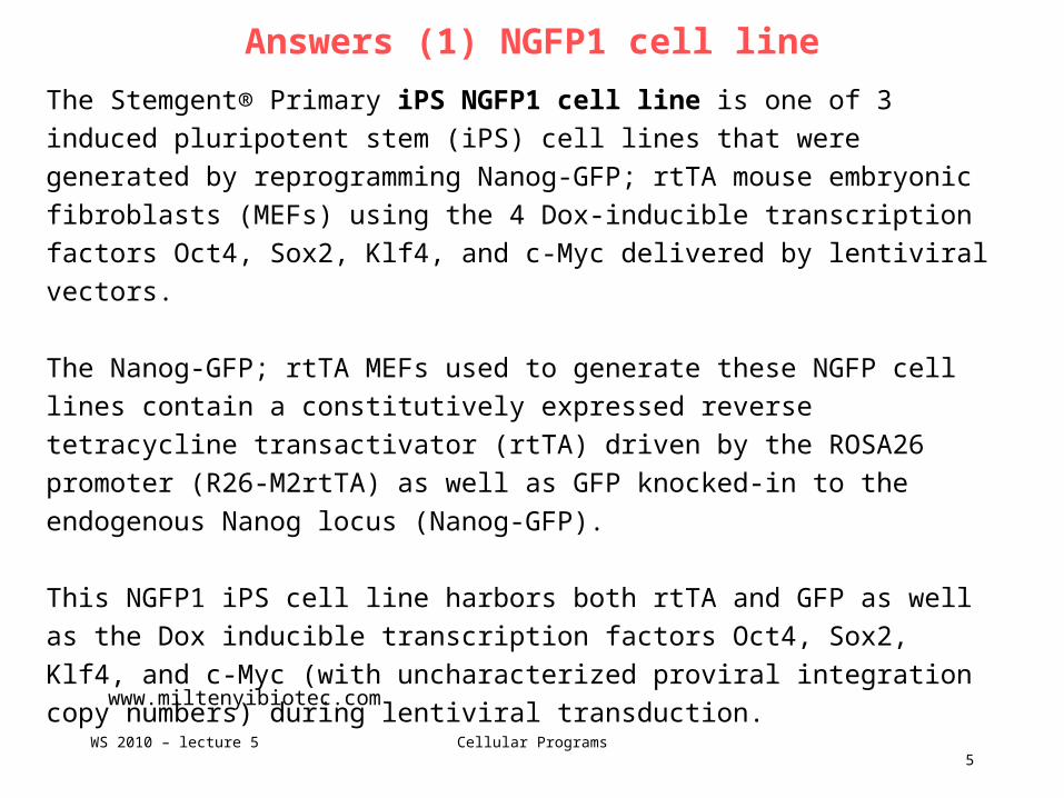
Answers (1) NGFP1 cell line
The Stemgent® Primary iPS NGFP1 cell line is one of 3 induced pluripotent
stem (iPS) cell lines that were generated by reprogramming Nanog-GFP; rtTA
mouse embryonic fibroblasts (MEFs) using the 4 Dox-inducible transcription
factors Oct4, Sox2, Klf4, and c-Myc delivered by lentiviral vectors.
The Nanog-GFP; rtTA MEFs used to generate these NGFP cell lines contain a
constitutively expressed reverse tetracycline transactivator (rtTA) driven by the
ROSA26 promoter (R26-M2rtTA) as well as GFP knocked-in to the endogenous
Nanog locus (Nanog-GFP).
This NGFP1 iPS cell line harbors both rtTA and GFP as well as the Dox
inducible transcription factors Oct4, Sox2, Klf4, and c-Myc (with uncharacterized
proviral integration copy numbers) during lentiviral transduction.
Cellular ProgramsWS 2010 – lecture 5
www.miltenyibiotec.com

6
Answers (3) Tet-On system
Cellular ProgramsWS 2010 – lecture 5
www.wikipedia.org
Tetracycline-Controlled Transcriptional Activation is a method of inducible
expression where transcription is reversibly turned on or off in the presence of
the antibiotic tetracycline or one of its derivatives (e.g. doxycycline).
In nature, the Ptet promoter expresses TetR, the repressor, and TetA, the
protein that pumps tetracycline antibiotic out of the cell.
The two most commonly used inducible expression systems for research of
eukaryote cell biology are named Tet-Off and Tet-On.
They consist of a fusion of the Tet repressor and a VP16 activation domain to
create a transcriptional activator protein (transactivator) rather than a repressor.

7
Answers (3) Tet-On system
Cellular ProgramsWS 2010 – lecture 5
www.wikipedia.org
Gene expression is activated as a result of binding of the Tet-Off or Tet-On
protein to tetracycline response elements (TREs) located within an inducible
promoter.
The difference between Tet-On and Tet-Off is not whether the transactivator
turns a gene on or off; rather, both proteins activate expression.
The difference relates to their respective response to doxycycline; Tet-Off
activates expression in the absence of Dox, whereas Tet-On activates in the
presence of Dox.
In the Tet-On system, the rtTA protein is capable of binding the operator only
when bound by doxycycline. Thus the introduction of doxycyline to the system
initiates the transcription of the genetic product.

8
Doxycycline
Doxycycline is a member of the
tetracycline antibiotics group and is
commonly used to treat a variety of
infections.
Cellular ProgramsWS 2010 – lecture 5
www.wikipedia.org

9
NGFP1 cell line
When cultured under standard mouse embryonic stem (ES) cell culture conditions, the
morphology of NGFP1 iPS cells is identical to that of mouse ES cells. The cells express
both GFP from the endogenous Nanog locus as well as the pluripotency marker SSEA-1.
Stemgent® Mouse Primary iPS cells-NGFP1 can be used to generate chimeric mice from
which cells can be isolated for secondary reprogramming studies.
Chimera:animal that has two or more different populations of genetically distinct cells that originated in different zygotes
involved with sexual reproduction. Chimeras are formed from four parent cells (two fertilized eggs or early embryos
fused together). Each population of cells keeps its own character and the resulting animal is a mixture of tissues.
The simple addition of Doxycycline (Dox) to the growth medium used to propagate the
isolated cell types from the chimeric mouse results in the upregulation in expression for
the 4 transcription factors implicated in the reprogramming process.
The Stemgent® Mouse Primary iPS cells-NGFP1 were generated in the lab of Dr. Rudolf
Jaenisch, M.D. at Whitehead Institute-MIT. Dr. Jaenisch is a recognized leader in the
study of epigenetic regulation of gene expression with numerous publications focused on
ES and iPS cellular mechanisms and methodologies.
Cellular ProgramsWS 2010 – lecture 5
www.miltenyibiotec.com

10
NGFP1 cell line
Derivation of iPS Cells Using
an Inducible Lentiviral System
(A) Somatic cells harboring a
GFP reporter driven by the
endogenous Oct4 or Nanog
promoters were infected with
tet-inducible lentiviral vectors
carrying the cDNAs of Oct4,
Sox2, Klf4, and c-Myc.
Cellular ProgramsWS 2010 – lecture 5
Brambrink et al. Cell Stem Cells 2, 151 (2008)
(B) The addition of dox after infection induced lentiviral expression and
subsequently led to reprogrammed, GFP-positive iPS cell colonies.
(C and D) The iPS cells were pluripotent and contributed to viable
chimeras after injection into BALB/C host blastocysts as indicated by coat
color. White mice are nonchimeric BALB/C animals, whereas mixed coat
color mice are chimeric.

11
What are B cells?
B cells are lymphocytes that play a large role in the humoral immune response (as
opposed to the cell-mediated immune response, which is governed by T cells).
The principal functions of B cells are to make antibodies against antigens, perform
the role of antigen-presenting cells (APCs) and eventually develop into memory B
cells after activation by antigen interaction.
B cells are an essential component of the adaptive immune system.
Cellular ProgramsWS 2010 – lecture 5
www.wikipedia.org

12
Development of B cells
Immature B cells are produced in the bone marrow of most mammals.
After reaching the IgM+ immature stage in the bone marrow, these immature B cells migrate
to the spleen, where they are called transitional B cells, and some of these cells differentiate
into mature B lymphocytes.
B cell development occurs through several stages, each stage representing a change in the
genome content at the antibody loci.
An antibody is composed of two identical light (L) and two identical heavy (H) chains, and the
genes specifying them are found in the 'V' (Variable) region and the 'C' (Constant) region.
In the heavy-chain 'V' region there are three segments; V, D and J, which recombine
randomly, in a process called VDJ recombination, to produce a unique variable domain in the
immunoglobulin of each individual B cell. (Random mutations also occur).
Similar rearrangements occur for light-chain 'V' region except there are only two segments
involved; V and J.
Cellular ProgramsWS 2010 – lecture 5
www.wikipedia.org

13
Development of B cells
Cellular ProgramsWS 2010 – lecture 5
www.wikipedia.org

14
p53 pathway
p53 (also known as protein 53 or tumor
protein 53), is a tumor suppressor
protein that in humans is encoded by the
TP53 gene.
p53 is important in multicellular
organisms, where it regulates the cell
cycle and, thus, functions as a tumor
suppressor that is involved in preventing
cancer.
P53 is called "the guardian of the
genome", referring to its role in
conserving stability by preventing
genome mutation.
Cellular ProgramsWS 2010 – lecture 5
www.rcsb.org
The name p53 refers to its apparent molecular
mass: It runs as a 53-kilodalton (kDa) protein on
SDS-PAGE. But the mass of p53 is actually only
43.7 kDa. The difference in speed on the gel is
due to the high number of proline residues in the
protein, which slow its migration on SDS-PAGE,
thus making it appear heavier than it actually is.

15
p53 signaling pathway
p53 is a transcription factor who's activity
is regulated by phosphorylation.
The function of p53 is to keep the cell
from progressing through the cell cycle if
there is damage to DNA present.
It may do this in multiple ways from
holding the cell at a checkpoint until
repairs can be made to causing the cell to
enter apoptosis if the damage cannot be
repaired.
The critical role of p53 is evidenced by the
fact that it is mutated in a very large
fraction of tumors from nearly all sources.
Cellular ProgramsWS 2010 – lecture 5
www.biocarta.com

16
p53:MDM2 complex
In a normal cell p53 is inactivated by its
negative regulator, mdm2.
Upon DNA damage or other stresses,
various pathways will lead to the
dissociation of the p53 and mdm2
complex.
Once activated, p53 will induce a cell
cycle arrest to allow either repair and
survival of the cell or apoptosis to
discard the damaged cell.
How p53 makes this choice is currently
unknown
Cellular ProgramsWS 2010 – lecture 5
www.wikipedia.org

17
Apoptotic signaling in response to DNA damage
The cellular activation of the caspase cascade
resulting in cell death is triggered by chemical
damage to DNA.
This stimulates a sequence resulting in the
cleavage of Bid in a manner similar to the binding
of so called “death-receptors” or directly initiates
the permeability transition of the mitochondrial
membrane. The permeability transition releases
several factors including cytochrome c, AIF and
other factors in to the cytoplasm.
Cellular ProgramsWS 2010 – lecture 5www.biocarta.com
Cytochrome c, a key protein in electron transport, is released from mitochondria in response to
apoptotic signals, and activates Apaf-1, a protease released from mitochondria. Activated
Apaf-1 activates caspase-9 and the rest of the caspase cascade. The caspases are a class of
cysteine proteases that includes several representatives involved in apoptosis. The caspases
convey the apoptotic signal in a proteolytic cascade, with caspases cleaving and activating
other caspases that then degrade other cellular targets that lead to cell death.

18
Cell cycle: G1/S checkpoint
The G1/S cell cycle checkpoint controls the
passage of eukaryotic cells from the first 'gap'
phase (G1) into the DNA synthesis phase (S).
Two cell cycle kinases, CDK4/6-cyclin D and
CDK2-cyclin E, and the transcription complex
that includes Rb and E2F are pivotal in controlling
this checkpoint.
During G1 phase, the Rb-HDAC repressor
complex binds to the E2F-DP1 transcription
factors, inhibiting the downstream transcription.
Phosphorylation of Rb by CDK4/6 and CDK2
dissociates the Rb-repressor complex, permitting
transcription of S-phase genes encoding for
proteins that amplify the G1 to S phase switch and
that are required for DNA replication ...
Cellular ProgramsWS 2010 – lecture 5
www.biocarta.com

19
Cell cycle: G2/M checkpoint
The G2/M DNA damage checkpoint prevents the cell
from entering mitosis (M phase) if the genome is
damaged.
Cdc2-cyclin B kinase is pivotal in regulating this
transition. During G2 phase, Cdc2 is maintained in an
inactive state by the kinases Wee1 and Mt1. As cells
approach M phase, the phosphatase Cdc25 is
activated. Cdc25 then activates Cdc2, establishing a
feedback amplification loop that efficiently drives the
cell into mitosis.
DNA damage activates the DNA-PK/ATM/ATR
kinases, initiating two parallel cascades that inactivate
Cdc2-cyclin B. The first cascade rapidly inhibits
progression into mitosis: the CHK kinases
phosphorylate and inactivate Cdc25, which can no
longer activate Cdc2.
Cellular ProgramsWS 2010 – lecture 5www.biocarta.com
The second cascade is slower. Phosphorylation of p53 dissociates it from MDM2, activating its DNA binding activity. Acetylation
by p300/PCAF further activates its transcriptional activity. The genes that are turned on by p53 constitute effectors of this second
cascade. They include 14-3-3s, which binds to the phosphorylated Cdc2-cyclin B kinase and exports it from the nucleus;
GADD45, which apparently binds to and dissociates the Cdc2-cyclin B kinase; and p21Cip1, an inhibitor of a subset of the
cyclin-dependent kinases including Cdc2 (CDK1).

20
Here: p53 was inhibited
p.597:
„Recently, p53 inhibition has been shown to enhance the efficiency of iPS cell
formation from fibroblasts ... by reducing apoptosis after initial transgene
infection.
...
NGFP1 iPS cells were infected with a constitutively expressed lentiviral vector
encoding a short interfering RNA (siRNA) hairpin for p53.
Infected cells were injected into hosts blastocysts and NGFP1-p53 knockdown
B cells were single-cell sorted and cultured in doxycycline.
Cellular ProgramsWS 2010 – lecture 5
www.wikipedia.org

21
Summary
Systematic study to understand mechanism of reprogramming.
Distinguish models for „elite cells“ vs. „all cells can do“.
Experiments started from an artificially generated „only little developed“ B cell.
This explains high success rates for reprogramming.
Cellular ProgramsWS 2010 – lecture 5
www.wikipedia.org